Tuberculosis in Camelids - USAHA · TUBERCULOSIS IN CAMELIDS Diagnosis in New and Old World...
26
SUNNY GEISER-NOVOTNY, VMD, MS USDA APHIS VS SPRS CATTLE HEALTH CENTER Tuberculosis in Camelids
Transcript of Tuberculosis in Camelids - USAHA · TUBERCULOSIS IN CAMELIDS Diagnosis in New and Old World...
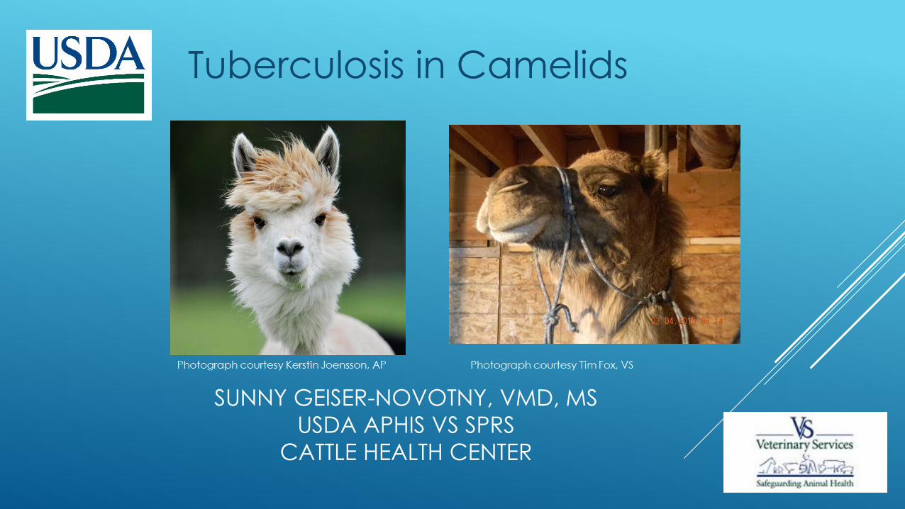
SUNNY GEISER-NOVOTNY, VMD, MS
USDA APHIS VS SPRS
CATTLE HEALTH CENTER
Tuberculosis in Camelids
TUBERCULOSIS IN CAMELIDS
Diagnosis in New and Old World Camelids
(NWC/OWC)
NWC – EU, South America, US
OWC – UAE, Africa, Pakistan, Kazakhstan, US
Isolation of M. bovis, M. tuberculosis & M. microti
from Mycobacterium Tuberculosis Complex
(MTBC)
M. avium, M. kansasii - environmental
Typing of M. bovis found in camelids reflects
locally predominant genotypes in cattle and
wildlife (Rhodes 2014)
Outbreaks rare in natural habitat
Frank Beard Bible Symbols or The Bible in Pictures (London, England: Hertel, Jenkins & Co., 1904)

CLINICAL SIGNS
Changes in behavior
Anorexia and cachexia
Respiratory distress, coughing
None
Sudden death or euthanasia for
deteriorating body condition
Photograph courtesy Kerstin Joensson, AP
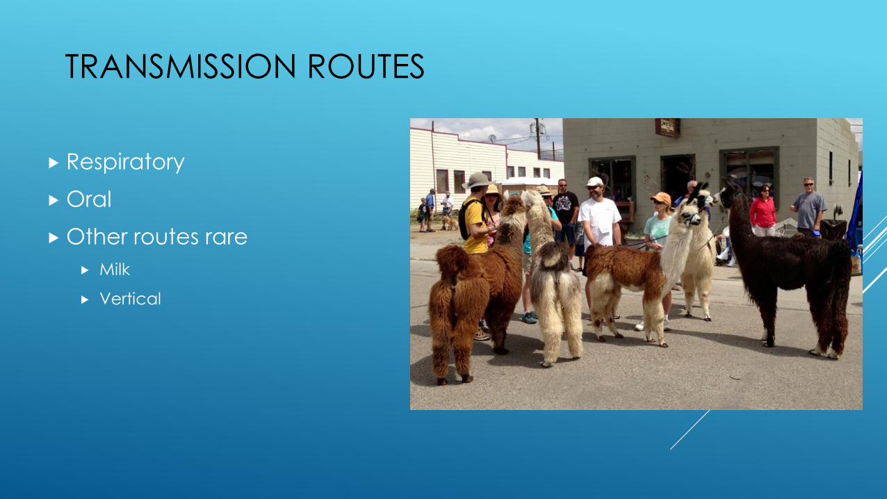
TRANSMISSION ROUTES
Respiratory
Oral
Other routes rare
Milk
Vertical
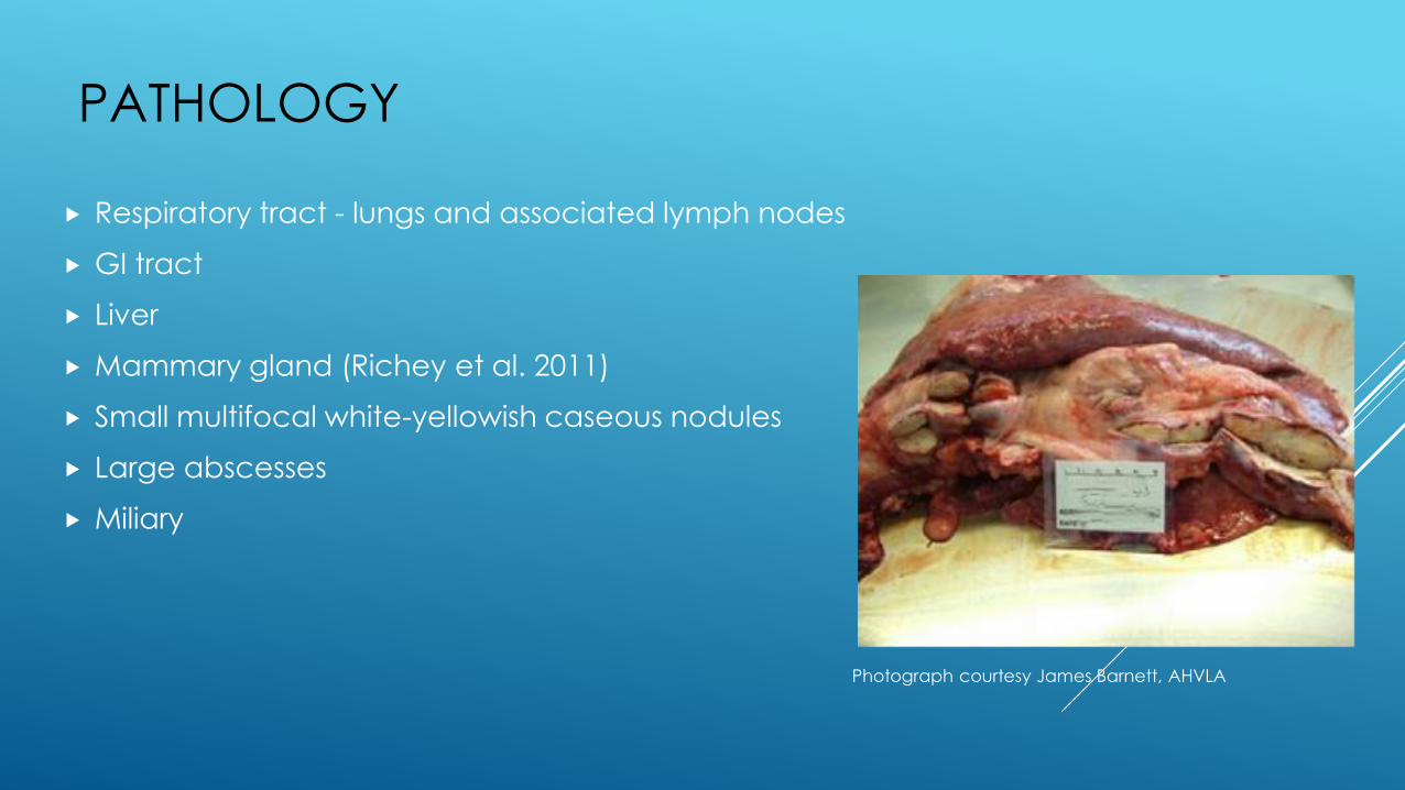
PATHOLOGY
Respiratory tract - lungs and associated lymph nodes
GI tract
Liver
Mammary gland (Richey et al. 2011)
Small multifocal white-yellowish caseous nodules
Large abscesses
Miliary
Photograph courtesy James Barnett, AHVLA
DIAGNOSIS
Post-mortem:
Compatible lesions found at necropsy/slaughter (usually first indication of issue in herd)
Histopathology – Acid-Fast Bacilli
Culture – M. microti difficult to isolate
Ante-mortem:
Cellular
Skin testing
For movement purposes
Required for herd test after confirmed positive at necropsy
Interferon-gamma (IFN-g)
Humoral (Serology)
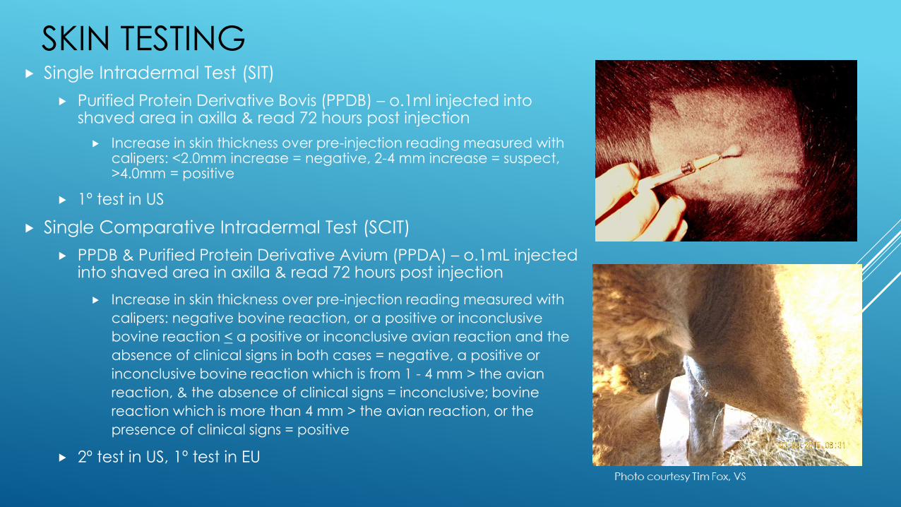
SKIN TESTING Single Intradermal Test (SIT)
Purified Protein Derivative Bovis (PPDB) – o.1ml injected into shaved area in axilla & read 72 hours post injection
Increase in skin thickness over pre-injection reading measured with calipers: <2.0mm increase = negative, 2-4 mm increase = suspect, >4.0mm = positive
1º test in US
Single Comparative Intradermal Test (SCIT)
PPDB & Purified Protein Derivative Avium (PPDA) – o.1mL injected into shaved area in axilla & read 72 hours post injection
Increase in skin thickness over pre-injection reading measured with
calipers: negative bovine reaction, or a positive or inconclusive
bovine reaction < a positive or inconclusive avian reaction and the
absence of clinical signs in both cases = negative, a positive or
inconclusive bovine reaction which is from 1 - 4 mm > the avian
reaction, & the absence of clinical signs = inconclusive; bovine
reaction which is more than 4 mm > the avian reaction, or the
presence of clinical signs = positive
2º test in US, 1º test in EU

SKIN TESTING
Lacks sensitivity and specificity in naturally infected animals (Dean et al. 2009)
SCIT used more to minimize false positives due to environmental mycobacteria
(Alvarez et al. 2011)
Several different injection sites used to enhance sensitivity – neck (pre-scapular),
axillary, tail
Variation in reading times used to evaluate skin fold increase (Bezos et al. 2013,
Wernery et al. 2007)
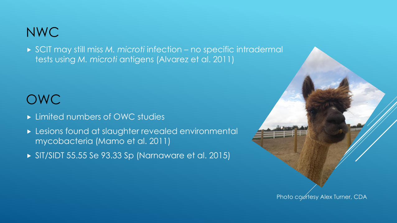
NWC SCIT may still miss M. microti infection – no specific intradermal
tests using M. microti antigens (Alvarez et al. 2011)
OWC Limited numbers of OWC studies
Lesions found at slaughter revealed environmental mycobacteria (Mamo et al. 2011)
SIT/SIDT 55.55 Se 93.33 Sp (Narnaware et al. 2015)
Photo courtesy Alex Turner, CDA
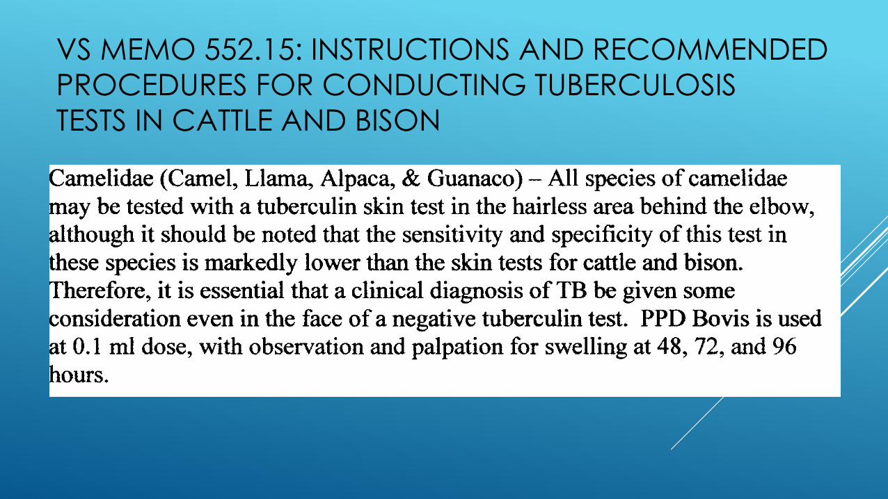
VS MEMO 552.15: INSTRUCTIONS AND RECOMMENDED
PROCEDURES FOR CONDUCTING TUBERCULOSIS
TESTS IN CATTLE AND BISON
TB TESTING REQUIREMENTS FOR CAMELIDS
TO CANADA 2.2 Tuberculosis
Negative results must be obtained on two (2) tuberculosis intradermal tests, using the post-axillary injection site, at least 90 days apart, with the second being performed no more than 30 days prior to export.
Testing procedures must be administered by a veterinarian competent in the specified procedure in the exported species.
The tuberculosis test to be conducted is the intradermal test with a dose rate of 0.1 ml of bovine PPD tuberculin (or product of equivalent potency approved by CFIA) injected at the post-axillary site, the injection site identified with a permanent ink marker, and the thickness of the skin recorded with calipers. The skin thickness will be measured seventy-two (72) hours post injection.
A responder is any animal in which there is an increase in the thickness of the skin greater than 1.5 mm at the site of injection in response to the initial injection of tuberculin.
Any responder animal to either the intradermal test is to be removed from the group of animals intended for export, and the entire testing protocol needs to begin again for the remainder of the group. A minimum of 90 days is always required between any intradermal test.
The results of all the tuberculin tests (including the dates of test readings) must be shown on the required health certificate for the animal to be imported.

Interferon-gamma (IFN-g)
Developed for use in South American Camelids (SAC) that would not be dependent on completion of prior skin test (Rhodes et al. 2012)
In vitro stimulation of blood cells
Responses on whole heparinized blood too low but significant responses seen when peripheral blood mononuclear cells (PMBCs) separated out and stimulated with PPDB, PPDA, ESAT6 – CFP10 (EC) peptide cocktail
80-98% Sp, 15-80% Se
Detects other MTBC mycobacteria
SEROLOGY
Infected animals can be detected before onset
of clinical signs (Lyashchenko et al. 2007)
2 animals seroreative 1-2 years before clinical
signs
Used alone or in combination with skin testing –
increased sensitivity (amanestic response)
(Stevens et al. 1998) (Bezos et al. 2013)

SEROLOGY MAPIA – Multiantigen print immunoassay (Chembio)
Uses range of M. bovis specific antigens including MPB70 & MPB83 printed on nitrocellulose
membrane incubated with serum samples in laboratory
Lateral flow based rapid test (RT)- VetTB Stat-Pak (Chembio)
No longer in production, replaced by DPP
Uses M. tuberculosis antigens - MPB83, ESAT-6 and CFP10
Dual Path Platform (DPP) VetTB assay (Chembio)
Licensed in US for cervids and elephants, animal-side test with results available in 15 minutes
Uses M. bovis recombinant MPB83 protein and CFP10/ESAT-6 fusion
Read by a DPP optical reader which measures reflectance
ELISA
IDEXX ELISA (IDEXX Laboratories) - plates pre-coated with MPB83 and MPB70 antigens
Enferplex antibody ELISA (Enfer Scientific) – includes seven antigens PPDB, SAT6, CFP10,
Rv3616c, MPB83, MPB70 and MPB70 peptide

EVALUATION OF STAT-PAK AND DPP IN SAC -
(LYASHCHENKO ET AL. 2011)
156 alpacas and 175 llamas from GB, Switzerland and US
Confirmed TB group (Culture +, and/or typical gross lesions or clinical signs)
35 alpacas/17 llamas
34 alpacas/10 llamas with M. Bovis (GB); 1 alpaca/7 llama with M. microti (Switzerland)
All except 2 M. bovis llamas negative on skin test
TB free group
Negative controls including 96 alpacas/122 llamas known to be TB free (Switzerland, US)
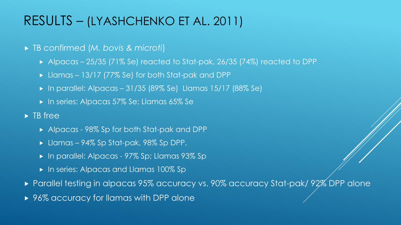
RESULTS – (LYASHCHENKO ET AL. 2011)
TB confirmed (M. bovis & microti)
Alpacas – 25/35 (71% Se) reacted to Stat-pak, 26/35 (74%) reacted to DPP
Llamas – 13/17 (77% Se) for both Stat-pak and DPP
In parallel: Alpacas – 31/35 (89% Se) Llamas 15/17 (88% Se)
In series: Alpacas 57% Se; Llamas 65% Se
TB free
Alpacas - 98% Sp for both Stat-pak and DPP
Llamas – 94% Sp Stat-pak, 98% Sp DPP,
In parallel: Alpacas - 97% Sp; Llamas 93% Sp
In series: Alpacas and Llamas 100% Sp
Parallel testing in alpacas 95% accuracy vs. 90% accuracy Stat-pak/ 92% DPP alone
96% accuracy for llamas with DPP alone
EVALUATION OF GAMMA INTERFERON AND
ANTIBODY TUBERCULOSIS TESTS IN ALPACAS -
(RHODES ET AL. 2012)
Research conducted by Animal Health and Veterinary Laboratories Agency
funded by industry
Analysis of IFN-g & 4 serological tests (STAT-PAK, DPP, IDEXX ELISA & multiplex ELISA)
All tests conducted on 48 diseased (typical gross visible lesions (VL) consistent with
mycobacteria) alpacas from 10 infected GB herds, 257 TB-free alpacas from 17 GB
herds, and 49 serum samples from the US
IFN-g assay: PPDB & PPDA used to generate comparative PPD response;
ESAT6/CFP10 peptides used for increased specificity for bovine TB
Data analyzed to suggest test combinations
RESULTS - (RHODES ET AL. 2012)
Camelid IFN-g detects MTBC (M. microti) – not specific
Serology - ~97% (Sp) & 60-70% (Se)
Combination of IFN-g with Stat—pak and DPP or IDEXX in parallel
provided greatest Se (100%)
Combination of tests in serial interpretation – 99.7% (Sp), 56% (Se)
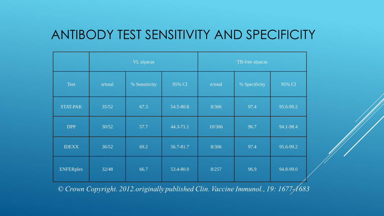
ANTIBODY TEST SENSITIVITY AND SPECIFICITY
VL alpacas TB-free alpacas
Test n/total % Sensitivity 95% CI n/total % Specificity 95% CI
STAT-PAK 35/52 67.3 54.5-80.8 8/306 97.4 95.6-99.2
DPP 30/52 57.7 44.3-71.1 10/306 96.7 94.1-98.4
IDEXX 36/52 69.2 56.7-81.7 8/306 97.4 95.6-99.2
ENFERplex 32/48 66.7 53.4-80.0 8/257 96.9 94.8-99.0
ASSESSMENT OF PCR TESTING OF NASAL SWABS AND FECES FOR
DETECTION OF M. BOVIS IN SAC – (CRAWSHAW ET AL. 2014)
44 Nasal swabs & fecal samples collected from 63 SAC carcasses from culture
confirmed M. bovis infected herds
Carcasses received a pathology score on PM. Values assigned according to tissues
affected & extent of lesions (min score 0, max score 12);carcasses reflected full
range of pathology scores, including 8 SACs with no gross lesions
Samples were tested for IS1081 PCR and RD4 Real Time PCR (to detect M. bovis
specifically)
23/44 positive on either test (64% Se),
Nasal swabs 14/44, Fecal swabs 21/44 positive to both IS1081 & RD4 PCR
confirming M. bovis
Culture performed on 14/44 (5 negative on culture & PCR)
9 positive on culture, fecal PCR detected 4 & the nasal PCR detected 5 (a
combination of the PCRs did not detect 3)
CAMELID TUBERCULOSIS IN THE UK
Legislation introduced in 2006 required veterinarians to notify veterinary authorities of all tuberculous lesions in carcasses of livestock and pets
Movement restrictions pending culture results
SCIT repeated at 90 day intervals
UK Voluntary camelid TB scheme (Hayton et al. 2014)
Defra, AHPA, Industry, British Veterinary Camelid Society, SureTest, Enfer Scientific
Camelids skin tested with SCIT 10-30 days before blood testing
Enferplex test – 2 antigen level: 66.7 (Se) 97 (Sp); 4 antigen level 100 (Sp)
Annual herd surveillance, pre-movement and export testing
TUBERCULOSIS IN OWC - (WERNERY ET AL. 2007)
Outbreak in UAE Dromedary racing herd Diagnosed on PM as M. bovis (antelope)
SIT and SCIT used to determine optimal test sites and reading times
Serum samples collected for Stat-pak and MAPIA 3 times within 6 months (2mo prior to
skin test, immediately before skin test, 6wk post skin test)
Serum samples from camels assayed for M. paratuberculosis using IDEXX-ELISA
modified for camelids

TUBERCULOSIS IN OWC - RESULTS
2 camels consistently positive to Stat-pak and MAPIA, positive on SCIT confirmed on PM
Strongest Stat-pak & MAPIA responses in index case,
moderate in number 1&2
Best responses for both skin tests – axillary read at 5 days
All 3 positives detected on Stat-pak /MAPIA, no false
positives
Presence of false positive SCIT responses in camelids may be
due to environmental mycobacteria
Photo courtesy Tim Fox, VS

CONCLUSIONS
Serology tests show promise for ante mortem detection of TB in Camelids
Current protocols for skin testing are unlikely to detect infection in herds
More data needed on appropriate protocol for skin testing in Camelids
More research needed on test performance in naturally infected and non-
infected camelids with known infection status
Additional research needed in OWC
True prevalence of TB in camelids is unknown due to lack of surveillance and
substandard tests

REFERENCES Alvarez, J., Bezos, J., de Juan, L., Vordermeier, M., Rodriguez, S., Fernandez-de-Mera, I.G. Mateos, A. and L. Dominguez. 2011. Diagnosis of tuberculosis in
camelids: old problems, current solutions and future challenges. Transbound. Emerg. Dis., 59(1): 1-10.
Bezos J, Casal C, Alvarez J, Díez-Guerrier A, Rodríguez-Bertos A, Romero B, Rueda P, López L, Domínguez L, de Juan L. 2013. Evaluation of the performance of cellular and serological diagnostic tests for the diagnosis of tuberculosis in an alpaca (Vicugna pacos) herd naturally infected with Mycobacterium bovis. Prev. Vet. Med, 111(3-4):304-313.
Bush, M. Montali, R. J., Phillips Jr., L.G., and Holobaugh, P.A. 1990. Bovine tuberculosis in a Bactrian Camel Herd: Clinical, Thearapeutic, and Pathologic Findings. Journal of Zoo and Wildlife Med., 21(2):171-179.
Crawshaw, T.R., Chanter, J.I., McGoldrick, A., Line, Kirsty. 2014. A proof of concept study to assess the potential of PCR testing to detect natural Mycobacterium bovis infection in South American camelids. Irish Vet. Journal, 67:5. doi:10.1186/2046-0481-67-5.
Dean, G.S., Crawshaw, T.R., de la Rua-Domenech, R., Farrant, L., Greenwald, R., Higgins, R.J., Lyashchenko, K., Vordermeier, H.M. and D.F. Twomey. 2009. Use of serological techniques for diagnosis of Mycobacterium bovis infection in a llama herd. Vet. Rec., 12;165(11):323-324.
Hayton, A. et al. Launch of a voluntary camelid TB scheme. Vet. Rec. 2014 175: 101-102. doi:10.1136/vr.g4758
Lyashchenko, K.P., Greenwald, R., Esfandiari, J., Meylan, M., Burri, I.H. and P. Zanolari. Antibody responses in New World camelids with tuberculosis caused by Mycobacterium microti. Vet. Microbiol., 2007 125(3-4):265-273.
Lyashchenko, K.P., Greenwald, R., Esfandiari, J., Rhodes. S., Dean, G., de la Rua-Domenech, R., Meylan, M., Vordermeier, H.M. and P. Zanolari. 2011. Diagnostic value of animal-side antibody assays for rapid detection of Mycobacterium bovis or Mycobacterium microti infection in South American camelids. Clin. Vaccine Immunol., 18(12):2143-2147.
Mamo, G., Bayleyegn, G., Tessema, T.S., Legesse, M., Medhin, G., Bjune, G., Abebe, F. and G. Ameni. 2011. Pathology of camel tuberculosis and molecular characterisation of its causative agents in pastoral regions of Ethiopia. PLoS One, 6(1): e15862.
Narnaware, S.d., Dahiya, S.S., Tuteja, F.C., Nagarajan, G., Nath, K., Patil, N.V. Pathology and diagnosis of Mycobacterium bovis in naturally infected dromedary camels (Camelus dromedaries) in India. Trop. Anim. Health Prod. 2015. DOI 10.1007/s11250-015-0905-5
Richey, M.J., Foster, A.P., Crawshaw, T.R. and A. Schok. 2011. Mycobacterium bovis mastitis in an alpaca and its implications. Vet. Rec., 169(8):214
Rhodes, S.G., Holder, T., Clifford, D., Dexter, I., Brewer, J., Smith, N., Waring, L., Crawshaw, T., Gillgan, S., Lyashchenko, K., Lawrence, J., Clarke, J., de la Rua-Domenech, R. and M. Vordermeier. 2012. Evaluation of gamma interferon and antibody tuberculosis tests in alpacas. Clinical and Vaccine Immunology, 19(10): 1677-1683.
Rhodes, S., Crawshaw, T., de la Rua-Domenech, R. Bradford, S., Lyashchenko, K.P., Mamo, G., Summers, D., Wernery, U. and Zanolari, P. (2014) Mycobacterial Infections in Camelids.
Stevens, J.B., Thoen, C.O., Rohonczy, E.B., Tessaro, S., Kelly, H.A. and J.R. Duncan. 1998. The immunological response of llamas (Lama glama) following experimental infection with Mycobacterium bovis. Can. J. Vet. Res., 62: 102-109.
Wernery, U. et al., Tuberulosis outbreak in a dromedary racing herd and rapid serological detection of infected camels,Vet. Microbiol. (2007), doi:10.1016/j.vetmic.2007.01.012

QUESTIONS?
Sunny Geiser-Novotny, VMD, MS
Staff Veterinarian
Cattle Health Center
USDA APHIS VS SPRS [email protected]
970-494-7372
Photograph courtesy Tim Fox, VS