Trypanosoma congolense - OpenAgrar · Hemato- logic, serum chemical, and serologic pro- cedures...
14
EXPERIMENTAL PARASITOLOGY 36, 6-19 ( 1974) Trypanosoma congolense I. Clinical Observations of Experimentally Infected Cattle B. WELLDE, R. L~TZSCH,~ G. DEINDL,~ E. SAWN, J. WILLIAMS, AND G. WARUI I. Department of Medical Zoology, Walter Reed Army Institute of Research, Washington, D.C. 20012 (Submitted for publication, April 30, 1973) WELLDE, B., LBIZSCH, R., DEINDL, G., SADUN, E., WILLIAMS, J., AND WARUI, G. 1974. Typanosoma congoknse: I. Clinical observations of experimental infection in cattle. Experimental Parasitology 36, 6-19. The course of disease was studied in 8 cattle infected with Typanosomu congolense. Although the onset of patency was dependent on the numbers of infecting organisms, the duration of the infection was not. High fevers were present on the day of or the day after initial patency. Suc- ceeding peaks of parasitemia, and a progressive weight loss of over 30% occurred. A ‘decrease in packed cell volume (PCV) beginning the first week after infection was observed. Early in the course of the developing anemia, many polychromato- philic erythrocytes and occasional normoblasts were found in the blood. A leucopenia persisted for the duration of the disease. Total serum protein concentrations fell sharply during the first 5 weeks of infection, then gradually increased to low normal levels. Serum albumin levels followed a similar pattern ,for the first 5 weeks, and remained at a relatively low level. Although gamma globulin levels also declined during the first 5 weeks, their levels gradually surpassed those of preinfection sam- ples. No marked changes in serum glucose were noted. A mild elevation of serum urea nitrogen values occurred early during infection, but subsided. The animals dying early after infection developed elevated total bilirubin levels. INDEX DESCRIPTORS: Typanosoma congoknse; Bovine trypanosomiads; Clinical studies; Packed cell ,volume; Erythrocytes; Proteins, serum; Nitrogen, serum; Bili- rubin; Cattle; Leukocytes; Enzymes; Transaminase; Glutamic oxaloacetic; Immuni- zation. INTRODUCTION The extent and severity of the problems caused by trypanosomiasis of domestic ani- mals in Africa have been> well known for many years, and various aspects of the dis- ease have received considerable attention by researchers. Although many pathologi- cal changes occurring in host animals have been attributed to trypanosomiasis, in some instances the findings ,appear to be of a 1 Veterinary Research Laboratory, Division of Veterinary Services, Kabete, Kenya. fragmentary and inconclusive nature. This may be due partially to differences in viru- lence of separate species of trypanosomes and of strains within ‘one species, as well as by host factors such as age, condition, sex, breed, and the presence of concomi- tant infections. Recently, the pathology of trypanosomiasis has been reviewed by Krampitz ( 1970), Fiennes ( 1970), Good- win ( MO), and Losos and Ikede ( 1972). In the course of experimental immuniza- tion studies, -we had an opportunity to 6 Copyright Q 1974 by Academic Press, Inc. All rights of reproduction in any form reerved.
Transcript of Trypanosoma congolense - OpenAgrar · Hemato- logic, serum chemical, and serologic pro- cedures...
EXPERIMENTAL PARASITOLOGY 36, 6-19 ( 1974)
Trypanosoma congolense I. Clinical Observations of Experimentally Infected Cattle
B. WELLDE, R. L~TZSCH,~ G. DEINDL,~ E. SAWN, J. WILLIAMS, AND G. WARUI I.
Department of Medical Zoology, Walter Reed Army Institute of Research, Washington, D.C. 20012
(Submitted for publication, April 30, 1973)
WELLDE, B., LBIZSCH, R., DEINDL, G., SADUN, E., WILLIAMS, J., AND WARUI, G. 1974. Typanosoma congoknse: I. Clinical observations of experimental infection in cattle. Experimental Parasitology 36, 6-19. The course of disease was studied in 8 cattle infected with Typanosomu congolense. Although the onset of patency was dependent on the numbers of infecting organisms, the duration of the infection was not. High fevers were present on the day of or the day after initial patency. Suc- ceeding peaks of parasitemia, and a progressive weight loss of over 30% occurred. A ‘decrease in packed cell volume (PCV) beginning the first week after infection was observed. Early in the course of the developing anemia, many polychromato- philic erythrocytes and occasional normoblasts were found in the blood. A leucopenia persisted for the duration of the disease. Total serum protein concentrations fell sharply during the first 5 weeks of infection, then gradually increased to low normal levels. Serum albumin levels followed a similar pattern ,for the first 5 weeks, and remained at a relatively low level. Although gamma globulin levels also declined during the first 5 weeks, their levels gradually surpassed those of preinfection sam- ples. No marked changes in serum glucose were noted. A mild elevation of serum urea nitrogen values occurred early during infection, but subsided. The animals dying early after infection developed elevated total bilirubin levels.
INDEX DESCRIPTORS: Typanosoma congoknse; Bovine trypanosomiads; Clinical studies; Packed cell ,volume; Erythrocytes; Proteins, serum; Nitrogen, serum; Bili- rubin; Cattle; Leukocytes; Enzymes; Transaminase; Glutamic oxaloacetic; Immuni- zation.
INTRODUCTION
The extent and severity of the problems caused by trypanosomiasis of domestic ani- mals in Africa have been> well known for many years, and various aspects of the dis- ease have received considerable attention by researchers. Although many pathologi- cal changes occurring in host animals have been attributed to trypanosomiasis, in some instances the findings ,appear to be of a
1 Veterinary Research Laboratory, Division of Veterinary Services, Kabete, Kenya.
fragmentary and inconclusive nature. This may be due partially to differences in viru- lence of separate species of trypanosomes and of strains within ‘one species, as well as by host factors such as age, condition, sex, breed, and the presence of concomi- tant infections. Recently, the pathology of trypanosomiasis has been reviewed by Krampitz ( 1970), Fiennes ( 1970), Good- win ( MO), and Losos and Ikede ( 1972).
In the course of experimental immuniza- tion studies, -we had an opportunity to
6
Copyright Q 1974 by Academic Press, Inc. All rights of reproduction in any form reerved.
T. hq&3l&? INFECTION IN CATTLE 7
document the course of disease in a group of cattle infected with Trypanosomu congo- Zense under similar conditions. Hemato- logic, serum chemical, and serologic pro- cedures were conducted ,at regular intervals following infection, and histopathological observations were made at death.
MATERIALS AND METHODS
Experimental Animuls
Two ,Hereford steers and 9 nonpregnant Hereford heifers, weighing from 505 to 845 pounds, were selected from the Veterinary Department herd at Kabete, Kenya, and used in these studies. The age of the ani- mals ranged from 1.3 to 2.8 yr. The cattle were stabled at the Trypanosomiasis Re- search Laboratory in Kabete, and grazed in a nearby pasture during the day. All ani- mals * were weighed and dipped in an acaracide weekly.
Parasites
The Trans Mara I strain of T. congobnse which was isolated from an infected cow in the Trans Mara area near the Kenya-Tan- zania border in 1966, was used throughout the experiment. The strain had been stored as a stabilate in dry ice and maintained in cattle by blood passage. For inoculation, trypanosome concentrations were calcu- lated from counts of the numbers of tryp- anosomes per 10,000 erythrocytes ( RBC’s ) on thin blood smears and total RBC counts per mm3. Blood for inoculations was di- luted with phosphate buffered saline (pH 7.8) containing 5% glucose, and injected into the jugular vein. Parasite concentra- tions of individual animals were deter- mined daily by counting the number of trypanosomes in 50 oil immersion fields
*In conducting; the research described in this report, the investigators adhered to the “Guide for Laboratory Animal Facilities and Care,” as promulgated ‘by the Committee on the Guide for Lajboratory Animal Facilities and Care of the In- stitute of Laborat‘ory Animal Resources, National Academy of Sciences, Natioial Research Council,
containing approximately 260 RBC’s per held in thin blood smears; if no parasites were seen at this level, 200 oil immersion fields on thick blood smears were observed.
Collection of Specimens
Thick and thin blood smears were usu- ally obtained 6 days per week at approxi- mately 9 AM by puncturing the tip of the tail. Blood for biochemical, hematological, and serolsogical determinations was col- lected (from the jugular vein) in tubes, allowed to clot overnight at 4 C and stored at -20 C until assayed. Animals found dead ,or moribund were transported to the Diagnostics Section of the Veterinary Re- search Laboratory for post mortem exami- nations. Impression smears were made from the cut surfaces of organs of recently killed animals. Tissue specimens were collected in buffered formalin for histopathological evaluation.
Hematology
Erythrocytes and leukocytes were counted in a Neubauer Chamber using Hayem’s solution as diluent for erythrocytes, and 1% acetic acid smolution for leukocytes. The mi- crohematocrit method was used to deter- mine packed cell volumes (PCV). Thin blood smears from each animal were fixed in methanol before staining with Giemsa’s stain. Differential white cell counts were done by classifying 100 leukocytes on thin smears.
Serum Chemistry
Serum glutamic oxaloacetic transaminase (SCOT), glucose, creatinine, and total pro- tein levels were determined by the meth- ods described by Williams et al. (1966). Levels of urea nitrogen were determined by a modification of the urease method ( Gentzkow 1942). Total serum Mlirubin values were obtained using a modification of the method described by Jendrassik and Grof ( 1936). Serum electrophoresis on cel-
WELLDE ET AL.
TABLE I
Trypanosoma congolense Infections in Cattle”
Animal No. Size of in- 1st Day Duration of in- Terminal conditions oculation of fection (days)
patency
Group I (donors)
62101, ZM 5 x 10⁸ 1 80 Persistent parasitemia, icteric ; died 1381. lF 3 x 10⁸ 1 216 Recrudescing parasitemia, recum-
bent ; killed 1621~ 2 x 10’ 2 213 Recrudescing parasitemia; died
suddenly 6193P 2 x 10⁷ 3 52 Persistent parasitemia ; died 1441F 1 x 10’ 3 204 Recrudescing parasitemia, recum-
bent ; killed
Group II (controls)
1401, ‘JF 2 x 10⁵ 151,F 2 x 10⁶
1331F 2 x 10⁶
5 95 Persistent parasitemia, died 5 Treated with Survived ; no parasitemia detected
Berenil on Day after treatment. 196
5 170 Recrudescing parasitemia, recum- bent; killed
Group III (immunized)
14&F 2x10⁵
1451F 2 x 10⁵ 149,F 2 x 10⁵
Treated with Berenil on Day
196 98 19
No parasitemia, recumbent ; killed on Day 200.
Persistent parasitemia ; died Persistent parasitemia, concurrent
anaplasma infection, recumbent, died
a 1, vaccinated (foot and mouth type C) ; 2, vaccinated (anaplasma) ; M, male ; F, Female.
lulose acetate strips was performed by the Beckman Microzone technique.
Zmmunization
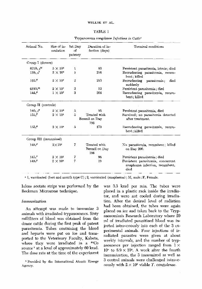
An attempt was made to immunize 3 animals with irradiated trypanosomes. Sixty milliliters of blood was obtained from the donor cattle during the first peak of patent parasitemia. Tubes containing the blood and heparin were put on ice and trans- ported to the Veterinary Faculty, Kabete, where they were irradiated in a WO source 3 at a level of approximately 60 krad. The dose rate at the time of the experiment
3 Provided by the International Atomic Energy Agency.
was 3.3 krad per min. The tubes were placed in a plastic rack inside the irradia- tor, and were not cooled during irradia- tion. After the desired level of radiation had been obtained, the tubes were again placed on ice and taken back to the Tryp- anosomiasis Research Laboratory where 20 ml of irradiated parasitized blood was in- jected intravenously into each of the 3 ex- perimental animals. Four injections of ir- radiated parasites were given at about weekly intervals, and the number of tryp- anosomes per injection ranged from 1 x
10y to 8.9 x 10⁸. A week after the fourth immunization, the 3 immunized as well as 3 control animals were challenged intrave- nously with 2 x lo5 viable T. congolense.
T. Congobnse INFECTION IN CAITLE 9
RESULTS
Clinical Observations
The animals in these experiments were separated into 3 groups. Group I consisted of 5 animals which were injected with via- ble T. congolense and used as donors of trypanosomes for immunization and chal- lenge. Group II contained 3 animals which served as controls for the immunized cattle which were designated as ,Group III (Table I ) , The only observable protective effect elicited by the irradiated parasites in the animals in Group III, when compared to their controls in Group II, was an increase of 2 days in the length of their prepatent periods and a corresponding delay in the development of’ fever. After the onset of ,parasitemia, the course of infection in 2 immunized animals was similar to that of the controls. The #other immunized animal died relatively soon after challenge, but its death was complicated by concomitant ana- plasmosis. Since the 8 cattle in Groups I and II received no preinfection treatment, the course of infection in these animals was studied closely.
The length of the prepatent periods, but not the duration of the disease, was related to the numbers of trypanosomes injected
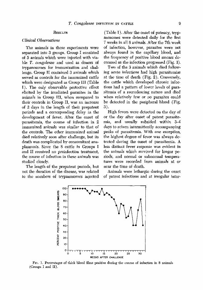
(Table I). After the onset ,of patency, tryp- anosomes were detected daily for the first 7 weeks in all 8 animals. After the 7th week of infection, however, parasites were not always f’ound in the capillary blood, and the frequency of positive blood smears de- creased as the infection progressed (Fig. 1).
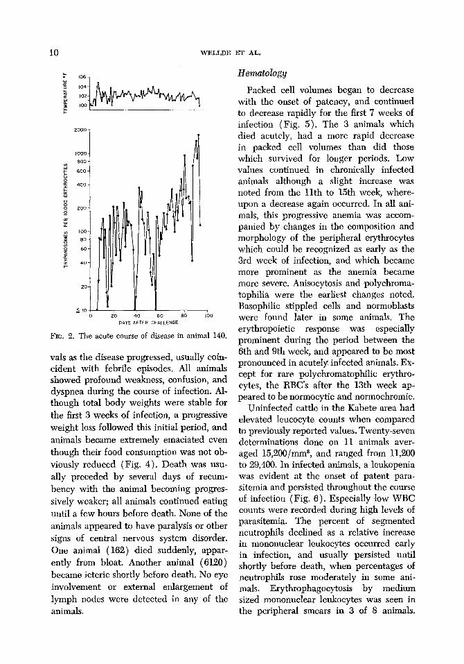
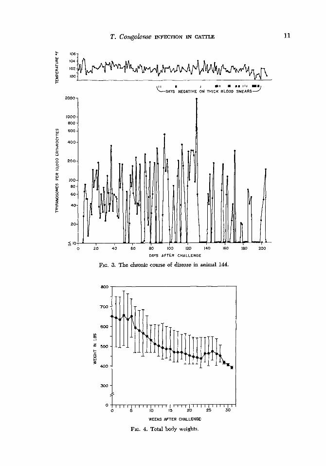
Two of the 3 animals which died follow- ing acute infections had high parasitemias at the time ‘of death (Fig. 2). Conversely, the cattle which developed chronic infec- tions had a pattern of lower levels of para- sitemia of a recrudescing nature and died when relatively few or no parasites could be detected in the peripheral blood (Fig.
3). High fevers were detected on the day of
or the day after onset of patent parasite- mia, and usually subsided within 34 days to return intermittently accompanying peaks of parasitemia. With one exception, the highest degree of fever was always de- tected during the onset of parasitemia. A less distinct fever response was evident in the animals which survived for longer pe- riods, and normal or subnormal tempera- tures were recorded from animals at or near the time of death.
Animals were lethargic during the onset of patent infections and at irregular inter-
100 - z 9 go-
z 60- 8 2 70-
5 60-
? I- 50-
Y F 40- iii
2 30-
!5 Y 20-
5 IO-
ok,,,,,,,,,,,,,,,,,,,,,,,,,,,,,,, 0 5 IO 15 20 25 30
WEEKS AFTER CHALLENGE
FIG. 1. Percentages of thick blood films positive during the course of infection in 8 animals (Groups I and II).
10 WELLDE ET AL.
FIG. 2. The acute course of disease in animal 140.
vals as the disease progressed, usually coin- cident with febrile episodes. All animals showed profound weakness, confusion, and dyspnea during the course of infection. Al- though total body weights were stable for the first 3 weeks of infection, ,a progressive weight loss followed this initial period, and animals became extremely emaciated even though their food consumption was not ob- viously reduced (Fig. 4). Death was usu- ally preceded by several days of recum- bency with the animal becoming progres- sively weaker; all animals continued eating until a few hours before death. None of the animals appeared to have paralysis or other signs of central nervous system disorder. One animal (162) ,died suddenly, appar- ently from bloat. Another animal (6120) became icteric shortly before death. No eye involvement or external enlargement of lymph nodes were detected in any of the animals.
Hematology
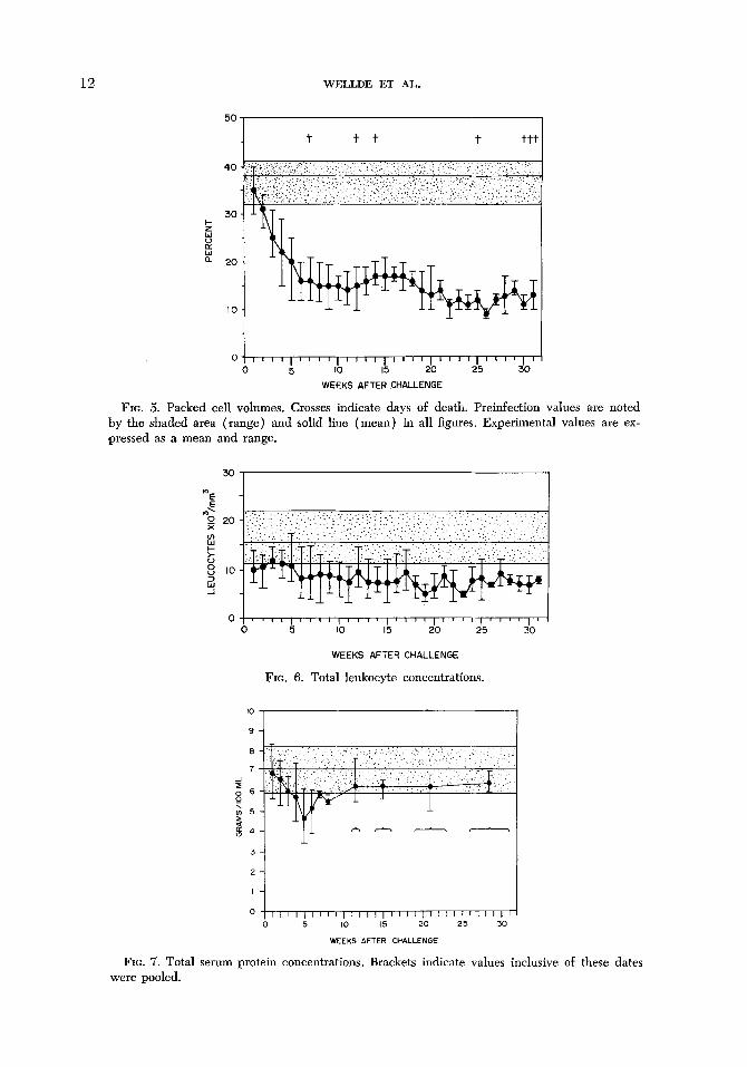
Packed cell volumes began to decrease with the onset of patency, and continued to decrease rapidly for the first 7 weeks of infection (Fig. 5). The 3 animals which died acutely, had a more rapid decrease in packed cell volumes than did those which survived for longer periods. Low values continued in chronically infected animals although a slight increase was noted from the 11th to 15th week, where- upon a decrease again occurred. In all ani- mals, this progressive anemia was accom- panied by changes in the composition and morphology of the peripheral erythrocytes which could be recognized as early as the 3rd week ‘of infection, and which became more prominent as the anemia became more severe. Anisocytosis and ,polychroma- tophilia were the earliest changes noted. Basophilic stippled cells and normoblasts were found later in some animals. The erythropoietic response was especially prominent during the period between the 6th and 9th week, and appeared to be most pronounced in acutely infected animals. Ex- cept for rare polychromatophilic erythro- cytes, the RBC’s after the 13th week ap- peared to be normocytic and normochromic.
Uninfected cattle in the Kabete area had elevated leucocyte counts when compared to previously reported values. Twenty-seven determinations done on 11 animals aver- aged 15,200/mm8, and ranged from 11,200 to 29,400. In infected animals, a leukopenia was evident at the onset of patent para- sitemia ‘and persisted throughout the course of infection (Fig. 6). Especially low WBC counts were recorded during high levels of parasitemia. The percent of segmented neutrophils declined as a relative increase in mononuclear leukocytes occurred early in infection, and usually persisted until shortly before death, when percentages of neutrophils rose moderately in some ani- mals. Erythrophagocytosis by medium sized mononuclear leukocytes was seen in the peripheral smears in 3 of 8 animals.
T. Congohse INFECTION IN CATTLE 11
b
2000
1000 600
v) k 600
k 400 I
k 8 200 g-
E 100 s 60 I w 60 z" % z 40 I-
20
6 IO 20 40
CL’ ’ l II . I, III I.
DAYS NEGATIVE ON THICK BLOOD SMEARS J
.
DAYS AFTER CHALLENGE
FIG. 3. The chronic course of disease in animal 144.
600
!i
z 500
g !i
400
300
0 ‘1 0
,,,l,,llll”l’l”lll”lll’l”l~ 5 IO 15 20 25 30
WEEKS AFTER CHALLENGE.
FIG. 4. Total body weights.
12 WELLDE ET AL.
0 ,,,,,,,,,,,,,,,,,,,,1’,‘,““,’ 0 5 IO 15 20 25 30
WEEKS AFTER CHALLENGE
FIG. 5. Packed cell volumes. Crosses indicate days of death. Preinfection values are noted by the shaded area (#range) and solid line (mean) in all figures. Experimental values are ex- pressed as a mean and range.
30 1
0 5 io I5 $0 is
WEEKS AFTER CHALLENGE
FIG. 6. Total leukocyte concentrations.
FIG. 7. Total serum protein concentrations. Brackets indicate values inclusive of these dates were pooled.
T. COngObW INFECTION IN CATTLE 13
Both macrophages and neutrophils with remnants of trypanosomes in their cyto- plasm were also seen in peripheral smears.
Serum Chemical Components
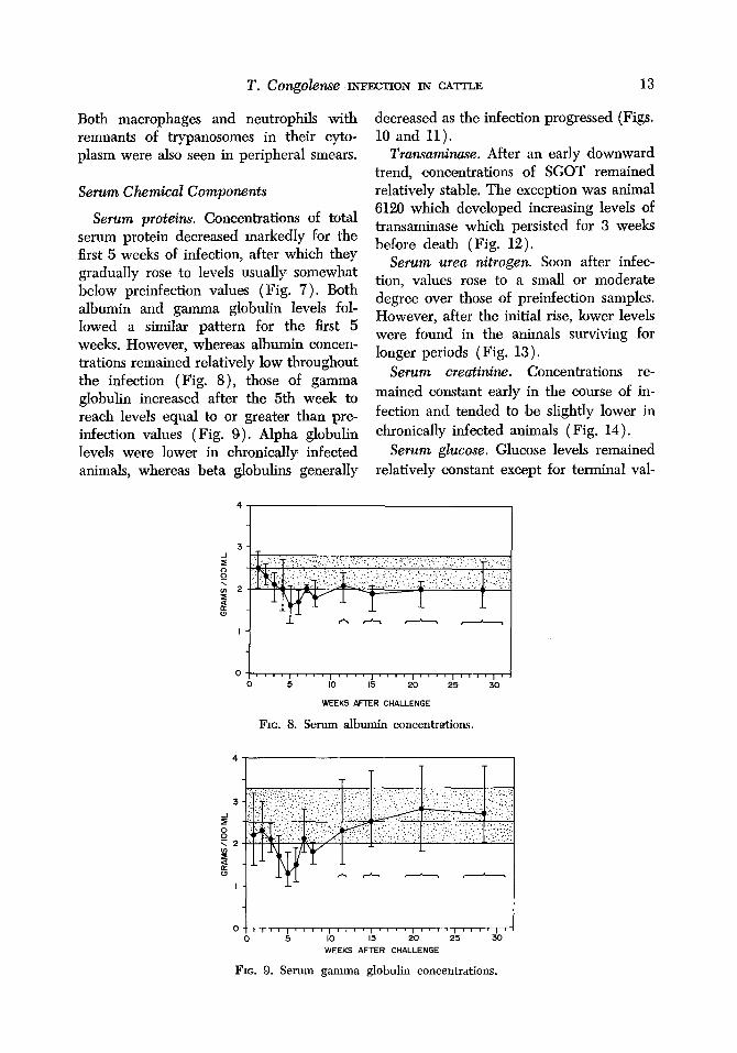
Serum proteins. Concentrations of total serum protein decreased markedly for the first 5 weeks of infection, after which they gradually rose to levels usually somewhat below preinfection values (Fig. 7). Both albumin and gamma globulin levels fol- lowed a similar pattern for the first 5 weeks. However, whereas albumin ‘concen- trations remained relatively low throughout the infection (Fig. 8), those of gamma globulin increased after the 5th week to reach levels equal to or greater than pre- infection values (Fig. 9). Alpha globulin levels were lower in chronically infected animals, whereas beta globulins generally
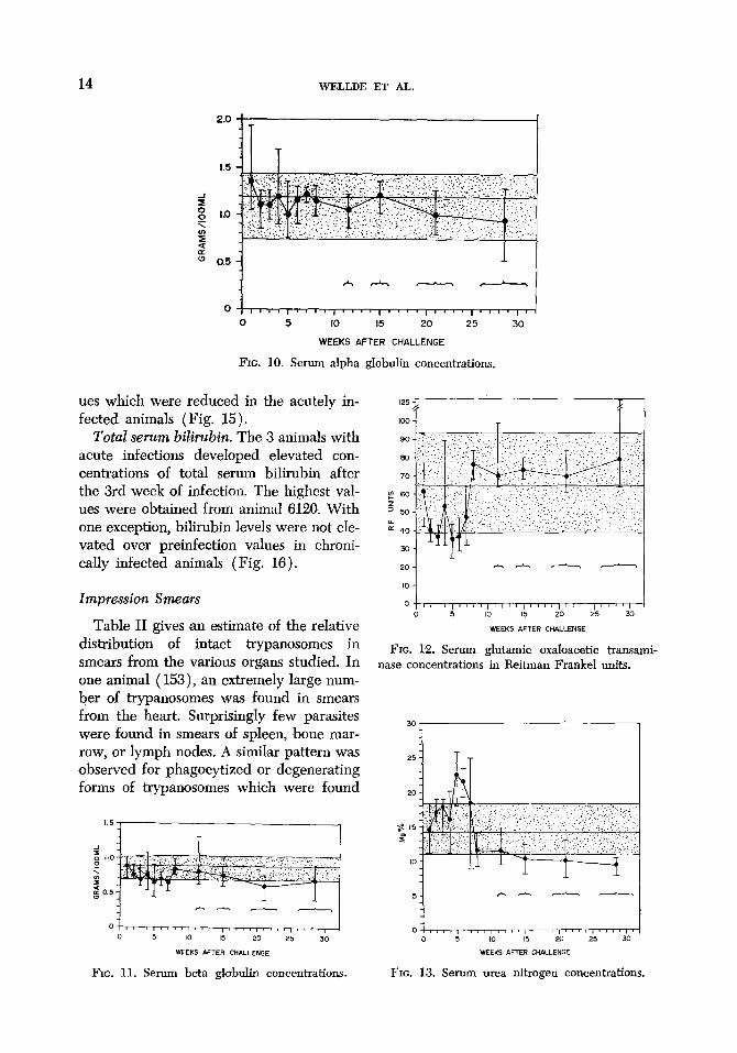
decreased as the infection progressed (Figs. 10 and 11).
Transaminase. After an early downward trend, concentrations of SGOT remained relatively stable. The exception was animal 6126 which developed increasing levels of transaminase which persisted for 3 weeks before death (Fig. 12).
Serum urea nitrogen. Soon after infec- tion, values rose to a small or moderate degree over those of preinfection samples. However, after the initial rise, lower levels were found in the animals surviving for longer periods (Fig. 13).
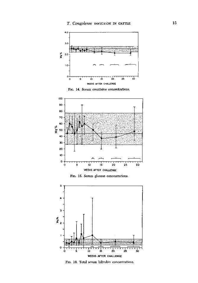
Serum creatinine. Concentrations re- mained constant early in the course of in- fection and tended to be slightly lower in chronically infected animals (Fig. 14).
Serum glucose. Glucose levels remained relatively constant except for terminal val-
0 5 IO 15 20 25 30
WEEKS AFTER CHALLENGE
FIG. 8. Serum albumin concentrations.
4
T T T
IL:: 0 5 IO 15 20 25
WEEKS AFTER CHALLENGE
FIG. 9. Serum gamma globulin concentrations.
14 WJZLLDE ET AL.
,,,,,,,,,(,,,‘,,,,,,,,,,,,,,,,,,
0 5 IO 15 20 25 30
WEEKS AFTER CHALLENGE
FIG. 10. Serum alpha globulin concentrations.
ues which were reduced in the acutely in- fected animals (Fig. 15).
Total serum bilirubin. The 3 animals with acute infections developed elevated con- centr,ations of total serum bilirubin after the 3rd week of infection. The highest val- ues were obtained from animal 6120. With one exception, bihrubin levels were not ele- vated over preinfection values in chroni- cally infected animals (Fig. 16).
Impression Smears
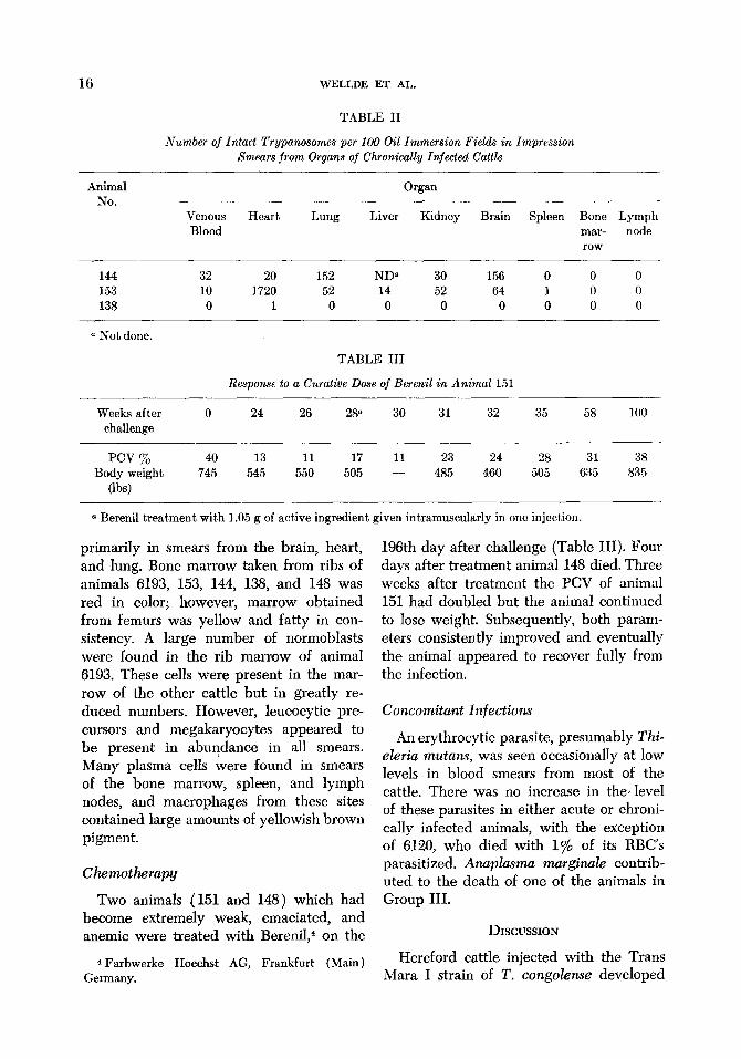
Table II gives an estimate of the relative distribution of intact trypanosomes in smears from the various organs studied. In one animal ( 153), an extremely large num- ber of trypanosomes was found in smears from the heart. Surprisingly few parasites were found in smears of spleen, bone mar- row, or lymph nodes. A similar pattern was observed for phagocytized or degenerating forms ,of trypanosomes which were found
1.5 1
FIG. 11. Serum beta globulin concentrations FIG. 13. Serum urea nitrogen concentrations.
FIG. 12. Serum glutamic oxaloacetic transami- nase concentrations in Reitman Frankel units.
T. COf&@IS~ INFECTIbN IN CA’ITLE 15
0 5 IO 15 20 25 30
WEEKS AFTER CHALLENGE
FIG. 14. Serum creatinine concentrations.
0 5 IO 15 20 25 30
WEEKS AFTER CHALLENGE
FIG. 15. Serum glucose concentrations.
5
4
3
s?
502
I
0
WEEKS AFTER CHALLENGE
FIG. 16. Total serum bihubin concentrations.
16 WELLDE ET AL.
TABLE II
Number of Intact Trypanosomes per 100 Oil Immersion Fields in Impression Smears from Organs of Chronically Injected Cattle
Animal No.
Venous Blood
Heart Lung
Organ
Liver Kidney Brain Spleen Bone Lymph mar- node row
144 32 20 152 ND5 30 156 0 0 0 153 10 1720 52 14 52 64 1 0 0 138 0 1 0 0 0 0 0 0 0
a Not done.
TABLE III
Response to a Curative Dose of Berenil in Animal 151
Weeks after challenge
0 24 26 28” 30 31 32 35 58 100
pm % 40 13 11 17 11 23 24 28 31 Body weight 745 545 550 505 - 485 460 505 635
(lb4
a Berenil treatment with 1.05 g of active ingredient given intramuscularly in one injection.
38 835
primarily in smears from the brain, heart, and lung. Bone marrow taken from ribs of animals 6193, 153, 144, 138, and I48 was red in color; however, marrow obtained from femurs was yellow and fatty in con- sistency. A large number of normoblasts were found in the rib marrow of animal 6193. These cells were present in the mar- row of the other cattle but in greatly re- duced numbers. However, leucocytic pre- cursors and megakaryocytes ‘appeared to be present in abuqdance in all smears. Many plasma cells were found in smears of the bone marrow, spleen, and lymph nodes, and macrophages from these sites contained large ,amounts of yellowish brown pigment.
Chemotlwapy
Two animals ( 151 and 148 ) which had become extremely weak, emaciated, and anemic were treated with Berenil,4 on the
4 Farbwerke Hoechst AG, Frankfurt (Main) Germany.
196th day after challenge (Table III). Four days after treatment animal 148 died. Three weeks after treatment the PCV of animal 151 had doubled but the animal continued to lose weight. Subsequently, both param- eters consistently improved and eventually the animal appeared to recover fully from the infection.
Concomitant Infections
An erythrocytic parasite, presumably Thi- eleria mutans, was seen occasionally at low levels in blood smears from most of the cattle. There was no increase in the, level of these parasites in either acute or chroni- cally infected animals, with the exception of 6120, who died with 1% of its RBC’s parasitized. Anuplasmu murginule contrib- uted to the death of one of the animals in Group III.
DISCUSSION
Hereford cattle injected with the Trans Mara I strain of T. congolense developed
T. COfZ@Jhe INFECTION IN CATTLE 17
acute and chronic diseases comparable to those described by Fiemms ( 1970). We did not see the hyperacute or recovery courses of infection described by this author in any of our animals. This strain of T. congolense produced a similar disease in young Hol- stein bulls (J. S. Anderson, unpubl. ) and extremely virulent hyperacute infections developed in Beagle dogs infected with this parasite (Johnson et al. unpubl.).
The most striking and puzzling aspect of T. congolense infections in Hereford cattle was the development and persistence of anemia. An erythropoietic response to this anemia was evident early in the course of infection, but fmor unknown reasons sub- sided wthout having resulted in greatly elevated packed cell volumes. Chronically infected cattle continued to be anemic without any further evidence of erythro- poietic response until the time of death, even though trypanosomes were being sup- pressed in large measure. It could be spec- ulated that the presence of live trypano- somes was needed to perpetuate the anemic condition, since after curative chemother- apy the level of packed cell volumes gradu- ally increased, and eventually regained pre- infectIon levels. The finding of elevated levels of total bilirubin in animals which underwent the most severe anemia was probably due to hemolysis of erythrocytes, since even in severe liver disease in cattle, serum bilirubin levels rise only slightly. All of the cattle in these studies also had large amounts of yellowish brown iron positive pigment in macrophages of the spleen, liver, and bone marrow. This pigment, thought to be hemosiderin, was seen in both impression smears ,and histological sections ( Kaliner 1974). The excessive ac- cumulations ‘of pigment have been reported by other authors and appears to be a rela- tively consistent finding, although Krampitz (1970) reported that only 1 of 16 cattle which died of trypanosomiasis in his stud- ies had excessive 8accumulation of pigment. These apparent discrepancies may be the
result of differences in severity of anemias and corresponding destruction of erythro- cytes among various experiments. While the initial fall in packed cell volumes seems to be associated primarily with destruction, i.e., hemolysis of erythrocytes and erythro- phagocytosis, it ,appears that later in the course ‘of infection a suppression of produc- tion ‘of erythrocytes also becomes a promi- nent factor. Studies of bone marrow ob- tained from the rib indicated that some erythropoesis was occurring, but overall erythropoiesis appeared to be suppressed, since marrow in the long bones had not regenerated. While the etiology of the ane- mia has not been defined, the suggestion that it is at least in part immunologically induced has been proposed by a number of workers (see Desowitz 1970 for review).
Concentration of total serum proteins also decreased rapidly early during the in- fection, and may be the result of increased protein breakdown, loss by proteinuria, dis- turbances in metabolism or absorption, or dilution as suggested by Fiennes ( 1970). The animals regained some of the early loss of total serum protein and in chronically infected animals the value was not severely lowered. The total body weight continued to decrease, however, and the relatively stable serum protein values found later were probably maintained at the expense of the tissue protein. The early depletion or dilution of gamma globulin is coincident with the decrease noted in the complement fixation and indirect fluorescent antibody titers of the same animals (Lotzsch and Deindl 1974). Levels of gamma globulin increased after the 5th week and may re- flect a more pronounced antibody response which in chronically infected cattle ap- parently influences the level of parasitemia; 7s gamma globulins obtained from the chronically infected animals at the terminal stage strongly inhibited parasitemia in mice infected with the same strain of T. congo- Zense ( Wellde and Rodriguez unpubl. ). Desowitz ( 1959) observed simil.ar fluctua-
18 WELLDE ET AL.
tions in serum protein concentrations in susceptible breeds of cattle infected with T. oiuaxr.
Excessive catabolism of serum protein could be responsible for the elevated urea nitrogen levels, since the loss of protein and increased urea nitrogen values occurred during the same #period. There was no cor- responding increase in creatinine levels which would be expected if renal impair- ment had occurred, although some degree of kidney pathology was present (Kaliner 1974). The slightly lower levels of urea nitrogen present in chronic animals may reflect a decrease in the rate of protein metabolism.
The reIativeIy stable leveIs of SGOT in- dicate that the liver was not severely af- fected by the infection. No severe histo- pathologic lesions were found there or in the heart or skeleta1 muscle (Kaliner 1974).
The spleen did not appear to be greatly involved ,during the ‘course of infection, since hypertrophy of this organ was not noted in either acute or chronically in- fected animals in our study. It would be reasonable to assume that the spleen, acting as the major lymphatic organ of the circu- latory system, would actively sequester, phagocytize, and ‘destroy ‘circulating tryp- anosomes, but evidence from our studies does not confirm this assumption. Plasma cells, however, were found in abundance in the spleen and the antibody forming role of the spleen may prove to be of an important nature. Krampitz (1970) has pointed ‘out that much conflicting informa- tion has been published on splenic hyper- trophy in bovine trypanosomiasis and the subject needs more concentrated study.
While the injections of irradiated T. con- golense initiated a detectable antibody re- sponse (Liitzsch and Deindl 1974), they did not produce a strong resist,ance. The effect of a 2 day ‘delay in onset of patent parasitemia corresponds to an approximate one hundredfold dilution of the challenge inoculum. This is in contrast to the strong
resistance produced in cattle by irradiated T. &.od&ense (Wellde et al. 1973). In general, T. congolense has proven to be a more ,difficult parasite to control by experi- mental immunization than T. rhodesiense, either by the use of attenuated parasites (Duxbury et al. 1973), or by infection and chemotherapeutic cure (F&on and Lourie 1946). Whether or not this indicates a greater propensity for antigenic vari’ation on the part ,of T. congolense (Wilson and Cunningham 1972), is not known.
ACKNOWLEDGMENTS
Dr. I. E. Muriithi, Director of Veterinary Ser- vices, is thanked for inviting us to proceed with these experiments in the facilities under his super- vision at Kabete, Kenya. We thank Dr. John Tremlett, Chief Veterinary Research Officer, Mr. Jan Le’Roux, Chief Zoologist, and Mr. Samuel Githatha, Zoologist, for their advice and assistance during these experiments. Dr. R. Schindler’s con- tribution to the planning of these experiments was also appreciated. Excellent technical assistance was provided by the workers at the Trypanosomiasis Research Laboratory, and we especially thank Samuel Gitchunge, Shem Kaiga, and Reuben Mu- tuaruhiu. We also thank Mr. Arthur Moon, Dr. Carter Diggs, Dr. Anthony Johnson, and Mrs. Genevieve Zivona, for their suggestions and help with the manuscript.
F~J~FFXIENCJB
DESOWITZ, R. S. 1959. ,Studies on Immunity and Has-Parasite Relationships. I. The Immuno- logical response of resistant and susceptible breeds of cattle to trypanosomal challenge. Annuls of Tropical Medicine and Parasitology 53, 293413.
DESOWITZ, R. S. 1970. African trypanosomes, In “Immunity to Parasitic An&n&,” Vol. 2, (G. J. Jackson, R. Rerman, and I. Singer, Eds.), pp. 551-596. Appleton-Century-Crofts, New York.
DUXBURY, R. E., ANDERSON, J. S., WELLDE, B. T., SADUN, E. H., AND MURIITHI, I. E. 1972. T ypanosomu congolense: Immunization of mice, dogs, and cattle with gamma irradiated parasites. Experimental Parasitobgy 31, X7- 533.
FIENNES, R. N. T.-W. 1970. Pathogenesis and pathology of animal trypanosomiases. In “The
T. cO~gOk??ISe INFECTION IN CA’ITLE 19
African Trypanosomes,” (H. W. Mulligan, Ed. ), pp. 729-756. Wiley-Interscience, New York.
FULTON, J. D., AND Lourux, E. M. 1946. The im- munity of mice cured of trypanosome infec- tions. Annals of Tropical Medicine and Para- sitology 40, 1-9.
GENTZKOW, C. J. 1942. An accurate method for the determination of blood urea nitrogen by direct nesslerization. Journal of Biological Chemistry 143, 531544.
GOODWIN, L. G. 1970. The pathology of African trypanosomiasis. Transactions of the Royal So- ciety of Tropical Medicine and Hygiene 64, 797-817.
JENDRASSIK, L., AND GROF, P. 1938. Vereinfachte photometrische methoden zur bestimmung des blutbilirubins. Biochemische Zeitschrift 297, 81-89.
KALINER, G. 1974. Typanosomu congolense. II. Histopathologic findings in experimentally in- fected cattle. Experimental Parasitology 36, 20-26.
K~MPITZ, H. E. 1970. Beobachtungen und ex- perimentellen infektionen ostafrikanischer ze- burinder mit Wildstammen von Tryputwsomu congohe. Zeitschrift fiir Tropenmdz.in und Pamsitologie 21, l-20.
Loses, G. J., AM) IKEDE, B. 0. 1972. Review of Pathology of Diseases in domestic and labora- tory animals caused #by Trypanosoma congo- lens. T. uivax, T. brucei, T. rhodesiense, and T. gambiense. Veterinary Pathology, Suple- mentum ad Vol. 9.
L&rzsc~, R., AND DEINDL, G. 1974. Typarwsomu congoIense. III. 8erological response of ex- perimentally infected cattle. Experimental Parasitology 36, 27-33.
WELLDE, B. T., DUXBURY, R. E., SAD&, E. H., LANGBEHN, H. R., LBrzscrr, R., DEINDL, G., AND WARUI, G. 1973. Experimental infections with African trypanosomes: IV. Immunization of cattle with gamma irradiated Typarwsomu rhoclesiense. Experimental Parasitology (in press).
WILLIAMS, J. S., MJZRONEY, F. C., Hmr, G., AND SADUN, E. H. 1966. #Serum chemical com- ponents in mice determined by the use of ultramicro techniques. .lournal of Applied Physiology 21, 1026-1030.
WILSON, A. J., AND GUNNINGHAM, M. P. 1972. Immunological Aspects of Bovine trypanoso- miasis. I. Immune response of cattle to infec- tion with Trypanosomu congolense and the antigenic variation of the infecting organism. Experimental Parasitology 32, 165-173.