TRP channels and organelles - University of...
16
The Department of Molecular, Cellular, and Developmental Biology, University of Michigan, Ann Arbor, Michigan, USA TRP channels and organelles Overview of TRP channels Transient receptor potential (TRP) is a cation channel superfamily with diverse physiological functions including thermosensation and mechanosensation (excellently re- viewed in Refs. Clapham 2003; Montell 2005; Nilius et al. 2007). Mammalian TRPs can be divided into six subfamilies: TRPC(1–7), TRPV(1–6), TRPM(1–8), TRPA(1), TRPP(1– 3), and TRPML(1–3) (Fig. 1). In past years, extensive efforts have been made to elucidate three basic aspects of TRP channels: the channel pore properties, the activation (gating) mechanisms, and the channels’ subcellular localization. Of these, the biophysical properties of the TRP channel pore have been best characterized. When activated, most TRPs conduct Ca 2+ , as well as Na + and K + ions. The activation/ gating mechanisms, however, have not been described in detail for most TRPs. In sensory cells such as somatosensory neurons, a subset of TRP channels (so-called ‘sensory’ TRPs) are activated by a variety of environmental cues such as temperature, mechanical force, and plant-derived volatiles (reviewed in Refs. Clapham 2003; Montell 2005; Nilius et al. 2007). Many of the sensory TRPs, along with the Received January 17, 2010; accepted January 25, 2010. Address correspondence and reprint requests to Haoxing Xu, The Department of Molecular, Cellular, and Developmental Biology, Uni- versity of Michigan, 3089 Natural Science Building (Kraus), 830 North University, Ann Arbor, MI 48109, USA. E-mail: [email protected] Abbreviations used: AM, acetoxymethyl; BAPTA, 1,2-bis(2-amino- phenoxy)ethane-N,N,N¢,N¢-tetraacetic acid; CaM, Calmodulin; CICR, Ca 2+ -induced Ca 2+ release; EPSP, excitatory postsynaptic potential; ER, endoplasmic reticulum; IP 3 R, inositol 1,4,5-trisphosphate receptor; LEL, late endosomal and lysosomal; LROs, lysosome-related organelles; MHCII, major histocompatibility complex II; ML4, type IV mucolipi- dosis; NAADP, nicotinic acid adenine dinucleotide phosphate; PIP, phosphoinositide; PM, plasma membrane; RyR, ryanodine receptor; SG, secretory granule; SNARE, soluble NSF attachment protein receptors; SR, sarcoplasmic reticulum; SV, secretory vesicle; SyV, synaptic vesicle; TG, thapsigargin; TGN, trans-Golgi network; TRP, transient receptor potential. Abstract Ion channels are classically understood to regulate the flux of ions across the plasma membrane in response to a variety of environmental and intracellular cues. Ion channels serve a number of functions in intracellular membranes as well. These channels may be temporarily localized to intracellular mem- branes as a function of their biosynthetic or secretory path- ways, i.e., en route to their destination location. Intracellular membrane ion channels may also be located in the endocytic pathways, either being recycled back to the plasma mem- brane or targeted to the lysosome for degradation. Several channels do participate in intracellular signal transduction; the most well known example is the inositol 1,4,5-trisphosphate receptor (IP 3 R) in the endoplasmic reticulum. Some organellar intracellular membrane channels are required for the ionic homeostasis of their residing organelles. Several newly-dis- covered intracellular membrane Ca 2+ channels actually play active roles in membrane trafficking. Transient receptor potential (TRP) proteins are a superfamily (28 members in mammal) of Ca 2+ -permeable channels with diverse tissue distribution, subcellular localization, and physiological func- tions. Almost all mammalian TRP channels studied thus far, like their ancestor yeast TRP channel (TRPY1) that localizes to the vacuole compartment, are also (in addition to their plasma membrane localization) found to be localized to intracellular membranes. Accumulated evidence suggests that intracellularly-localized TRP channels actively participate in regulating membrane traffic, signal transduction, and vesicular ion homeostasis. This review aims to provide a summary of these recent works. The discussion will also be extended to the basic membrane and electrical properties of the TRP-residing compartments. Keywords: endosomes, intracellular channel, lysosomes, membrane traffic, TRP channel, TRPML. J. Neurochem. (2010) 10.1111/j.1471-4159.2010.06626.x JOURNAL OF NEUROCHEMISTRY | 2010 doi: 10.1111/j.1471-4159.2010.06626.x ȑ 2010 The Authors Journal Compilation ȑ 2010 International Society for Neurochemistry, J. Neurochem. (2010) 10.1111/j.1471-4159.2010.06626.x 1
Transcript of TRP channels and organelles - University of...
The Department of Molecular, Cellular, and Developmental Biology, University of Michigan,
Ann Arbor, Michigan, USA
TRP channels and organelles
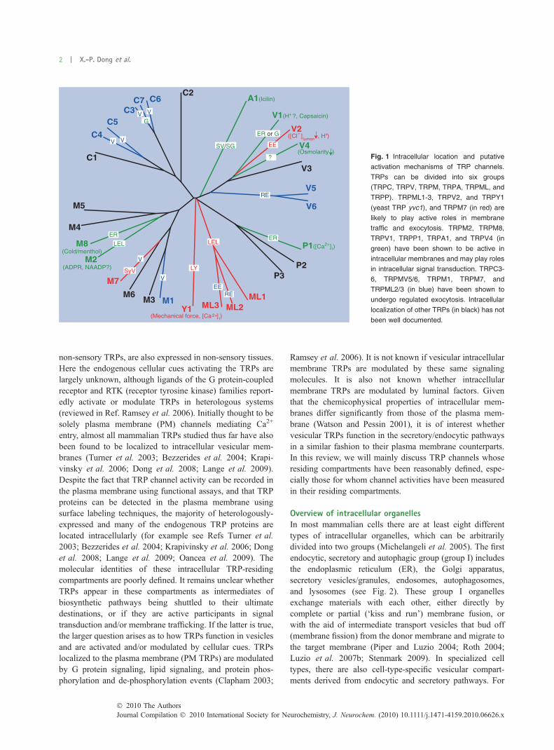
Overview of TRP channelsTransient receptor potential (TRP) is a cation channelsuperfamily with diverse physiological functions includingthermosensation and mechanosensation (excellently re-viewed in Refs. Clapham 2003; Montell 2005; Nilius et al.2007). Mammalian TRPs can be divided into six subfamilies:TRPC(1–7), TRPV(1–6), TRPM(1–8), TRPA(1), TRPP(1–3), and TRPML(1–3) (Fig. 1). In past years, extensive effortshave been made to elucidate three basic aspects of TRPchannels: the channel pore properties, the activation (gating)mechanisms, and the channels’ subcellular localization. Ofthese, the biophysical properties of the TRP channel porehave been best characterized. When activated, most TRPsconduct Ca2+, as well as Na+ and K+ ions. The activation/gating mechanisms, however, have not been described indetail for most TRPs. In sensory cells such as somatosensoryneurons, a subset of TRP channels (so-called ‘sensory’
TRPs) are activated by a variety of environmental cues suchas temperature, mechanical force, and plant-derived volatiles(reviewed in Refs. Clapham 2003; Montell 2005; Niliuset al. 2007). Many of the sensory TRPs, along with the
Received January 17, 2010; accepted January 25, 2010.Address correspondence and reprint requests to Haoxing Xu, The
Department of Molecular, Cellular, and Developmental Biology, Uni-versity of Michigan, 3089 Natural Science Building (Kraus), 830 NorthUniversity, Ann Arbor, MI 48109, USA. E-mail: [email protected] used: AM, acetoxymethyl; BAPTA, 1,2-bis(2-amino-
phenoxy)ethane-N,N,N¢,N¢-tetraacetic acid; CaM, Calmodulin; CICR,Ca2+-induced Ca2+ release; EPSP, excitatory postsynaptic potential; ER,endoplasmic reticulum; IP3R, inositol 1,4,5-trisphosphate receptor; LEL,late endosomal and lysosomal; LROs, lysosome-related organelles;MHCII, major histocompatibility complex II; ML4, type IV mucolipi-dosis; NAADP, nicotinic acid adenine dinucleotide phosphate; PIP,phosphoinositide; PM, plasma membrane; RyR, ryanodine receptor; SG,secretory granule; SNARE, soluble NSF attachment protein receptors;SR, sarcoplasmic reticulum; SV, secretory vesicle; SyV, synaptic vesicle;TG, thapsigargin; TGN, trans-Golgi network; TRP, transient receptorpotential.
Abstract
Ion channels are classically understood to regulate the flux of
ions across the plasma membrane in response to a variety of
environmental and intracellular cues. Ion channels serve a
number of functions in intracellular membranes as well. These
channels may be temporarily localized to intracellular mem-
branes as a function of their biosynthetic or secretory path-
ways, i.e., en route to their destination location. Intracellular
membrane ion channels may also be located in the endocytic
pathways, either being recycled back to the plasma mem-
brane or targeted to the lysosome for degradation. Several
channels do participate in intracellular signal transduction; the
most well known example is the inositol 1,4,5-trisphosphate
receptor (IP3R) in the endoplasmic reticulum. Some organellar
intracellular membrane channels are required for the ionic
homeostasis of their residing organelles. Several newly-dis-
covered intracellular membrane Ca2+ channels actually play
active roles in membrane trafficking. Transient receptor
potential (TRP) proteins are a superfamily (28 members in
mammal) of Ca2+-permeable channels with diverse tissue
distribution, subcellular localization, and physiological func-
tions. Almost all mammalian TRP channels studied thus far,
like their ancestor yeast TRP channel (TRPY1) that localizes
to the vacuole compartment, are also (in addition to their
plasma membrane localization) found to be localized to
intracellular membranes. Accumulated evidence suggests
that intracellularly-localized TRP channels actively participate
in regulating membrane traffic, signal transduction, and
vesicular ion homeostasis. This review aims to provide a
summary of these recent works. The discussion will also be
extended to the basic membrane and electrical properties of
the TRP-residing compartments.
Keywords: endosomes, intracellular channel, lysosomes,
membrane traffic, TRP channel, TRPML.
J. Neurochem. (2010) 10.1111/j.1471-4159.2010.06626.x
JOURNAL OF NEUROCHEMISTRY | 2010 doi: 10.1111/j.1471-4159.2010.06626.x
� 2010 The AuthorsJournal Compilation � 2010 International Society for Neurochemistry, J. Neurochem. (2010) 10.1111/j.1471-4159.2010.06626.x 1
non-sensory TRPs, are also expressed in non-sensory tissues.Here the endogenous cellular cues activating the TRPs arelargely unknown, although ligands of the G protein-coupledreceptor and RTK (receptor tyrosine kinase) families report-edly activate or modulate TRPs in heterologous systems(reviewed in Ref. Ramsey et al. 2006). Initially thought to besolely plasma membrane (PM) channels mediating Ca2+
entry, almost all mammalian TRPs studied thus far have alsobeen found to be localized to intracellular vesicular mem-branes (Turner et al. 2003; Bezzerides et al. 2004; Krapi-vinsky et al. 2006; Dong et al. 2008; Lange et al. 2009).Despite the fact that TRP channel activity can be recorded inthe plasma membrane using functional assays, and that TRPproteins can be detected in the plasma membrane usingsurface labeling techniques, the majority of heterologously-expressed and many of the endogenous TRP proteins arelocated intracellularly (for example see Refs Turner et al.2003; Bezzerides et al. 2004; Krapivinsky et al. 2006; Donget al. 2008; Lange et al. 2009; Oancea et al. 2009). Themolecular identities of these intracellular TRP-residingcompartments are poorly defined. It remains unclear whetherTRPs appear in these compartments as intermediates ofbiosynthetic pathways being shuttled to their ultimatedestinations, or if they are active participants in signaltransduction and/or membrane trafficking. If the latter is true,the larger question arises as to how TRPs function in vesiclesand are activated and/or modulated by cellular cues. TRPslocalized to the plasma membrane (PM TRPs) are modulatedby G protein signaling, lipid signaling, and protein phos-phorylation and de-phosphorylation events (Clapham 2003;
Ramsey et al. 2006). It is not known if vesicular intracellularmembrane TRPs are modulated by these same signalingmolecules. It is also not known whether intracellularmembrane TRPs are modulated by luminal factors. Giventhat the chemicophysical properties of intracellular mem-branes differ significantly from those of the plasma mem-brane (Watson and Pessin 2001), it is of interest whethervesicular TRPs function in the secretory/endocytic pathwaysin a similar fashion to their plasma membrane counterparts.In this review, we will mainly discuss TRP channels whoseresiding compartments have been reasonably defined, espe-cially those for whom channel activities have been measuredin their residing compartments.
Overview of intracellular organellesIn most mammalian cells there are at least eight differenttypes of intracellular organelles, which can be arbitrarilydivided into two groups (Michelangeli et al. 2005). The firstendocytic, secretory and autophagic group (group I) includesthe endoplasmic reticulum (ER), the Golgi apparatus,secretory vesicles/granules, endosomes, autophagosomes,and lysosomes (see Fig. 2). These group I organellesexchange materials with each other, either directly bycomplete or partial (‘kiss and run’) membrane fusion, orwith the aid of intermediate transport vesicles that bud off(membrane fission) from the donor membrane and migrate tothe target membrane (Piper and Luzio 2004; Roth 2004;Luzio et al. 2007b; Stenmark 2009). In specialized celltypes, there are also cell-type-specific vesicular compart-ments derived from endocytic and secretory pathways. For
C2C6C7
C3C5
C4
C1
V1(H ?, Capsaicin)
([Cl ] , H )
(Osmolarity )
V3
V5
V6
P1([Ca ] )
P2P3
ML1ML2ML3Y1
M1M3M6
M7
M2(ADPR, NAADP?)
M8 (Cold/menthol)
M4
M5
2+i
(Mechanical force, [Ca ] ) 2+i
+
lumen
LEL LEL
LY
V
V
V
RE
EE
?
ER
EERE
ER or GV
V
ER
V
+V2
V4
-
SyV
A1(Icilin)
SV/SG
G
Fig. 1 Intracellular location and putative
activation mechanisms of TRP channels.
TRPs can be divided into six groups
(TRPC, TRPV, TRPM, TRPA, TRPML, and
TRPP). TRPML1-3, TRPV2, and TRPY1
(yeast TRP yvc1), and TRPM7 (in red) are
likely to play active roles in membrane
traffic and exocytosis. TRPM2, TRPM8,
TRPV1, TRPP1, TRPA1, and TRPV4 (in
green) have been shown to be active in
intracellular membranes and may play roles
in intracellular signal transduction. TRPC3-
6, TRPMV5/6, TRPM1, TRPM7, and
TRPML2/3 (in blue) have been shown to
undergo regulated exocytosis. Intracellular
localization of other TRPs (in black) has not
been well documented.
2 | X.-P. Dong et al.
Journal Compilation � 2010 International Society for Neurochemistry, J. Neurochem. (2010) 10.1111/j.1471-4159.2010.06626.x� 2010 The Authors
example melanosomes, which release melanin in response tolight, are specialized lysosome-related organelles (LROs) inmelanocytes (Blott and Griffiths 2002). In neurons, synapticvesicles that release neurotransmitters upon electrochemicalstimulation are derived from endosomes (Suudhof 2008).The second group of intracellular organelles includes mito-chondria, peroxisomes, and the nucleus. Both group I andgroup II organelles function as intracellular Ca2+ stores withthe luminal Ca2+ concentration ([Ca2+]lumen) ranging frommicromolar (lM) to millimolar (mM) values, 10- to 10,000-fold higher than the level of resting cytosolic Ca2+([Ca2+]cyt,� 100 nM) (Michelangeli et al. 2005). The Ca2+ gradient ineach of these organelles is actively established and main-tained by various Ca2+ pumps and/or secondary Ca2+
transporters (Fig. 2; see reviews Camello et al. 2002;Michelangeli et al. 2005).
Role of Ca2+ in signal transduction and organellarhomeostasisIntra-organellar Ca2+ release (efflux) has been shown to havean important role in signal transduction. For example, theinositol 1,4,5- trisphosphate receptor (IP3R) and the ryano-dine receptor (RyR) in the ER and sarcoplasmic reticulum(SR), respectively, couple numerous extracellular signals tointracellular events such as hormone secretion and musclecontraction (Berridge et al. 2003). Alternatively other secondmessengers, such as nicotinic acid adenine dinucleotidephosphate (NAADP) and sphingolipid-derived messengers,may induce Ca2+ release from intracellular stores via novelmechanisms (Berridge et al. 2003; Zhang and Li 2007;Calcraft et al. 2009). Intra-organellar Ca2+ release, effectinga reduction of [Ca2+]lumen, may also modulate the function oforganelles. For example, a reduction of [Ca2+]lumen mayaffect the protein folding and lipid synthesis in the ER(Corbett and Michalak 2000).
Ca2+-dependence of membrane trafficNumerous in vitro and in vivo studies suggest that Ca2+
release (efflux) from group I organelles (ER, Golgi, endo-somes, and lysosomes) is essential for membrane trafficking,fusion and fission (Burgoyne and Clague 2003; Hay 2007;Luzio et al. 2007b). The basic steps of fusion (tethering,docking, priming, and bilayer fusion) and the fusionmachinery (SNAREs, phosphoinositides, and Rabs) involvedare similar for fusion between intracellular membranes andfor plasma membrane exocytosis (Martens and McMahon2008; Suudhof 2008), suggesting that a similar mechanismmay regulate both processes. Ca2+-dependence has been welldocumented for regulated exocytotic events, i.e. fusion ofintracellular organelles such as synaptic vesicles, secretoryvesicles/granules, or lysosomes with the plasma membrane(Reddy et al. 2001; Blott and Griffiths 2002; Suudhof 2008).On the other hand, intracellular membrane traffic has beentraditionally classified to be ‘constitutive’ (Roth 2004; Hay
2007). Evidence now exists, however, suggesting thatintracellular traffic is also highly regulated. The final triggerof intracellular membrane fusion is also likely to be a rise of[Ca2+]cyt, whose amplitude and duration might be in a scaledifferent from fusion at plasma membrane (Burgoyne andClague 2003; Hay 2007). Both in vitro and in vivo studiessuggest that [Ca2+]cyt in the vicinity of organelles plays acritical role in most, but not all, fusion events during thebiosynthetic, secretory and endocytic pathways (Holroydet al. 1999; Luzio et al. 2000, 2007b; Pryor et al. 2000;Chen et al. 2002; Burgoyne and Clague 2003; Hay 2007).Using cell extracts from yeast or mammalian cells, in vitrofusion (content mixing) has been successfully reconstitutedbetween various endosomal compartments: early endosome-early endosome, late endosome – late endosome, lateendosome – lysosome, and lysosome-lysosome (Peters andMayer 1998; Holroyd et al. 1999; Pryor et al. 2000). In allcases, fusion is inhibited by 1,2-bis(2-aminophenoxy)ethane-N,N,N¢,N¢-tetraacetic acid (BAPTA), but not EGTA.Although both are strong chelators for Ca2+, BAPTA bindsCa2+ ions at least one hundred times faster than EGTA (Chenet al. 2002; Hay 2007). The increased sensitivity of endoso-mal fusion to BAPTA versus EGTA has been widelyinterpreted as evidence to support that intraluminal Ca2+
release is essential for triggering intracellular membranefusion, and that the putative action site is extremely close(estimated to be < 20 nm) to the Ca2+ release site (Pryoret al. 2000; Chen et al. 2002; Hay 2007). Using membranepermeable forms of chelators, i.e., BAPTA-acetoxymethyl(AM) ester and EGTA-AM, it was shown that, in intactmammalian cells, intraluminal Ca2+ release is required formany steps of intracellular transport. For example, antero-grade intra-Golgi transport and retrograde endosome-to-transGolgi network (TGN) transport require intraluminal Ca2+
release (Chen et al. 2002; Burgoyne and Clague 2003; Hay2007). The Ca2+-sensitive transport steps in the secretory andendocytotic pathways are summarized in Fig. 2 (whitearrows). Upon localized juxta-organellar Ca2+ elevation,two kinds of Ca2+ sensor proteins are activated to initiatemembrane fusion. Synaptotagmins (Syts), a family of proteinswith C2-type Ca2+ binding sites, are involved in theexocytosis of secretory vesicles, secretory granules, synapticvesicles and lysosomes (Reddy et al. 2001; Hay 2007;Luzio et al. 2007b; Suudhof 2008). Calmodulin (CaM), anEF-hand cytosolic protein, has been shown to play importantroles in multiple transport steps of secretory (for example,intra-Golgi transport) and endocytic pathways (forexample, early and late endosomal fusions) (Peters andMayer 1998; Burgoyne and Clague 2003; Hay 2007). HowCaM is recruited to various intracellular vesicles is still notclear.
Juxta-organellar luminal Ca2+ release also regulatesmembrane fission/budding events (Hay 2007; Luzio et al.2007a,b). Fission events share many common mechanisms
TRP calcium channels in intracellular organelles | 3
� 2010 The AuthorsJournal Compilation � 2010 International Society for Neurochemistry, J. Neurochem. (2010) 10.1111/j.1471-4159.2010.06626.x

with fusion events, although the outcome of fission orbudding is to extract, rather than add, membrane. Forexample, Rab proteins and phosphoinositides (PIPs) canregulate both membrane fission and fusion (Roth 2004).Membrane fission is also dependent upon Ca2+ (Hay 2007;Luzio et al. 2007a,b). For instance BAPTA-AM, but notEGTA-AM, inhibited vesicle budding in vitro (Ahluwaliaet al. 2001). The Ca2+ effector protein involved in thefission process is not known. In the TGN, both neuronalcalcium sensor 1 and Arf proteins are implicated (Hay2007).
Although the importance of organellar Ca2+ release insignal transduction, organelle homeostasis, and membranetraffic has been established, the molecular identities ofthe Ca2+ release proteins resident in these compartments haveremained elusive. Furthermore, the mechanisms that activatethe release channels are largely unknown. As many TRPproteins are Ca2+-permeable channels localized in themembranes of vesicles involved in the secretory and endo-cytic pathways, they are natural candidates to mediateorganellar Ca2+ release. A list of TRP channels with theirreported intracellular locations and potential functions isprovided in Fig. 1 and will be discussed in detail below.
TRP channels in the endocytic and autophagicpathways
Biogenesis of endosomes and lysosomesThe primary functions of endosomes and lysosomes (endo-lysosomes, collectively) are degradation, membrane traffick-ing, protein transport and signal transduction (for review, seeRefs. Berridge et al. 2003; Luzio et al. 2007b). Endosomesare endocytotic vesicles derived from the plasma membrane(Fig. 2). Primary endocytic vesicles undergo maturation andtrafficking to become early endosomes initially and lateendosomes or multi-vesicular bodies later with a time scaleof minutes or tens of minutes (Maxfield and McGraw 2004).Derived from late endosomes, lysosomes are membrane-enclosed compartments containing hydrolases for the intra-cellular digestion of macro-molecules (Luzio et al. 2007b).Lysosomes may fuse with late endosomes or multi-vesicularbodies to degrade macro-molecules (for example, the ligand-bound epidermal growth factor receptor) that are taken upfrom the plasma membrane via endocytosis (Luzio et al.2007b). Alternatively, lysosomes may fuse with obsoleteparts of the cell via autophagy (Luzio et al. 2007b). In bothcases, ingested materials are degraded to obtain energy and to
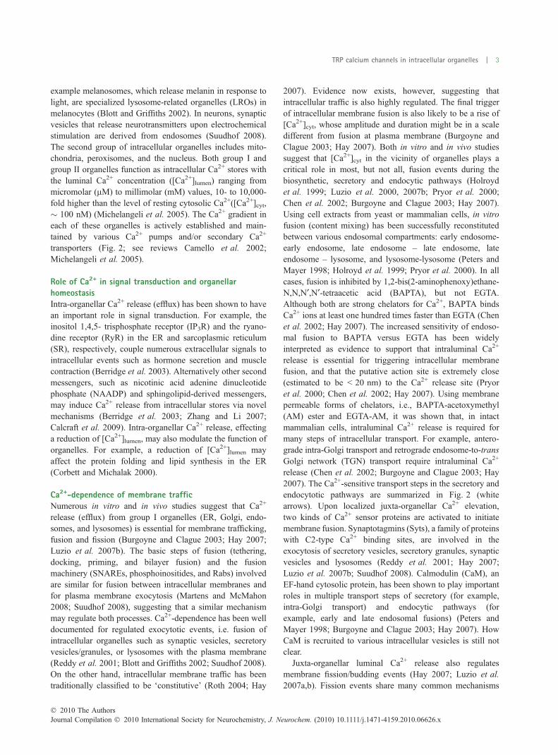
Fig. 2 Intracellular TRP channels in the secretory and endocytic
pathways. Intracellular compartments undergo membrane fusion and
fission/budding. There are two kinds of membrane fusions: ‘kiss and
run’ and complete fusion. In some steps, transport vesicles fission off
from the source membranes and fuse with the target membranes to
delivery cargos. Ca2+-sensitive membrane fusion and fission steps are
indicated with white arrows. The molecular identities of intracellular
compartments are defined by specific recruitment of small G proteins
(Rab and Arf GTPases) and the composition of phosphoinositides
(PIPs). The luminal ionic (H+ and Ca2+) composition is indicated for
each organelle. (a) The Biosynthetic Pathway. Essentially all TRPs
(labeled in mixed color) in the biosynthetic pathway may be present in
the ER (pH 7.2; [Ca2+]ER � 0. 7 mM; PI(4)P + PI(4,5)P2) and the Golgi
apparatus (trans-Golgi network; TGN; pH 6.4; [Ca2+]Golgi � 0. 3–
0.7 mM; PI(4)P + PI(4,5)P2). The Ca2+ gradients are established by
the thapsigargin (TG)-sensitive SERCA (sarco/endoplasmic reticulum
Ca2+) pump in the ER, and by both SERCA and TG-insensitive SPCA
(secretory pathway Ca2+) pumps in the Golgi. TRPV1 is reportedly
functional in the ER or Golgi. There are intermediate transport vesicles
derived from the ER and the Golgi apparatus. These transport vesicles
may deliver cargos to early endosomes (EE; pH 6.0; [Ca2+]EE �0.003–2 mM; PI(3)P; Rab5), late endosomes (LE; pH 5.5; [Ca2+]LE �0.5 mM; PI(3)P + PI(3,5)P2; Rab7 and Rab9), or the PM through
secretory vesicles (SV; pH6.4) and/or secretory granules (SG; pH 6.4).
[Ca2+]EE changes significantly during the maturation of early EE,
dropping from 2 mM in the primary endocytic vesicles to � 0.003 mM
20 min after endocytosis (Gerasimenko et al. 1998). SVs and SGs
deliver the newly synthesized TRP channels to the PM. (b) The En-
docytic Pathway. EEs are derived from the primary endocytic vesicles
after endocytosis. In addition to the late endocytic pathway, contents in
the EE can also be sorted into recycling endosomes (RE; pH 6.4;
[Ca2+]RE � 0. 003–2 mM; PI(3)P + PI(4)P + PI(4,5)P2; Rab11/Rab4),
which are subsequently recycled back to the PM. TRP channels may
be detected in the EE and RE as cargos during this cycle of endo-
cytosis and recycling. In addition, TRPV2, TRPML2, and TRPML3
may play active roles in the early endocytic pathways. TRPV2 is
activated by low pH and a reduction of intra-endosomal [Cl)]. The
channel activity of TRPML2 (in RE) may regulate the activation of
small GTPase Arf6, an important regulator of the recycling pathway.
The activation mechanism of TRPML3 (in EE) is still not known. In
sympathetic neurons, TRPM7 is localized in synaptic vesicles (SyV;
pH 6.4) that are derived from EEs. The channel activity of TRPM7
plays a role in controlling neurotransmitter release. In EEs, intra-en-
dosomal Ca2+ release may activate Ca2+ sensor proteins such as
Synaptotagmin (Syt) and calmodulin (CaM). Subsequently, homotypic
and heterotypic fusion events occur. In the late endocytic pathways,
late endosomes (LEs) may ‘kiss and run’ or completely fuse with other
LE or lysosomes (LY; pH 4.5; [Ca2+]LY � 0.5 mM; PI(3)P + PI(3,5)P2;
Rab7). TRPML1-3 channels are predominantly localized in LEs and
LYs. Activation of TRPML channels by unidentified cellular cues may
induce intralysosomal Ca2+ release. LEs, LYs, or hybrids of LEs and
LYs, will then undergo calmodulin (CaM)- or synaptotagmin (Syt)-s
dependent membrane fusion or fission/budding. Membrane proteins
enter the degradation pathway following membrane invagination to
form multi-vesicular bodies (MVB) in LEs. The inward budding of
internal vesicles into MVBs is a Ca2+-dependent process. In addition,
MVBs may also undergo Ca2+-dependent exocytosis to release
internal vesicles (exosomes) (Savina et al. 2003). Retrograde (retro-
mer) transport vesicles (TVs), derived from EEs, LEs, or LYs upon
membrane fission, transport lipids and proteins retrogradely to the
TGN. In addition to fusion with LEs, LYs can also undergo fusion with
autophagosomes (APs) to form autolysosomes (ALs), or with the PM,
i.e., lysosomal exocytosis. TRPM2 in LEL compartments is activated
by ADPR, and likely by NAADP as well.
4 | X.-P. Dong et al.
Journal Compilation � 2010 International Society for Neurochemistry, J. Neurochem. (2010) 10.1111/j.1471-4159.2010.06626.x� 2010 The Authors
recycle building materials which are then released into thecytosol or delivered to the TGN via a retrograde route.Internalized membrane proteins that are not targeted fordegradation, for example, transferrin receptor, are recycledback to the plasma membrane. In this case, the protein cargois sorted into the recycling endosome (Fig. 2).
Structural and regulatory mechanisms of membrane trafficTo control the direction and specificity of transport, acollection of structural and signaling proteins are recruitedduring various stages of the endocytic pathway. The fusion oftwo distinct vesicular compartments requires the contact ofone v-SNARE (soluble NSF attachment protein receptors)and two or three t-SNARE proteins (Martens and McMahon2008). SNAREs and their accessory proteins are regulated byboth G proteins and lipid signaling (Roth 2004; Stenmark2009). Rabs are small GTPases that are involved inmediating the identification and transport of organelles(review see Ref. Stenmark 2009). There are more than sixtyRabs in the secretory and endocytic pathways. The mech-anisms for recruiting specific Rabs are not clear, although the
cargos involved are known to play a crucial role (Stenmark2009). The GTP-GDP cycle controls the recruitment of Rabsand their effector proteins to vesicles (Stenmark 2009). Tocontrol the transport specificity, various lipid kinases andphosphatases are often recruited to generate a set oforganelle-specific PIPs (Roth 2004; Poccia and Larijani2009). Rabs and PIPs collaboratively determine the vesicularidentities (see Fig. 2). For example, Rab5 and PI(3)P defineearly endosomes; Rab7 and PI(3,5)P2 define late endosomes(Roth 2004; Poccia and Larijani 2009). Dysregulated Rabsignaling or PIP levels result in defective membranetrafficking (Roth 2004; Poccia and Larijani 2009; Stenmark2009). While Rabs and PIPs can determine the direction ofmembrane traffic, the final step(s), i.e., the fusion of twovesicular compartments, depends on a brief increase in juxta-organellar [Ca2+](Luzio et al. 2007a,b).
Ionic composition and electrical properties ofendolysosomesThe properties of endo-lysosomes (see Fig. 3) fit well withtheir primary functions. For example, the degradative
PI(4,5)P2PI(3)P PI(3,5)P2
Ca 2+
Rab7
Rab5
TRPML
Syt or CaM
Arf6 PI(4)P
Rab11
H +
V-ATPase
SERCA SPCA
- Ca 2+ H +
Exchanger
T R P
Rab27
Rab9
AP: Autophagosome EE: Early endosome ER: Endoplasmic recticulum LE: Late endosome LY: Lysosome SG: Secretory granule SV: Secretory vesicle SyV: Synaptic vesicle RE: Recycling endosome TGN: Trans-Golgi networkTV: Transport vesicle
TGN
EE
s i s o t y c o x E
s i s o t y c o d n E
LE
ER
LY
pH 6.4
M7 + +
+ + + +
RE
SyV SV or SG
M2 ML1-3
pH 4.5 0.5 mM Ca 2+
?
ADPR NAADP?
+ +
+ +
? 1 V
4 . 6 H p
a C
M
m
7 .
0 -
3 .
0
+ 2
pH 6.4 0.003 - 2 mM Ca 2+
ML2
T R P
0 . 6 H p
a C M m 2 - 3 0 0 . 0 + 2
ML3 2 V + +
Cl - - [ [
V1?
P1
a C
M
m
7 .
0
+ 2
2 .
7
H
p
+
M8?
AP
pH 5.5
0.5 mM Ca 2+
ML1-3 + + ?
M2
T R P
pH 6.4 A1
TGN
ER
S V o r SG
?1 V
4. 6 H p 4
aC
M
m
7 .
0 -
3 .
0
+2 a
C
P1
aC
M
m
7 .
0
+2
2 .
7
H
p
++++++
TRP
pH 6.4 A1
TV
TV
TV
TV
TV
TRP calcium channels in intracellular organelles | 5
� 2010 The AuthorsJournal Compilation � 2010 International Society for Neurochemistry, J. Neurochem. (2010) 10.1111/j.1471-4159.2010.06626.x
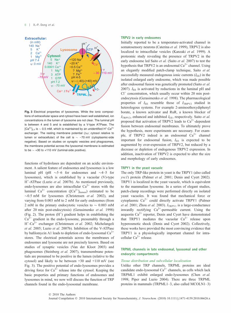
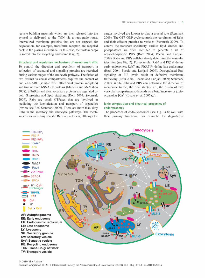
functions of hydrolases are dependent on an acidic environ-ment. A salient feature of endosomes and lysosomes is a lowluminal pH (pH �5–6 for endosomes and �4–5 forlysosomes), which is established by a vacuolar (V)-typeH+-ATPase (Luzio et al. 2007b). As mentioned previously,endo-lysosomes are also intracellular Ca2+ stores with theluminal Ca2+ concentration ([Ca2+]lumen) estimated to be�0.5 mM for lysosomes (Christensen et al. 2002), andvarying from 0.003 mM to 2 mM for early endosomes (from2 mM in the primary endocytotic vesicles to � 0.003 mMafter 20 min post-endocytosis) (Gerasimenko et al. 1998)(Fig. 2). The proton (H+) gradient helps in establishing theCa2+ gradient in the endo-lysosome, presumably through aH+-Ca2+ exchanger (Christensen et al. 2002; Michelangeliet al. 2005; Luzio et al. 2007b). Inhibition of the V-ATPaseby bafilomycin A1 leads to depletion of endo-lysosomal Ca2+
stores. The electrical potentials across the membranes ofendosomes and lysosome are not precisely known. Based onstudies of synaptic vesicles (Van der Kloot 2003) andphagosomes (Steinberg et al. 2007), transmembrane poten-tials are presumed to be positive in the lumen (relative to thecytosol) and likely to be between +30 and +110 mV (seeFig. 3). The positive potential of endo-lysosomes provides adriving force for Ca2+ release into the cytosol. Keeping thebasic properties and primary functions of endosomes andlysosomes in mind, we now will discuss the function of TRPchannels found in the endo-lysosomal membrane.
TRPV2 in early endosomesInitially reported to be a temperature-activated channel insomatosensory neurons (Caterina et al. 1999), TRPV2 is alsolocalized to intracellular vesicles (Kanzaki et al. 1999). Aproteomic study revealing the presence of TRPV2 in theearly endosome led Saito et al. (Saito et al. 2007) to test thehypothesis that TRPV2 is an endosomal Ca2+ channel. Usingan elegantly modified patch-clamp technique, Saito et al.successfully measured endogenous ionic currents (IEE) in theisolated enlarged early endosome, which was made possibleafter endosomal fusion was genetically promoted (Saito et al.2007). IEE is activated by reductions in the luminal pH andCl) concentration, which usually occur within 20 min post-endocytosis (Gerasimenko et al. 1998). The pharmacologicalproperties of IEE resemble those of ITRPV2 studied inheterologous systems. For example 2-aminoethoxydiphenylborate, a known activator and RuR, a known blocker ofITRPV2, enhanced and inhibited IEE, respectively. Saito et al.proposed that activation of TRPV2 leads to Ca2+-dependentfusion between endosomal membranes. To ultimately provethe hypothesis, more experiments are necessary. For exam-ple, if TRPV2 indeed is an endosomal Ca2+ channelimportant for endosomal fusion, IEE is expected to beaugmented by over-expression of TRPV2, but reduced by adecrease or depletion of endogenous TRPV2 expression. Inaddition, inactivation of TRPV2 is expected to alter the sizeand morphology of early endosomes.
TRPY1 in the yeast vacuoleThe only TRP-like protein in yeast is the TRPY1 (also calledyvc1) protein (Palmer et al. 2001; Denis and Cyert 2002).TRPY1 is localized in the yeast vacuole, which is equivalentto the mammalian lysosome. In a series of elegant studies,patch-clamp recordings were performed directly on isolatedyeast vacuoles. It was found that mechanical force orcytoplasmic Ca2+ could directly activate TRPY1 (Palmeret al. 2001; Zhou et al. 2003). ITRPY1 is a large-conductanceinwardly rectifying Ca2+-permeable current. Using theaequorin Ca2+ reporter, Denis and Cyert have demonstratedthat TRPY1 mediates the vacuolar Ca2+ release uponhyperosmotic shock (Denis and Cyert 2002). Collectively,these works have provided the most convincing evidence thatTRPY1 is a physiologically important channel for intra-cellular Ca2+ release.
TRPML channels in late endosomal, lysosomal and otherendocytic compartments
Tissue distribution and subcellular localizationUnlike other TRP channels, TRPML proteins are idealcandidate endo-lysosomal Ca2+ channels, as cells which lackTRPML1 exhibit enlarged endo-lysosomes (Chen et al.1998; Piper and Luzio 2004). There are three TRPMLproteins in mammals (TRPML1–3, also called MCOLN1–3)
Fig. 3 Electrical properties of lysosomes. While the ionic composi-
tions of extracellular space and cytosol have been well established, ion
concentrations in the lumen of lysosome are not clear. The luminal pH
is between 4 and 5 and is established by a V-type ATPase. The
[Ca2+]LY is � 0.5 mM, which is maintained by an unidentified H+-Ca2+
exchanger. The resting membrane potential (Du; cytosol relative to
lumen or extracellular) of the cell is � )70 mV (cytoplasmic-side
negative). Based on studies on synaptic vesicles and phagosomes,
the membrane potential across the lysosomal membrane is estimated
to be � +30 to +110 mV (luminal-side positive).
6 | X.-P. Dong et al.
Journal Compilation � 2010 International Society for Neurochemistry, J. Neurochem. (2010) 10.1111/j.1471-4159.2010.06626.x� 2010 The Authors
(Puertollano and Kiselyov 2009). TRPML1 is expressed inmost tissues (Slaugenhaupt 2002; Cheng et al. 2010) andco-localizes exclusively with late endosomal and lysosomal(LEL) markers (Dong et al. 2009; Puertollano and Kiselyov2009; Cheng et al. 2010) (see Fig. 1). The LEL localizationof TRPML1 is also supported by gradient fractionationstudies on both endogenous and heterologously-expressedproteins (Kim et al. 2009; Zeevi et al. 2009). In addition,TRPML2 and TRPML3 exhibit more restrictive tissuedistribution patterns, but are also located in the LELcompartment (Cuajungco and Samie 2008; Puertollano andKiselyov 2009; Zeevi et al. 2009). TRPML2 and TRPML3are also found in recycling endosomes (Arf6-positivepathway) and early endosomes, respectively (Karacsonyiet al. 2007; Kim et al. 2009; Martina et al. 2009). TRPMLdistribution and localization are reviewed in detail recently(Puertollano and Kiselyov 2009; Cheng et al. 2010).
Pore properties of TRPML channelsAll three TRPMLs have been shown to be Ca2+-permeable(LaPlante et al. 2002; Xu et al. 2007; Dong et al. 2008; Kimet al. 2008, 2009), making them candidates to be lysosomalCa2+ release channels. The first electrophysiologically char-acterized wild-type TRPML channel was TRPML3 (Kimet al. 2007, 2008; Xu et al. 2007). Although the majority ofheterologously-expressed TRPML3 proteins are vesicular, asmall portion of them are able to traffic to the plasmamembrane and give rise to whole-cell currents (ITRPML3)(Grimm et al. 2007; Xu et al. 2007; Cuajungco and Samie2008; Nagata et al. 2008; Kim et al. 2009; Martina et al.2009; Puertollano and Kiselyov 2009). ITRPML3 is aninwardly-rectifying Ca2+-permeable current that is inhibitedby low extracellular (analogous to the luminal side) pH butincreased by Na+ manipulation (removal followed by re-addition) (Kim et al. 2007, 2008). Mutations in the mouseTRPML3 (A419P) result in the varitint-waddler (Va) pheno-type (Cuajungco and Samie 2008; Puertollano and Kiselyov2009). Va mice are deaf, exhibit circling behavior and havepigmentation defects. Compared with wild-type TRPML3,much larger TRPML3-mediated currents are seen in cellsexpressing TRPML3A419P (TRPML3Va). The TRPML3Va
channel exhibits similar pore properties as wild-typeTRPML3, but with altered gating behavior, suggesting thatVa is a channel gain-of-function mutation (Grimm et al.2007; Kim et al. 2007, 2008; Xu et al. 2007; Cuajungco andSamie 2008; Nagata et al. 2008; Puertollano and Kiselyov2009). By artificially introducing a Va-like mutation in theanalogous position of TRPML1 (V432P), Xu et al. were ableto characterize the pore properties of TRPML1 (Xu et al.2007). Like TRPML3, TRPML1Va is an inwardly rectifyingCa2+-permeable but proton-impermeable channel. But, un-like ITRPML3, ITRPML1-Va is potentiated by low pH. Similarly,ITRPML2-Va is also a proton-potentiated inwardly rectifyingCa2+-permeable current (Dong et al. 2008). It remains a
possibility that Va-like mutations change the pore propertiesof TRPML1 and TRPML2, although this is least likely.Recently, Dong et al. developed a patch-clamp method torecord currents directly from isolated endo-lysosomes whichwere pharmacologically enlarged (from 0.1–0.5 to 2–3 lm)using the small molecule compound vacuolin-1 (Dong et al.2008). With this method, Dong et al. were able to recordITRPML1 in enlarged endo-lysosomes. Lysosomal ITRPML1,although much smaller in amplitude than ITRPML1-Va, largelyresembles ITRPML1-Va, suggesting that although TRPML1 islikely to be gated by other, unidentified, cellular cues, theactivating mutation is still a valid approach for characterizingthe pore properties of TRPML1 and TRPML2. In summary,TRPMLs constitute a family of inwardly rectifying, Ca2+-permeant but proton-impermeant cation channels. TRPMLchannel properties are reviewed in detail recently (Puertol-lano and Kiselyov 2009; Cheng et al. 2010).
Dual roles of TRPMLs in membrane trafficking: fusion andfissionThe role of the TRPMLs in post-endocytic membranetrafficking and organelle dynamics in the late endocyticpathway has been extensively studied both in vitro andin vivo (reviewed in Refs. Puertollano and Kiselyov 2009;Cheng et al. 2010). Mutations in human TRPML1 cause typeIV mucolipidosis (ML4), a devastating neurodegenerativedisease in young children (Slaugenhaupt 2002). ML4patients exhibit motor defects, mental retardation, retinaldegeneration, and iron-deficiency anemia. Loss-of-functionstudies revealed a role of TRPML1 in membrane fission fromthe LEL compartment. Fibroblasts derived from ML4patients (ML4 cells), expressing loss-of-function TRPML1mutations, exhibit enlarged, swollen lysosome-like vacuoles(Chen et al. 1998; Slaugenhaupt 2002). Endocytic deliveryto lysosomes involves direct fusion between late endosomesand lysosomes to produce late endosome–lysosome hybridorganelles, from which the lysosomes are re-formed (Luzioet al. 2000, 2007b; Pryor et al. 2000). Genetic studies of thecup-5 mutant, a C. elegans orthologue of mammalianTRPMLs, reveal that the enlarged vacuoles of cup-5 cellscontain markers for both late endosomes and lysosomes andare likely to be hybrid organelles (Treusch et al. 2004).Mechanistically, enlarged endo-lysosomes could result fromuncontrolled and excessive fusion, defective membranefission, or impaired organellar osmoregulation (Luzio et al.2007a). In most cases, enlarged endosomes and lysosomesare suggestive of defective trafficking. For example, dysre-gulation of Rabs and PIPs, two essential regulators ofmembrane trafficking, often leads to similar phenotypes, i.e.,enlarged vacuoles containing hybrid markers (Roth 2004;Poccia and Larijani 2009; Stenmark 2009). While overactiveRab5 causes enlarged endosomes, loss-of-function mutationsof PIKfyve/Fab1, a LEL-specific lipid kinase synthesizingPI(3,5)P2 from PI(3)P, causes enlarged LEL compartments.
TRP calcium channels in intracellular organelles | 7
� 2010 The AuthorsJournal Compilation � 2010 International Society for Neurochemistry, J. Neurochem. (2010) 10.1111/j.1471-4159.2010.06626.x
ML4 cells also accumulate a variety of undigested lipids andwater-soluble substances in the LEL compartment (Chenet al. 1998). Lipid accumulation may result from thedefective degradation of cellular components and/or disrup-tions in membrane trafficking. Defective degradation couldin turn be because of either a lack of specific hydrolaseenzymes, as revealed in most lysosome storage diseases(Puertollano and Kiselyov 2009), or an impaired ionhomeostasis (especially H+) of organelles. However, inTRPML1-deficient (TRPML1–/–) cells, accumulated lipidsand storage materials are of heterogeneous origins (Chenet al. 1998; Treusch et al. 2004; Venugopal et al. 2007;Venkatachalam et al. 2008), suggesting that the defectsmight be related to the homeostasis of LEL compartmentsand/or disruption of broad spectrum lipid metabolism. Thehydrolase-catalyzed lysosomal degradation of storage mate-rials, however, is largely unaffected by the TRPML1 defectin ML4 cells (Chen et al. 1998). Thus the most likely defectof ML4 cells is membrane trafficking. Consistent with this,the use of labeled lipids such as fluorescent lactosylceramidein pulse-chase experiments demonstrated a delay in theretrograde transport of lipids from lysosomes to the TGN,consistent with the role of TRPML1 in late endocyticpathway trafficking (Chen et al. 1998; Pryor et al. 2006;Thompson et al. 2007). Although lipid accumulation phe-notype may be caused by secondary effects because of thechronic accumulation of undigested lipids (Miedel et al.2008), the lipid accumulation in ML4 cells, however, can beacutely rescued by introduction of the wild-type TRPML1gene (Pryor et al. 2006). Thus the defective lipid exit fromthe LEL compartment may be a direct consequence ofTRPML1-deficiency.
The simplest model to reconcile most results in theliterature is that the formation of transport vesicles from theLEL compartment to the TGN, and the reformation oflysosomes from the late endosome-lysosome hybrid organ-elles, are blocked in ML4 or cup-5 cells (Piper and Luzio2004; Treusch et al. 2004; Thompson et al. 2007). In otherwords, TRPML1 may be required for the biogenesis oflysosomes and LEL-derived transport vesicles. Consistentwith this idea, TRPML1 gene expression is significantlyelevated when lysosome biogenesis is induced (Sardielloet al. 2009). As membrane fission is Ca2+-dependent, it islikely that TRPML1 and, more specifically, its Ca2+
conduction, is required for the membrane fission from LELcompartments or late endosome-lysosome hybrids. Notably,intraluminal Ca2+ release has been demonstrated to play anessential role in the in vitro re-formation of lysosomes fromendosome–lysosome hybrids (Pryor et al. 2000; Luzio et al.2007a). Consistent with the requirement of TRPML1 in lipidmigration from late endosomes and lysosomes to the TGN,Ca2+ has been shown to be required for the formation andstabilization of specific transport vesicles (Ahluwalia et al.2001; Luzio et al. 2007a,b).
TRPML1 is also involved in the membrane fusion ofendo-lysosomes. Both the transport of fluid-phase markers tolysosomes and the lysosomal degradation of internalizedgrowth factor receptors are delayed in TRPML1–/– cells(Treusch et al. 2004; Thompson et al. 2007), suggesting adefect in trafficking of endocytosed materials into the LELcompartment. These findings could indicate that the Ca2+
permeability of TRPML1 is required for the Ca2+-dependentmembrane fusion between early endocytic compartments, orfor the formation of transport vesicles from early to lateendosomes. In addition to its interaction with the lateendosome, a lysosome can also incompletely (‘kiss-and-run’)or completely fuse with the plasma membrane (Luzio et al.2007b) resulting in the exocytosis of lysosomal contents(lysosomal exocytosis). This process has been implicated incellular waste elimination, membrane repair, and neurotrans-mitter release (Reddy et al. 2001; Zhang et al. 2007).Lysosomal exocytosis is triggered by a rise of [Ca2+]cyt andthe subsequent binding of Ca2+ to the C2 domains ofsynaptotagmin VII localized in the lysosomal membrane.
The lumen of the lysosome is a major sources of the Ca2+
involved in lysosomal exocytosis (Luzio et al. 2007a,b). Themechanisms underlying lysosomal Ca2+ release, however,are still unclear. Moreover, the molecular identities of theputative lysosomal Ca2+ release channels remain elusive.Lysosomal exocytosis is reduced in ML4 cells (LaPlanteet al. 2006) but increased in HEK cells expressing gain-of-function TRPML1 mutations (Dong et al. 2009). Increasedlysosomal exocytosis results in increased cell surfaceexpression of TRPML1, which may explain the measurablewhole-cell current of TRPML1Va (Dong et al. 2009). Toelaborate this hypothesis, it is necessary to investigatewhether or not the blockade of lysosomal exocytosis reducesITRPML1-Va amplitude. Consistent with a role of TRPML1 inexocytosis, shRNA knockdown of TRPML1 leads to thereduced transport of major histocompatibility complex II(MHCII) to the plasma membrane in macrophages (Thomp-son et al. 2007). In summary, TRPML1 participates inmultiple transport steps of late endocytic pathways by itsinvolvement in the mechanisms of both membrane fusionand fission.
TRPML2 and TRPML3 appear to play similar roles toTRPML1 in post-endocytic membrane trafficking. Zeeviet al. recently reported that siRNA knockdown of endoge-nous TRPML2 in HEK or Hela cells results in inclusionbodies in the LEL compartment (Zeevi et al. 2009). Inacti-vation of TRPML2 by a dominant-negative approachreduced recycling of internalized plasma membrane proteinsback to the plasma membrane (Karacsonyi et al. 2007).Conversely, over-expression of TRPML2 caused a constit-utive activation of Arf6, leading to increased exocytosis(Karacsonyi et al. 2007). These studies suggest thatTRPML2 is required for membrane fusion in the earlyendocytic pathways, and membrane fusion and fission in the
8 | X.-P. Dong et al.
Journal Compilation � 2010 International Society for Neurochemistry, J. Neurochem. (2010) 10.1111/j.1471-4159.2010.06626.x� 2010 The Authors
late endocytic pathways. Consistent with a positive role ofTRPML3 in the late endocytic pathways, siRNA knockdownof TRPML3 leads to inclusion bodies in the LEL compart-ment of HEK and Hela cells (Zeevi et al. 2009). Two recentstudies report that over-expression of TRPML3 results inenlarged endo-lysosomes, decreased endocytosis, and re-duced lysosomal degradation (Kim et al. 2009; Martina et al.2009). These results initially appear inconsistent with Zeeviet al.’s TRPML3 findings and the proposed functions forTRPML1 and TRPML2. These apparent disparities can bereconciled if we consider the dual functions of TRPMLs inmembrane fusion and fission. As mentioned above, enlargedendo-lysosomes might result from either excessive fusion orreduced fission. TRPML3 is expressed in both earlyendosomes and the LEL compartment (Kim et al. 2009;Martina et al. 2009). Dysregulated organellar Ca2+ releaseresulting from TRPML3 over-expression may possiblyincrease membrane fusion events in the early and/or lateendocytic pathways. The vacuolar phenotype resulting fromover-expression may not be limited to TRPML3. Indeed, wehave observed enlarged LEL compartments in HEKcells expressing high levels of TRPML1 (Cheng and Xu,unpublished observation; also see Ref. Vergarajauregui et al.2009). An over-expression of C-terminal, but not N-terminal,enhanced green fluorescence protein fusion constructs ofTRPML1 or TRPML2 in B-lymphocytes was reported tocause enlarged LEL compartments (Song et al. 2006). TheseC-terminal fusion constructs appear to exhibit high levels ofexpression in B-lymphocytes (personal communication withScharenberg A.). Consistent with a role of TRPML3 inmembrane fusion, TRPML3 over-expression also leads to anincreased number of autolysosomes, which results from thefusion of autophagosomes and lysosomes (Kim et al. 2009).TRPMLs may thus play important roles in membrane trafficby regulating both Ca2+-dependent membrane fusion andfission. As different Ca2+ sensors and effectors are implicatedin fusion and fission, selective inhibitors of Ca2+ sensors mayprove informative regarding the nature (effecting fusion vs.fission) of the defects leading to enlarged endolysosomes.
A key open question is how TRPML channels areregulated by various cytosolic and luminal factors, and/orproteins and lipids in the endo-lysosomal membranes,especially those which are known to be involved in endo-lysosomal trafficking. In addition to Ca2+, PIPs and Gproteins have been found to regulate intracellular trafficking(Roth 2004; Stenmark 2009). It is conceivable that thesesignaling cascades may be involved in relaying extracellularsignals to those intracellular pathways that mediate mem-brane trafficking. Signaling by lipid and G proteins is knownto modulate plasma membrane TRP channels (Clapham2003; Montell 2005; Nilius et al. 2007); it is likely that PIPsand Rabs regulate membrane traffic by modulating vesicularTRPMLs. Direct evidence to support this hypothesis is stilllacking.
TRPMLs in signal transductionTRPML1 has been proposed to be an important participant inlysosome-mediated signal transduction. As mentionedpreviously, Ca2+ release from lysosomes or lysosome-relatedacidic stores, similar to Ca2+ release from the ER, plays anindispensable role in the transduction of many extracellularsignals (Galione et al. 2009). For example, endothelin-1 andintegrin ligands mobilize lysosome-related Ca2+ stores insmooth muscle cells (Galione et al. 2009). Accumulatedevidence suggests that NAADP may act as a common secondmessenger downstream of these receptors, and that theputative NAADP receptor is likely to be found in lysosomes(Zhang and Li 2007; Calcraft et al. 2009; Galione et al.2009). As Ca2+ channels in the LEL compartment, TRPMLsare natural candidates to mediate the NAADP response. Byreconstituting lysosomal membranes into a lipid bilayer,Zhang et al. recently reported that NAADP up-regulates acationic current that is dependent on TRPML1 expression(Zhang and Li 2007). The pharmacological properties ofINAADP in the lipid bilayer are similar to those of the putativeNAADP receptor. However, INAADP is a Cs+-permeablecurrent and exhibits a linear I-V curve, two properties that areinconsistent with ITRPML1. It is possible that other lysosomalmembrane proteins may form heteromultimers withTRPML1, leading to a novel current. For example, TPC2(two pore calcium channel protein 2) protein has beenrecently shown to mediate NAADP-induced Ca2+ releasefrom lysosomes (Calcraft et al. 2009; Galione et al. 2009).Future work may reveal the relative contributions ofTRPML1, TRPM2 (see following), and TPC2 to theendogenous NAADP response in different cell types.
TRPMLs in vesicular ion homeostasisIn the endo-lysosome system, H+, Ca2+, and membranefusion have been found to be interconnected (Luzio et al.2007a,b). In addition to Ca2+, TRPML channels arepermeable to other cations in the LEL lumen (Fig. 3) andthus may have functions distinct from Ca2+ signaling.Although these findings are highly controversial, ML4 orTRPML1 knockdown cells (TRPML1–/–) cells appear tohave an overly acidified pH in LEL compartments (Soyomboet al. 2006; Miedel et al. 2008). Given that the TRPML1Va
channel exhibits no permeability to protons, it is notimmediately clear what may cause lysosomal hyperacidifi-cation in the absence of TRPML1. Although the TRPML3channel is not proton-permeable and is indeed inhibited bylow pH, over-expression of TRPML3 results in the alkali-zation of endo-lysosomes (Martina et al. 2009). Therefore, itis likely that endo-lysosomal acidification is secondary toCa2+ release (see Ref. (Cheng et al. 2010) for discussion).
TRPML1 and TRPML2, but not TRPML3, are alsopermeable to Fe2+, Mn2+, and other heavy trace metals (Donget al. 2008). ML4 cells exhibit a cytosolic Fe2+ deficiencyand a concurrent lysosomal Fe2+ overload, suggesting that
TRP calcium channels in intracellular organelles | 9
� 2010 The AuthorsJournal Compilation � 2010 International Society for Neurochemistry, J. Neurochem. (2010) 10.1111/j.1471-4159.2010.06626.x

the iron efflux pathway is blocked in ML4 cells and thatTRPML1 is essential for lysosomal Fe2+ release (Dong et al.2008). Under oxidative conditions, lysosomal Fe2+ overloadmay dramatically increase the production of reactivehydroxyl radicals (OH); Fenton reaction), which in turnfacilitate the formation of lipofuscin (also called agingpigment) (Kurz et al. 2008).
In summary, TRPMLs participate in multiple endo-lyso-some-mediated functions including signal transduction, ionichomeostasis, and more than one aspect of membranetrafficking. A major challenge of TRPML research is tounderstand how one single TRPML protein can play suchdiverse roles. Multiple ionic conductances in a singlemembrane channel may certainly contribute to multifacetedfunctions. In addition to their divalent permeability, TRPMLsare also permeable to Na+ and K+ (Xu et al. 2007). TRPMLsmay therefore regulate organelle dynamics by regulatingendo-lysosomal membrane potentials. Rabs and PIPs exist in‘microdomains’ in the membranes of endo-lysosomes, par-ticipating in multiple functions by recruiting distinct effectorproteins (Poccia and Larijani 2009; Stenmark 2009).TRPMLs may differentially associate with Rabs, PIPs, andCa2+ sensors (for example, CaM, Syt, and ALG-2), givingthem the ability to generate multiple cellular outputs.
TRPM2 in the LEL compartmentInitially characterized as a plasma membrane channel gatedby free cytosolic ADP-ribose (ADPR) (Perraud et al. 2001),recent evidence suggests that TRPM2 is also localized in theLEL compartment (Lange et al. 2009). Rather than simplybeing cargo in the degradative pathway, TRPM2 can functionas a lysosomal Ca2+ release channel in response to cytosolicADP-ribose (Lange et al. 2009). Plasma membrane TRPM2is reportedly activated by NAADP (Beck et al. 2006). It isconceivable that TRPM2 also has a role in lysosomalNAADP signaling (see above). In addition, plasma mem-brane TRPM2 is activated by increases in [Ca2+]i (Du et al.2009). Thus, TRPM2 might mediate Ca2+-induced Ca2+
release (CICR) in the LEL compartment, analogous to IP3R/RyR in the ER/SR (Berridge et al. 2003). Although CICRhas not been demonstrated in mammalian LELs, suchmechanism has been proposed to exist in yeast vacuoles(Palmer et al. 2001).
TRP channels in recycling endosomesLike the transferrin receptor, plasma membrane TRP chan-nels constantly undergo endocytosis and enter the recyclingpathway (Maxfield and McGraw 2004). The steady-statelocation of TRPs is determined by the balance of endocytosisand membrane insertion, which is under tight regulation by avariety of extracellular signals and cellular cues (Maxfieldand McGraw 2004). TRPV5 and TRPV6 are Ca2+-selectivechannels involved in Ca2+ transport in the kidney (van deGraaf et al. 2006). In epithelial cells, TRPV5 and 6 are found
to be localized to recycling endosomes and physicallyinteract with Rab11, a small G protein that is predominantlylocalized in recycling endosomes (van de Graaf et al. 2006).Locking Rab11 in an inactive GDP-bound state results in thereduced surface expression of TRPV5 and 6 (van de Graafet al. 2006).
Several TRPs have been shown to undergo regulatedexocytosis. In hippocampal neurons, TRPC5 is localized inthe intracellular vesicles of neurites (Bezzerides et al. 2004).Using total internal reflection fluorescence microscopy toimage events within 100 nm of the plasma membrane,Berrerides et al. demonstrated that, in response to growthfactor stimulation, TRPC5-residing vesicles undergo rapidinsertion into the plasma membrane (Bezzerides et al. 2004).Similarly TRPC3, residing in vesicle-associated membraneprotein 2-positive compartments, was reported to undergo Gprotein-coupled receptor-stimulated translocation to theplasma membrane (Singh et al. 2004). In response tomechanical shear stress, TRPM7-residing vesicles undergotranslocation to the plasma membrane of vascular smoothmuscle cells (Oancea et al. 2006). TRPV1 is found inintracellular vesicles and physically interacts with thevesicular proteins Snapin and Synaptotagmin IX (Morenil-la-Palao et al. 2004). Upon stimulation, TRPV1-containingvesicles undergo protein kinase C-dependent exocytosis,allowing a novel form of channel regulation (Morenilla-Palaoet al. 2004). Although the molecular identities of thesevesicles are poorly defined, they can be tentatively classifiedas belonging to an intracellular pool that is derived fromrecycling endosomes.
Are these vesicular TRPs functional? It was shown thatactivation of TRPC3 by diacylglycerol may further increasethe cell surface expression of the channel (Singh et al. 2004).It is not clear whether diacylglycerol was activating plasmamembrane or vesicular TRPC3, or perhaps both. Conductingthis experiment in the presence of a membrane-impermeableTRPC3 inhibitor (not reported yet) would prove informative.If vesicular TRPs were purely cargo that did not activelyparticipate in vesicular trafficking, it would be important thatthey keep inactive during transport to avoid the misregulationof membrane traffic. There are several reasons to suggest thatthis might be the case. The lipid composition of intracellularmembranes is quite different from that of the plasmamembrane. Many TRPs require PI(4,5)P2 for their function(Ramsey et al. 2006). Although abundant in the plasmamembrane, this lipid is usually excluded from endocyticvesicles (Roth 2004; Poccia and Larijani 2009). In addition,most TRPs are inhibited by low pH (Clapham 2003), such asthat found in many vesicles. Therefore, it is likely that eventhe presence of agonist may not sufficiently activate TRPs inthe intracellular reserve pool. This hypothesis could be testedby measuring the [Ca2+]cyt response upon stimulating thecells using membrane-permeable agonists (if available) in theabsence of extracellular Ca2+. As intracellular vesicles may
10 | X.-P. Dong et al.
Journal Compilation � 2010 International Society for Neurochemistry, J. Neurochem. (2010) 10.1111/j.1471-4159.2010.06626.x� 2010 The Authors
be heavily dependent on extracellular Ca2+ to fill/refill theirCa2+ stores, caution is necessary regarding to the time courseof such experiments.
TRP channels in the biosynthetic/secretory pathway
Like all other plasma membrane proteins, TRPs have to gothrough various stages of biosynthetic pathways before theyreach their destination. For example, in order to carry outnormal functions, plasma membrane TRPs are glycosylatedin the ER and Golgi apparatus (Cohen 2006). Recent works,however, indicate that several TRPs are constitutivelylocalized in the ER and Golgi and, more importantly, canbe activated in these compartments by their agonists (Koulenet al. 2002; Turner et al. 2003; Thebault et al. 2005; Prasadet al. 2008). As multiple transport steps in the ER and theGolgi are regulated by intraluminal Ca2+ release (Ahluwaliaet al. 2001; Hay 2007), these studies point to active roles ofTRPs in ER or Golgi-mediated signal transduction and/ormembrane trafficking.
Ca2+ release from organelles in the secretory pathways(ER and Golgi) is important for signal transduction (Berridgeet al. 2003). For example, the ‘classic’ Ca2+ release channels,i.e., IP3Rs/RyRs in the ER/SR, couple numerous extracellularsignals to intracellular events such as hormone secretion(Berridge et al. 2003). Several TRP Ca2+ channels (seebelow) in the ER and Golgi may also be activated tocompletely or partially deplete the Ca2+ stores, which may becoupled with unidentified cellular events. A most likelypossibility is that TRP-mediated ER Ca2+ release activatesIP3R/RyR-mediated CICR (Berridge et al. 2003). In addi-tion, there exist poorly characterized ‘ER leak’ pathways thatare known to be important in regulating ER Ca2+ homeo-stasis, although their molecular identities are not known(Camello et al. 2002; Berridge et al. 2003).
Ca2+ release from the ER and the Golgi is also importantfor membrane trafficking. In vivo studies revealed distinctsensitivities to BAPTA-AM versus EGTA-AM for manysteps of membrane trafficking in the secretory pathwaysincluding, for example, anterograde intra-Golgi transport andretrograde endosome-to-TGN transport (Ahluwalia et al.2001; Chen et al. 2002; Burgoyne and Clague 2003; Hay2007). Inhibitory effects by BAPTA but not EGTAwere alsoobserved in cell-free intra-Golgi transport assays (Porat andElazar 2000). CaM inhibitors have also been shown to blockthe intra-Golgi transport (Porat and Elazar 2000; Ahluwaliaet al. 2001; Chen et al. 2002). Therefore, it is likely thatuncharacterized Ca2+ release channels in the ER or Golgimay be activated by trafficking cues to regulate membranefusion/fission events in the secretory pathways.
TRPP1 channels in the endoplasmic reticulumTRPP1 (also called polycystin-2, PC2, and TRPP2) is one oftwo genes mutated in polycystic kidney disease (Zhou 2009).
TRPP1 is localized to the ciliary membranes of kidneyepithelial cells where it mediates fluid flow-induced Ca2+
influx (Nilius et al. 2007). However, both heterologously-expressed and endogenous TRPP1 proteins are also found tobe localized to the ER (Koulen et al. 2002; Geng et al.2008). By reconstitution into the lipid bilayer, TRPP1 wasshown to be activated by Ca2+ on the cytoplasmic side(Koulen et al. 2002; Geng et al. 2008). TRPP1 maytherefore function as a calcium-induced calcium releasechannel like the IP3R and the RyR, participating in signalamplification. Consistent with this, receptor-mediated IP3-dependent ER Ca2+ release is increased by the over-expression of wild-type, but not the disease-causing mutantTRPP1 (Koulen et al. 2002; Geng et al. 2008). In TRPP1-deficient cells, the basal [Ca2+]cyt is significantly lower thanin control cells (Geng et al. 2008). In contrast, over-expression of TRPP1 leads to an increase of [Ca2+]cyt anda concurrent decrease of ER Ca2+ content (Geng et al. 2008).These results suggest that TRPP1 may function as an ER leakchannel. TRPP1 is also found to interact with Syntaxin-5, anER and Golgi-associated t-SNARE; this interaction inhibitsthe ITRPP1(Geng et al. 2008). While Ca2+ is presumed toregulate SNARE complex formation, this study suggests thatSNARE proteins can in turn regulate Ca2+ release channels.
TRPM8 in the endoplasmic reticulumTRPM8 is a cold- and menthol-activated channel in theplasma membrane of somatosensory neurons (Ramseyet al. 2006). In human prostate epithelial cells, however,TRPM8 is highly localized to the ER (Thebault et al.2005). In the absence of extracellular Ca2+, activation ofTRPM8 by menthol or cold induces [Ca2+]i increases thatis sensitive to thapsigargin (TG), suggesting that the ERstore is the source of the Ca2+ release (Thebault et al.2005). TRPM8-agonist-inducced ER Ca2+ store depletionresults in the activation of the store-operated Ca2+ release-activated Ca2+ channel (Thebault et al. 2005). Therefore, insome cell types, TRPM8 may function as an ER Ca2+
release channel similar to IP3R and RyR. It is not clearwhether TRPM8 can function as a Ca2+ release channel inthe ER of other cell types, such as sensory neurons.Although the cellular mechanism activating TRPM8 in theER is not known, it is conceivable that endogenousmenthol-like molecules exist, functioning as novel Ca2+-mobilizing second messengers.
TRPV1 in the endoplasmic reticulum and the trans-GolginetworkTRPV1 is a true sensory channel that is expressed insomatosensory neurons and is activated by heat, protons,and capsaicin (Clapham 2003). Subcellular localizationstudies suggest that both heterologously-expressed andendogenous TRPV1 are also localized to the ER and Golgicompartments (Turner et al. 2003). Activation of TRPV1 by
TRP calcium channels in intracellular organelles | 11
� 2010 The AuthorsJournal Compilation � 2010 International Society for Neurochemistry, J. Neurochem. (2010) 10.1111/j.1471-4159.2010.06626.x
capsaicin induces Ca2+ release from an IP3-sensitive butTG-insensitive store. While ER Ca2+ stores are maintainedby the TG-sensitive SERCA (sarco/endoplasmic reticulumCa2+) pump, Ca2+ gradients in the Golgi are established byboth SERCA and the TG-insensitive SPCA (secretorypathway Ca2+) pump. Like the ER, the Golgi also containsfunctional IP3Rs (Michelangeli et al. 2005). Because of theTG-insensitivity of TRPV1-induced Ca2+ release, the mostlikely location for TRPV1 is in SPCA-positive Golgicompartments. Mobilization of TRPV1-containing Ca2+
stores, however, is not sufficient to activate Ca2+ release-activated Ca2+ channels (Turner et al. 2003). Nevertheless,the evidence sufficiently supports a role of TRPV1 inintracellular Ca2+ release.
The endogenous agonist of TRPV1 in intracellular mem-branes has remained elusive. As the Golgi is acidic relative tothe ER (Fig. 1), transport of TRPV1-positive vesicles from theER to the Golgi may readily activate or sensitize TRPV1.Anandamide, a weak agonist of TRPV1, is known to induceintracellular Ca2+ release via a phospholipase C-independentmechanism (Felder et al. 1993). TRPV1 in the Golgi com-partments may serve as a natural candidate for this release.
TRPA1 in secretory vesicles and secretory granulesTRPA1 is expressed in somatosensory neurons and respondsto a variety of noxious sensory compounds (Ramsey et al.2006). In mast cells, TRPA1 is localized in secretory vesicles(SVs) and secretory granules (SGs) and physically interactswith vesicular proteins in SGs (Prasad et al. 2008). Whenheterologously expressed in HEK cells, TRPA1 can mediateintracellular Ca2+ release, presumably from SVs, in responseto the TRPA1 agonist icilin (Prasad et al. 2008). Asintracellular Ca2+ activates TRPA1 (Zurborg et al. 2007),TRPA1 may function as another CICR-like channel in SV orSG. As SVs or SGs undergo Ca2+-regulated exocytosis, thismechanism may allow a feed-forward induction of cell-surface expression of TRPA1, contributing to a suddenincrease in the ITRPA1.
Other TRP channels in the endoplasmic reticulum,trans-Golgi network, secretory vesicles and granulesAll plasma membrane TRP channels need to go through thebiosynthetic pathways, i.e. ER, TGN, and secretory vesicles/granules before being inserted into the plasma membrane.Are these nascent TRP channels functional in these com-partments? As many synthetic and natural products havebeen found to activate specific TRPs in the plasma membrane(Ramsey et al. 2006), for these TRPs, these relativelyselective agonists may be useful for future studies to revealwhether they are functional in intracellular compartments.
All four TRPs mentioned above (TRPP1, TRPM8, TRPV1,and TRPA1) are active intracellularly. Is intracellular activa-tion a general feature for TRPs in the biosynthetic pathways?While PI(4,5)P2 is a limiting factor to prevent the activation of
TRPs in the recycling pathways, the ER and the Golgi docontain significant amounts of PI(4,5)P2 in their membranes(Roth 2004). There are factors, however, that may prevent thefull functionality of TRPs in the ER and Golgi membranes.For example, the lipid bilayers of the ER and the Golgi favorproteins with shorter transmembrane domains than those ofthe plasma membrane (Watson and Pessin 2001). In addition,several TRPs require post-translational modification such asglycosylation for function (Cohen 2006). Nevertheless, as allfour TRPs studied so far are active intracellularly, it isconceivable that TRPs in the biosynthetic pathways might beactive. Although activation of many other vesicular TRPs(Fig. 1) has not been reported, some of them, for example,TRPC3 and TRPC7, have been shown to actively participatein trafficking/exocytosis (Lavender et al. 2008).
TRP channels in the cell-type specificcompartments
Several TRPs are found in cell-type specific vesicularcompartments, for example, LROs and synaptic vesicles(SyVs). These may participate in cell-type specific functionsin these compartments independent of their general roles.
TRPM7 in synaptic vesiclesTRPM7 is a protein with two functional domains: a cationchannel module and a kinase domain (Ramsey et al. 2006).Under physiological conditions, ITRPM7 is outwardly rectify-ing with an inward non-selective (Ca2+, Mg2+, Na+) conduc-tance and an outward monovalent conductance (Clapham2003). Although ITRPM7-like current can be recorded inalmost every cell, in sympathetic neurons and PC12 cells,TRPM7 is found to be mainly localized in acetylcholine-containing SyVs (Krapivinsky et al. 2006; Brauchi et al.2008). Consistent with its vesicular localization, Kapivinskyet al. provided additional biochemical evidence that TRPM7forms a molecular complex with the fusion machinerycomponents snapsin, a SNARE protein, and synaptotagminI (Krapivinsky et al. 2006). Electrophysiological analysesrevealed that both amplitude and frequency of excitatorypostsynaptic potential (EPSP) are reduced in neurons trans-fected with TRPM7-specific siRNA or dominant-negativeTRPM7 constructs. Further analysis suggest that the reducedEPSP amplitude is because of a smaller quantal size ofneurotransmitter release, which reflects a reduced amount ofacetylcholine release in a single fusion event (fusion of a SyVwith the plasma membrane). As TRPM7 is outwardlyrectifying, one possibility is that TRPM7 may providecounter ions for the release of positively-charged neurotrans-mitters (Krapivinsky et al. 2006). However, vesicular ITRPM7
is presumed to exhibit a linear I-Vas low pH from the luminalside may potentiate the inward (cations flowing out thevesicular lumen) conductance (Jiang et al. 2005). An alter-native possibility is that the ITRPM7 controls the ionic
12 | X.-P. Dong et al.
Journal Compilation � 2010 International Society for Neurochemistry, J. Neurochem. (2010) 10.1111/j.1471-4159.2010.06626.x� 2010 The Authors

homeostasis and membrane potential of the SyV, which mayindirectly affect the neurotransmitter release. The role ofTRPM7 in controlling transmitter release probability and/orfrequency has been confirmed using a direct assay for thefusion of acidic vesicles with the plasma membrane (Brauchiet al. 2008). In this assay, total internal reflection fluores-cence microscopy was employed to monitor the fluorescentreporter pHluorin, which is quenched in the acidic lumen ofthe vesicle but increases in fluorescence upon exposure toextracellular pH, thus indicating fusion of the vesicle with theplasma membrane. Reduced EPSP amplitude may cause aportion of the fusion events to fall out of the assay detectionlimit, which might indirectly cause the reduced frequencyphenotype. It is also possible that TRPM7 may play a directrole in controlling the probability of transmitter release. Forexample, the Ca2+ permeability of TRPM7 and its associationwith synaptotagmin may allow TRPM7 to regulate mem-brane fusion. A Ca2+-impermeable TRPM7 pore mutant withintact outward conductance would allow a test of thispossibility. Finally, the kinase domain of TRPM7 appears toplay a role in vesicle mobility (Brauchi et al. 2008). ThusTRPM7 may be a key regulator of multiple steps ofexocytosis involved in neurotransmitter release.
The key question remains unanswered is how TRPM7 isactivated in the SyV. Synaptic, like endocytic, vesicles aredeprived of the PI(4,5)P2 that is abundant in the plasmamembrane (Krapivinsky et al. 2006). PI(4,5)P2 bindingactivates TRPM7 in the plasma membrane (Runnels et al.2002). One interesting possibility is that when a SyV istethered to the plasma membrane via the SNARE complex,the cytoplasmic portion of TRPM7 is exposed to PI(4,5)P2from the inner leaf of the plasma membrane, and is activated.PIP2 is indispensable for exocytosis and neurotransmission(Suudhof 2008). Upon Ca2+ binding, the cytoplasmic part ofthe synaptotagmins may interact with PIP2 in the plasmamembrane, and this interaction is important for the mem-brane penetration of synaptotagmin and subsequent vesiclefusion (Bai et al. 2004). As TRPM7 has been shown tocomplex with synaptotagmin I (Krapivinsky et al. 2006), it ispossible that TRPM7-mediated Ca2+ release further enhancesthe interaction of synaptotagmin and the plasma membranewhich is necessary for bilayer fusion.
TRPs in lysosome-related organellesTRPMLs are also localized in cell-type specific LROs, whichresemble LEL compartments but have distinct morphologies,content, and functions (Blott and Griffiths 2002). Forexample, TRPML3 is localized in melanosomes of melano-cytes (Xu et al. 2007). In response to light stimulation,melanosomes undergo rapid translocation to the plasmamembrane and release melanin (Blott and Griffiths 2002).The pigmentation defects seen in the Va mice could indicate arole of TRPML3 in melanin release. However, Va is a severegain-of-function phenotype and may cause melanocyte cell
death because of Ca2+ overload in the cytosol. To dissect themechanism underlying the pigmentation defect, it is neces-sary to conduct loss-of-function studies of TRPML3 using,for example, TRPML3 knockout mice. Rab38 and Rab27 aretwo small G proteins that co-localize with TRPML3 in themelanosomes (Stenmark 2009). Mice with mutations ofRab38 and Rab27 exhibit pigmentation phenotypes (Blottand Griffiths 2002). This is suggestive of an interactionbetween Rab proteins and TRPML3 being involved in theregulation of melanosome trafficking. Recently, Oanceaet al. reported that TRPM1, although not localized inmelanosomes, can regulate the melanin content and pigmen-tation in melanocytes (Oancea et al. 2009). It is intriguinghow vesicular TRPs indirectly regulate functions of LROs.
Gastric parietal cells contains a specialized vesicularcompartment that is involved in the regulated transport ofvesicles containing an H+/K+-ATPase for acid secretion intothe gastric lumen (Slaugenhaupt 2002; Puertollano andKiselyov 2009). ML4 patients exhibit reduced gastric acidsecretion, suggesting that TRPML1 might be required for thetranslocation (Slaugenhaupt 2002). In addition, TRPML1,and probably TRPML2 as well, is localized in the MHCIIcompartments of antigen presenting cells such as macrophag-es and B-lymphocytes (Song et al. 2006; Thompson et al.2007). Following endocytosis or phagocytosis of antigenreceptors, MHCII compartments undergo translocation to theplasma membrane (Thompson et al. 2007). Over-expressionof TRPML1 or TRPML2 results in enlarged compartmentsthat are positive for MHCII (Song et al. 2006). Consistentwith this trafficking defect, knockdown of TRPML1 results ina delayed translocation of MHCII compartments to theplasma membrane in cultured macrophages.
Summary, perspectives, and future directions
Many TRP channels have been found to be localized tointracellular vesicles and to interact with a variety ofvesicular proteins. Functional studies suggest that manyintracellularly-localized TRPs are not simply passive cargo,but instead play active roles in membrane fusion and fission,signal transduction, and vesicular homeostasis. In the future,we hope to see the advancement of our knowledge ofintracellular TRPs in the following areas:
(i) Real-time live imaging methods will be used to capturethe local Ca2+ transients mediated by release from vesicles.These seemingly spontaneous events might be able to becorrelated with the membrane fusion events, which can bemonitored with fluorescence imaging approaches. Ca2+
release could possibly be altered by genetic or pharmaco-logical manipulation of the vesicular proteins involved inmembrane fusion and fission.
(ii) Organellar identities of TRP-resident compartmentswill be defined for most TRPs. It will be revealed whetherTRPs are also in group II organelles.
TRP calcium channels in intracellular organelles | 13
� 2010 The AuthorsJournal Compilation � 2010 International Society for Neurochemistry, J. Neurochem. (2010) 10.1111/j.1471-4159.2010.06626.x
(iii) More information will be provided for the electricproperties of intracellular compartments and vesicles. Ionimaging methods will be applied to accurately measureluminal ion concentrations at both basal and stimulatedstates.
(iv) Molecular identification will be performed of Ca2+
release channels and respective activation mechanisms.Agonists and antagonists of intracellular TRPs may provideuseful tools for altering membrane traffic.
Acknowledgements
We apologize to colleagues whose works are not cited because of
space limitations; in many cases review articles were referenced at
the expense of original contributions. The work in the authors’
laboratory is supported by startup funds to H.X. from the
Department of MCDB and Biological Science Scholar Program,
the University of Michigan, a NIH RO1 grant (NS062792 to H.X.),
pilot grants (to H.X.) from the UM Initiative on Rare Disease
Research, Michigan Alzheimer’s Disease Research Center (NIH
grant P50-AG08671 to Gilman), and National Multiple Sclerosis
Society. We appreciate the encouragement and helpful comments
from other members of the Xu laboratory.
References
Ahluwalia J. P., Topp J. D., Weirather K., Zimmerman M. and StamnesM. (2001) A role for calcium in stabilizing transport vesicle coats.J. Biol. Chem. 276, 34148–34155.
Bai J., Tucker W. C. and Chapman E. R. (2004) PIP2 increases the speedof response of synaptotagmin and steers its membrane-penetrationactivity toward the plasma membrane. Nat. Struct. Mol. Biol. 11,36–44.
Beck A., Kolisek M., Bagley L. A., Fleig A. and Penner R. (2006)Nicotinic acid adenine dinucleotide phosphate and cyclic ADP-ribose regulate TRPM2 channels in T lymphocytes. FASEB J. 20,962–964.
Berridge M. J., Bootman M. D. and Roderick H. L. (2003) Calciumsignalling: dynamics, homeostasis and remodelling. Nat. Rev. 4,517–529.
Bezzerides V. J., Ramsey I. S., Kotecha S., Greka A. and Clapham D. E.(2004) Rapid vesicular translocation and insertion of TRP chan-nels. Nat. Cell Biol. 6, 709–720.
Blott E. J. and Griffiths G. M. (2002) Secretory lysosomes. Nat. Rev. 3,122–131.
Brauchi S., Krapivinsky G., Krapivinsky L. and Clapham D. E. (2008)TRPM7 facilitates cholinergic vesicle fusion with the plasmamembrane. Proc. Natl Acad. Sci. USA 105, 8304–8308.
Burgoyne R. D. and Clague M. J. (2003) Calcium and calmodulin inmembrane fusion. Biochim. Biophys. Acta 1641, 137–143.
Calcraft P. J., Ruas M., Pan Z. et al. (2009) NAADP mobilizes calciumfrom acidic organelles through two-pore channels. Nature 459,596–600.
Camello C., Lomax R., Petersen O. H. and Tepikin A. V. (2002) Calciumleak from intracellular stores–the enigma of calcium signalling.Cell calcium 32, 355–361.
Caterina M. J., Rosen T. A., Tominaga M., Brake A. J. and Julius D.(1999) A capsaicin-receptor homologue with a high threshold fornoxious heat. Nature 398, 436–441.
Chen C. S., Bach G. and Pagano R. E. (1998) Abnormal transport alongthe lysosomal pathway in mucolipidosis, type IV disease. Proc.Natl Acad. Sci. USA 95, 6373–6378.
Chen J. L., Ahluwalia J. P. and Stamnes M. (2002) Selective effects ofcalcium chelators on anterograde and retrograde protein transportin the cell. J. Biol. Chem. 277, 35682–35687.
Cheng X., Shen D., Samie M. and Xu H. (2010) Mucolipins: intracellularTRPML1-3 channels. FEBS Lett., in press.
Christensen K. A., Myers J. T. and Swanson J. A. (2002) pH-dependentregulation of lysosomal calcium in macrophages. J. Cell Sci. 115,599–607.
Clapham D. E. (2003) TRP channels as cellular sensors. Nature 426,517–524.
Cohen D. M. (2006) Regulation of TRP channels by N-linked glyco-sylation. Semin. Cell Dev. Biol. 17, 630–637.
Corbett E. F. and Michalak M. (2000) Calcium, a signaling molecule inthe endoplasmic reticulum? Trends Biochem. Sci. 25, 307–311.
Cuajungco M. P. and Samie M. A. (2008) The varitint-waddler mousephenotypes and the TRPML3 ion channel mutation: cause andconsequence. Pflugers Arch. 457, 463–473.
Denis V. and Cyert M. S. (2002) Internal Ca(2+) release in yeast istriggered by hypertonic shock and mediated by a TRP channelhomologue. J. Cell Biol. 156, 29–34.
Dong X. P., Cheng X., Mills E., Delling M., Wang F., Kurz T. and Xu H.(2008) The type IV mucolipidosis-associated protein TRPML1 isan endolysosomal iron release channel. Nature 455, 992–996.
Dong X. P., Wang X., Shen D. et al. (2009) Activating mutations of theTRPML1 channel revealed by proline-scanning mutagenesis.J. Biol. Chem. 284, 32040–32052.
Du J., Xie J. and Yue L. (2009) Intracellular calcium activates TRPM2and its alternative spliced isoforms. Proc. Natl Acad. Sci. USA 106,7239–7244.
Felder C. C., Briley E. M., Axelrod J., Simpson J. T., Mackie K. andDevane W. A. (1993) Anandamide, an endogenous cannabimi-metic eicosanoid, binds to the cloned human cannabinoid receptorand stimulates receptor-mediated signal transduction. Proc. NatlAcad. Sci. USA 90, 7656–7660.
Galione A., Evans A. M., Ma J., Parrington J., Arredouani A., Cheng X.and Zhu M. X. (2009) The acid test: the discovery of two-porechannels (TPCs) as NAADP-gated endolysosomal Ca(2+) releasechannels. Pflugers Arch. 458, 869–876.
Geng L., Boehmerle W., Maeda Y. et al. (2008) Syntaxin 5 regulates theendoplasmic reticulum channel-release properties of polycystin-2.Proc. Natl Acad. Sci. USA 105, 15920–15925.
Gerasimenko J. V., Tepikin A. V., Petersen O. H. and Gerasimenko O. V.(1998) Calcium uptake via endocytosis with rapid release fromacidifying endosomes. Curr. Biol. 8, 1335–1338.
van de Graaf S. F., Chang Q., Mensenkamp A. R., Hoenderop J. G. andBindels R. J. (2006) Direct interaction with Rab11a targets theepithelial Ca2+ channels TRPV5 and TRPV6 to the plasmamembrane. Mol. Cell. Biol. 26, 303–312.
Grimm C., Cuajungco M. P., van Aken A. F., Schnee M., Jors S., KrosC. J., Ricci A. J. and Heller S. (2007) A helix-breaking mutation inTRPML3 leads to constitutive activity underlying deafness in thevaritint-waddler mouse. Proc. Natl Acad. Sci. USA 104, 19583–19588.
Hay J. C. (2007) Calcium: a fundamental regulator of intracellularmembrane fusion? EMBO Rep. 8, 236–240.
Holroyd C., Kistner U., Annaert W. and Jahn R. (1999) Fusion of en-dosomes involved in synaptic vesicle recycling. Mol. Biol. Cell 10,3035–3044.
Jiang J., Li M. and Yue L. (2005) Potentiation of TRPM7 inward cur-rents by protons. J. Gen. Physiol. 126, 137–150.
14 | X.-P. Dong et al.
Journal Compilation � 2010 International Society for Neurochemistry, J. Neurochem. (2010) 10.1111/j.1471-4159.2010.06626.x� 2010 The Authors
Kanzaki M., Zhang Y. Q., Mashima H., Li L., Shibata H. and Kojima I.(1999) Translocation of a calcium-permeable cation channel ind-uced by insulin-like growth factor-I. Nat. Cell Biol. 1, 165–170.
Karacsonyi C., Miguel A. S. and Puertollano R. (2007) Mucolipin-2localizes to the Arf6-associated pathway and regulates recycling ofGPI-APs. Traffic (Copenhagen, Denmark) 8, 1404–1414.
Kim H. J., Li Q., Tjon-Kon-Sang S., So I., Kiselyov K. and Muallem S.(2007) Gain-of-function mutation in TRPML3 causes the mouseVaritint-Waddler phenotype. J. Biol. Chem. 282, 36138–36142.
Kim H. J., Li Q., Tjon-Kon-Sang S., So I., Kiselyov K., Soyombo A. A.and Muallem S. (2008) A novel mode of TRPML3 regulation byextracytosolic pH absent in the varitint-waddler phenotype. EMBOJ. 27, 1197–1205.
Kim H. J., Soyombo A. A., Tjon-Kon-Sang S., So I. and Muallem S.(2009) The Ca(2+) channel TRPML3 regulates membrane traf-ficking and autophagy. Traffic (Copenhagen, Denmark) 10, 1157–1167.
Koulen P., Cai Y., Geng L., Maeda Y., Nishimura S., Witzgall R., EhrlichB. E. and Somlo S. (2002) Polycystin-2 is an intracellular calciumrelease channel. Nat. Cell Biol. 4, 191–197.
Krapivinsky G., Mochida S., Krapivinsky L., Cibulsky S. M. andClapham D. E. (2006) The TRPM7 ion channel functions incholinergic synaptic vesicles and affects transmitter release. Neu-ron 52, 485–496.
Kurz T., Terman A., Gustafsson B. and Brunk U. T. (2008) Lysosomes iniron metabolism, ageing and apoptosis. Histochem. Cell Biol. 129,389–406.
Lange I., Yamamoto S., Partida-Sanchez S., Mori Y., Fleig A. andPenner R. (2009) TRPM2 functions as a lysosomal Ca2+-releasechannel in beta cells. Sci. Signal. 2, ra23.
LaPlante J. M., Falardeau J., Sun M., Kanazirska M., Brown E. M.,Slaugenhaupt S. A. and Vassilev P. M. (2002) Identification andcharacterization of the single channel function of human mucoli-pin-1 implicated in mucolipidosis type IV, a disorder affecting thelysosomal pathway. FEBS Lett. 532, 183–187.
LaPlante J. M., Sun M., Falardeau J., Dai D., Brown E. M., Sla-ugenhaupt S. A. and Vassilev P. M. (2006) Lysosomal exocytosis isimpaired in mucolipidosis type IV. Mol. Genet. Metab. 89, 339–348.
Lavender V., Chong S., Ralphs K., Wolstenholme A. J. and Reaves B. J.(2008) Increasing the expression of calcium-permeable TRPC3 andTRPC7 channels enhances constitutive secretion. Biochem. J. 413,437–446.
Luzio J. P., Rous B. A., Bright N. A., Pryor P. R., Mullock B. M. andPiper R. C. (2000) Lysosome-endosome fusion and lysosomebiogenesis. J. Cell Sci. 113 (Pt 9), 1515–1524.
Luzio J. P., Bright N. A. and Pryor P. R. (2007a) The role of calcium andother ions in sorting and delivery in the late endocytic pathway.Biochem. Soc. Trans. 35, 1088–1091.
Luzio J. P., Pryor P. R. and Bright N. A. (2007b) Lysosomes: fusion andfunction. Nat. Rev. 8, 622–632.
Martens S. and McMahon H. T. (2008) Mechanisms of membrane fu-sion: disparate players and common principles. Nat. Rev. 9, 543–556.
Martina J. A., Lelouvier B. and Puertollano R. (2009) The calciumchannel mucolipin-3 is a novel regulator of trafficking along theendosomal pathway. Traffic (Copenhagen, Denmark) 10, 1143–1156.
Maxfield F. R. and McGraw T. E. (2004) Endocytic recycling. Nat. Rev.5, 121–132.
Michelangeli F., Ogunbayo O. A. and Wootton L. L. (2005) A plethoraof interacting organellar Ca2+ stores. Curr. Opin. Cell Biol. 17,135–140.
Miedel M. T., Rbaibi Y., Guerriero C. J., Colletti G., Weixel K. M.,Weisz O. A. and Kiselyov K. (2008) Membrane traffic and turn-over in TRP-ML1-deficient cells: a revised model for mucolipi-dosis type IV pathogenesis. J. Exp. Med. 205, 1477–1490.
Montell C. (2005) The TRP superfamily of cation channels. Sci. STKE2005, re3.
Morenilla-Palao C., Planells-Cases R., Garcia-Sanz N. and Ferrer-Montiel A. (2004) Regulated exocytosis contributes to proteinkinase C potentiation of vanilloid receptor activity. J. Biol.Chem. 279, 25665–25672.
Nagata K., Zheng L., Madathany T., Castiglioni A. J., Bartles J. R. andGarcia-Anoveros J. (2008) The varitint-waddler (Va) deafnessmutation in TRPML3 generates constitutive, inward rectifyingcurrents and causes cell degeneration. Proc. Natl Acad. Sci. USA105, 353–358.
Nilius B., Owsianik G., Voets T. and Peters J. A. (2007) Transientreceptor potential cation channels in disease. Physiol. Rev. 87, 165–217.
Oancea E., Wolfe J. T. and Clapham D. E. (2006) Functional TRPM7channels accumulate at the plasma membrane in response to fluidflow. Circ. Res. 98, 245–253.
Oancea E., Vriens J., Brauchi S., Jun J., Splawski I. and Clapham D. E.(2009) TRPM1 forms ion channels associated with melanin contentin melanocytes. Sci. Signal. 2, ra21.
Palmer C. P., Zhou X. L., Lin J., Loukin S. H., Kung C. and Saimi Y.(2001) A TRP homolog in Saccharomyces cerevisiae forms anintracellular Ca(2+)-permeable channel in the yeast vacuolarmembrane. Proc. Natl Acad. Sci. USA 98, 7801–7805.
Perraud A. L., Fleig A., Dunn C. A. et al. (2001) ADP-ribose gating ofthe calcium-permeable LTRPC2 channel revealed by Nudix motifhomology. Nature 411, 595–599.
Peters C. and Mayer A. (1998) Ca2+/calmodulin signals the completionof docking and triggers a late step of vacuole fusion. Nature 396,575–580.
Piper R. C. and Luzio J. P. (2004) CUPpling calcium to lysosomalbiogenesis. Trends Cell Biol. 14, 471–473.
Poccia D. and Larijani B. (2009) Phosphatidylinositol metabolism andmembrane fusion. Biochem. J. 418, 233–246.
Porat A. and Elazar Z. (2000) Regulation of intra-Golgi membranetransport by calcium. J. Biol. Chem. 275, 29233–29237.
Prasad P., Yanagihara A. A., Small-Howard A. L., Turner H. and StokesA. J. (2008) Secretogranin III directs secretory vesicle biogenesisin mast cells in a manner dependent upon interaction with chro-mogranin A. J. Immunol. 181, 5024–5034.
Pryor P. R., Mullock B. M., Bright N. A., Gray S. R. and Luzio J. P.(2000) The role of intraorganellar Ca(2+) in late endosome-lyso-some heterotypic fusion and in the reformation of lysosomes fromhybrid organelles. J. Cell Biol. 149, 1053–1062.
Pryor P. R., Reimann F., Gribble F. M. and Luzio J. P. (2006) Mucolipin-1 is a lysosomal membrane protein required for intracellular lac-tosylceramide traffic. Traffic (Copenhagen, Denmark) 7, 1388–1398.
Puertollano R. and Kiselyov K. (2009) TRPMLs: in sickness and inhealth. Am. J. Physiol. 296, F1245–F1254.
Ramsey I. S., Delling M. and Clapham D. E. (2006) An introduction toTRP channels. Annu. Rev. Physiol. 68, 619–647.
Reddy A., Caler E. V. and Andrews N. W. (2001) Plasma membranerepair is mediated by Ca(2+)-regulated exocytosis of lysosomes.Cell 106, 157–169.
Roth M. G. (2004) Phosphoinositides in constitutive membrane traffic.Physiol. Rev. 84, 699–730.
Runnels L. W., Yue L. and Clapham D. E. (2002) The TRPM7 channel isinactivated by PIP(2) hydrolysis. Nat. Cell Biol. 4, 329–336.
TRP calcium channels in intracellular organelles | 15
� 2010 The AuthorsJournal Compilation � 2010 International Society for Neurochemistry, J. Neurochem. (2010) 10.1111/j.1471-4159.2010.06626.x
Saito M., Hanson P. I. and Schlesinger P. (2007) Luminal chloride-dependent activation of endosome calcium channels: patch clampstudy of enlarged endosomes. J. Biol. Chem. 282, 27327–27333.
Sardiello M., Palmieri M., di Ronza A. et al. (2009) A gene networkregulating lysosomal biogenesis and function. Science (New York,NY) 325, 473–477.
Savina A., Furlan M., Vidal M. and Colombo M. I. (2003) Exosomerelease is regulated by a calcium-dependent mechanism in K562cells. J. Biol. Chem. 278, 20083–20090.
Singh B. B., Lockwich T. P., Bandyopadhyay B. C. et al. (2004)VAMP2-dependent exocytosis regulates plasma membrane inser-tion of TRPC3 channels and contributes to agonist-stimulatedCa2+ influx. Mol. Cell 15, 635–646.
Slaugenhaupt S. A. (2002) The molecular basis of mucolipidosis type IV.Curr. Mol. Med. 2, 445–450.
Song Y., Dayalu R., Matthews S. A. and Scharenberg A. M. (2006)TRPML cation channels regulate the specialized lysosomal com-partment of vertebrate B-lymphocytes. Eur. J. Cell Biol. 85, 1253–1264.
Soyombo A. A., Tjon-Kon-Sang S., Rbaibi Y., Bashllari E., Bisceglia J.,Muallem S. and Kiselyov K. (2006) TRP-ML1 regulates lysosomalpH and acidic lysosomal lipid hydrolytic activity. J. Biol. Chem.281, 7294–7301.
Steinberg B. E., Touret N., Vargas-Caballero M. and Grinstein S. (2007)In situ measurement of the electrical potential across the phagos-omal membrane using FRET and its contribution to the proton-motive force. Proc. Natl Acad. Sci. USA 104, 9523–9528.
Stenmark H. (2009) Rab GTPases as coordinators of vesicle traffic. Nat.Rev. 10, 513–525.
Suudhof T. C. (2008) Neurotransmitter release. Handb. Exp. Pharmacol.184, 1–21.
Thebault S., Lemonnier L., Bidaux G. et al. (2005) Novel role of cold/menthol-sensitive transient receptor potential melastatine familymember 8 (TRPM8) in the activation of store-operated channels inLNCaP human prostate cancer epithelial cells. J. Biol. Chem. 280,39423–39435.
Thompson E. G., Schaheen L., Dang H. and Fares H. (2007) Lysosomaltrafficking functions of mucolipin-1 in murine macrophages. BMCCell Biol. 8, 54.
Treusch S., Knuth S., Slaugenhaupt S. A., Goldin E., Grant B. D. andFares H. (2004) Caenorhabditis elegans functional orthologue ofhuman protein h-mucolipin-1 is required for lysosome biogenesis.Proc. Natl Acad. Sci. USA 101, 4483–4488.
Turner H., Fleig A., Stokes A., Kinet J. P. and Penner R. (2003)Discrimination of intracellular calcium store subcompartments
using TRPV1 (transient receptor potential channel, vanilloidsubfamily member 1) release channel activity. Biochem. J. 371,341–350.
Van der Kloot W. (2003) Loading and recycling of synaptic vesicles inthe Torpedo electric organ and the vertebrate neuromuscularjunction. Prog. Neurobiol. 71, 269–303.
Venkatachalam K., Long A. A., Elsaesser R., Nikolaeva D., Broadie K.and Montell C. (2008) Motor deficit in a Drosophila model ofmucolipidosis type IV due to defective clearance of apoptotic cells.Cell 135, 838–851.
Venugopal B., Browning M. F., Curcio-Morelli C., Varro A., MichaudN., Nanthakumar N., Walkley S. U., Pickel J. and Slaugenhaupt S.A. (2007) Neurologic, gastric, and opthalmologic pathologies in amurine model of mucolipidosis type IV. Am. J. Hum. Genet. 81,1070–1083.
Vergarajauregui S., Martina J. A. and Puertollano R. (2009) Identifica-tion of the penta-EF-hand protein ALG-2 as a Ca2+-dependentinteractor of mucolipin-1. J. Biol. Chem. 284, 36357–36366.
Watson R. T. and Pessin J. E. (2001) Transmembrane domain lengthdetermines intracellular membrane compartment localization ofsyntaxins 3, 4, and 5. Am. J. Physiol. Cell Physiol. 281, C215–C223.
Xu H., Delling M., Li L., Dong X. and Clapham D. E. (2007) Activatingmutation in a mucolipin transient receptor potential channel leadsto melanocyte loss in varitint waddler mice. Proc. Natl Acad. Sci.USA 104, 18321–18326.
Zeevi D. A., Frumkin A., Offen-Glasner V., Kogot-Levin A. and BachG. (2009) A potentially dynamic lysosomal role for the endoge-nous TRPML proteins. J. Pathol. 219, 153–162.
Zhang F. and Li P. L. (2007) Reconstitution and characterization of anicotinic acid adenine dinucleotide phosphate (NAADP)-sensitiveCa2+ release channel from liver lysosomes of rats. J. Biol. Chem.282, 25259–25269.
Zhang Z., Chen G., Zhou W., Song A., Xu T., Luo Q., Wang W., Gu X.S. and Duan S. (2007) Regulated ATP release from astrocytesthrough lysosome exocytosis. Nat. Cell Biol. 9, 945–953.
Zhou J. (2009) Polycystins and primary cilia: primers for cell cycleprogression. Annu. Rev. Physiol. 71, 83–113.
Zhou X. L., Batiza A. F., Loukin S. H., Palmer C. P., Kung C. and SaimiY. (2003) The transient receptor potential channel on the yeastvacuole is mechanosensitive. Proc. Natl Acad. Sci. USA 100,7105–7110.
Zurborg S., Yurgionas B., Jira J. A., Caspani O. and Heppenstall P. A.(2007) Direct activation of the ion channel TRPA1 by Ca2+. Nat.Neurosci. 10, 277–279.
16 | X.-P. Dong et al.
Journal Compilation � 2010 International Society for Neurochemistry, J. Neurochem. (2010) 10.1111/j.1471-4159.2010.06626.x� 2010 The Authors