
Trinit Student Sientifi Reie ol I Immunoglobulin-binding ...
14
Trinity Student Scientific Review Vol. I 76 Immunoglobulin-binding Proteins of Staphylococcus Aureus Microbiology Kelly Murray Junior Sophister Antibiotic resistance is developing faster than the rate of discovery of effective new treatments against Staphylococcus aureus (S. aureus) infections, meaning research into the virulence factors of S. aureus is more important than ever. The four Immunoglobulin-binding proteins of S. aureus play a crucial role in virulence, as well as enabling the pathogen to evade the host immune defences. Infection with S. aureus does not result in lasting immunological memory, due to the B-cell superantigenic activity of immunoglobulin-binding protein A. By understanding the roles these immunoglobulin-binding proteins play in infection, we can devise potential treatments and preventative strategies against them. Introduction Staphylococcus aureus is a Gram-positive pathogen that causes recurrent skin and soft tissue infections (SSTI) and more serious infections such as pneumonia, sepsis, endocarditis or bacteremia (Blot et al. 1998). 20-30% of the human population are carriers of S. aureus on their skin and nasal cavity, however it only poses a threat when it enters the body, as it is an invasive pathogen. Due to the overuse of antibiotics, S. aureus strains have become resistant to most treatments, and are known as methicillin-resistant S. aureus (MRSA). MRSA has become a major public health problem, as even with treatment the survival rates are poor. To devise new treatments and preventative strategies, it is therefore crucial to understand how S. aureus evades the host immune defences and to determine the
Transcript of Trinit Student Sientifi Reie ol I Immunoglobulin-binding ...

Trinity Student Scientific Review Vol. I
76
Immunoglobulin-binding Proteins of Staphylococcus Aureus
MicrobiologyKelly MurrayJunior Sophister
Antibiotic resistance is developing faster than the rate of discovery of effective new treatments against Staphylococcus aureus (S. aureus) infections, meaning research into the virulence factors of S. aureus is more important than ever. The four Immunoglobulin-binding proteins of S. aureus play a crucial role in virulence, as well as enabling the pathogen to evade the host immune defences. Infection with S. aureus does not result in lasting immunological memory, due to the B-cell superantigenic activity of immunoglobulin-binding protein A. By understanding the roles these immunoglobulin-binding proteins play in infection, we can devise potential treatments
and preventative strategies against them.
IntroductionStaphylococcus aureus is a Gram-positive pathogen that causes recurrent skin and soft tissue infections (SSTI) and more serious infections such as pneumonia, sepsis, endocarditis or bacteremia (Blot et al. 1998). 20-30% of the human population are carriers of S. aureus on their skin and nasal cavity, however it only poses a threat when it enters the body, as it is an invasive pathogen. Due to the overuse of antibiotics, S. aureus strains have become resistant to most treatments, and are known as methicillin-resistant S. aureus (MRSA). MRSA has become a major public health problem, as even with treatment the survival rates are poor. To devise new treatments and preventative strategies, it is therefore crucial to understand how S. aureus evades the host immune defences and to determine the

Biology
77
virulence factors involved. The immunoglobulin-binding proteins of S. aureus are Protein A, Staphylococcal binder of immunoglobulin and Superantigen-like binding proteins 7 and 10. All of these proteins play a crucial role in virulence in S. aureus infection (Yamamoto et al. 2013, Zecconi & Scali 2013, Foster et al. 2014). Protein A in particular, is crucial in pneumonia and sepsis. Here we review how these four immunoglobulin-binding proteins evade the complement system. Even though a recently discovered antibiotic has no known resistant mutants, it is not the end of the era of antibiotic resistance (Ling et al. 2015).
Immunoglobulin-binding Proteins
Protein AProtein A (SpA) is a surface protein of S. aureus with two functionally distinct halves, the c-terminal domain and the N-terminal domain. The C-terminal domain anchors SpA to the surface of the cell wall via LPXTG motif (Fig1). The N-terminal contains five protein binding domains, each consisting of three helices: E, D, A, B and C (Schneewind et al. 1992). Recently, the crystal structures of these five binding domains have been established (Deis et al. 2014). These crystal structures confirmed that a single domain can bind multiple partners at the same time, such as Fab and Fc. (Graille et al. 2000). SpA is involved in both adhesion to host cells and evading innate immune responses relating to immunoglobulins and opsonisation. SpA is a major microbial surface recognizing adhesive matrix molecule (MSCRAMM). Experiments with spa-deletion mutants, show decreased adhesion which corroborates this (Hartleib et al. 2000, Foster et al. 2014). SpA also acts as a B-cell superantigen as it can interact with the Fab region of VH3 type B cell receptors on IgM. This causes the proliferation of B-cells by super clonal expansion and apoptosis of activated B-cells (Goodyear & Silverman 2003, Palmqvist et al. 2005). The destruction of B-cells prevents the host from developing immunological memory.
Trinity Student Scientific Review Vol. I
78
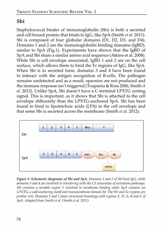
SbiStaphylococcal binder of immunoglobulin (Sbi) is both a secreted and cell bound protein that binds to IgG, like SpA (Smith et al. 2011). Sbi is composed of four globular domains (D1, D2, D3, and D4). Domains 1 and 2 are the immunoglobulin binding domains (IgBD), similar to SpA (Fig.1). Experiments have shown that the IgBD of SpA and Sbi share a similar amino acid sequence (Atkins et al. 2008). While Sbi is cell envelope associated, IgBD 1 and 2 are on the cell surface, which allows them to bind the Fc regions of IgG, like SpA. When Sbi is in secreted form, domains 3 and 4 have been found to interact with the antigen recognition of B-cells. The pathogen remains undetected and as a result, opsonins are not produced and the immune response isn’t triggered (Toapanta & Ross 2006, Smith et al. 2012). Unlike SpA, Sbi doesn’t have a C-terminal LPXTG sorting signal. This is important, as it shows that Sbi is attached to the cell envelope differently than the LPXTG-anchored SpA. Sbi has been found to bind to lipoteichoic acids (LPA) in the cell envelope and that some Sbi is secreted across the membrane (Smith et al. 2012).
Figure 1: Schematic diagrams of Sbi and SpA. Domains 1 and 2 of Sbi bind IgG, while domains 3 and 4 are involved in interfering with the C3 convertase of activation pathways. Sbi contains a variable region Y involved in membrane binding while SpA contains an LPXTG, a wall anchoring motif and transmembrane domain M. The Wr and Xc regions are proline rich. Domains 1 and 2 share structural homology with regions E, D, A, B and C of SpA. Adapted from Smith et al. (Smith et al. 2011)

Biology
79
Superantigen-like ProteinSuperantigen-like proteins (SSL), formally known as staphylococcal exotoxin-like proteins, are family of exotoxins. SSL proteins have no superantigenic activity, even though they are structurally similar to enterotoxins, which do show superantigenic activity. SSL7 and SSL10 are the only SSL proteins that bind to immunoglobulins, therefore these will be the focus of our discussion. SSL7 binds to IgA while SSL10 binds to human IgG and this may be beneficial in preventing FcR-mediated leukocyte activation (Langley et al. 2005). SSL10 only binds to primate IgG, specifically to the Fc part through its N-terminal oligonucleotide binding fold domain (Patel et al. 2010). When bound to IgG, SSL10 has been found to act as an anticoagulant, however, more research is needed to establish if this plays a role in S. aureus virulence (Itoh et al. 2013). SSL10 also binds to phosphatidylserine (PS) to the surface of apoptotic cells, and may interfere with the clearance of these cells (Itoh et al. 2012). Additionally, both SSL7 and SSL10 appear to act as virulence factors in S. aureus infection by interfering with the classical pathway in the complement system.
Immunoglobulin-binding Proteins in Evasion of ComplementThe complement system is comprised of a family of proteins and proteolytic fragments. Complement proteins bind to the surface of the pathogen to identify it as a target for opsonisation by macrophages. The activation of complement can occur through three different pathways; classical, alternative and lectin pathways (Fig. 2). All of the pathways activate C3 by cleaving it into C3a and C3b. In the Classical and lectin pathways, C4bC2a also called C3 convertase, cleaves C3 whereas C3bBb is the C3 convertase in the Alternative pathway.
The Classical pathway is initiated by the binding of antibodies to the antigens displayed on the surface of the pathogen, and a complement complex (C1S,C1q,C1r) attaches on to the Fc region of immunoglobulins. SSL10 competes with C1q to bind to IgG and in

Trinity Student Scientific Review Vol. I
80
doing so can inhibit activation of the complement pathway (Itoh et al. 2010, Patel et al. 2010).
SpA aids the pathogen in evading the host immune defences by binding the Fc receptors on Immunoglobulin G (IgG).This acts as a decoy by binding the complement binding portion of the IgG the wrong way round and coating it with antibodies, so it can’t trigger the complement system. This prevents the S. aureus being killed by opsonophagocytosis. SpA binding to IgG is essential to avoid the host immune defences as mouse models with a SpA deficient strain of S. aureus suffered less severe arthritis and septic death. This indicated that SpA is a virulence factor (Palmqvist et al. 2002). SpA can also bind to von Willebrand factor (vWF) which is a large glycoprotein, that when bound to SpA is involved in the adhesion to platelets. This is very important in S. aureus caused endocarditis, as it allows for the adherence of the pathogen to the host platelets (Hartleib et al. 2000)
Sbi is involved in disrupting the alternative pathway by causing the sequestration of C3. The function of C3b is to attach to the surface of the pathogen through thioester bonds, which causes them to be phagocytosed. The crystallized structure of Sbi has shown that it binds to C3 via domain 3 and 4 which prevents it binding to its target, thus preventing the initiation of phagocytosis (Atkins et al., 2008). Sbi works in tandem with extracellular fibrinogen-binding protein (Efb) to sequester C3 with plasminogen. Alternatively, the C3b protein can from a complex with C4b2a to form the C3/C5 convertase, causing the formation of the membrane attack complex (MAC). The MAC causes a pore to form on the pathogens surface, resulting in cell lysis. Sbi also prevents the formation of the MAC by binding to it, preventing it reaching its target surface and form a complex with C4b2a (Laursen et al. 2010).
SSL7 binds both IgA and complement C5. The C5 convertase cleaves C5 to give C5a and C5b. Both of these molecules are a threat to the survival of S. aureus as C5a stimulates a proinflammatory response and attracts leukocytes, while C5b is responsible for the formation of the MAC. The crystal structure of SLL7 interacting with IgA, showed that the C-terminal β–domain binds to C5 and that the OB domain interacts with IgA (Laursen et al. 2010). Although IgA

Biology
81
bound SSL has shown enhanced inhibitory activity in complement systems in mice, it is not essential that they are bound to each other (Lorenz et al. 2013)2013. SSL 7 binding to C5, has been shown to prevent the cleavage of C5 by the C5 convertase, which inhibits both the classical and alternative pathway.
Figure 2: Complement cascade pathways; A is the Classical pathway which is initiated by the binding of antibodies to antigens present on the surface, Ig then binds to this complex. Subsequent arrows represent cleavage events by which the next complement protein becomes activated in the cascade. B is the Lectin pathway, which is initiated by mannose binding. It follows the same mechanism of action as the Classical pathway once C3 is cleaved. C is the alternative pathway which isn’t discussed, but it provides an amplification loop binding C3b to the bacteria surface which increases the production of more C3b until the pathogen is covered with them, which attracts macrophages to destroy them. This diagram was taken from ’’Immune evasions by Staphylococci’’ (Foster 2005)

Trinity Student Scientific Review Vol. I
82
SpA induced signalling pathways in pneumonia and sepsisProtein A is a virulence factor in both S. aureus induced pneumonia and sepsis (Palmqvist et al. 2002, Bubeck Wardenburg et al. 2007, Kim et al. 2012). SpA is multifunctional as it can aid S. aureus in evading the complement system and establish infection, but it can also regulate inflammation. SpA is thought to act as a virulence factor by interacting with signalling pathways, such as Tumour necrosis factor receptor 1 (TNFR1) and epidermal growth factor receptor (EGFR) (Gomez et al. 2004). SpA can use its IgG binding domains to bind with TNFR1, which activates the expression of chemoattractant cytokines, such as interleukin-8 (IL-8).This interaction also initiates the production of CXCL10 in airway epithelial cells, and attracts type 1 interferon (IFN) (Ahn et al. 2014). This results in inflammation and tissue damage to the lungs.
Figure 3: The induction and repression of inflammation. A, binding of SpA to the TNFR1 receptor results in the production of IL-8 through a

Biology
83
signalling cascade, which attracts neutrophils, resulting in inflammation. B, TNF-α is neutralized upon TNFR1 binding as the TNFR1 receptors on the cell surface have been cleaved off by SpA induced ADAM17 and EGFR resulting in reduced inflammation. Adapted from Gomez et al. (Gomez et al. 2004).
TNFR1 signalling can also be activated when TNF-α binds to it. TNF-α is a pro-inflammatory signalling cytokine that binds to its receptor (TNFR1) present on the surface of cells, which triggers the production IL-8, recruits neutrophils, and results in inflammation (Gomez et al. 2004, Gomez et al. 2007). TNFR1 can be positively or negatively regulated. Negative regulation of TNFR1, in relation to pneumonia and sepsis, is carried out by an enzyme called ADAM17 (also known as TNF-α converting enzyme, TACE). ADAM17 cleaves the TNFR1 off the surface of cells so TNF-α has less receptors to bind to, which dampens the inflammation response (Giai et al. 2013). EGFR is triggered by SpA, and activates ADAM17 to cleave off the TNFR1 on the surface of macrophages (Fig. 3). This suggests that SpA can promote inflammatory events as well as inhibit them, depending on which receptor it binds to. In pulmonary infections such as pneumonia, SpA normally binds to TNFR1 which signals for the production of IL-8, causing a proinflammatory event in the lungs (Giai et al. 2013). Early action of TNF-α is very limited in the lung as airway epithelial cells don’t produce significant levels of it so SpA binds to TNFR1. However, in other S. aureus infections, such as sepsis, there are plenty of immune cells that can generate TNF-α. A novel mechanism has been suggested to account for this, and suggests that soluble TNFR1 may neutralize circulating TNF-α, by causing the early shedding of TNFR1, which reduces inflammation. This has been shown using SpA deficient mutants, and tnfr1-/- in mice (Gomez et al. 2004).
Preventative Therapies and Treatments Successful vaccination is dependent on the induction of immune memory. SpA has been found to prevent the production of specific antibodies to other staphylococcal antigens (Pauli et al. 2014), so for vaccination to be successful , SpA must first be supressed. However,

Trinity Student Scientific Review Vol. I
84
experiments using SpAKKAA mutant, which lacks the Fab and Fc binding regions, elicited antibody production against SpA in mouse models (Kim et al. 2010).This mutant also resulted in partial immune protection from the wild-type SpA. However, it is important to note that past clinical vaccine trials have all failed and that they all used a single antigen target, be it SpA or any of the immunoglobulin-binding proteins. It is highly likely that a successful vaccine strategy would incorporate multiple antigens as different types of S. aureus caused infections contain different virulence factors that aid its evasion of the host immune system (Bagnoli et al. 2012). Experiments in mice have already shown that vaccines with a combination of protein targets, provide more protection. Four surface proteins; IsdB, IsdA, Sdr+ and SdrE provided better protection to mice than any single component vaccine (Stranger-Jones et al. 2006, Kim et al. 2012, Rauch et al. 2014).
Recent experiments using phage therapy in mouse models found promising results for the use of phage therapy as a treatment to septicaemia, and potentially against MRSA pneumonia. Mice infected with lung derived septicaemia and then treated with S. aureus phage S13, showed lower levels of the pro-inflammatory cytokines, IL-6 and TNF-α which implies reduced inflammation and disease severity (Takemura-Uchiyama et al. 2014).
Treatment of S. aureus infections with antibiotics is becoming increasing difficult, due to the rapid rate of strains developing resistance. However the recent discovery of a new antibiotic that so far has no detectable resistant strains, is showing hope as a treatment to resistant S. aureus strains. In MRSA caused septicaemia mouse models, with a 90% mortality rate, all mice treated with teixobactin survived (Ling et al. 2015). Further experiments also showed that teixobactin is nontoxic to human cells, which indicates it may be a successful therapeutic agent in humans against S. aureus infection. Patients suffering from MRSA infections are currently treated with vancomycin, but it has poor bactericidal activity, which often leads to patient death (Kollef 2007). Teixobactin has excellent bactericidal properties as it is thought to interfere with the formation of Teichoic acid, which is responsible for anchoring autolysins. Teixobactin binds to lipid II which is the precursor for peptidoglycan formation,

Biology
85
and lipid III which is the precursor for cell wall teichoic acids. This mode of action results in excellent bactericidal activity against S. aureus, as it frees the autolysins which then digest the cell wall and cause bacterial cell lysis (Ling et al. 2015). Teixobactin is only effective against gram-positive bacteria, so it may also be used against Clostridium difficile and Bacillus anthracis.
ConclusionsIn this review I have described the major strategies in which S. aureus uses its immunoglobulin-binding proteins to evade the host immune system, particularly the complement system. SpA is the most studied, and also appears to carry out multiple functions involved in immune evasion, such as inflammation, prevention of opsonisation and platelet adhesion. However, there is little known about the more recently discovered immunoglobulin-binding proteins SSL10 and SSL7. Further research is needed to determine their roles in immune evasion and to confirm that they are involved in the prevention of leukocyte activation.
Although teixobactin has no resistant strains yet, it is thought that some may evolve over time. It took more than 30 years for vancomycin resistant strains to appear so resistant strains of teixobactin may eventually emerge (Ling et al. 2015). This recent antibiotic discovery suggests that there may still be more compounds with potential therapeutic affects against S. aureus, waiting to be discovered in nature. The recent finding that phage therapy is successful in mouse models, needs to be confirmed by other studies, but it is promising. It is accepted that a possible S. aureus vaccine would need to incorporate multiple antigens in a single vaccine to target a specific S. aureus induced infection. This indicates that a universal vaccine against S. aureus will not be possible until we fully understand the different virulence factors and their mechanisms of action in each S. aureus infection.

Trinity Student Scientific Review Vol. I
86
AcknowledgementsThis work was adapted from an essay “Immunoglobulin-binding proteins of Staphylococcus aureus “for course MI3MO3.
ReferencesAHN, D. S., PARKER, D., PLANET, P. J., NIETO, P. A., BUENO, S. M. & PRINCE,
A. (2014) Secretion of IL-16 through TNFR1 and calpain-caspase signaling contributes to MRSA pneumonia. Mucosal Immunology, 7, 1366-1374.
ATKINS, K. L., BURMAN, J. D., CHAMBERLAIN, E. S., COOPER, J. E., POUTREL, B., BAGBY, S., JENKINS, A. T. A., FEIL, E. J. & VAN DEN ELSEN, J. M. H. (2008) S. aureus IgG-binding proteins SpA and Sbi: Host speciticity and mechanisms of immune complex formation. Molecular Immunology, 45, 1600-1611.
BAGNOLI, F., BERTHOLET, S. & GRANDI, G. (2012) Inferring reasons for the failure of Staphylococcus aureus vaccines in clinical trials. Frontiers in Cellular and Infection Microbiology, 2, 4.
BLOT, S., VANDEWOUDE, K. & COLARDYN, F. (1998) Staphylococcus aureus infections. New England Journal of Medicine, 339, 2025-2026.
BUBECK WARDENBURG, J., PATEL, R. J. & SCHNEEWIND, O. (2007) Surface proteins and exotoxins are required for the pathogenesis of Staphylococcus aureus pneumonia. Infection and immunity, 75, 1040-4.
DEIS, L. N., PEMBLE, C. W., QI, Y., HAGARMAN, A., RICHARDSON, D. C., RICHARDSON, J. S. & OAS, T. G. (2014) Multiscale Conformational Heterogeneity in Staphylococcal Protein A: Possible Determinant of Functional Plasticity. Structure, 22, 1467-1477.
FOSTER, T. J. (2005) Immune evasion by Staphylococci. Nature Reviews Microbiology, 3, 948-958.
FOSTER, T. J., GEOGHEGAN, J. A., GANESH, V. K. & HOOK, M. (2014) Adhesion, invasion and evasion: the many functions of the surface proteins of Staphylococcus aureus. Nature Reviews Microbiology, 12, 49-62.
GIAI, C., GONZALEZ, C., LEDO, C., GAROFALO, A., DI GENARO, M. S., SORDELLI, D. O. & GOMEZ, M. I. (2013) Shedding of tumor necrosis factor receptor 1 induced by protein A decreases tumor necrosis factor alpha availability and inflammation during systemic Staphylococcus aureus infection. Infect Immun, 81, 4200-7.

Biology
87
GOMEZ, M. I., LEE, A., REDDY, B., MUIR, A., SOONG, G., PITT, A., CHEUNG, A. & PRINCE, A. (2004) Staphylococcus aureus protein A induces airway epithelial inflammatory responses by activating TNFR1. Nature Medicine, 10, 842-848.
GOMEZ, M. I., SEAGHDHA, M. O. & PRINCE, A. S. (2007) Staphylococcus aureus protein A activates TACE through EGFR-dependent signaling. Embo j, 26, 701-9.
GOODYEAR, C. S. & SILVERMAN, G. J. (2003) Death by a B cell superantigen: In vivo V-H-targeted apoptotic supraclonal B cell deletion by a staphylococcal toxin. Journal of Experimental Medicine, 197, 1125-1139.
GRAILLE, M., STURA, E. A., CORPER, A. L., SUTTON, B. J., TAUSSIG, M. J., CHARBONNIER, J. B. & SILVERMAN, G. J. (2000) Crystal structure of a Staphylococcus aureus protein A domain complexed with the Fab fragment of a human IgM antibody: Structural basis for recognition of B-cell receptors and superantigen activity. Proceedings of the National Academy of Sciences of the United States of America, 97, 5399-5404.
HARTLEIB, J., KOHLER, N., DICKINSON, R. B., CHHATWAL, G. S., SIXMA, J. J., HARTFORD, O. M., FOSTER, T. J., PETERS, G., KEHREL, B. E. & HERRMANN, M. (2000) Protein A is the von Willebrand factor binding protein on Staphylococcus aureus. Blood, 96, 2149-2156.
ITOH, S., HAMADA, E., KAMOSHIDA, G., YOKOYAMA, R., TAKII, T., ONOZAKI, K. & TSUJI, T. (2010) Staphylococcal superantigen-like protein 10 (SSL10) binds to human immunoglobulin G (IgG) and inhibits complement activation via the classical pathway. Molecular Immunology, 47, 932-938.
ITOH, S., YOKOYAMA, R., KAMOSHIDA, G., FUJIWARA, T., OKADA, H., TAKII, T., TSUJI, T., FUJII, S., HASHIZUME, H. & ONOZAKI, K. (2013) Staphylococcal Superantigen-like Protein 10 (SSL10) Inhibits Blood Coagulation by Binding to Prothrombin and Factor Xa via Their gamma-Carboxyglutamic Acid (Gla) Domain. Journal of Biological Chemistry, 288, 21569-21580.
ITOH, S., YOKOYAMA, R., MURASE, C., TAKII, T., TSUJI, T. & ONOZAKI, K. (2012) Staphylococcal superantigen-like protein 10 binds to phosphatidylserine and apoptotic cells. Microbiology and Immunology, 56, 363-371.
KIM, H. K., CHENG, A. G., KIM, H.-Y., MISSIAKAS, D. M. & SCHNEEWIND, O. (2010) Nontoxigenic protein A vaccine for methicillin-resistant Staphylococcus aureus infections in mice. Journal of Experimental Medicine, 207, 1863-1870.
KIM, H. K., EMOLO, C., DEDENT, A. C., FALUGI, F., MISSIAKAS, D. M. & SCHNEEWIND, O. (2012) Protein A-Specific Monoclonal Antibodies

Trinity Student Scientific Review Vol. I
88
and Prevention of Staphylococcus aureus Disease in Mice. Infection and Immunity, 80, 3460-3470.
KOLLEF, M. H. (2007) Limitations of vancomycin in the management of resistant staphylococcal infections. Clin Infect Dis, 45 Suppl 3, S191-5.
LANGLEY, R., WINES, B., WILLOUGHBY, N., BASU, I., PROFT, T. & FRASER, J. D. (2005) The staphylococcal superantigen-like protein 7 binds IgA and complement C5 and inhibits IgA-Fc alpha RI binding and serum killing of bacteria. Journal of Immunology, 174, 2926-2933.
LAURSEN, N. S., GORDON, N., HERMANS, S., LORENZ, N., JACKSON, N., WINES, B., SPILLNER, E., CHRISTENSEN, J. B., JENSEN, M., FREDSLUND, F., BJERRE, M., SOTTRUP-JENSEN, L., FRASER, J. D. & ANDERSEN, G. R. (2010) Structural basis for inhibition of complement C5 by the SSL7 protein from Staphylococcus aureus. Proceedings of the National Academy of Sciences of the United States of America, 107, 3681-3686.
LING, L. L., SCHNEIDER, T., PEOPLES, A. J., SPOERING, A. L., ENGELS, I., CONLON, B. P., MUELLER, A., SCHABERLE, T. F., HUGHES, D. E., EPSTEIN, S., JONES, M., LAZARIDES, L., STEADMAN, V. A., COHEN, D. R., FELIX, C. R., FETTERMAN, K. A., MILLETT, W. P., NITTI, A. G., ZULLO, A. M., CHEN, C. & LEWIS, K. (2015) A new antibiotic kills pathogens without detectable resistance. Nature, advance online publication.
LORENZ, N., CLOW, F., RADCLIFF, F. J. & FRASER, J. D. (2013) Full functional activity of SSL7 requires binding of both complement C5 and IgA. Immunology and Cell Biology, 91, 469-476.
PALMQVIST, N., FOSTER, T., TARKOWSKI, A. & JOSEFSSON, E. (2002) Protein A is a virulence factor in Staphylococcus aureus arthritis and septic death. Microbial Pathogenesis, 33, 239-249.
PALMQVIST, N., SILVERMAN, G. J., JOSEFSSON, E. & TARKOWSKI, A. (2005) Bacterial cell wall-expressed protein A triggers supraclonal B-cell responses upon in vivo infection with Staphylococcus aureus. Microbes and Infection, 7, 1501-1511.
PATEL, D., WINES, B. D., LANGLEY, R. J. & FRASER, J. D. (2010) Specificity of Staphylococcal Superantigen-Like Protein 10 toward the Human IgG1 Fc Domain. Journal of Immunology, 184, 6283-6292.
PAULI, N. T., KIM, H. K., FALUGI, F., HUANG, M., DULAC, J., DUNAND, C. H., ZHENG, N.-Y., KAUR, K., ANDREWS, S. F., HUANG, Y., DEDENT, A., FRANK, K. M., CHARNOT-KATSIKAS, A., SCHNEEWIND, O. & WILSON, P. C. (2014) Staphylococcus aureus infection induces protein A-mediated immune evasion in humans. Journal of Experimental Medicine,

Biology
89
211, 2331-2339.
RAUCH, S., GOUGH, P., KIM, H. K., SCHNEEWIND, O. & MISSIAKAS, D. (2014) Vaccine Protection of Leukopenic Mice against Staphylococcus aureus Bloodstream Infection. Infection and Immunity, 82, 4889-4898.
SCHNEEWIND, O., MODEL, P. & FISCHETTI, V. A. (1992) Sorting of protein a to the staphylococcal cell wall. Cell, 70, 267-281.
SMITH, E. J., CORRIGAN, R. M., VAN DER SLUIS, T., GRUENDLING, A., SPEZIALE, P., GEOGHEGAN, J. A. & FOSTER, T. J. (2012) The immune evasion protein Sbi of Staphylococcus aureus occurs both extracellularly and anchored to the cell envelope by binding lipoteichoic acid. Molecular Microbiology, 83, 789-804.
SMITH, E. J., VISAI, L., KERRIGAN, S. W., SPEZIALE, P. & FOSTER, T. J. (2011) The Sbi Protein Is a Multifunctional Immune Evasion Factor of Staphylococcus aureus. Infection and Immunity, 79, 3801-3809.
STRANGER-JONES, Y. K., BAE, T. & SCHNEEWIND, O. (2006) Vaccine assembly from surface proteins of Staphylococcus aureus. Proceedings of the National Academy of Sciences of the United States of America, 103, 16942-16947.
TAKEMURA-UCHIYAMA, I., UCHIYAMA, J., OSANAI, M., MORIMOTO, N., ASAGIRI, T., UJIHARA, T., DAIBATA, M., SUGIURA, T. & MATSUZAKI, S. (2014) Experimental phage therapy against lethal lung-derived septicemia caused by Staphylococcus aureus in mice. Microbes and Infection, 16, 512-517.
TOAPANTA, F. R. & ROSS, T. M. (2006) Complement-mediated activation of the adaptive immune responses - Role of C3d in linking the innate and adaptive immunity. Immunologic Research, 36, 197-210.
YAMAMOTO, T., HUNG, W. C., TAKANO, T. & NISHIYAMA, A. (2013) Genetic nature and virulence of community-associated methicillin-resistant Staphylococcus aureus. BioMedicine (Netherlands), 3, 2-18.
ZECCONI, A. & SCALI, F. (2013) Staphylococcus aureus virulence factors in evasion from innate immune defenses in human and animal diseases. Immunology Letters, 150, 12-22.RAUCH, S., GOUGH, P., KIM, H. K., SCHNEEWIND, O. & MISSIAKAS, D. (2014) Vaccine Protection of Leukopenic Mice against Staphylococcus aureus Bloodstream Infection. Infection and Immunity 82, 4889-4898.