Trilobite biostratigraphy of the Stairsian Stage …...Trilobite biostratigraphy of the Stairsian...
48
Trilobite biostratigraphy of the Stairsian Stage (upper Tremadocian) of the Ibexian Series, Lower Ordovician, western United States JONATHAN M. ADRAIN, STEPHEN R. WESTROP, TALIA S. KARIM & ED LANDING LOWER AND Middle Ordovician trilobite faunas from the western part of northern Laurentia (in Ordovician palaeogeographic terms) are remarkable in that they feature very common preservation via secondary silicification across a broad region. This permits recovery of exceptionally preserved material via acid digestion. These faunas were originally treated by Ross (1949, 1951, 1953; Garden City Formation, southeastern Idaho and northern Utah) and Hintze (1951, 1953; Pogonip Group, western Utah and eastern Nevada). The faunas received very little attention for a half century following these classic studies, but a modern field based revision founded on extensive new collections is now in progress (Adrain et al. 2003, 2009, 2011a, b, 2012; Adrain & Westrop 2006a, b, 2007a, b; McAdams & Adrain 2009a, b, 2010, 2011a, b, c). A revised, high resolution zonation for most of the Floian was given by Adrain et al. (2009), a preliminary scheme for the upper Floian by Adrain & McAdams (2012), and for the Dapingian and lower Darriwilian by Adrain et al. (2012). The history of study of the faunas and previous attempts at biostratigraphy were reviewed by Adrain et al. (2009). The goal of the present work is to extend the new zonation to cover the upper Tremadocian Stairsian Stage of northern Laurentia, based mainly on faunas sampled from the lower Fillmore Formation in the Ibex area, western Utah (original work by Hintze [1951, 1953, 1973]), and the Garden City Formation, southeastern Idaho (original work by Ross [1949, 1951]). The Stairsian Stage was defined by Ross et al. (1997) on the basis of trilobites, with its base drawn at the base of the now-obsolete “Zone D” of Ross (1949, 1951) and Hintze (1953), which was termed the “Leiostegium-Kainella Zone” by Ross et al. (1997) and the “Leiostegium Zone” by Taylor et al. (2012). The base of the stage was moved by Taylor et al. (2012) to the base of the old “Zone C”/“Paraplethopeltis Zone”, an action with which we agree. The top of the stage is the base of the (revised) lowest Tulean Litzicurus shawi Zone of Adrain et al. (2009). As now defined, the Stairsian is bracketed by significant mass extinctions, and its trilobite faunal content consists in large part of clades restricted in distribution to the interval. LOCALITIES A general overview of the history of study and the main field regions in western Utah and southeastern Idaho was given by Adrain et al. (2009, p. 544-548) and will not be repeated. Parts of the Stairsian portions of the sections were treated ADRAIN, J.M., WESTROP, S.R., KARIM, T.S. & LANDING, E., 2014:04:25. Trilobite biostratigraphy of the Stairsian Stage (upper Tremadocian) of the Ibexian Series, Lower Ordovician, western United States. Memoirs of the Association of Australasian Palaeontologists 45, 167-214. ISSN 0810-8889. New field collections from sections in western Utah and southeastern Idaho permit the development of a high resolution trilobite biostratigraphy for the northern Laurentian Lower Ordovician (upper Tremadocian) Stairsian Stage similar to that proposed previously for the overlying Tulean and Blackhillsian stages. Four zones recognised previously are replaced with 11 formally proposed zones, most of which are new in concept. The new zonal scheme in ascending order includes the Paraplethopeltis genacurva Zone (replaces “Paraplethopeltis Zone”/“Zone C”), Paraplethopeltis helli Zone (new), Hystricurus zanderi Zone (new), Rossaspis leboni Zone (new), Unnamed Zone 1 (new), Bearriverops loganensis Zone (new; contains much of the diversity previously assigned to “Tesselacauda Zone”/“Zone E”), Bearriverops deltaensis Zone (new), Bearriverops alsacharovi Zone (new), Pseudoclelandia weymouthae Zone (new), Pseudoclelandia cornupsittaca Zone (new) and Pseudohystricurus obesus Zone (new). The latter two zones were previously lumped as the “Rossaspis superciliosa Zone”/“Zone F”. Four of the name bearers of the new zones, Paraplethopeltis helli sp. nov., Hystricurus zanderi sp. nov., Rossaspis leboni sp. nov. and Pseudoclelandia weymouthae sp. nov., are formally described. Jonathan M. Adrain ([email protected]), Department of Earth and Environmental Sciences, University of Iowa, 121 Trowbridge Hall, Iowa City, Iowa 52242, USA; Stephen R. Westrop, Oklahoma Museum of Natural History and School of Geology and Geophysics, University of Oklahoma, Norman, Oklahoma 73072, USA; Talia S. Karim, University of Colorado Museum of Natural History, 265 UCB, University of Colorado, Boulder, Colorado 80309, USA; Ed Landing, New York State Museum, 222 Madison Avenue, Albany, New York 12230, USA. Received 5 September 2013. Keywords: Ordovician, trilobite, biostratigraphy, Tremadocian, Laurentia, silicified.
Transcript of Trilobite biostratigraphy of the Stairsian Stage …...Trilobite biostratigraphy of the Stairsian...

Trilobite biostratigraphy of the Stairsian Stage (upper Tremadocian) of the Ibexian Series, Lower Ordovician, western United States
JONATHAN M. ADRAIN, STEPHEN R. WESTROP, TALIA S. KARIM & ED LANDING
LOWER AND Middle Ordovician trilobite faunas from the western part of northern Laurentia (in Ordovician palaeogeographic terms) are remarkable in that they feature very common preservation via secondary silicification across a broad region. This permits recovery of exceptionally preserved material via acid digestion. These faunas were originally treated by Ross (1949, 1951, 1953; Garden City Formation, southeastern Idaho and northern Utah) and Hintze (1951, 1953; Pogonip Group, western Utah and eastern Nevada). The faunas received very little attention for a half century following these classic studies, but a modern field based revision founded on extensive new collections is now in progress (Adrain et al. 2003, 2009, 2011a, b, 2012; Adrain & Westrop 2006a, b, 2007a, b; McAdams & Adrain 2009a, b, 2010, 2011a, b, c). A revised, high resolution zonation for most of the Floian was given by Adrain et al. (2009), a preliminary scheme for the upper Floian by Adrain & McAdams (2012), and for the Dapingian and lower Darriwilian by Adrain et al. (2012). The history of study of the faunas and previous attempts at biostratigraphy were reviewed by Adrain et al. (2009). The goal of the present work is to extend the new zonation to cover the upper Tremadocian Stairsian Stage of northern Laurentia,
based mainly on faunas sampled from the lower Fillmore Formation in the Ibex area, western Utah (original work by Hintze [1951, 1953, 1973]), and the Garden City Formation, southeastern Idaho (original work by Ross [1949, 1951]).
The Stairsian Stage was defined by Ross et al. (1997) on the basis of trilobites, with its base drawn at the base of the now-obsolete “Zone D” of Ross (1949, 1951) and Hintze (1953), which was termed the “Leiostegium-Kainella Zone” by Ross et al. (1997) and the “Leiostegium Zone” by Taylor et al. (2012). The base of the stage was moved by Taylor et al. (2012) to the base of the old “Zone C”/“Paraplethopeltis Zone”, an action with which we agree. The top of the stage is the base of the (revised) lowest Tulean Litzicurus shawi Zone of Adrain et al. (2009). As now defined, the Stairsian is bracketed by significant mass extinctions, and its trilobite faunal content consists in large part of clades restricted in distribution to the interval.
LOCALITIESA general overview of the history of study and the main field regions in western Utah and southeastern Idaho was given by Adrain et al. (2009, p. 544-548) and will not be repeated. Parts of the Stairsian portions of the sections were treated
ADRAIN, J.M., WESTROP, S.R., KARIM, T.S. & LANDING, E., 2014:04:25. Trilobite biostratigraphy of the Stairsian Stage (upper Tremadocian) of the Ibexian Series, Lower Ordovician, western United States. Memoirs of the Association of Australasian Palaeontologists 45, 167-214. ISSN 0810-8889.
New field collections from sections in western Utah and southeastern Idaho permit the development of a high resolution trilobite biostratigraphy for the northern Laurentian Lower Ordovician (upper Tremadocian) Stairsian Stage similar to that proposed previously for the overlying Tulean and Blackhillsian stages. Four zones recognised previously are replaced with 11 formally proposed zones, most of which are new in concept. The new zonal scheme in ascending order includes the Paraplethopeltis genacurva Zone (replaces “Paraplethopeltis Zone”/“Zone C”), Paraplethopeltis helli Zone (new), Hystricurus zanderi Zone (new), Rossaspis leboni Zone (new), Unnamed Zone 1 (new), Bearriverops loganensis Zone (new; contains much of the diversity previously assigned to “Tesselacauda Zone”/“Zone E”), Bearriverops deltaensis Zone (new), Bearriverops alsacharovi Zone (new), Pseudoclelandia weymouthae Zone (new), Pseudoclelandia cornupsittaca Zone (new) and Pseudohystricurus obesus Zone (new). The latter two zones were previously lumped as the “Rossaspis superciliosa Zone”/“Zone F”. Four of the name bearers of the new zones, Paraplethopeltis helli sp. nov., Hystricurus zanderi sp. nov., Rossaspis leboni sp. nov. and Pseudoclelandia weymouthae sp. nov., are formally described.
Jonathan M. Adrain ([email protected]), Department of Earth and Environmental Sciences, University of Iowa, 121 Trowbridge Hall, Iowa City, Iowa 52242, USA; Stephen R. Westrop, Oklahoma Museum of Natural History and School of Geology and Geophysics, University of Oklahoma, Norman, Oklahoma 73072, USA; Talia S. Karim, University of Colorado Museum of Natural History, 265 UCB, University of Colorado, Boulder, Colorado 80309, USA; Ed Landing, New York State Museum, 222 Madison Avenue, Albany, New York 12230, USA. Received 5 September 2013.
Keywords: Ordovician, trilobite, biostratigraphy, Tremadocian, Laurentia, silicified.

AAP Memoir 45 (2014)168
Figure 1. Maps showing lines of sections through the lower Fillmore Formation in the Ibex area, western Utah. A, index map. B, northern House Range, showing line of Section AAA. C, Middle Mountain, showing line of Section MME. D, southern House Range, showing line of base of Section C and Section B-TOP. E, southern Confusion Range, showing line of Section G.
Figure 2. Maps showing lines of sections through the Garden City Formation in the Bear River Range, southeastern Idaho. A, index map showing location of sections. B, lines of sections of sections HC5 and HC6, Hillyard Canyon, and Section FB7, Franklin Basin.
AAP Memoir 45 (2014) 169
previously by Adrain & Westrop (2007a, b), but herein we provide complete graphical logs for all sections that we have sampled.
Ibex area, western UtahThe Stairsian Stage comprises rocks mainly of Hintze’s (1953, 1973) “basal ledge-forming limestone member” of the Fillmore Formation. We sampled the unit at our Section MME (Fig. 1C; Appendix 1), Middle Mountain, and parts of Hintze’s Section G (Fig. 1E; log given by Adrain et al. [2009, appendix 1]), southern Confusion Range, our Section B-TOP (equivalent to the high part of Hintze’s [1953, 1973] Section B) and Hintze’s Section C (Fig. 1D; Appendix 2), southern House Range, and our Section AAA (Fig. 1B, Appendix 3; approximately equivalent to Section AA of Terrell [1973]), northern House Range. Hintze (1973, p. 10) considered that “Except for abundant silicified asaphid trilobites in the brown-weathering beds of the upper 100 feet of this member, it is the least fossiliferous portion of the Fillmore Formation.” Terrell (1973) attempted to correlate the Stairisan faunas of Sections AA (probably the same as our AAA) and E (just to the south of our MME), also remarking (Terrell 1973, p. 67) that “identifiable specimens are sparse” and concluding that “Paleontologic work completed on these two sections is perhaps more detailed that has been done on any of the other sections in the area, yet refinement in correlation has not been proportionally improved.”
While it is certainly true that other parts of the Pogonip Group contain more regularly spaced silicified faunas, many
lower Fillmore Formation horizons with prolific faunas, apparently previously overlooked, were encountered in the present study, and a biostratigraphic zonation comparable in resolution to that published earlier for the Floian (Adrain et al. 2009) is possible.
Section MME contains the most complete sequence of Stairsian faunas, lacking only evidence of the Pseudoclelandia weymouthae Zone. Section G is mainly Floian, but approximately 40-50 m at the bottom of the section represent the upper part of the Stairsian. The richest collections of the Bearriverops alsacharovi Zone were obtained from G 26.6 m and G 27.0 m, and of the Pseudohystricurus obesus Zone at G 48.5 m. Section B-TOP yielded the most prolific samples of the lowest Stairsian Paraplethopeltis genacurva Zone, and it and Section C contain important Paraplethopeltis helli Zone horizons. Section AAA is likely the same as Terrell’s (1973) AA. His difficulty in correlating it with the succession at Middle Mountain (his Section EE) is understandable, as Section AAA is mostly covered. Nevertheless, it yielded important material of the relatively poorly known Bearriverops deltaensis and Pseudoclelandia weymouthae zones.
Bear River Range, southeastern IdahoWe sampled Stairsian faunas from three sections of the Garden City Formation in the Bear River Range (Fig. 2). Section HC5, on the east side of Hillyard Canyon (Appendix 4) is Ross’s (1949, 1951) Locality 5, Section HC6, on the west crest of Hillyard Canyon (Appendix 5), is his Locality
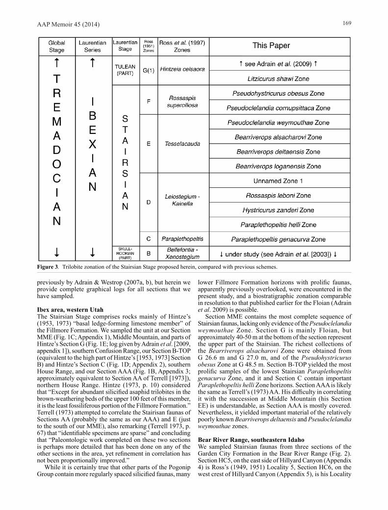
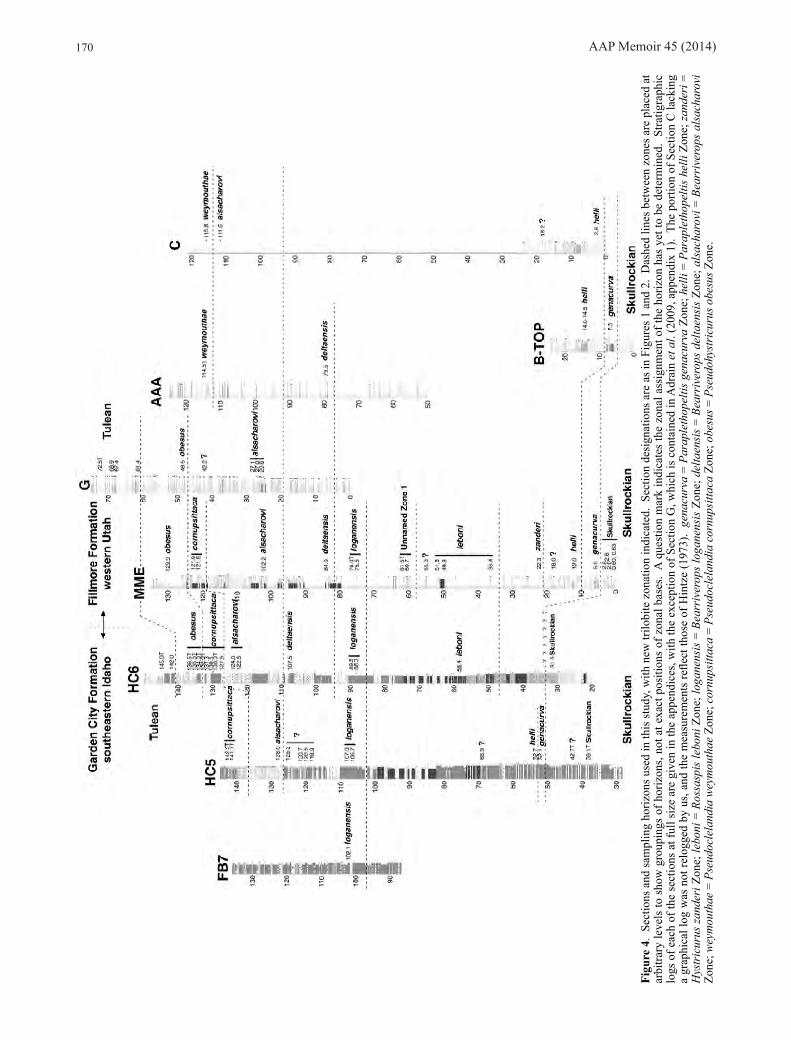
Figure 3. Trilobite zonation of the Stairsian Stage proposed herein, compared with previous schemes.
AAP Memoir 45 (2014)170
Figu
re 4
. Se
ctio
ns a
nd sa
mpl
ing
horiz
ons u
sed
in th
is st
udy,
with
new
trilo
bite
zon
atio
n in
dica
ted.
Sec
tion
desi
gnat
ions
are
as i
n Fi
gure
s 1 a
nd 2
. D
ashe
d lin
es b
etw
een
zone
s are
pla
ced
at
arbi
trary
leve
ls to
sho
w g
roup
ings
of h
oriz
ons,
not a
t exa
ct p
ositi
ons
of z
onal
bas
es.
A q
uest
ion
mar
k in
dica
tes
the
zona
l ass
ignm
ent o
f the
hor
izon
has
yet
to b
e de
term
ined
. St
ratig
raph
ic
logs
of e
ach
of th
e se
ctio
ns a
t ful
l siz
e ar
e gi
ven
in th
e ap
pend
ices
, with
the
exce
ptio
n of
Sec
tion
G, w
hich
is c
onta
ined
in A
drai
n et
al.
(200
9, a
ppen
dix
1).
The
porti
on o
f Sec
tion
C la
ckin
g a
grap
hica
l log
was
not
relo
gged
by
us, a
nd th
e m
easu
rem
ents
refle
ct th
ose
of H
intz
e (1
973)
. ge
nacu
rva
= Pa
rapl
etho
pelti
s ge
nacu
rva
Zone
; hel
li =
Para
plet
hope
ltis
helli
Zon
e; z
ande
ri =
H
ystr
icur
us z
ande
ri Z
one;
lebo
ni =
Ros
sasp
is le
boni
Zon
e; lo
gane
nsis
= B
earr
iver
ops
loga
nens
is Z
one;
del
taen
sis
= Be
arri
vero
ps d
elta
ensi
s Zo
ne; a
lsac
haro
vi =
Bea
rriv
erop
s al
sach
arov
i Zo
ne; w
eym
outh
ae =
Pse
udoc
lela
ndia
wey
mou
thae
Zon
e; c
ornu
psitt
aca
= Ps
eudo
clel
andi
a co
rnup
sitta
ca Z
one;
obe
sus =
Pse
udoh
ystr
icur
us o
besu
s Zon
e.
AAP Memoir 45 (2014) 171
6, and Section FB7, at Franklin Basin (Appendix 6), is his Locality 7. The lower part of the Stairsian seems genuinely poorly fossiliferous at these localities, with the exception of small but well preserved samples from HC6 58.4 m representing the Rossaspis leboni Zone. Rich horizons representing the Bearriverops loganensis Zone occur at all three sections, and from this point upward, good samples of all of the zones occurring at Ibex were found, with the exception of the Pseudoclelandia weymouthae Zone.
ZONATIONThe results of new field sampling for the Stairsian are very similar to those for the Floian (Adrain et al. 2009) in that they indicate large portions of the available trilobite diversity have not previously been noticed and traditional zones have had huge stratigraphic thicknesses that are not justified by actual data. There are many more stratigraphically successive, almost entirely distinct, faunas than previously realised, and as a result a more highly resolved zonal biostratigraphy is both possible and required. In the case of the Floian (Adrain et al. 2009), five traditionally recognised zones were replaced by a new 15 zone scheme (and more work remains to be carried out, see Adrain & McAdams [2012]). Here, the traditional upper Skullrockian and Stairsian scheme of four zones (Ross 1951; Hintze 1953; Ross et al. 1997) is replaced by a new 11 zone scheme (Fig. 3).
As argued by Adrain et al. (2009, p. 542-544), this increased resolution is not achieved through differing taxonomic approaches but simply by the discovery of more data. Species ranges given by Ross et al. (1997) in defense of the now obsolete zones were mostly undocumented. When these undocumented intervals have been sampled and the faunas investigated in detail, they have proven to contain entirely new, undescribed assemblages.
The approximate correlation of the sections, along with an indication of which fossiliferous horizons are assigned to each zone, is given in Figure 4. Once again, it bears emphasis that while many of the new species encountered are illustrated, details of their taxonomy and comparisons with similar species from other zones is far beyond the scope of this work. In some cases species distinctions are
subtle and not easily conveyed by a single dorsal view of a cranidium and/or pygidium. Many systematic studies are in preparation in which the morphology of the species listed will be documented in detail.
Conventions used in the following lists follow those of Adrain et al. (2009). Horizon designations followed by the letter “T” indicate talus or float samples, generally considered to be weathering in place but not from definite outcrop. Nameable new species awaiting description are identified as “sp. nov.” with a number following. Species which are definitely new but for which material adequate for formal naming has yet to be obtained are identified as “sp. nov.” with a capital letter following. Species which are likely new and distinct but for which this judgement cannot be made with absolute certainty are identified as “sp.” with a number following if there were more than one encountered belonging to the same genus. Most systematic work on the faunas remains to be carried out, and genus (and even family, in some cases) assignments should be regarded as provisional.
1. Paraplethopelt is genacurva Zone (replaces “Paraplethopeltis Zone”/“Zone C”) (Fig. 5)
Horizons. B-TOP 7.5, HC5 52.1 m. The interval is present at sections LDN, approximately 150 m, and MME 5.9 m, but trilobites are generally not silicified at these localities.
Species (5):Asaphidae
Symphysurina sp.Dimeropygidae
Dimeropygidae gen. nov. 2 sp. 1Hystricuridae
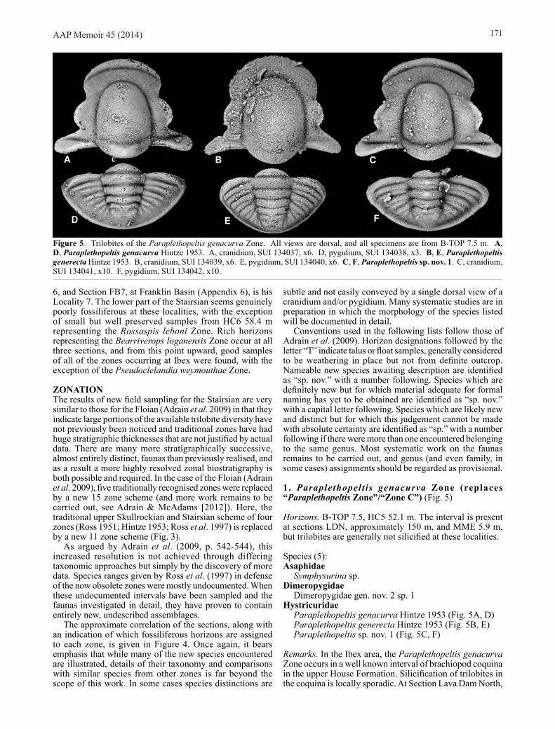
Paraplethopeltis genacurva Hintze 1953 (Fig. 5A, D)Paraplethopeltis generecta Hintze 1953 (Fig. 5B, E)Paraplethopeltis sp. nov. 1 (Fig. 5C, F)
Remarks. In the Ibex area, the Paraplethopeltis genacurva Zone occurs in a well known interval of brachiopod coquina in the upper House Formation. Silicification of trilobites in the coquina is locally sporadic. At Section Lava Dam North,
Figure 5. Trilobites of the Paraplethopeltis genacurva Zone. All views are dorsal, and all specimens are from B-TOP 7.5 m. A, D, Paraplethopeltis genacurva Hintze 1953. A, cranidium, SUI 134037, x6. D, pygidium, SUI 134038, x3. B, E, Paraplethopeltis generecta Hintze 1953. B, cranidium, SUI 134039, x6. E, pygidium, SUI 134040, x6. C, F, Paraplethopeltis sp. nov. 1. C, cranidium, SUI 134041, x10. F, pygidium, SUI 134042, x10.
AAP Memoir 45 (2014)172
for example, there is only sparse and partial silicification, whereas a short distance to the north at Section B-Top silicification is pervasive and preservation excellent. At Ibex, the fauna is overwhelmingly dominated by three species of Paraplethopeltis Bridge & Cloud 1947. Hintze (1953, pl. 7, figs 1–9) illustrated sclerites belonging to all three, but assigned them to only two species. The material he assigned to P. genacurva (Hintze 1953, pl. 7, figs 1–5) is correctly associated. However, the material assigned to P. generecta represents two species. The two librigenae (one of which is the holotype of P. generecta) belong together with the illustrated pygidium (i.e., Hintze 1953, pl. 7, figs 6, 7, 9). The cranidium (Hintze 1953, pl. 7, fig. 8) represents a third (new) species. The correct cranidium of P. generecta is illustrated herein for the first time (Fig. 5B). All three species will be dealt with in detail in a forthcoming work.
A fauna also dominated by Paraplethopeltis occurs in a similar stratigraphic position in the Garden City Formation at Section HC5 52.1 m. The trilobites are not silicified, but well preserved crackout material has been sampled. While it may be a function of a much smaller sample size owing to calcareous preservation requiring mechanical preparation, thus far the only species recovered is P. genacurva.
Taylor et al. (2012) characterised the content of the zone as “dominated by a survivor of the stage-boundary extinction that decimated the diverse fauna of the underlying Bellefontia trilobite Zone....” In the present state of knowledge, however, the sister group of Paraplethopeltis is obscure. The diverse pre-extinction faunas feature common species belonging to Hintzecurinae (see Adrain et al. 2003) and there is no obvious close phylogenetic relationship between them and Paraplethopeltis. A few taxa certainly did cross the extinction horizon. A very rare species of Symphysurina Ulrich in Walcott 1924, occurs in the zone, the youngest known. A rare dimeropygid has clear phylogenetic connections to Skullrockian and later Stairsian species. And although absent from the lower two Stairsian zones, Hystricurus s.s. Raymond 1913, is known with certainty from the upper Skullrockian (work in progress) and reappears as a common faunal element in the Hystricurus zanderi Zone.
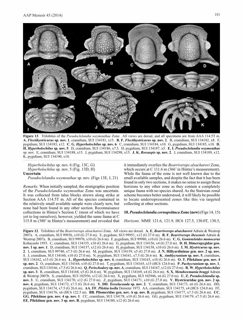
2. Paraplethopeltis helli Zone (new) (Fig. 6)
Horizons. B-TOP 14.0–14.5, C 2.6T m. The zone is also present at MME 12.0 m, but the trilobites are not silicified.
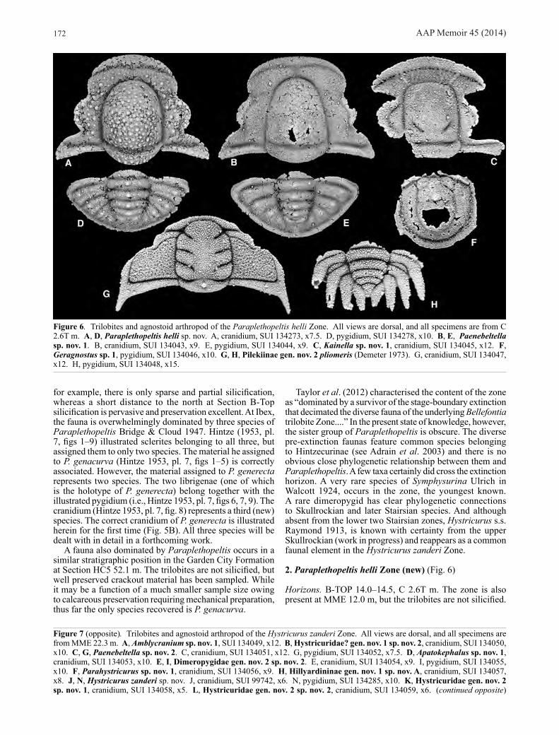
Figure 6. Trilobites and agnostoid arthropod of the Paraplethopeltis helli Zone. All views are dorsal, and all specimens are from C 2.6T m. A, D, Paraplethopeltis helli sp. nov. A, cranidium, SUI 134273, x7.5. D, pygidium, SUI 134278, x10. B, E, Paenebeltella sp. nov. 1. B, cranidium, SUI 134043, x9. E, pygidium, SUI 134044, x9. C, Kainella sp. nov. 1, cranidium, SUI 134045, x12. F, Geragnostus sp. 1, pygidium, SUI 134046, x10. G, H, Pilekiinae gen. nov. 2 pliomeris (Demeter 1973). G, cranidium, SUI 134047, x12. H, pygidium, SUI 134048, x15.
Figure 7 (opposite). Trilobites and agnostoid arthropod of the Hystricurus zanderi Zone. All views are dorsal, and all specimens are from MME 22.3 m. A, Amblycranium sp. nov. 1, SUI 134049, x12. B, Hystricuridae? gen. nov. 1 sp. nov. 2, cranidium, SUI 134050, x10. C, G, Paenebeltella sp. nov. 2. C, cranidium, SUI 134051, x12. G, pygidium, SUI 134052, x7.5. D, Apatokephalus sp. nov. 1, cranidium, SUI 134053, x10. E, I, Dimeropygidae gen. nov. 2 sp. nov. 2. E, cranidium, SUI 134054, x9. I, pygidium, SUI 134055, x10. F, Parahystricurus sp. nov. 1, cranidium, SUI 134056, x9. H, Hillyardininae gen. nov. 1 sp. nov. A, cranidium, SUI 134057, x8. J, N, Hystricurus zanderi sp. nov. J, cranidium, SUI 99742, x6. N, pygidium, SUI 134285, x10. K, Hystricuridae gen. nov. 2 sp. nov. 1, cranidium, SUI 134058, x5. L, Hystricuridae gen. nov. 2 sp. nov. 2, cranidium, SUI 134059, x6. (continued opposite)

AAP Memoir 45 (2014) 173
M, Q, Perischodory sp. nov. 1. M, cranidium, SUI 134060, x10. Q, pygidium, SUI 134061, x7.5. O, S, Kainella sp. nov. 2. O, cranidium, SUI 134062, x10. S, pygidium, SUI 134063, x8. P, T, Geragnostus sp. nov. 1. P, cephalon, SUI 134064, x12. T, pygidium, SUI 134065, x12. R, V, Leiostegium sp. nov. 1. R, cranidium, SUI 134066, x10. V, pygidium, SUI 134067, x5. U, Y, Pilekia sp. nov. 1. U, cranidium, SUI 134068, x7.5. Y, pygidium, SUI 134069, x7.5. W, AA, Rossaspis sp. nov. 1. W, cranidium, SUI 134070, x9. AA, pygidium, SUI 134071, x12. X, Dimeropygidae gen. nov. 2 sp. nov. 1, cranidium, SUI 134072, x12. Z, Pilekiinae sp. 1, pygidium, SUI 134073, x7.5. BB, Pilekiinae gen. nov. 3 sp. nov. A, pygidium, SUI 134074, x4.5.
AAP Memoir 45 (2014)174
Species (6):Agnostoid Arthropods
Geragnostus sp. 1 (Fig. 6F)Cheiruridae
Pilekiinae gen. nov. 2 pliomeris (Demeter 1973) (Fig. 6G, H)
HystricuridaePaenebeltella sp. nov. 1 (Fig. 6B, E)Paraplethopeltis helli sp. nov. (Figs 6A, D, 18)
LeiostegiidaeLeiostegium sp. nov. A
RemopleurididaeKainella sp. nov. 1 (Fig. 6C)
Remarks. Well preserved silicified trilobites occur in very thin bedded lime mudstone in a recessive interval near the base of the Fillmore Formation. The most prolific samples occur near the base of Section C, from which Demeter (1973) described his new “Rossaspis” pliomeris. The fauna has also been located with slightly poorer silicification at B-TOP 14.0–14.5 m and in calcareous preservation at MME 12.0 m. Apart from “R.” pliomeris, the two most common species are Paraplethopeltis helli, described below, and an apparently phylogenetically basal species of Paenebeltella Ross 1951 (=Glabretina Lochman 1965). This zone represents the basal part of the obsolete “Leiostegium Zone” and equivalents of previous authors. None of its species range into the overlying zone. Leiostegiid trilobites are present sporadically at many horizons of the “Leiostegium Zone”, but they also occur as rare elements throughout the Stairsian and into the Tulean at Ibex.
3. Hystricurus zanderi Zone (new) (Fig. 7)
Horizon. MME 23.3 m.
Species (20):Agnostoid Arthropods
Geragnostus sp. nov. 1 (Fig. 7P, T)Cheiruridae
Pilekia sp. nov. 1 (Fig. 7U, Y)Pilekiinae gen. nov. 3 sp. nov. A (Fig. 7BB)Pilekiinae sp. 1 (Fig. 7Z)Rossaspis sp. nov. 1 (Fig. 7W, AA)Rossaspis sp.
DimeropygidaeAmblycranium sp. nov. 1 (Fig. 7A)
Dimeropygidae gen. nov. 2 sp. nov. 2 (Fig. 7E, I)Dimeropygidae gen. nov. 2 sp. nov. 1 (Fig. 7X)Parahystricurus sp. nov. 1 (Fig. 7F)
HystricuridaeHillyardininae gen. nov. 1 sp. nov. A (Fig. 7H)Hystricuridae? gen. nov. 1 sp. nov. 2 (Fig. 7B)Hystricuridae gen. nov. 2 sp. nov. 2 (Fig. 7L)Hystricuridae gen. nov. 2 sp. nov. 1 (Fig. 7K)Hystricurus zanderi sp. nov. (Figs 7J, N, 19)Paenebeltella sp. nov. 2 (Fig. 7C, G)
LeiostegiidaeLeiostegium sp. nov. 1 (Fig. 7R, V)Perischodory sp. nov. 1 (Fig. 7M, Q)
RemopleurididaeApatokephalus sp. nov. 1 (Fig. 7D)Kainella sp. nov. 2 (Fig. 7O, S)
Remarks. A sample from Section MME 22.3 m at Middle Mountain features the stratigraphically lowest of the high diversity (20–24 species) faunas typical of the remainder of the Stairsian (lower diversity exceptions below are almost certainly due to small sample sizes). These faunas tend to have common species of Hystricurus s.s., and are dominated by species presently assigned to Cheiruridae, Hystricuridae and Dimeropygidae. The fauna of the Hystricurus zanderi Zone was apparently overlooked during earlier sampling. All of its species are new and unique to the zone, which has yet to be sampled in any other locality or section. The selected name bearer is a large and distinctive member of Hystricurus s.s., described below.
4. Rossaspis leboni Zone (new) (Fig. 8)
Horizons. MME 36.4, 49.8, 51.8, HC6 58.4 m.
Species (22):Agnostoid Arthropods
Geragnostus sp. 2 (Fig. 8BB)Cheiruridae
Pilekia sp. nov. APilekia sp. nov. B (Fig. 8X)Pilekiinae gen. nov. 3 sp. nov. 1 (Fig. 8Q, U)Pilekiinae sp. 2 (Fig. 8Y)Rossaspis leboni sp. nov. (Figs 8B, F, 20)Tesselacauda sp. nov. 1 (Fig. 8A, E)
DimeropygidaeAmblycranium sp. nov. 3 (Fig. 8L)
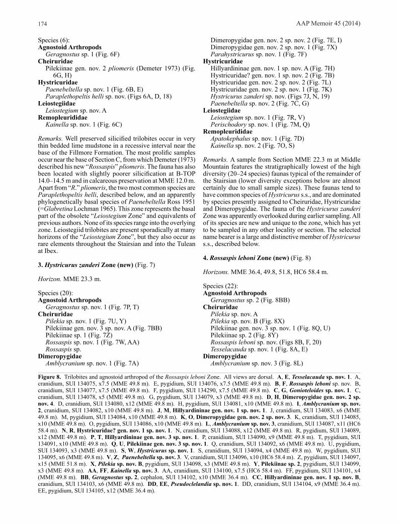
Figure 8. Trilobites and agnostoid arthropod of the Rossaspis leboni Zone. All views are dorsal. A, E, Tesselacauda sp. nov. 1. A, cranidium, SUI 134075, x7.5 (MME 49.8 m). E, pygidium, SUI 134076, x7.5 (MME 49.8 m). B, F, Rossaspis leboni sp. nov. B, cranidium, SUI 134077, x7.5 (MME 49.8 m). F, pygidium, SUI 134290, x7.5 (MME 49.8 m). C, G, Gonioteloides sp. nov. 1. C, cranidium, SUI 134078, x5 (MME 49.8 m). G, pygidium, SUI 134079, x3 (MME 49.8 m). D, H, Dimeropygidae gen. nov. 2 sp. nov. 4. D, cranidium, SUI 134080, x12 (MME 49.8 m). H, pygidium, SUI 134081, x10 (MME 49.8 m). I, Amblycranium sp. nov. 2, cranidium, SUI 134082, x10 (MME 49.8 m). J, M, Hillyardininae gen. nov. 1 sp. nov. 1. J, cranidium, SUI 134083, x6 (MME 49.8 m). M, pygidium, SUI 134084, x10 (MME 49.8 m). K, O, Dimeropygidae gen. nov. 2 sp. nov. 3. K, cranidium, SUI 134085, x10 (MME 49.8 m). O, pygidium, SUI 134086, x10 (MME 49.8 m). L, Amblycranium sp. nov. 3, cranidium, SUI 134087, x11 (HC6 58.4 m). N, R, Hystricuridae? gen. nov. 1 sp. nov. 1. N, cranidium, SUI 134088, x12 (MME 49.8 m). R, pygidium, SUI 134089, x12 (MME 49.8 m). P, T, Hillyardininae gen. nov. 3 sp. nov. 1. P, cranidium, SUI 134090, x9 (MME 49.8 m). T, pygidium, SUI 134091, x10 (MME 49.8 m). Q, U, Pilekiinae gen. nov. 3 sp. nov. 1. Q, cranidium, SUI 134092, x6 (MME 49.8 m). U, pygidium, SUI 134093, x3 (MME 49.8 m). S, W, Hystricurus sp. nov. 1. S, cranidium, SUI 134094, x4 (MME 49.8 m). W, pygidium, SUI 134095, x6 (MME 49.8 m). V, Z, Paenebeltella sp. nov. 3. V, cranidium, SUI 134096, x10 (HC6 58.4 m). Z, pygidium, SUI 134097, x15 (MME 51.8 m). X, Pilekia sp. nov. B, pygidium, SUI 134098, x3 (MME 49.8 m). Y, Pilekiinae sp. 2, pygidium, SUI 134099, x3 (MME 49.8 m). AA, FF, Kainella sp. nov. 3. AA, cranidium, SUI 134100, x7.5 (HC6 58.4 m). FF, pygidium, SUI 134101, x4 (MME 49.8 m). BB, Geragnostus sp. 2, cephalon, SUI 134102, x10 (MME 36.4 m). CC, Hillyardininae gen. nov. 1 sp. nov. B, cranidium, SUI 134103, x6 (MME 49.8 m). DD, EE, Pseudoclelandia sp. nov. 1. DD, cranidium, SUI 134104, x9 (MME 36.4 m). EE, pygidium, SUI 134105, x12 (MME 36.4 m).

AAP Memoir 45 (2014) 175

AAP Memoir 45 (2014)176
Amblycranium sp. nov. 2 (Fig. 8I)Dimeropygidae gen. nov. 2 sp. nov. 4 (Fig. 8D, H)Dimeropygidae gen. nov. 2 sp. nov. 3 (Fig. 8K, O)Gonioteloides sp. nov. 1 (Fig. 8C, G)
HystricuridaeHillyardininae gen. nov. 1 sp. nov. 1 (Fig. 8J, M)Hillyardininae gen. nov. 1 sp. nov. B (Fig. 8CC)Hillyardininae gen. nov. 3 sp. nov. 1 (Fig. 8P, T)Hystricuridae? gen. nov. 1 sp. nov. 1 (Fig. 8N, R)Hystricurus sp. nov. 1 (Fig. 8S, W)Paenebeltella sp. nov. 3 (Fig. 8V, Z)
LeiostegiidaeLeiostegium sp. nov. B
RemopleurididaeApatokephalus sp.Kainella sp. nov. 3 (Fig. 8AA, FF)
UncertainPseudoclelandia sp. nov. 1 (Fig. 8DD, EE)
Remarks. This zone is named for a common species of the cheirurid Rossaspis Harrington 1957, R. leboni, described
below. It is notable in that it is the lowest Stairsian zone which can be unambiguously located in the Garden City Formation, as all species occurring in a limited but well preserved sample at HC6 58.4 m are shared with MME 49.8 m. The zone would occupy the middle part of the obsolete “Leiostegium Zone”, but does not appear to have been detected in previous sampling. All of its species are new, and all are unique to the zone.
5. Unnamed Zone 1 (Fig. 9)
Horizon. MME 60.7 m.
Species (7):Dimeropygidae
Amblycranium sp. (Fig. 9B)Dimeropygidae gen. nov. 2 sp. 2 (Fig. 9C)Gonioteloides sp. nov. A (Fig. 9D)
HystricuridaeHillyardininae gen. nov. 1 sp. nov. C (Fig. 9A)Hystricuridae gen. nov. 3 sp. (Fig. 9G)
Figure 9. Trilobites of Unnamed Zone 1. All views are dorsal, and all specimens are from MME 60.7 m. A, Hillyardininae gen. nov. 1 sp. nov. C, cranidium, SUI 134106, x12. B, Amblycranium sp., cranidium, SUI 134107, x12. C, Dimeropygidae gen. nov. 2 sp. 2, cranidium, SUI 134108, x7.5. D, Gonioteloides sp. nov. A, pygidium, SUI 134109, x6. E, Pliomeridae gen. nov. A sp. nov. A, pygidium, SUI 134110, x10. F. Paenebeltella sp., left librigena, SUI 134111, x10. G. Hystricuridae gen. nov. 3 sp., left librigena, SUI 134112, x7.5.
Figure 10. Trilobites of the Bearriverops loganensis Zone. All views are dorsal. A, E, Bearriverops loganensis Adrain & Westrop 2007a. A, cranidium, SUI 99887, x7.5 (MME 75.5 m). E, pygidium, SUI 99901, x7.5 (MME 75.5 m). B, Hillyardininae gen. nov. 1 carinata (Ross 1951), cranidium, SUI 134113, x9 (MME 75.5 m). C, F, Gonioteloides sp. nov. 2. C, cranidium, SUI 134114, x7.5 (MME 75.5 m). F, pygidium, SUI 134115, x10 (MME 75.5 m). D, G, Paenebeltella vultulata Ross 1951. D, cranidium, SUI 134116, x7.5 (MME 75.5 m). G, pygidium, SUI 134117, x7.5 (MME 75.5 m). H, L, Hystricurus eos Kobayashi 1955. H, cranidium, SUI 134118, x6 (MME 75.5 m). L, pygidium, SUI 134119, x6 (MME 75.5 m). I, M, Hystricuridae gen. nov. 4 sp. nov. 1. I, cranidium, SUI 134120, x7.5 (MME 75.5 m). M, pygidium, SUI 134121, x7.5 (MME 75.5 m). J, Hillyardininae gen. nov. 1 robusta (Ross 1951), cranidium, SUI 134122, x7.5 (MME 75.5 m). K, Hyperbolochilus sp. nov. 1, cranidium, SUI 134123, x6 (MME 75.5 m). N, R, Amblycranium variabile Ross 1951. N, cranidium, SUI 134124, x7.5 (MME 75.5 m). R, pygidium, SUI 134125, x12 (MME 75.5 m). O, S, Hystricuridae gen. nov. 2 sp. nov. 3. O, cranidium, SUI 134126, x6 (HC5 106.7 m). S, pygidium, SUI 134127, x6 (HC5 106.7 m). P, Apatokephalus sp. nov. 2, cranidium, SUI 134128, x7.5 (MME 75.5 m). Q, U, Tesselacauda depressa Ross 1951. Q, cranidium, SUI 134129, x5 (MME 75.5 m). U, pygidium, SUI 134130, x6 (MME 75.5 m). T, X, Bearriverops borderinnensis Adrain & Westrop 2007a. T, cranidium, SUI 99927, x10 (MME 75.5 m). X, pygidium, SUI 99935, x10 (MME 75.5 m). V, Pseudoclelandia sp. 1, cranidium, SUI 134131, x12 (MME 75.5 m). W, Hystricuridae gen. nov. 3 populus (Ross 1951), cranidium, SUI 134132, x5 (MME 75.5 m). Y, Dimeropygidae gen. nov. 1 sp. nov. A, pygidium, SUI 134133, x15 (HC5 106.7 m). Z, CC, Pilekiinae gen. nov. 3 sp. nov. 2. Z, cranidium, SUI 134134, x10 (FB7 102.1 m). CC, pygidium, SUI 134135, x8 (HC5 106.7 m). AA, Parahystricurus sp. nov. 2, cranidium, SUI 134136, x12 (HC5 106.7 m). BB, FF, Pilekiinae gen. nov. 4 trio (Hintze 1953). BB, cranidium, SUI 134137, x15 (FB7 102.1 m). FF, pygidium, SUI 134138, x10 (HC5 106.7 m). DD, Hillyardininae gen. nov. 1 sp. nov. 2, cranidium, SUI 134139, x12 (HC5 106.7 m). EE, Hillyardina sp. nov. 1, cranidium, SUI 134140, x9 (HC5 106.7 m).
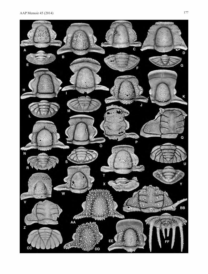
AAP Memoir 45 (2014) 177

AAP Memoir 45 (2014)178
Paenebeltella sp. (Fig. 9F)Pliomeridae
Pliomeridae gen. nov. A sp. nov. A (Fig. 9E)
Remarks. A very limited sample from MME 60.7 contains overall diversity similar to that of the underlying and overyling zones. However, for all comparisons that can be made, the species occurring appear to be distinct and unique to the horizon. For example, the species of Gonioteloides present (Fig. 9D) has a huge terminal pygidial spine much larger than that of any other known species. Unfortunately, none of the species are currently well enough known to formally name, and hence the zone itself is left unnamed. Preservation is reasonably good, but trilobite sclerites are rare in the sample. Very large field samples were made recently in the hope that material adequate to name some of the species present can eventually be accumulated. This zone represents
the highest horizon that would previously have been assigned to the obsolete “Leiostegium Zone”.
6. Bearriverops loganensis Zone (new) (Fig. 10)
Horizons. MME 75.5, 76.0T, HC5 106.7, HC6 88.3, 89.5, FB7 102.1 m.
Species (24):Cheiruridae
Pilekiinae gen. nov. 3 sp. nov. 2 (Fig. 10Z, CC)Pilekiinae n gen. 4 trio (Hintze 1953) (Fig. 10BB, FF)Tesselacauda depressa Ross 1951 (Fig. 10Q, U)
DimeropygidaeAmblycranium variabile Ross 1951 (Fig. 10N, R)Bearriverops borderinnensis Adrain & Westrop 2007a
(Fig. 10T, X)
Figure 11. Trilobites of the Bearriverops deltaensis Zone. All views are dorsal, and all specimens are from MME 84.0 m. A, E, Bearriverops deltaensis Adrain & Westrop 2007a. A, cranidium, SUI 99908, x10. E, pygidium, SUI 99920, x10. B, Hillyardininae gen. nov. 1 sp. nov. 4, cranidium, SUI 134141, x6. C, Amechilus sp., cranidium, SUI 134142, x15. D, I, Hystricurus sp. nov. A. D, cranidium, SUI 134143, x5. I, pygidium, SUI 134144, x12. F, Pseudoclelandia sp. nov. 2, cranidium, SUI 134145, x12. G, Hillyardininae gen. nov. 1 sp. nov. D, cranidium, SUI 134146, x7.5. H, Gonioteloides sp. nov. B, pygidium, SUI 134147, x12. J, Hyperbolochilus sp. nov. 2, cranidium, SUI 134148, x12. K, Hillyardininae gen. nov. 1 sp. nov. 3, cranidium, SUI 134149, x10. L, Amblycranium sp. nov. 4, cranidium, SUI 134150, x12. M, N, Paenebeltella vultulata Ross 1951. M, pygidium, SUI 134151, x10. N, cranidium, SUI 134152, x12. O, Pilekiinae gen. nov. 3 sp. nov. 2?, pygidium, SUI 134153, x12. P, Hystricuridae gen. nov. 4 sp., pygidium, SUI 134154, x15.

AAP Memoir 45 (2014) 179
Bearriverops loganensis Adrain & Westrop 2007a (Fig. 10A, E)
Dimeropygidae gen. nov. 1 sp. nov. A (Fig. 10Y)Gonioteloides sp. nov. 2 (Fig. 10C, F)Parahystricurus sp. nov. 2 (Fig. 10AA)
HystricuridaeHillyardina sp. nov. 1 (Fig. 10EE)Hillyardininae gen. nov. 1 carinata (Ross 1951) (Fig. 10B)Hillyardininae gen. nov. 1 robusta (Ross 1951) (Fig. 10J)Hillyardininae gen. nov. 1 sp. nov. 2 (Fig. 10DD)Hyperbolochilus sp. nov. 1 (Fig. 10K)Hyperbolochilus sp. nov. AHystricuridae gen. nov. 2 sp. nov. 3 (Fig. 10O, S)Hystricurus eos Kobayashi 1955 (Fig. 10H, L)Hystricuridae gen. nov. 3 populus (Ross 1951) (Fig. 10W)Hystricuridae gen. nov. 4 sp. nov. 1 (Fig. 10I, M)Paenebeltella vultulata Ross 1951 (Fig. 10D, G)
RemopleurididaeApatokephalus sp. nov. 2 (Fig. 10P)
UncertainAmechilus palaora Ross 1951Pseudoclelandia lenisora Ross 1951Pseudoclelandia sp. 1 (Fig. 10V)
Remarks. This zone is represented as a single prolifically yielding horizon (or two, very closely spaced) at all relevant sections. A single horizon, HC5 106.7 m, likely yielded most of the species previously assigned to the obsolete “Zone E”/“Tesselacauda Zone” by Ross (1951). Based on taphonomic evidence, the same horizon may be represented by HC6 89.5 m and FB7 102.1 m, which feature the same taxon-abundances, very similar styles of silicified preservation, and the unusual prevalence of articulated specimens of the same handful of species.
7. Bearriverops deltaensis Zone (new) (Fig. 11)
Horizons. MME 84.0, AAA 79.5, HC6 107.5 m.
Species (14):Cheiruridae
Pilekiinae gen. nov. 3 sp. nov. 2? (Fig. 11O)Dimeropygidae
Amblycranium sp. nov. 4 (Fig. 11L)Bearriverops deltaensis Adrain & Westrop 2007a (Fig.
11A, E)Bearriverops sp. cf. B. ibexensis of Adrain & Westrop
(2007a)Gonioteloides sp. nov. B (Fig. 11H)
HystricuridaeHillyardininae gen. nov. 1 sp. nov. 4 (Fig. 11B)Hillyardininae gen. nov. 1 sp. nov. 3 (Fig. 11K)Hillyardininae gen. nov. 1 sp. nov. D (Fig. 11G)Hyperbolochilus sp. nov. 2 (Fig. 11J)Hystricuridae gen. nov. 4 sp. (Fig. 11P)Hystricurus sp. nov. A (Fig. 11D, I)Paenebeltella vultulata Ross 1951 (Fig. 11M, N)
UncertainAmechilus sp. (Fig. 11C)Pseudoclelandia sp. nov. 2 (Fig. 11F)
Remarks. Species from this zone are typically very similar to, yet clearly differentiated from, congenerics in the underlying Bearriverops loganensis Zone. This was first demonstrated by Adrain & Westrop (2007a) who noted the
subtle distinctions between B. deltaensis and B. loganensis. As photography has advanced, similar distinctions have become apparent for almost all of the species present. Only Paenebeltella vultulata Ross 1951, appears to be shared between the zones. Lower diversity of this zone is likely a result of the limited sample sizes thus far available.
8. Bearriverops alsacharovi Zone (new) (Fig. 12)
Horizons. G 26.6, 27.0, 27.1, C 111.6, MME 102.2, HC5 128.0, HC6 122.5, 124.0 m.
Species (21):Cheiruridae
Pilekia loella Demeter 1973 (Fig. 12AA, FF)Pilekiinae gen. nov. 3 sp. nov. B (Fig. 12EE)Pilekiinae n gen. 4 sp. nov. 2 (Fig. 12O, T)Pilekiinae n gen. 4 sp. nov. 1 (Fig. 12CC, GG)Tesselacauda sp. nov. 2 (Fig. 12Y, DD)
DimeropygidaeAmblycranium sp. nov. 5 (Fig. 12K)Bearriverops alsacharovi Adrain & Westrop 2007a (Fig.
12A, E)Bearriverops ibexensis Adrain & Westrop 2007a (Fig.
12B, F)Dimeropygidae gen. nov. 1 sp. nov. 1 (Fig. 12D, H)Gonioteloides monoceras Kobayashi 1955 (Fig. 12C, G)
HystricuridaeHillyardininae gen. nov. 2 sp. nov. 1 (Fig. 12J, N)Hyperbolochilus sp. nov. 4 (Fig. 12L)Hyperbolochilus sp. nov. 3 (Fig. 12R, W)Hystricuridae gen. nov. 2 sp. nov. 4 (Fig. 12V)Hystricurus sp. nov. 2 (Fig. 12I, M)Metabowmania braggi Adrain & Westrop 2007b (Fig.
12S, X)Pachycranium sp. nov. 1 (Fig. 12P)
LeiostegiidaePerischodory sp. nov. A (Fig. 12Q)
PliomeridaePliomeridae gen. nov. A sp. nov. B (Fig. 12BB)
UncertainPseudoclelandia sp. nov. 3 (Fig. 12U, Z)Shumardiidae sp. indet.
Remarks. Rich collections from G 26.6 m and G 27.0 m yielded the only previously named species belonging to this zone, Pilekia loella Demeter 1973. New sampling has shown that almost all of the associated species are new (the exception is Hystricurus eos, which appears to be the same species described by Kobayashi (1955) and Dean (1989) from further to the (Ordovician) east along the northern Laurentian margin. Samples representing the zone have also been discovered at Sections HC5 and HC6 in the Garden City Formation.
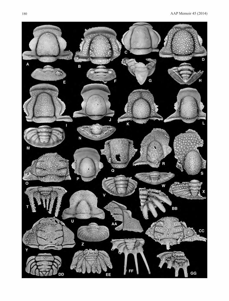
9. Pseudoclelandia weymouthae Zone (new) (Fig. 13)
Horizons. AAA 114.5T, C 115.8 m.
Species (6):Cheiruridae
Rossaspis sp. nov. 2 (Fig. 13J, K)Hystricuridae
Flectihystricurus sp. nov. 2 (Fig. 13B, F)Flectihystricurus sp. nov. 1 (Fig. 13A)
AAP Memoir 45 (2014)180
AAP Memoir 45 (2014) 181
Hyperbolochilus sp. nov. 6 (Fig. 13C, G)Hyperbolochilus sp. nov. 5 (Fig. 13D, H)
UncertainPseudoclelandia weymouthae sp. nov. (Figs 13E, I, 21)
Remarks. When initially sampled, the stratigraphic position of the Pseudoclelandia weymouthae Zone was uncertain. It was collected from talus blocks strewn along strike at Section AAA 114.5T m. All of the species contained in the relatively small available sample were clearly new, but none had been found in any other section. Reconnaisance collections in Hintze’s Section C (most of which we have yet to log ourselves), however, yielded the same fauna at C 115.8 m (380’ in Hintze’s measurement) and revealed that
it immediately overlies the Bearriverops alsacharovi Zone, which occurs at C 111.6 m (366’ in Hintze’s measurement). While the fauna of the zone is not well known due to the small available samples, and despite the fact that it has been found in only two sections, it makes no sense to assign these horizons to any other zone as they contain a completely unique fauna with no species shared. As the Stairsian zonal scheme becomes better understood, it will likely be possible to locate underrepresented zones like this via targeted collecting at other sections.
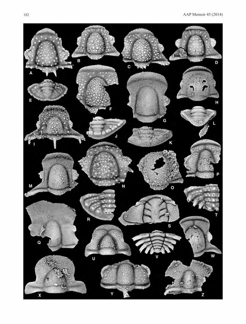
10. Pseudoclelandia cornupsittaca Zone (new) (Figs 14, 15)
Horizons. MME 121.6, 121.9, HC6 127.5, 130.0T, 130.5,
Figure 13. Trilobites of the Pseudoclelandia weymouthae Zone. All views are dorsal, and all specimens are from AAA 114.5T m. A, Flectihystricurus sp. nov. 1, cranidium, SUI 134181, x15. B, F, Flectihystricurus sp. nov. 2. B, cranidium, SUI 134182, x8. F, pygidium, SUI 134183, x12. C, G, Hyperbolochilus sp. nov. 6. C, cranidium, SUI 134184, x10. G, pygidium, SUI 134185, x10. D, H, Hyperbolochilus sp. nov. 5. D, cranidium, SUI 134186, x7.5. H, pygidium, SUI 134187, x5. E, I, Pseudoclelandia weymouthae sp. nov. E, cranidium, SUI 134188, x15. I, pygidium, SUI 134298, x15. J, K, Rossaspis sp. nov. 2. J, cranidium, SUI 134189, x12. K, pygidium, SUI 134190, x10.
Figure 12. Trilobites of the Bearriverops alsacharovi Zone. All views are dorsal. A, E, Bearriverops alsacharovi Adrain & Westrop 2007a. A, cranidium, SUI 99936, x10 (G 27.0 m). E, pygidum, SUI 99951, x12 (G 27.0 m). B, F, Bearriverops ibexensis Adrain & Westrop 2007a. B, cranidium, SUI 99970, x10 (G 26.6 m). F, pygidium, SUI 99980, x10 (G 26.6 m). C, G, Gonioteloides monoceras Kobayashi 1955. C, cranidium, SUI 134155, x10 (G 26.6 m). G, pygidium, SUI 134156, x4 (G 27.0 m). D, H, Dimeropygidae gen. nov. 1 sp. nov. 1. D, cranidium, SUI 134157, x12 (G 26.6 m). H, pygidium, SUI 134158, x10 (G 26.6 m). I, M, Hystricurus sp. nov. 2. I, cranidium, SUI 99740, x7.5 (G 26.6 m). M, pygidium, SUI 134159, x5 (G 27.0 m). J, N, Hillyardininae gen. nov. 2 sp. nov. 1. J, cranidium, SUI 134160, x10 (G 27.0 m). N, pygidium, SUI 134161, x7.5 (G 26.6 m). K, Amblycranium sp. nov. 5, cranidium, SUI 134162, x15 (G 26.6 m). L, Hyperbolochilus sp. nov. 4, cranidium, SUI 134163, x10 (G 26.6 m). O, T, Pilekiinae gen. nov. 4 sp. nov. 2. O, cranidium, SUI 134164, x10 (G 27.0 m). T, pygidium, SUI 134165, x15 (HC6 124.0 m). P, Pachycranium sp. nov. 1, cranidium, SUI 134166, x7.5 (G 26.6 m). Q, Perischodory sp. nov. A, cranidium, SUI 134167, x12 (G 27.0 m). R, W, Hyperbolochilus sp. nov. 3. R, cranidium, SUI 134168, x5 (G 26.6 m). W, pygidium, SUI 134169, x4 (G 26.6 m). S, X, Metabowmania braggi Adrain & Westrop 2007b. S, cranidium, SUI 102936, x12 (G 26.6 m). X, pygidium, SUI 102946, x6 (G 27.0 m). U, Z, Pseudoclelandia sp. nov. 3. U, cranidium, SUI 134170, x12 (G 27.0 m). Z, pygidium, SUI 134171, x10 (G 27.0 m). V, Hystricuridae gen. nov. 2 sp. nov. 4, pygidium, SUI 134172, x7.5 (G 26.6 m). Y, DD, Tesselacauda sp. nov. 2. Y, cranidium, SUI 134173, x6 (G 26.6 m). DD, pygidium, SUI 134174, x7.5 (G 26.6 m). AA, FF, Pilekia loella Demeter 1973. AA, cranidium, SUI 134175, x4 (HC6 124.0 m). FF, pygidium, SUI 134176, x6 (HC6 122.5 m). BB, Pliomeridae gen. nov. A sp. nov. B, pygidium, SUI 134177, x7.5 (G 26.6 m). CC, GG, Pilekiinae gen. nov. 4 sp. nov. 1. CC, cranidium, SUI 134178, x10 (G 26.6 m). GG, pygidium, SUI 134179, x7.5 (G 26.6 m). EE, Pilekiinae gen. nov. 3 sp. nov. B, pygidium, SUI 134180, x12 (G 26.6 m).
AAP Memoir 45 (2014)182

AAP Memoir 45 (2014) 183
131.0T, 131.3, 132.0T m.
Species (29):Cheiruridae
Rossaspis superciliosa (Ross 1951) (Fig. 15B, E)Rossaspis sp. nov. 3 (Fig. 14S, V)
DimeropygidaeParahystricurus fraudator Ross 1951 (Figs 14B, 15Q)Parahystricurus oculirotundus Ross 1951 (Fig. 14D)Parahystricurus pustulosus Ross 1951 (Figs 14C, 15R)Pseudohystricurus sp. (Fig. 14O)
HarpetidaeHypothetica rawi Ross 1951 (Fig. 14Z)
HystricuridaeFlectihystricurus flectimembrus (Ross 1951) (Fig. 15A,
D)Flectihystricurus sp. nov. 3 (Fig. 14A, E)Flectihystricurus sp. nov. B (Fig. 14N)Flectihystricurus sp. nov. A (Fig. 15G, J)Hillyardina semicylindrica Ross 1951 (Fig. 15F, I)Hillyardina sp. nov. 2 (Fig. 14I)Hillyardininae gen. nov. 1 sp. nov. E (Fig. 14F, J)Hyperbolochilus marginauctum Ross 1951 (Figs 14G,
K, 15O, S)Hyperbolochilus sp. nov. 8 (Fig. 15H, K)Hyperbolochilus sp. nov. 7 (Fig. 14M)Hystricurus sp. nov. C (Fig. 14R)Hystricurus sp. nov. B (Figs 14P, T, 15P)Metabowmania morgani Adrain & Westrop 2007bMetabowmania sp. nov. A of Adrain & Westrop (2007b)
(Fig. 14Q)Metabowmania cf. M. latilimbata of Adrain & Westrop
(2007b) (Fig. 15T)Pachycranium faciclunis Ross 1951 (Figs 14W, 15N)
TelephinidaePyraustocranium orbatum Ross 1951 (Fig. 15C)Pyraustocranium sp. nov. A (Fig. 14Y)
UncertainAffinities uncertain gen. nov. 2 sp. nov. 1 (Fig. 14H, L)Pseudoclelandia cornupsittaca Ross 1951 (Figs 14U,
15M)Pseudoclelandia fluxafissura Ross 1951 (Figs 14X, 15L)Pseudoclelandia sp. 2
Remarks. Where they are represented by large collections in either region, the zones beneath the Pseudoclelandia cornupsittaca Zone contain virtually identical faunas in the Fillmore and the Garden City formations. Species present
are the same, and relative abundance is in most cases directly comparable. For reasons not yet clear, this is not true of the uppermost two Stairsian zones, in which there are considerable differences between the two regions. Enough species are clearly shared that correlation is possible and the same zonal nomenclature can be applied to either succession. Within each zone, however, there is far more differentiation encountered than is the case lower in the sections. Species of Pyraustocranium Ross 1951, are clearly different between the regions, as are species of Metabowmania Kobayashi 1955, Hystricurus, Hyperbolochilus Ross 1951, Hillyardina Ross 1951, Flectihystricurus Adrain, Lee, Westrop, Chatterton & Landing 2003, and Rossaspis Harrington 1957. On the other hand, species of Pseudoclelandia (including the zonal name bearer), Parahystricurus Ross 1951 and Pachycranium Ross 1951, seem definitely to be shared. The inter-regional differences could perhaps be stratigraphic in nature, with collections from either region sampling different time intervals, and the species distinctions recording taxonomic turnover. The zone is not very thick, however, and it is difficult to explain why such distinctions are not encountered lower in the Stairsian (nor in the Floian zones documented by Adrain et al. [2009]). Alternatively, the differences may record geographic diversification in response to as yet unknown extrinsic factors.
11. Pseudohystricurus obesus Zone (new) (Figs 16, 17)
Horizons. G 48.5, MME 129.9, HC6 134.0T, 135.0T m.
Species (24):Cheiruridae
Pilekiinae gen. nov. 1 sp. nov. 1 (Fig. 16Q, U)Dimeropygidae
Parahystricurus bispicatus Hintze 1953 (Fig. 16X)Parahystricurus sp. nov. 6 (Fig. 17E)Parahystricurus sp. nov. 5 (Fig. 16W)Parahystricurus sp. nov. 4 (Fig. 16Z)Parahystricurus sp. nov. 3 (Fig. 16Y)Pseudohystricurus obesus Ross 1951 (Fig. 16A, E)
HarpetidaeHypothetica sp. nov. 1 (Fig. 17M)
HystricuridaeFlectihystricurus acumennasus (Ross 1951) (Fig. 17F, I)Flectihystricurus sp. nov. 4 (Fig. 16K, N)Hillyardina sp. nov. 3 (Fig. 17A)Hyperbolochilus marginauctum Ross 1951 (Figs 16O,
S, 17G, J)
Figure 14. Trilobites of the Pseudoclelandia cornupsittaca Zone from the Ibex area, western Utah. All views are dorsal. A, E, Flectihystricurus sp. nov. 3. A, cranidium, SUI 134191, x10 (MME 121.6 m). E, pygidium, SUI 134192, x10 (MME 121.6 m). B, Parahystricurus fraudator Ross 1951, cranidium, SUI 134193, x10 (MME 121.6 m). C, Parahystricurus pustulosus Ross 1951, cranidium, SUI 134194, x10 (MME 121.6 m). D, Parahystricurus oculirotundus Ross 1951, cranidium, SUI 134195, x12 (MME 121.9 m). F, J, Hillyardininae gen. nov. 1 sp. nov. E. F, cranidium, SUI 134196, x7.5 (MME 121.9 m). J, pygidium, SUI 134197, x15 (MME 121.9 m). G, K, Hyperbolochilius marginauctum Ross 1951. G, cranidium, SUI 134198, x7.5 (MME 121.6). K, pygidium, SUI 134199, x6 (MME 121.6 m). H, L, Affinities uncertain gen. nov. 2 sp. nov. 1. H, cranidium, SUI 134200, x10 (MME 121.6 m). L, pygidium, SUI 134201, x15 (MME 121.6 m). I, Hillyardina sp. nov. 2, cranidium, SUI 134202, x12 (MME 121.6 m). M, Hyperbolochilus sp. nov. 7, cranidium, SUI 134203, x15 (MME 121.6 m). N, Flectihystricurus sp. nov. B, cranidium, SUI 134204, x9 (MME 121.6 m). O, Pseudohystricurus sp., cranidium, SUI 134205, x16 (MME 121.6 m). P, T, Hystricurus sp. nov. B. P, cranidium, SUI 134206, x10 (MME 121.6 m). T, pygidium, SUI 134207, x5 (MME 121.9 m). Q, Metabowmania sp. nov. A of Adrain & Westrop (2007b), cranidium, SUI 102950, x10 (MME 121.6 m). R, Hystricurus sp. nov. C, pygidium, SUI 134208, x4 (MME 121.9 m). S, V, Rossaspis sp. nov. 3. S, cranidium, SUI 134209, x7.5 (MME 121.9 m). V, pygidium, SUI 134210, x7.5 (MME 121.9 m). U, Pseudoclelandia cornupsittaca Ross 1951, cranidium, SUI 134211, x15 (MME 121.9 m). W, Pachycranium faciclunis Ross 1951, cranidium, SUI 134212, x12 (MME 121.6 m). X, Pseudoclelandia fluxafissura Ross 1951, cranidium, SUI 134213, x15 (MME 121.9 m). Y, Pyraustocranium sp. nov. A, cranidium, SUI 134214, x12 (MME 121.9 m). Z, Hypothetica rawi Ross 1951, cranidium, SUI 134215, x15 (MME 121.6 m).

AAP Memoir 45 (2014)184
Figure 15. Trilobites of the Pseudoclelandia cornupsittaca Zone from the Bear River Range, southeastern Idaho. All views are dorsal. A, D, Flectihystricurus flectimembrus (Ross 1951). A, cranidium, SUI 134216, x7.5 (HC6 127.5 m). D, pygidium, SUI 134217, x7.5 (HC6 130.5 m). B, E, Rossaspis superciliosa (Ross 1951). B, cranidium, SUI 134218, x7.5 (HC6 127.5 m). E, pygidium, SUI 134219, x7.5 (HC6 127.5 m). C, Pyraustocranium orbatum Ross 1951, cranidium, SUI 134220, x10 (HC6 131.3 m). F, I, Hillyardina semicylindrica Ross 1951. F, cranidium, SUI 134221, 12 (HC6 130.5 m). I, pygidium, SUI 134222, x12 (HC6 130.5 m). G, J, Flectihystricurus sp. nov. A. G, cranidium, SUI 134223, x5 (HC6 132.0T m). J, pygidium, SUI 134224, x7.5 (HC6 132.0T m). H, K, Hyperbolochilus sp. nov. 8. H, cranidium, SUI 134225, x7.5 (HC6 127.5 m). K, pygidium, SUI 134226, x7.5 (HC6 131.3 m). L, Pseudoclelandia fluxafissura Ross 1951, cranidium, SUI 134227, x17 (HC6 127.5 m). M, Pseudoclelandia cornupsittaca Ross 1951, cranidium, SUI 134228, x15 (HC6 127.5 m). N, Pachycranium faciclunis Ross 1951, cranidium, SUI 134229, x10 (HC6 132.0T m). O, S, Hyperbolochilus marginauctum Ross 1951. O, cranidium, SUI 134230, x12 (HC6 127.5 m). S, pygidium, SUI 134231, x8 (HC6 127.5 m). P, Hystricurus sp. nov. B, cranidium, SUI 134232, x12 (HC6 127.5 m). Q, Parahystricurus fraudator Ross 1951, cranidium, SUI 134233, x10 (HC6 127.5 m). R, Parahystricurus pustulosus Ross 1951, cranidium, SUI 134234, x8 (HC6 127.5 m). T, Metabowmania cf. M. latilimbata of Adrain & Westrop (2007b), cranidium, SUI 134235, x7.5 (HC6 130.5 m).

AAP Memoir 45 (2014) 185
Hyperbolochilus sp. nov. 9 (Fig. 16P, T)Hystricuridae gen. nov. 2 sp. nov. A (Fig. 16V)Hystricurus oculilunatus Ross 1951 (Fig. 16J, M)Hystricurus sp. nov. 3 (Fig. 16I, L)Metabowmania morgani Adrain & Westrop 2007b (Fig.
16R)Metabowmania sp. nov. B (Fig. 17K, L)
TelephinidaeGoniophrys prima Ross 1951 (Fig. 17H)Goniophrys sp. nov. 1 (Fig. 16D, H)
UncertainAffinities uncertain gen. nov. 1 sp. nov. 1 (Fig. 16B, F)Affinities uncertain gen. nov. 2 sp. nov. 2 (Figs 16C, G,
17D)Pseudoclelandia sp. nov. 4 (Fig. 17C)Pseudoclelandia sp. 3 (Fig. 17B)
Remarks. Even fewer species are shared between the Fillmore Formation and the Garden City Formation in the Pseudohystricurus obesus Zone than in the underlying Pseudoclelandia cornupsittaca Zone (see above), with only the zonal name bearer and Hyperbolochilus marginauctum confirmed at present. Ross (1951) lumped the Pseudoclelandia cornupsittaca Zone and the overlying Pseudohystricurus obesus Zone as his “Zone F” and at HC6 they together only occupy a few meters of section. However, the faunas of the zones in the Garden City Formation are almost entirely different from one another, with only H. marginauctum apparently shared between them.
COMPARISON WITH OTHER REGIONSThe only other significant northern Laurentian Stairsian faunas to have been described in the literature are those from British Columbia dealt with by Kobayashi (1955), and from Alberta by Dean (1989), who revised some of Kobayashi’s taxa. In either case the amount of data and scope of illustration is limited, but some species (e.g., Gonioteloides monoceros Kobayashi 1955; Hystricurus eos [Kobayashi 1955]) are clearly shared and there is good reason to expect that were more information available the biostratigraphic scheme established herein would be applicable to these localities lying to the east along the northern Laurentian margin. Pratt (1988) described a low diversity fauna from the Rabbitkettle Formation of the southern Mackenzie Mountains, Northwest Territories, which contains Leiostegium and Kainella and is likely of early Stairsian age. It may be possible to correlate this fauna directly with the scheme established herein once detailed taxonomic work on the relevant groups is undertaken. Similarly, material from Colorado described by Berg & Ross (1959) could well be directly correlated if better known, as it contains representatives of leiostegiids, Kainella, Paraplethopeltis and Gonioteloides.
Stairsian-equivalent faunas from southern Laurentia (in terms of Ordovician geography) have been described from, for example, Texas (Bridge & Cloud 1947), Missouri (Heller 1956), New York (Flower 1968; Landing et al. 2012), Oklahoma (Stitt 1983), western Newfoundland (Boyce 1989), and from the Laurentian-derived Norwegian Caledonides by Gobbett (1960). However, no benthic trilobite species have been confirmed to occur in both northern and southern Laurentia, as it appears that the Transcontinental Arch formed a strong distributional barrier through most of the Early Ordovician. This was the basis of Loch’s (1995, 2007) advocacy of separate stage nomenclature and zonations for northern versus southern Laurentia. Boyce’s (1989)
assignment of material from the Boat Harbour Formation of Newfoundland to Hystricurus oculilunatus Ross 1951, for example, was rejected by Westrop (in Landing et al. 2003, p. 93). While the extinction marking the base of the Stairsian can be recognised in southern Laurentia, there are few if any southern Laurentia data from the interval of the extinction marking the base of the Tulean. Nevertheless, all known data are consistent with the notion that the higher extinction also took place across the entire continent (at minimum), and recognition of stadial equivalence does not seem problematic (Landing et al. [2012], for example, apply the Skullrockian and Stairsian stages to southern Laurentian successions in New York State). (Note that the same is not true of the Tulean and Blackhillsian stages; as pointed out by Adrain et al. [2009, pp. 551–552], a distinction between these stages even in northern Laurentia is of limited utility.) Stairsian correlation between southern and northern Laurentia at species/zonal level thus far does not seem possible. The appearance of pelagic telephinids in the upper Stairsian (Goniophrys Ross 1951) offers limited hope, but the taxon has not been discovered in southern Laurentia.
Stairsian-equivalent faunas with otherwise “typical” Laurentian elements can be recognised from tropical regions of other palaeocontinents, but thus far the record is very limited. The best described occurrence with the most species belonging to groups known in Laurentia only from the Stairsian is a fauna from Tasmania described by Jell & Stait (1985). This assemblage contains hillyardinines, hystricurines, leiostegiids and cheirurids directly comparable with those occurring in northern Laurentia. Hystricurus penchiensis (Lu in Lu et al. 1976), from South China appears to represent Flectihystricurus. Jell & Stait (1985, p. 4) assigned material from Tasmania to this species, but on their plate caption (Jell & Stait 1985, p. 28) the species was identified as “Hystricurus timsheaensis sp. nov.”. The Tasmanian species seems clearly distinct from F. penchiensis, and it, too, should be assigned to Flectihystricurus. The material assigned by Jell & Stait (1985, p. 5, pl. 2, figs 1–15, pl. 3, figs 9, 10, 13) to Hystricurus lewisi (Kobayashi 1940) represents at least three species. Four cranidia (Jell & Stait 1985, pl. 2, figs 1–4), including the holotypes of both Tasmanaspis lewisi Kobayashi 1940, and T. longa Kobayashi 1940, represent T. lewisi, of which T. longa is a junior subjective synonym. These cranidia have unambiguously different morphology from the remainder assigned by Jell & Stait: there is only a fine granulate sculpture, the glabella is shorter relative to its width, the anterior border is much longer and the anterior border furrow more shallow, the anterior sections of the facial suture are strongly anteriorly divergent, and the palpebral lobe is large and the palpebral furrow very shallow. Jell & Stait (1985, p. 7) ascribed the differences in the anterior region to flattening of Kobayashi’s specimens, but the specimens have multiple obvious differences which cannot be explained by differential preservation. Tasmanaspis Kobayashi 1940, should be recognised as a monotypic, inadequately known, hystricurid genus. For the other material assigned by Jell & Stait, some (pl. 2, figs 5–9, 12) seem to represent a species of Flectihystricurus. One librigena (Jell & Stait 1985, pl. 2, fig. 10A, B) represents Hystricurus s.s. of Adrain et al. (2003), and likely should be associated with a Hystricurus pygidium assigned by Jell & Stait to “Hystricurus cf. H. robustus” (Jell & Stait 1985, pl. 3, fig. 14). The remaining specimens (Jell & Stait, pl. 2, figs 11, 13–15, pl. 3, figs 9, 10, 13) certainly represent a hillyardinine. Of the other material assigned

AAP Memoir 45 (2014)186
Figure 16. Trilobites of the Pseudohystricurus obesa Zone from the Ibex area, western Utah. All views are dorsal, and all specimens are from G 48.5 m. A, E, Pseudohystricurus obesus Ross 1951. A, cranidium, SUI 134236, x19. E, pygidium, SUI 134237, x20. B, F, Affinities uncertain gen. nov. 1 sp. nov. 1. B, cranidium, SUI 134238, x9. F, pygidium, SUI 134239, x12. C, G, affinities uncertain gen. nov. 2 sp. nov. 2. C, cranidium, SUI 134240, x12. G, pygidium, SUI 134241, x10. D, H, Goniophrys sp. nov. 1. D, cranidium, SUI 134242, x10. H, pygidium, SUI 134243, x10. I, L, Hystricurus sp. nov. 3. I, cranidium, SUI 134244, x10. (continued opposite)

AAP Memoir 45 (2014) 187
to “Hystricurus cf. H. robustus”, an articulated thorax and partial cranidium (Jell & Stait, pl. 3, fig. 12) is difficult to identify, but the pygidia (Jell & Stait, pl. 3, figs 8, 11) and at least some of the cranidia (Jell & Stait 1985, pl. 4, figs 3, 4, 6) are definitely hillyardinine. Hystricurus robustus Ross 1951, is a hillyardinine and will be assigned to a new genus in a forthcoming work on that subfamily. Finally, Tanybregma tasmaniensis Jell & Stait 1985, is a hillyardinine.
Goniophrys laifengensis Zhou in Zhou et al. 1977, and G. subcylindricus Xiang & Zhou 1987, both from South China, appear to be correctly assigned. In all of these cases the species are associated with numerous taxa not shared with Laurentia. The Tasmanian fauna, for example, contains common asaphids, whereas asaphids are entirely unknown
from the Laurentian Stairsian.
SYSTEMATIC PALAEONTOLOGYNote. Where possible, new and revised zones treated herein have used a previously named species as name bearer. In four cases (five counting Unnamed Zone 1) this has proven impossible as no adequate potential name bearing species has been described. Hence we name and describe four zonal name bearers below. In each case, extended taxonomic works on the respective group are in progress and each species will be treated in more detail in those works. In particular, we confine ourselves here to a level of illustration adequate to demonstrate the novelty and general morphology of each species. This represents a fraction of that which we
Figure 17. Trilobites of the Pseudohystricurus obesa Zone from the Bear River Range, southeastern Idaho. All views are dorsal, and all specimens are from HC6 134.0T m. A, Hillyardina sp. nov. 3, cranidium, SUI 134260, x10. B, Pseudoclelandia sp. 3, cranidium, SUI 134261, x20. C, Pseudoclelandia sp. nov. 4, cranidium, SUI 134262, x22. D, affinities uncertain gen. nov. 2 sp. nov. 2, cranidium, SUI 134263, x20. E, Parahystricurus sp. nov. 6, cranidium, SUI 134264, x8. F, I, Flectihystricurus acumennasus (Ross 1951). F, cranidium, SUI 134265, x9. I, pygidium, SUI 134266, x12. G, J, Hyperbolochilius marginauctum Ross 1951. G, cranidium, SUI 134267, x10. J, pygidium, SUI 134268, x6. H, Goniophrys prima Ross 1951, cranidium, SUI 134269, x9. K, L, Metabowmania sp. nov. B. K, pygidium, SUI 134270, x10. L, cranidium, SUI 134271, x12. M, Hypothetica sp. nov. 1, cranidium, SUI 134272, x15.
L, pygidium, SUI 134245, x7.5. J, M, Hystricurus oculilunatus Ross 1951. J, cranidium, SUI 134246, x5. M, pygidium, SUI 99737, x4. K, N, Flectihystricurus sp. nov. 4. K, cranidium, SUI 134247, x10. N, pygidium, SUI 134248, x10. O, S, Hyperbolochilus marginauctum Ross 1951. O, cranidium, SUI 134249, x5. S, pygidium, SUI 134250, x3. P, T, Hyperbolochilus sp. nov. 9. P, cranidium, SUI 134251, x7. T, pygidium, SUI 134252, x6. Q, U, Pilekiinae gen. nov. 1 sp. nov. 1. Q, cranidium, SUI 134253, x8. U, pygidium, SUI 134254, x7. R, Metabowmania morgani Adrain & Westrop 2007b, cranidium, SUI 102931, x7.5. V, Hystricuridae gen. nov. 2 sp. nov. A, pygidium, SUI 134255, x7.5. W, Parahystricurus sp. nov. 5, cranidium, SUI 134256, x12. X, Parahystricurus bispicatus Hintze 1953, cranidium, SUI 134257, x8. Y, Parahystricurus sp. nov. 3, cranidium, SUI 134258, x8. Z, Parahystricurus sp. nov. 4, cranidium, SUI 134259, x9.

AAP Memoir 45 (2014)188
have used to summarise the full range of intraspecific and ontogenetic variation as well as geographic and stratigraphic distribution in other recent work (e.g., Adrain et al. 2011b; Adrain et al. 2012).
Repository. Figured material is housed in the Paleontology Repository, Department of Geoscience, University of Iowa, Iowa City, with specimen number prefix SUI.
Family HYSTRICURIDAE Hupé 1953
Paraplethopeltis Bridge & Cloud 1947
Type species. Paraplethopeltis obesa Bridge & Cloud 1947, from the Tanyard Formation, Texas, USA.
Other species. Paraplethopeltis carinifera Flower 1968, Tribes Hill Formation, New York; P. depressa Bridge & Cloud 1947, Tanyard Formation, Texas; P. genacurva Hintze 1953, House Formation, Utah; P. generecta Hintze 1953, House Formation, Utah; P. helli sp. nov., Fillmore Formation, Utah; Hystricurus (Hystricurus) scrofulosus Fortey & Peel 1989, Christian Elv Formation, Daugaard-Jensen Land, Greenland; Hystricurus (Paraplethopeltis) sp. nov. A of Fortey & Peel (1989), Christian Elv Formation, Daugaard-Jensen Land, Greenland; Hystricurus? sp. aff. H.? genacurvus (Hintze) of Berg & Ross (1959), Manitou Formation, Colorado; “Undetermined hystricurid(?) species” of Ross (1970, p. 73, pl. 10, figs 32-34), Goodwin Formation, Nevada (assignment tentative, as Ross’s photographs are tiny); “P. genacurva Hintze” of Stitt (1983, p. 22), Mackenzie Hill Formation, Oklahoma (P. obesa of Stitt [1983, p. 22], also from the Mackenzie Hill Formation, appears to be correctly assigned); P. sp. of Strauss et al. (2013, fig. 3E, F), Nanook Formation, Alaska; P. sp. 1 and P. sp. 2 of Taylor et al. (2004, fig. 10E and 10W respectively), both from the Hitt Canyon Formation, New Mexico; P. sp. nov. 1 (herein), House Formation, Utah.
Remarks. Paraplethopeltis and related genera will be the subject of a forthcoming work. As far as is known, the genus is restricted in stratigraphic occurrence to the lower part of the Stairsian, and in western Laurentia occurs only in the lowest two zones. Misassociation of sclerites illustrated by Hintze (1953) was discussed above under the P. genacurva Zone.
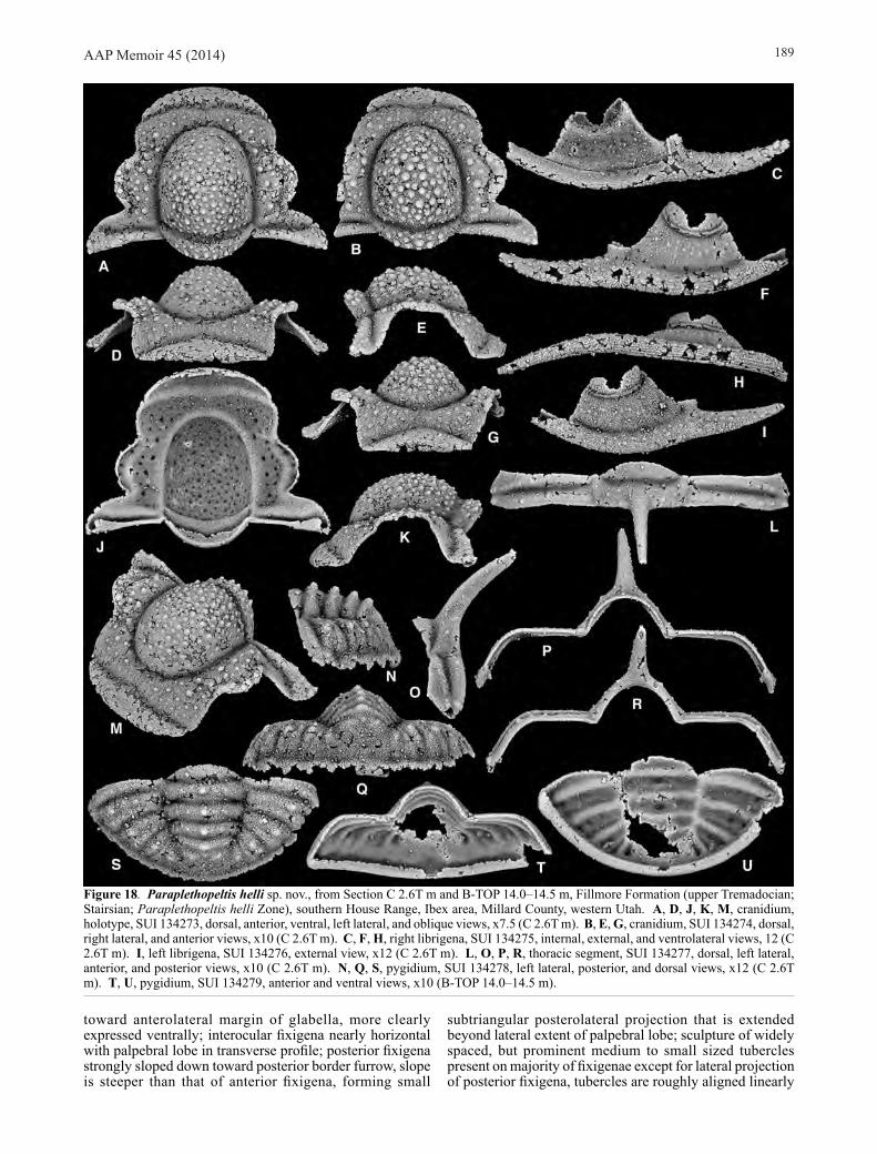
Paraplethopeltis helli sp. nov. (Figs 6A, D, 18)
Material. Holotype, cranidium, SUI 134273 (Fig. 18A, D, J, K, M), assigned specimens SUI 134274–134278 from Section C 2.6T m, and assigned specimen SUI 134279 from Section B-TOP 14.0–14.5 m, both Fillmore Formation (upper Tremadocian; Stairsian; Paraplethopeltis helli Zone), southern House Range, Ibex area, Millard County, western Utah.
Etymology. After Richard Hell.
Diagnosis. Most dorsal surfaces with distinct tubercles, ranging in expression from subdued to prominent and densely scattered; cranidial anterior border furrow, axial furrows, preglabellar furrow, SO, and librigenal lateral border furrow all deeply impressed; librigenal field with faint scattered tubercles, mainly on adaxial two thirds; genal spine
about as long as field (exsag.); pygidium with prominent median nodes on axial rings and prominent larger tubercle set near the fulcrum on each posterior pleural band.
Description. Cranidial measurements were made on the two cranidia of Figure 18 and an additional specimen from C 2.6T m that will be figured in an expanded work of the genus. Cranidium with maximum cranidial width across posterior projections 144.2% (140.0–148.8) sagittal length; cranidial width across ∂ 110.7% (106.3–114.0) cranidial sagittal length; cranidial width across β 82.0% (80.0–84.5) cranidial sagittal length; distance across γ 81.1% (80.4–82.4) cranidial sagittal length; distance across ε 94.7% (93.5–96.3) cranidial sagittal length; pronounced cranidial convexity produced by strongly dorsally inflated glabella and posterior fixigena, anterior fixigena, preglabellar field, and anterior border strongly downturned from horizontal plane; anterior border longest sagittally, progressively shorter abaxially so that border tapers to a point at intersection with anterior border furrow, sagittal length longer than preglabellar field (sag.) in plan view, anterior margin describing gentle smooth arc, posterior margin also gently arched, but disrupted medially by a prominent posteriorly directed depression; anterior border nearly subtriangular in transverse profile, with dorsal margin strongly arched forming a rounded peak medially, ventral margin very gently arched to nearly transverse medially with lateral portions more strongly concave; sculpture of fine raised anastomosing lines covers anterior portion of anterior border in plan view and majority of anterior border in anterior view, lines are arranged subparallel to external margin of border, secondary sculpture of very fine granules present on posterior portion of border adjacent to anterior border furrow; anterior border gently sloping downward away from preglabellar field in lateral profile, with thin strip of doublure curving under border visible on some specimens (Fig. 18K); anterior border furrow moderately long, dorsally concave, slightly shallower medially at break in course where furrow is directed posteriorly; anterior sections of facial sutures laterally convex opposite anterior border, laterally concave opposite anterior border furrow and adjacent portions of anterior border and frontal areas, strongly laterally bowed around anterolateral corner of frontal area, and then becoming nearly straight to slightly sinuous and directed just slightly posteromedially to γ; anterior fixigena sloped strongly downward from interocular fixigena; preglabellar field similarly strongly downturned, in plan view field is shortest sagittally and lengthened exsagittally, sagittal length 7.6% (6.3–9.8) cranidial sagittal length; ventral surface of preglabellar field with small swollen sector medially between anterior border furrow and preglabellar furrow; anterior fixigena and lateral portions of preglabellar field are covered with medium and small sized widely spaced tubercles, tubercles become smaller and more faint anteriorly, medial portion of preglabellar field mostly lacking tubercles, faint caecal pitting present; palpebral lobes long and moderately narrow, occupying about half of cranidial sagittal length, lobe moderately strongly laterally bowed, lobe not particularly well preserved on largest specimens, but appears to have background sculpture of fine granules (Fig. 18B), the presence of a few larger tubercles arranged along the external margin of the lobe is suggested (Fig. 18A, B), but they are not well preserved; lobe set off from interocular fixigena by distinct palpebral furrow, which is largely effaced anteriorly, furrow strongly bowed similar to lobe; eye ridge very faint, directed strongly anterolaterally
AAP Memoir 45 (2014) 189
toward anterolateral margin of glabella, more clearly expressed ventrally; interocular fixigena nearly horizontal with palpebral lobe in transverse profile; posterior fixigena strongly sloped down toward posterior border furrow, slope is steeper than that of anterior fixigena, forming small
subtriangular posterolateral projection that is extended beyond lateral extent of palpebral lobe; sculpture of widely spaced, but prominent medium to small sized tubercles present on majority of fixigenae except for lateral projection of posterior fixigena, tubercles are roughly aligned linearly
Figure 18. Paraplethopeltis helli sp. nov., from Section C 2.6T m and B-TOP 14.0–14.5 m, Fillmore Formation (upper Tremadocian; Stairsian; Paraplethopeltis helli Zone), southern House Range, Ibex area, Millard County, western Utah. A, D, J, K, M, cranidium, holotype, SUI 134273, dorsal, anterior, ventral, left lateral, and oblique views, x7.5 (C 2.6T m). B, E, G, cranidium, SUI 134274, dorsal, right lateral, and anterior views, x10 (C 2.6T m). C, F, H, right librigena, SUI 134275, internal, external, and ventrolateral views, 12 (C 2.6T m). I, left librigena, SUI 134276, external view, x12 (C 2.6T m). L, O, P, R, thoracic segment, SUI 134277, dorsal, left lateral, anterior, and posterior views, x10 (C 2.6T m). N, Q, S, pygidium, SUI 134278, left lateral, posterior, and dorsal views, x12 (C 2.6T m). T, U, pygidium, SUI 134279, anterior and ventral views, x10 (B-TOP 14.0–14.5 m).

AAP Memoir 45 (2014)190
along the length of the fixigena following the curve of the lateral margin of the glabella, background sculpture of fine granules present on all portions of fixigena; posterior section of facial suture directed posterolaterally, forming a broad convex arc across anterior margin of posterior projection; posterior border furrow distinct, deeply impressed, but shallower abaxially from fulcrum, shortest adaxially and longer abaxially (Fig. 18B); posterior border shortest (exsag.) proximally, significantly lengthened distally, forming distinct broad and rounded distal tip; sculpture of sparse smaller sized distinct tubercles present on distal tip, with larger sized tubercles arranged closer to posterolateral margin, one or two very small faint tubercles present on main portion of border; posterior margin of posterior border between axial furrow and fulcrum directed slightly posterolaterally and nearly transverse, portion of margin from fulcrum distally similarly transverse and directed more strongly posterolaterally; thin strip of doublure present beneath posterior border, shortest proximally, lengthening distally so that strip forms a triangle distally; glabella large, roughly elliptical outline in plan view, maximum width 88.7% (86.4–92.9) sagittal length excluding LO, sagittal length 62.8% (60.4–65.5) cranidial sagittal length, strongly dorsally inflated, with apex sitting high above fixigena and strongly vaulted in both transverse and sagittal profiles; sculpture of densely spaced, prominent, small and medium sized tubercles covers majority of glabella, tubercles are largest and most densely spaced posteromedially, with the lateral and anterior margins possessing fewer and smaller tubercles, background sculpture of fine granules also present; axial furrows deep opposite glabella, slightly deeper at fossulae (Fig. 18A), slightly shallower opposite LO, where axial furrows intersect posterior border furrow, width across posterior contact of furrows with posterior margin 49.3% (49.2–49.5) cranidial sagittal length; axial furrows narrowest opposite LO, wider opposite palpebral lobes, slightly narrower anteriorly, running without obvious distinction or change in course into preglabellar furrow; preglabellar furrow deep and moderately narrow, slightly shallower medially (Fig. 18B, G), describing strongly curved arc; glabellar furrows not clearly expressed; LO moderately short (sag.), with sagittal length 9.6% (9.0–10.5) that of cranidium, and sagittal length 74.8% (68.8–84.5) that of anterior border, progressively shorter (exsag.) abaxially tapering almost to a point, anterior and posterior margins very strongly posteriorly bowed, with sculpture similar to that on main glabella of densely spaced, medium and small sized tubercles; distal tips of LO lacking tubercles; entire LO with background sculpture of fine granules; doublure beneath LO short (sag.; exsag.), extending only about halfway across length of LO, longest sagittally and strongly tapering abaxially, anterior margin strongly posteriorly bowed as anterior margin of LO; SO deep, long (sag.; exsag.), strongly bowed posteriorly, but slightly less so than posterior margin of LO, confluent with axial furrow and forming obtuse angle at intersection; tuberculate sculpture present on dorsal surface of cranidium expressed ventrally as small, prominent pits.
Librigenal measurements were made on Figure 18F, I. Main body of librigena (excluding anterior projection and genal spine) with width opposite median point of visual surface 33.6% (33.6–33.6) exsagittal length; main body with exsagittal length about 64% total librigenal length; anterior section almost transversely straight opposite field, with very slight outward bow just before eye socle, strong bend across lateral border furrow, and very gently curved
to almost transverse across lateral border and anterior projection; anterior projection with length about 20% exsagittal librigenal length; posterior section very gently bowed opposite main portion of field, very strongly bent opposite posteriormost portion of field and posterior border furrow, almost straight across posterior border; visual surface incomplete, but long, occupying 35.3% (34.6–36.0) of exsagittal librigenal length, bounded by deep, narrow furrow; socle narrow, with sculpture of fine raised lines running subparallel to base of visual surface; socle separated from field by deep, narrow furrow; field with very faint caecal sculpture, overlain by fine granules, and a band of more prominent tubercles running across proximal portion of field and oriented subparallel to base of visual surface; lateral border furrow moderately broad, deepest anteriorly, significantly shallower posteriorly toward genal angle; posterior border furrow deepest at base of genal spine; lateral border broad, widens slightly toward base of genal spine, with lateral margin moderately curved, curve strongest at midlength, strongly inflated, with ventrolateral margin forming broad surface; primary sculpture of prominent raised lines running subparallel with margin, lines run from anterior projection along lateral border and onto genal spine, lines cover all of lateral border and anterior projection in external and ventrolatral views, lines cover outer portion of doublure beneath anterior projection and lateral border, inner portion smooth; secondary sculpture of fine granules present on lateral border, and genal spine, a few small scattered tubercles arranged linearly subparallel to margin also present along lateral border adjacent to lateral border furrow, row of tubercles continues onto genal spine (Fig. 18I); posterior border very small, subtriangular, does not extend far beyond base of genal spine; doublure beneath lateral and posterior borders moderately wide, extending to ventral expression of lateral and posterior border furrows; inner margin of doublure under posterior border slightly thickened; genal spine moderately long, with length about 36% total librigenal length, blade like, subelliptical in section, tapered to a blunt point distally, with sculpture of fine raised lines continuing from lateral and posterior borders running down length of spine on all aspects (Fig. 18C, F, H); base of genal spine and margin of posterior border intersect at slightly obtuse angle.
Hypostome and rostral plate not identified.Thoracic segments with axial ring wider anteriorly than
posteriorly, sculpture of scattered small sized tubercles; segment possess a prominent axial spine that appears derived from the posteromedial portion of the axial segment in plan view (Fig. 18B), but from the entire segment in sagittal profile (Fig. 18O), spine is subcircular in cross section, directed strongly posterodorsally, and moderately arched; ring furrow long, deep, broadly posteriorly bowed, transverse medially with distal tips directed anterolaterally on segment bearing axial spine; articulating half ring large, lenticular to subcrescentic in outline; axial furrows very shallow and narrow; pleural furrow moderately long (exsag.) and impressed; anterior pleural band distinct, and forming inflated ridge adjacent to pleural furrow, sculpture of very small faint tubercles present on ridge-like portion from fulcrum distally; anterolateral corner of anterior pleural band developed into a prominent triangular facet; posterior pleural band also distinct, but less ridge like and lacking inflation of anterior band, sculpture of small faint tubercles arranged linearly along middle portion of band; anterior margin of pleural region forms a narrow transverse rim which articulates with a narrow groove along the posterior edge
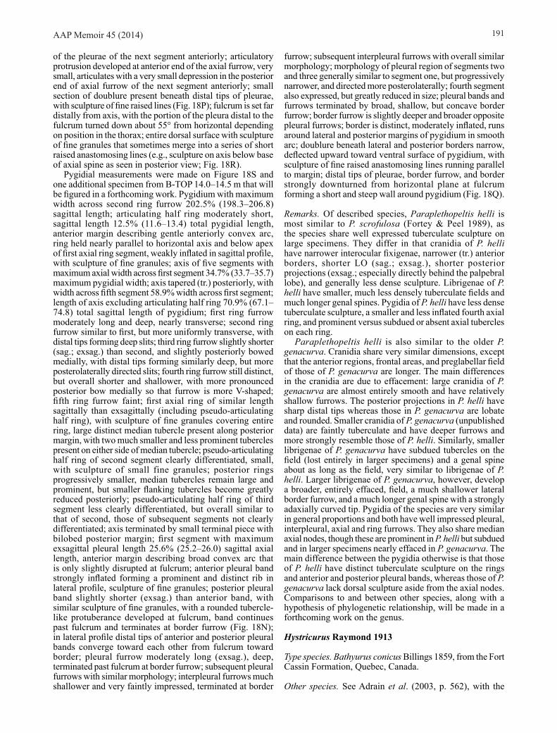
AAP Memoir 45 (2014) 191
of the pleurae of the next segment anteriorly; articulatory protrusion developed at anterior end of the axial furrow, very small, articulates with a very small depression in the posterior end of axial furrow of the next segment anteriorly; small section of doublure present beneath distal tips of pleurae, with sculpture of fine raised lines (Fig. 18P); fulcrum is set far distally from axis, with the portion of the pleura distal to the fulcrum turned down about 55° from horizontal depending on position in the thorax; entire dorsal surface with sculpture of fine granules that sometimes merge into a series of short raised anastomosing lines (e.g., sculpture on axis below base of axial spine as seen in posterior view; Fig. 18R).
Pygidial measurements were made on Figure 18S and one additional specimen from B-TOP 14.0–14.5 m that will be figured in a forthcoming work. Pygidium with maximum width across second ring furrow 202.5% (198.3–206.8) sagittal length; articulating half ring moderately short, sagittal length 12.5% (11.6–13.4) total pygidial length, anterior margin describing gentle anteriorly convex arc, ring held nearly parallel to horizontal axis and below apex of first axial ring segment, weakly inflated in sagittal profile, with sculpture of fine granules; axis of five segments with maximum axial width across first segment 34.7% (33.7–35.7) maximum pygidial width; axis tapered (tr.) posteriorly, with width across fifth segment 58.9% width across first segment; length of axis excluding articulating half ring 70.9% (67.1–74.8) total sagittal length of pygidium; first ring furrow moderately long and deep, nearly transverse; second ring furrow similar to first, but more uniformly transverse, with distal tips forming deep slits; third ring furrow slightly shorter (sag.; exsag.) than second, and slightly posteriorly bowed medially, with distal tips forming similarly deep, but more posterolaterally directed slits; fourth ring furrow still distinct, but overall shorter and shallower, with more pronounced posterior bow medially so that furrow is more V-shaped; fifth ring furrow faint; first axial ring of similar length sagittally than exsagittally (including pseudo-articulating half ring), with sculpture of fine granules covering entire ring, large distinct median tubercle present along posterior margin, with two much smaller and less prominent tubercles present on either side of median tubercle; pseudo-articulating half ring of second segment clearly differentiated, small, with sculpture of small fine granules; posterior rings progressively smaller, median tubercles remain large and prominent, but smaller flanking tubercles become greatly reduced posteriorly; pseudo-articulating half ring of third segment less clearly differentiated, but overall similar to that of second, those of subsequent segments not clearly differentiated; axis terminated by small terminal piece with bilobed posterior margin; first segment with maximum exsagittal pleural length 25.6% (25.2–26.0) sagittal axial length, anterior margin describing broad convex arc that is only slightly disrupted at fulcrum; anterior pleural band strongly inflated forming a prominent and distinct rib in lateral profile, sculpture of fine granules; posterior pleural band slightly shorter (exsag.) than anterior band, with similar sculpture of fine granules, with a rounded tubercle-like protuberance developed at fulcrum, band continues past fulcrum and terminates at border furrow (Fig. 18N); in lateral profile distal tips of anterior and posterior pleural bands converge toward each other from fulcrum toward border; pleural furrow moderately long (exsag.), deep, terminated past fulcrum at border furrow; subsequent pleural furrows with similar morphology; interpleural furrows much shallower and very faintly impressed, terminated at border
furrow; subsequent interpleural furrows with overall similar morphology; morphology of pleural region of segments two and three generally similar to segment one, but progressively narrower, and directed more posterolaterally; fourth segment also expressed, but greatly reduced in size; pleural bands and furrows terminated by broad, shallow, but concave border furrow; border furrow is slightly deeper and broader opposite pleural furrows; border is distinct, moderately inflated, runs around lateral and posterior margins of pygidium in smooth arc; doublure beneath lateral and posterior borders narrow, deflected upward toward ventral surface of pygidium, with sculpture of fine raised anastomosing lines running parallel to margin; distal tips of pleurae, border furrow, and border strongly downturned from horizontal plane at fulcrum forming a short and steep wall around pygidium (Fig. 18Q).
Remarks. Of described species, Paraplethopeltis helli is most similar to P. scrofulosa (Fortey & Peel 1989), as the species share well expressed tuberculate sculpture on large specimens. They differ in that cranidia of P. helli have narrower interocular fixigenae, narrower (tr.) anterior borders, shorter LO (sag.; exsag.), shorter posterior projections (exsag.; especially directly behind the palpebral lobe), and generally less dense sculpture. Librigenae of P. helli have smaller, much less densely tuberculate fields and much longer genal spines. Pygidia of P. helli have less dense tuberculate sculpture, a smaller and less inflated fourth axial ring, and prominent versus subdued or absent axial tubercles on each ring.
Paraplethopeltis helli is also similar to the older P. genacurva. Cranidia share very similar dimensions, except that the anterior regions, frontal areas, and preglabellar field of those of P. genacurva are longer. The main differences in the cranidia are due to effacement: large cranidia of P. genacurva are almost entirely smooth and have relatively shallow furrows. The posterior projections in P. helli have sharp distal tips whereas those in P. genacurva are lobate and rounded. Smaller cranidia of P. genacurva (unpublished data) are faintly tuberculate and have deeper furrows and more strongly resemble those of P. helli. Similarly, smaller librigenae of P. genacurva have subdued tubercles on the field (lost entirely in larger specimens) and a genal spine about as long as the field, very similar to librigenae of P. helli. Larger librigenae of P. genacurva, however, develop a broader, entirely effaced, field, a much shallower lateral border furrow, and a much longer genal spine with a strongly adaxially curved tip. Pygidia of the species are very similar in general proportions and both have well impressed pleural, interpleural, axial and ring furrows. They also share median axial nodes, though these are prominent in P. helli but subdued and in larger specimens nearly effaced in P. genacurva. The main difference between the pygidia otherwise is that those of P. helli have distinct tuberculate sculpture on the rings and anterior and posterior pleural bands, whereas those of P. genacurva lack dorsal sculpture aside from the axial nodes. Comparisons to and between other species, along with a hypothesis of phylogenetic relationship, will be made in a forthcoming work on the genus.
Hystricurus Raymond 1913
Type species. Bathyurus conicus Billings 1859, from the Fort Cassin Formation, Quebec, Canada.
Other species. See Adrain et al. (2003, p. 562), with the

AAP Memoir 45 (2014)192
following additions: Bathyurus cordai Billings 1860, Lévis Formation, Quebec; Bathyurus? crotalifrons Dwight 1884, Rochdale Formation, New York (see Landing et al. 2012); Bathyurus seelyi Whitfield 1889, Rochdale Formation, New York; Hystricurus sp. nov. 1 (herein), Fillmore Formation, Utah; Hystricurus sp. nov. 2 (herein), Fillmore Formation, Utah (Hystricurus sp. nov. B of Adrain et al. [2003]); Hystricurus sp. nov. 3 (herein), Fillmore Formation, Utah; Hystricurus sp. nov. A (herein), Fillmore Formation, Utah; Hystricurus sp. nov. B (herein), Fillmore Formation, Utah; Hystricurus sp. nov. C (herein), Fillmore Formation, Utah. The status of the species Vermilionites bisulcatus Kobayashi 1955, and Hystricurus platypleurus Kobayashi 1955, both from the McKay Group, British Columbia, is uncertain.
Diagnosis. See Adrain et al. (2003, p. 562).
Remarks. Hystricurus had been used as a nearly meaningless form taxon for Lower Ordovician trilobites now assigned to numerous different subfamilies and genera. Adrain et al. (2003) restricted the concept of the genus to a putative clade of usually strongly tuberculate species with “fiddle-shaped” glabellae and distinctive pygidia with large fulcral nodes/spines overlying a steep, nearly vertical region bounded by a strong, rim-like border. A forthcoming work will describe all of the new species illustrated briefly herein and consider the phylogenetic structure of the genus and its potential sister group.
Hystricurus zanderi sp. nov. (Figs 7J, N, 19)
2003 Hystricurus cf. deflectus Heller; Adrain et al., p. 563, fig. 4D, H, L.
Material. Holotype, pygidium, SUI 134283 (Fig. 19N, P, R, S, W) and assigned specimens SUI 99742, 134280–134282, 134284–134286, from Section MME 22.3 m, Fillmore Formation (upper Tremadocian; Stairsian; Hystricurus zanderi Zone), Middle Mountain, Ibex area, Millard County, western Utah.
Etymology. After Robin Zander.
Diagnosis. Glabella relatively narrow, especially anteriorly, and weakly dorsally inflated; palpebral lobes short (exsag.); pygidium with fulcral spines and pair of spines on rear of axis large and overhanging rear margin; vertical area above border tall.
Description. Recovered cranidia incomplete, but with sagittal length less than maximum width across posterior projection; anterior border with distinct dorsal inflation, longest sagittally, with sagittal length nearly equal to that of preglabellar field in plan view, progressively shorter abaxially, anterior margin describing moderately to strongly bowed arc in plan view, gently dorsally bowed in transverse
profile just barely obscuring anterior border furrow, with sculpture of about five or six fine raised lines on anterior portions, lines run subparallel to margin, posterior portion of border covered by fine granules; anterior border furrow is longest sagittally, with slight lenticular bulge medially, progressively shorter exsagittally, deep along entire course, but slightly shallower medially, anterior contact with anterior border and posterior contact with preglabellar field both appear sharp in plan view; anterior sections of facial sutures laterally convex opposite anterior border, laterally concave opposite anterior border furrow and adjacent portion of frontal area, strongly, but unevenly convex opposite frontal area; preglabellar furrow deep, confluent with axial furrows; preglabellar field shorter sagittally than exsagittally, very faint median furrow present, furrow more deeply impressed anteriorly than posteriorly, also expressed ventrally (Fig. 19H); frontal area and preglabellar field very strongly (Fig. 19E, I) to moderately (Fig. 19AA) sloped downward toward anterior border furrow; anterior border is directed upward from horizontal plane in contrast to preglabellar field and frontal areas; anterolateral corners of frontal area mostly facing anteriorly with anterolateral corners turned slightly obliquely; palpebral lobes long (exsag.), lobe strongly laterally bowed, curve slightly stronger posteriorly so that lobe appears slung posteriorly, lobe with very narrow, rim-like border around margin (Fig. 19L); palpebral furrow distinct, sinuous, deepest opposite posterior margin of lobe, shallowest medially, and deep again opposite anterior portion of lobe, but anterior portion of furrow shallower than posterior portion; interocular fixigena wide, with width about 49% maximum glabellar width (measured on Fig. 19A), held nearly horizontal, adaxial portion sloping slightly toward axial furrow in transverse profile; eye ridge weakly expressed, running obliquely from anterior margin of palpebral lobe to anterolateral corner of glabella, furrow bounding anterior and posterior margins clearly discernible dorsally, more deeply impressed on some specimens (Fig. 19B) than others (Fig. 19Q), clearly expressed ventrally; posterior fixigena known from only one larger specimen (Fig. 19A), strongly sloped downward from horizontal plane, portion distal to fulcrum facing more obliquely, abaxial portion developed into small triangular posterolateral projection extending a short distance beyond lateral extent of palpebral lobe; posterior section of facial suture directed posterolaterally at about 115° across posterior fixigena, describing convex arc across posterior fixigena and posterior border, with a small disruption in course at posterior border furrow; posterior border furrow distinct, long (exsag.), proximal portion shallower than distal portion, of similar length along entire course, almost exactly transverse and directed just slightly anterolaterally; posterior border strongly dorsally inflated, proximally very short (exsag.), lengthened distally, terminating in semi-lobate distal tip, sculpture of very faint tubercles are present on proximal portion of border; distinct ridge running along transverse length of border proximally and turned posterolaterally
Figure 19. Hystricurus zanderi sp. nov., from Section MME 22.3 m, Fillmore Formation (upper Tremadocian; Stairsian; Hystricurus zanderi Zone), Middle Mountain, Ibex area, Millard County, western Utah. A, D, H, I, L, cranidium, SUI 99742, dorsal, anterior, ventral, right lateral, and oblique views, x7.5. B, E, M, cranidium, SUI 134280, dorsal, right lateral, and anterior views, x5. C, F, G, right librigena, SUI 134281, external, ventrolateral, and internal views, x4. J, K, O, left librigena, SUI 134282, internal, ventrolateral, and external views, x7.5. N, P, R, S, W, pygidium, holotype, SUI 134283, posterior, dorsal, anterior, ventral, and left lateral views, x4. Q, V, AA, cranidium, SUI 134284, dorsal, anterior, and left lateral views, x7.5. T, U, Y, pygidium, SUI 134285, posterior, dorsal, and left lateral views, x12. X, BB, CC, pygidium, SUI 99743, dorsal, left lateral, and posterior views, x4. Z, DD, EE, pygidium, SUI 134286, right lateral, dorsal, and posterior views, x7.5.
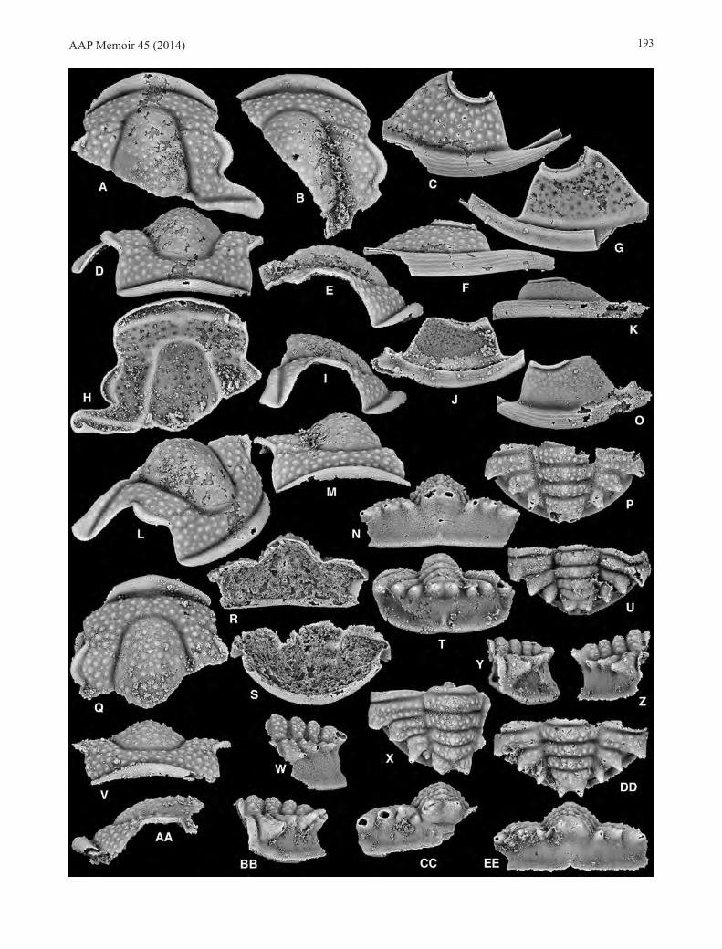
AAP Memoir 45 (2014) 193
AAP Memoir 45 (2014)194
distally, ridge divides border into two sectors, a smaller subtriangular anterior portion that faces anterobliquely, and a larger posterior portion that faces posterodorsally; posterior margin of posterior border directed posterolaterally at about 100° angle; small triangular portion of glabella present at posterolateral tip of posterior border (Fig. 19H); glabella broadly subtriangular, but slightly waisted anteriorly, with broadly rounded anterior margin in plan view, sagittal length greater than maximum width, strong independent inflation, with apex reached at posterior margin and sagittal profile strongly sloping downward toward preglabellar furrow; axial furrows moderately narrow and deep; short portion of axial furrow between posterolateral corner of glabella and intersection with posterior border furrow straight (Fig. 19A), directed posterolaterally around anterolateral corner of LO, axial furrow continues around posterolatral corner of LO to margin; glabellar furrows not clearly expressed; glabella, fixigenae, frontal areas, and preglabellar field with sculpture of small and medium sized tubercles; sculpture of densely spaced fine granules covers palpebral lobe and furrow, granules merged on anterior portion of lobe to form a short series of anastomosing lines (Fig. 19L).
Librigena with socle strongly inflated, with sculpture of fine, closely spaced raised lines running subparallel to margin of visual surface, set off from visual surface by narrow and shallow furrow (Fig. 19C), separated from field by slightly longer and deeper furrow; field longer than wide, broadly subrectangular, with portion opposite anterior facial suture shorter than portion opposite posterior facial suture, with background sculpture of fine granules, overlain by medium and small sized tubercles roughly arranged into rows oriented sub parallel to socle margin; anterior section of facial suture with slight anterior bow opposite proximal portion of field just below base of visual surface, distinct almost 90° change in course at lateral border furrow and proximal portion of lateral border, gently curved across border and anterior projection; posterior section describing gentle convex curve opposite field, strongly curved at border furrow across posterior border; anterior projection long; lateral border furrow broad, deepest medially with anterior and posterior portions slightly shallower; lateral border widest medially opposite field, slightly tapered anteriorly and posteriorly, fine granules present on border along margin adjacent to lateral border furrow; lateral margin gently curved; anterior projection and lateral border strongly convex, forming moderately tall face in ventrolateral view (Fig. 19F); sculpture of prominent raised lines running subparallel with margin present on external and ventrolatral aspects of anterior projection and lateral border, lines more closely spaced anteriorly toward tip of anterior projection, orientation of lines slightly disrupted at base of genal spine; posterior border on smaller specimens greatly reduced to small protuberance adjacent to base of genal spine (Fig. 19O), set off from genal spine by shallow, but distinct furrow; doublure beneath anterior projection and lateral border broad, reaching external margin of lateral border furrow beneath lateral border and beyond proximal extent of anterior projection so that the anterior portion of the doublure is visible in external view; doublure appears to continue beneath posterior border (Fig. 19J), forming a small shallow depressed area at genal angle; sculpture of very fine raised lines present along length of doublure.
Hypostome, rostral plate and thorax not identified.Pygidial measurements were made on Figure 19U, X
and one additional specimen from MME 22.3 m that will be figured in a separate expanded treatment of the genus.
Pygidium wider (tr.) than long (sag.), with maximum width achieved across posterior portion of second axial segment on two largest specimens; fulcrum is set very far distally from axis, with portion of pygidium distal to fulcrum strongly downturned from horizonal plane forming a nearly vertical face, posteromedian portion of face is angled slightly anteriorly in transverse profile; face is bounded dorsally by pygidial spines and ventrally by a moderately narrow, but well inflated border that bounds lateral and posterior margins of pygidium; pygidial border set off from vertical face by shallow, but concave border furrow, in plan view furrow forms broad concave region below pygidial spines; main surface of vertical face with sculpture of densely spaced, fine granules; pygidial border with sculpture of fine raised lines arranged parallel to margin; doublure present ventrally beneath posterior and at least part of lateral borders; axis composed of four segments and distinct terminal axial piece, maximum axial width across first segment 34.6% maximum pygidial width (measured on Fig. 19X), width across anterior margin of terminal axial piece 62.6% (62.1–63.1) width across first segment; length of axis excluding articulating half ring 43.5% maximum pygidial width (measured on Fig. 19X); axis with strong to moderate dorsal convexity, with apex reached at first axial segment and gently sloping posteroventrally in transverse profile; articulating half ring broken on all recovered specimens, but apparently very short (sag., exsag.), posterior margin almost exactly transverse, strong dorsal convexity in sagittal profile and sitting below apex of first axial segment, with sculpture of fine granules; first ring furrow deep, short, and almost exactly transverse or slightly anteriorly bowed; second ring furrow similar to first, forming a deeper pit adjacent to intersection with axial furrow; third ring furrow similar in all aspects to second, except that distal tips are directed strongly posterolaterally; fourth ring furrow slightly shallower, but also deep, distal tips directed strongly posterolaterally as third ring furrow, and median portion posteriorly bowed; fifth ring furrow shallow, more strongly posteriorly bowed medially than fourth ring furrow; first axial ring of similar length sagittally and exsagittally, with pseudo-articulating half ring of second segment largely merged and only faintly set off by narrow and faint furrow; second ring with morphology similar to first, but distal tips along posterior margin directed posterolaterally and pseudo-articulating half ring not developed; third ring overall shorter (sag.; exsag.), distal tips more directed posterolaterally, median portion with slight posterior bow; fourth ring slightly shorter exsagittally than sagittally, median portion more strongly posteriorly bowed than third segment; first to fourth axial segments with sculpture of mostly medium and a few smaller sized, densely spaced tubercles, large and distinct median tubercle present on each segment; axis terminated by distinct terminal piece developed into two, short, subconical posterodorsally directed spines, spines are set off from each other by very shallow median furrow, tuberculate sculpture of previous rings more subdued on terminal piece; shallow pit developed medially below base of terminal axial piece (best seen in posterior view); short median ridge is developed behind axis (Fig. 19T) from border, ridge does not reach posterior margin of axis in posterior view, but is visible behind axis in plan view (Fig. 19U); first segment with anterior margin describing gently convex arc to fulcrum, directed slightly posterolaterally, portion from fulcrum distally directed slightly anterorlaterally; anterior pleural band longest (exsag.) distally, tapering to a point adaxially at intersection
AAP Memoir 45 (2014) 195
with axial furrow, anterior margin bounded by very narrow rim-like ridge, moderately inflated, distal tip square in plan view, with sculpture of very fine granules, some specimens (e.g., Fig. 19U) with very faint row of tubercles present along posterior margin of band; posterior pleural band longer (sag.; exsag.) than anterior band, longer (exsag.) distally than main portion of band, with sculpture of fine granules overlain by more prominent medium and small sized tubercles; posterior pleural band is merged distally with subsequent anterior pleural band and developed into a prominent spine, which is directed posterolaterally and also slightly dorsally, spine is conical in cross section; pleural furrow deep, shallower adjacent to axial furrow, moderately long along entire course, terminated just past start of subvertical face of pygidium, directed posterolaterally in plan view; interpleural furrow significantly shallower than pleural furrow, deepest medially abaxial to fulcrum, shallower but discernible adjacent to axis, progressively effaced toward base of spine becoming completely effaced on spine so that posterior pleural band of segment one and anterior pleural band of segment two appear merged, directed just slightly posterolaterally; interpleural furrow between segments two and three slightly impressed, with that between segments three and four very faintly visible on largest pygidium; morphology of second pleural segment similar to first, but overall shorter and with shorter pygidial spine; third pleural segment even shorter and more strongly posterolaterally directed, with third posterior pleural band and fourth anterior pleural band expressed solely as prominent subconical spine, strongly posterolaterally directed; very short and minute fourth pygidial spine developed on one specimen (Fig. 19X, CC) between third spine and spine of terminal axial piece.
Remarks. At present, no other member of the genus is really adequately known, though many of the species illustrated herein are represented by extensive collections. The morphologically closest species to Hystricurus zanderi is Hystricurus sp. nov. 1 (Fig. 8S, W) from the Rossaspis leboni Zone. Hystricurus zanderi differs from H. sp. nov. 1 in having a cranidium with a more anteriorly bowed anterior margin, a longer preglabellar field, more subdued tuberculate sculpture, and a narrower, less dorsally inflated glabella. The pygidia of H. zanderi differ from those of H. sp. nov. 1 in the possession of more robust fulcral spines with more posteriorly extended tips, and taller vertical regions above the border with less expression of pleural and interpleural furrows.
Family CHEIRURIDAE Hawle & Corda 1847
Rossaspis Harrington 1957
Type species. Protopliomerops superciliosa Ross 1951, from the Garden City Formation (upper Tremadocian; Stairsian; Pseudoclelandia cornupsittaca Zone), Idaho, USA.
Other species. Rossaspis leboni sp. nov., Fillmore Formation (upper Tremadocian; Stairsian; Rossaspis leboni Zone), Utah, USA; Rossaspis sp. 1 (herein), Fillmore Formation, Utah; Rossaspis sp. nov. 2 (herein), Fillmore Formation, Utah; Rossaspis sp. nov. 3 (herein), Fillmore Formation, Utah; Rossaspis sp. (herein), Fillmore Formation, Utah.
Remarks. Rossaspis appears to represent a clade, but full diagnosis and discussion is deferred until the type species is
revised and three additional new species (illustrated in open nomenclature herein) are described.
Rossaspis leboni sp. nov. (Figs 8B, F, 20)
Material. Holotype, cranidium, SUI 134287 (Fig. 20A, D, E, G, K) and assigned specimens SUI 134077, 134288, 134290, 134291, from Section MME 49.8 m, Fillmore Formation (upper Tremadocian; Stairsian; Rossaspis leboni Zone), Middle Mountain, Ibex area, Millard County, western Utah. Assigned specimen SUI 134289 from Section HC6 58.4 m, Garden City Formation (upper Tremadocian; Stairsian; Rossaspis leboni Zone), west side of Hillyard Canyon, Bear River Range, Franklin County, southeastern Idaho.
Etymology. After Simon Le Bon.
Diagnosis. Cranidium with S1 and S2 nearly transverse; glabella with even scattering of very dense, small tubercles; axial and glabellar furrows narrow; anterior border short (sag.; exsag.); pygidial pleural spines laterally extended, turned posteriorly near tips, tips blunt; pygidial terminal piece prominent and inflated.
Description. Cranidial measurements were made on Fig. 20A, C, and four addititional specimens from MME 49.8 m that will be figured in a forthcoming work. Cranidium with maximum cranidial sagittal length 40.9% (37.0–44.3) maximum width across posterior projections; width across across γ 106.9% (100.6–111.5) maximum width sagittal length, and 43.7% (40.0–46.8) maximum width across posterior projections; anterior margin of anterior border describing broad, anteriorly convex arc in plan view, with anterior sections of facial sutures following this curve opposite anterior border; facial suture making sudden change in course at intersection with anterior border furrow, strongly anterolaterally bowed opposite anterior portion of small palpebral lobe, with sudden change in course opposite posterior portion of palpebral lobe so that suture abruptly cuts laterally across fixigena and lateral border; main portion of palpebral lobe situated opposite L3, posterior margin defined by deep and narrow palpebral furrow that runs from point opposite middle of L3 laterally to point just before facial suture, in anterior view posterior margin of lobe is strongly deflected upward from horizontal plane (Fig. 20G); eye ridge prominent and confluent with palpebral lobe, together they possess slight independent inflation from adjacent areas of cranidium, anterior margin running obliquely from palpebral lobe to fossulae with anterior margin bounded by anterior border furrow, posteriorly bounded palpebral furrow; anterior border very short in front of main portion of glabella, lengthened exsagittally forming small subtriangular area adjacent to anterolateral corner of glabella and eye ridge, border sits below glabella in lateral profile and is angled downward from horizontal plane; in anterior profile anterior border describing broad concave arc medially opposite rostral suture, with general bow of this arc continued abaxially to palpebral lobes with only slight disruptions along course; sculpture of fine, densely spaced granules overlain by small sized, scattered tubercles covers border; anterior border furrow short (sag.), deep, and confluent with preglabellar furrow in front of glabella, defining anterior margin of eye ridge abaxially, significantly narrower and shallower along this portion; anterior border furrow, axial furrow, and abaxial tip of S3 intersect at fossulae, which

AAP Memoir 45 (2014)196
are deep and large; fixigena large, with exsagittal length adjacent to axial furrow 55.9% (53.3–60.2) cranidial sagittal length, maximum width 83.4% (78.5–87.2) glabellar width, portion adaxial to fulcrum held nearly parallel to horizontal plane, portion abaxial to fulcrum strongly downturned from
horizontal plane, covered with sculpture of fine granules and prominent small pits, some cranidia with a scattering of small tubercles on fixigena (e.g., Fig. 20I); lateral border broad, confluent with posterior border around genal angle, which is developed into a short posterolaterally directed
Figure 20. Rossaspis leboni sp. nov., from Section MME 22.3 m, Fillmore Formation, Middle Mountain, Ibex area, Millard County, western Utah, and Section HC6 58.4 m, Garden City Formation, west side of Hillyard Canyon, Bear River Range, Franklin County, southeastern Idaho, both upper Tremadocian, Stairsian, Rossaspis leboni Zone. A, D, E, G, K, cranidium, holotype, SUI 134287, dorsal, ventral, right lateral, anterior, and oblique views, x12 (MME 49.8 m). B, C, F, cranidium, SUI 134288, right lateral, dorsal, and anterior views, x12 (MME 49.8 m). H, I, L, cranidium, SUI 134289, left lateral, dorsal, and anterior views, x12 (HC6 58.4 m). J, M, N, P, Q, pygidium, SUI 134290, dorsal, ventral, left lateral, posterior, and anterior views, x15 (MME 49.8 m). O, R, S, pygidium, SUI 134291, left lateral, posterior, and dorsal views, x12 (MME 49.8 m).
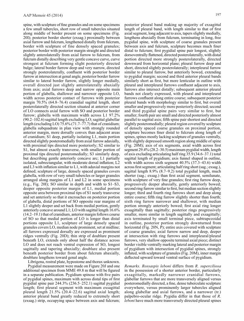
AAP Memoir 45 (2014) 197
spine, with sculpture of fine granules and on some specimens a few small tubercles, short row of small tubercles situated along middle of border present on some specimens (Fig. 20I); posterior border shorter (exsag.) proximally between axial furrow and fulcrum, lengthened distally from fulcrum, border with sculpture of fine densely spaced granules; posterior border with posterior margin straight and directed slightly anterolaterally from axial furrow to fulcrum, from fulcrum distally describing very gentle concave curve, curve strongest at fulcrum forming slight posteriorly directed bulge; lateral border furrow deep and very narrow, directed strongly posterolaterally, confluent with posterior border furrow at intersection at genal angle, posterior border furrow similar to lateral border furrow, slightly longer medially, overall directed just slightly anterolaterally abaxially from axis; axial furrows deep and narrow opposite main portion of glabella, shallower and narrower opposite LO, width across posterior contact of furrows with posterior margin 70.5% (64.8–76.4) cranidial sagittal length, short posterolaterally directed section situated at anterior corner of LO connects axial furrow with SO and posterior border furrow; glabella with maximum width across L1 97.2% (90.2–102.6) sagittal length excluding LO; sagittal glabellar length (excluding LO) 75.6% (71.7–77.7) that of cranidium; glabella subquadrate in plan view with strongly rounded anterior margin, more dorsally convex than adjacent areas of cranidium; S1 deep, partially isolating L1, main portion directed slightly posteromedially from axial furrow adaxially, with proximal tips directed more posteriorly; S2 similar to S1, but almost exactly transverse, with smaller portion of proximal tips directed posteromedially; S3 similarly deep, but describing gentle anteriorly concave arc; L1 partially isolated, subrectangular, with moderate dorsal inflation; L2 and L3 with inflation similar to L1, with adaxial margins not defined; sculpture of large, densely spaced granules covers glabella, with row of very small tubercles or larger granules arranged along margins of L1 and L2 in some specimens (e.g., Fig. 20I); SO similar in depth and width to S1–S3, deeper opposite posterior margin of L1, medial portion opposite area between proximal tips of S1 nearly transverse and set anteriorly so that it encroaches on posterior portion of glabella, distal portions of SO opposite rear margins of L1 slightly deeper and set back from medial portion, gently anteriorly concave around L1; LO with sagittal length 16.6% (14.2–19.1) that of cranidium, anterior margin follows course of SO so that medial portion of LO is longer than distal portions opposite L1, sculpture of large densely spaced granules covers LO, median node prominent, set at midline; all furrows expressed dorsally are expressed as prominent ridges ventrally (Fig. 20D); thin strip of doublure present beneath LO, extends only about half the distance across LO and does not reach ventral expression of SO, longest sagittally and tapering abaxially; doublure also present beneath posterior border from about fulcrum abaxially, doublure lengthens toward genal angle.
Librigena, rostral plate, hypostome and thorax unknown.Pygidial measurement were made on Figure 20J and one
additional specimen from MME 49.8 m that will be figured in a separate publication. Pygidium spinose with five pairs of pygidial spines, maximum with across distal tips of first pygidial spine pair 244.3% (236.5–252.1) sagittal pygidial length; first pleural segment with maximum exsagittal pleural length 21.5% (20.4–22.6) sagittal axial length; anterior pleural band greatly reduced to extremely short (exsag.) strip, occupying space between axis and fulcrum;
posterior pleural band making up majority of exsagittal length of pleural band, with length similar to that of first axial segment, long adjacent to axis, tapers slightly medially, lengthens abaxially from fulcrum, terminating in long, free pygidial spine, with sculpture of coarse granules present between axis and fulcrum, sculpture becomes much finer distal to fulcrum; first pygidial spine pair longest, slightly dorsoventrally flattened, directed posterolaterally, with distal portion directed more strongly posterolaterally, directed downward from horizontal plane; pleural furrow deep and short, directed slightly posterolaterally; interpleural furrow similar to pleural furrow, but anteriorly bowed, extending to pygidial margin; second and third anterior pleural bands similarly short as first, but more lenticular in outline with pleural and interpleural furrows confluent adjacent to axis, furrows also intersect distally; subsequent anterior pleural bands not clearly expressed, with pleural and interpleural furrows confluent along entire course; subsequent posterior pleural bands with morphology similar to first, but overall smaller and progressively more posteriorly directed; second and third pygidial spine pairs very similar to first, but smaller; fourth pair are small and directed posteriorly almost parallel to sagittal axis; fifth spine pair shortest and directed slightly posteromedially; pleural region covered by sculpture of densely spaced coarse granules on proximal portion, sculpture becomes finer distal to fulcrum along length of spines, spines mostly lacking sculpture of granules ventrally, with slightly depressed smooth region along middle of spine (Fig. 20M); axis of six segments, axial width across first segment 29.4% (28.2–30.5) maximum pygidial width, length of axis excluding articulating half ring 35.9 (34.6–37.1) total sagittal length of pygidium; axis funnel shaped in outline, with width across sixth segment 40.5% (37.3–43.6) width across first segment; articulating half ring short (sag.; exsag.), sagittal length 8.9% (8.7–9.2) total pygidial length, much shorter (sag.; exsag.) than first axial segment, semilunate, with sculpture of very fine granules; first ring furrow deep, progressively deeper abaxially, gently anteriorly bowed; second ring furrow similar to first, but median section slightly longer; third and fourth ring furrows short and deep along entire course; fifth ring furrow slightly shorter than fourth; sixth ring furrow narrower and shallower, with median portion strongly anteriorly bowed; first axial ring longer exsagittally than sagittally; posterior rings progressively smaller, more similar in length sagittally and exsagittally; axis terminated by small terminal piece, subtrapezoidal in outline, posterior portion strongly downturned from horizontal (Fig. 20N, P); entire axis covered with sculpture of coarse granules; axial furrow narrow and deep, deeper at intersection with ring furrows and interpleural/pleural furrows, very shallow opposite terminal axial piece; distinct border visible ventrally marking lateral and posterior margin of pygidium with intersection of pygidial spines, strongly inflated, with sculpture of granules (Fig. 20M), inner margin deflected upward toward ventral surface of pygidium.
Remarks. Rossaspis leboni differs from R. superciliosa in the possession of a shorter anterior border, particularly exsagittally, markedly narrower cranidial furrows, glabellar furrows that are more transversely aligned versus posteromedially directed, a fine, dense tuberculate sculpture everywhere, versus prominently larger tubercles aligned along glabellar and axial furrows, and a narrower (tr.) palpebro-ocular ridge. Pygidia differ in that those of R. leboni have much more transversely directed pleural spines

AAP Memoir 45 (2014)198
which are only turned posteriorly near the tips. Those of R. superciliosa are even curved and much more posteriorly directed. The tips of the spines in R. leboni and quite blunt whereas those in R. superciliosa are sharply pointed. The terminal piece in R. superciliosa is weakly inflated whereas in R. leboni it is strongly inflated and separated from the fifth pair of pleural spines by a well impressed continuation of the axial furrows.
Family Uncertain
Pseudoclelandia Ross 1951
Type species. Pseudoclelandia cornupsittaca Ross 1951, from the Garden City Formation, Idaho, USA.
Other species. Pseudoclelandia fluxafissura Ross 1951, Garden City Formation, Idaho; P. lenisora Ross 1951, Garden City Formation, Idaho; P. weymouthae sp. nov., Fillmore Formation, Utah; Pseudoclelandia sp. nov. 1 (herein), Fillmore Formation, Utah; Pseudoclelandia sp. nov. 2 (herein), Fillmore Formation, Utah; Pseudoclelandia sp. nov. 3 (herein), Fillmore Formation, Utah; Pseudoclelandia sp. nov. 4 (herein), Garden City Formation, Idaho; Pseudoclelandia sp. 1, Fillmore Formation, Utah; Pseudoclelandia sp. 2, Fillmore Formation, Utah; Pseudoclelandia sp. 3, Garden City Formation, Idaho.
Remarks. Apart from Pseudoclelandia weymouthae, described herein, no members of the genus are adequately known. Nevertheless the taxon is common throughout most of the Stairsian. The known species will be revised based on new collections and several new species described in a forthcoming work. In the present state of knowledge, detailed comparisons are difficult.
Pseudoclelandia weymouthae sp. nov. (Figs 13E, I, 21)
Material. Holotype, cranidium, SUI 134292 (Fig. 21A, D, G, K) and assigned specimens SUI 134294, 134297, 134300, from Section C 115.8 m, southern House Range, and assigned specimens SUI 134188, 134293, 134295, 134296, 134298, 134299, from Section AAA 114.5T m, northern House Range, both Fillmore Formation (upper Tremadocian; Stairsian; Pseudoclelandia weymouthae Zone), Ibex area, Millard County, western Utah.
Etymology. After Tina Weymouth.
Diagnosis. Cephalon lacking tuberculate sculpture, with prominent raised lines on cranidial anterior and librigenal lateral borders, fine punctate sculpture on most external surfaces; glabella elongate, lobation weakly defined; pygidium with paired tubercles on axial rings and rows of tubercles on posterior pleural bands.
Description. Cranidial measurements were made on Figure 21A, B, C and four additional specimens from AAA 114.5T m. Cranidium strongly vaulted with portion of fixigena anterior to posterolateral projection, preglabellar field, and anterior border forming broad field, sloped strongly downward from horizontal plane, facing mostly anteriorly, but slightly anterolaterally; maximum cranidial width across posterior projections 145.0% (142.0–152.2) sagittal length; anterior border moderately long (sag.; exsag.), slightly shorter abaxially, with sagittal length 11.9% (10.7–13.3) sagittal length of cranidium, much shorter than LO with sagittal length 64.4% (54.8–72.3) length of LO, prominent sculpture of five to six raised lines running almost parallel to margin in plan view, posteriormost line slightly peaked medially in anterior view (Fig. 21F) and marking posterior extent of anterior border, there is a short gap between this line and the next line anteriorly on most specimens (e.g., Fig. 21F), remaining lines are all more closely spaced, border sloping strongly downward; anterior margin of anterior border describing gentle arc in plan view, medial portion almost exactly transverse in anterior view; anterior border furrow very short (sag.; exsag.) and shallow, in sagittal profile expressed as very slight disruption in slope between preglabellar field and anterior border, with curve similar to that of anterior border in plan view, very slightly medially bowed in anterior view; anterior sections of facial sutures straight and strongly posterolaterally directed opposite anterior border, more laterally convex opposite frontal area, with only a very slight hint of disruption across anterior border furrow; sutures continue posteriorly with slight lateral concavity before distinct change in course at start of posterolateral projection of posterior fixigena; posterior section of facial suture laterally convex across posterolateral projection of fixigena; preglabellar field long (sag.; exsag.), with sagittal length 18.5% (16.5–20.7) that of cranidium, slope similar to that of anterior fixigena, very faint median furrow present on some specimens (e.g., Fig. 13E); sculpture of small pits present on preglabellar field and anterior fixigena (Fig. 21F), sculpture expressed ventrally (Fig. 21J); posterior fixigena inflated opposite proximal portion of posterior border furrow, extended laterally forming short (exsag.), subtriangular, posterolateral projection; posterior border furrow moderately long (exsag.) and shallow, deepest at fulcrum, contact anteriorly with posterior fixigena opposite fulcrum sharp, more gradational contact laterally along anterior margin and posteriorly with posterior border; posterior border long (exsag.) proximally, progressively longer distally, flattened in lateral profile, posterior margin of border almost exactly transverse to fulcrum, portion distal to fulcrum strongly directed posterolaterally so that distal tip forms a posterolaterally directed projection, lateral margin of distal tip gently laterally convex; glabella subtriangular, strongly dorsally inflated, with maximum width posteriorly 90.3% (84.6–99.5) sagittal length excluding LO, sagittal length (excluding LO) 49.5% (46.0–56.0) that of cranidium; in lateral profile highest point of glabella reached posteriorly
Figure 21. Pseudoclelandia weymouthae sp. nov., from Section AAA 114.5T m, northern House Range, and Section C 115.8 m, southern House Range, both Fillmore Formation (upper Tremadocian; Stairsian; Pseudoclelandia weymouthae Zone), Ibex area, Millard County, western Utah. A, D, G, K, cranidium, holotype, SUI 134292, dorsal, anterior, oblique, and right lateral views, x12 (C 115.8 m). B, E, H, J, cranidium, SUI 134293, dorsal, anterior,left lateral, and ventral views, x15 (AAA 114.5T m). C, F, I, cranidium, SUI 134294, dorsal, anterior, and right lateral views, x14 (C 115.8 m). L, M, O, right librigena, SUI 134295, external, internal, and ventrolateral views, x15 (AAA 114.5T m). N, Q, left librigena, SUI 134296, external and internal views, x15 (AAA 114.5T m). P, left librigena, SUI 134297, external view, x15 (C 115.8 m). R, W, X, AA, BB, pygidium, SUI 134298, left lateral, dorsal, ventral, posterior, and anterior views, x14 (AAA 114.5T m). S, V, Z, CC, DD, pygidium, SUI 134299, x14 (AAA 114.5T m). T, U, Y, pygidium, SUI 134300, left lateral, posterior, and dorsal views, x17 (C 115.8 m).
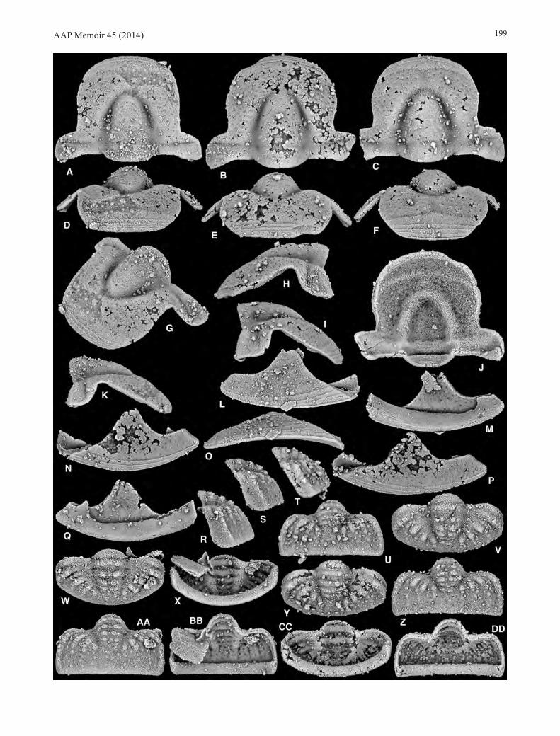
AAP Memoir 45 (2014) 199

AAP Memoir 45 (2014)200
opposite SO, with posterior margin of LO sitting even slightly higher, glabellar profile strongly sloped downward from horizontal plane anteriorly so that anterior margin is nearly inline with slope of preglabellar field and anterior border; axial furrows very broad and deep opposite main anterior portion of glabella, narrower anteriorly joining preglabellar furrow in a smooth continuous arc, much shallower posteriorly opposite posterolateral corners of glabella, shallower still opposite LO, with width across posterior contact of furrows with posterior margin 50.6% (48.2–53.7) cranidial sagittal length; intersection of axial and posterior border furrows with SO forming broad shallow depression that encroaches onto posterolateral margins of glabella; preglabellar furrow shallower and short, strongly anteriorly bowed medially; glabella smooth with furrows not expressed; LO long (sag.), with sagittal length 18.6% (16.4–20.8) that of cranidium, much longer sagittally than exsagittally, posterior margin strongly posteriorly bowed, anterior margin almost transverse; SO very faint, distal tips deeper, nearly transverse; doublure broad and semicircular under LO, anterior margin just reaching posterior margin of SO as expressed ventrally, with sculpture of fine raised lines running subparallel to margin; additional small triangular section of doublure present ventrally at posterolateral tip of posterior border (Fig. 21J).
Librigenal measurements were made on Figure 21L and one additional specimen from AAA 114.5T m to be figured in a forthcoming work. Librigena with maximum width behind eye 48.1% (45.3–51.0) total librigenal length (including anterior projection and genal spine); anterior section of facial suture with length 62.8% (60.0–65.6) total length librigenal length, gently curved across field and border, with curve slightly stronger opposite distal portion of field just before border furrow; posterior section of facial suture with length 45.5% (41.0–50.0) total librigenal length, strongly curved; field subtriangular in outline, with maximum width 38.8% (37.4–40.2) total librigenal length; lateral border furrow shallow, expressed mainly as slightly concave region between field and border; posterior border furrow similar to lateral border furrow, very faint; lateral border with lateral margin gently laterally bowed, widest anteriorly, tapering to a point distally and termination of genal spine, with sculpture of about five prominent raised lines running roughly parallel to margin, proximal line prominent, clearly setting border off from field, all lines more widely spaced anteriorly and becoming progressively closer toward distal tip of genal spine so that only two lines continue onto tip of genal spine, line along lateral margin more prominent that others (Fig. 21O); lateral border in ventrolateral view gently arched (Fig. 21O), sloping upward toward genal field; cuticle thickened at lateral margin of librigena (Fig. 21L, N); posterior border very short, sculpture of raised lines fainter than on lateral border, proximal line from lateral border continues around tip of genal spine onto lateral border setting it off from field; genal spine short, tapered to a blunt point; area in front of base of genal spine (confluence of lateral and posterior borders) forming broad flattened area, sculpture of scattered pits present in this region (Fig. 21N) and continuing onto field, clearly visible on ventral surface of field (Fig. 21M); anterior projection short, sculpture on lateral border continues onto projection; doublure broad, portion beneath anterior projection visible in external view (Fig. 21N, P), broadest anteriorly and tapering toward genal angle, external third of doublure with sculpture of fine raised lines, lines finer than those on dorsal surface of lateral border, inner two
thirds of doublure more smooth, inner margin gently curved, with strong bend at genal angle and across posterior border; portion of doublure beneath posterior border developed into a short triangular process (Fig. 21Q).
Hypostome and thorax not identified.Pygidial measurements were made on Figures 13I, 21W,
V, Y. Pygidium with maximum with across second segment 187.0% (176.7–193.3) sagittal length; axis triangular in plan view, strongly convex (Fig. 21DD), composed of five segments, first four segments clearly expressed and very small terminal piece best seen in lateral profile, maximum axial width across first segment 31.9% (29.9–33.7) maximum pygidial width, length of axis excluding articulating half ring 60.0% (64.5–71.6) total sagittal length of pygidium; articulating half ring large and long with sagittal length 15.5% (13.5–18.5) total pygidial length, anterior margin strongly convex, posterior margin very gently anteriorly convex, ring sloped posteroventrally in line with general sagittal profile of axis; first ring furrow moderately short (sag.; exsag.) and shallow; second ring furrow nearly as deep as first, but slightly posteriorly bowed medially; third and fourth ring furrows shallow, but longer than first two; fifth ring furrow mostly effaced; first axial ring slightly longer sagittally than exsagittally (including partially merged second pseudo-articulating half ring), with sculpture of two prominent nodes present on either side of sagittal line; pseudo-articulating half ring of second segment clearly visible, but poorly developed, set off by narrow short furrow; posterior rings progressively smaller, but with essentially the same morphology and sculpture, width across fourth segment 67.3% (64.6–71.4) width across first segment; pseudo-articulating half rings of subsequent segments not clearly expressed; axis terminated by very small terminal piece, just barely visible in plan view as two very small nodes, more clearly identified in lateral profile by change in slope between fourth axial segment and post-axial region; anterior margin of first pleural band anteriorly concave opposite axial furrow, strongly anteriorly convex across fulcrum, with small subtriangular facet developed at anterolateral corner; first pleural segment with maximum exsagittal length 30.6% (27.3–33.2) sagittal axial length; anterior pleural band of first segment longest adaxially from fulcrum, much shorter distal to fulcrum and adjacent to axial furrow; posterior pleural band longer than anterior band, more prominent with stronger inflation than anterior band, very faint sculpture of small tubercles visible in lateral profile, tubercles continue past fulcrum almost to pygidial margin; posterolateral faces of pygidium from fulcrum distally form just slightly less than vertical “wall” (Fig. 21R–T); pleural furrow short (exsag.), nearly effaced between fulcrum and axial furrow, deeper from fulcrum distally, but terminated just past fulcrum before pygidial margin, directed posterolaterally; interpleural furrows similar to pleural furrows but more clearly define almost to pygidial margin; morphology of pleural region of segments two and three generally similar to segment one, but progressively narrower, more weakly expressed, turned slightly more posterolaterally, and with sculpture of small tubercles, sculpture stops just past fulcrum; pleura of fourth segment barely expressed as small node adjacent to terminal axial piece, in posterior view expressed as prominent round nodes with sculpture of faint smaller tubercles continuing onto “wall;” pleural bands terminate at pygidial border; border most clearly expressed in posterior view as short section with very slight independent inflation and distinct sculpture of closely spaced fine granules; ventral pygidial

AAP Memoir 45 (2014) 201
margin describing very slightly medially bowed margin, distal portion of “wall” with sculpture of fine raised granules and dashes arranged in an anastomosing pattern; doublure broad around lateral and posterior pygidial margins, inner margin describing smooth arc similar to that of pygidial margin, oriented subvertical (Fig. 21R) against ventral surface of pygidium, sculpture of fine raised lines covering entire doublure (Fig. 21S).
Remarks. Of the three species previously described by Ross (1951), two were based on a single cranidium and one on a single cranidium and a single librigena. Of these, Pseudoclelandia cornupsittaca is not closely similar to P. weymouthae, as it has a deep anterior border furrow which connects medially to the preglabellar furrow and has strong tuberculate sculpture, among other prominent differences. Pseudoclelandia lenisora (from the Bearriverops logani Zone) is similar to P. weymouthae in general dimensions, but is also densely tuberculate. Pseudoclelanida fluxafissura is the closest presently named species. It differs from P. weymouthae in the possession of a shorter cranidium, more laterally extended anterior facial sutures, and a more subtriangular, versus anteriorly extended, glabella. Much more satisfactory comparisons will be facilitated when the species are revised on the basis of extensive new material in a forthcoming work, and further comments are deferred until then.
ACKNOWLEDGEMENTSResearch has been supported by NSF grants EAR 9973065, EAR 0308685, and DEB 0716065. Tiffany Adrain provided assistance with curation and specimen numbers. Arne T. Nielsen and John F. Taylor provided helpful reviews of the mansuscript.
REFERENCESAdrAin, J.M., Lee, d.-C., Westrop, s.r., ChAtterton, B.d.e. &
LAnding, e., 2003. Classification of the trilobite subfamilies Hystricurinae and Hintzecurinae subfam. nov., with new genera from the Lower Ordovician (Ibexian) of Idaho and Utah. Memoirs of the Queensland Museum 48, 553–586.
AdrAin, J.M. & MCAdAMs, n.e.B., 2012. The Lower Ordovician (upper Floian) bathyurid trilobite Aponileus Hu, with species from Utah, Texas, and Greenland. Zootaxa 3293, 1–67.
AdrAin, J.M., MCAdAMs, n.e.B. & KAriM, t.s., 2012. The Middle Ordovician bathyurid trilobite Pseudoolenoides, with a revised trilobite biostratigraphy of the Dapingian and lower Darriwilian of western Laurentia. Zootaxa 3467, 1–97.
AdrAin, J.M., MCAdAMs, n.e.B. & Westrop, s.r., 2009. Trilobite biostratigraphy and revised bases of the Tulean and Blackhillsian Stages of the Ibexian Series, Lower Ordovician, western United States. Memoirs of the Association of Australasian Palaeontologists 37, 541–610.
AdrAin, J.M., MCAdAMs, n.e.B. & Westrop, s.r., 2011a. Affinities of the Lower Ordovician (Tulean; lower Floian) trilobite Gladiatoria, with species from the Great Basin, western United States. Memoirs of the Association of Australasian Palaeontologists 42, 321–367.
AdrAin, J.M., MCAdAMs, n.e.B., Westrop, s.r. & KAriM, t.s., 2011b. Systematics and affinity of the Lower Ordovician (Tulean; lower Floian) trilobite Psalikilopsis. Memoirs of the Association of Australasian Palaeontologists 42, 369–416.
AdrAin, J.M. & Westrop, s.r., 2006a. New earliest Ordovician trilobite genus Millardicurus: The oldest known hystricurid. Journal of Paleontology 80, 650–671.
AdrAin, J.M. & Westrop, s.r., 2006b. New genus of dimeropygid trilobites from the earliest Ordovician of Laurentia. Acta Palaeontologica Polonica 51, 541–550.
AdrAin, J.M. & Westrop, s.r., 2007a. Bearriverops, a new Lower Ordovician trilobite genus from the Great Basin, western USA, and classification of the family Dimeropygidae. Canadian Journal of Earth Sciences 44, 337–366.
AdrAin, J.M. & Westrop, s.r., 2007b. The hystricurid trilobite Metabowmania in the Lower Ordovician (Ibexian; Stairsian) of the Great Basin, Utah and Idaho, USA. Memoirs of the Association of Australasian Palaeontologists 34, 227–242.
Berg, r.r. & ross, r.J., 1959. Trilobites from the Peerless and Manitou formations, Colorado. Journal of Paleontology 33, 106–119.
BiLLings, e., 1859. Fossils of the Calciferous Sandrock, including those of a deposit of white limestone at Mingan, supposed to belong to the formation. The Canadian Naturalist and Geologist, and Proceedings of the Natural History Society of Montreal 4, 345–367.
BiLLings, e., 1860. On some new species of fossils from the limstones near Point Levi opposite Quebec. The Canadian Naturalist and Geologist, and Proceedings of the Natural History Society of Montreal 5, 301–324.
BoyCe, W.d., 1989. Early Ordovician trilobite faunas of the Boat Harbour and Catoche Formations (St. George Group) in the Boat Harbour-Cape Norman area, Great Northern Peninsula, western Newfoundland. Newfoundland Department of Mines and Energy, Geological Survey Branch, Report, 89-2, 1–169.
Bridge, J. & CLoud, p.e., 1947. New gastropods and trilobites critical in the correlation of Lower Ordovician rocks. American Journal of Science 245, 545–559.
deAn, W.t., 1989. Trilobites from the Survey Peak, Outram and Skoki formations (Upper Cambrian-Lower Ordovician) at Wilcox Pass, Jasper National Park, Alberta. Geological Survey of Canada Bulletin 389, 1–141.
deMeter, e.J., 1973. Lower Ordovician pliomerid trilobites from western Utah. Brigham Young University Geology Studies 20, 37–65.
dWight, W.B., 1884. Recent explorations in the Wappinger Valley limestone of Dutchess County, New York. American Journal of Science, 3rd series 27, 249–259.
FLoWer, r.h., 1968. Fossils from the Smith Basin Limestone of the Fort Ann Region, New York. New Mexico Bureau of Mines and Mineral Resources Memoir 22, 21–27.
Fortey, r.A. & peeL, J.s., 1989. Stratigraphy and hystricurid trilobites of the Christian Elv Formation (Lower Ordovician) of western North Greenland. Rapport. Grønlands Geologiske Undersøgelse 144, 5-15.
goBBett, d.J., 1960. A new species of trilobite from the Lower Oslobreen Limestone. Geological Magazine 107, 457–459.
hArrington, h.J., 1957. Notes on new genera of Pliomeridae (Trilobita). Journal of Paleontology 31, 811–812.
hAWLe, i. & CordA, A.J.C., 1847. Prodrom einer Monographie der böhmischen Trilobiten. J. G. Calve, Prague, 176 pp.
heLLer, r.L., 1956. Stratigraphy and paleontology of the Roubidoux Formation of Missouri. Missouri Geological Survey and Water Resources, Series 2 35, 1–113. [for 1954]
hintze, L.F., 1951. Lower Ordovician detailed stratigraphic sections for western Utah. Utah Geological and Mineralogical Survey Bulletin 39, 1–99.
hintze, L.F., 1953. Lower Ordovician trilobites from western Utah and eastern Nevada. Utah Geological and Mineralogical Survey Bulletin, 48, 1–249. [for 1952]
hintze, L.F., 1973. Lower and Middle Ordovician stratigraphic sections in the Ibex area, Millard County, Utah. Brigham Young
AAP Memoir 45 (2014)202
University Geology Studies 20, 3–36. hupé, p., 1953. Classification des Trilobites. Annales de
Paléontologie 39, 59–168. JeLL, p.A. & stAit, B., 1985. Tremadoc trilobites from the Florentine
Valley Formation, Tim Shea Area, Tasmania. Memoirs of the Museum of Victoria 46, 1–34.
KoBAyAshi, t., 1940. Lower Ordovician fossils from Junee, Tasmania. Papers and Proceedings of the Royal Society of Tasmania 1939, 61–66. [for 1939]
KoBAyAshi, t., 1955. The Ordovician fossils of the McKay Group in British Columbia western Canada, with a note on the early Ordovician palaeogeography. Journal of the Faculty of Science, Tokyo University, Section 2 9, 355–493.
LAnding, e., AdrAin, J.M., Westrop, s.r. & Kröger, B., 2012. Tribes Hill–Rochdale formations in east Laurentia: proxies for Early Ordovician (Tremadocian) eustasy on a tropical passive margin (New York and west Vermont). Geological Magazine 149, 93–123.
LAnding, e., Westrop, s.r. & VAn ALLer herniCK, L., 2003. Uppermost Cambrian-Lower Ordovician faunas and Laurentian platform sequence stratigraphy, eastern New York and Vermont. Journal of Paleontology 77, 78–98.
LoCh, J.d., 1995. An affirmation of the Jeffersonian Stage (Ibexian) of North America and a proposed boundary stratotype. 45–48 in Cooper, J.D., Droser, M.L. & Finney, S.C. (eds), Ordovician Odyssey: Short Papers for the Seventh International Symposium on the Ordovician System. Pacific Section Society for Sedimentary Geology (SEPM), Fullerton, California.
LoCh, J.d., 2007. Trilobite biostratigraphy and correlation of the Kindblade Formation (Lower Ordovician) of Carter and Kiowa Counties, Oklahoma. Oklahoma Geological Survey Bulletin, 149, 1–157.
LoChMAn, C., 1965. Lower Ordovician (Zone D) faunules from the Williston Basin, Montana. Journal of Paleontology 39, 466–486.
Lu, y.-h., zhu, z.-L., QiAn, y.-y., zhou, z.-y., Chen, J.-y., Liu, g.-W., yü, W., Chen, X. & Xu, h.-K., 1976. Ordovician biostratigraphy and palaeozoogeography of China. Memoirs of the Nanjing Institute of Geology and Palaeontology 7, 1–83.
MCAdAMs, n.e.B. & AdrAin, J.M., 2009a. Heckethornia, a new genus of dimeropygid trilobites from the Lower Ordovician (Ibexian; Tulean and Blackhillsian) of the Great Basin, western USA. Canadian Journal of Earth Sciences 46, 875–914.
MCAdAMs, n.e.B. & AdrAin, J.M., 2009b. New pliomerid trilobite genus Lemureops from the Lower Ordovician (Ibexian; Tulean, Blackhillsian) of western Utah, USA. Memoirs of the Association of Australasian Palaeontologists 37, 491–540.
MCAdAMs, n.e.B. & AdrAin, J.M., 2010. A new species of the Lower Ordovician pliomerid trilobite Pseudocybele and its biostratigraphic significance. Zootaxa 2550, 21–38.
MCAdAMs, n.e.B. & AdrAin, J.M., 2011a. Panisaspis, a new genus of pliomerid trilobites from the Lower Ordovician (Ibexian; Tulean and Blackhillsian) of the Great Basin, western USA. Zootaxa 2969, 1–68.
MCAdAMs, n.e.B. & AdrAin, J.M., 2011b. Revision of the Lower Ordovician (lower Floian; Tulean) pliomerid trilobite Protopliomerella, with new species from the Great Basin, western USA. Zootaxa 3144, 1–113.
MCAdAMs, n.e.B. & AdrAin, J.M., 2011c. Systematics of the Lower Ordovician pliomerid trilobite Hintzeia, with species from the Great Basin, western USA. Zootaxa 2910, 1–45.
prAtt, B.r., 1988. An Ibexian (Early Ordovician) trilobite faunule from the type section of the Rabbitkettle Formation (southern Mackenzie Mountains, Northwest Territories). Canadian Journal of Earth Sciences 25, 1595–1607.
rAyMond, p.e., 1913. A revision of the species which have been referred to the genus Bathyurus. Bulletin of the Victoria Memorial Museum 1, 51–69.
ross, r.J., 1949. Stratigraphy and trilobite faunal zones of the Garden City Formation, northeastern Utah. American Journal of Science 247, 472–491.
ross, r.J., 1951. Stratigraphy of the Garden City Formation in northeastern Utah, and its trilobite faunas. Peabody Museum of Natural History, Yale University, Bulletin, 6, 1–161.
ross, r.J., 1953. Additional Garden City (Early Ordovician) trilobites. Journal of Paleontology 27, 633–646.
ross, r.J., 1970. Ordovician brachiopods, trilobites, and stratigraphy in eastern and central Nevada. United States Geological Survey Professional Paper 639, 1–103.
ross, r.J., hintze, L.F., ethington, r.L., MiLLer, J.F., tAyLor, M.e. & repetsKi, J.e., 1997. The Ibexian, lowermost series in the North American Ordovician. United States Geological Survey Professional Paper 1579, 1–50.
stitt, J.h., 1983. Trilobites, biostratigraphy, and lithostratigraphy of the McKenzie Hill Limestone (Lower Ordovician), Wichita and Arbuckle Mountains, Oklahoma. Oklahoma Geological Survey Bulletin 134, 1–54.
strAuss, J.V., MACdonALd, F.A., tAyLor, J.F., repetsKi, J.e. & MCCLeLLAnd, W.C., 2013. Laurentian origin for the North Slope of Alaska: Implications for the tectonic evolution of the Arctic. Lithosphere, doi:10.1130/L284.1.
tAyLor, J.F., MyroW, p.M., ripperdAn, r.L., LoCh, J.d. & ethington, r.L., 2004. Paleoceanographic events and faunal crises recorded in the Upper Cambrian and Lower Ordovician of west Texas and southern New Mexico. 167–183 in Nelson, E.P. & Erslev, E.A. (s). Field Trips in the Southern Rocky Mountains, USA. Geological Society of America Field Guide 5, 167–183.
tAyLor, J.F., repetsKi, J.e., LoCh, J.d. & LesLie, s.A., 2012. Biostratigraphy and chronostratigraphy of the Cambrian-Ordovician great American carbonate bank. 15–35 in Derby, J.R., Fritz, R.D., Longacre, S.A., Morgan, W.A. & Sternbach, C.A. (eds). The great American carbonate bank: The geology and economic resources of the Cambrian-Ordovician Sauk megasequence of Laurentia. American Association of Petroleum Geologists Memoir 98.
terreLL, F.M., 1973. Silicified trilobite zonation in the Lower Fillmore Formation in western Utah. Brigham Young University Geology Studies 20, 67–90.
WhitFieLd, r.p., 1889. Observations on some imperfectly known fossils from the Calciferous sandrock of Lake Champlain and descriptions of several new forms. Bulletin of the American Museum of Natural History 2, 41–63.
XiAng, L.-W. & zhou, t.-M., 1987. Trilobites. 294–335 in Wang, X.-F., Xiang, L.-W., Ni, S.-Z., Zeng, Q.-L., Xu, G.-H., Zhou, T.-M., Lai, C.-G. & Li, Z.-H. (eds), Biostratigraphy of the Yangtze Gorges Area (2). Early Palaeozoic Era. Geological Publishing House, Beijing.
zhou, t.-M., Liu, y.-r., Meng, X.-s. & sun, z.-h., 1977. Trilobita. 104–266 in Palaeontological Atlas of Central and South China. I. Early Palaeozoic. Geological Publishing House, Beijing.

AAP Memoir 45 (2014) 203
APPENDIX 1. Stratigraphic log of Ibex Section MME through the upper House Formation and lower part of the Fillmore Formation (measured and logged by Westrop and Adrain). See Figure 1 for position and line of section. Our new sampling horizons are indicated in metres to the right of the columns. Legend is on page 205.

AAP Memoir 45 (2014)204
APPENDIX 1 (continued)
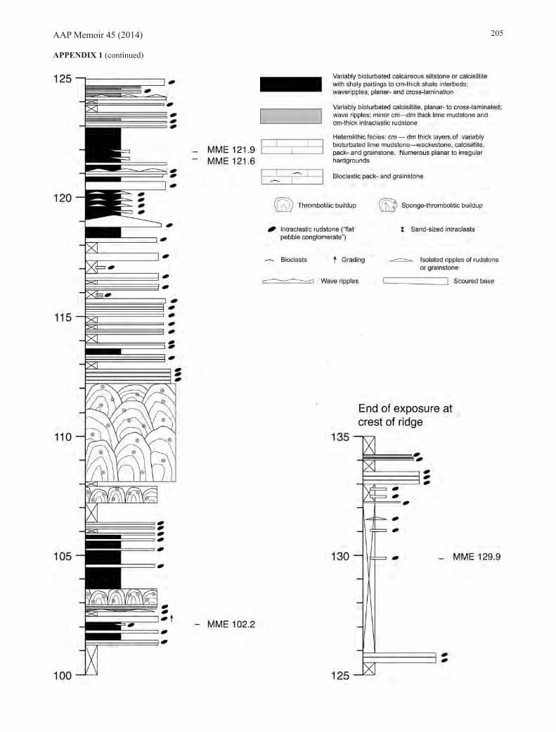
AAP Memoir 45 (2014) 205
APPENDIX 1 (continued)

AAP Memoir 45 (2014)206
APPENDIX 2. Stratigraphic logs of Ibex Sections B-TOP and basal C (measured and logged by Westrop and Adrain), with lithological correlations indicated. See Figure 1 for position and line of section. Legend given in Appendix 1.

AAP Memoir 45 (2014) 207
APPENDIX 3. Stratigraphic log of the upper part of Ibex Section AAA (measured and logged by Westrop and Adrain). See Figure 1 for position and line of section. Legend given in Appendix 1.
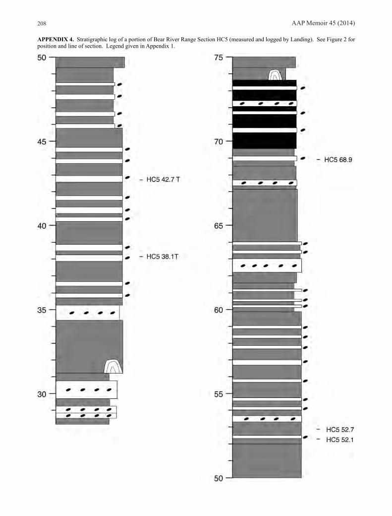
AAP Memoir 45 (2014)208
APPENDIX 4. Stratigraphic log of a portion of Bear River Range Section HC5 (measured and logged by Landing). See Figure 2 for position and line of section. Legend given in Appendix 1.

AAP Memoir 45 (2014) 209
APPENDIX 4 (continued)
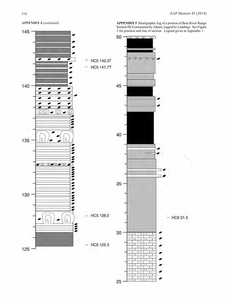
AAP Memoir 45 (2014)210
APPENDIX 4 (continued) APPENDIX 5 Stratigraphic log of a portion of Bear River Range Section HC6 (measured by Adrain, logged by Landing). See Figure 2 for position and line of section. Legend given in Appendix 1.

AAP Memoir 45 (2014) 211
APPENDIX 5 (continued)

AAP Memoir 45 (2014)212
APPENDIX 5 (continued)

AAP Memoir 45 (2014) 213
APPENDIX 6 Stratigraphic log of a portion of Bear River Range Section FB7 (measured and logged by Landing). See Figure 2 for position and line of section. Legend given in Appendix 1.

AAP Memoir 45 (2014)214
APPENDIX 6 (continued)