Trigeminal Nerve Anatomy
110
Department of Oral & Maxillofacial Surgery NARSINHBHAI PATEL DENTAL COLLEGE AND HOSPITAL VISNAGAR Guided By : Dr. Arvind Agarwal , HOD and Professor Dr.Anil Mannagutti , Professor Dr.Shreedevi Bhoi Presented by: Dr. Harsh Patel 1 st year PG 1/20/2015 Oral And Maxillofacial Surgery 1
-
Upload
drharshpatel21 -
Category
Health & Medicine
-
view
1.164 -
download
3
Transcript of Trigeminal Nerve Anatomy
Department of Oral & Maxillofacial Surgery
NARSINHBHAI PATEL DENTAL COLLEGE AND HOSPITAL VISNAGAR
Guided By :
Dr. Arvind Agarwal , HOD and ProfessorDr.Anil Mannagutti , ProfessorDr.Shreedevi Bhoi
Presented by: Dr. Harsh Patel1st year PG
1/20/2015 Oral And Maxillofacial Surgery 1
Contents: Introduction
Trigeminal Nuclei
Functional Components
Course & Distribution
Trigeminal Ganglion
Divisions of Trigeminal Nerve
Clinical Examination of V Nerve
Applied Anatomy
Summary
References
1/20/2015 Oral And Maxillofacial Surgery 2
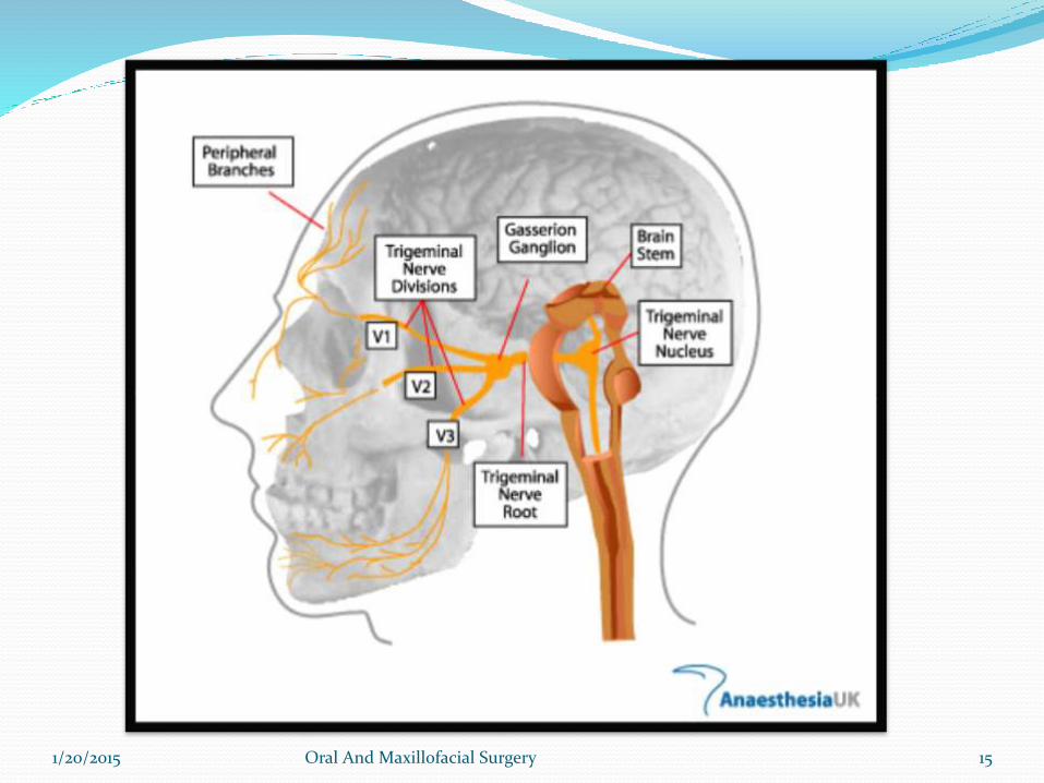
INTRODUCTION The largest cranial nerve
It is mixed nerve ( sensory and motor )
Sensory to – Skin of face
-Mucosa of cranial viscera
-Except base of tongue and pharynx
Motor to –Muscles of Mastication
-Tensor ville palatini,Tensor tympany
-Anterior belly of digastric
-Mylohyoid1/20/2015 Oral And Maxillofacial Surgery 3
NUCLEI
1/20/2015 Oral And Maxillofacial Surgery 4
TRIGEMINAL NUCLEIo A cranial nerve nucleus is a collection
of neurons (gray matter) in the brain stem that is associated with one or more cranial nerves.
o Axons carrying information to and from the cranial nerves form a synapse first at these nuclei.
o Lesions occurring at these nuclei can lead to effects resembling those seen by the severing of nerve(s) they are associated with.
1/20/2015 Oral And Maxillofacial Surgery 5
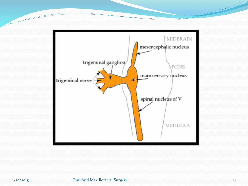
SENSORY NUCLEI :
1.Mesencephalic nucleus
- Cell body of Pseudounipolarneuron
- Relay proprioception from muscles of mastication,
Extra ocular Muscles, Facial muscles.
Situated in Midbrain just latetral to Aqueduct.
1/20/2015 Oral And Maxillofacial Surgery 6
2.Principal sensory nucleus-
Lies in Pons lateral to Motor nucleus
Relays touch sensation
1/20/2015 Oral And Maxillofacial Surgery 7
3.Spinal nucleus- Extends from caudal end of principal sensory Nucles
in pons to 2nd or 3rd spinal
segment
It relys Pain and Temperature
1/20/2015 Oral And Maxillofacial Surgery 8
MOTOR NUCLEUS : Innervates muscles of mastication and tensor
tympani and tensor palatini
Derived from first branchial arch.
Located in pons medial to principle sensory nucleus.
1/20/2015 Oral And Maxillofacial Surgery 9
1/20/2015 Oral And Maxillofacial Surgery 10
1/20/2015 Oral And Maxillofacial Surgery 11
FUNCTIONAL COMPONENTS
Sensory Root
Motor Root
1/20/2015 Oral And Maxillofacial Surgery 12
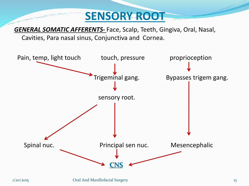
SENSORY ROOTGENERAL SOMATIC AFFERENTS- Face, Scalp, Teeth, Gingiva, Oral, Nasal,
Cavities, Para nasal sinus, Conjunctiva and Cornea.
Pain, temp, light touch touch, pressure proprioception
Trigeminal gang. Bypasses trigem gang.
sensory root.
1/20/2015 Oral And Maxillofacial Surgery 13
Spinal nuc. Principal sen nuc. Mesencephalic
CNS
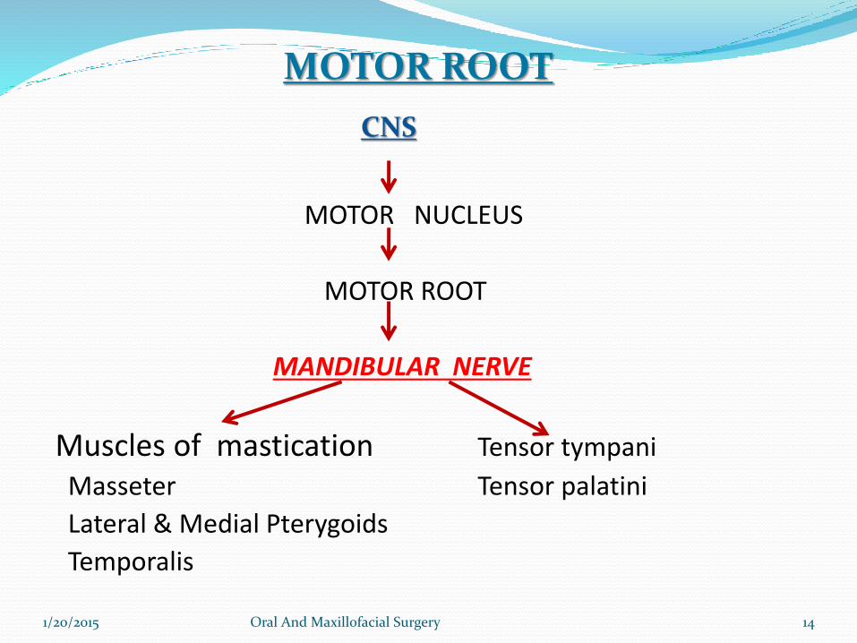
MOTOR NUCLEUS
MOTOR ROOT
MANDIBULAR NERVE
Muscles of mastication Tensor tympani
Masseter Tensor palatini
Lateral & Medial Pterygoids
Temporalis
CNS
1/20/2015 Oral And Maxillofacial Surgery 14
MOTOR ROOT
1/20/2015 Oral And Maxillofacial Surgery 15
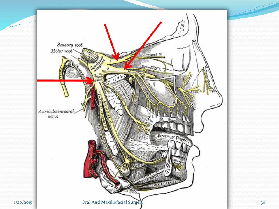
COURSE & DISTRIBUTION
Both motor and sensory root are attached ventrally to junction
of pons and middle cerebellar peduncle with motor root lying
ventromedially to the sensory root.
Pass anteriorly in middle cranial fossa to lie below tentorium
cerebelli in cavum trigeminale, here motor root lies inferior
to sensory root.
1/20/2015 Oral And Maxillofacial Surgery 16
Sensory root connected to postromedial concave
border of the trigeminal ganglion.
Convex antrolatateral margin of the ganglion gives
attachment to the 3 div. Of the trigeminal nerve.
1/20/2015 Oral And Maxillofacial Surgery 17
1/20/2015 Oral And Maxillofacial Surgery 19
1/20/2015 Oral And Maxillofacial Surgery 20
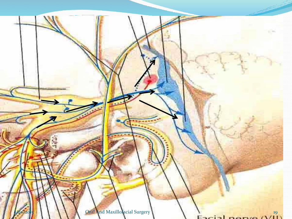
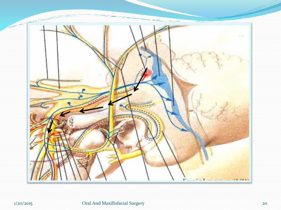
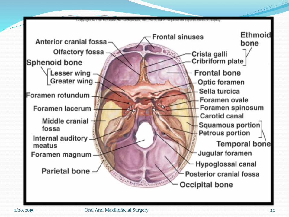
Motor root turns further inferior with sensory component of
V3 to emerge out of foramen Ovale as Mandibular
nerve.
Ophthalmic and Maxillary division emerges through
Superior orbital fissure and foramen Rotundum
respectively.
1/20/2015 Oral And Maxillofacial Surgery 21
1/20/2015 Oral And Maxillofacial Surgery 22
GANGLION
1/20/2015 Oral And Maxillofacial Surgery 23
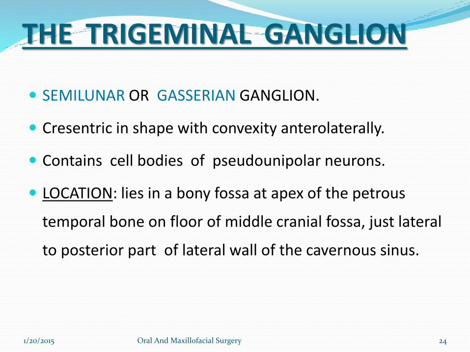
THE TRIGEMINAL GANGLION
SEMILUNAR OR GASSERIAN GANGLION.
Cresentric in shape with convexity anterolaterally.
Contains cell bodies of pseudounipolar neurons.
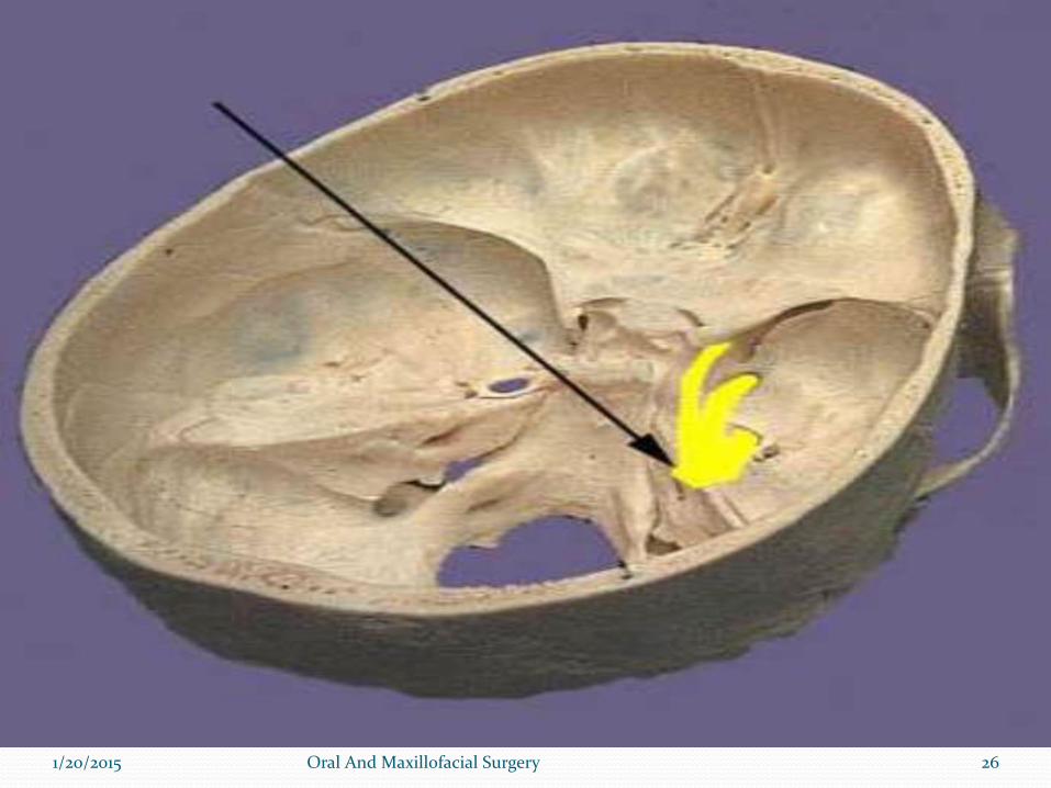
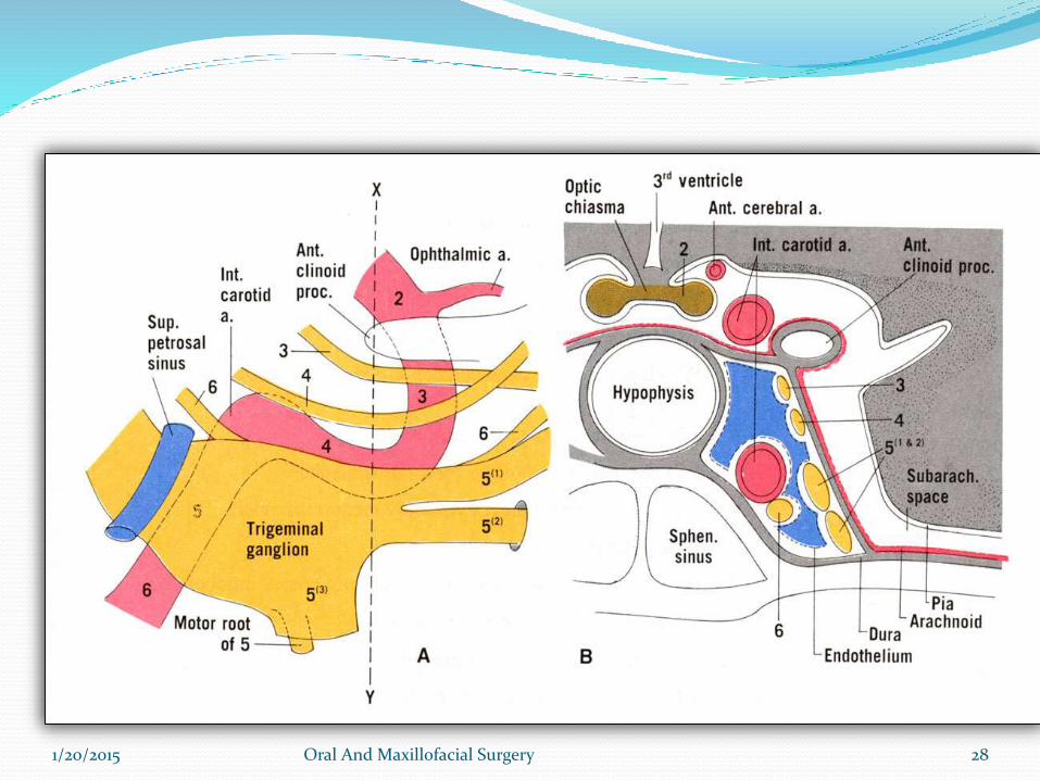
LOCATION: lies in a bony fossa at apex of the petrous
temporal bone on floor of middle cranial fossa, just lateral
to posterior part of lateral wall of the cavernous sinus.
1/20/2015 Oral And Maxillofacial Surgery 24
COVERINGS: covered by dural pouch = MECKLES CAVE or CAVUM TRIGEMINALE.
cave lined by pia and arachnoid thus the
ganglion is bathed in CSF.
ARTERIAL SUPPLY: Ganglionic branches of Internal Carotid Artery, middle meningeal artery and accessory meningealartery.
1/20/2015 Oral And Maxillofacial Surgery 25
1/20/2015 Oral And Maxillofacial Surgery 26
RELATIONS:SUPERIORLY: *superior petrosal sinus
*free margin of tentorium cerebelli
INFERIORLY: *motor root
*greater petrosal nerve
*petrous apex
*foramen lacerum
MEDIALLY: *posterior part of lateral wall of cavernous sinus
*Internal Carotid Artery with its sympathetic plexus
LATERALLY: *uncus of temporal lobe
*middle meningeal artery and vein
*nervous spinosum
1/20/2015 Oral And Maxillofacial Surgery 27
1/20/2015 Oral And Maxillofacial Surgery 28
DIVISIONS OF TRIGEMINAL NERVE
1. Ophthalmic nerve
2. Maxillary nerve
3. Mandibular nerve
1/20/2015 Oral And Maxillofacial Surgery 29
1/20/2015 Oral And Maxillofacial Surgery 30
OPTHALMIC NERRVE
1/20/2015 Oral And Maxillofacial Surgery 31
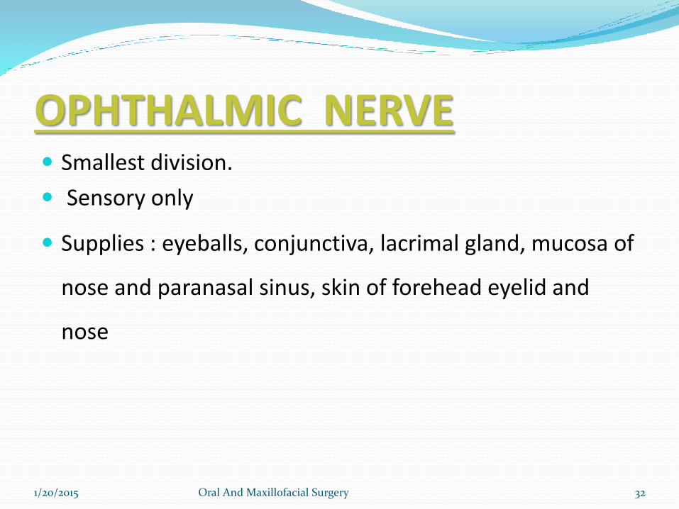
OPHTHALMIC NERVE Smallest division.
Sensory only
Supplies : eyeballs, conjunctiva, lacrimal gland, mucosa of
nose and paranasal sinus, skin of forehead eyelid and
nose
1/20/2015 Oral And Maxillofacial Surgery 32
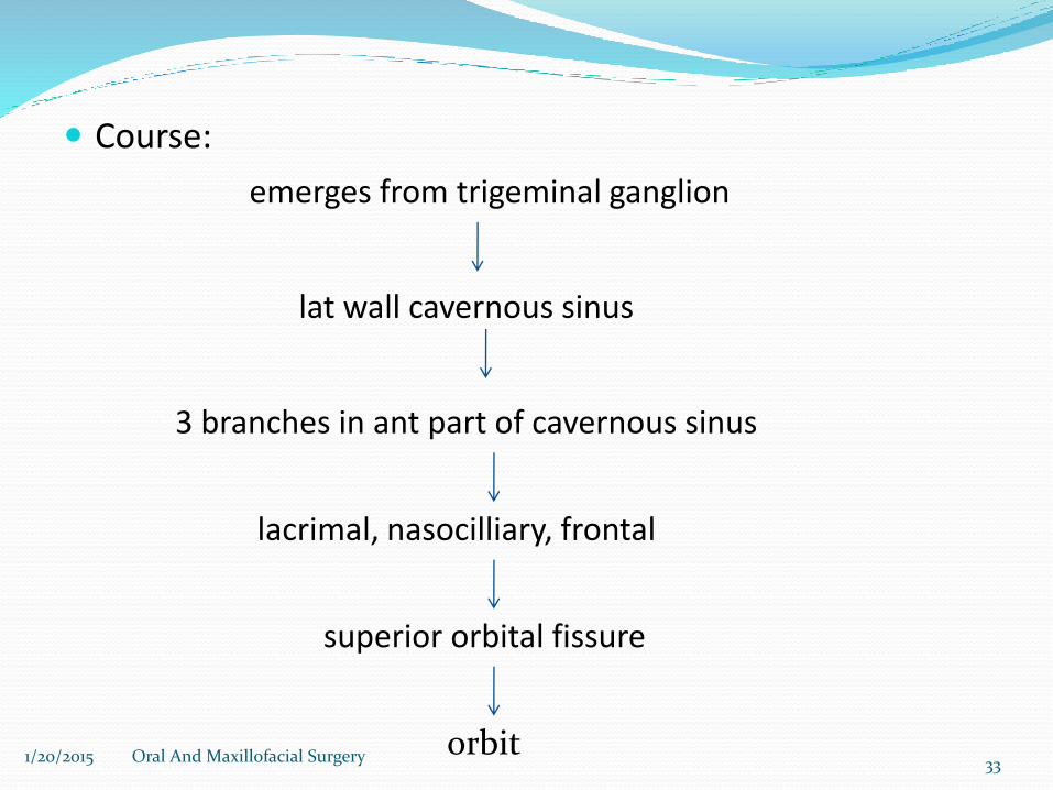
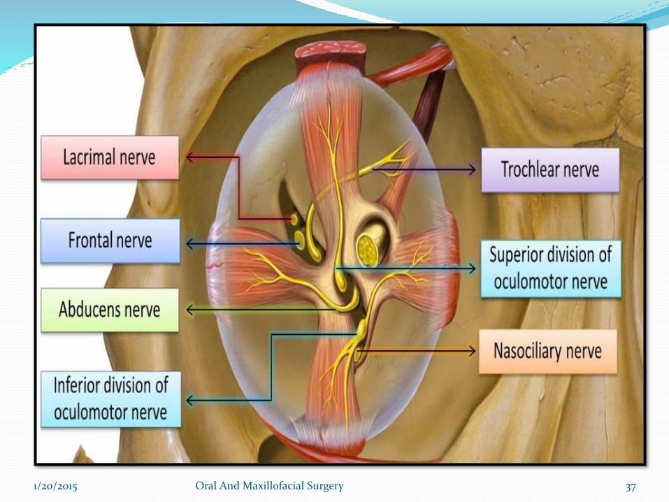
Course:
3 branches in ant part of cavernous sinus
superior orbital fissure
lat wall cavernous sinus
orbit
lacrimal, nasocilliary, frontal
emerges from trigeminal ganglion
1/20/2015 Oral And Maxillofacial Surgery 33
1/20/2015 Oral And Maxillofacial Surgery 34
LACRIMAL NERVE
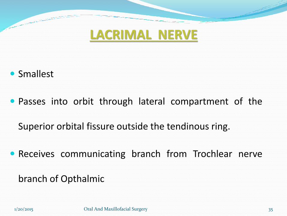
Smallest
Passes into orbit through lateral compartment of the
Superior orbital fissure outside the tendinous ring.
Receives communicating branch from Trochlear nerve
branch of Opthalmic
1/20/2015 Oral And Maxillofacial Surgery 35
Receives branch from Zygomaticotemporal nerve branch
of maxillary
Sensory to lateral conjunctiva, Upper Lid, lacrimal gland
Post synaptic parasympathetic fibers from pterigopalatine
ganglion to lacrimal gland (parasym secretomotor).
1/20/2015 Oral And Maxillofacial Surgery 36
1/20/2015 Oral And Maxillofacial Surgery 37
FRONTAL NERVE
Largest
Enters orbit through lateral part of superior orbital fissure
outside tendinous ring
Passes forward between roof of orbit and Levator Palpebral
Superioris
Supratrochlear Nerve
Divides midway into :
Supraorbital Nerve
1/20/2015 Oral And Maxillofacial Surgery 38
1/20/2015 Oral And Maxillofacial Surgery 39
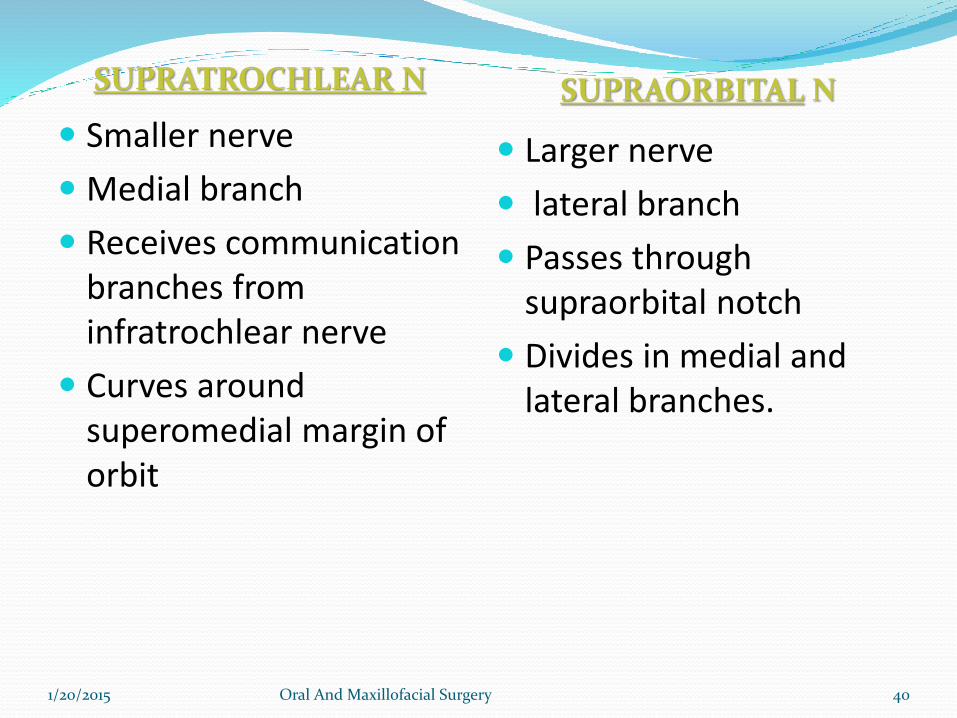
SUPRATROCHLEAR N SUPRAORBITAL N
Smaller nerve
Medial branch
Receives communication branches from infratrochlear nerve
Curves around superomedial margin of orbit
Larger nerve
lateral branch
Passes through supraorbital notch
Divides in medial and lateral branches.
1/20/2015 Oral And Maxillofacial Surgery 40
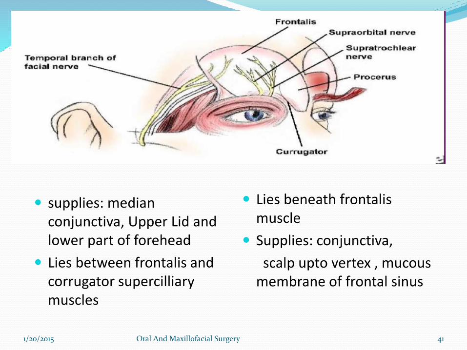
supplies: median conjunctiva, Upper Lid and lower part of forehead
Lies between frontalis and corrugator supercilliarymuscles
Lies beneath frontalismuscle
Supplies: conjunctiva,
scalp upto vertex , mucous membrane of frontal sinus
1/20/2015 Oral And Maxillofacial Surgery 41
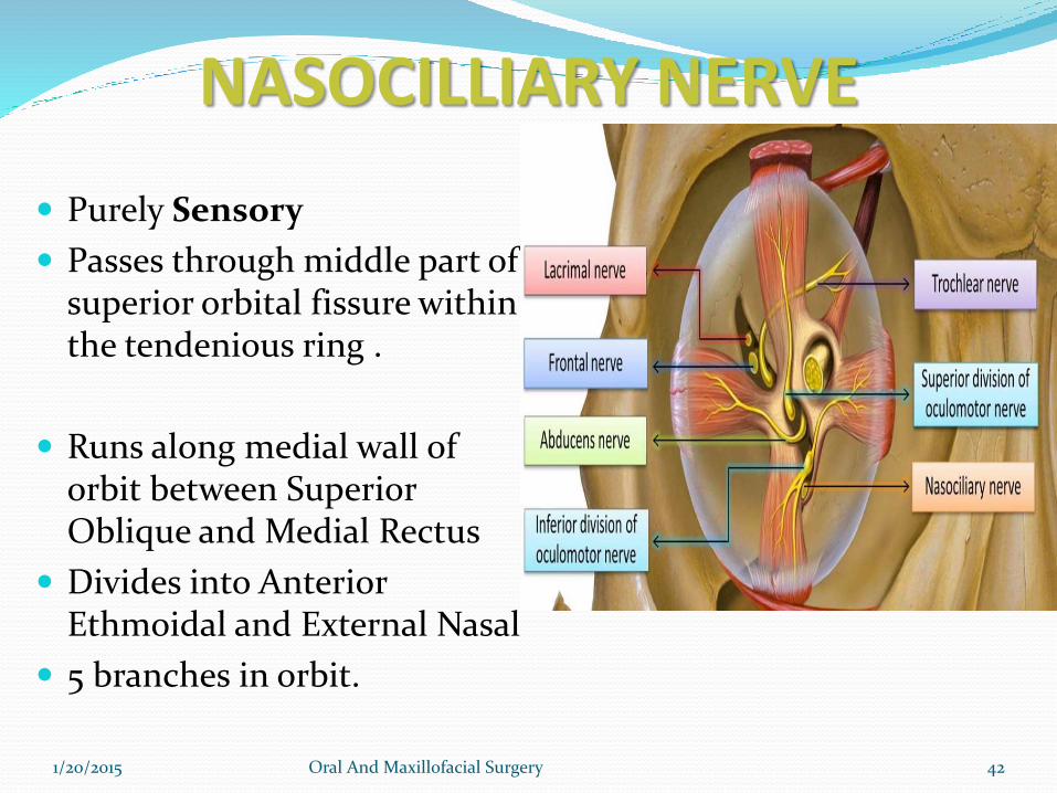
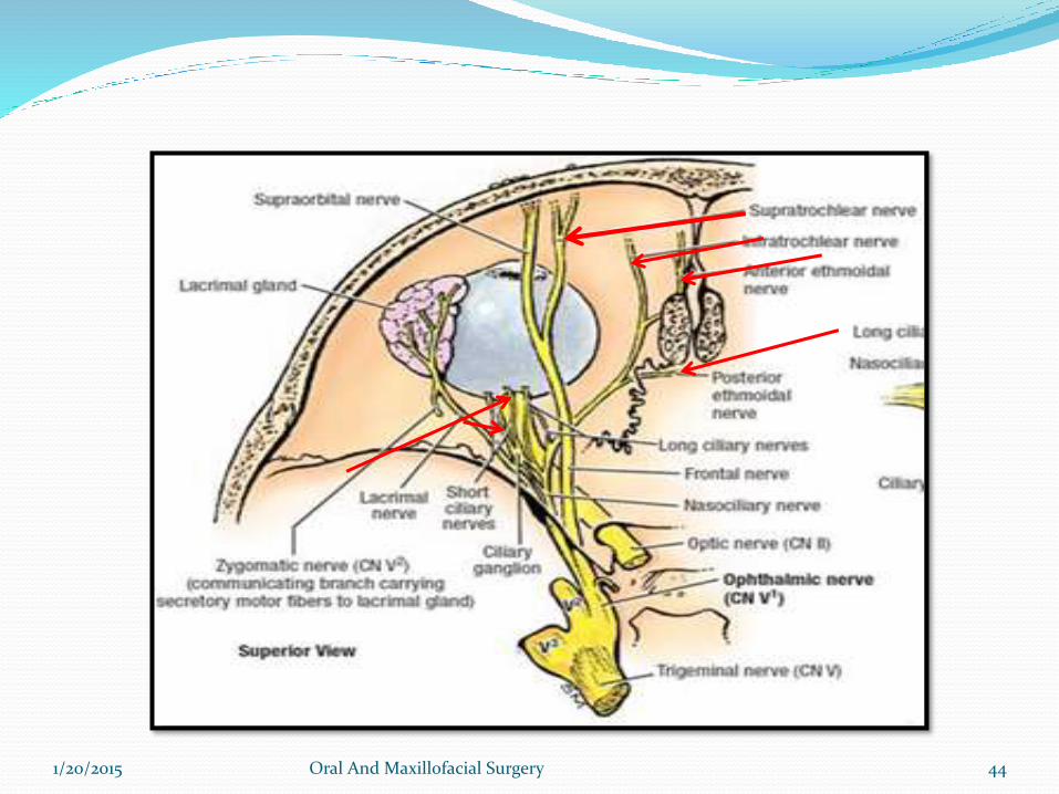
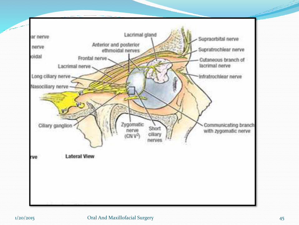
NASOCILLIARY NERVE
Purely Sensory
Passes through middle part of superior orbital fissure within the tendenious ring .
Runs along medial wall of orbit between Superior Oblique and Medial Rectus
Divides into Anterior Ethmoidal and External Nasal
5 branches in orbit.
1/20/2015 Oral And Maxillofacial Surgery 42
1. Short Clliary Nerves: Fibers reaches eyeball and also
contains fibers from Cilliary Ganglion
2. Long Cilliary Nerves : 2 or 3in no. supply to Iris and
Cornea.
3. Post Ethmoidal Nerve: passes through posterior
ethmoidal foramen to supply the Ethmoid and Sphenoid
PNS.
4. Infratrochlear Nerve: appears on face above med angle
the eye. Supplies to skin of lacrimal sac and caruncle.
1/20/2015 Oral And Maxillofacial Surgery 43
1/20/2015 Oral And Maxillofacial Surgery 44
1/20/2015 Oral And Maxillofacial Surgery 45
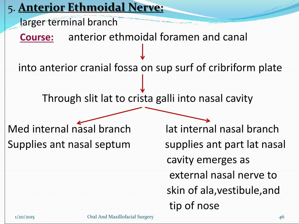
l5. Anterior Ethmoidal Nerve:
larger terminal branch
Course: anterior ethmoidal foramen and canal
into anterior cranial fossa on sup surf of cribriform plate
Through slit lat to crista galli into nasal cavity
Med internal nasal branch lat internal nasal branch
Supplies ant nasal septum supplies ant part lat nasal
cavity emerges as
external nasal nerve to
skin of ala,vestibule,and
tip of nose 1/20/2015 Oral And Maxillofacial Surgery 46
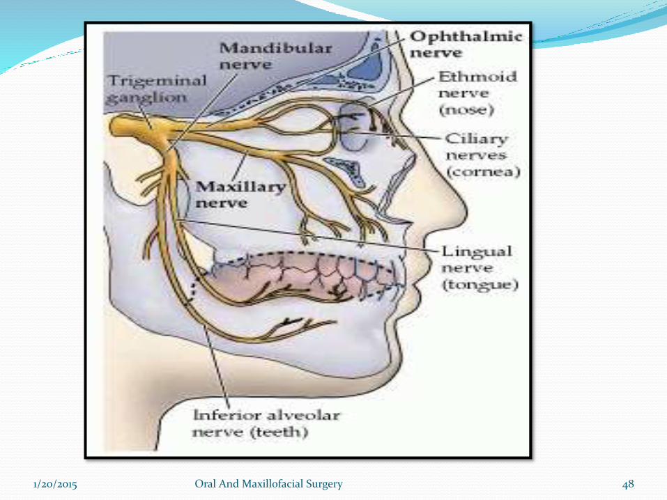
MAXILLARY NERVE
1/20/2015 Oral And Maxillofacial Surgery 47
1/20/2015 Oral And Maxillofacial Surgery 48
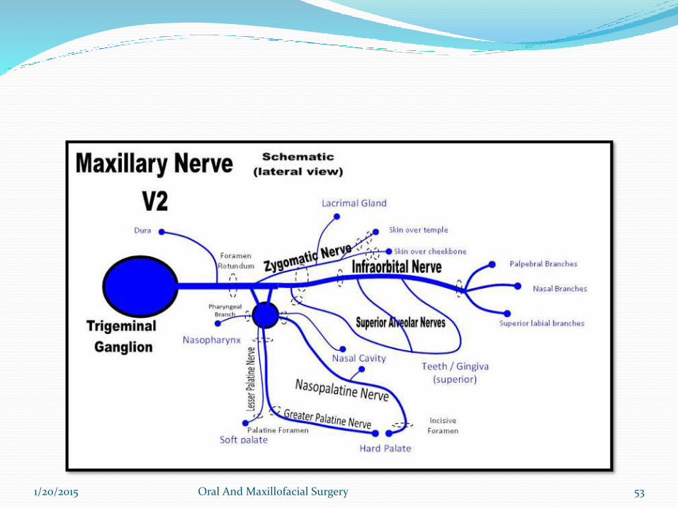
MAXILLARY NERVE Second division of trigeminal nerve
Pure sensory Supplies derivatives of maxillary process and frontonasal
process
1/20/2015 Oral And Maxillofacial Surgery 49
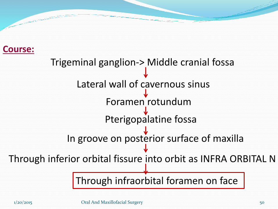
Course:
1/20/2015 Oral And Maxillofacial Surgery 50
Trigeminal ganglion-> Middle cranial fossa
Lateral wall of cavernous sinus
Foramen rotundum
Pterigopalatine fossa
In groove on posterior surface of maxilla
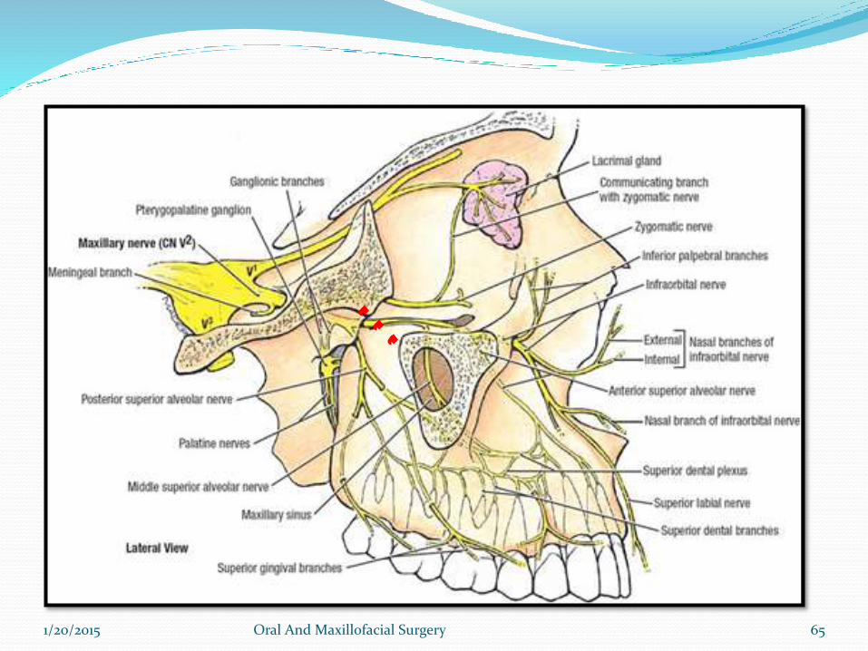
Through inferior orbital fissure into orbit as INFRA ORBITAL N
Through infraorbital foramen on face
After leaving foramen rotundum it moves anteriorly in
the uppermost part of pterygopalatine fossa.
As it passes through pterygopalatine fossa it also gives
branches to sphnopalatine ganglion, posterior superior
alveolar nerve and zygomatic branches.
1/20/2015 Oral And Maxillofacial Surgery 51
It then moves laterally and moves in a groove on
posterior surface of maxilla.
Then enters orbit through infra orbital fissure and
moves through infra orbital groove where it is called as
Infraorbital nerve and emerges on face from infra
orbital foramen.
1/20/2015 Oral And Maxillofacial Surgery 52
1/20/2015 Oral And Maxillofacial Surgery 53
1/20/2015 Oral And Maxillofacial Surgery 54
BRANCHES
IN MIDDLE CRANIAL FOSSA:
- Meningeal branch:Travels along the middle meningealartery and provides sensory innervation to cranial duramatter.
1/20/2015 Oral And Maxillofacial Surgery 55

IN PTERIGOPALATINE FOSSA:
1. Ganglionic branches-
Arises as 2trunks.Trunks join to form single root within pterygopalatine ganglion.
Gives Orital branches,Palatine branches,Pharyngealbranches,Nasal branches
Gives postganglionic secretomotor fibers to lacrimal gland via zygomaticotemporal and lacrimal.
1/20/2015 Oral And Maxillofacial Surgery 56
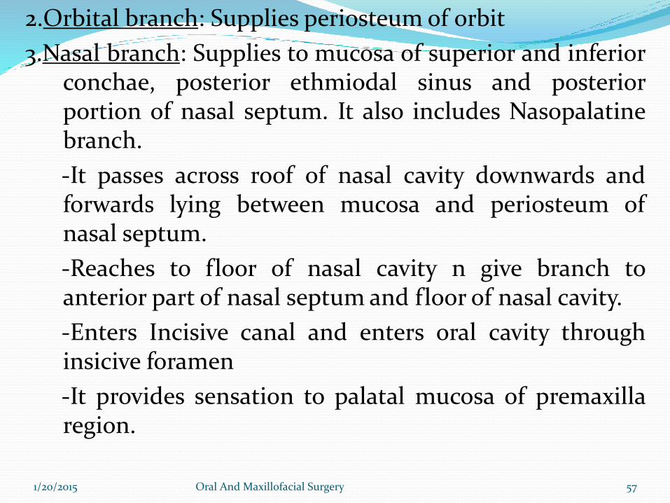
2.Orbital branch: Supplies periosteum of orbit
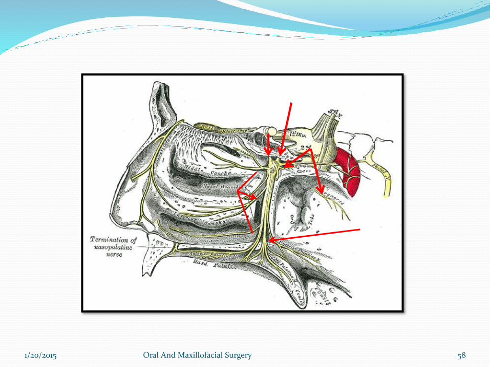
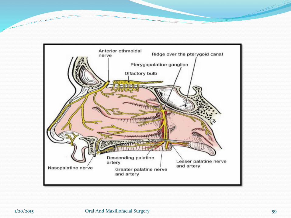
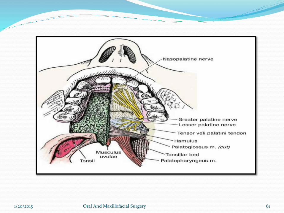
3.Nasal branch: Supplies to mucosa of superior and inferiorconchae, posterior ethmiodal sinus and posteriorportion of nasal septum. It also includes Nasopalatinebranch.
-It passes across roof of nasal cavity downwards andforwards lying between mucosa and periosteum ofnasal septum.
-Reaches to floor of nasal cavity n give branch toanterior part of nasal septum and floor of nasal cavity.
-Enters Incisive canal and enters oral cavity throughinsicive foramen
-It provides sensation to palatal mucosa of premaxillaregion.
1/20/2015 Oral And Maxillofacial Surgery 57
1/20/2015 Oral And Maxillofacial Surgery 58
1/20/2015 Oral And Maxillofacial Surgery 59
4. Palatine branch: Arise as greater palatine (anterior) and lesser
palatine (middle and posterior)
-Greater palatine nerve descends through pterygopalatine canal
from the ganglion and emerges from greater palatine foramen of
hard palate.
-Then moves anteriorly between mucoperiostem and hard palate
upto 1st premolar supplying sensory innervation to palatal soft
tissue and bone. Then communicates with nasopalatine
-Middle palatine and posterior palatine emerges from lesser
palatine foramen and supply soft palate and tonsilar region
respectively.
1/20/2015 Oral And Maxillofacial Surgery 60
1/20/2015 Oral And Maxillofacial Surgery 61
5. Pharyngeal branch: It leaves the posterior part of
pterygopalatine ganglion and passes through the
phryngeal canal
It is distributed to the mucous mambreane of the nasal
part of pharynx, posterior to eustachian tube.
1/20/2015 Oral And Maxillofacial Surgery 62
POST. SUPERIOR ALVEOLAR NERVE
-It arises from the main trunk of maxillary nerve in the
petrygopalatine fossa just before the nerve enters the inferior
orbital canal
- Usually arises as 2 trunks.
- Passes downwards and crosses the pterygoplatine fossa reaching
infratemporal surface of maxilla.
- 1st trunk continues downwards on posterior surface of maxilla
and provide sensory innervation to buccal gingiva in maxillary
molar region and adjacent facial mucosal surface
1/20/2015 Oral And Maxillofacial Surgery 63
-2nd trunk enters maxila through PSA canal to travel to
posterolateral wall of maxillary sinus providing sensory
innervation to sinus mucosa. Continuing downwards this
also provides sensory innervation to alveoli, PDL, pulp of
molar tooth.
1/20/2015 Oral And Maxillofacial Surgery 64
1/20/2015 Oral And Maxillofacial Surgery 65
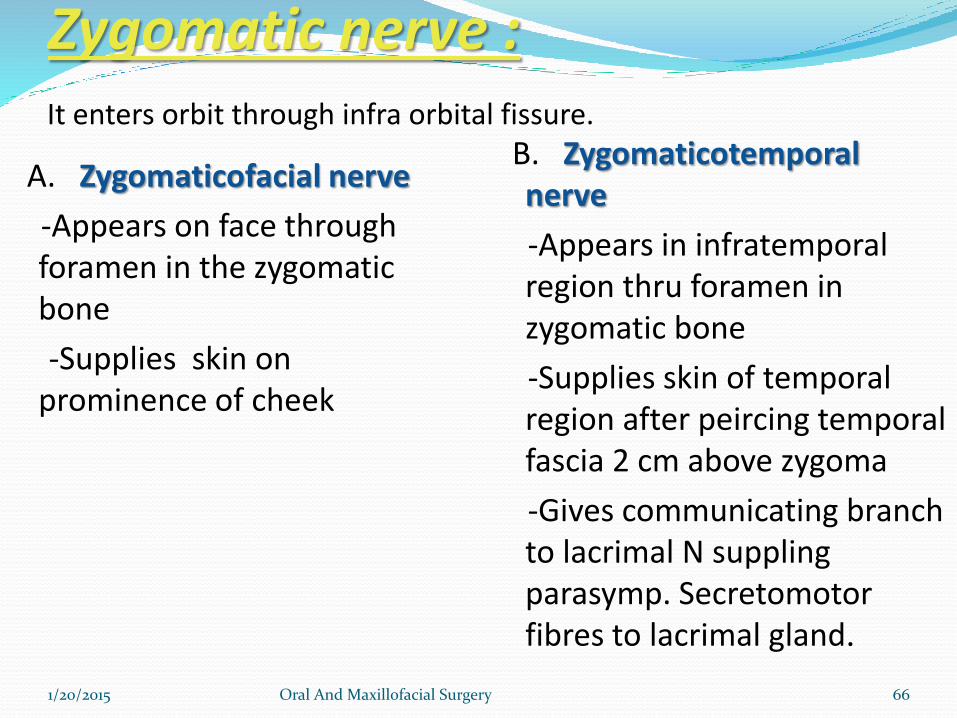
Zygomatic nerve :
A. Zygomaticofacial nerve
-Appears on face through foramen in the zygomaticbone
-Supplies skin on prominence of cheek
B. Zygomaticotemporalnerve
-Appears in infratemporalregion thru foramen in zygomatic bone
-Supplies skin of temporal region after peircing temporal fascia 2 cm above zygoma
-Gives communicating branch to lacrimal N supplingparasymp. Secretomotorfibres to lacrimal gland.
1/20/2015 Oral And Maxillofacial Surgery 66
It enters orbit through infra orbital fissure.
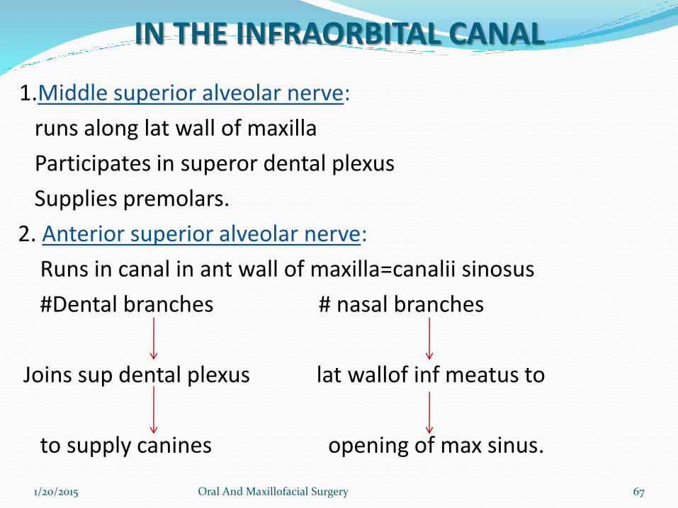
IN THE INFRAORBITAL CANAL
1.Middle superior alveolar nerve:
runs along lat wall of maxilla
Participates in superor dental plexus
Supplies premolars.
2. Anterior superior alveolar nerve:
Runs in canal in ant wall of maxilla=canalii sinosus
#Dental branches # nasal branches
Joins sup dental plexus lat wallof inf meatus to
to supply canines opening of max sinus.
1/20/2015 Oral And Maxillofacial Surgery 67
3. FACIAL BRANCHES:
1.Palpebral nerves-pierces Orbicularis Occuli and supplies skin of lower lid.
2.Nasal branches-supplies skin of lat wall nose and mobile part of septum.
3. Superior labial nerve- forms infraorbital plexus
supplies skin and mm of upper lip, cheek and labial glands.
1/20/2015 Oral And Maxillofacial Surgery 68
1/20/2015 Oral And Maxillofacial Surgery 69
MANDIBULAR NERVE
1/20/2015 Oral And Maxillofacial Surgery 70
MANDIBULAR NERVE Largest
Mixed
Nerve of 1st branchial arch
Motor root- from motor nucleus in pons
sensory root- gasserianganglion a
a small ant. Division
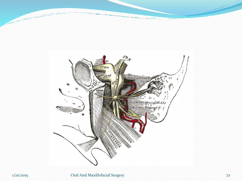
exit through foramen ovale in greater wing of sphenoid
from trunk which remain 2-3 mm undivided in infratemporalfossa
travels between lat. Pterygoid and Otic ganglion laterally and tensor palatine medially anteriorly to med. Meningeal A.
large post. division1/20/2015 Oral And Maxillofacial Surgery 71
1/20/2015 Oral And Maxillofacial Surgery 72
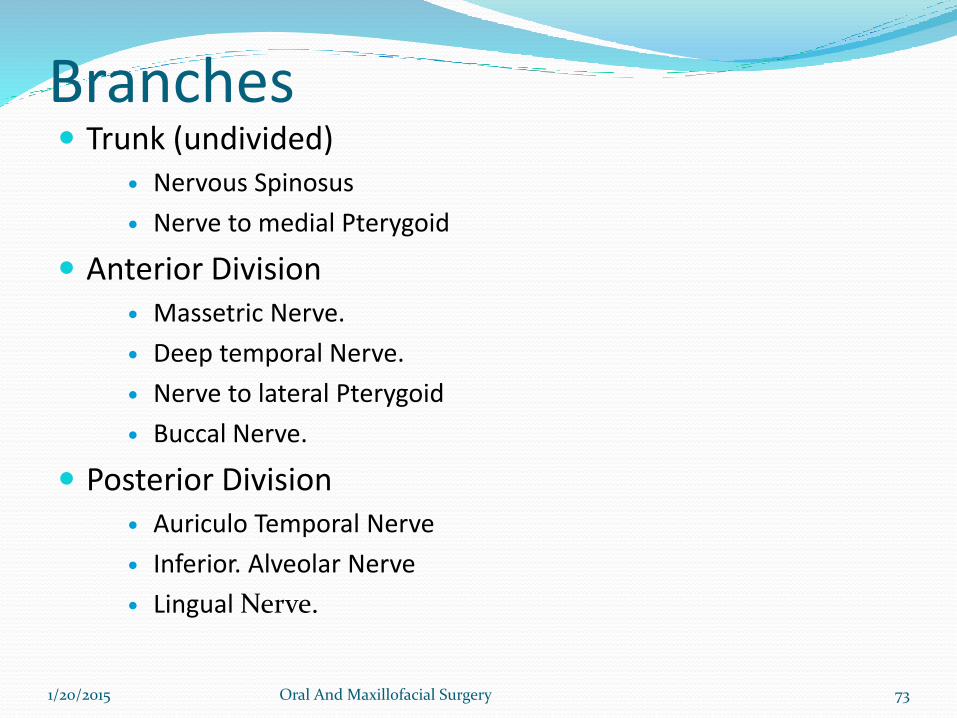
Branches Trunk (undivided)
Nervous Spinosus
Nerve to medial Pterygoid
Anterior Division Massetric Nerve.
Deep temporal Nerve.
Nerve to lateral Pterygoid
Buccal Nerve.
Posterior Division Auriculo Temporal Nerve
Inferior. Alveolar Nerve
Lingual Nerve.
1/20/2015 Oral And Maxillofacial Surgery 73
Branches from trunk Before dividing into anterior and posterior division it gives 2 branches
during its 2-3mm path
1.Nervous spinosus or Meningeal branch of Mandibular nerve
It reenters cranial cavity through foramen spinosus along with middle
meningial artery
Supply Dura matter of middle cranial fossa and mastoid air sinus
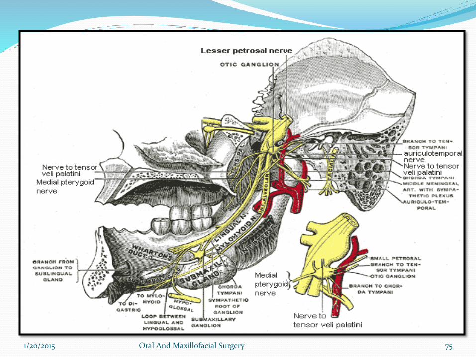
2.Nerve to mededial Pterygoid
Supplies medial pterygoid
Through Otic ganglion without interruption to
Tensor tympani
Tensor palatini
1/20/2015 Oral And Maxillofacial Surgery 74
1/20/2015 Oral And Maxillofacial Surgery 75
Branches from the anterior division
The anterior division is significantly smaller than posterior.
After dividing from the main trunk. It runs anteriorly and below
the lateral pterygoid muscle to over its upper border. After this
the nerve is buccal nerve. reach its external surface of muscle by
either passing through two heads or winding
1.Nerve to lateral pterygoid: It enters the deep surface of the
muscle. It may arise as independent branch or may arise in
common with buccal nerve.
1/20/2015 Oral And Maxillofacial Surgery 76
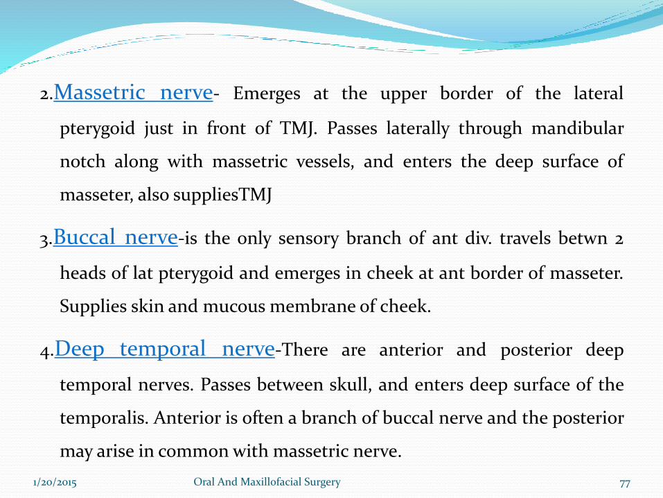
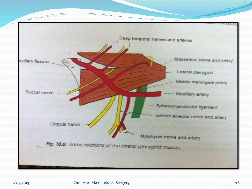
2.Massetric nerve- Emerges at the upper border of the lateral
pterygoid just in front of TMJ. Passes laterally through mandibular
notch along with massetric vessels, and enters the deep surface of
masseter, also suppliesTMJ
3.Buccal nerve-is the only sensory branch of ant div. travels betwn 2
heads of lat pterygoid and emerges in cheek at ant border of masseter.
Supplies skin and mucous membrane of cheek.
4.Deep temporal nerve-There are anterior and posterior deep
temporal nerves. Passes between skull, and enters deep surface of the
temporalis. Anterior is often a branch of buccal nerve and the posterior
may arise in common with massetric nerve.
1/20/2015 Oral And Maxillofacial Surgery 77
1/20/2015 Oral And Maxillofacial Surgery 78
Branches Of Posterior Division
1/20/2015 Oral And Maxillofacial Surgery 79
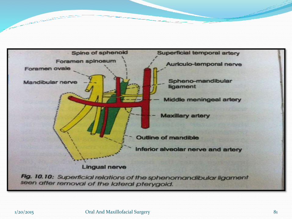
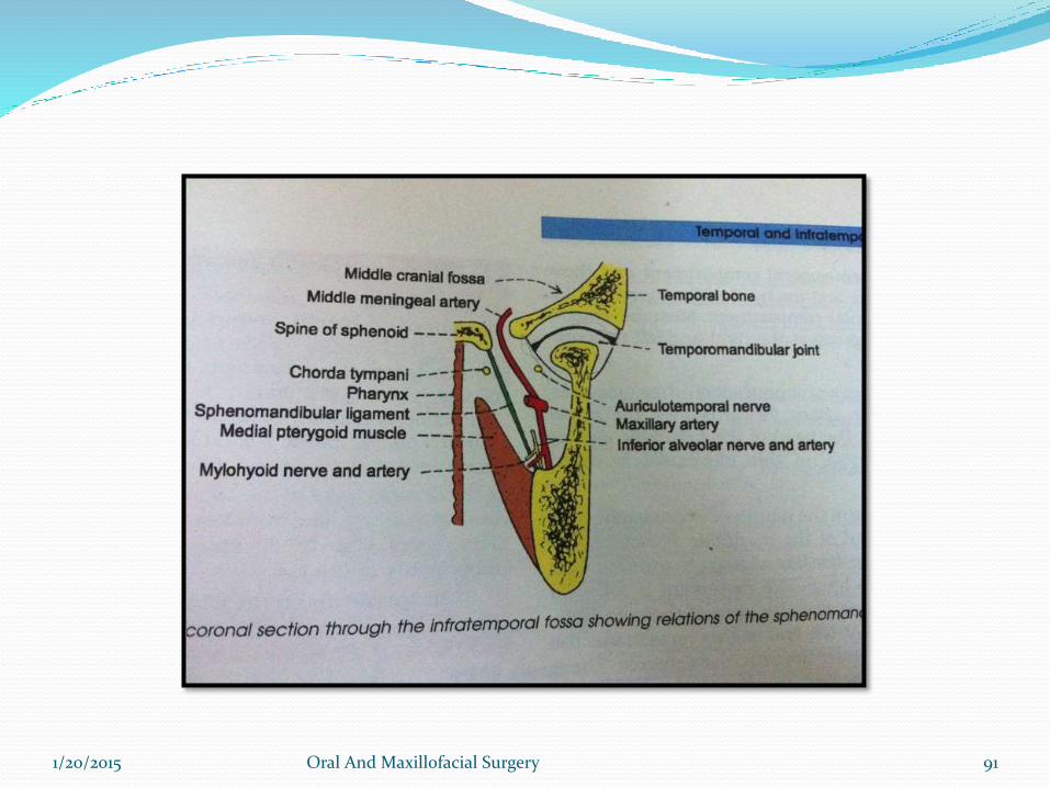
1.Auriculotemporal nerve-Arises from 2 roots which run backwards and encircle the
middle meningeal artery and form single trunk
The trunk passes posterior to lateral pterygoid between neck ofmandible and sphenomandibular ligament superior to 1st partof maxillary art.
Lies behind the TMJ close to the parotid
Ascends behind superficial temporal vessels and then intemporal region divides into superficial temporal branches.
1/20/2015 Oral And Maxillofacial Surgery 80
1/20/2015 Oral And Maxillofacial Surgery 81
Branches Of Auriculotemporal Nerve
Auricular branches- supply tragus, upper part of aurical,roof of external auditory meatus, anterosuperior part of tympanic membrane
Superficial temporal branches-supply skin of temple
It also supply sensory and secretomotor to parotid.
Articular branches-supply the TMJ.
1/20/2015 Oral And Maxillofacial Surgery 82
2. Inferior alveolar nerve:• Is mixed nerve
• Runs vertically downwards medial to lateral ptrygoid and
lateroposterior to lingual nerve. Then moves between the
sphenomandibular ligament and medial surface of mandibular
ramus
• Enters mandible through mandibular foramen to run in a bony
canal below the teeth
1/20/2015 Oral And Maxillofacial Surgery 83
Branches:1.Mylohyoid: Arises just before the nerve enters mandibular foramen.It pierces the
sphenomandibular ligament along with mylohyoid muscle and runs in the mylohyoid
goove. Supplies to mylohyoid muscle and anterior belly of digastric. It is also sensory to
skin on inferior and anterior surface surfaces of mental protuberence. It may provide
sensory innervation to mandibular incisors. There is also evidence that mylohyoid supply
to mesial root of mandibular frist molar.
2.Branches to lower teeth and gums.
3.Mental nerve : It exits canal and divides into three branches innervating skin of chin and
skin and mucous membrane of the lower lip.
4.Incisive nerve : It remains within the canal and form plexus that innervates pulpal tissue of
first premolar canine and incisors through dental branches.
1/20/2015 Oral And Maxillofacial Surgery 84
1/20/2015 Oral And Maxillofacial Surgery 85

1/20/2015 Oral And Maxillofacial Surgery 86
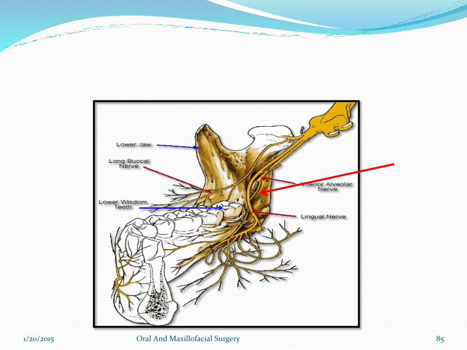
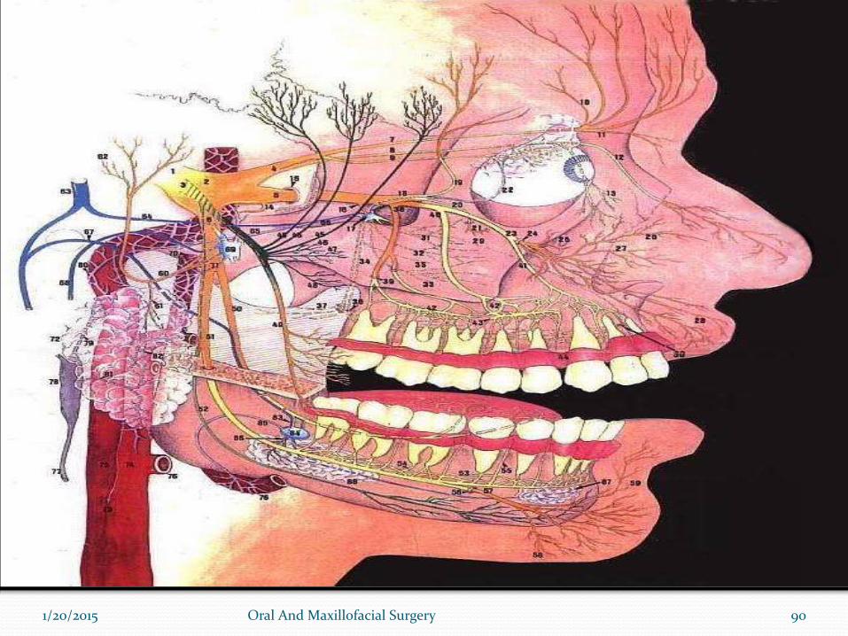
3.Lingual nerve: lies anterior to inferior alveolar n between lateral
pterygoid and tensor palatini
receives chorda tympani (SVA)
Emerges from inferior border of lateral pterygoid to lie between ramus and medial pterygoid in peterygomandibular space
moves downwards and forwards deep to pterygomandibularraphe between origins of supirior constrictor and mylohyoid
Reach to side of base of tongue 1 cm below and behind 3rd
molar just below mucous membrane of lateral lingual sulcus
1/20/2015 Oral And Maxillofacial Surgery 87
1/20/2015 Oral And Maxillofacial Surgery 88
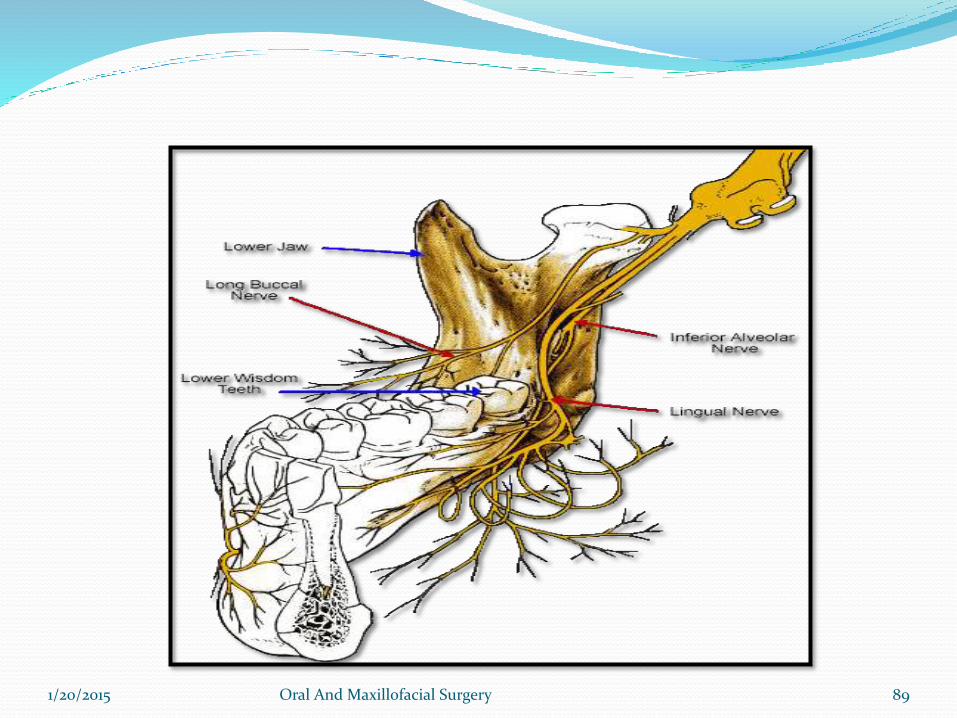
-Then proceeds anteriorly across the muscles of tongue ,looping
medially and downwards to submandibular duct to deep surface of
submandibular gland where it break in terminal branches
-Sensory to anterior 2/3 of tonge along with special sensation also
sensory to floor of mouth and gingiva on lingual side of mandible.
1/20/2015 Oral And Maxillofacial Surgery 89
1/20/2015 Oral And Maxillofacial Surgery 90
1/20/2015 Oral And Maxillofacial Surgery 91
Branches of lingual nerve and its communications:
1.Chorda tympani
2.Communications with submandibular ganglion
3.Hypoglossal nerve
1/20/2015 Oral And Maxillofacial Surgery 92
Ganglia Associated With The Trigeminal Nerve
1.Cilliary Ganglion: connected with nasocilliary nerve by ganglionic branchesin orbit,non synapsingsensory for orbit
2.Pterygopalatine Ganglion: connected to maxillary nerve in infratemporalfossasensory to orbital septum, orbicularis and nasal cavity, max sinus, palate,nasopharynx.
3. Otic Ganglion: betwn trunk of mandibular n and tensor palatini, nerve tomed pterygoid passes thru but does not synapse in the ganglion.
4.Submandibular Ganglion: related to lingual n, rests on hypoglossussupplies post gang. Parasym secretomotor fibres to submandibular andsublingual gland.
1/20/2015 Oral And Maxillofacial Surgery 93
CUTANEOUS DISTRIBUTION OF TRIGEMINAL NERVE
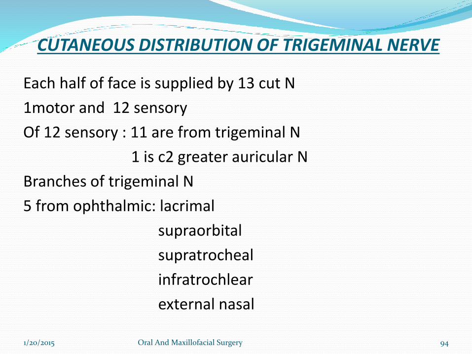
Each half of face is supplied by 13 cut N
1motor and 12 sensory
Of 12 sensory : 11 are from trigeminal N
1 is c2 greater auricular N
Branches of trigeminal N
5 from ophthalmic: lacrimal
supraorbital
supratrocheal
infratrochlear
external nasal
1/20/2015 Oral And Maxillofacial Surgery 94
1/20/2015 Oral And Maxillofacial Surgery 95
3 from maxillary N: infra orbital N
zygomaticofacial N
zygomaticotemporal N
3 from mandibular N: buccal N
auriculotemporal N
mental N
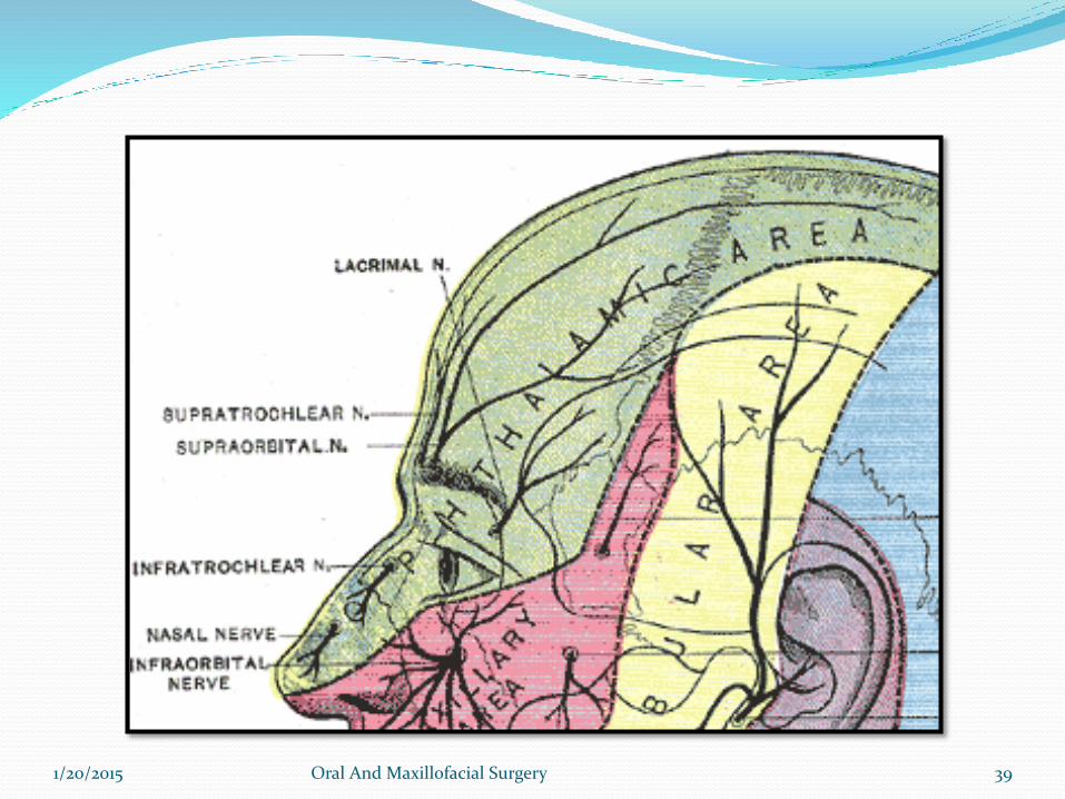
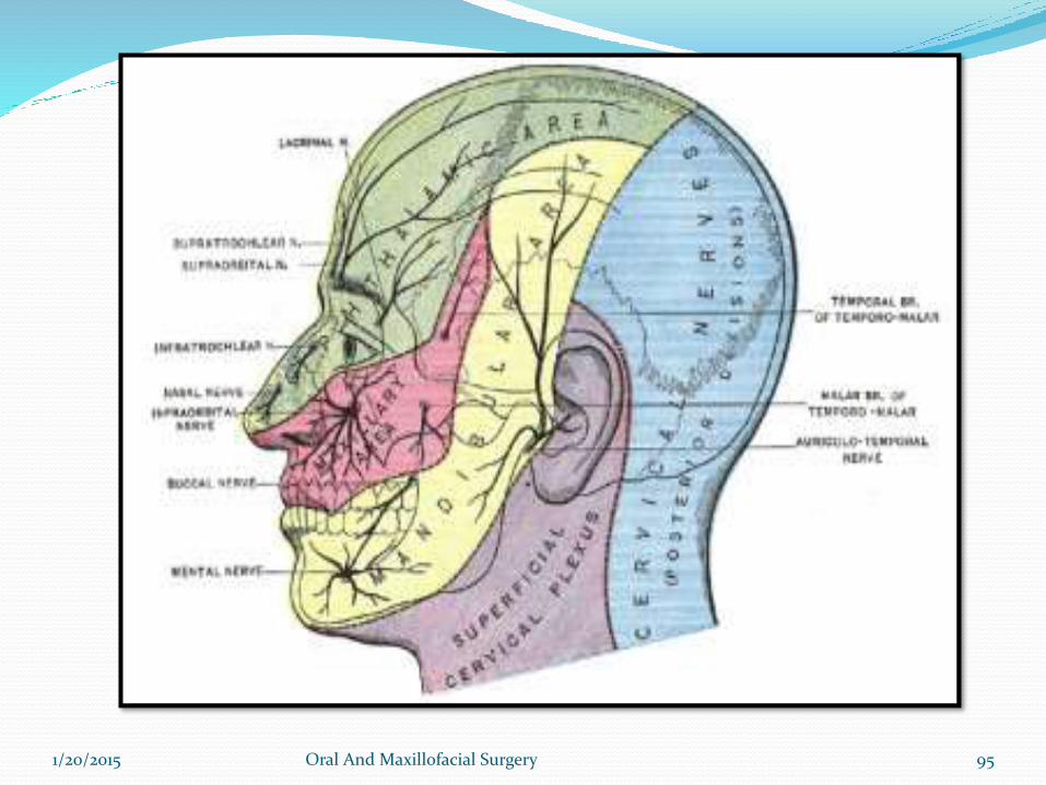
DIVISIONAL SUPPLY:
From lat canthus to vertex- ophthalmic N
From angle of mouth to vertex- mandibular N
Between the two areas-maxillary N
1/20/2015 Oral And Maxillofacial Surgery 96
Examination of trigeminal nerve
1/20/2015 Oral And Maxillofacial Surgery 97
Examination of trigeminal nerve1- Sensation Function
2- Motor Function
3- Corneal reflex
4- Test jaw jerk
1/20/2015 Oral And Maxillofacial Surgery 98
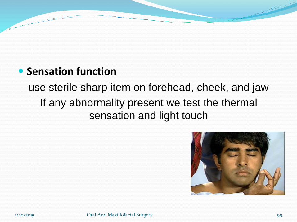
Sensation function
use sterile sharp item on forehead, cheek, and jaw
If any abnormality present we test the thermal
sensation and light touch
1/20/2015 Oral And Maxillofacial Surgery 99
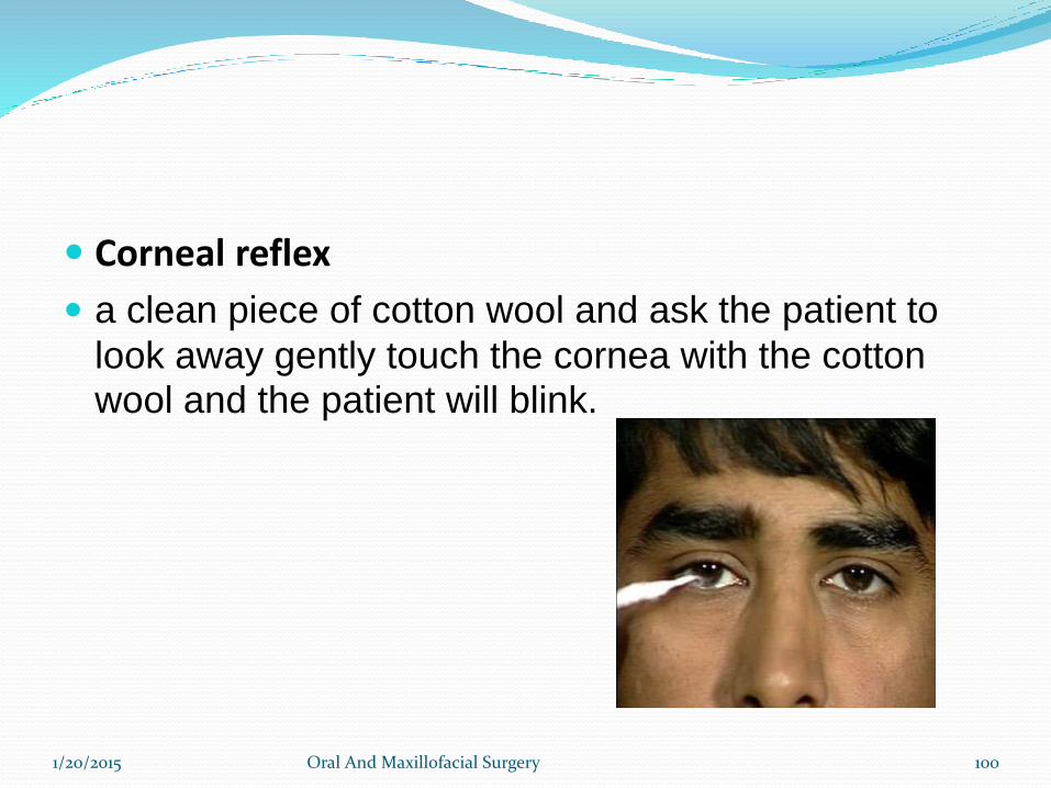
Corneal reflex
a clean piece of cotton wool and ask the patient to
look away gently touch the cornea with the cotton wool and the patient will blink.
1/20/2015 Oral And Maxillofacial Surgery 100
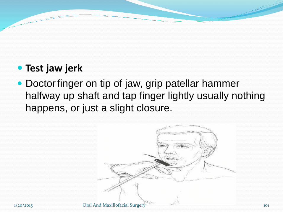
Test jaw jerk
Doctor finger on tip of jaw, grip patellar hammer
halfway up shaft and tap finger lightly usually nothing
happens, or just a slight closure.
1/20/2015 Oral And Maxillofacial Surgery 101
APPLIED ANATOMY
1/20/2015 Oral And Maxillofacial Surgery 102
1. Trigeminal Neuralgia – Tic Douloureux
• Sudden, usually unilateral severe, brief, stabbing
lancinating, recurring pain in the distribution of one or
more branches of the 5th Nerve
1/20/2015 Oral And Maxillofacial Surgery 103
2. TRIGEMINAL NEUROPATHY
• sensory loss of face or weakness of the jaw muscles
• causes- sjogren syndrome
• herpes zoster, leprosy
• meningioma,schwanomma
1/20/2015 Oral And Maxillofacial Surgery 104
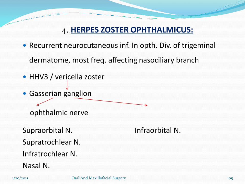
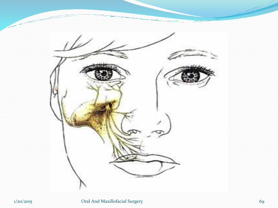
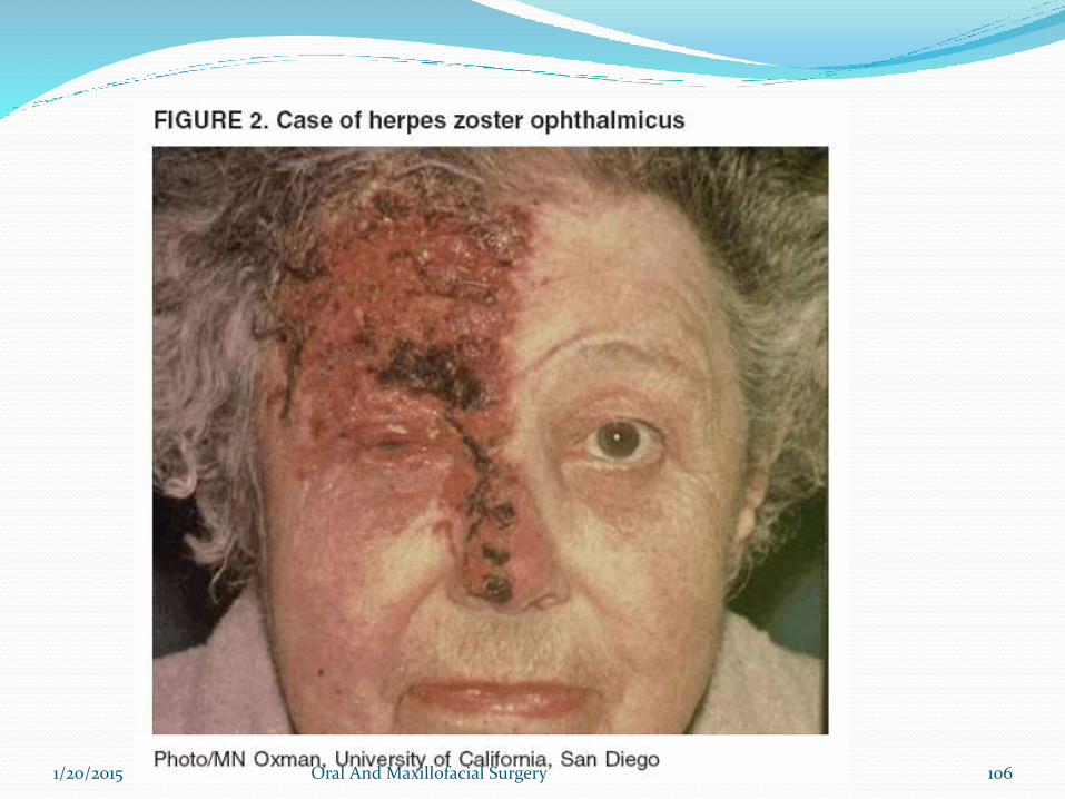
4. HERPES ZOSTER OPHTHALMICUS:
Recurrent neurocutaneous inf. In opth. Div. of trigeminal
dermatome, most freq. affecting nasociliary branch
HHV3 / vericella zoster
Gasserian ganglion
ophthalmic nerve
Supraorbital N. Infraorbital N.
Supratrochlear N.
Infratrochlear N.
Nasal N.
1/20/2015 Oral And Maxillofacial Surgery 105
1/20/2015 Oral And Maxillofacial Surgery 106
5. Cavernous sinus syndrome
• Cavernous sinus syndrome• Multiple cranial neuropathies• Exophthalmos, ocular motor defects, sensory loss in V1
and / or V2.• Pupils may be spared or involved.
causes: bacterial thrombophlebitisactinomycosisrhinocerebellar mucormycosisaspergillosistolosa hunt syndromeneoplasmsvascular lesions
1/20/2015 Oral And Maxillofacial Surgery 107
6.Gradenigos syndrome
Petrous bone osteitis due to otitis media
Characterized by I/L trigeminal N palsy (Va, Vb)
retro orbital pain
I/L sixth N palsy.
1/20/2015 Oral And Maxillofacial Surgery 108
Conclusion Since Trigeminal nerve is mixed nerve, suplies mainly
head and neck region. Hence as a Oral and Maxillofacial surgeon one should know throughlyabout itracranial and extracranial course and distribution of Trigeminal nerve,to diagnose the pathologies associated with Trigeminal nerveand for appropriate treatment.
1/20/2015 Oral And Maxillofacial Surgery 109
Refrences:
Greys anatomy
Snells anatomy
Head and Neck Anatomy-BD Chourasia
Textbook of Local Anesthesia-Stenly F Malamed
1/20/2015 Oral And Maxillofacial Surgery 110
THANK YOU
1/20/2015 Oral And Maxillofacial Surgery 111