Treatment of Syndesmotic Injuries - COA · Treatment of Syndesmotic Injuries Kenneth J. Hunt, M.D....
80
Treatment of Syndesmotic Injuries Kenneth J. Hunt, M.D. Stanford University Department of Orthopaedic Surgery
Transcript of Treatment of Syndesmotic Injuries - COA · Treatment of Syndesmotic Injuries Kenneth J. Hunt, M.D....
Treatment of Syndesmotic Injuries
Kenneth J. Hunt, M.D. Stanford University
Department of Orthopaedic Surgery
Syndesmosis Injuries
My disclosures are up to date on AAOS web site
No disclosures pertinent to this topic
Introduction Syndesmosis Injury (High Ankle Sprain)
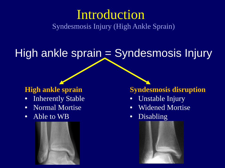
High ankle sprain = Syndesmosis Injury
Introduction Syndesmosis Injury (High Ankle Sprain)
High ankle sprain • Inherently Stable • Normal Mortise • Able to WB
Syndesmosis disruption • Unstable Injury • Widened Mortise • Disabling
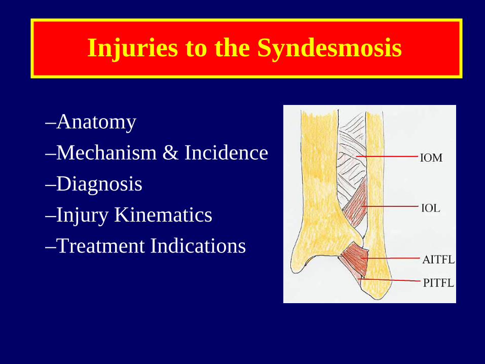
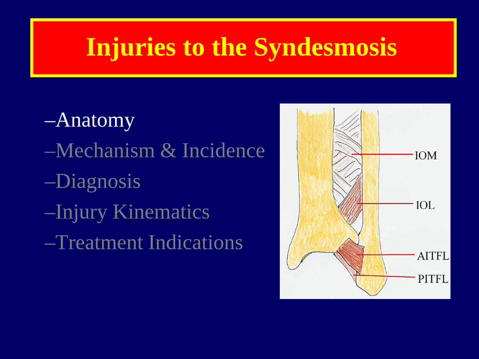
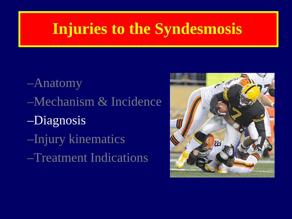
Injuries to the Syndesmosis
–Anatomy –Mechanism & Incidence –Diagnosis –Injury Kinematics –Treatment Indications
Injuries to the Syndesmosis
–Anatomy –Mechanism & Incidence –Diagnosis –Injury Kinematics –Treatment Indications
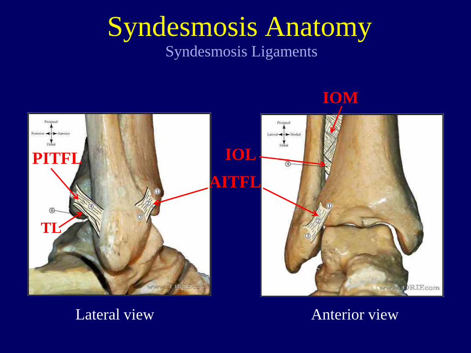
Syndesmosis Anatomy
AITFL PITFL
IOM
IOL
Syndesmosis Ligaments
Lateral view Anterior view
TL
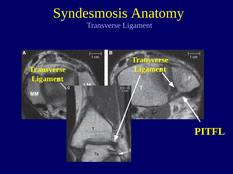
Syndesmosis Anatomy Transverse Ligament
Transverse Ligament
PITFL PITFL
Transverse Ligament
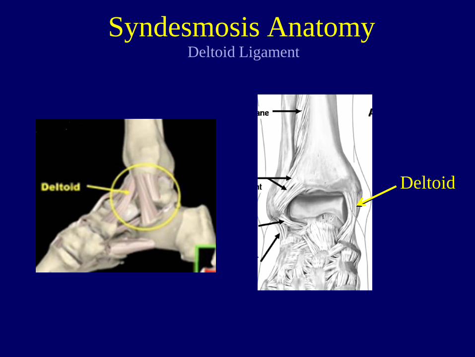
Syndesmosis Anatomy Deltoid Ligament
Deltoid
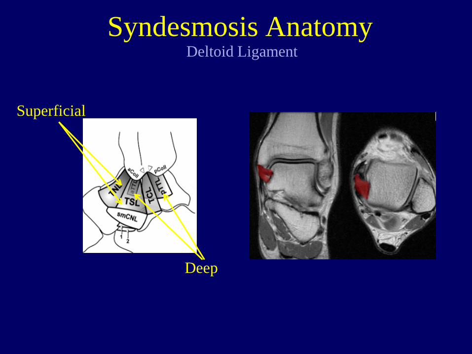
Syndesmosis Anatomy Deltoid Ligament
Superficial
Deep
Injuries to the Syndesmosis
–Anatomy –Mechanism & Incidence –Diagnosis –Injury kinematics –Treatment Indications
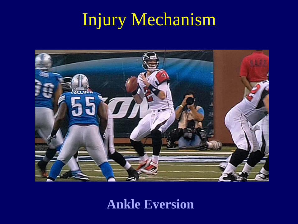
Injury Mechanism
Ankle Eversion
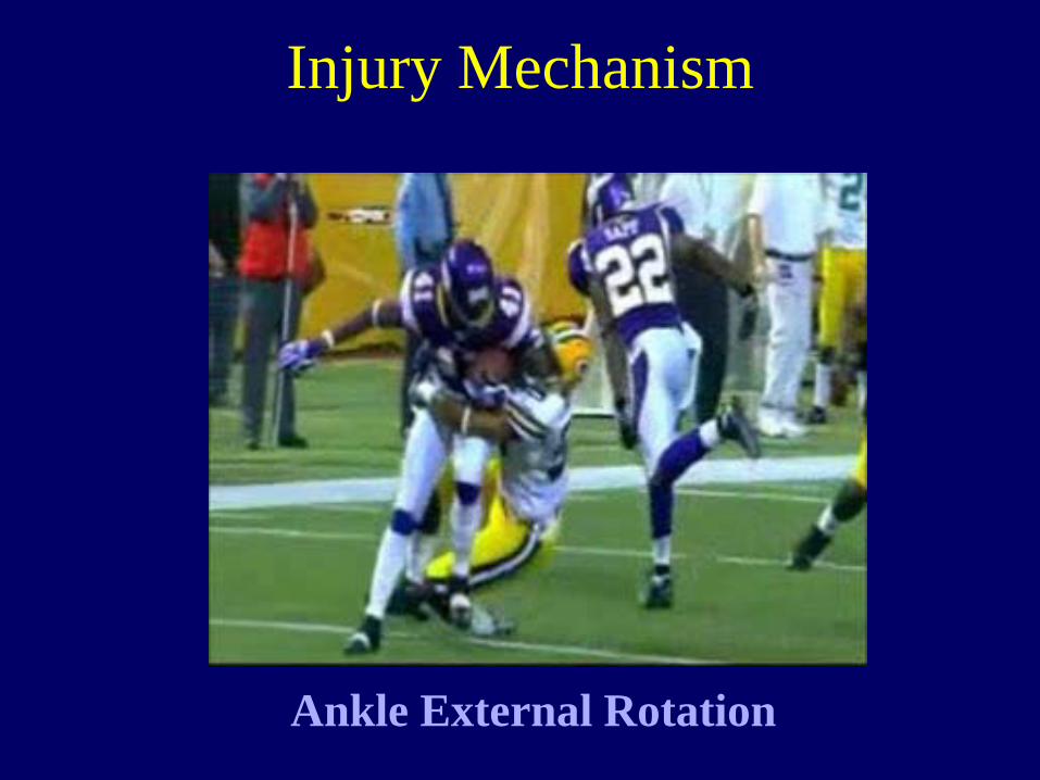
Injury Mechanism
Ankle External Rotation
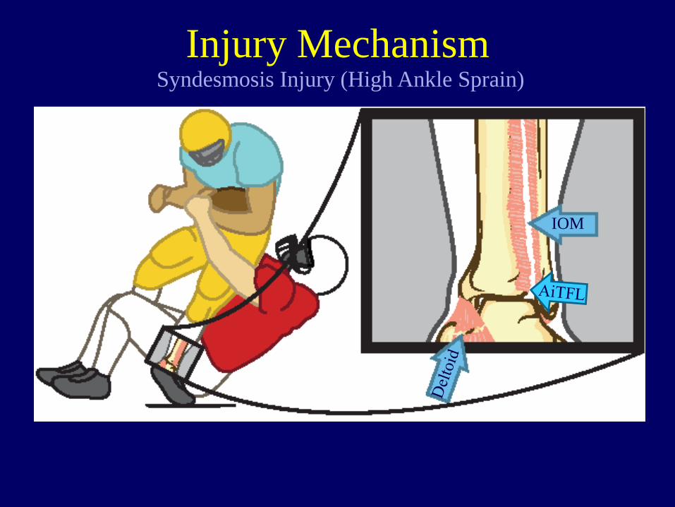
Syndesmosis Injury (High Ankle Sprain)
IOM
Injury Mechanism
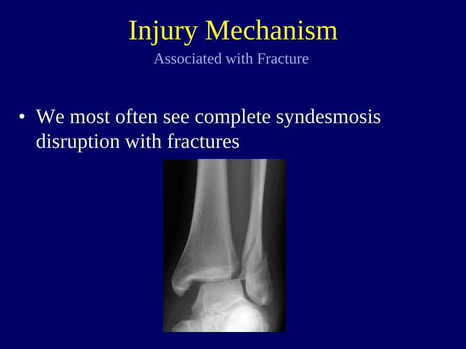
• We most often see complete syndesmosis disruption with fractures
Associated with Fracture Injury Mechanism

http://www.youtube.com/
Pronation External Rotation Injury Mechanism
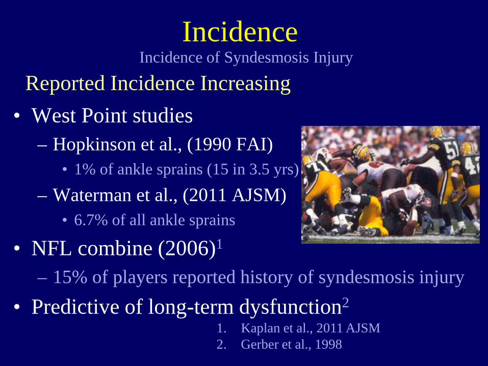
• West Point studies – Hopkinson et al., (1990 FAI)
• 1% of ankle sprains (15 in 3.5 yrs) – Waterman et al., (2011 AJSM)
• 6.7% of all ankle sprains
• NFL combine (2006)1
– 15% of players reported history of syndesmosis injury • Predictive of long-term dysfunction2
Reported Incidence Increasing
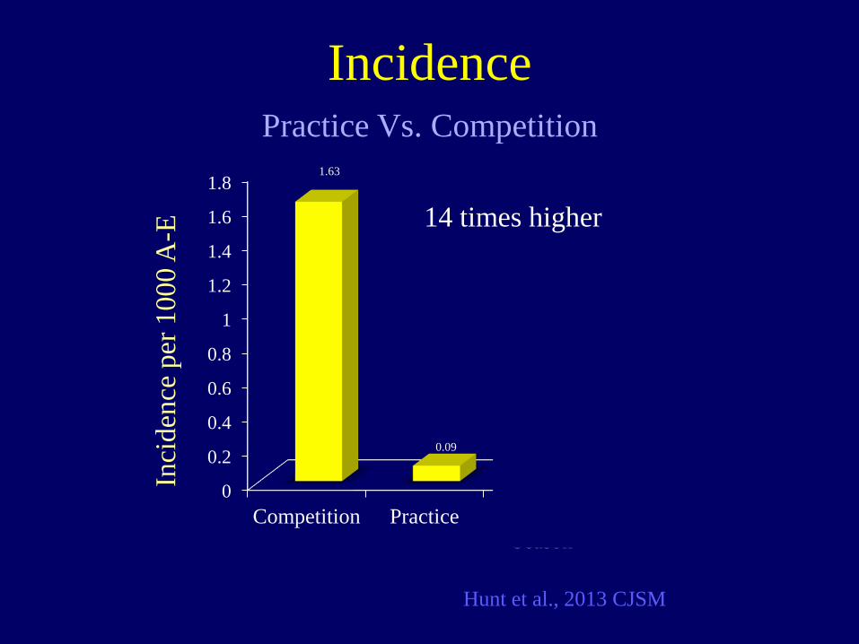
Incidence Incidence of Syndesmosis Injury
1. Kaplan et al., 2011 AJSM 2. Gerber et al., 1998
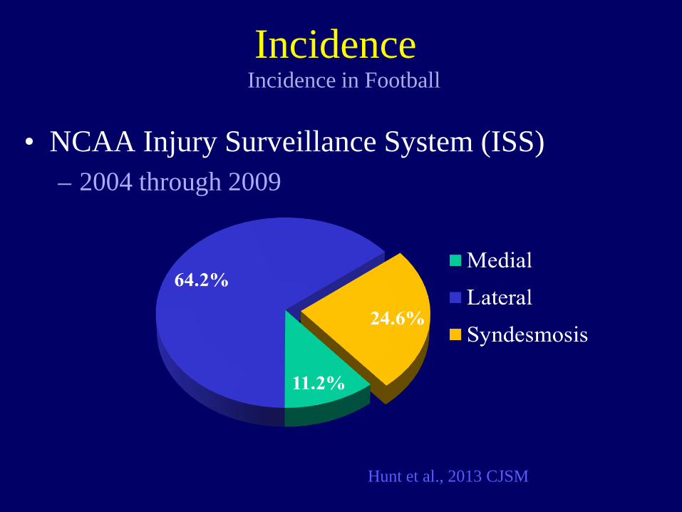
• NCAA Injury Surveillance System (ISS) – 2004 through 2009
Hunt et al., 2013 CJSM
Incidence Incidence in Football
0
0.2
0.4
0.6
0.8
1
1.2
1.4
1.6
1.8
Competition Practice Regular Season
Pre-season Post-Season
1.63
0.09
0.28 0.18 0.15
Inci
denc
e pe
r 100
0 A
-E
Incidence Practice Vs. Competition
Hunt et al., 2013 CJSM
14 times higher
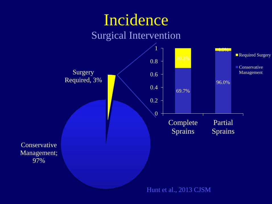
Surgery Required, 3%
Conservative Management;
97%
69.7% 96.0%
30.3% 4.0%
0
0.2
0.4
0.6
0.8
1
Complete Sprains
Partial Sprains
Required Surgery
Conservative Management
Incidence Surgical Intervention
Hunt et al., 2013 CJSM
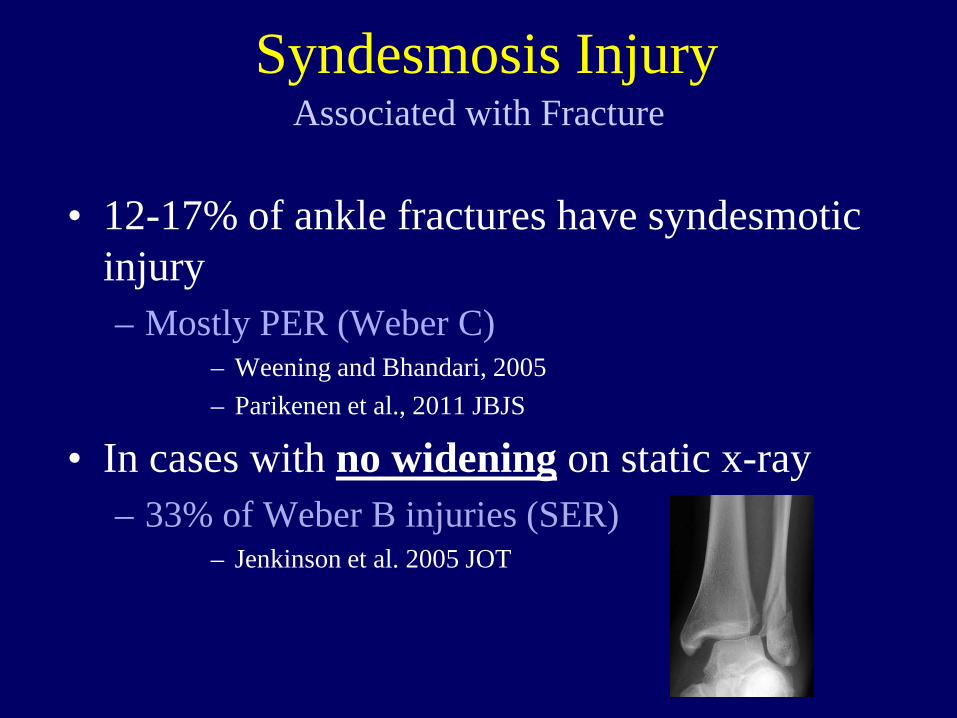
• 12-17% of ankle fractures have syndesmotic injury – Mostly PER (Weber C)
– Weening and Bhandari, 2005 – Parikenen et al., 2011 JBJS
• In cases with no widening on static x-ray – 33% of Weber B injuries (SER)
– Jenkinson et al. 2005 JOT
Associated with Fracture Syndesmosis Injury
Injuries to the Syndesmosis
–Anatomy –Mechanism & Incidence –Diagnosis –Injury kinematics –Treatment Indications
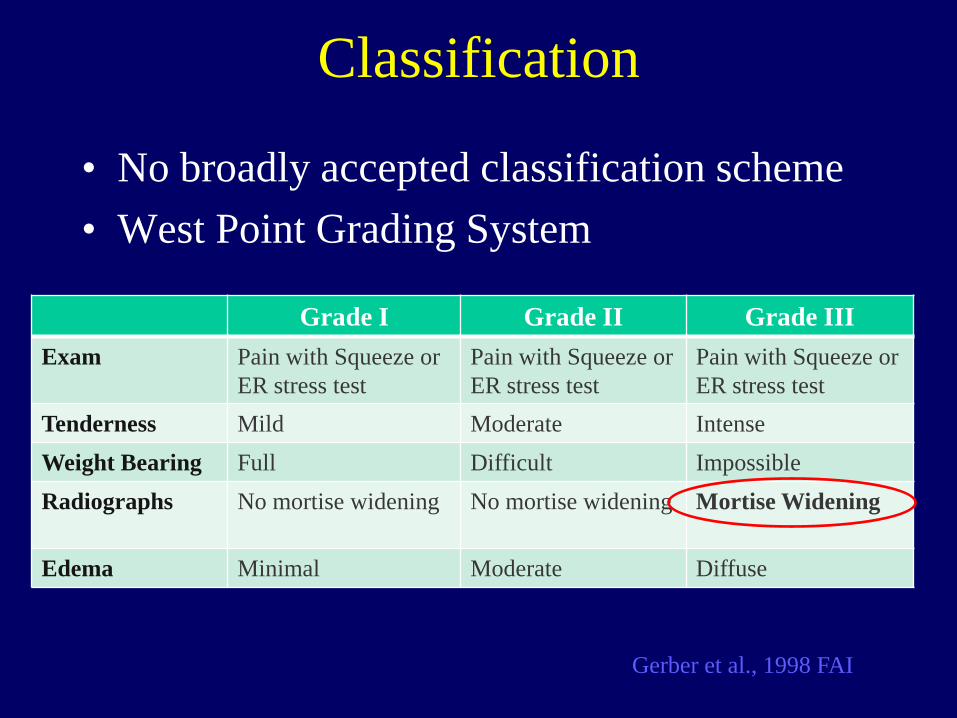
Classification
• No broadly accepted classification scheme • West Point Grading System
Grade I Grade II Grade III Exam Pain with Squeeze or
ER stress test Pain with Squeeze or ER stress test
Pain with Squeeze or ER stress test
Tenderness Mild Moderate Intense Weight Bearing Full Difficult Impossible Radiographs No mortise widening No mortise widening Mortise Widening
Edema Minimal Moderate Diffuse
Gerber et al., 1998 FAI
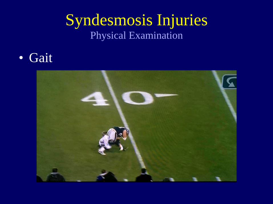
• Gait
Syndesmosis Injuries Physical Examination
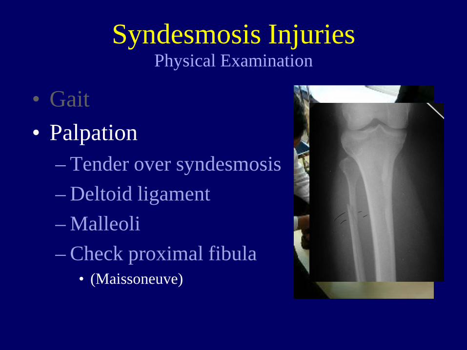
• Gait • Palpation
– Tender over syndesmosis – Deltoid ligament – Malleoli – Check proximal fibula
• (Maissoneuve)
Syndesmosis Injuries Physical Examination
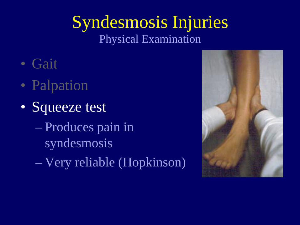
• Gait • Palpation • Squeeze test
– Produces pain in syndesmosis
– Very reliable (Hopkinson)
Syndesmosis Injuries Physical Examination
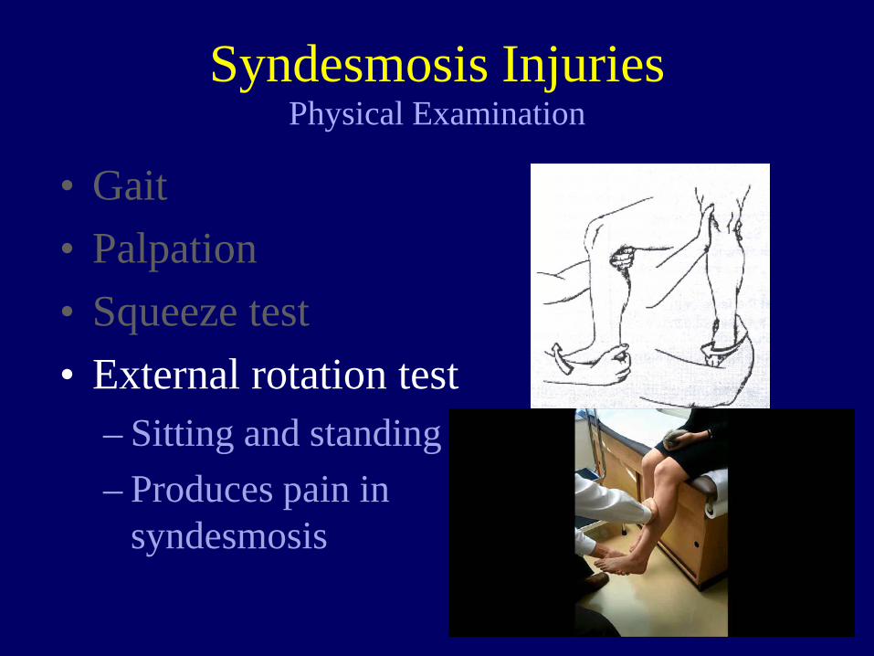
• Gait • Palpation • Squeeze test • External rotation test
– Sitting and standing – Produces pain in
syndesmosis
Syndesmosis Injuries Physical Examination
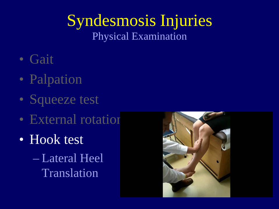
• Gait • Palpation • Squeeze test • External rotation test • Hook test
– Lateral Heel Translation
Syndesmosis Injuries Physical Examination
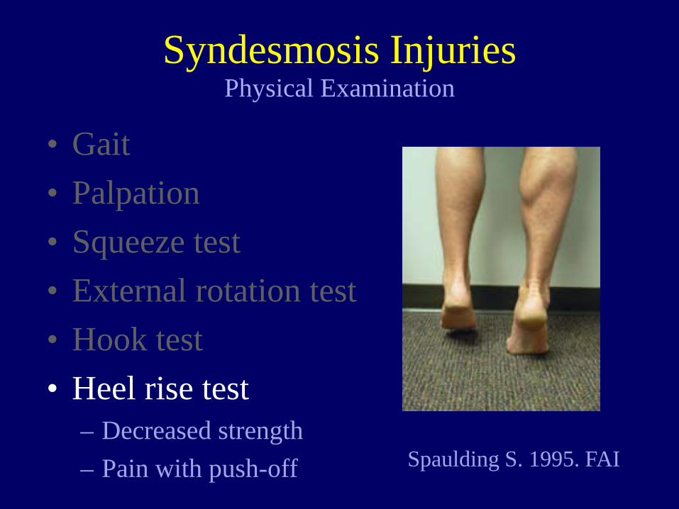
Spaulding S. 1995. FAI
• Gait • Palpation • Squeeze test • External rotation test • Hook test • Heel rise test
– Decreased strength – Pain with push-off
Syndesmosis Injuries Physical Examination
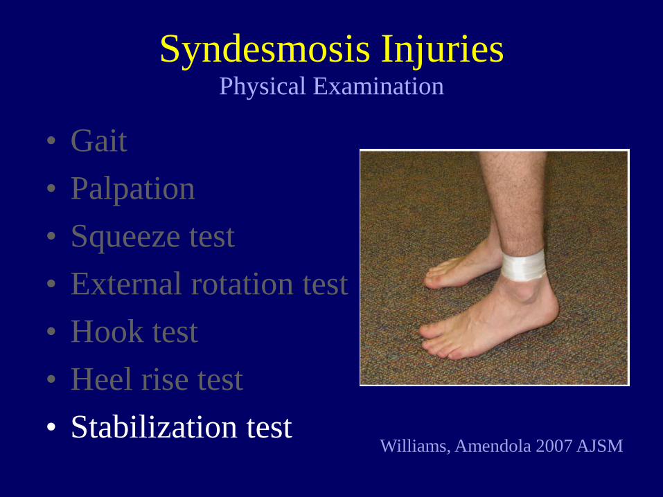
• Gait • Palpation • Squeeze test • External rotation test • Hook test • Heel rise test • Stabilization test
Syndesmosis Injuries Physical Examination
Williams, Amendola 2007 AJSM
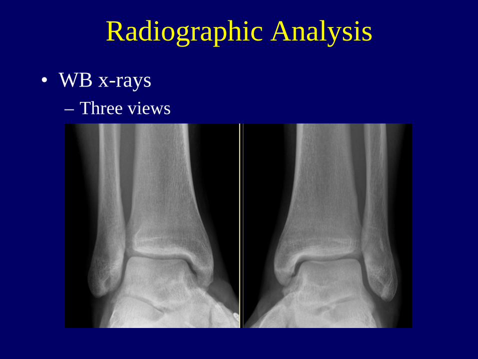
Radiographic Analysis • WB x-rays
– Three views
• Radiographs
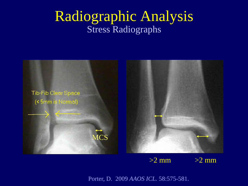
Stress Radiographs
Porter, D. 2009 AAOS ICL. 58:575-581.
>2 mm >2 mm
MCS
Radiographic Analysis
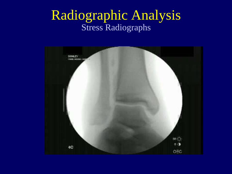
• Radiographs
Stress Radiographs Radiographic Analysis
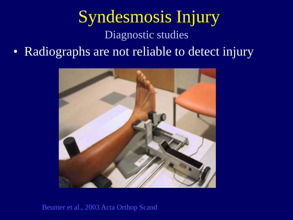
• Radiographs are not reliable to detect injury
Beumer et al., 2003 Acta Orthop Scand
Diagnostic studies Syndesmosis Injury
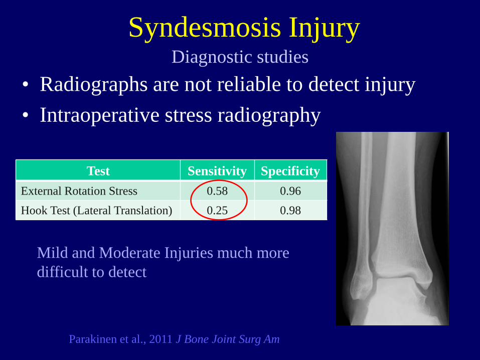
• Radiographs are not reliable to detect injury • Intraoperative stress radiography
Parakinen et al., 2011 J Bone Joint Surg Am
Test Sensitivity Specificity External Rotation Stress 0.58 0.96 Hook Test (Lateral Translation) 0.25 0.98
Mild and Moderate Injuries much more difficult to detect
Diagnostic studies Syndesmosis Injury
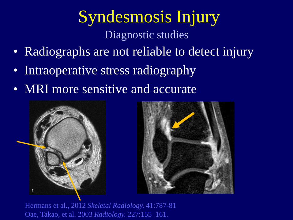
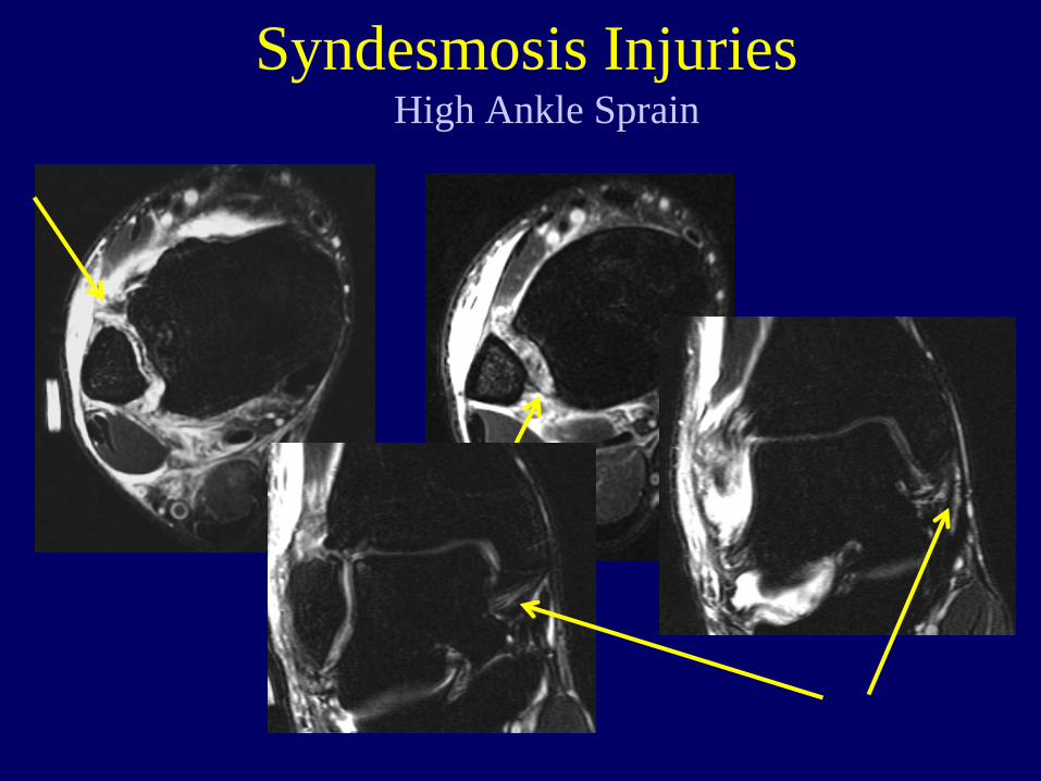
• Radiographs are not reliable to detect injury • Intraoperative stress radiography • MRI more sensitive and accurate
Hermans et al., 2012 Skeletal Radiology. 41:787-81 Oae, Takao, et al. 2003 Radiology. 227:155–161.
Diagnostic studies Syndesmosis Injury
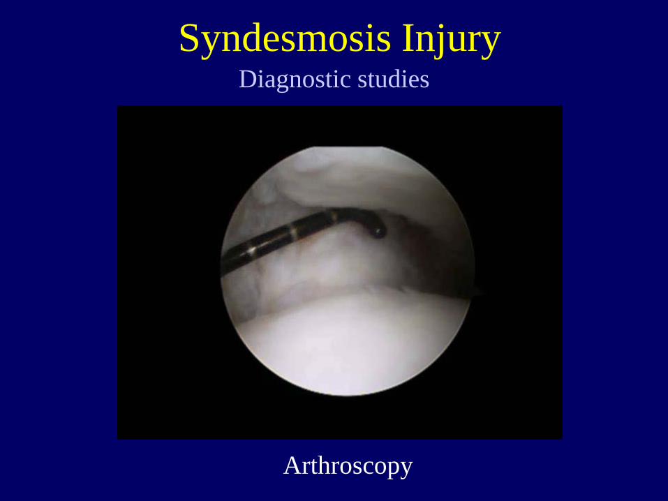
Diagnostic studies Syndesmosis Injury
Arthroscopy
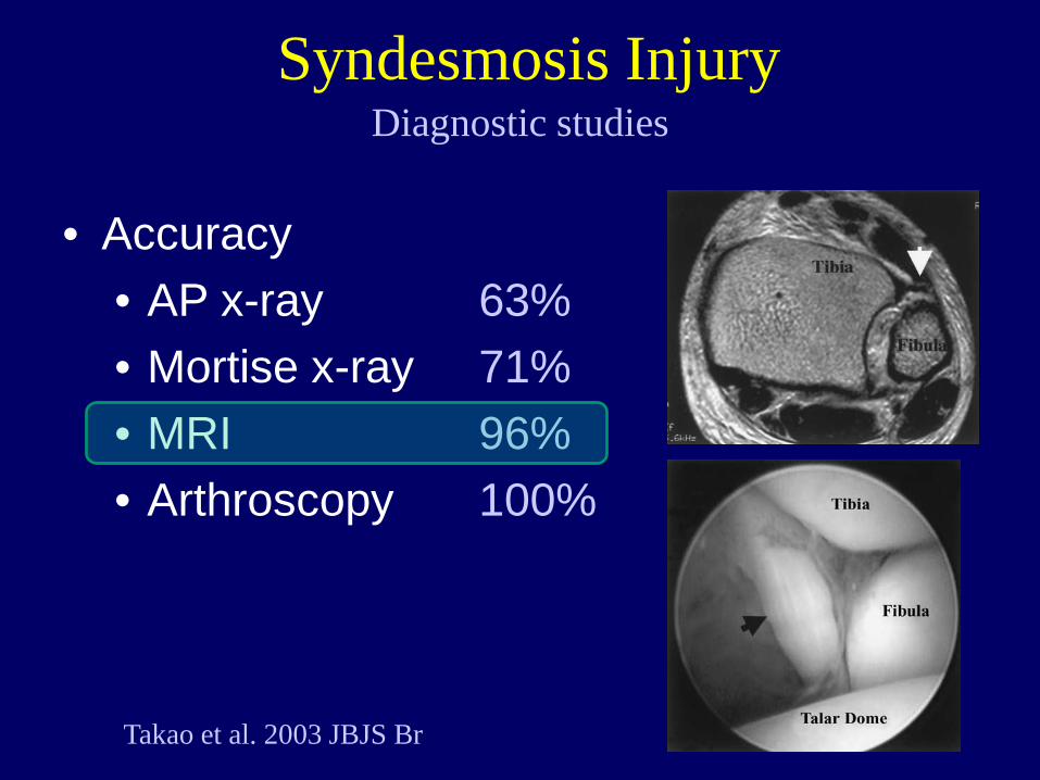
• Accuracy • AP x-ray 63% • Mortise x-ray 71% • MRI 96% • Arthroscopy 100%
Diagnostic studies
Takao et al. 2003 JBJS Br
Syndesmosis Injury
Injuries to the Syndesmosis
–Anatomy –Mechanism & Incidence –Diagnosis –Injury Kinematics –Treatment Indications
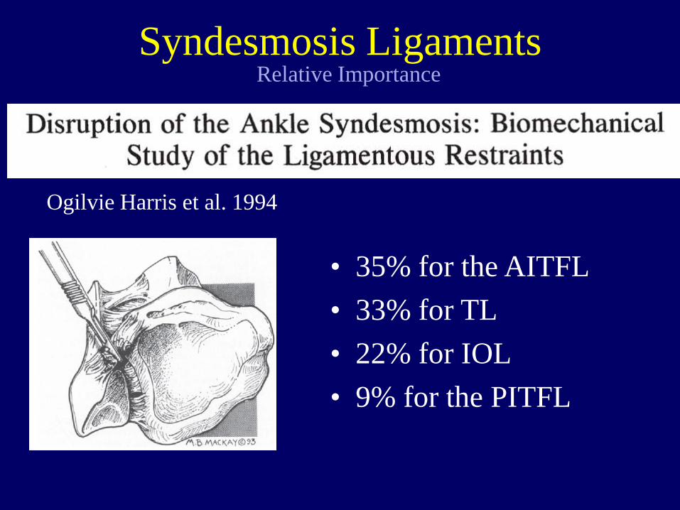
Syndesmosis Ligaments
Ogilvie Harris et al. 1994
• 35% for the AITFL • 33% for TL • 22% for IOL • 9% for the PITFL
Relative Importance
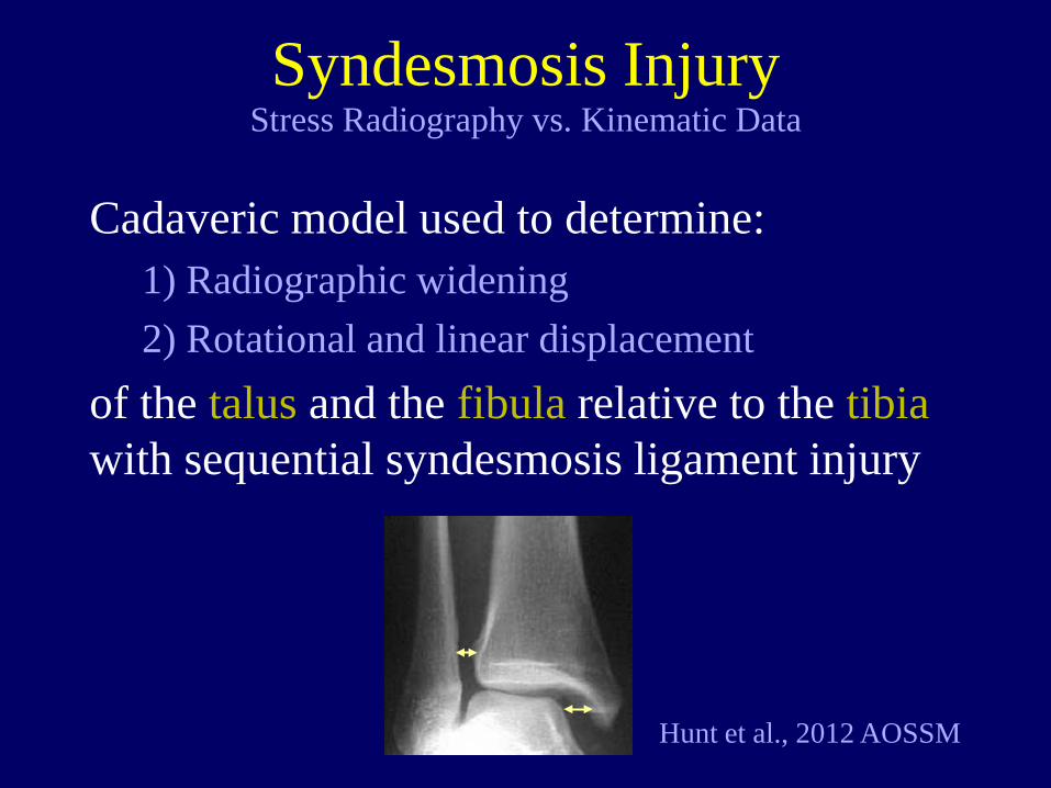
Syndesmosis Injury
Cadaveric model used to determine: 1) Radiographic widening 2) Rotational and linear displacement
of the talus and the fibula relative to the tibia with sequential syndesmosis ligament injury
Stress Radiography vs. Kinematic Data
Hunt et al., 2012 AOSSM
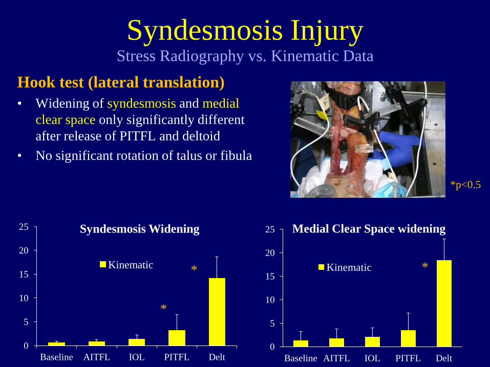
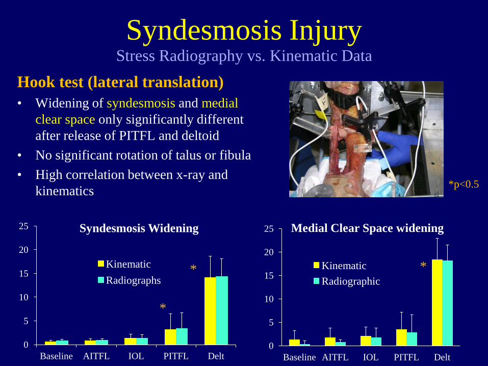
Hook test (lateral translation) • Widening of syndesmosis and medial
clear space only significantly different after release of PITFL and deltoid
• No significant rotation of talus or fibula
0
5
10
15
20
25
Baseline AITFL IOL PITFL Delt
Medial Clear Space widening
Kinematic
0
5
10
15
20
25
Baseline AITFL IOL PITFL Delt
Syndesmosis Widening
Kinematic * *
*
*p<0.5
Syndesmosis Injury Stress Radiography vs. Kinematic Data
Hook test (lateral translation) • Widening of syndesmosis and medial
clear space only significantly different after release of PITFL and deltoid
• No significant rotation of talus or fibula • High correlation between x-ray and
kinematics
0
5
10
15
20
25
Baseline AITFL IOL PITFL Delt
Medial Clear Space widening
Kinematic Radiographic
0
5
10
15
20
25
Baseline AITFL IOL PITFL Delt
Syndesmosis Widening
Kinematic Radiographs
* *
*
*p<0.5
Syndesmosis Injury Stress Radiography vs. Kinematic Data
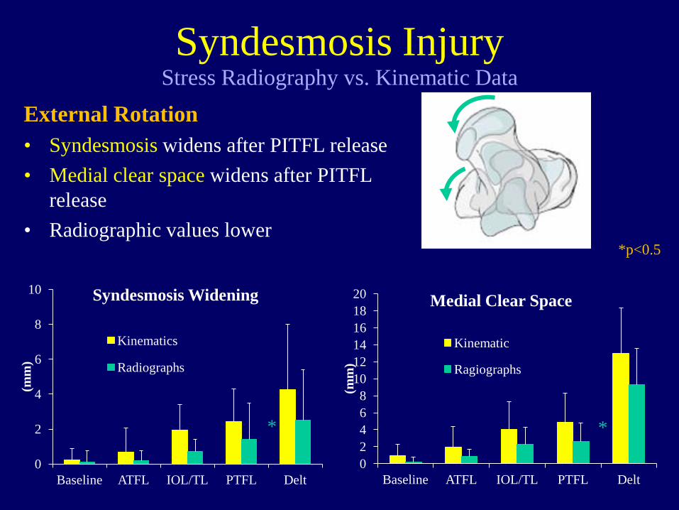
External Rotation • Syndesmosis widens after PITFL release • Medial clear space widens after PITFL
release • Radiographic values lower
0 2 4 6 8
10 12 14 16 18 20
Baseline ATFL IOL/TL PTFL Delt
(mm
)
Medial Clear Space
Kinematic
Ragiographs
0
2
4
6
8
10
Baseline ATFL IOL/TL PTFL Delt
(mm
)
Syndesmosis Widening
Kinematics
Radiographs
* *
*p<0.5
Syndesmosis Injury Stress Radiography vs. Kinematic Data
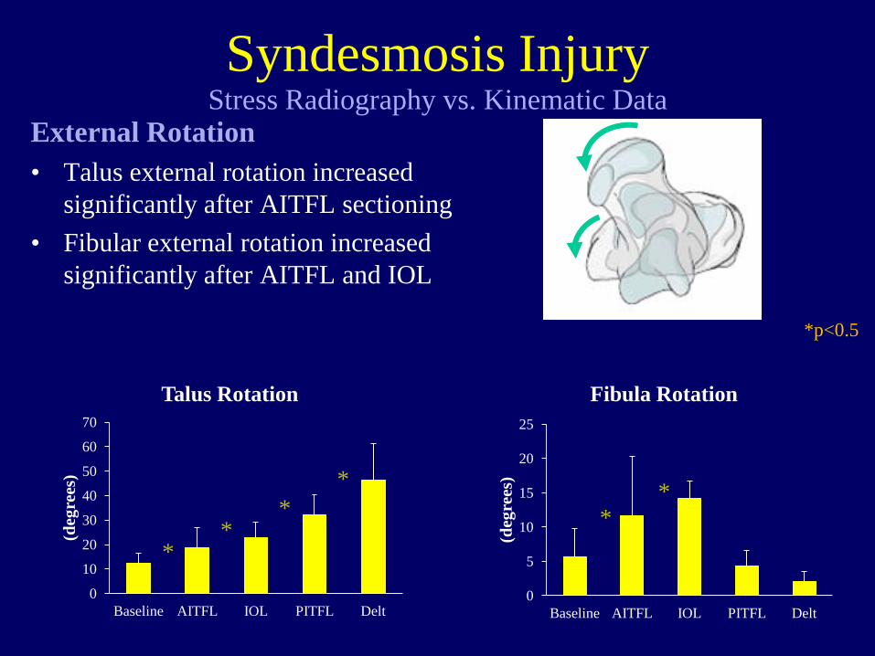
External Rotation • Talus external rotation increased
significantly after AITFL sectioning • Fibular external rotation increased
significantly after AITFL and IOL
0
10
20
30
40
50
60
70
Baseline AITFL IOL PITFL Delt
(deg
rees
)
Talus Rotation
0
5
10
15
20
25
Baseline AITFL IOL PITFL Delt
(deg
rees
)
Fibula Rotation
* *
* *
* *
*p<0.5
Syndesmosis Injury Stress Radiography vs. Kinematic Data
Point 1: Stress radiography not a reliable indicator of mild or moderate syndesmosis injuries. Particularly External Rotation Stress
Syndesmosis Injury Stress Radiography vs. Kinematic Data
Hunt et al., 2012 AOSSM
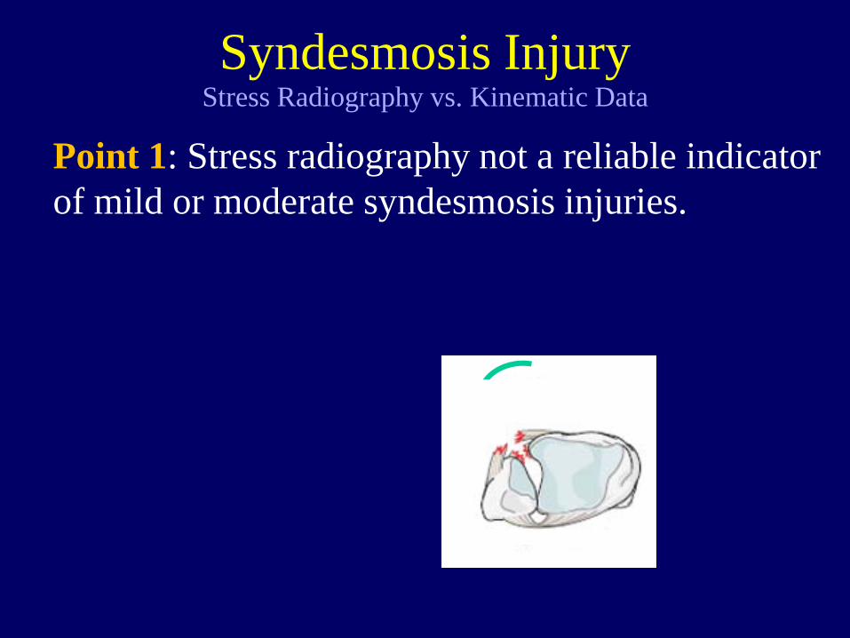
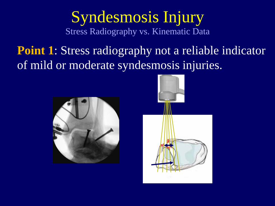
Point 1: Stress radiography not a reliable indicator of mild or moderate syndesmosis injuries.
Syndesmosis Injury Stress Radiography vs. Kinematic Data
Point 1: Stress radiography not a reliable indicator of mild or moderate syndesmosis injuries.
Syndesmosis Injury Stress Radiography vs. Kinematic Data
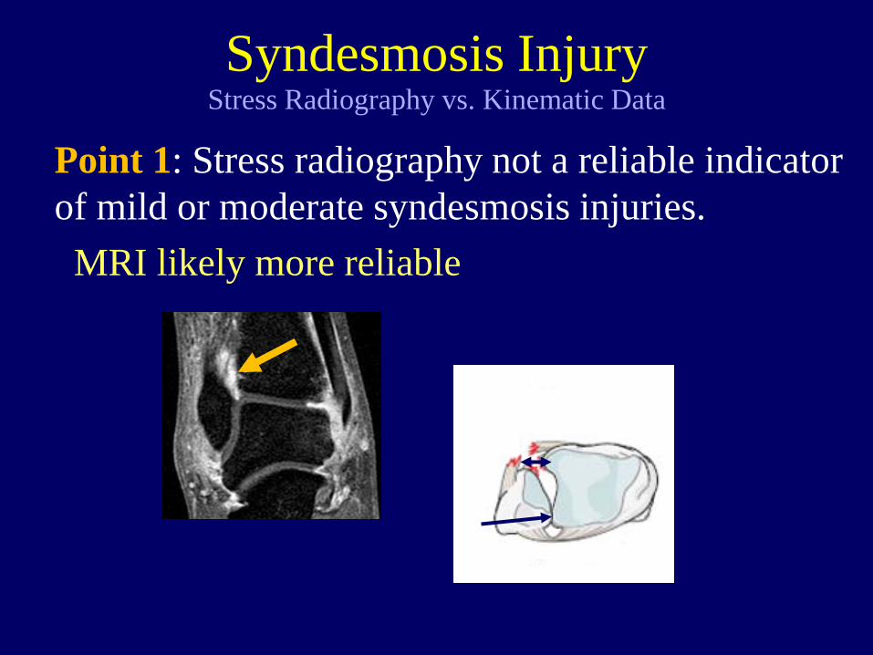
Point 1: Stress radiography not a reliable indicator of mild or moderate syndesmosis injuries. MRI likely more reliable
Syndesmosis Injury Stress Radiography vs. Kinematic Data
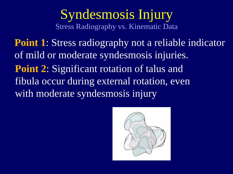
Point 1: Stress radiography not a reliable indicator of mild or moderate syndesmosis injuries. Point 2: Significant rotation of talus and fibula occur during external rotation, even with moderate syndesmosis injury
Syndesmosis Injury Stress Radiography vs. Kinematic Data
Injuries to the Syndesmosis
–Anatomy –Mechanism & Incidence –Diagnosis –Injury Kinematics –Treatment Indications
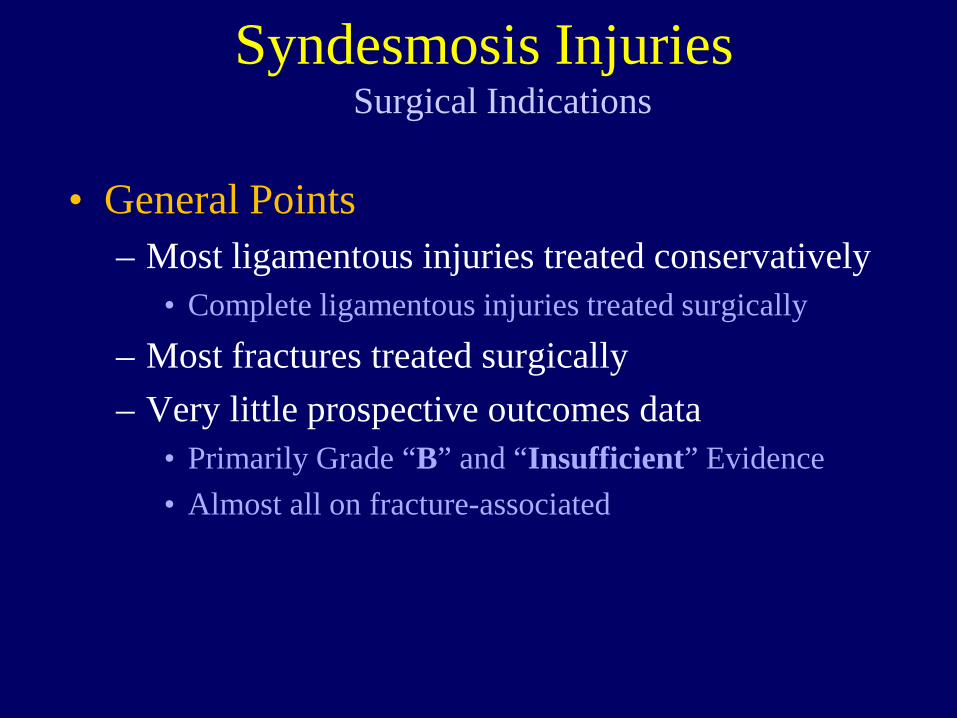
• General Points – Most ligamentous injuries treated conservatively
• Complete ligamentous injuries treated surgically – Most fractures treated surgically – Very little prospective outcomes data
• Primarily Grade “B” and “Insufficient” Evidence • Almost all on fracture-associated
Surgical Indications Syndesmosis Injuries
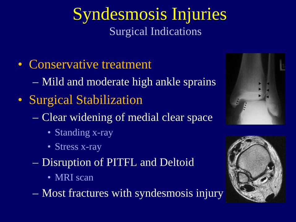
• Conservative treatment – Mild and moderate high ankle sprains
• Surgical Stabilization – Clear widening of medial clear space
• Standing x-ray • Stress x-ray
– Disruption of PITFL and Deltoid • MRI scan
– Most fractures with syndesmosis injury
Surgical Indications Syndesmosis Injuries
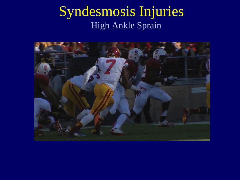
High Ankle Sprain Syndesmosis Injuries
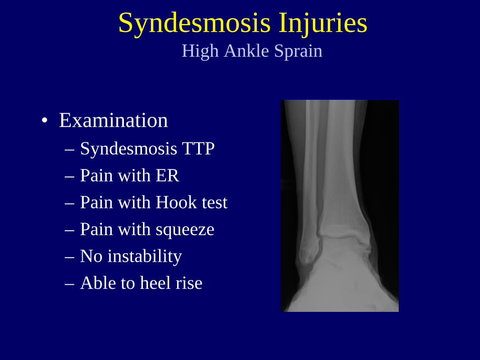
• Examination – Syndesmosis TTP – Pain with ER – Pain with Hook test – Pain with squeeze – No instability – Able to heel rise
Syndesmosis Injuries High Ankle Sprain
Syndesmosis Injuries High Ankle Sprain
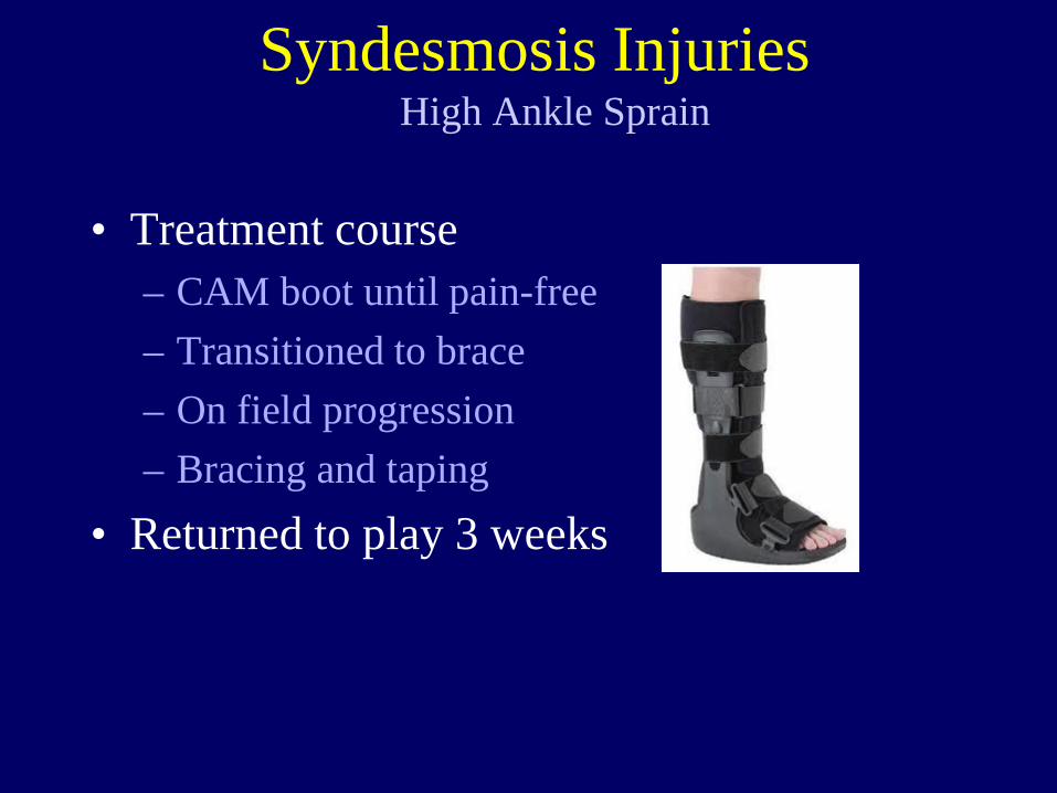
• Treatment course – CAM boot until pain-free – Transitioned to brace – On field progression – Bracing and taping
• Returned to play 3 weeks
Syndesmosis Injuries High Ankle Sprain
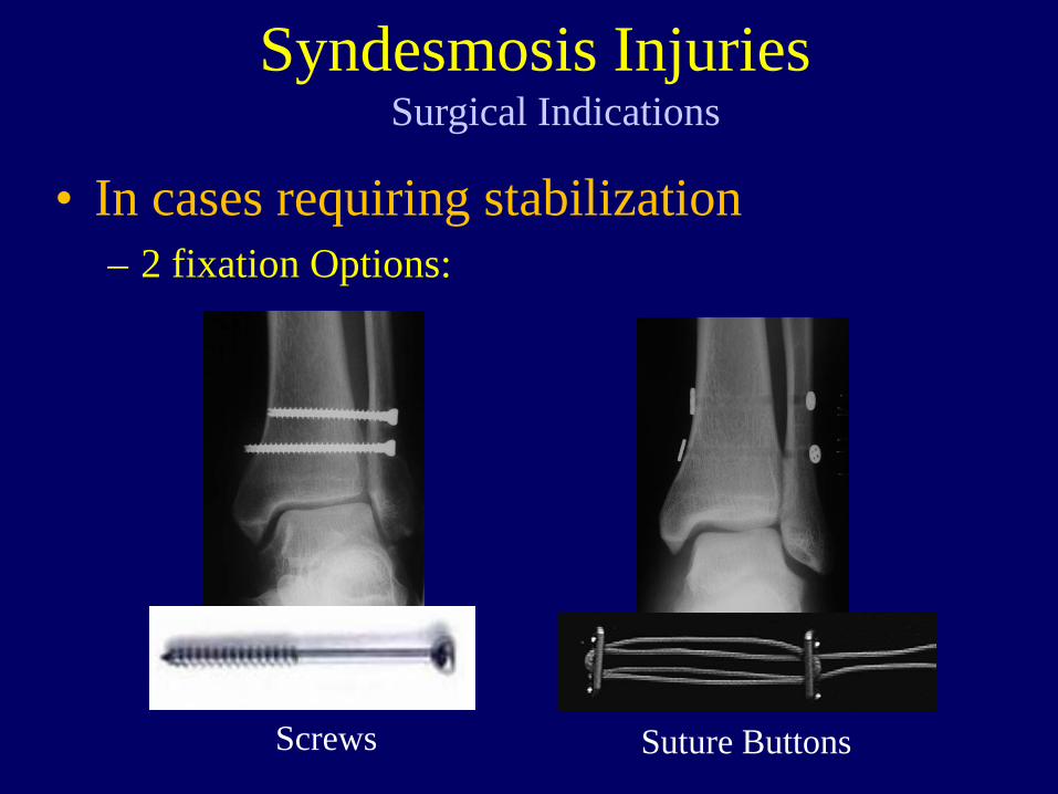
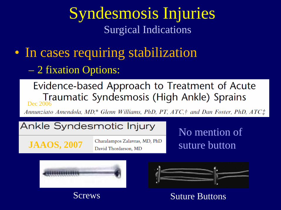
• In cases requiring stabilization – 2 fixation Options:
Surgical Indications Syndesmosis Injuries
Screws Suture Buttons
• In cases requiring stabilization – 2 fixation Options:
Surgical Indications Syndesmosis Injuries
Screws Suture Buttons
Dec 2006
JAAOS, 2007 No mention of suture button
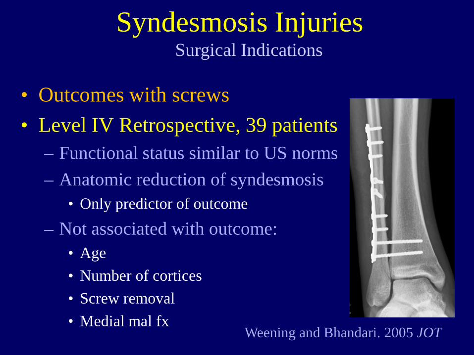
• Outcomes with screws • Level IV Retrospective, 39 patients
– Functional status similar to US norms – Anatomic reduction of syndesmosis
• Only predictor of outcome – Not associated with outcome:
• Age • Number of cortices • Screw removal • Medial mal fx
Surgical Indications Syndesmosis Injuries
Weening and Bhandari. 2005 JOT
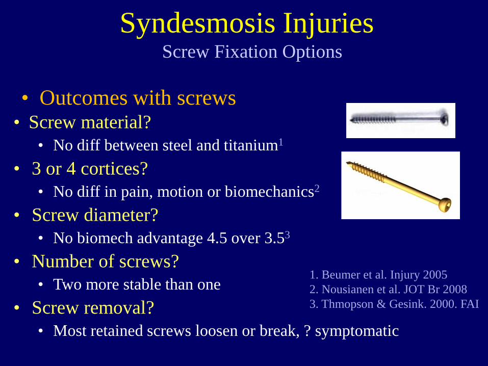
• Screw material? • No diff between steel and titanium1
• 3 or 4 cortices? • No diff in pain, motion or biomechanics2
• Screw diameter? • No biomech advantage 4.5 over 3.53
• Number of screws? • Two more stable than one
• Screw removal? • Most retained screws loosen or break, ? symptomatic
Screw Fixation Options Syndesmosis Injuries
1. Beumer et al. Injury 2005 2. Nousianen et al. JOT Br 2008 3. Thmopson & Gesink. 2000. FAI
• Outcomes with screws
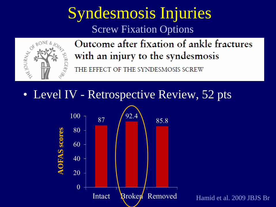
Hamid et al. 2009 JBJS Br
Screw Fixation Options Syndesmosis Injuries
AO
FAS
scor
es
• Level IV - Retrospective Review, 52 pts
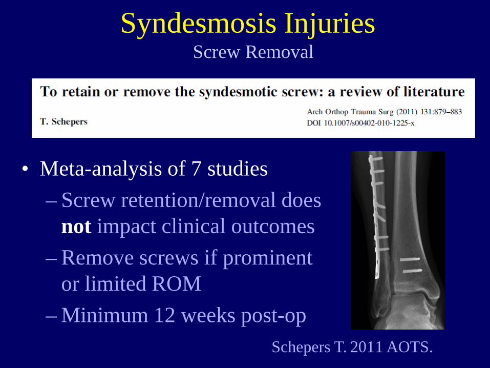
• Meta-analysis of 7 studies – Screw retention/removal does
not impact clinical outcomes – Remove screws if prominent
or limited ROM – Minimum 12 weeks post-op
Screw Removal Syndesmosis Injuries
Schepers T. 2011 AOTS.
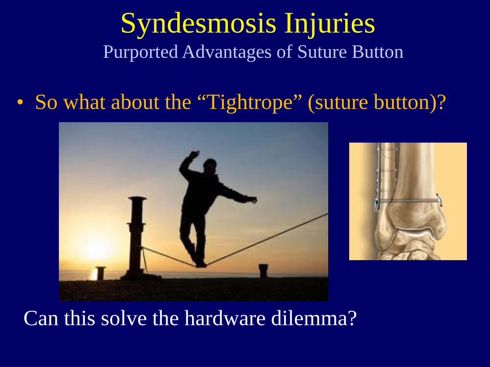
• So what about the “Tightrope” (suture button)?
Can this solve the hardware dilemma?
Purported Advantages of Suture Button Syndesmosis Injuries
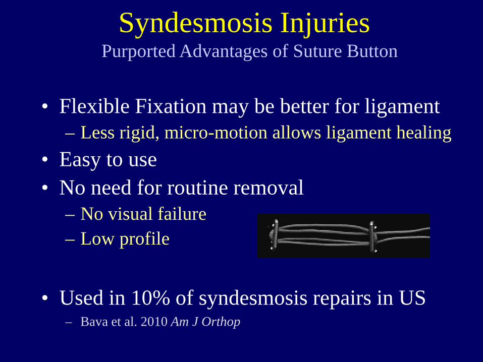
• Flexible Fixation may be better for ligament – Less rigid, micro-motion allows ligament healing
• Easy to use • No need for routine removal
– No visual failure – Low profile
Purported Advantages of Suture Button Syndesmosis Injuries
• Used in 10% of syndesmosis repairs in US – Bava et al. 2010 Am J Orthop
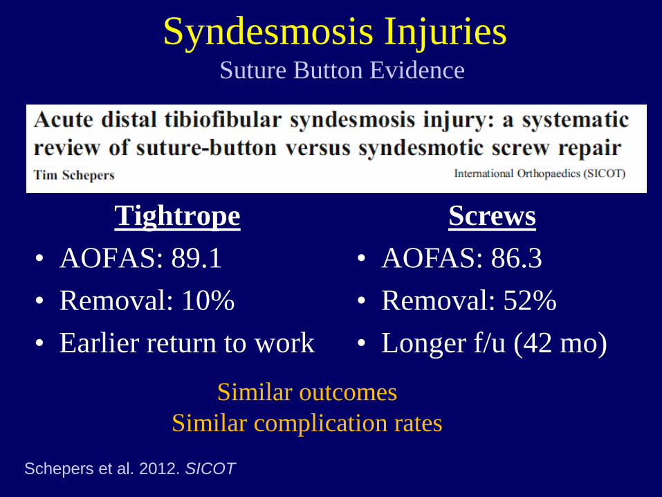
Suture Button Evidence Syndesmosis Injuries
Tightrope • AOFAS: 89.1 • Removal: 10% • Earlier return to work
Screws • AOFAS: 86.3 • Removal: 52% • Longer f/u (42 mo)
Similar outcomes Similar complication rates
Schepers et al. 2012. SICOT

• Clinical outcomes: – Level III Retrospective Cohort, 32 patients
• Suture-button vs. 3.5 screws – Suture Button:
• Better AOFAS scores • Faster return to work – 2.8 months vs 4.6 months • Reduction maintained (CT scan) • No additional surgery
Suture Button Evidence Syndesmosis Injuries
Thornes et al. CORR 2005
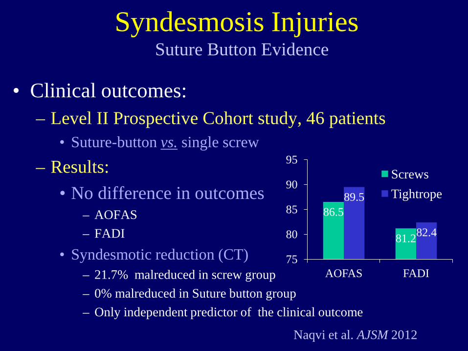
• Clinical outcomes: – Level II Prospective Cohort study, 46 patients
• Suture-button vs. single screw – Results:
• No difference in outcomes – AOFAS – FADI
• Syndesmotic reduction (CT) – 21.7% malreduced in screw group – 0% malreduced in Suture button group – Only independent predictor of the clinical outcome
Suture Button Evidence Syndesmosis Injuries
Naqvi et al. AJSM 2012
86.5
81.2
89.5
82.4
75
80
85
90
95
AOFAS FADI
Screws Tightrope
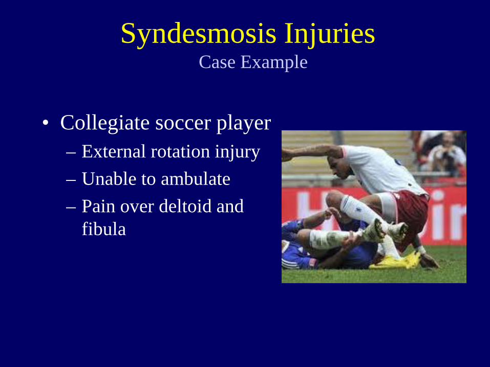
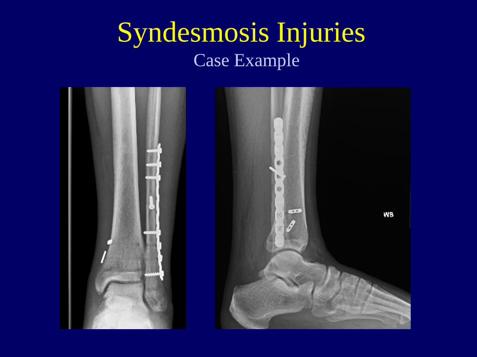
• Collegiate soccer player – External rotation injury – Unable to ambulate – Pain over deltoid and
fibula
Case Example Syndesmosis Injuries
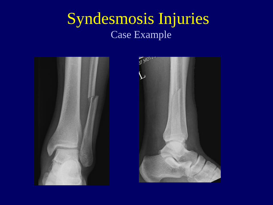
Case Example Syndesmosis Injuries
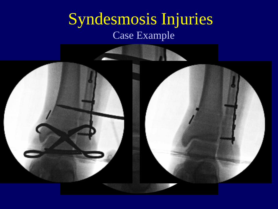
Case Example Syndesmosis Injuries
Case Example Syndesmosis Injuries
• Surgery rare for purely ligamentous injuries – Common for fractures
• Radiographs not reliable for moderate injuries • Reduction of syndesmosis is key • Screws and suture buttons both effective
– Suture button may provide advantages – More evidence needed
Take Away Points

Thank You
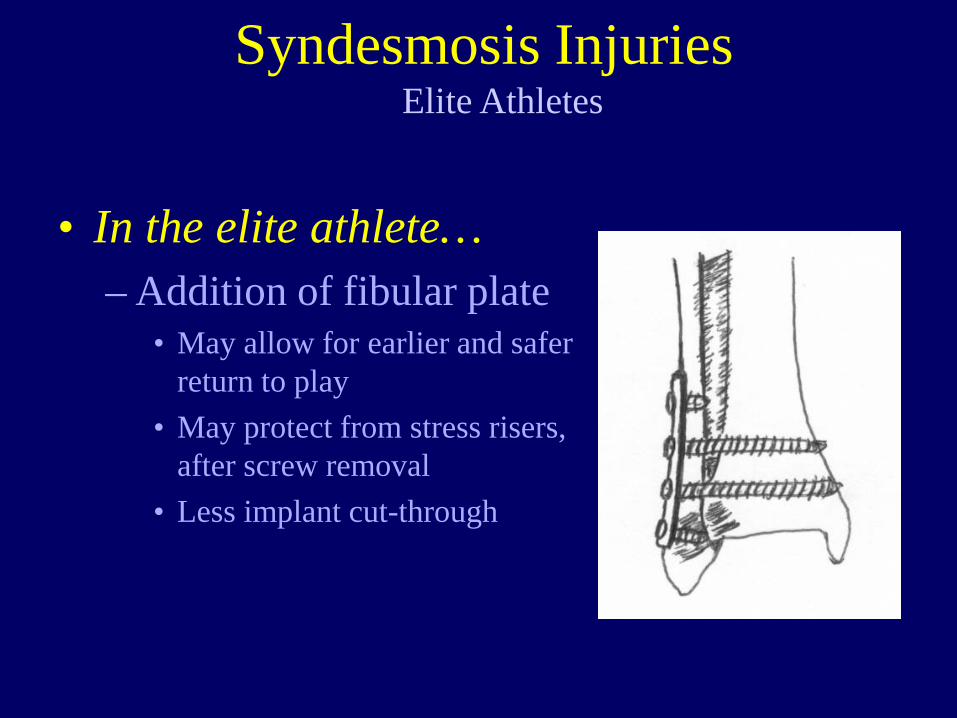
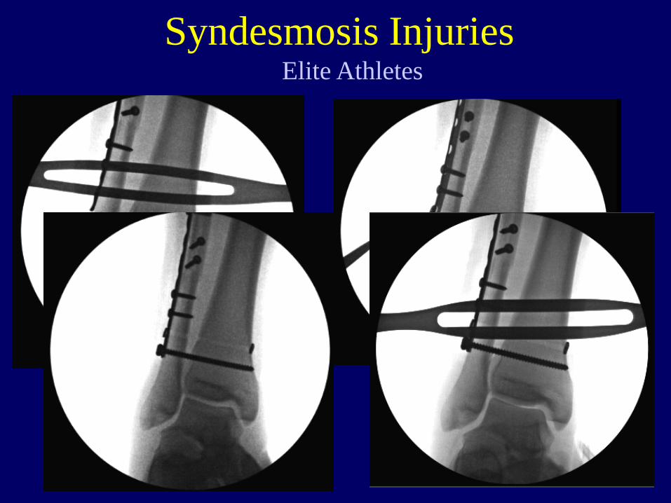
• In the elite athlete… – Addition of fibular plate
• May allow for earlier and safer return to play
• May protect from stress risers, after screw removal
• Less implant cut-through
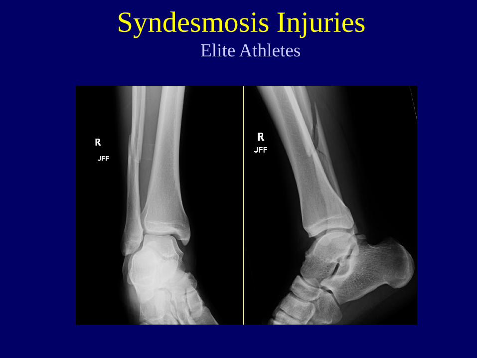
Elite Athletes Syndesmosis Injuries
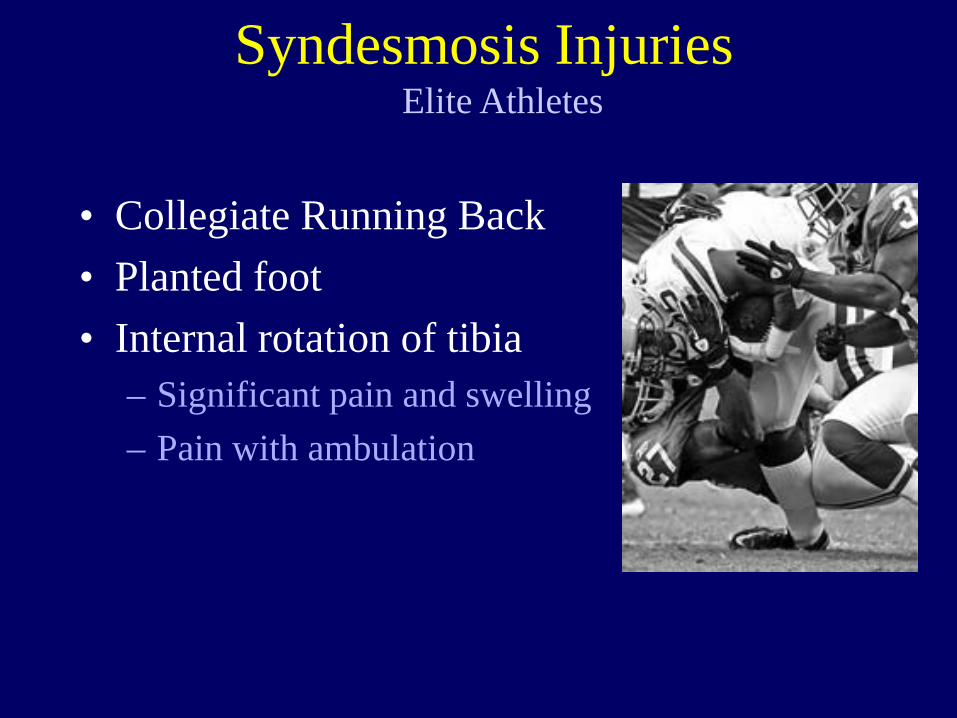
• Collegiate Running Back • Planted foot • Internal rotation of tibia
– Significant pain and swelling – Pain with ambulation
Elite Athletes Syndesmosis Injuries
Elite Athletes Syndesmosis Injuries
Elite Athletes Syndesmosis Injuries
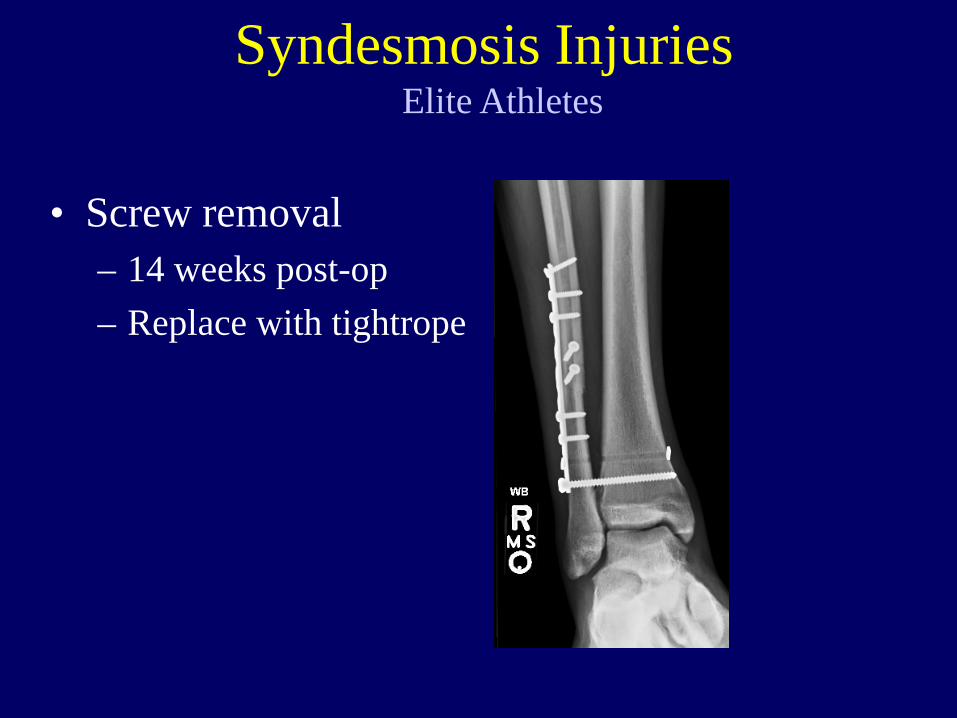
• Screw removal – 14 weeks post-op – Replace with tightrope
Elite Athletes Syndesmosis Injuries

• WBAT in boot • Begin rehab • Training 2 weeks
post-HWR (4 months post-injury)
Elite Athletes Syndesmosis Injuries