TREATMENT OF FIFTFI METATARSAL Guidelines … by means of a smooth, radiolucent line running...
7
CHAPTER 28 TREATMENT OF FIFTFI METATARSAL FRACTURE.S: Guidelines for Decision-Making Stephen J. Milleu DPM, FACFAS INTRODUCTION Fractures of the fifth methtarsal present a unique set of challenges for treatment to the foot and ankle specialist. Understanding the local anatomy, vascular supply, function, and dynamic stresses placed upon the bone, as well as fracture classifications, mechanisms of injury, and expected responses to treatment aid immensely in the decision-making processes. The author will briefly summarize the large amount of literature that has been generated on this unique topic and leave rhe reader with a succinct set of guidelines for treatment. EPIDEMIOLOGY Metatarsal fractures are among the most common injuries to the foot. They are roughly 10 times the frequency of LisFranct fracture-dislocations. Specifically, in 2 reviews, of 10,988 foot fractures, the proximal fifth metatarsal diaphl,5s2fmetaphyseal Jones fracture (acute and stress) had a frequency of 0.7o/o-7.9o/o.','In children,6T0/o of all foot fractures involve the metatarsal bones and most (41%) are of the fifth ray. In a study of industrial injuries, the fifth metatarsal - including base fractures - was the most commonly fractured at 23o/o followed by the third metatarsal. Stress fractures, which are seen especially in military, athletic and dancer populations, most commonly affect the second, third and fifth metatarsals at their proximal third.' Fractures of the base of the fifth metatarsal are more common than distal fractures of the shaft or head and neck. The proximal fifth metatarsal has been divided into 3 zones. In one study of 237 patients with fractures of the proximal fifth metatarsal,620/o were female and 3B%o were male. The most common proximal acute disruption was tuberosity (zone I) fractures (93o/o) in this general orthopedic population.a The incidence of true acute ]ones (zone II) fractures was found to be higher in the military population (25o/o).t Fractures in the more distal zone III close to the diaphysis were mosr commonly stress- induced fractures. ANATOMYAND DYNAMICS The fifth metatarsal is a long bone consisting of a head, neck, shaft, base and tuberosity or styloid process. The metaphyseal base tapers distally to the more tubular diaphysis which, besides being more convex dorsally, is actually wider in cross-section from medial to lateral than it is from dorsal to plantar. Also, the diaphyseal cortices tend to be thinner on the dorsal and plantar sides than on the medial and lateral sides. The bone often bows laterally.o These are significant considerations when planning intrameduilary screw placement. The tuberosity protrudes Iaterally and plantarward from the base.7 Proximal articulations of the fifth metatarsal are with the cuboid bone and adjacent base of the fourth metatarsal. Sturdy ligaments both dorsally and plantarly connect the cuboid to the base of the fifth metatarsal as well as to the base of the fourth metatarsai. The 2 adjacent bases are also connected by ligaments. The long plantar ligament extends from the distal calcaneous across the cuboid and inserts into the base of the fifth metatarsal, while more superficially, the lateral band of the plantar fascia sends a slip into the plantar tuberosiq,. It has been suggested to be more responsible for tuberosiq, fractures than the more prominent dorsal insertion of the peroneus brevis tendon into the tuberosiry.'r The insertion of the peroneus tertius tendon more distally onto the drorsal base of the fifth metatarsal is thought to have minimal influence as a fracture force. There are 3 main muscle slips that arise from the fifth metatarsal: the flexor digiti minimi brevis from the plantar base and the dorsal and plantar interossei from the medial surface with only the dorsal interossei opposed by its counterpart attached to the fourth metatarsal. Occasional variations from the normal muscle anatomy include: part of the abductor digiti minimi muscle can originate from the plantar aspect of the fifth metatarsal base; if present, the abductor ossi metatarsi quinti muscle inserts into the styloid process; and, when present, the opponens digiti minimi muscle inserts into the lateral border of the fifth metatarsal shaft.'o
-
Upload
duonghuong -
Category
Documents
-
view
214 -
download
0
Transcript of TREATMENT OF FIFTFI METATARSAL Guidelines … by means of a smooth, radiolucent line running...
CHAPTER 28
TREATMENT OF FIFTFI METATARSAL FRACTURE.S:Guidelines for Decision-Making
Stephen J. Milleu DPM, FACFAS
INTRODUCTION
Fractures of the fifth methtarsal present a unique set ofchallenges for treatment to the foot and ankle specialist.
Understanding the local anatomy, vascular supply,function, and dynamic stresses placed upon the bone, as
well as fracture classifications, mechanisms of injury, andexpected responses to treatment aid immensely in thedecision-making processes. The author will brieflysummarize the large amount of literature that has been
generated on this unique topic and leave rhe reader with a
succinct set of guidelines for treatment.
EPIDEMIOLOGY
Metatarsal fractures are among the most common injuriesto the foot. They are roughly 10 times the frequency ofLisFranct fracture-dislocations. Specifically, in 2 reviews, of10,988 foot fractures, the proximal fifth metatarsaldiaphl,5s2fmetaphyseal Jones fracture (acute and stress)
had a frequency of 0.7o/o-7.9o/o.','In children,6T0/o of allfoot fractures involve the metatarsal bones and most (41%)are of the fifth ray. In a study of industrial injuries, thefifth metatarsal - including base fractures - was the mostcommonly fractured at 23o/o followed by the thirdmetatarsal. Stress fractures, which are seen especially inmilitary, athletic and dancer populations, most commonlyaffect the second, third and fifth metatarsals at theirproximal third.'
Fractures of the base of the fifth metatarsal are morecommon than distal fractures of the shaft or head andneck. The proximal fifth metatarsal has been divided into3 zones. In one study of 237 patients with fractures of theproximal fifth metatarsal,620/o were female and 3B%o were
male. The most common proximal acute disruption was
tuberosity (zone I) fractures (93o/o) in this generalorthopedic population.a The incidence of true acute ]ones(zone II) fractures was found to be higher in the militarypopulation (25o/o).t Fractures in the more distal zone
III close to the diaphysis were mosr commonly stress-
induced fractures.
ANATOMYAND DYNAMICS
The fifth metatarsal is a long bone consisting of a head,
neck, shaft, base and tuberosity or styloid process. Themetaphyseal base tapers distally to the more tubulardiaphysis which, besides being more convex dorsally, isactually wider in cross-section from medial to lateral thanit is from dorsal to plantar. Also, the diaphyseal cortices
tend to be thinner on the dorsal and plantar sides than onthe medial and lateral sides. The bone often bows laterally.o
These are significant considerations when planningintrameduilary screw placement. The tuberosity protrudesIaterally and plantarward from the base.7
Proximal articulations of the fifth metatarsal are withthe cuboid bone and adjacent base of the fourthmetatarsal. Sturdy ligaments both dorsally and plantarlyconnect the cuboid to the base of the fifth metatarsal as
well as to the base of the fourth metatarsai. The 2 adjacentbases are also connected by ligaments. The long plantarligament extends from the distal calcaneous across the
cuboid and inserts into the base of the fifth metatarsal,
while more superficially, the lateral band of the plantarfascia sends a slip into the plantar tuberosiq,. It has been
suggested to be more responsible for tuberosiq, fractures
than the more prominent dorsal insertion of the peroneus
brevis tendon into the tuberosiry.'r The insertion of the
peroneus tertius tendon more distally onto the drorsal base
of the fifth metatarsal is thought to have minimalinfluence as a fracture force.
There are 3 main muscle slips that arise from the
fifth metatarsal: the flexor digiti minimi brevis from the
plantar base and the dorsal and plantar interossei from themedial surface with only the dorsal interossei opposed byits counterpart attached to the fourth metatarsal.
Occasional variations from the normal muscle anatomyinclude: part of the abductor digiti minimi muscle can
originate from the plantar aspect of the fifth metatarsal
base; if present, the abductor ossi metatarsi quinti muscleinserts into the styloid process; and, when present, theopponens digiti minimi muscle inserts into the lateralborder of the fifth metatarsal shaft.'o

r56 CHAPTER 28
Biomechanically, the fifth metatarsal functions withan independent axis of motion that allows primarilydorsiflexion and plantarflexion with inversion-eversion as
potential movements as well. Strong soft tissue
attachments that contain the base stabilize it against acute
and repetitive force attacks. Excessive acute and repetitive
strain loads on the bone are usually flexural, whereas
torque can occur with inversion injuries.
Two studies have investigated the vascular supply tothe fifth metararsal."''' The tuberosity is well supplied byfrom numerous random vessels that are directed from the
metaphysis. There is a nutrient artery supplying the
diaphysis but the proximal diaphyseal region contains a
watershed "no man's land" where there is a run-out of thenutrient artery before the metaphyseal vessels are
encountered. This area of poor vascular supply is thoughtto be the etiology of delayed union or nonunion offractures in this area, especially if the nutrient arteryis disrupted.'3
There are anatomic variations that may result inconfusion with fractures of the tuberosity of the fifthmetatarsal. The os peroneum, within the peroneus iongus
tendon, is usually situated lateral to the cuboid bone
whereas the os vesalianum within the peroneus brevis
tendon sits adjacent to its insertion. Also, the apophysis,
or growth center may be confused with a nondisplaced
tuberosity fracture in a child. The apophysis can be
identified by means of a smooth, radiolucent line runningparallel to the metatarsal shaft. This growth center first
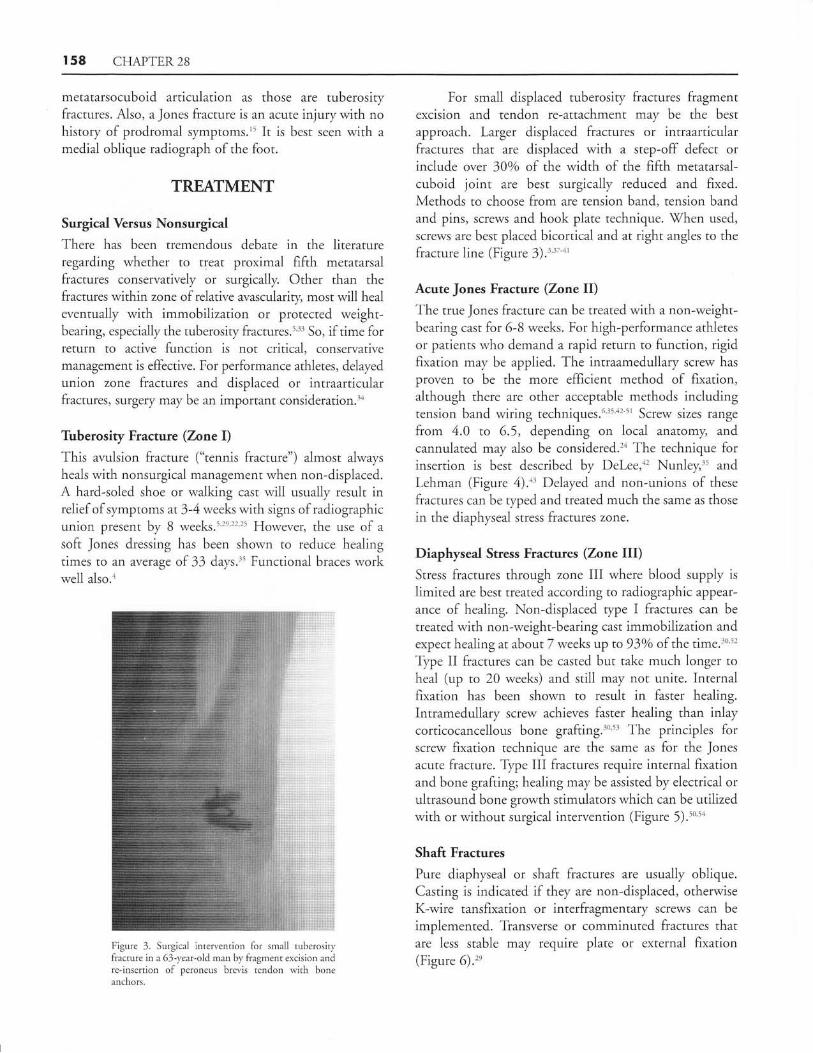
Figure 1. Diaphyseal stress lracture in a 64-year-olddiabetic female n'ith metatarsus adductus and Charcotneuroarthropathv.
appears in males between ages 9-11 years and in females
from 11-14 years. The line obliterates by fusing with the
tubersiry 2-3 years after its appearance."'" \fhen the
apophysis is afflicted with osteochodrosis it is known as
Iselins disease; its pain and tenderness may mimic a
tuberosity fracture.'6't
MECHANISM OF INJURY
Foot plantarflexion with an adduction force applied tothe forefoot is the source of most acute injuries to the base
of the fifth metatarsal whether it causes a tuberosityfracture or Jones fracture or even a cervical fracture.
Hence, these injuries are often encountered in such sports
as basketball, football, soccer and tennis as well as danc-
ing and gymnastics. They are also seen in the general
population as the result of sudden inversion injuries such
as slipping while going down stairs or stepping over an
edge. The locking configuration of all the soft tissue con-
straints about the base make dislocation of the fifthmetatarsal-cuboid joint an exceedingly rare occurrence.
Stress fractures, which usually disrupt the proximaldiaphysis, are the result of repeated submaximaldistraction forces. One biomechanical study revealed thatthe peak stress point occurs approximately 3.38 to 4.05
centimeters distal to the tuberosiry when the load is
directed 30 to 60 degrees from the horizontal plane
relative to the long axis of the metatarsal.'' Structural and
bio-mechanical abnormalities can contribute to the
production of these repetitive load concentrations.Examples include uncompensated forefoot varus,'e rigidpes cavus, metatarsus adductus,'u talipes equinovarus,"
post Evan's caicaneal osteotomy," Charcot foot,"''a and
Charcot-Marie-Tooth disease." (Figure 1) Dancers,
distance runners and military trainees are especially
subject to fifth metatarsal stress fractures at the base or
proximal diaphysis although stress fractures of the second
metatarsal are more common.'3't6''6'"
CI/.SSIFICATION
Quill presented a useful classification for fracture patterns
thar occur in the proximal fifth metatarsal." He dividedthe fractures in this region of the bone into 3 areas,
sometimes referred to as zones': tuberosity avulsion
fractures (ZoneI),Jones fractures (Zone II) and proximaldiaphyseal stress fractures (Zone III). (Figure) Vogler
added three other iocations in his anatomic classification
to better define treatment options for the variousfractures of the complete fifth metatarsal: capitumfractures, cervical fractures and shaft fractures (Thble 1)."
CHAPTER 28 157
Each of the fracture patterns has its own unique location,mechanism of injury, treatment options, and prognosisregarding delayed union and nonunion.
Torg further subdivided the proximal diaphysealstress fractures, also applicable to Jones fractures, intothree subtypes based on their radiographic signs ofhealing: type I - acute or early union characterized by a
narrow fracture line and absence of intrameduilarysclerosis; type II - delayed union, involving fractures withwidening of the fracture line and evidence ofintramedullary sclerosis; and type III - nonunion, whichincludes fractures with complete sclerotic obliteration ofthe intramedullary canal (Thble 2).'"
There has been considerable confusion regardingthe exact definition ofthe "Jones fracture." Jones sufferedthe fracture himself in 1902 while dancing around a tentpole and recognized it as the result of an indirect force.3'
Stewart defined the true Jones fracture as a transverse
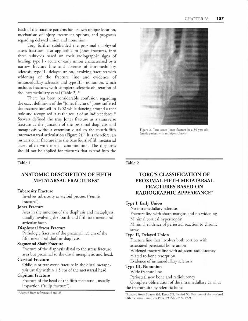
fracture at the junction of the proximal diaphysis andmetaphysis without extension distal to the fourth-fifthintermetatarsal articulation (Figure 2).3'lt is therefore, an
intraarticular fracture into the base fourth-fifth metatarsalfacet, often with medial comminution. The diagnosisshould not be applied for fractures that extend into the
Thble 1
ANAIOMIC DESCRIPTION OF FIFTHMETAIARSAL FRACTURESX
Tirberosity FractureInvolves tuberosity or sryloid process ("tennis
fracture").
Jones FractureArea in the junction of the diaphysis and metaphysis,usually involving the fourth and fifth intermetatarsalarticular facet.
Diaphyseal Stress FracturePathologic fracture of the proximal 1.5 cm of thefifth metatarsal shaft or diaphysis.
Segmental Shaft FractureFracture ofthe diaphysis distal to the stress fracturearea but proximal to the distal metaphysic and head.
Cervical FractureOblique or transverse fracture in the distal metaph-ysis usually within 1.5 cm of the metatarsal head.
Capitum FractureFracture of the head of the fifth metatarsal. usuallvimpaction ("tulip fracture").
Figure 2. True acute Jones fracture in a 50-year-oldlemale patient with muJtiple sclerosis.
Thble 2
TORG'S CLASSIFICATION OFPROXIMAL FIFTH METATARSAL
FRACTURES BASED ONRADIOGRAPHIC APPEARANCEX
Type I, Early UnionNo intramedullary sclerosis
Fracture line with sharp margins and no wideningMinimal cortical hypertrophyMinimal evidence of periosteal reaction to chronicstress
Type II, Delayed UnionFracture line that invoives both cortices withassociated periosteal bone unionWidened fracture line with adjacent radiolucencyrelated to bone resorptionEvidence of intramedullary sclerosis
Type III, Nonunion\7ide fracture linePeriosteal new bone and radiolucencyComplete obliteration of the intramedullary canal at
the fracture site by sclerotic bone
*Adapted fromfifth metatarsal
Strayer SM, Reece SG, Petrizzi NJ: Fractures of the proximalAm Fam Phvs, 59:25 16-2522,1999.
'Ad:pLed lr.m relerence' 5 anJ JJ
I58 CHAPTER28
metatarsocuboid articulation as those are tuberosityfractures. Also, a Jones fracture is an acute injury with nohistory of prodromal symptoms.'5 It is best seen with a
medial oblique radiograph of the foot.
TREAIMENT
Surgical Versus Nonsurgical
There has been tremendous debate in the literatureregarding whether to treat proximal fifth metatarsalfractures conservatively or surgically. Other than thefractures within zone of relative avascularity, most will heal
eventually with immobilization or protected weight-bearing, especially the tuberosity fractures.5'33 So, if time forreturn to active function is not critical, conservative
management is effective. For performance athletes, delayed
union zone fractures and displaced or intraarticularfractures, surgery may be an important consideration.'a
Tirberosity Fracture (Znne l)This avulsion fracture ("tennis fracture") almost always
heals with nonsurgical management when non-displaced.A hard-soled shoe or walking cast will usually result inrelief of symptoms at 3-4 weeks with signs of radiographicunion present by 8 weeks.5're'r2'25 However, the use of a
soft Jones dressing has been shown to reduce healingtimes to an average of 33 days.35 Functional braces workwell also.'
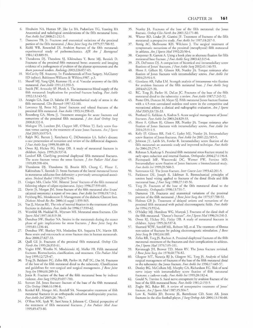
Figure 3. Surgical intervention for small tuberosiwfracture in;r 63-year-old man bv fragment excision andre-insertion of peroneus brevis tendon with bonea nchors-
For small displaced tuberosity fractures fragmentexcision and tendon re-attachment may be the best
approach. Larger displaced fractures or intraarticularfractures that are displaced with a step-off defect orinclude over 30o/o of the width of the fifth metatarsal-
cuboid joint are best surgicaily reduced and fixed.Methods to choose from are tension band, tension bandand pins, screws and hook plate technique. When used,
screws are best placed bicortical and at right angles to thefracture line (Figure 3).:'tt'tt
Acute Jones Fracture (Zone Il)The true Jones fracture can be treated with a non-weight-bearing cast for 6-8 weeks. For high-performance athletes
or patients who demand a rapid return to function, rigidfixation may be applied. The intraamedullary screw has
proyen to be the more efficient method of fixation,although there are other acceptable methods includingtension band wiring techniques.o'35'12 t, Screw sizes range
from 4.0 to 6.5, depending on local anatomy, and
cannulated may also be considered." The technique forinsertion is best described by Del-ee,a' Nunley,.5 and
Lehman (Figure 4).a' Delayed and non-unions of these
fractures can be typed and treated much the same as those
in the diaphyseal stress fractures zone.
Diaphyseal Stress Fractures (Zone III)Stress fractures through zone III where blood supply islimited are best treated according to radiographic appear-
ance of healing. Non-displaced type I fractures can be
treated with non-weight-bearing cast immobilization and
expect healing at about 7 weeks up to 93o/o of the time.3o'5'
Type II fractures can be casted but take much longer toheal (up to 20 weeks) and still may not unite. Internalfixation has been shown to resuit in faster healing.
Intramedullary screw achieves faster healing than inlaycorticocancellous bone grafting.3"'53 The principles forscrew fixation technique are the same as for the Jonesacute fracture. Type III fractures require internal fixationand bone grafting; healing may be assisted by electrical orultrasound bone growth stimulators which can be utilizedwith or without surgical intervention (Figure 5).to'5a
Shaft Fractures
Pure diaphyseal or shaft fractures are usually oblique.Casting is indicated if they are non-displaced, otherwiseK-wire tansfixation or interfragmentary screws can be
implemented. flansverse or comminuted fractures thatare less stable may require plate or external fixation(Figure 6)."

CHAPTER 28 r59
Figure 4A. Preop. Acute Jones lracture inlemale construction lvorker, successfullyintramedullary screw fixatjon.
26-vear-oldtreated lvith
Figure 48. Postop healed at 8 weeks.
Figure 5. Typical diaphyseal stress fracture fullnonunion. Notice its location distal to the fourth-fifthmetatatsal base articulation.
Figure 6. Diaphyseal or sha€t fiacture.

T60 CHAPTER28
Figure 7. Ceruical fracture of the fifth metatarsal.
Cervical Fractures
This fracture can present as impaction, transverse, orspiral "dancert fracture."5' It can be treated by closed
reduction with traction under local anesthesia followedby casting and usually heals well. Fixation may be
considered for failure to reduce, dorsal displacement, orspiral shearing (Figure 7).''"
Capitum Fractures
Fractures of the head of the fifth metatarsal are oftencaused by distal impaction ("tulip fracture") or directvertical impact. They usually heal well with protectedweightbearing. Severe comminution may require head
resection (Figure 8)."
COMPLICATIONS
The most difficult complications in treating proximalfifth metatarsal fractures are delayed or non-union. \X4ren
either occurs, treatment choices include ultrasound orelectrical bone growth stimulationt. 54'56'57 or surgical repair
with autogenous or prepared bone graft. Refracture can
occur after return to activity or when the screw fixationhas been removed, especially in athletes.5t Screw fractures,
screws missing the intramedullary canal, fracturemalalignment, and pain over the prominent screw head
have all been reported as complications.42'5e'60
One of the most frustrating complications ofsurgery on the base of the fifth metatarsal occurs whenthere is inadvertent damage to the sural nerve. Since thenerye courses and branches at the tuberosity, it is a
Figure 8. Capitum or "tulip" impaction factureinvolving the filth metatarsal head.
structure usually encountered during dissection.Knowledge of its usual expected location and careful
atraumatic technique helps avoid such complicationsand the need for further surgery to release and
decompress the nerve.61 62
CONCLUSION
The true Jones fracture is a very specific and defined
subtype of fifth metatarsal base fractures. Evaluation and
treatment of fifth metatarsal fractures in general are best
accomplished by dividing the long bone into anatomic
regions, each with its own characteristics. fleatment can
range from completely nonoperative care for all types ofthese fractures seen in a general orthopaedic practice33'63
to surgical repair for ail Jones fractures in elite athletes.6a
This paper provides information to help in the
decision-making for all other situations involving fifthmetatarsal fractures.
REFERENCES
1. losefsson PO, Karlsson M, Redlund-Johnell I, \Wendeberg B. Joneslracture. Surgical versus nonsurgical treatment. Clin Orthop Relat
Resl.994;299:252-5.2. Jossefsson PO, Karlsson M, Redlund-Johnell I, tll'endeberg B: Closed
treatment ofJones fracture. Good results in 40 cases after 11-26 years.
Actd Orthop Scand 199465:545-7.3. Rammelt S, Heineck J, Zwipp H. Metatarsal fractures. Injury. 20043)
Suppl 2:SB77-86.4. Dameron TB. Fractures of the proximal flfth metatarsal: selecting the
best treatment option. J Am Acad Orthop Surg 1995:3:710-4.5. Clapper MF, O'Brien TJ, Lyons PM. Fractures of the fifth metatarsal.
Analysis ofa fracture registry. Clin Orthop Relat Res 1995;315:238-41.
CHAPTER 28 t6t
21.
12.
14
15
16.
17
18.
19
t1.
10.
t3
8.
25.
27.
31.
\3.
Ebraheim NA, Haman SP, Jike Lu BA, Padanilam TG, Yeasting RAAnatomical and radiological considerations ofthe fifth metatarsal bone.Foot Ankle Intl 2000;21:212-5.Dameron TB, Jr. Fractures and anatomical r.ariations of the proximalportion ofthe fifth metatarsal. / Bone Joint SurgAm 1975;57:788-92.fuchli \X/R, Rosenthal DI. Avulsion fracture of the fifth metatarsal:experimental study of pathomechanics. AJR Am J Roentgenol1 984:143:889-9 1.
Theodorou DJ, Theodoru SJ, Kikitsubata Y, Botte MJ, Resnick D.Fractures of the proximal fifth metatarsal bone: anatomic and imagingevidence ofa pathogenesis ofavulsion ofthe plantar aponeurosis and theshort peroneal rcndon. Radiologt 2003;226:857 -65.McCarthy DJ. Anatomy. In: Fundamentals of Foot Surgery. McGlamryED (editor), Baltimore:-{/illiams & \X/ilkins;
1 987. p.3.Shereff MJ, Yang QM, Kummer FJ, et al. Vascular anaromy of the fifthmetatarsal. Foot Ankle 199 1; 1 1 :350-3.Smith J\Xr, Amoczky SP, Hersh A. The intraosseous blood supplv of thefifth metatarsal: Implications for proximal fracture healing. Foot Ankle1992;13:143-52.Arangio GA, Xao D, Salathe EP. Biomechanical study of stress in thefifth metatarsal. Clin Biomech 1997 12:160.Lawrence SJ, Botte MJ. Jones' fractures and related fractures of theproximal fifth metatarsal. Foot Ankle 1993 14:358-65.Rosenberg GA, Sferra ]J. Treatment strategies for acute fractures andnonunions of the proxirnal fifth metatarsal. J Am Acad Orthop Swrg2000;8:132-8.Mologne TS, Lundeen JM, Clapper MF, O'Brien TJ. Early screw fixa-tion versus casting in the treatment of acute Jones fractures. Am J SportsMed 2005:33:970-5.Ralph BC, Barrett -f, Kenyhercz C, DiDomenico lA. Iselin's disease:
a case presentation of nonunion and review of the differential diagnosis.
J Foot Ankle Surg 1999;38:40')-16.Orven RJ, Hickey FG, Finlay DB. A study of metatarsal fractures inchildren. Inj ury. 199 5 ;26:5 37 -8.Landorf KB. Clarifi.ing proximal diaphyseal fifth metatarsal fractures.The acute fracture versus the stress ftacwe. J Am Pocliatr Med Asoc1 999r89:398-404.f'heodorou DJ, Theodorou SJ, Boutin RD, Chung C, Fliszar E,
Kakitsubata Y, Resnick D. Stress fiactures ofthe lateral metatarsal bonesin metatarsus adductns foot deformity: a previously unrecognized associ-
ation. Sleeletal Radiol 1999 ;28:67 9 -84.Craigen MA, Clarke NM. Bilateral'Jones' fractures of the fifth metatarsal
following relapse of talipes equinovarus. Injury 199627 :599 -601.Davitt JS, Morgan JM. Stress fracture of the fifth metatusal after Evans'
calcaneal osteotomy: a report ofnvo cases. Foot Ankle Int 1998 19:710-2.Frykberg RG, Mendeszoon E: Management of the diabetic Charcot foot.Diabetes Metab Res Reu 2000;15 suppl 1:S59-565.Yue JJ, Marcus RE. The role of internal fixation in the treatment of Jonesfractures in diabetics. Foot Ankle Int 1996 17:559-62.\(einfeld SB, Haddad SL, Myerson MS. Metatarsal stress fractures. C/lzSporx Med 1997 ;16:319-38.Donahue S1tr, Sharkey NA. Strains in the metatarsals during the stance
phase of gait: implications for stress fractures. J Bone Joint Surg Am1999$1:1236-44.Donahue S-tr, Sharkey NA, Modanlou K{, Sequeira LN, Martin RB.Bone strain and microcracks at stress fracture sites in human metatarsals.
Bone 2000;27:827-33.
Quill GE Jr. Fractures of the proximal fifth metatarsal. Orthop ClinNorth Am 1995;26:353-61.Vogler H-{/, \Testlin N, Mlodzienski AJ, Moller FB. Fifrh metatarsalfractures. Biomechanics, classification, and treatment. Clin Podiatr MetlSurg 1995;12:725-47.TorgJS, Balduini FC, Zelko RR, Pavlov H, PeffTC, Das M. Fracturesof the base of the fifth metatarsal distal to the tuberosiry. Classificationand guidelines for non-surgical and surgical management. J Bone JointSurg Am 1984;66:209 -14.
Jones R. Fracture of the base of the fifth metatarsal bone b). indirectviolence. Ann Surg 1992;35:697 -7 00.Stewart IM. Jones fracture: fracture of the base of the fifth metatarsal.Clin Orthop 1960;16:190-8.Konkel KF, Menger AG, Retzlaff SA. Nonoperative treatment of fifthmetatarsa] fractures in an orthopaedic suburban multispecialq. practjce.Foor Ankle lnrl )00\;)o: -04--.O'Shea MI(, Spak V, Sant'Anna S, Johnson C. Clinical perspectiye ofthe treatment of fifth metatarsal fractures. J Am Podiatr Med AsocI 995:85:473-80.
Nunley JA. Fractures of the base of the fifth metatarsal: the Jonesfractwe. Orthop Clin North Am 2001;32:171-80.\iliener BD, Linder JF, Giattini JF. Treatment of fractures of the fifthmetatarsal: a prospective study. Foot Ankle Int 1997 18:267-9.Rettig AC, Shelbourne KD, ]X/ilckens J. The surgical treatment ofsymptomatic nonunions of the proximal (metaphyseal) fifth metatarsalin athletes. Am J Sports Med 7992,20:50-4.Carpenter B, Garrett A. Using a hook plate as alternate fixation for fifthmetatarsal base fracture.,/ -Fo ot Ankle Surg 2003;42:315-6.ZS, DeFronzo DJ. A comparison of bicortical and intramedullary screw
fixations of Jones' fracwes. J Foot Ankb Surg 2002;41:146-53.Horst F, Gilben BJ, Glisson RR, Nunley JA. Torque resistance afterfixation of Jones fractures with intramedullary screws. Foot Ankle Int2004;25:914-9.Giordano AR, Fallat LM. Strength analysis of intraosseous wire fixationfor avulsion fractures of the fifth metatarsal 6ase. J Foot Ankle Sury2004:43:225-30.RC, Torg JS, Pavlov H, Delee JC. Fractures of the base of the fifthmetatarsal distal to the tuberosity: a review. Foot Ankle 1987 7:245-52.Porter DA, Duncan M, Meyer S]. Fifth metatarsalJones fracture fixationwith a 4.5-mm cannulated stainless steel screw in the competitive andrecreational athlete: a clinical and radiographic evaluation. Am J Sports
Med 2005;33:726-33.Portland G, Kelikian A, Kodros S. Acute surgical management of Jones'fractures. Foot Ankk Int 2003;24:829 -33.Horst F, Gilbert BJ, Glisson RR, Nunley JA. Torque resistance afterfixation of Jones fractures with intramedullary screws. Foot Ankle Int2004.25:914-9.Kelly IP, Glisson RR, Fink C, Easley ME, Nunley JA. Intramedullaryscrew fixation ofJones lractures . Foot Ankle Int 2001;22;:585-9.
Johnson JT, Labib SA, Fowler R. Intramedullary screw fixation of thefifth metatarsal: an anatomic study and improved technique. Foot AnkleInt 2004:25:274-7.Rehman S, Kashyap S. Proximai fifth metatarsal stress fracture treated byearly open reduction and internal fsation. Orthopedic: 2004;27 :1 196-8.Pietropaoli MP, \(/norowski DC, \flerner F\7, Fortino MD.Intramedullary screw fixation ofJones fractures: a biomechanical study.Foot Anble Int 1999:20:560-3.Sammarco GJ. The Jones fracttre. Inst Course Lect 1993 42:201-5.Parkinson DE, Joseph R, Edelman R. Biomechanical principles oftension band wiring applied to fractures of the distal fibula and fifthmetatarsal base. J Foot Surg 1988;27:149-56.Torg JS. Fractures of the base of the fifth metatarsal distal to thetuberosiry. Orthopetlics 1990;13:737-7.Dameron TB. Fractures and anatomical variations of the proximalportion ofthe fifth metatarsal./ BoneJoint SurgAm 1975 57:788-92.Holmes GB Jr. Treatment of delayed unions and nonunions of theproximal fifth metatarsal with pulsed electromagnetic fields. Foot AnkleInt 199415:552-6.O'Mallev MJ, Hamilton VG, Munyak J. Fractures of the distal shaft ofthe fifth metatarsal. "Dancer's fracture". Am J Sports Med 1996:24:240-3.Owen RJ, Hickey FG, Finlay DB. A study of metatarsal fractures inchildren. Injury 199 5 :26:537 -8.Sharrard \X{rW, Sutcliff ML, Robson MJ, et al. The treatment of fibrousnon-union of fi'actures by pulsing electromagnetic stimulation. /.BazrJoint Surg Br 1982;64:189.Zelko RR, TorgJS, Rachun A. Proximal diaphyseal lractures ofthe fifthmetatarsal-trearment of the fractures and their complications in athletes.
Am J Sports Med 1979:7:95-701.Kavanaugh JH, Brower TD, Mann RV. The Jones lracture revisited.
J Bone Joint Surg Am 1978;60:776-82.Glasgow MT, Naranja RJ Jr, Glasgow SG, Torg JS. Aralysis of failedsurgical management of lractures of the base of the fifth metatarsal distalto the tuberosity: the Jones fracttre. Foot Ankle Int 1996;17:449-57.Donley BG, McCollum MJ, Murphy GA, Richardson EG. fusk of suralnerve injury with intramedullary screw fixation of fifth metatarsalfi'actures: a cadaver strdy. Foot Ankle Int 1999 20:182-4.Gould N, Trevino S. Sural nerue entrapment by avulsion lracture of thebase of the fifth metatarsal bone. Foot Ankle 1981 2:153-5.Zogby RG, Baker BE. A review of nonoperative treatment of Jones'fracwe. Am J Sports Med 1987;15:304-7.Low K, Noblin lD, Browne JE, Barnthouse CD, Scott AR. Jonesfractures in the elite football player. / Surg Orthop Adu 2004;13:156-60.
35
36
38
40.
41.
44
39.
41 .
42.
17
57.
46.
47
48
49
50.
51.
52.
53
54
55
56.
20
22
23
24
26
58
59
60
6l
52
63
28.
2)
30
32.
34.
64.