Treatment of Congenital Vertical Talus: Comparison...
12
Treatment of Congenital Vertical Talus: Comparison of Minimally Invasive and Extensive Soft-Tissue Release Procedures at Minimum Five-Year Follow-up Justin S. Yang, MD, and Matthew B. Dobbs, MD Investigation performed at St. Louis Shriner’ s Hospital for Children, St. Louis, St. Louis Children’ s Hospital, St. Louis, and the Department of Orthopaedic Surgery, Washington University School of Medicine, St. Louis, Missouri Background: The most common historical treatment method for congenital vertical talus is extensive soft-tissue release surgery. A minimally invasive treatment approach that relies primarily on serial cast correction was introduced almost ten years ago, with promising early results. The purpose of this study was to assess the long-term outcome of patients with congenital vertical talus managed with the minimally invasive technique and compare them with a cohort treated with extensive soft-tissue release surgery. Methods: The records of twenty-seven consecutive patients with vertical talus (forty-two feet) were retrospectively reviewed at a mean of seven years (range, five to 11.3 years) after initial correction was achieved. The minimally invasive method was used to treat sixteen patients (twenty-four feet), and extensive soft-tissue release surgery was used to treat eleven patients (eighteen feet). Patient demographics, ankle range of motion, the PODCI (Pediatric Outcomes Data Collection Instrument) questionnaire, and radiographic measurements were analyzed. Results: At the latest follow-up, the mean range of motion of patients treated with the minimally invasive method was 42.4° compared with 12.7° for patients treated with extensive surgery (p < 0.0001). The PODCI normative pain and global function scores were superior in the minimally invasive treatment group compared with the extensive soft-tissue release group. Greater correction of hindfoot valgus (anteroposterior talar axis-first metatarsal base angle) was achieved in the minimally invasive treatment group compared with the extensive surgery group (40.1° versus 27.9°,p = 0.03), although all other radiographic values were similar between the two groups (p > 0.1 for all). Subgroup analysis of patients with isolated vertical talus also showed superior range of motion and PODCI normative global function scores in the minimally invasive group. Conclusions: The minimally invasive treatment method for vertical talus resulted in better long-term ankle range of motion and pain scores compared with extensive soft-tissue release surgery. Longer-term studies are necessary to determine whether the improved outcomes are maintained into adulthood and whether the superior outcome is related to reduced scarring. Level of Evidence: Therapeutic Level III. See Instructions for Authors for a complete description of levels of evidence. C ongenital vertical talus is a rare flatfoot deformity that is present at birth and is characterized by a fixed dorsal dislocation of the navicular on the talus with associated Achilles tendon and dorsolateral soft-tissue contractures as well as calcaneocuboid joint subluxation and/or dislocation 1 . The estimated prevalence of vertical talus is one in 10,000 2 , Disclosure: One or more of the authors received payments or services, either directly or indirectly (i.e., via his or her institution), from a third party in support of an aspect of this work. In addition, one or more of the authors, or his or her institution, has had a financial relationship, in the thirty-six months prior to submission of this work, with an entity in the biomedical arena that could be perceived to influence or have the potential to influence what is written in this work. No author has had any other relationships, or has engaged in any other activities, that could be perceived to influence or have the potential to influence what is written in this work. The complete Disclosures of Potential Conflicts of Interest submitted by authors are always provided with the online version of the article. Peer Review: This article was reviewed by the Editor-in-Chief and one Deputy Editor, and it underwent blinded review by two or more outside experts. It was also reviewed by an expert in methodology and statistics. The Deputy Editor reviewed each revision of the article, and it underwent a final review by the Editor-in-Chief prior to publication. Final corrections and clarifications occurred during one or more exchanges between the author(s) and copyeditors. 1354 COPYRIGHT Ó 2015 BY THE J OURNAL OF BONE AND J OINT SURGERY,I NCORPORATED J Bone Joint Surg Am. 2015;97:1354-65 d http://dx.doi.org/10.2106/JBJS.N.01002
Transcript of Treatment of Congenital Vertical Talus: Comparison...

Treatment of Congenital Vertical Talus: Comparisonof Minimally Invasive and Extensive Soft-Tissue
Release Procedures at Minimum Five-Year Follow-upJustin S. Yang, MD, and Matthew B. Dobbs, MD
Investigation performed at St. Louis Shriner’s Hospital for Children, St. Louis, St. Louis Children’s Hospital, St. Louis, and the Department ofOrthopaedic Surgery, Washington University School of Medicine, St. Louis, Missouri
Background: Themost common historical treatment method for congenital vertical talus is extensive soft-tissue releasesurgery. A minimally invasive treatment approach that relies primarily on serial cast correction was introduced almost tenyears ago, with promising early results. The purpose of this study was to assess the long-term outcome of patients withcongenital vertical talus managed with the minimally invasive technique and compare them with a cohort treated withextensive soft-tissue release surgery.
Methods: The records of twenty-seven consecutive patients with vertical talus (forty-two feet) were retrospectivelyreviewed at a mean of seven years (range, five to 11.3 years) after initial correction was achieved. The minimally invasivemethod was used to treat sixteen patients (twenty-four feet), and extensive soft-tissue release surgery was used to treateleven patients (eighteen feet). Patient demographics, ankle range of motion, the PODCI (Pediatric Outcomes DataCollection Instrument) questionnaire, and radiographic measurements were analyzed.
Results: At the latest follow-up, themean range ofmotion of patients treatedwith theminimally invasivemethod was 42.4�compared with 12.7� for patients treated with extensive surgery (p < 0.0001). The PODCI normative pain and global functionscores were superior in the minimally invasive treatment group compared with the extensive soft-tissue release group.Greater correction of hindfoot valgus (anteroposterior talar axis-first metatarsal base angle) was achieved in the minimallyinvasive treatment group compared with the extensive surgery group (40.1� versus 27.9�, p = 0.03), although all otherradiographic values were similar between the two groups (p > 0.1 for all). Subgroup analysis of patients with isolated verticaltalus also showed superior range of motion and PODCI normative global function scores in the minimally invasive group.
Conclusions: The minimally invasive treatment method for vertical talus resulted in better long-term ankle range ofmotion and pain scores compared with extensive soft-tissue release surgery. Longer-term studies are necessary todetermine whether the improved outcomes are maintained into adulthood and whether the superior outcome is related toreduced scarring.
Level of Evidence: Therapeutic Level III. See Instructions for Authors for a complete description of levels of evidence.
Congenital vertical talus is a rare flatfoot deformity thatis present at birth and is characterized by a fixed dorsaldislocation of the navicular on the talus with associated
Achilles tendon and dorsolateral soft-tissue contractures aswell as calcaneocuboid joint subluxation and/or dislocation1.The estimated prevalence of vertical talus is one in 10,0002,
Disclosure: One or more of the authors received payments or services, either directly or indirectly (i.e., via his or her institution), from a third party insupport of an aspect of this work. In addition, one or more of the authors, or his or her institution, has had a financial relationship, in the thirty-six monthsprior to submission of this work, with an entity in the biomedical arena that could be perceived to influence or have the potential to influence what is writtenin this work. No author has had any other relationships, or has engaged in any other activities, that could be perceived to influence or have the potential toinfluence what is written in this work. The complete Disclosures of Potential Conflicts of Interest submitted by authors are always provided with theonline version of the article.
Peer Review: This article was reviewed by the Editor-in-Chief and one Deputy Editor, and it underwent blinded review by two or more outside experts. It was also reviewedby an expert in methodology and statistics. The Deputy Editor reviewed each revision of the article, and it underwent a final review by the Editor-in-Chief prior to publication.Final corrections and clarifications occurred during one or more exchanges between the author(s) and copyeditors.
1354
COPYRIGHT � 2015 BY THE JOURNAL OF BONE AND JOINT SURGERY, INCORPORATED
J Bone Joint Surg Am. 2015;97:1354-65 d http://dx.doi.org/10.2106/JBJS.N.01002
although this is likely an underestimation because of lackof recognition of vertical talus in the neonatal period. Verticaltalus is etiologically heterogeneous. Nearly half of all casesoccur as an isolated condition, whereas the remaining casesare associated with known genetic3 and/or neuromuscularconditions, including arthrogryposis and myelomeningo-cele, and are referred to as “non-isolated.”4 Although thecause of many cases of isolated vertical talus is unknown,there is growing evidence to support a genetic etiology, as>20% of reported cases in some series are familial5,6. Mu-tations in the HOXD107 and GDF58 genes have been iden-tified in some patients with isolated vertical talus, with manyadditional genetic factors remaining unknown. Indeed, pri-mary muscle abnormalities have been found on muscle biop-sies in some cases9. Although both isolated and non-isolatedvertical tali pose treatment challenges, it is generally acceptedthat non-isolated cases are more rigid and less responsive totreatment10,11.
The challenge in treating vertical talus is how to bestachieve the desired outcome of a mobile, plantigrade, pain-free, and functional foot. Bracing and/or shoe modificationsalone do not provide correction and often result in pain andlong-term disability1,12. The traditional surgical approachinvolving extensive soft-tissue release, while effective for gaininginitial correction in many cases, is associated with severalpotential complications, including wound necrosis, osteo-necrosis, inadequate correction of the deformity, stiffness ofthe ankle and subtalar joints, and amputation in extremecases2,13-16.
A minimally invasive technique for correcting verticaltalus that relies primarily on serial casting was introducedalmost ten years ago17,18. Multiple centers have reproducedthe effectiveness of this technique in achieving initial correction(both radiographically and clinically), while maintaining excel-lent motion in the foot and ankle, for patients with both isolated
and non-isolated vertical talus10,19-26. In the present study, wecompare the long-term outcomes of clinical and radiographiccorrection, foot function, and foot and ankle flexibility in pa-tients with vertical talus (isolated and non-isolated) treated witheither the minimally invasive method17 or extensive soft-tissuerelease surgery.
Materials and Methods
After institutional review board approval, we retrospectively reviewed therecords of thirty-two consecutive patients (fifty feet) who were treated for
congenital vertical talus at a single institution between 1998 and 2007. In-clusion criteria were (1) diagnosis of vertical talus confirmed by a lateralradiograph made with the foot in maximum plantar flexion (Figs. 1-A and1-B) that demonstrated persistent dislocation of the navicular on the talarhead with a talar axis-first metatarsal base angle of >35�, (2) follow-up for aminimum of five years after correction was achieved, and (3) availability ofcomplete pretreatment and post-treatment radiographs. Two patients werelost to follow-up, and two patients did not have the required pretreatment andpost-treatment radiographs. One patient was excluded as an outlier on thebasis of age at initiation of treatment. The remaining twenty-seven patientswere available for analysis. Patients with bilateral involvement had one footrandomly selected for statistical analysis. Patients with isolated vertical taluswere also examined separately in a subgroup analysis.
Patient demographics were recorded (Table I); fifteen were male andtwelve were female. Seventeen patients (twenty-four feet) had a diagnosis ofisolated vertical talus (Figs. 2-A and 2-B), and ten patients (eighteen feet) hadnon-isolated vertical talus (vertical talus occurring in association with a knowngenetic or neuromuscular condition). Mean age at the start of serial casting was6.6 months for the minimally invasive group and 15.2 months for the extensiverelease group. Mean follow-up duration was seven years (range, five to 11.3years). The choice of treatmentmethod utilized for each patient was based strictlyon surgeon preference and not on the severity of the deformity. The senior authorutilized the minimally invasive method (sixteen consecutive patients), and theremaining two surgeons utilized an extensive soft-tissue release.
TreatmentThe minimally invasive technique has previously been described for treatingboth isolated and non-isolated vertical talus and consists of serial foot
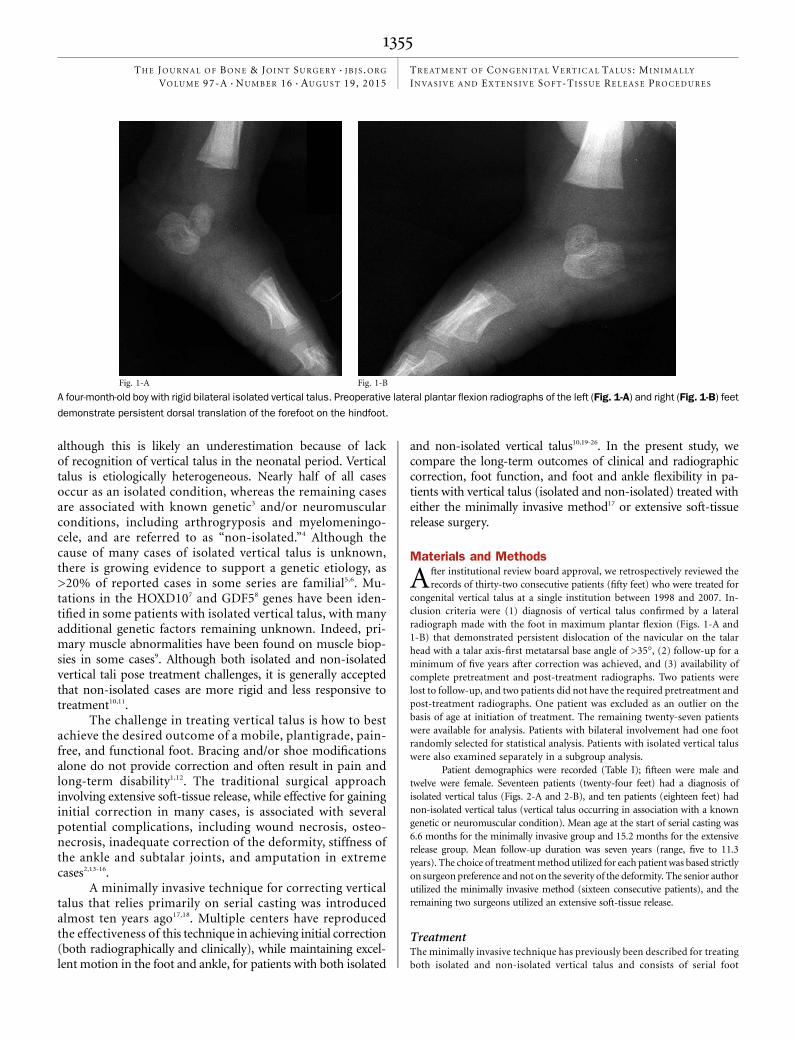
Fig. 1-A Fig. 1-B
A four-month-old boy with rigid bilateral isolated vertical talus. Preoperative lateral plantar flexion radiographs of the left (Fig. 1-A) and right (Fig. 1-B) feet
demonstrate persistent dorsal translation of the forefoot on the hindfoot.
1355
THE JOURNAL OF BONE & JOINT SURGERY d J B J S .ORG
VOLUME 97-A d NUMBER 16 d AUGUST 19, 2015TREATMENT OF CONGENITAL VERTICAL TALUS : MINIMALLY
INVAS IVE AND EXTENS IVE SOFT-TIS SUE RELEASE PROCEDURES

manipulation and casting followed by percutaneous talonavicular joint pinningand percutaneous Achilles tenotomy to correct hindfoot equinus, followed byshoe-and-bar bracing once casting is complete and the pin is removed
17,18,27. All
of the isolated vertical tali in this study were reduced with the above treatmentprotocol. In those non-isolated vertical tali inwhich serial casting did not result incomplete correction, a limited anterior subtalar joint capsulotomy, performedthrough a 1-cm dorsal skin incision, allowed the placement of an elevator tocomplete the reduction, which was followed by the treatment outlinedabove, involving pin fixation and tenotomy of the Achilles tendon. Werecommend that surgeons who are first utilizing this technique make thesmall dorsal skin incision to visualize reduction and aid in pin placement. Ifthe talonavicular joint is reduced under fluoroscopic visualization, capsu-lotomy is not necessary; if the joint is not reduced, then the surgeon pro-ceeds with a limited capsulotomy as outlined above.
For the patient cohort treated with extensive soft-tissue release sur-gery, this procedure was performed as a single-stage surgery and included
posterior capsulotomy of the ankle and subtalar joints, sectioning of thecalcaneofibular ligament, and capsulotomies of the calcaneocuboid and ta-lonavicular joints
28,29. Only one patient had release of the talocalcaneal in-
terosseous ligament.
Follow-up EvaluationsRecurrences were defined radiographically as any loss of correction of thetalonavicular reduction as measured on the lateral standing foot radio-graph. Patients diagnosed radiographically with evidence of recurrencehad a corresponding loss of ‡10� of plantar flexion. Radiographs of thefeet were made at the time of presentation, immediately postoperatively,and on an annual basis thereafter
30. Radiographic angles were measured
twice by the same examiner three weeks apart, and the mean of the twomeasurements was recorded. The examiner was blinded with regard tothe treatment group and previous measurement results and was not in-volved in the treatment of any of the patients. The same examiner
Fig. 2-A Fig. 2-B
Preoperative clinical photographs of both feet with vertical talus shown in the previous figures, also at the patient age of four months. Fig. 2-A The plantar
aspect of the right foot is convex. Fig 2-B There is a fixed forefoot adduction and hindfoot valgus deformity.
TABLE I Demographics of Patients with Vertical Talus Included in This Study
Minimally Invasive Extensive Surgery
Age at initial treatment* (mo) 6.6 (1.1 to 28.8) 15.2 (1.6 to 37.6)
Sex (no. of patients)
Male 10 5
Female 6 6
Diagnosis (no. of patients [no. of feet])
Isolated 10 (14) 7 (10)
Non-isolated 6 (10) 4 (8)
No. of casts†
Preop. 4 (2 to 6) 0 (0 to 2)
Postop. 2 (2 to 4) 2 (2 to 4)
*Values are given as the mean, with the range in parentheses. †Values are given as the median, with the interquartile range in parentheses.
1356
THE JOURNAL OF BONE & JOINT SURGERY d J B J S .ORG
VOLUME 97-A d NUMBER 16 d AUGUST 19, 2015TREATMENT OF CONGENITAL VERTICAL TALUS : MINIMALLY
INVAS IVE AND EXTENS IVE SOFT-TIS SUE RELEASE PROCEDURES

measured ankle range of motion of all patients with a handheldgoniometer.
At the latest visit, the PODCI (Pediatric Outcomes Data CollectionInstrument) questionnaire was completed by the parents or guardian
31. Both
standardized and normative scores were calculated on the basis of publishedguidelines (http://www.aaos.org/research/outcomes/outcomes_documentation.asp#pedsref). Standardized scores are raw scores reported on the range of 0 to100, with 100 being the best possible score; interpretation of the standardizedscore is not consistent among scales because of differences in how thegeneral healthy population scored. To make the scores comparable acrossvarious scales, the normative score was calculated on the basis of data fromthe general healthy population, which has a mean normative score of 50.Thus, a patient scoring >50 is above the mean of the general healthypopulation.
Statistical AnalysisPreoperative and postoperative limb-specific range of motion and preoper-ative radiographic measurements were compared for the four combinationsof treatment method utilized and presence or absence of an isolated verticaltalus using one-way analysis of variance (ANOVA). When the overall modelwas significant (p < 0.05), least-squares means were used to perform allpairwise between-group comparisons, with particular interest in the com-
parison between the two treatments for both isolated and non-isolated ver-
tical talus. These pairwise comparisons were adjusted for the performance of
multiple comparisons with the Tukey-Kramer method. Within the ANOVA, a
statistical contrast was used to test the a priori hypothesis that values for the
minimally invasive method were similar to those for the extensive-surgery
group, regardless of syndrome. For bilaterally affected patients, one foot
was randomly selected for analysis. Each foot was treated as an independent
observation.The change in radiographic measurements was compared across
groups using analysis of covariance (ANCOVA) in which the value at thelatest follow-up was the dependent variable, the four combinations oftreatment method and presence or absence of an isolated vertical taluswere the independent variables, and the preoperative value was the co-variate. Specific between-group comparisons were performed as described
above. A subset of PODCI domain scores was compared between patientstreated with minimally invasive and extensive surgery, regardless of syn-drome, by ANOVA. Because of violations of the assumptions required for
TABLE II Postoperative Ankle Range of Motion in Patients with Vertical Talus Included in This Study
Minimally Invasive Extensive Surgery P Value
All patients
No. of patients 16 11
Dorsiflexion* (deg) 18.5 ± 7.9 (5 to 30) 5.0 ± 3.9 (0 to 10) <0.0001
Plantar flexion* (deg) 23.9 ± 11.8 (0 to 35) 7.7 ± 5.2 (0 to 15) 0.0006
Total range of motion (deg)* 42.4 ± 18.0 (5 to 60) 12.7 ± 6.8 (5 to 25) <0.0001
Isolated vertical talus
No. of patients 10 7
Dorsiflexion* (deg) 21.1 ± 6.2 (10 to 30) 6.4 ± 3.8 (0 to 10) <0.0001
Plantar flexion* (deg) 30.7 ± 4.3 (20 to 35) 9.3 ± 5.3 (0 to 15) <0.0001
Total range of motion* (deg) 51.8 ± 6.9 (40 to 60) 15.7 ± 6.1 (10 to 25) <0.0001
Non-isolated vertical talus
No. of patients 6 4
Dorsiflexion* (deg) 14.2 ± 9.2 (5 to 30) 2.5 ± 2.9 (0 to 5) 0.03
Plantar flexion* (deg) 12.5 ± 11.7 (0 to 30) 5 ± 4.1 (0 to 10) 0.5
Total range of motion (deg)* 26.7 ± 20.4 (5 to 60) 7.5 ± 5.0 (5 to 15) 0.04
*Values are given as the mean and standard deviation, with the range in parentheses.
Fig. 3
Eleven years after correction of the vertical talus, thepatient in the previous
figures demonstrates neutral alignment of the hindfeet in stance.
1357
THE JOURNAL OF BONE & JOINT SURGERY d J B J S .ORG
VOLUME 97-A d NUMBER 16 d AUGUST 19, 2015TREATMENT OF CONGENITAL VERTICAL TALUS : MINIMALLY
INVAS IVE AND EXTENS IVE SOFT-TIS SUE RELEASE PROCEDURES
ANOVA, some variables were rank-transformed prior to analysis. TheStudent t test was used for comparison of subsequent procedures betweenthe minimally invasive and extensive soft-tissue release groups, and be-tween the isolated and non-isolated groups. Data for normally distributedvariables are reported as the mean and standard deviation. Variables thatwere not normally distributed are reported as the median and inter-quartile range (defined as the difference between the 25th and 75thpercentiles).
Source of FundingOne of the authors (J.S.Y.) received an OREF (Orthopaedic Research and Ed-ucation Foundation) Resident Research Grant.
ResultsRange of Motion
The mean postoperative ankle arc of motion was greater inpatients treated with the minimally invasive method com-
paredwith those treated with extensive soft-tissue release surgery(42.4� versus 12.7�, p < 0.0001) (Table II). Mean dorsiflexionwas 18.5� in the minimally invasive group compared with 5.0�in the extensive soft-tissue release group (p < 0.0001). Meanplantar flexion was 23.9� in the minimally invasive groupcompared with 7.7� in the extensive soft-tissue release group(p = 0.0006) (Fig. 3).
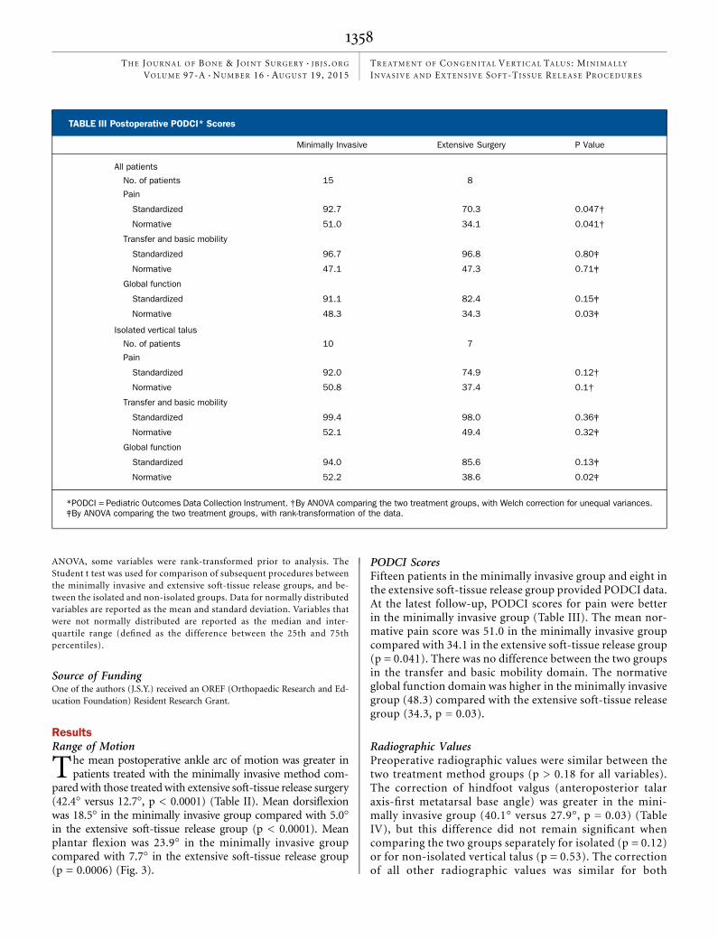
PODCI ScoresFifteen patients in the minimally invasive group and eight inthe extensive soft-tissue release group provided PODCI data.At the latest follow-up, PODCI scores for pain were betterin the minimally invasive group (Table III). The mean nor-mative pain score was 51.0 in the minimally invasive groupcompared with 34.1 in the extensive soft-tissue release group(p = 0.041). There was no difference between the two groupsin the transfer and basic mobility domain. The normativeglobal function domain was higher in the minimally invasivegroup (48.3) compared with the extensive soft-tissue releasegroup (34.3, p = 0.03).
Radiographic ValuesPreoperative radiographic values were similar between thetwo treatment method groups (p > 0.18 for all variables).The correction of hindfoot valgus (anteroposterior talaraxis-first metatarsal base angle) was greater in the mini-mally invasive group (40.1� versus 27.9�, p = 0.03) (TableIV), but this difference did not remain significant whencomparing the two groups separately for isolated (p = 0.12)or for non-isolated vertical talus (p = 0.53). The correctionof all other radiographic values was similar for both
TABLE III Postoperative PODCI* Scores
Minimally Invasive Extensive Surgery P Value
All patients
No. of patients 15 8
Pain
Standardized 92.7 70.3 0.047†
Normative 51.0 34.1 0.041†
Transfer and basic mobility
Standardized 96.7 96.8 0.80‡
Normative 47.1 47.3 0.71‡
Global function
Standardized 91.1 82.4 0.15‡
Normative 48.3 34.3 0.03‡
Isolated vertical talus
No. of patients 10 7
Pain
Standardized 92.0 74.9 0.12†
Normative 50.8 37.4 0.1†
Transfer and basic mobility
Standardized 99.4 98.0 0.36‡
Normative 52.1 49.4 0.32‡
Global function
Standardized 94.0 85.6 0.13‡
Normative 52.2 38.6 0.02‡
*PODCI = Pediatric Outcomes Data Collection Instrument. †By ANOVA comparing the two treatment groups, with Welch correction for unequal variances.‡By ANOVA comparing the two treatment groups, with rank-transformation of the data.
1358
THE JOURNAL OF BONE & JOINT SURGERY d J B J S .ORG
VOLUME 97-A d NUMBER 16 d AUGUST 19, 2015TREATMENT OF CONGENITAL VERTICAL TALUS : MINIMALLY
INVAS IVE AND EXTENS IVE SOFT-TIS SUE RELEASE PROCEDURES

treatment method groups (p > 0.1 for all variables) (Figs. 4-Aand 4-B).
Isolated Vertical TalusDorsiflexion was significantly greater in the minimally invasivegroup (21.1� versus 6.4�, p < 0.0001), as were plantar flexion(30.7� versus 9.3�, p < 0.0001) and total range of motion (51.8�versus 15.7�, p < 0.0001). Analysis of the PODCI scores showed
that the normative score for global function was signifi-cantly higher in the minimally invasive group (52.2 versus 38.6,p = 0.02).
Radiographic measures of severity were greater pre-operatively in the minimally invasive group, although theamount of correction achieved was the same in both groupsfor most measures (Table V). The only exception was that thecorrection of the anteroposterior talocalcaneal angle was
Fig. 4-A Fig. 4-B
Lateral standing radiographs of the patient in the previous figures, also eleven years after correction, demonstrate normal relationships between the talus
and the calcaneus and between the tibia and the calcaneus.
TABLE IV Radiographic Values of All Feet with Vertical Talus Included in This Study
AngleMinimally Invasive
(N = 16 Patients)* (deg)Extensive Surgery
(N = 11 Patients)* (deg) P Value†
Anteroposterior talocalcaneal
Preop. 42.6 ± 12.1 39.4 ± 18.4 0.97‡
Latest follow-up 15.8 ± 9.2 9.3 ± 11.2
Correction 226.8 ± 9.7 230.1 ± 24.3 0.26§
Anteroposterior talar axis-first metatarsal base
Preop. 46.6 ± 25.7 35.0 ± 35.7 0.59‡
Latest follow-up 6.4 ± 4.0 7.1 ± 14.5
Correction 240.1 ± 25.5 227.9 ± 29.9 0.03§
Lateral talocalcaneal
Preop. 59.4 ± 15.5 54.5 ± 15.3 0.18‡
Latest follow-up 30.8 ± 8.6 27.5 ± 11.5
Correction 228.7 ± 14.3 227.0 ± 20.7 0.68§
Lateral talar axis-first metatarsal base
Preop. 78.3 ± 14.2 81.2 ± 9.6 0.44‡
Latest follow-up 5.3 ± 9.6 17.3 ± 26.8
Correction 273.0 ± 16.8 263.9 ± 28.7 0.10§
Lateral tibiocalcaneal
Preop. 109.1 ± 14.0 106.1 ± 22 0.56‡
Latest follow-up 76.4 ± 12.7 78.7 ± 13.1
Correction 232.6 ± 23.1 227.4 ± 29.8 0.93§
*Values are given as the mean and standard deviation. †Data were rank-transformed prior to analysis. ‡By ANOVA statistical contrast comparingall four of the subgroups: isolated and non-isolated vertical talus treated with minimally invasive and extensive surgery. §By ANCOVA statisticalcontrast comparing all four of the subgroups.
1359
THE JOURNAL OF BONE & JOINT SURGERY d J B J S .ORG
VOLUME 97-A d NUMBER 16 d AUGUST 19, 2015TREATMENT OF CONGENITAL VERTICAL TALUS : MINIMALLY
INVAS IVE AND EXTENS IVE SOFT-TIS SUE RELEASE PROCEDURES
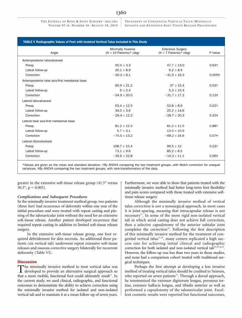
greater in the extensive soft-tissue release group (41.5� versus30.3�, p = 0.005).
Complications and Subsequent ProceduresIn the minimally invasive treatment method group, two patients(three feet) had recurrence of deformity within one year of theinitial procedure and were treated with repeat casting and pin-ning of the talonavicular joint without the need for an extensivesoft-tissue release. Another patient developed recurrence thatrequired repeat casting in addition to limited soft-tissue releasesurgery.
In the extensive soft-tissue release group, one foot re-quired debridement for skin necrosis. An additional three pa-tients (six vertical tali) underwent repeat extensive soft-tissuereleases and osseous corrective surgery bilaterally for recurrentdeformity (Table VI).
Discussion
The minimally invasive method to treat vertical talus wasdeveloped to provide an alternative surgical approach so
that a more mobile, functional foot could ultimately result17. Inthe current study, we used clinical, radiographic, and functionaloutcomes to demonstrate the ability to achieve correction usingthe minimally invasive method for isolated and non-isolatedvertical tali and to maintain it at a mean follow-up of seven years.
Furthermore, we were able to show that patients treated with theminimally invasive method had better long-term foot flexibilityand pain scores compared with those treated with extensive soft-tissue release surgery.
Although the minimally invasive method of verticaltalus correction is not a nonsurgical approach, in most casesit is joint-sparing, meaning that intracapsular release is notnecessary17. In some of the more rigid non-isolated verticaltali in which serial casting does not achieve full correction,then a selective capsulotomy of the anterior subtalar jointcompletes the correction10. Following the first descriptionof this minimally invasive method for the treatment of con-genital vertical talus17,18, many centers replicated a high suc-cess rate for achieving initial clinical and radiographiccorrection for both isolated and non-isolated vertical tali10,19-26,32.However, the follow-up was less than two years in those studies,and none had a comparison cohort treated with traditional sur-gical techniques.
Perhaps the first attempt at developing a less invasivemethod of treating vertical talus should be credited to Seimon,who reported on seven patients33. Through a dorsal approach,he tenotomized the extensor digitorum longus, peroneus ter-tius, extensor hallucis longus, and tibialis anterior as well asperformed a capsulotomy of the talonavicular joint. Excel-lent cosmetic results were reported but functional outcomes,
TABLE V Radiographic Values of Feet with Isolated Vertical Talus Included in This Study
AngleMinimally Invasive
(N = 10 Patients)* (deg)Extensive Surgery
(N = 7 Patients)* (deg) P Value
Anteroposterior talocalcaneal
Preop. 50.4 ± 3.3 47.7 ± 13.0 0.63†
Latest follow-up 20.1 ± 8.9 6.2 ± 8.9
Correction 230.3 ± 8.1 241.5 ± 16.3 0.005‡
Anteroposterior talar axis-first metatarsal base
Preop. 60.9 ± 21.2 37 ± 15.4 0.03†
Latest follow-up 6 ± 2.4 5.3 ± 15.4
Correction 254.9 ± 20.0 231.7 ± 17.2 0.12‡
Lateral talocalcaneal
Preop. 63.4 ± 12.5 53.8 ± 8.9 0.02†
Latest follow-up 34.0 ± 3.6 25.2 ± 14.6
Correction 229.4 ± 12.3 228.7 ± 20.3 0.42‡
Lateral talar axis-first metatarsal base
Preop. 81.2 ± 12.3 81.2 ± 11.5 0.88†
Latest follow-up 5.7 ± 3.1 13.0 ± 10.5
Correction 275.5 ± 13.2 268.2 ± 16.6 0.07‡
Lateral tibiocalcaneal
Preop. 108.7 ± 15.4 99.5 ± 12 0.23†
Latest follow-up 73.1 ± 9.6 85.2 ± 8.5
Correction 235.6 ± 22.8 214.3 ± 11.1 0.06‡
*Values are given as the mean and standard deviation. †By ANOVA comparing the two treatment groups, with Welch correction for unequalvariances. ‡By ANOVA comparing the two treatment groups, with rank-transformation of the data.
1360
THE JOURNAL OF BONE & JOINT SURGERY d J B J S .ORG
VOLUME 97-A d NUMBER 16 d AUGUST 19, 2015TREATMENT OF CONGENITAL VERTICAL TALUS : MINIMALLY
INVAS IVE AND EXTENS IVE SOFT-TIS SUE RELEASE PROCEDURES

TABLE VI Detailed Patient Data
Patient
Age atInitiation ofTreatment
(mo)AssociatedAnomalies Side
No. ofCastsPrior toSurgery
PrimaryProcedures*
Age atInitial
Revision†(mo)
SubsequentProcedures
Age atFinal
Follow-up(mo)
Minimallyinvasivemethod
1 29 None Right 2 A, B 33 Limitedcalcaneocuboidjoint capsulotomy;tibialis anteriortendon, peroneusbrevis, andextensordigitorumlongus tendonlengthening
124
2 1 None Right 6 A, B NA None 81
2 1 None Left 6 A, B NA None 81
3 4 None Right 4 A, B NA None 78
4 3 None Right 6 A, B NA None 78
4 3 None Left 6 A, B NA None 78
5 4 None Right 5 A, B NA None 62
6 4 None Right 6 A, B NA None 65
7 2 Brachydactyly,facialdysmorphism
Right 4 A, B 3 Revision ofpin stickingout of skin
85
7 2 Brachydactyly,facialdysmorphism
Left 4 A, B 7 None 85
8 2 Sacralagenesis,fatty filum
Left 7 A, B, C NA None 88
9 15 Arthrogryposis Right 6 A, B, C 48 Talonavicularandcalcaneocuboidjointcapsulotomies
97
9 15 Arthrogryposis Left 6 A, B, C 48 Talonavicular andcalcaneocuboidjointcapsulotomies
97
10 2 Complexpolydactylyand syndactylyof hands andfeet, amnioticband syndrome
Right 6 A, B 3 Revision of pinsticking out ofskin
63
10 2 Complexpolydactyly andsyndactyly ofhands and feet,amniotic bandsyndrome
Left 6 A, B 3 Revision of pinsticking out ofskin
63
11 4 Sacral agenesis,caudal regression
Left 7 A, B, C NA None 80
continued
1361
THE JOURNAL OF BONE & JOINT SURGERY d J B J S .ORG
VOLUME 97-A d NUMBER 16 d AUGUST 19, 2015TREATMENT OF CONGENITAL VERTICAL TALUS : MINIMALLY
INVAS IVE AND EXTENS IVE SOFT-TIS SUE RELEASE PROCEDURES
TABLE VI (continued)
Patient
Age atInitiation ofTreatment
(mo)AssociatedAnomalies Side
No. ofCastsPrior toSurgery
PrimaryProcedures*
Age atInitial
Revision†(mo)
SubsequentProcedures
Age atFinal
Follow-up(mo)
12 15 Myelodysplasia,choanal atresia,tracheomalacia
Right 5 A, B, C NA None 80
12 15 Myelodysplasia,choanal atresia,tracheomalacia
Left 5 A, B, C NA None 80
13 5 None Right 5 A, B NA None 82
13 5 None Left 5 A, B NA None 82
14 7 None Right 5 A, B NA None 117
14 7 None Left 4 A, B NA None 117
15 5 None Right 4 A, B NA None 123
16 5 None Left 5 A, B NA None 111
Extensivesoft-tissuerelease
17 7 None Right 0 D 24 Medial cuneiformosteotomy,circumferentialsubtalar release,calcanealosteotomy,lateral columnlengthening
113
17 7 None Left 0 D 24 Medial cuneiformosteotomy,circumferentialsubtalar release,calcanealosteotomy,lateral columnlengthening
113
18 31 Escobarsyndrome,vertical talus,kyphosis
Right 0 D, E 73 Partial calcanealexcision, medialand plantarexostosis excision
134
18 31 Escobarsyndrome,vertical talus,kyphosis
Left 0 D, E 73 Partial calcanealexcision, medialand plantarexostosis excision
134
19 24 Arthrogryposis Right 0 D NA None 88
19 24 Arthrogryposis Left 0 D NA None 88
20 37 Arthrogryposis Right 0 D NA None 86
20 37 Arthrogryposis Left 0 D NA None 86
21 14 Congenitalmusculardystrophy
Right 0 D NA None 84
21 14 Congenitalmusculardystrophy
Left 0 D NA None 84
22 31 None Left 0 D NA None 159
23‡ 106 None Right 0 D, F NA None 214continued
1362
THE JOURNAL OF BONE & JOINT SURGERY d J B J S .ORG
VOLUME 97-A d NUMBER 16 d AUGUST 19, 2015TREATMENT OF CONGENITAL VERTICAL TALUS : MINIMALLY
INVAS IVE AND EXTENS IVE SOFT-TIS SUE RELEASE PROCEDURES

including the ability to dorsiflex toes or ankles, were not de-scribed. The advantage of the minimally invasive approach thatwe utilized in the current study is that it relies on serial casting togradually stretch the dorsolateral soft tissues so that, unlike withSeimon’s approach, tenotomies of the dorsolateral tendons arenot necessary and in most cases the talonavicular joint is fullyreduced with casting alone.
Since Seimon published his original article, there havebeen no further published studies assessing the efficacy of thetechnique, to our knowledge. Instead, more extensive soft-tissue release procedures have been developed15,28,34-36. Althoughgood correction can be achieved with these extensive surgicalprocedures, long-term problems are reported, including stiff-ness of the ankle and subtalar joints13-15. Patients with clubfoottreated with extensive soft-tissue releases have similar long-term problems, and this recognition contributed to the pop-ularity of the Ponseti method of clubfoot management,whereby intracapsular joint surgery is avoided in the majorityof patients37. It has, in fact, been hypothesized that the mini-
mization of scar tissue formation in the growing foot achievedwith the Ponseti method results in long-term improvement offoot mobility, foot function, and quality of life37 compared withclubfeet treated with extensive soft-tissue release surgery38,39.On the basis of our findings, the goal of vertical talus treatmentshould also be to provide correction with the least invasivemethod possible.
Although recurrences occurred in both the minimally in-vasive method and the extensive surgery group, two of the threepatients in the minimally invasive method group were treatedwith repeat casting and did not require extensive soft-tissue re-lease, whereas all three patients in the extensive-surgery groupwith recurrences went on to have more extensive soft-tissuereleases and osseous surgical procedures. Applying the principlesof the minimally invasive method to treat recurrences is thuseffective and can minimize the amount of surgery required withthe goal of maintaining mobility.
The results of this study must be interpreted in light of thefollowing limitations. First, our study design is retrospective.
TABLE VI (continued)
Patient
Age atInitiation ofTreatment
(mo)AssociatedAnomalies Side
No. ofCastsPrior toSurgery
PrimaryProcedures*
Age atInitial
Revision†(mo)
SubsequentProcedures
Age atFinal
Follow-up(mo)
23‡ 135 None Left 0 D NA None 214
24 12 None Right 0 D NA None 73
24 12 None Left 0 D NA None 73
25 11 None Right 0 D NA None 75
26 12 None Right 0 D 13 Debridementof skin necrosis
84
27 8 None Left 1 D NA None 123
28 15 None Right 2 D 77 Medial cuneiformexcision, medialsubtalar release,peroneal tendonrelease, lateralcolumnlengtheningthroughcalcaneocuboidjoint
141
28 15 None Left 2 D 77 Medial cuneiformexcision, medialsubtalar release,peroneal tendonrelease, lateralcolumnlengtheningthroughcalcaneocuboidjoint
141
*A = percutaneous Achilles tenotomy, B = percutaneous pinning of talonavicular joint, C = limited anterior subtalar joint capsulotomy,D = capsulotomies of posterior ankle and subtalar joints, calcaneocuboid joint, and talonavicular joint, E = interosseous talocalcaneal ligamentrelease, and F = navicular excision. †NA = not applicable. ‡This patient was excluded on account of age.
1363
THE JOURNAL OF BONE & JOINT SURGERY d J B J S .ORG
VOLUME 97-A d NUMBER 16 d AUGUST 19, 2015TREATMENT OF CONGENITAL VERTICAL TALUS : MINIMALLY
INVAS IVE AND EXTENS IVE SOFT-TIS SUE RELEASE PROCEDURES

It would now be difficult to perform a prospective study, as theminimally invasive method has become the standard of care forinitial treatment of vertical talus because of the more favorableshort-term results. Second, one patient who was to receiveminimally invasive surgery crossed over to the extensive-surgerytreatment arm; this patient was analyzed in the original treat-ment group. Although we are not able to quantify the potentialbias that this introduces in our conclusions, we note that thiswould likely have biased our results against the noninvasivemethod since the patient was analyzed in that group even thoughboth feet eventually needed more extensive surgery. Third, wedid not record subtalar motion. Fourth, the age at the start oftreatment differed between the two groups. Finally, as a conse-quence of the rarity of congenital vertical talus, the number ofpatients in this study was small. As sample size limitations pre-cluded an examination of the possible interaction between thetreatment group and the underlying syndrome and/or neuro-muscular condition, we were unable to statistically determinewhether the underlying etiology impacts treatment outcomes.However, because vertical talus, like many musculoskeletal dis-orders, is genetically and etiologically heterogeneous, the un-derlying cause is likely to affect outcomes, as studies (includingours) suggest10,19,20,22,23,25.
We hypothesize that by minimizing intracapsular surgerythrough theminimally invasive treatmentmethod, less scar tissuewill be generated in the growing foot, leading to improved footand ankle mobility. Better motion is thought to lead to superiorlong-term outcomes with the Ponseti method37 and is likely thefactor also contributing to improved outcomes with the mini-mally invasive method for vertical talus correction. Longer-termstudies are necessary to determine if the improved outcomes aremaintained into adulthood. nNOTE: The authors thank Karen Steger-May for her statistical analysis and Perry Schoenecker andMargaret Rich for their contribution of patients to this study.
Justin S. Yang, MDDepartment of Orthopaedic Surgery,Washington University School of Medicine,660 South Euclid Avenue,St. Louis, MO 63110
Matthew B. Dobbs, MDSt. Louis Children’s Hospital,1 Children’s Place, Suite 4S-60,St. Louis, MO 63110.E-mail address: [email protected]
References
1. Drennan JC. Congenital vertical talus. Instr Course Lect. 1996;45:315-22.2. Jacobsen ST, Crawford AH. Congenital vertical talus. J Pediatr Orthop. 1983 Jul;3(3):306-10.3. Townes PL, Dehart GK Jr, Hecht F, Manning JA. Trisomy 13-15 in a male infant.J Pediatr. 1962 Apr;60:528-32.4. Sharrard WJ, Grosfield I. Themanagement of deformity and paralysis of the foot inmyelomeningocele. J Bone Joint Surg Br. 1968 Aug;50(3):456-65.5. Ogata K, Schoenecker PL, Sheridan J. Congenital vertical talus and its familialoccurrence: an analysis of 36 patients. Clin Orthop Relat Res. 1979 Mar-Apr;139:128-32.6. Dobbs MB, Schoenecker PL, Gordon JE. Autosomal dominant transmission ofisolated congenital vertical talus. Iowa Orthop J. 2002;22:25-7.7. Dobbs MB, Gurnett CA, Pierce B, Exner GU, Robarge J, Morcuende JA, ColeWG, Templeton PA, Foster B, Bowcock AM. HOXD10 M319K mutation in afamily with isolated congenital vertical talus. J Orthop Res. 2006 Mar;24(3):448-53.8. Dobbs MB, Gurnett CA, Robarge J, Gordon JE, Morcuende JA, Bowcock AM.Variable hand and foot abnormalities in family with congenital verticaltalus and CDMP-1 gene mutation. J Orthop Res. 2005 Nov;23(6):1490-4. Epub2005 Jul 11.9. Merrill LJ, Gurnett CA, Connolly AM, Pestronk A, Dobbs MB. Skeletal muscleabnormalities and genetic factors related to vertical talus. Clin Orthop Relat Res.2011 Apr;469(4):1167-74. Epub 2010 Jul 20.10. Chalayon O, Adams A, Dobbs MB. Minimally invasive approach for the treat-ment of non-isolated congenital vertical talus. J Bone Joint Surg Am. 2012 Jun 6;94(11):e73.11. Yan G, Yu Z, Yang Z, Lu M, Zhang J. [Surgical correction of congenital verticaltalus by one-stage comprehensive soft-tissue release and peritalar reduction in-corporating tibialis anterior transfer]. Zhonghua Yi Xue Za Zhi. 2014 May 6;94(17):1322-5. Chinese.12. Coleman SS, Stelling FH 3rd, Jarrett J. Pathomechanics and treatment of con-genital vertical talus. Clin Orthop Relat Res. 1970 May-Jun;70:62-72.13. Dodge LD, Ashley RK, Gilbert RJ. Treatment of the congenital vertical talus: aretrospective review of 36 feet with long-term follow-up. Foot Ankle. 1987 Jun;7(6):326-32.14. Zorer G, Bagatur AE, Dogan A. Single stage surgical correction of congenitalvertical talus by complete subtalar release and peritalar reduction by using theCincinnati incision. J Pediatr Orthop B. 2002 Jan;11(1):60-7.15. Mazzocca AD, Thomson JD, Deluca PA, Romness MJ. Comparison of the pos-terior approach versus the dorsal approach in the treatment of congenital verticaltalus. J Pediatr Orthop. 2001 Mar-Apr;21(2):212-7.
16. Hootnick DR, Dutch WM Jr, Crider RJ Jr. Ischemic necrosis leading to amputa-tion following surgical correction of congenital vertical talus. Am J Orthop (BelleMead NJ). 2005 Jan;34(1):35-7.17. Dobbs MB, Purcell DB, Nunley R, Morcuende JA. Early results of a new methodof treatment for idiopathic congenital vertical talus. J Bone Joint Surg Am. 2006Jun;88(6):1192-200.18. Dobbs MB, Purcell DB, Nunley R, Morcuende JA. Early results of a new methodof treatment for idiopathic congenital vertical talus. Surgical technique. J Bone JointSurg Am. 2007 Mar;89(Suppl 2 Pt.1):111-21.19. Sweet LA, OʼNeill LM, Dobbs MB. Serial casting for neuromuscular flatfoot andvertical talus in an adolescent with hereditary spastic paraplegia. Pediatr Phys Ther.2014 Summer;26(2):253-64.20. Eberhardt O, Fernandez FF, Wirth T. [Treatment of vertical talus with theDobbs method]. Z Orthop Unfall. 2011 Apr;149(2):219-24. Epub 2011 Apr 5.German.21. Eberhardt O, Fernandez FF, Wirth T. The talar axis-first metatarsal base anglein CVT treatment: a comparison of idiopathic and non-idiopathic cases treatedwith the Dobbs method. J Child Orthop. 2012 Dec;6(6):491-6. Epub 2012 Nov10.22. Aslani H, Sadigi A, Tabrizi A, Bazavar M, Mousavi M. Primary outcomes of thecongenital vertical talus correction using the Dobbs method of serial casting andlimited surgery. J Child Orthop. 2012 Aug;6(4):307-11. Epub 2012 Aug 18.23. Wright J, Coggings D, Maizen C, Ramachandran M. Reverse Ponseti-typetreatment for children with congenital vertical talus: comparison between idiopathicand teratological patients. Bone Joint J. 2014 Feb;96-B(2):274-8.24. Khader A, Huntley JS. Congenital vertical talus in cri du chat syndrome: a casereport. BMC Res Notes. 2013;6(1):270. Epub 2013 Jul 13.25. Aydın A, Atmaca H, Muezzinoglu US. Bilateral congenital vertical talus withsevere lower extremity external rotational deformity: treated by reverse Ponsetitechnique. Foot (Edinb). 2012 Sep;22(3):252-4. Epub 2012 May 4.26. Bhaskar A. Congenital vertical talus: treatment by reverse Ponseti technique.Indian J Orthop. 2008 Jul;42(3):347-50.27. Alaee F, Boehm S, Dobbs MB. A new approach to the treatment of congenitalvertical talus. J Child Orthop. 2007 Sep;1(3):165-74. Epub 2007 Aug 1.28. Stricker SJ, Rosen E. Early one-stage reconstruction of congenital vertical talus.Foot Ankle Int. 1997 Sep;18(9):535-43.29. Colton CL. The surgical management of congenital vertical talus. J Bone JointSurg Br. 1973 Aug;55(3):566-74.30. Vanderwilde R, Staheli LT, Chew DE, Malagon V. Measurements on radiographsof the foot in normal infants and children. J Bone Joint Surg Am. 1988 Mar;70(3):407-15.
1364
THE JOURNAL OF BONE & JOINT SURGERY d J B J S .ORG
VOLUME 97-A d NUMBER 16 d AUGUST 19, 2015TREATMENT OF CONGENITAL VERTICAL TALUS : MINIMALLY
INVAS IVE AND EXTENS IVE SOFT-TIS SUE RELEASE PROCEDURES

31. Daltroy LH, Liang MH, Fossel AH, Goldberg MJ; Pediatric Outcomes InstrumentDevelopment Group. Pediatric Orthopaedic Society of North America. The POSNApediatric musculoskeletal functional health questionnaire: report on reliability,validity, and sensitivity to change. J Pediatr Orthop. 1998 Sep-Oct;18(5):561-71.32. Eberhardt O, Wirth T, Fernandez FF. [Minimally invasive treatment of congenitalfoot deformities in infants: new findings and midterm-results]. Orthopade. 2013Dec;42(12):1001-7. German.33. Seimon LP. Surgical correction of congenital vertical talus under the age of 2years. J Pediatr Orthop. 1987 Jul-Aug;7(4):405-11.34. Duncan RD, Fixsen JA. Congenital convex pes valgus. J Bone Joint Surg Br.1999 Mar;81(2):250-4.
35. NapiontekM. Congenital vertical talus: a retrospective and critical review of 32 feetoperated on by peritalar reduction. J Pediatr Orthop B. 1995;4(2):179-87.36. Wirth T, Schuler P, Griss P. Early surgical treatment for congenital vertical talus.Arch Orthop Trauma Surg. 1994;113(5):248-53.37. Cooper DM, Dietz FR. Treatment of idiopathic clubfoot. A thirty-year follow-upnote. J Bone Joint Surg Am. 1995 Oct;77(10):1477-89.38. Dobbs MB, Nunley R, Schoenecker PL. Long-term follow-up of patients withclubfeet treated with extensive soft-tissue release. J Bone Joint Surg Am. 2006May;88(5):986-96.39. Ippolito E, Farsetti P, Caterini R, Tudisco C. Long-term comparative results inpatients with congenital clubfoot treated with two different protocols. J Bone JointSurg Am. 2003 Jul;85(7):1286-94.
1365
THE JOURNAL OF BONE & JOINT SURGERY d J B J S .ORG
VOLUME 97-A d NUMBER 16 d AUGUST 19, 2015TREATMENT OF CONGENITAL VERTICAL TALUS : MINIMALLY
INVAS IVE AND EXTENS IVE SOFT-TIS SUE RELEASE PROCEDURES