TREATMENT CONCEPTS FOR MINOR BONE …oss.dentalead.co.jp/download/TreatmentConcepts_Minor...2 –...
24
1 TREATMENT CONCEPTS FOR MINOR BONE AUGMENTATION
Transcript of TREATMENT CONCEPTS FOR MINOR BONE …oss.dentalead.co.jp/download/TreatmentConcepts_Minor...2 –...

1
TREATMENT CONCEPTS FOR
MINOR BONE AUGMENTATION

2 – TREATMENT CONCEPTS MINOR BONE AUGMENTATION
TABLE OF CONTENTS
3 Minor bone defects: key factors for successful treatment
5 Long-term success with the dream-team
6–9 Clinical Cases Class 0 Prof. Ronald Jung Dr. Benoit Brochery & Dr. Gary Finelle Dr. Raffaele Cavalcanti Dr. Paul Rosen
10–11 Clinical Cases Class I
Dr. Ueli Grunder Dr. Su Yu Cheng
12–13 Treatment Decisions in Minor Bone Augmentation
14 Clinical Cases Class I
PD Dr. Dr. Markus Schlee
15–20 Clinical Cases Class II Dr. Teppei Tsukiyama Prof. Ui-Won Jung Dr. João Batista César Neto Dr. Colin Campbell Dr. Marlene Teo Prof. Saso Ivanovski
21 Recommendend material combinations I References
22 Product Range
“The GBR technique with the combination of autologous bone chips, Geistlich Bio-Oss® and Geistlich Bio-Gide® offers predictable outcomes for contour augmentation. It’s important that the products are both clinically and scientifically well documented.” Prof. Daniel Buser | Switzerland
2

3
Minor Bone Augmentation: Bone defects around implants
Guided Bone Regeneration (GBR) is a well-documented and successful method for bone augmentation. The term Minor Bone Augmentation is used to describe GBR procedures around an implant when a bony defect leaves part of the implant surface exposed. Therefore Minor Bone Augmentation is defined as the treatment of a bone defect around an implant and amongst others includes dehiscences and apical fenestrations.1
In the present treatment concepts we focus on minor bone augmen-tations and differentiate them from major bone augmentation and ridge preservation procedures. Major bone augmentation includes staged approaches for implant placement. Ridge preservation may be accomplished by grafting of the extraction socket with biomate-rial in order to avoid ridge resorption after tooth extraction. (Tab. 1)
Decision criteria for implant placement with a simultaneous GBRBesides general factors like smoking behavior, age, disease, etc. local factors play an important role for a successful GBR procedure. General factors cannot be influenced by the clinician whereas
MINOR BONE DEFECTS: KEY FACTORS FOR SUCCESSFUL TREATMENT
l ocal factors are influenced by the decision-making of the clini-cian. The first and most important local factor is the ratio between the surface area of the exposed bone and the defect volume to be regenerated. Whereas the bone regeneration depends on the number of bone walls available that can contribute to new bone formation, a simple rule summarizes: the more bone walls are avail-able in a defect area, the better the healing potential in a given defect site. One-wall defects are more demanding than two- or three-wall defects.The 3 most important criteria for implant placement with simul-taneous GBR are:2
1. CORRECT 3D-IMPLANT POSITION(Fig. 1–3) The implant must be placed in a correct three-dimen-sional position in order to obtain an optimal functional and esthetic outcome.2
2. PRIMARY STABILITYPrimary implant stability has to be achieved for a predictable osseointegration.2
3. FAVORABLE MORPHOLOGY(Fig. 4–6) The peri-implant bone defect must have a favorable defect morphology to allow predictable bone regeneration of the defect area and successful implant placement.2
Theo
rie
EXTRACTION SOCKETMANAGEMENT
MINOR BONE AUGMENTATION
MAJOR BONEAUGMENTATION
Ridge Preservation procedures irrespective of the prosthetic restoration time point (implant/bridge).
Implant placement combined with GBR
GBR prior to implant placement / GBR with form stable compon-tents / sinus elevation
See “Treatment Concepts for Extraction Sockets”
See online version of the present “Treatment Concepts for Minor Bone Augmentations”
See “Innovative Treatment Con-cepts in Oral and Maxillofacial Surgery”
TAB. 1: DIFFERENT THERAPEUTIC AREAS WITH VARIOUS TREATMENT SOLUTIONS:

4 – TREATMENT CONCEPTS MINOR BONE AUGMENTATION
“I have been practicing implant dentistry for more than 20 years. The majority of that time I have exclusively used Geistlich products for treatment involving bone regeneration. The results, year after year, are exceptionally predictable.” Jay Beagle | USA
Favorable defect morphology1 and treatment with Geistlich biomaterials Successful Minor Bone Augmentation with Geistlich Bio-Oss® and Geistlich Bio-Gide® can be expected if the following defect morphology is prevalent:
FIG. 4 CONTOUR DEFICIT: CLASS 0Implant is placed in a bony envelope, but a contour augmenta-tion is necessary for an esthetic outcome. GBR procedure with Geistlich Bio-Oss® and Geistlich Bio-Gide® is performed in these situations. (Picture by courtesy of Dr. Raffaele Cavalcanti)
FIG. 5 INTRA-ALVEOLAR DEFECT: CLASS IImplant is placed and an intra-alveolar gap defect is visible. A GBR procedure is performed with Geistlich Bio-Oss®17 and Geistlich Bio-Gide® to fill the gap between the implant surface
and the intact bone walls. If necessary, combined GBR procedure is performed with Geistlich Bio-Oss® and Geistlich Bio-Gide® to also account for filling the gap as well as for contour augmenta-tion. (Picture by courtesy of Dr. Su Yu Cheng)
FIG. 6 DEHISCENCE-TYPE DEFECT: CLASS IIAfter implant placement a peri-implant defect is visible with deficient bone wall. The GBR procedure is performed with Geistlich Bio-Oss® and Geistlich Bio-Gide® to fill the defect and adapt the contour augmentation.3,9 (Picture by courtesy of Dr. Teppei Tsukiyama)
4 5 6
Correct 3D implant position The placement of implants in a correct three-dimensional position is one of the keys to an esthetic and functional treatment outcome:
FIG.1 MESIODISTAL DIMENSIONMinimal distance to neighboring teeth or implants needs to be respected at implant placement to prevent vertical bone loss on ad-jacent teeth in the mesiodistal dimension. Correct implant position in the comfort zone (blue zone fig. 1) avoiding the danger zone (red zone fig. 1), which is dependent on the nature of adjacent structures.3
FIG. 2 OROFACIAL DIMENSIONIn the orofacial dimension, the implant shoulder should be po-sitioned in the comfort zone (blue zone fig. 2) which measures about 1.5–2.0 mm in width when measured from the ideal point
of emergence. The danger zones (red zone fig. 2) are located both facially and palatally from the comfort zone.3
FIG. 3 CORONOAPICAL DIMENSIONIn the coronoapical dimension, the comfort zone (blue zone fig. 3) is a narrow band of about 1.0 mm and is dependent on implant systems and manufacturers recommendations. In general, the implant shoulder should be positioned approximately 2.0 mm apical to the mid-facial gingival margin of the implant restoration. Apical and coronal to this narrow band the danger zone (red zone fig. 3) is positioned.3
1
1.5 mm 1.5 mm
2
1.5–2.0 mm
3
2.0 mm
1.0 mm

5
LONG-TERM SUCCESS WITH THE DREAM-TEAM
GBR with the dream-team: Geistlich Bio-Oss® and Geistlich Bio-Gide® for outstanding and predictable clinical outcomesImplant placement and simultaneous GBR with Geistlich Bio-Oss® and Geistlich Bio-Gide® perform as well as implant placement into native bone with respect to implant survival, marginal bone height and peri-implant soft tissue parameters (cross-sectional retrospec-tive study).4 According to studies on simultaneous implantation and GBR with Geistlich Bio-Oss® and Geistlich Bio-Gide® results suggest:
› Comparable implant survival rate after at least 5 years.4 The implant survival rate remains comparable after 12–14 years.6
› Successful osseointegration of implants and high stability of simultaneous augmented peri-implant bone volume.5
› Maintained facial bone wall in 95 % of the patients for up to 5–9 years follow-up.7–9
› Stable peri-implant soft tissue.8
› High predictability for successful esthetic outcomes and good long-term stability of the established facial bone wall.9
IMPLANT SURVIVAL RATE AT UP TO 13 YEARS6
0 20 40 60 80 100
Implant survival rate (%)
mid-to late
1980 19861980s early
1990 1996 1998 2004 2007 2016
1st clinical results with Geistlich Bio-Oss®
1st resorbable col-lagen membrane Geistlich Bio-Gide®
on the market18
Geistlich Bio-Oss® is the most-used bone substitute worldwide in oral and maxillofacial surgery19
96% Implant survival rate with Geistlich Bio-Oss® and Geistlich Bio-Gide®13
30 years of Geistlich Bio-Oss® 20 years of Geistlich Bio-Gide®
Proven long-term success with Geistlich Bio-Oss® and Geistlich Bio-Gide® 5–8,13
GBR as stan-dard procedure with ePTFE and auto genous bone grafts2
Introduction of GBR utilizing resorbable barrier membranes2
Hürzeler et al. pub-lished about the native collagen membrane Geistlich Bio-Gide® 10
MARKET DEMAND TODAY12
for successful osseo-integration of dental implants: › Sufficient alveolar ridge bone height › Sufficient alveolar crest width
Milestones in GBR & Geistlich Biomaterials history go hand in hand
Reliable91.9 % Implant survival rate after 12–14 years 6
Drawbacks of the utilization of ePTFE membranes in combination with bone grafts or bone substitutes: Tissue dehiscences | Local infections | Compromised GBR treatment outcome2
GBR with Geistlich Bio-Oss® and Geistlich Bio-Gide® 91.9
MILESTONES OF GEISTLICH BIOMATERIALS
MILESTONES OF GBR
Implants in intact bone 94.6
Theo
rie
LESS DEHISCENCESwith Geistlich Bio-Gide® in comparison with ePTFE11

6 – TREATMENT CONCEPTS MINOR BONE AUGMENTATION
CONTOUR AUGMENTATION WITH L-SHAPE TECHNIQUE
Prof. Ronald Jung, PhD | Switzerland
1 2 3 4
5 6 7 8
DEFECT REGION TREATMENT ADDITIONAL MEANS
Class 0 Class II anterior maxilla Geistlich Bio-Oss® | Geistlich Bio-Oss® Collagen Autogenous bone chips
Class I posterior mandible Geistlich Bio-Gide® Resorbable pins
CAPTION
1. Initial situation before extraction of teeth 11 and 21. A fistula apically of tooth 21 is visible.
2. After implant placement a small dehiscence defect was visible at the buccal aspect of implant site 11 and a large buccal bone dehiscence was present at the implant in region 21.
3. In order to cover the implant surfaces, a mixture of autog-enous bone chips from the surrounding area was combined with Geistlich Bio-Oss® particles.
4. Preparation of the Geistlich Bio-Oss® Collagen by cutting into an L-shape. The cutting process is easier when it is wet.
5. Occlusal view displaying how nicely Geistlich Bio-Oss® Collagen
CLINICAL CHALLENGE
For buccal, peri-implant defects in the esthetic region, Geistlich Combi-Kit Collagen offers the possibility of building up volume and of imitating the natural root prominence.
AIM / APPROACH
Geistlich Bio-Oss® Collagen is cut into an L-shape and is adapted to the defect using Geistlich Bio-Gide® and resorbable pins. This supports the peri-implant soft tissue and mimics the natural root contour at the implant site.
L-shape was used for contour augmentation in regions 11 and 21. Geistlich Bio-Oss® particles are used additionally to smooth the contour.
6. The defect is covered with a Geistlich Bio-Gide® membrane, which is tacked and stabilized with two resorbable pins made of polylactic acid placed at the apical part of the collagen membrane.
7. After abutment connection with subsequent soft tissue condition-ing using screw retained temporary crowns an excellent emergence profile was achieved 4 months after implant placement.
8. A very natural appearance was achieved with two all-ceramic screw retained crowns 11 and 21. An optimal result for the ridge contour 8 years after crown insertion.
CONCLUSION
The 10% collagen component probably supports stabilization of the blood coagulum and keeps the Geistlich Bio-Oss® particles together.

7
CONTOUR AUGMENTATION WITH RIDGE PRESERVATION
Dr. Benoit Brochery & Dr. Gary Finelle | France
Cli
nica
l Cas
es C
lass
0
1 2 3 4
5 6 7 8
DEFECT REGION TREATMENT ADDITIONAL MEANS
Class 0 Class II anterior maxilla Geistlich Bio-Oss® Connective Tissue Graft (CTG)
Class I posterior mandible Geistlich Bio-Gide®
CAPTION
1. Buccal view of the bridge 21–23: the deep buccal probing on tooth 21 indicates the presence of a root fracture and absence of buccal cortical bone.
2. First stage: the surgery reveals a prominent horizontal bone defect after extraction of 21. Placement of implant at 22.
3. Buccal view of guided bone reconstruction with Geistlich Bio-Oss® and Geistlich Bio-Gide® of site 21 and of the buccal deficit of the implant 22.
4. Occlusal view of Geistlich Bio-Gide® covering the bone reconstruc-tion of site 21–22.
CLINICAL CHALLENGE
This case illustrates a proposition of implant protocol adapted to the initial tissue conditions and to the esthetic and functional requirements of previ-ous restorations. Tooth 21 presents a root fracture causing a total loss of buccal cortical bone, while this tooth is the anterior abutment of a bridge replacing 22 and relying on 23. The clinical challenge to be overcome is the reconstruction of lost tissue and the choice of temporary restoration.
AIM / APPROACH
The loss of buccal cortical bone requires reconstruction by GBR. A 2-step surgical approach is performed in order to optimize the reconstruction of the missing bone and to keep a fixed temporary restoration, firstly tooth-supported and then implant-supported, used to shape the morphology of soft tissues.
5. Re-entry at 8 months showing the good results of reconstruction at site 21 and integration of implant 22.
6. Implant placement at 21, use of CTG for soft tissue augmentation and impression taking of implant 22 to change the temporary resto-ration.
7. 18-month follow-up: Situation with the final restorations.
8. Retroalveolar x-ray taken at 18-month follow-up
CONCLUSION
The esthetic objective of anterior implant restorations is made possible by obtaining a thick bony and soft tissue environment in the long term.
Case source: “Anterior maxillary implant rehabilitation: tissue management and protocol of temporization; Réalités Cliniques – HS, June 2016, B. Brochery, G. Finelle”

8 – TREATMENT CONCEPTS MINOR BONE AUGMENTATION
CONTOUR AUGMENTATION IN A DEMANDING ESTHETIC SITUATION
Dr. Raffaele Cavalcanti | Italy
1 2 3 4
5 6 7b7a 8
CAPTION
1. Three-month follow-up situation after major bone augmentation treatment with Geistlich Bio-Oss® and Geistlich Bio-Gide®.
2. Six-month follow-up situation showing the regenerated ridge in the intraoperative view.
3. The residual small bone defect after implant placement.
4. Contour augmentation using Geistlich Bio-Oss® in order to meet the high esthetic demand.
CLINICAL CHALLENGE
Replacement of a compromised upper lateral incisor (due to external root resorption) in a young woman with high expectations in an esthetically demanding area.
AIM / APPROACH
To achieve, after tooth extraction and residual major bone defect, regen-eration of a sufficient amount of bone to place, after six months healing period, a dental implant with simultaneous bone recontouring and soft tissue graft, in order to optimize functional and esthetic result.
5. Simultaneous soft tissue augmentation using a connective tissue graft.
6. Immediate post-operative picture after suture placement.
7a Clinical picture after second surgical stage and tissue conditioning by mean of screw-retained provisional crown.
7b Radiographic picture after 18-month
8. Final clinical picture. Follow-up shows a very nice situation.
CONCLUSION
The 18-month follow-up shows a remarkable tissue stability which is very important in an esthetic area.
DEFECT REGION TREATMENT ADDITIONAL MEANS
Class 0 Class II anterior maxilla Geistlich Bio-Oss® Connective Tissue Graft (CTG)
Class I posterior mandible Geistlich Bio-Gide®

9
DEFECT REGION TREATMENT ADDITIONAL MEANS
Class 0 Class II anterior maxilla Geistlich Bio-Oss®
Class I posterior mandible Geistlich Bio-Gide®
1 2 3 4
5 6 7 8
CLINICAL CHALLENGE
A 49-year-old male with no contributory medical conditions presented with maxillary incisors that had undergone endodontics during his teenage years. Tooth 11 had mobility of ½ degree, no significant probing depths, but failed endodontics while tooth 21 presented with a 6 mm mesial pocket. Previous surgery to correct the apical lesion on tooth 11 and the infrabony defect on tooth 21 had failed, leaving a gingival, black triangle deformity.
AIM / APPROACH
Extraction and degranulation revealed fairly intact labial bony walls but a bony fenestration on tooth 11. Geistlich Bio-Oss® pen was mixed with sterile saline, and defects around the immediately placed dental implants were overfilled. Geistlich Bio-Gide® was trimmed to cover the grafting area but to be slightly relieved from the implant/ healing abutment. Interrupted sutures (ePTFE) were used to coronally advance the labial gingiva from its preexisting height. The implants were stable, and radiographs revealed what appeared to be new interproximal and coronal bone.
RIDGE OVER-CONTOUR FOR IMPROVED ANTERIOR ESTHETICS
1 2 3 4
5 6 7 8
CONCLUSION
A good example of how predominantly intact coronal extensions of extraction socket, buccal bony walls (despite bony dehiscences) provide an opportunity for immediate implant placement, grafting and good soft tissue healing, using Geistlich Biomaterials. Prosthetic planning and soft tissue contouring, on a solid graft foundation, can provide patients with a relatively quick solution, good function and improved anterior esthetics.
Dr. Paul Rosen | USA
CAPTION
1. Preoperative view of maxillary incisors
2. Degranulated extraction sockets
3. Immediate implants in place. Resonance frequency analysis (RFA) was 69–70 for tooth 11 and 70–75 for tooth 21.
4. Geistlich Bio-Oss® Pen used to fill defects and over-contour the ridge.
5. Geistlich Bio-Gide® placed to cover the graft but also relieved from implant / healing abutment.
6. ePTFE interrupted suture used to pull gingiva labially from palate.
7. Good gingival healing and contouring around healing abutments
8. Final crowns with good gingival contouring after 6-months postop-erative. The GBR procedure provided a bone foundation for good soft tissue esthetics.
Cli
nica
l Cas
es C
lass
0

CONTOUR AUGMENTATION WITH GEISTLICH BIO-OSS® COLLAGEN
Dr. Ueli Grunder | Switzerland
1 2 3 4
5 6 7 8
DEFECT REGION TREATMENT ADDITIONAL MEANS
Class 0 Class II anterior maxilla Geistlich Bio-Oss® Collagen Collagen Fleece
Class I posterior mandible Geistlich Bio-Gide®
CAPTION
1. Preoperative radiograph showing the severe bone defect around the tooth.
2. Six weeks after tooth extraction an uneventful soft-tissue healing in the post-operative phase is visible.
3. After flap preparation an implant was placed in a site with a severe bone defect.
4. In order to fill up the defect and provide volume stability Geistlich Bio-Oss® Collagen was applied.
CLINICAL CHALLENGE
The upper premolar had to be removed due to an advanced periodontal disease and severe bone loss around the infected tooth. The bone defect was an intra-alveolar defect without dehiscence or fenestration.
AIM / APPROACH
An early implant placement approach – with a healing time of six weeks before implant placement – was chosen. The bone augmentation with Geistlich Bio-Oss® Collagen was conducted simultaneously with implant placement. As this patient was treated in 1991, the case is one of the very first clinical applications of Geistlich Bio-Oss® Collagen.
5. After augmentation with Geistlich Bio-Oss® Collagen the site was covered with a collagen fleece.
6. Final crown restoration after 6 months post-operative.
7. The radiograph after 25 years shows a stable bone situation.
8. The clinical picture after 25 years follow-up presents a very nice and stable bone and soft tissue situation.
CONCLUSION
A premolar grafted with Geistlich Bio-Oss® Collagen during implant placement showed good long-term result after 25 years. Satisfactory hard and soft tissue contour are present 25 years after implantation.
25 years follow up
10 – TREATMENT CONCEPTS MINOR BONE AUGMENTATION

11
CONTOUR AUGMENTATION OF AN INTRABONY DEFECT
Dr. Su Yu Cheng | China
1 2 3 4
5 6 7 8
DEFECT REGION TREATMENT ADDITIONAL MEANS
Class 0 Class II anterior maxilla Geistlich Bio-Oss® None
Class I posterior mandible Geistlich Bio-Gide®
CAPTION
1. Preoperative situation, upper right central incisor, showing the soft tissue situation.
2. Status following implant placement. Probing at buccal alveolar wall is shown. Note the local bone defect between the implant and buccal wall.
3. Use of Geistlich Bio-Oss® granules in the local bone defect. Bone anatomy was improved at the same time.
4. Covering of the Geistlich Bio-Oss® granules with a Geistlich Bio-Gide® membrane in accordance with the GBR principle.
CLINICAL CHALLENGE
The maxillary central incisor had to be extracted and was replaced with an implant immediately after tooth extraction. The clinical challenge in this situation is to maintain the ridge volume – which is crucial not only from a functional, but also from an esthetic point of view.
AIM / APPROACH
The implant was placed immediately after extraction of tooth 11. To minimize bone resorption and volume loss, the space between the implant and the alveolar bone walls was filled with Geistlich Bio-Oss® and the area was covered with Geistlich Bio-Gide® membrane.
5. Occlusal view, the augmented site is protected by the membrane, extending its margins to the native bone. Flaps are prepared to obtain primary closure of the regenerated site.
6. Clinical situation after 6 months before restoration.
7. At 12 months: lateral view of the restoration. An optimal ridge con-tour is achieved.
8. Frontal view, successful esthetic outcome in the upper right central incisor site, status at the 12-month follow-up.
CONCLUSION
By using regenerative surgery, predictable esthetic outcomes were achieved for immediate implant placement in esthetic area.
Cli
nica
l Cas
es C
lass
I

12 – TREATMENT CONCEPTS MINOR BONE AUGMENTATION
Peri-implant dehiscence, in which the volume stability of the area to be augmented is pro-vided by the adjacent bone walls.
Peri-implant bone atrophy and soft tissue recession in the es-thetic area.
Extraction Socket Management or Major Bone Augmentation(scan the QR-code on page 3)
Implant can be placed in a bony envelope, but to increase the contour of the ridge a contour augmentation is necessary. Side with a ridge contour deficit and sufficient bone volume for stan-dard implant placement
Intra-alveolar defect between the implant surface and intact bone walls (gap-grafting)
IS AN IMPLANT PLACEMENT TOGETHER WITH GBR POSSIBLE ?
YES
YES
NO
WHICH CLASS OF DEFECT EXISTS?
WAS THE IMPLANT PLACED PREVIOUSLY?
Bony defect around implantMinor Bone Augmentation is defined as a GBR at a bone defect around an implant, mainly de-hiscences and apical fenestrations.
TREATMENT DECISIONS IN MINOR BONE AUGMENTATION
CLASS 0 DEFECT CONTOUR AUGMENTATION BUCCAL BONE
CLASS I DEFECT FILL-THE-GAP INTRA ALVEOLAR
CLASS II DEFECT CONTOUR AUGMENTATION DEHISCENCE DEFECT / FENESTRATION DEFECT
PERI-IMPLANT FAILURE CONTOUR AUGMENTATION OF PERI-IMPLANT DEFECT AFTER PRIOR DECONTAMINATION OF THE IMPLANT SURFACE
NO
Defect morphology1
12 – TREATMENT CONCEPTS MINOR BONE AUGMENTATION

13
CLASS 0 DEFECT CONTOUR AUGMENTATION BUCCAL BONE
CLASS I DEFECT FILL-THE-GAP INTRA ALVEOLAR
CLASS II DEFECT CONTOUR AUGMENTATION DEHISCENCE DEFECT / FENESTRATION DEFECT
PERI-IMPLANT FAILURE CONTOUR AUGMENTATION OF PERI-IMPLANT DEFECT AFTER PRIOR DECONTAMINATION OF THE IMPLANT SURFACE
Clinical example Recommended material
Geistlich Bio-Gide® or Geistlich Bio-Gide® Compressed
+
Geistlich Bio-Oss® or Geistlich Bio-Oss® Collagen
Geistlich Bio-Gide® or Geistlich Bio-Gide® Compressed
+
Geistlich Bio-Oss® or Geistlich Bio-Oss® Collagen
Geistlich Bio-Gide® or Geistlich Bio-Gide® Compressed
+
Geistlich Bio-Oss® or Geistlich Bio-Oss® Collagen
Geistlich Bio-Gide® or Geistlich Bio-Gide® Compressed
+
Geistlich Bio-Oss® or Geistlich Bio-Oss® Collagen
Online content: Treatment of Class 0 Defect with Geistlich Bio-Gide® Compressed and Geistlich Bio-Oss® as esthetic follow-up of a previous Major Bone Augmentation Procedure. (Dr. Luca De Stavola, Italy)
Register now for THE BOX https://box.osteology.org/
13

14 – TREATMENT CONCEPTS MINOR BONE AUGMENTATION
IMMEDIATE IMPLANT PLACEMENT INVOLVING THIN BUCCAL BONE LAMELLA
PD Dr. Dr. Markus Schlee | Germany
DEFECT REGION TREATMENT ADDITIONAL MEANS
Class 0 Class II anterior maxilla Geistlich Bio-Oss® None
Class I posterior mandible Geistlich Bio-Gide®
1 2 3 4
5 6 7 8
CLINICAL CHALLENGE
The torn, perforated buccal bone lamella is very thin due to the horizontal absorption distally and the angulation of the tooth occurring as a result of the extraction.
AIM / APPROACH
Preserving the extraction socket and alveolar ridge volume in imme-diate implant placement and simultaneous bone augmentation with Geistlich Bio-Oss®.
1 2 3 4
5 6 7 8
CONCLUSION
Augmentation with Geistlich Bio-Oss® enabled the ridge volume and the esthetics to be preserved.
CAPTION
1. Tooth needs to be extracted due to insufficient crown length.
2. The buccal bone wall is very thin due to the horizontal absorption distally and the angulation of the tooth; it is apically perforated and torn due to the extraction. The immediate implant in region 43 is aligned to the lingual bone wall.
3. Discongruences of shape between the socket and buccal bone lamella were augmented with Geistlich Bio-Oss®. The gingiva former tabilizes the graft.
4. Postoperative state following 2 weeks of healing.
5. X-ray of the implants in region 43, 44 and 46, 5 months post- operative.
6. X-ray picture 6 years postoperative.
7. Clinical situation 6 years after treatment.
8. The occlusal view shows the preservation of the buccal contour 6 years postoperative.
Cli
nica
l Cas
es C
lass
I

15
CONTOUR AUGMENTATION AND DOUBLE LAYER TECHNIQUE
Dr. Teppei Tsukiyama | Japan
1 2 3 4
5 6 7 8
DEFECT REGION TREATMENT ADDITIONAL MEANS
Class 0 Class II anterior maxilla Geistlich Bio-Oss® Connective Tissue Graft (CTG)
Class I posterior mandible Geistlich Bio-Gide®
CAPTION
1. Preoperative frontal view. Extraction of tooth 11 due to the acute apical periodontitis and vertical root facture. After 8 weeks of uneventfully soft tissue healing a significant horizontal volume reduction of the alveolar ridge is present.
2. Despite the ridge deficiency, it was possible to stabilize the implant in the apical bone. Implant fixture was placed on a correct 3D posi-tion based on the future restorative margin of tooth 11.
3. Geistlich Bio-Oss® was grafted over the implant to correct the hori-zontal bony defect.
4. Geistlich Bio-Gide® was placed using a double-layer technique to exclude the soft tissue invagination and allow for bone formation.
CLINICAL CHALLENGE
34 year old female complained about swollen gingiva and a loose tooth. After the atraumatic removal of tooth 11, the buccal bone was already lost due to the acute infection. A horizontal alveolar ridge reduction was observed. Soft tissue healing was uneventful 4 weeks after extraction; therefore, early implant placement together with guided bone regen-eration was performed. The patients risk profile according to the ITI SAC classification is Moderate-High, which requires delicate tissue handling and meticulous restorative planning.
AIM / APPROACH
After correct 3D implant placement a decortication of the bone was per-formed. Geistlich Bio-Oss® granules were grafted over the exposed implant surface and covered with Geistlich Bio-Gide® double-layer technique. A periosteal incision was performed to release the tension of the flaps and achieve adequate primary closure of the flaps. After 4-months uneventful healing, an incision for implant exposure was performed to uncover the implant. The patient was satisfied with the final restorations on teeth 11 and 21. The esthetic and functional result has been followed for 18 months.
5. Horizontal mattress suture and simple interrupted sutures were per-formed after releasing incision allowing tension free primary closure.
6. After a punch incision to uncover the implant, a pouch technique was utilized to create the space for connective tissue insertion. A healing abutment was placed into the fixture.
7. Tooth 21 was prepared for a ceramic veneer in order to match the symmetry with tooth 11. A customized Impression was placed on implant 11. Adequate amount of peri-implant tissue can be observed.
8. A natural esthetic outcome was achieved as shown after months following the insertion of the final restorations. The horizon-tal defect was corrected with the use of Geistlich Bio-Oss® and Geistlich Bio-Gide®.
CONCLUSION
Treatment planning for implant replacement in the esthetic zone, requires a comprehensive understanding of biology, basic biomaterial concepts, restorative concepts and delicate tissue management techniques. The surgical treatment phase has to be led by the final prosthetic desired results, proper implant positioning and hard and soft tissue management to obtain proper long-term and ideal soft tissue environment surrounding the implant restoration.
Cli
nica
l Cas
es C
lass
II

16 – TREATMENT CONCEPTS MINOR BONE AUGMENTATION
CONTOUR AUGMENTION AFTER SOFT TISSUE PUNCH
Prof. Ui-Won Jung | South Korea
1 2 3 4
5 6 7 8
DEFECT REGION TREATMENT ADDITIONAL MEANS
Class 0 Class II anterior maxilla Geistlich Bio-Oss® Soft Tissue Punch
Class I posterior mandible Geistlich Bio-Gide®
CAPTION
1. Inflamed labial gingiva due to root fracture of the left maxillary central incisor.
2. Soft tissue punch technique from palatal donor site (diameter 6 mm)
3. Implant placement 3 months after healing.
4. Placing Geistlich Bio-Oss® on labial bone defect and covering Geistlich Bio-Gide® over bone graft material
CLINICAL CHALLENGE
Restoration of anterior area is intimately concerned with esthetic aspect as well as functional aspects. When incisors are extracted due to periodontal or endodontic problems, the labial wall of the sockets frequently partially or completely resorb. This results in loss of bone tissue volume, also nega-tively affecting soft tissue contour. Moreover, following extraction, often thin mucosa can be observed in the extraction site.
AIM / APPROACH
The soft tissue punch technique was used immediately following maxillary incisor extraction. After 3 months, the implant was placed with the GBR using a Geistlich Bio-Oss® and Geistlich Bio-Gide®.
5. 6 month after implantation and GBR on the left maxillary central incisor area. The right maxillary central incisor was extracted due to trauma. The defect was augmented with Geistlich Bio-Oss®.
6. Placing Geistlich Bio-Gide® over Geistlich Bio-Oss®
7. 1.5-year follow-up (occlusal view)
8. 3-year follow-up (facial view)
CONCLUSION
Successful and esthetic outcome has been obtained showing augmentation of edentulous ridge volume on the maxillary incisor area.

17
Cli
nica
l Cas
es C
lass
II
TREATMENT OF A FENESTRATION DEFECT WITH GBR AND CT GRAFT
Dr. João Batista César Neto, Dr. Roberto Zangirolami*, Dr. Ricardo Takiy Sekiguchi* | Brazil
DEFECT REGION TREATMENT ADDITIONAL MEANS
Class 0 Class II anterior maxilla Geistlich Bio-Oss® Connective Tissue Graft (CTG)
Class I posterior mandible Geistlich Bio-Gide®
CAPTION
1. Preoperative buccal view of the intended treatment area. The right lateral incisor is absent and a provisional restoration is replacing it.
2. Implant site preparation: occlusal view of the perforation showing a favorable prosthetic position.
3. Implant in place showing a fenestration with apical implant threads exposed.
4. Geistlich Bio-Gide® trimmed according to the anatomy of bone defect.
CLINICAL CHALLENGE
Replacement of tooth 12 with predictable procedures, comfortable to the patient and, achieving good esthetic results.
AIM / APPROACH
Implant placement simultaneously to GBR was adopted due to the pos-sibility of placing an implant in a prosthetic driven position with primary stability.
5. Note that the palatal bone was thin. Then, Geistlich Bio-Oss® was placed both on top of buccal exposed threads and on palatal region. Geistlich Bio-Gide® was initially placed in palatal region and pre-pared to cover buccal defect.
6. Geistlich Bio-Gide® positioned on buccal defect to act as a barrier and favor bone formation on the exposed threads.
7. Nine-months after implant placement: a connective tissue graft was used to improve soft tissue volume.
8. Final restoration 21 months after implant placement associated with GBR.
CONCLUSION
A careful clinical and CBCT examination may identify situations in which the simultaneous approach is favorable. Such an approach may reduce the treatment time and number of surgical procedures.
*In collaboration with Dr. Roberto Zangirolami (Restorative Dentistry) and Dr. Ricardo Takiy Sekiguchi (Crown Lengthening Procedure)
1 2 3
5 6 7 8
4
REGISTER NOW FOR THE BOX HTTPS://BOX.OSTEOLOGY.ORG
Movie to the present case
Another example of treatment

18 – TREATMENT CONCEPTS MINOR BONE AUGMENTATION
SINGLE IMPLANT PLACEMENT WITH GBR
Dr. Colin Campbell | UK
1 2 3 4
5 6 7 8
DEFECT REGION TREATMENT ADDITIONAL MEANS
Class 0 Class II anterior maxilla Geistlich Bio-Oss® Autogenous Bone Chips
Class I posterior mandible Geistlich Bio-Gide®
CAPTION
1. Image of area to be treated immediately following local anaesthetic administration. Note pre-existing recession of tooth 12 and early recession of tooth 21.
2. Retraction of flap shows considerable bone loss associated with tooth 12 and early bone loss associated with tooth 21. Vertical and horizontal defects associated with 11 implant site also clearly visible.
3. Additional picture with surgical guide in position demonstrating correct vertical position of implant.
4. Implant placed with cover screw in position. Autogenous bone chips applied which were harvested locally.
CLINICAL CHALLENGE
Patient presented following extensive periodontal treatment within our practice. Periodontist seeking to replace tooth 11 with fixed restoration.The patient was keen to retain as many teeth as possible and although prognosis of tooth 12 was questionable, it was agreed to provide a single implant replacement with GBR and attempt long term maintenance of tooth 21.
AIM / APPROACH
Type 2 implant placement tooth 11 with autogenous bone and Geistlich Geistlich Bio-Oss® and Geistlich Bio-Gide® GBR procedure to provide stable mucosal result around implant restoration for the long term.
5. Geistlich Bio-Oss® granules applied over autogenous bone to act as slow substituting filler to provide stability to graft volume.
6. Double layer Geistlich Bio-Gide® application to protect graft during healing.
7. View immediately postoperatively following application of sutures.
8. Photograph of patient at 2-year follow-up with stable gingival position.
CONCLUSION
Even in an extremely difficult esthetic case including horizontal and vertical bone and tissue loss with associated recession (teeth 12 and 21), this procedure allows a predictable outcome which is acceptable for the patient and gives good long term chances of success.

19
Cli
nica
l Cas
es C
lass
II
DEFECT REGION TREATMENT ADDITIONAL MEANS
Class 0 Class II anterior maxilla Geistlich Bio-Oss® Autogenous Bone Chips
Class I posterior mandible Geistlich Bio-Gide®
CLINICAL CHALLENGE
The CT scan showed barely sufficient bone to achieve an implant stability. The patient was suggested a bridge spanning tooth 11 to tooth 22, but did not like the idea of having three frontal teeth linked together. Furthermore, there was a recurring abscess at tooth 11 due to an unresolved PA lesion. Root canal treatment on tooth 11 was done 3 years prior to the consultation at our clinic. Tooth 11 would not have served as a proper bridge abutment without treating the recurrent periodical abscess.
AIM / APPROACH
This case required a multidisciplinary approach. Our endodontist sug-gested an apisectomy procedure instead of retreatment for tooth 11 as the crown was intact and the root canal fill looked dense. Hence, we decided to raise a flap to see if we could augment the bone at tooth 21 and place an implant on the day of the apisectomy surgery. If the implant could be stabilized at the time of surgery, the implant could be used as a tenting screw to support bone regeneration around the implant.
TREATMENT OF FENESTRATION AND OF PERIAPICAL BONE DEFECT
1 2 3 4
5 6 7 8
1 2 3 4
5 6 7 8
CONCLUSION
Treatment successfully cleared off the recurrent infection at tooth 11 with an apisectomy procedure. Stable implant placement at tooth 21 in a thin buccal piece of native bone. Regeneration of bone in both regions with good clinical results using Geistlich Bio-Oss® and Geistlich Bio-Gide®.
Dr. Marlene Teo | Singapore
CAPTION
1. Tooth 21 was extracted 2 months ago. Tooth 11 has recurrent periapical infections.
2. An apisectomy was done and the root tip was retro-filled with mineral trioxide aggregate (MTA).
3. The buccal view showing exposure of the implant threads with good primary stability.
4. The buccal defects in regions 11 and 21 were filled with Geistlich Bio-Oss® particles.
5. The augmentation was covered with Geistlich Bio-Gide®.
6. The site was sutured with non-resorbable sutures.
7. Follow-up picture 4 months postoperative.
8. Implant crown at region 21 and a crown at tooth 22 after 20-month follow-up situation.
20 – TREATMENT CONCEPTS MINOR BONE AUGMENTATION
1 2 3 4
5 6 7 8
DEFECT REGION TREATMENT ADDITIONAL MEANS
Class 0 Class II anterior maxilla Geistlich Bio-Oss® None
Class I posterior mandible Geistlich Bio-Gide®
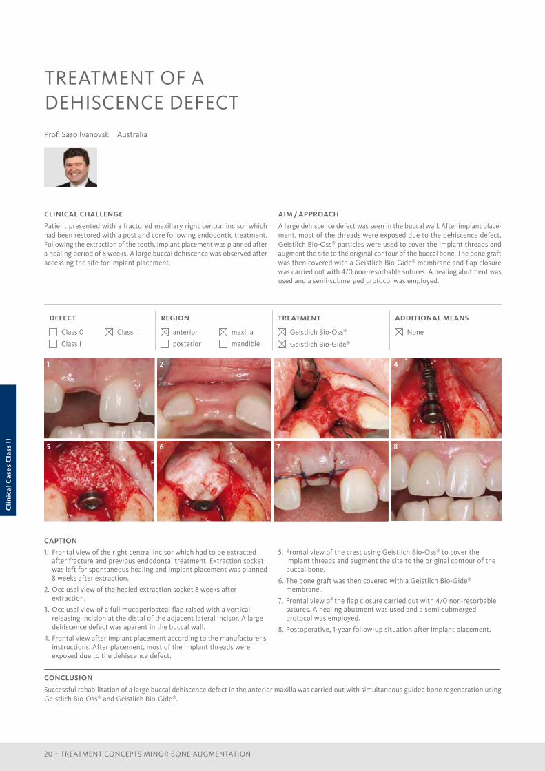
CAPTION
1. Frontal view of the right central incisor which had to be extracted after fracture and previous endodontal treatment. Extraction socket was left for spontaneous healing and implant placement was planned 8 weeks after extraction.
2. Occlusal view of the healed extraction socket 8 weeks after extraction.
3. Occlusal view of a full mucoperiosteal flap raised with a vertical releasing incision at the distal of the adjacent lateral incisor. A large dehiscence defect was aparent in the buccal wall.
4. Frontal view after implant placement according to the manufacturer’s instructions. After placement, most of the implant threads were exposed due to the dehiscence defect.
CLINICAL CHALLENGE
Patient presented with a fractured maxillary right central incisor which had been restored with a post and core following endodontic treatment. Following the extraction of the tooth, implant placement was planned after a healing period of 8 weeks. A large buccal dehiscence was observed after accessing the site for implant placement.
AIM / APPROACH
A large dehiscence defect was seen in the buccal wall. After implant place-ment, most of the threads were exposed due to the dehiscence defect. Geistlich Bio-Oss® particles were used to cover the implant threads and augment the site to the original contour of the buccal bone. The bone graft was then covered with a Geistlich Bio-Gide® membrane and flap closure was carried out with 4/0 non-resorbable sutures. A healing abutment was used and a semi-submerged protocol was employed.
TREATMENT OF A DEHISCENCE DEFECT
5. Frontal view of the crest using Geistlich Bio-Oss® to cover the implant threads and augment the site to the original contour of the buccal bone.
6. The bone graft was then covered with a Geistlich Bio-Gide® membrane.
7. Frontal view of the flap closure carried out with 4/0 non-resorbable sutures. A healing abutment was used and a semi-submerged protocol was employed.
8. Postoperative, 1-year follow-up situation after implant placement.
CONCLUSION
Successful rehabilitation of a large buccal dehiscence defect in the anterior maxilla was carried out with simultaneous guided bone regeneration using Geistlich Bio-Oss® and Geistlich Bio-Gide®.
Prof. Saso Ivanovski | Australia
Cli
nica
l Cas
es C
lass
II

21
RECOMMENDED MATERIAL COMBINATIONS
CLASS 0 DEFECT CLASS I DEFECT CLASS II DEFECT PERI-IMPLANT FAILURE
BONE REPLACEMENT MATERIALS
GEISTLICH BIO-OSS® Granules 0.25–1 mm0.25 g ~ 0.5 cm3
GEISTLICH BIO-OSS® Granules 0.25–1 mm0.5g ~ 1.0 cm3
GEISTLICH BIO-OSS® PEN Granules 0.25–1 mm0.25 g ~ 0.5 cm3
GEISTLICH BIO-OSS® PEN Granules 0.25–1 mm0.5 g ~ 1.0 cm3
GEISTLICH BIO-OSS® COLLAGEN100 mg ~ 0.2–0.3 cm3
MEMBRANES
GEISTLICH BIO-GIDE®
25 × 25 mm, 30 × 40 mm
GEISTLICH BIO-GIDE® COMPRESSED*13 × 25 mm
GEISTLICH BIO-GIDE® COMPRESSED*20 × 30 mm
COMBI
GEISTLICH COMBI-KIT COLLAGENGeistlich Bio-Oss® Collagen 100 mg + Geistlich Bio-Gide® 16 × 22 mm
*Product availability may vary from country to country
References
1 Benić GI & Hämmerle C. Periodontol 2000. 2014 Oct;66(1):13–40.
2 Buser D. 20 Years of Guided Bone Regeneration in Implant Dentistry. 2009.
3 ITI Treatment Guide Vol. 1–6.
4 Benić GI et al., Clin Oral Implants Res. 2009 May;20(5):507–13.
5 Buser D et al., J Periodontol. 2011 Mar;82(3):342–9.
6 Jung RE et al., Clin Oral Implants Res. 2013 Oct;24(10):1065–73.
7 Buser D et al., J Periodontol. 2013 Nov;84(11):1517–27.
8 Jensen SS et al., J Periodontol. 2014 Nov;85(11):1549–56.
9 Buser D et al., J Dent Res. 2013 Dec;92(12 Suppl):176S–82S.
10 Hürzeler M et al., Deutsche Zahnärztliche Zeitschrift. 1996; 51.
11 Zitzmann NU et al., Oral Surg Oral Med Oral Pathol Oral Radiol Endod. 1998 Jan;85(1):8–17.
12 Geistlich Regeneration Leaders’ Meeting 2016.
13 Aghaloo TL & Moy PK. Int J Oral Maxillofac Implants. 2007;22 Suppl:49–70.
14 Garber DA & Belser UC. Compend Contin Educ Dent. 1995 Aug;16(8):796, 798–802, 804.
15 Buser D et al., Int J Oral Maxillofac Implants. 2004;19 Suppl:43–61.
16 Grunder U et al., Int J Periodontics Restorative Dent. 2005 Apr;25(2):113–9.
17 Cardaropoli D et al., Int J Periodontics Restorative Dent. 2015 Mar–Apr;35(2):191–8.
18 Geistlich Bio-Gide® is the first resorbable collagen membrane specifically for use in guided tissue regeneration. Data on file, Geistlich Pharma AG (Wolhusen, Switzerland).
19 Millennium Research Group, Dental Bone Graft Substitutes and Tissue Regeneration 2005, AP/US/EU.
Theo
rie

22 – TREATMENT CONCEPTS MINOR BONE AUGMENTATION
PRODUCT RANGE
GEISTLICH BIO-OSS®
Small granules (0.25–1 mm) | Quantities: 0.25 g, 0.5 g, 1.0 g, 2.0 g (1 g ~ 2.05 cm3)Large granules (1–2 mm) | Quantities: 0.5 g, 1.0 g, 2.0 g (1 g ~ 3.13 cm3)
The small Geistlich Bio-Oss® granules are recommended for smaller 1–2 socket defects and for contouring auto genous block grafts. The large Geistlich Bio-Oss® granules enable improved regeneration over large distances and provide enough space for the in-growing bone.
GEISTLICH BIO-OSS® COLLAGEN
Geistlich Bio-Oss® (small granules) + 10% collagen (porcine) Sizes: 100 mg (0.2–0.3 cm3), 250 mg (0.4–0.5 cm3), 500 mg (0.9–1.1 cm3)
Geistlich Bio-Oss® Collagen is indicated for use in periodontal defects and extraction sockets. Through the addition of collagen, Geistlich Bio-Oss® Collagen can be tailored to the morphology of the defect and is particularly easy to apply.
GEISTLICH BIO-OSS® PEN
Small granules (0.25–1 mm) | Quantities: 0.25 g ~ 0.5 cm3, 0.5 g ~ 1.0 cm3 Large granules (1–2 mm) | Quantity: 0.5 g ~ 1.5 cm3
Geistlich Bio-Oss® granules are available in an applicator. It allows the bone substitute material to be applied more precisely to the surgical site. Geistlich Bio-Oss® Pen is available with either the small granules or the large granules.
GEISTLICH BIO-GIDE®
Geistlich Bio-Gide® | Sizes: 25 × 25 mm, 30 × 40 mm
Geistlich Bio-Gide® protects the grafted area and supports soft tissue healing with its bilayer structure – a rough side that faces the bone and a smooth side that faces the soft tissue. Geistlich Bio-Gide® is easy to handle: it can be positioned easily, adheres well to the defect, and is resistant to tension and tearing.
GEISTLICH BIO-GIDE® COMPRESSED
Geistlich Bio-Gide® Compressed* | Sizes: 13 × 25 mm, 20 × 30 mm
Geistlich Bio-Gide® Compressed is the product twin to Geistlich Bio-Gide®. It combines the proven biofunctionality of Geistlich Bio-Gide® with a different feel. Its bilayer structure protects the graft and supports wound healing. Geistlich Bio-Gide® Compressed is easy to handle and can be positioned easily.
GEISTLICH COMBI-KIT COLLAGEN
Geistlich Bio-Oss® Collagen 100 mg+ Geistlich Bio-Gide® 16 × 22 mm
When used in combination, the system has optimized properties for Ridge Preservation and minor bone augmentations according to the GBR principle.
Pro
duct
Ran
ge
23
Geistlich Expertise
Geistlich has specialized in bone mineral purification processes and collagen purification processes involving natural tissues for over 160 years. Manufacturing processes and systems have been con-tinually optimized to result in cutting-edge products in their field.
Today’s product line comprises bone regeneration materials such as Geistlich Bio-Oss® and many specialized collagen products such as a membrane with a barrier function (Geistlich Bio-Gide®), a matrix with a scaffold function for soft tissue regeneration (Geistlich Mucograft®) and products designed for easy handling (Geistlich Bio-Oss® Collagen).
23
FROM A TO Z: THE WHOLE PRODUCT KNOWLEDGE IS IN OUR HANDS

MANUFACTURER© Geistlich Pharma AGBusiness Unit BiomaterialsBahnhofstrasse 40CH-6110 WolhusenTel. +41 41 4 92 55 55Fax +41 41 4 92 56 39www.geistlich-biomaterials.com
6016
01/
1611
/EN