Traumatic Dental Injuries: Clinical Case Presentation and ...
8
Case Report Traumatic Dental Injuries: Clinical Case Presentation and a 10- Year Epidemiological Investigation in an Italian Dental Emergency Service Alberto Murri Dello Diago , Luigi Generali , Roberto Apponi , Vittorio Colombini , and Vittorio Checchi Department of Surgery, Medicine, Dentistry and Morphological Sciences-Unit of Dentistry and Oral-Maxillo-Facial Surgery, University of Modena and Reggio Emilia, Modena, Italy Correspondence should be addressed to Alberto Murri Dello Diago; [email protected] Received 3 May 2021; Accepted 18 June 2021; Published 30 June 2021 Academic Editor: Leandro Napier de Souza Copyright © 2021 Alberto Murri Dello Diago et al. This is an open access article distributed under the Creative Commons Attribution License, which permits unrestricted use, distribution, and reproduction in any medium, provided the original work is properly cited. Traumatic dental injuries (TDIs) are very common in the world population, and international literature reports several studies which helped in the definition of international guidelines. The aim of this study is to present two clinical cases of TDI and to investigate epidemiological and etiological aspects of TDIs in patients treated in Modena, Italy, between January 2010 and December 2020. The presented case reports are two explicative clinical cases of successful TDI management with a long-term follow-up. The epidemiological analysis was performed on patients who visited the Dental Emergency Service of the Dentistry and Oral-Maxillo-Facial Surgery Unit of Modena (Italy) over a period of 10 years. Data relating to age, gender, type of trauma, and place of accident were collected. Five-hundred-sixty-five TDIs that occurred to patients from 1 to 68 years old were reported, with a total of 860 injured teeth. The peak age at which TDIs are most represented varies between 2 and 3 years old, and they occurred frequently from 1 up to 7 years old. 57.5% were male, while 42.5% were female. The most common trauma resulted to be the uncomplicated crown fracture (20%), immediately followed by lateral luxation (19%), intrusive luxation (18%), avulsion (17%), and complicated crown fracture (15%). TDIs occurred at home in 44% of cases. The need for more prevention training must be highlighted, due to the fact that many TDIs occur at home and in a preschool age. 1. Introduction Traumatic dental injuries (TDIs) are common in the worldwide population. Although the oral cavity represents a small component of the human body, TDIs represent 5% of all health injuries and up to 17% in pediatric patients [1]. A recent study shows that more than one billion living people have had TDIs [1]. Scientific literature shows a great variability regarding the incidence and prevalence of TDIs in the world population [2–4]. Although the International Association of Dental Traumatology (IADT) had codified precise guidelines [5–7], this variability is related to several reasons, such as the diversity in TDI classification methods and parameter recording, and the different cultural and social contexts in which the various studies had been performed. TDIs mainly affect the pediatric population, with a high percentage of preschool children and adolescents, and hardly concern elderly patients, if not in a small percentage [1]. The most frequent TDIs in patients with deciduous teeth are peri- odontal injuries and luxation, while hard tissue injuries and consequent crown fractures are more specific and related to patients with permanent dentition [7]. Trauma such as avul- sion and complicated fractures determine functional and esthetic issues that could affect social relationships; numer- ous studies have shown how TDIs could lead to relationship difficulties and therefore be the reason for personal, domes- tic, and social issues [3, 8, 9]. Hindawi Case Reports in Dentistry Volume 2021, Article ID 8649663, 8 pages https://doi.org/10.1155/2021/8649663
Transcript of Traumatic Dental Injuries: Clinical Case Presentation and ...

Case ReportTraumatic Dental Injuries: Clinical Case Presentation and a 10-Year Epidemiological Investigation in an Italian DentalEmergency Service
Alberto Murri Dello Diago , Luigi Generali , Roberto Apponi , Vittorio Colombini ,and Vittorio Checchi
Department of Surgery, Medicine, Dentistry and Morphological Sciences-Unit of Dentistry and Oral-Maxillo-Facial Surgery,University of Modena and Reggio Emilia, Modena, Italy
Correspondence should be addressed to Alberto Murri Dello Diago; [email protected]
Received 3 May 2021; Accepted 18 June 2021; Published 30 June 2021
Academic Editor: Leandro Napier de Souza
Copyright © 2021 Alberto Murri Dello Diago et al. This is an open access article distributed under the Creative CommonsAttribution License, which permits unrestricted use, distribution, and reproduction in any medium, provided the original workis properly cited.
Traumatic dental injuries (TDIs) are very common in the world population, and international literature reports several studieswhich helped in the definition of international guidelines. The aim of this study is to present two clinical cases of TDI and toinvestigate epidemiological and etiological aspects of TDIs in patients treated in Modena, Italy, between January 2010 andDecember 2020. The presented case reports are two explicative clinical cases of successful TDI management with a long-termfollow-up. The epidemiological analysis was performed on patients who visited the Dental Emergency Service of the Dentistryand Oral-Maxillo-Facial Surgery Unit of Modena (Italy) over a period of 10 years. Data relating to age, gender, type of trauma,and place of accident were collected. Five-hundred-sixty-five TDIs that occurred to patients from 1 to 68 years old werereported, with a total of 860 injured teeth. The peak age at which TDIs are most represented varies between 2 and 3 years old,and they occurred frequently from 1 up to 7 years old. 57.5% were male, while 42.5% were female. The most common traumaresulted to be the uncomplicated crown fracture (20%), immediately followed by lateral luxation (19%), intrusive luxation(18%), avulsion (17%), and complicated crown fracture (15%). TDIs occurred at home in 44% of cases. The need for moreprevention training must be highlighted, due to the fact that many TDIs occur at home and in a preschool age.
1. Introduction
Traumatic dental injuries (TDIs) are common in theworldwide population. Although the oral cavity representsa small component of the human body, TDIs represent 5%of all health injuries and up to 17% in pediatric patients[1]. A recent study shows that more than one billion livingpeople have had TDIs [1]. Scientific literature shows agreat variability regarding the incidence and prevalenceof TDIs in the world population [2–4]. Although theInternational Association of Dental Traumatology (IADT)had codified precise guidelines [5–7], this variability isrelated to several reasons, such as the diversity in TDIclassification methods and parameter recording, and the
different cultural and social contexts in which the variousstudies had been performed.
TDIs mainly affect the pediatric population, with a highpercentage of preschool children and adolescents, and hardlyconcern elderly patients, if not in a small percentage [1]. Themost frequent TDIs in patients with deciduous teeth are peri-odontal injuries and luxation, while hard tissue injuries andconsequent crown fractures are more specific and related topatients with permanent dentition [7]. Trauma such as avul-sion and complicated fractures determine functional andesthetic issues that could affect social relationships; numer-ous studies have shown how TDIs could lead to relationshipdifficulties and therefore be the reason for personal, domes-tic, and social issues [3, 8, 9].
HindawiCase Reports in DentistryVolume 2021, Article ID 8649663, 8 pageshttps://doi.org/10.1155/2021/8649663

The aim of this article is to present two explicative clinicalcases of TDI management and to investigate TDI frequency,patterns, and causes in patients treated in Modena, Italy,between January 2010 and December 2020.
2. Case Presentation
Making use of a digital clinical chart archiving system, TDIcases that occurred at the Dental Emergency Service of theDental Clinic of Modena University, between 1 January2010 and 31 December 2020, were selected. All patients havebeen treated according to the protocols established by theIADT guidelines [5–7], enhanced with photographic docu-mentation, radiographic examination, pulp status evaluation,and instrumental investigations. According to the principleof minimum invasiveness and with the aim of optimizingthe oral health-related quality of life, individualized treat-ment plans for each patient were implemented in line withscientific evidence [10–14].
All the anamnestic questionnaires and the TDI-relatedinformation were collected, and the following data wereobtained: demographic data (gender and age); causes oftrauma (sports injuries, accidents at work, collisions, violenceand abuse, unintentional falls, and road accidents); locationof the trauma (home, school, leisure activities, sports envi-ronment, and work); type of trauma, classified according toIADT classification [5–7]; and teeth involved (permanentor deciduous, maxillary or mandibular, and anterior or pos-terior). All data were registered into Microsoft Excel version14.1.0 for Macintosh.
Among all selected TDIs, the management of two expli-cative and challenging clinical cases is described, followingCARE reporting guidelines for case reports.
2.1. Clinical Case #1. A 15-year-old female patient wasreferred due to traumatic avulsion of her right upper centralincisor that occurred during a volleyball game (Figure 1).
This tooth had been kept in an extraoral dry environmentfor two hours and had a mature apex, and for this reason,nonviable soft tissues were removed from the root with agauze and endodontic therapy was carried out prior toreplantation. After local anesthesia administration, thesocket was irrigated with saline solution and carefullychecked to exclude the presence of bony fractures. The toothwas then replanted applying slight but firm pressure. Thecorrect position of the tooth was verified clinically and radio-graphically, and it was stabilized through a passive flexiblesplint made by a 0.4mm diameter metal wire bonded to thetooth and to adjacent teeth (Figure 2).
Postoperative instructions included antibiotic therapywith amoxicillin, soft diet for 2 weeks, and soft-bristle tooth-brush and chlorhexidine 0.12% mouth rinses, twice a day for2 weeks. The splint was kept in place for 2 weeks, and thepatient was visited after 2, 3, and 6 months and yearly. Atthe 3-year follow-up visit, the tooth appeared asymptomatic,with physiological mobility, no sensitivity to percussion, andnormal percussion sound. No radiotransparency and noradiographic evidence of root resorption were detected(Figure 3).
At the 8-year follow-up visit instead, the tooth presentedno mobility, metallic percussion sound, and clinical infrapo-sition. Radiographically, there was evidence of ankylosis-related resorption (Figure 4).
2.2. Clinical Case #2. A 20-year-old male patient was referreddue to traumatic root fracture of his left upper central incisorafter a bike accident (Figure 5).
A passive and flexible buccal splint with metal stainlesssteel wire and composite patches was immediately performedand kept for 4 weeks, aimed at stabilizing the mobile coronalsegment and maintaining the vitality of the tooth. Soft dietwas suggested for 1 week and oral hygiene instructions weredelivered, brushing with a soft-bristle toothbrush and rinsingwith chlorhexidine 0.12% mouthwash to prevent accumula-tion of plaque and biofilm.
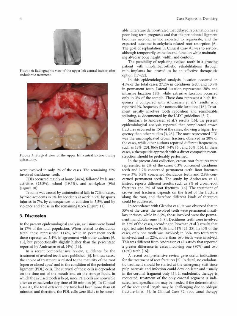
However, after one month, the tooth still did not respondto electrometric and thermal pulp testing, due to plausiblepulp necrosis development. For this reason, an endodontictreatment of the coronal tooth segment to the fracture linehad to be performed (Figure 6).
After a further 4 weeks, the patient still referred pain anddiscomfort of the area, and the decision for a surgicalapproach had to be taken. An apicectomy with a retrogradecanal obturation was performed in order to remove thesymptomatology and obtain a complete healing of the area(Figure 7).
Postsurgical instructions included prevention of furtherinjury by avoidance of contact sports, meticulous oralhygiene, and rinsing with an antibacterial agent such aschlorhexidine gluconate 0.12%.
Healing was uneventful, and the patient did not referpain nor swelling. At the 4-year follow-up visit, clinical andradiographic analysis showed a healed area with a good ossi-fication of the periradicular bone (Figure 8).
2.3. Epidemiological Results. Out of a total of 26355 patientswho accessed the Dental Emergency Service unit in theperiod from January 2010 to December 2020, 565 TDIs wereregistered. Patients’ age varied from 0 to 68 years, and thepeak age at which TDIs were most represented variedbetween 2 and 3 years old; however, it was highly representedfrom 1 up to 7 years old.
In 40% of the cases, only one tooth was involved, in 38%two teeth, in 15% three teeth, and in 7% four teeth, for a totalof 860 injured teeth.
The most frequent injury resulted in an uncomplicatedcrown fracture (20% of cases), immediately followed by lat-eral luxation. Successively, this was followed by 18% of intru-sive luxation, 17% of avulsion, and 15% of complicatedcrown fracture (Figure 9). Crown-root fractures were repre-sented in the 2% of the cases. The 57.5% of affected patientswere male, while 42.5% were female. The most frequentTDI among males resulted in uncomplicated crown fracture,while the most frequent TDI among females resulted in lat-eral luxation.
TDIs involved permanent dentition in 63% of patients:maxillary anterior teeth involvement was observed in 55.5%of cases, mandibular anterior in 6.5%, while posterior teeth
2 Case Reports in Dentistry

(a) (b)
Figure 1: Posttraumatic (a) clinical and (b) radiographic view showing the absence of the upper right central incisor.
(a) (b)
Figure 2: (a) Clinical and (b) radiographic view of the upper incisor after reimplantation and splint.
(a) (b)
Figure 3: (a) Clinical and (b) radiographic view of the upper incisor at the 3-year follow-up visit.
(a) (b)
Figure 4: (a) Clinical and (b) radiographic view of the upper incisor at the 8-year follow-up visit: tooth infraposition and root resorption areevident.
(a) (b)
Figure 5: Posttraumatic (a) clinical and (b) radiographic view. Gingival tissues around the upper left central incisor are swollen and red, whilea horizontal root fraction is detectable from the periapical X-ray.
3Case Reports in Dentistry
were involved in only 1% of the cases. The remaining 37%involved deciduous teeth.
TDIs occurred mainly at home (44%), followed by leisureactivities (23.5%), school (19.5%), and workplace (9%)(Figure 10).
Trauma was caused by unintentional falls in 72% of cases,by road accidents in 8%, by accidents at work in 7%, by sportsinjuries in 7%, by consequences of collision in 5.5%, and byviolence and abuse in the remaining 0.5% (Figure 11).
3. Discussion
In the present epidemiological analysis, avulsions were foundin 17% of the total population. When related to deciduousteeth, these represented 11.6%, while in permanent teeththese represented 5.4%, in agreement with other authors [6,15], but proportionally slightly higher than the percentagereported by Andreasen et al. (4%) [16].
In a recent comprehensive review, guidelines for thetreatment of avulsed teeth were published [6]. In these cases,the choice of treatment is related to the maturity of the root(open or closed apex) and to the condition of the periodontalligament (PDL) cells. The survival of these cells is dependenton the time out of the mouth and on the storage liquid inwhich the avulsed tooth is kept, since PDL cells are nonviableafter an extraalveolar dry time of 30 minutes [6]. In ClinicalCase #1, the total extraoral dry time had been more than 60minutes, and therefore, the PDL cells were likely to be nonvi-
able. Literature demonstrated that delayed replantation has apoor long-term prognosis and that the periodontal ligamentbecomes necrotic, is not expected to regenerate, and theexpected outcome is ankylosis-related root resorption [6].The goal of replantation in Clinical Case #1 was to restore,although temporarily, esthetics and function while maintain-ing alveolar bone height, width, and contour.
The possibility of replacing avulsed tooth in a growingpatient with implant-prosthetic rehabilitations throughmini-implants has proved to be an effective therapeuticoption [17–22].
In this epidemiological analysis, luxation occurred in41% of the total cases: 27.2% in deciduous teeth and 13.9%in permanent teeth. Lateral luxation represented 20% andintrusive luxation 18%, while extrusive luxation occurredonly in 3% of the sample. These data represent a high fre-quency if compared with Andreasen et al.’s results whoreported 9% frequency for nonspecific luxations [16]. Treat-ment usually involves tooth reposition and semiflexiblesplinting, as documented by the IADT guidelines [5–7].
Similarly to Andreasen et al.’s results [16], the presentepidemiological analysis reported that complicated crownfractures occurred in 15% of the cases, showing a higher fre-quency than other studies [3, 23]. The most represented TDIwas the uncomplicated crown fracture, observed in 20% ofthe cases, while other authors reported different frequencies,such as 13% [23], 86% [24], 94% [6], and 50% [16]. In thesecases, a therapeutic approach with a direct composite recon-struction should be preferably performed.
In the present data collection, crown-root fractures wererepresented in 2% of the cases: 0.3% concerned deciduousteeth and 1.7% concerned permanent teeth. Root fractureswere 3%: 0.2% concerned deciduous teeth and 2.8% con-cerned permanent teeth. The study by Andreasen et al.instead reports different results, such as 9% of crown-rootfractures and 7% of root fractures [16]. The treatment ofcrown-root fractures depends on the level of the fracturealong the root, and therefore different kinds of therapiescould be addressed.
In accordance with Glendor et al., it was observed that in55% of the cases, the involved teeth were permanent maxil-lary incisors, while in 6.5%, those involved were the perma-nent mandibular ones [3, 8]. Deciduous teeth were involvedin 37% of the cases, according to Piovesan et al.’s results thatreported rates between 9.4% and 41% [24, 25]. In 40% of thecases, only one tooth was involved; in 36%, two teeth wereinvolved; and in 22%, more than two teeth were involved.This was different from Andreasen et al.’s study that reporteda greater difference in cases involving one (80%) and two(18%) teeth [16].
A recent comprehensive review gave useful indicationsfor the treatment of root fractures [5]. In detail, no endodon-tic treatment should be started at the emergency visit sincepulp necrosis and infection could develop later and usuallyin the coronal fragment only [5]. If endodontic therapy isrequested, treatment of the only coronal segment is indi-cated, and apexification may be needed if the determinationof the root canal length may be challenging due to obliquefracture lines [5]. In Clinical Case #2, root canal therapy
Figure 6: Radiographic view of the upper left central incisor afterendodontic treatment.
Figure 7: Surgical view of the upper left central incisor duringapicectomy.
4 Case Reports in Dentistry

had to be performed after one month since the tooth did notrespond to thermal pulp testing nor electrometric test. Fol-lowing IADT guidelines, only the coronal part of the frac-
tured tooth was treated. However, after another month, asurgical approach had to be chosen in order to solve thepatient’s symptomatology that was still present.
(a) (b)
Figure 8: (a) Clinical and (b) radiographic view 4 years after the surgical therapy.
0Uncompli
catedcrown
fracture
Complicatedcrown
fracture
Uncomplicated
crown-radicularfracture
Radicularfracture
Intrusiveluxation
Type of TDI
Lateralluxation
Avulsion OtherExtrusiveluxation
Complicated
crown-radicularfracture
172Teeth
Teeth
129 9 26 154 172 146 9349
20406080
100120140160180200
Figure 9: TDI frequency based on the type of injury.
Place where TDIs occurred
TDI 249 109 51 17 133 6
Home School Workplace Sportenvironment
Leisureactivities
Other
TDI
0
50
100
150
200
250
300
Figure 10: TDI frequency based on the place where injuries occurred.
5Case Reports in Dentistry

The results of this epidemiological analysis show thatTDI frequency treated in the Dental Emergency Service ofthe Dentistry and Oral-Maxillo-Facial Surgery Unit ofModena, Italy, are in line with what is recorded in othernational and international data.
Concerning a patient’s age, the results of this epidemio-logical investigation are only partially in agreement with pre-vious researches that, although with a larger populationsample, revealed a TDI average age incidence of about 15years [3, 8]. In our case, the peak diagnosis of TDIs has beenobserved between 2 and 3 years old. The mobility skills of achild between 2 and 3 years of age are still to be developedsince they must still acquire an adult confidence. However,the desire to overcome personal limits often pushes the childto test himself, thus risking his own safety, and these aspectscould explain the TDI peak incidence in this age group.Other possible causes, such as lack of supervision or childabuse, have to be considered.
In accordance with previous studies [3, 8, 9], about 46%of TDIs were however found in the age group between 1and 7 years (preschool). At this stage of life, children are verydynamic and sports activities are increasingly practiced, lead-ing to TDIs being consequently much more frequent.
As demonstrated by several studies [3, 8, 9], a prevalenceof TDI has been observed among male subjects (57.5%),rather than female (42.5%). This prevalence appears equalto the findings of Glendor et al.’s results that show a 2 : 1 ratioof TDIs between males and females [7].
Concerning TDI reasons, it emerged that the main causewas an unintentional fall, especially in preschool age, whichmost frequently caused luxation and avulsion. Domestic fallsand accidental impacts against objects, in particular interiorfurnishings such as tables, chairs, and sinks, were recordedin 72% of total cases.
Road accidents, which range from falling from a bicycleor moped to, more rarely, car accidents, were reported in8% of the cases. According to these data, accidents at workare also frequent along with sports injuries, causing a signif-
icant incidence of avulsions. Different types of TDIs can beobserved based on the type of sport activity and thereforeof the impact dynamic. High-speed sports seem to lead tobone fractures, while low-speed sports lead to dental injuries.These data are in agreement with previous studies on the eti-ology of trauma [22, 26–33]. In fact, sports injuries have beenobserved in patients aged 12 to 32, road accidents have beenobserved in patients aged 17 to 62, and workplace accidentshave been observed only in adult patients, as well as colli-sions. In a similar way, in a recent study, the majority ofTDI patients were adolescents (27.9%), or younger than 10years (23.2%). The main cause of TDI was cycling (43.5%),followed by sports such as football and baseball [34].
Although there are well-coded international preventionguidelines, TDIs are still very relevant in pediatric patients.Therefore, there is a need for the development of trainingand prevention programs for TDI, organizing adequateemergency services, and planning awareness campaigns.
Data Availability
Data supporting the conclusions of the study can be accessedby asking the corresponding author.
Ethical Approval
Clinical cases were conducted in accordance with the Decla-ration of Helsinki (1964).
Disclosure
The funding source has no role in conceiving and performingthe study.
Conflicts of Interest
The authors declare that there is no conflict of interestregarding the publication of this article.
Causes of TDIs
407TDI
TDI
46 39 39 31 3
Unintentionalfalls
Road accident Accident atwork
Sport injuries Collision Violence andabuse
0
50
100
150
200
250
300
350
400
450
Figure 11: TDI frequency based on the cause of injury.
6 Case Reports in Dentistry

References
[1] S. Petti, U. Glendor, and L. Andersson, “World traumatic den-tal injury prevalence and incidence, a meta-analysis—one bil-lion living people have had traumatic dental injuries,” DentalTraumatology, vol. 34, no. 2, pp. 71–86, 2018.
[2] S. Petti, J. O. Andreasen, U. Glendor, and L. Andersson, “Thefifth most prevalent disease is being neglected by public healthorganizations,” The Lancet Global Health, vol. 6, no. 10,pp. e1070–e1071, 2018.
[3] U. Glendor, “Epidemiology of traumatic dental injuries—a 12year review of the literature,” Dental Traumatology, vol. 24,no. 6, pp. 603–611, 2008.
[4] GBD 2015 Disease and Injury Incidence and Prevalence Col-laborators, “Global, regional, and national incidence, preva-lence, and years lived with disability for 310 diseases andinjuries, 1990-2015: a systematic analysis for the Global Bur-den of Disease Study 2015,” Lancet, vol. 388, no. 10053,pp. 1545–1602, 2016.
[5] C. Bourguignon, N. Cohenca, E. Lauridsen et al., “Interna-tional Association of Dental Traumatology guidelines for themanagement of traumatic dental injuries: 1. Fractures and lux-ations,” Dental Traumatology, vol. 36, no. 4, pp. 314–330,2020.
[6] A. F. Fouad, P. V. Abbott, G. Tsilingaridis et al., “InternationalAssociation of Dental Traumatology guidelines for the man-agement of traumatic dental injuries: 2. Avulsion of permanentteeth,” Dental Traumatology, vol. 36, no. 4, pp. 331–342, 2020.
[7] P. F. Day, M. T. Flores, A. C. O'Connell et al., “InternationalAssociation of Dental Traumatology guidelines for the man-agement of traumatic dental injuries: 3. Injuries in the primarydentition,” Dental Traumatology, vol. 36, no. 4, pp. 343–359,2020.
[8] R. Lam, “Epidemiology and outcomes of traumatic dental inju-ries: a review of the literature,” Australian Dental Journal,vol. 61, Suppl 1, pp. 4–20, 2016.
[9] U. Glendor, “Aetiology and risk factors related to traumaticdental injuries—a review of the literature,” Dental Traumatol-ogy, vol. 25, no. 1, pp. 19–31, 2009.
[10] L. Giannetti, A. Murri Dello Diago, G. Silingardi, andE. Spinas, “Superficial infiltration to treat white hypominera-lized defects of enamel: clinical trial with 12-month follow-up,” Journal of Biological Regulators and Homeostatic Agents,vol. 32, no. 5, pp. 1335–1338, 2018.
[11] M. Mazur, S. Westland, F. Guerra et al., “Objective and subjec-tive aesthetic performance of icon treatment for enamel hypo-mineralization lesions in young adolescents: a retrospectivesingle center study,” Journal of Dentistry, vol. 68, pp. 104–108, 2018.
[12] L. Giannetti, A. Murri Dello Diago, E. Corciolani, andE. Spinas, “Deep infiltration for the treatment of hypominera-lized enamel lesions in a patient with molar incisor hypomi-neralization: a clinical case,” Journal of Biological Regulatorsand Homeostatic Agents, vol. 32, no. 3, pp. 751–754, 2018.
[13] K. Bakes, S. Amend, J. Priller, C. Zamek, T. Stamm, andN. Krämer, “Changes in oral health-related quality of life aftertreatment of hypersensitive molar incisor hypomineralization-affected molars with a sealing,” Clinical Oral Investigations,2021.
[14] A. Murri Dello Diago, M. Cadenaro, R. Ricchiuto et al.,“Hypersensitivity in molar incisor hypomineralization: super-
ficial infiltration treatment,” Applied Sciences, vol. 11, no. 4,p. 1823, 2021.
[15] L. Giannetti and A. Murri, “Clinical evidence and literature tocompare two different therapeutic protocols in tooth avul-sion,” European Journal of Paediatric Dentistry, vol. 7, no. 3,pp. 122–130, 2006.
[16] J. O. Andreasen, F. M. Andreasen, and L. Andersson, Textbookand Color Atlas of Traumatic Injuries to the Teeth, Wiley-Blackwell, 4th edition, 2013, ISBN: 978-1-118-69990.
[17] L. Giannetti, A. Murri Dello Diago, F. Vecci, and U. Consolo,“Mini-implants in growing patients: a case report,” PediatricDentistry, vol. 32, no. 3, pp. 239–244, 2010.
[18] J. B. Cope and D. McFadden, “Temporary replacement ofmissing maxillary lateral incisors with orthodontic miniscrewimplants in growing patients: rationale, clinical technique,and long-term results,” Journal of Orthodontics, vol. 41, Suppl.1, pp. 62–74, 2014.
[19] L. Giannetti, R. Apponi, A. Murri Dello Diago, andF. Mintrone, “Rehabilitation of a patient with mini-implantsafter avulsion of the upper incisors: a 13-year follow up,” Den-tal Traumatology, vol. 37, no. 2, pp. 354–359, 2021.
[20] D. Re, D. Augusti, G. Paglia, G. Augusti, and E. Cotti, “Treat-ment of traumatic dental injuries: evaluation of knowledgeamong Italian dentists,” European Journal of Paediatric Den-tistry, vol. 15, no. 1, pp. 23–28, 2014.
[21] E. Spinas, M. Aresu, F. Canargiu, and L. Giannetti, “Preventivetreatment of post-traumatic dental infraocclusion: study onthe knowledge of dental decoronation in a sample of Italiandental students and dentists,” European Journal of PaediatricDentistry, vol. 16, no. 4, pp. 279–283, 2015.
[22] E. Spinas, L. Generali, A. Mameli, C. Demontis, D. Martinelli,and L. Giannetti, “Delayed tooth replantation and inflamma-tory root resorption in childhood and adolescence,” Journalof Biological Regulators and Homeostatic Agents, vol. 33,no. 2, pp. 623–627, 2019.
[23] D. Locker, “Prevalence of traumatic dental injury in grade 8children in six Ontario communities,” Canadian Journal ofPublic Health, vol. 96, no. 1, pp. 73–76, 2005.
[24] C. Piovesan, R. S. Guedes, L. Casagrande, and T. M. Ardenghi,“Socioeconomic and clinical factors associated with traumaticdental injuries in Brazilian preschool children,” Brazilian OralResearch, vol. 26, no. 5, pp. 464–470, 2012.
[25] C. Piovesan, C. Abella, and T. M. Ardenghi, “Child oral health-related quality of life and socioeconomic factors associatedwith traumatic dental injuries in schoolchildren,” Oral Health& Preventive Dentistry, vol. 9, no. 4, pp. 405–411, 2011.
[26] J. Traebert, M. A. Peres, V. Blank, S. Böell Rda, and J. A. Pie-truza, “Prevalence of traumatic dental injury and associatedfactors among 12-year-old school children in Florianópolis,Brazil,” Dental Traumatology, vol. 19, no. 1, pp. 15–18,2003.
[27] E. Spinas, M. Aresu, and L. Giannetti, “Use of mouth guard inbasketball: observational study of a group of teenagers withand without motivational reinforcement,” European Journalof Paediatric Dentistry, vol. 15, no. 4, pp. 392–396, 2014.
[28] D. Stanbouly, R. Stanbouly, K. C. Lee, and S. K. Chuang, “Prev-alence of dentofacial injuries and concussions among collegeathletes and their perceptions of mouthguards,” Journal ofCraniofacial Surgery, vol. 32, no. 4, pp. 1600–1603, 2021.
[29] E. Spinas, A. Mameli, and L. Giannetti, “Traumatic dentalinjuries resulting from sports activities; immediate treatment
7Case Reports in Dentistry

and five years follow-up: an observational study,” The OpenDentistry Journal, vol. 12, no. 1, pp. 1–10, 2018.
[30] G. Shirani, M. H. Kalantar Motamedi, A. Ashuri, and P. S. Esh-kevari, “Prevalence and patterns of combat sport related max-illofacial injuries,” Journal of Emergencies, Trauma, and Shock,vol. 3, no. 4, pp. 314–317, 2010.
[31] E. Spinas, L. Giannetti, A. Mameli, and D. Re, “Dental injuriesin young athletes, a five-year follow-up study,” European jour-nal of paediatric dentistry, vol. 19, no. 3, pp. 187–193, 2018.
[32] Y. Z. Tan, L. Levin, W. Guo, and Y. Chen, “Dental injuries atthe Xi’an, China Stomatological Hospital: a retrospectivestudy,”Dental Traumatology, vol. 36, no. 5, pp. 505–509, 2020.
[33] M. Oliveira Werlich, L. R. Honnef, J. V. Silva Bett et al., “Prev-alence of dentofacial injuries in contact sports players: a sys-tematic review and meta-analysis,” Dental Traumatology,vol. 36, no. 5, pp. 477–488, 2020.
[34] H. K. Park, J. Y. Park, N. R. Choi, U. K. Kim, and D. S. Hwang,“Sports-related oral and maxillofacial injuries: a 5-year retro-spective study, Pusan National University Dental Hospital,”Journal of Oral and Maxillofacial Surgery, vol. 79, 2021.
8 Case Reports in Dentistry