Trauma and Orthopaedic - ACT Health Newsletter2015... · postgraduate students. A memorandum of...
20
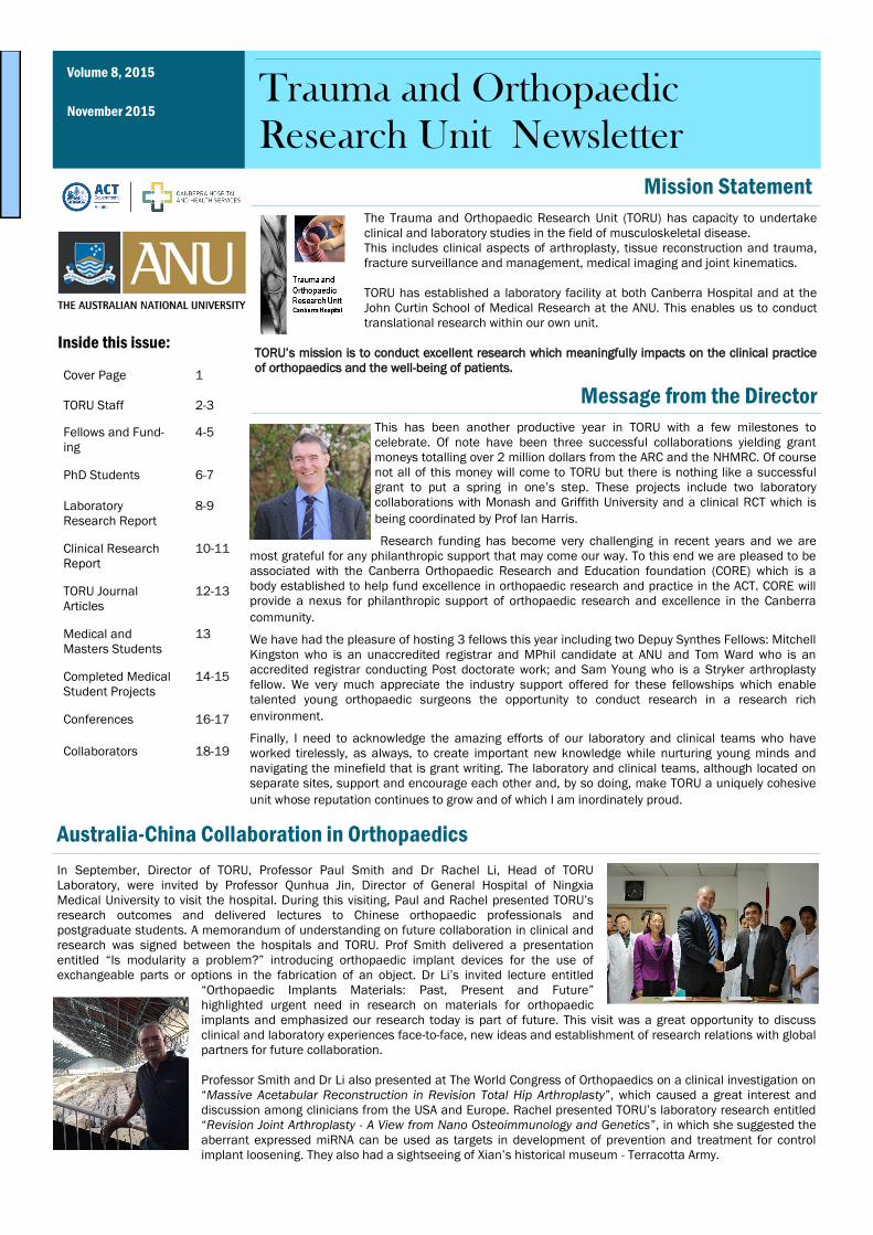
This has been another productive year in TORU with a few milestones to celebrate. Of note have been three successful collaborations yielding grant moneys totalling over 2 million dollars from the ARC and the NHMRC. Of course not all of this money will come to TORU but there is nothing like a successful grant to put a spring in one’s step. These projects include two laboratory collaborations with Monash and Griffith University and a clinical RCT which is being coordinated by Prof Ian Harris. Research funding has become very challenging in recent years and we are most grateful for any philanthropic support that may come our way. To this end we are pleased to be associated with the Canberra Orthopaedic Research and Education foundation (CORE) which is a body established to help fund excellence in orthopaedic research and practice in the ACT. CORE will provide a nexus for philanthropic support of orthopaedic research and excellence in the Canberra community. We have had the pleasure of hosting 3 fellows this year including two Depuy Synthes Fellows: Mitchell Kingston who is an unaccredited registrar and MPhil candidate at ANU and Tom Ward who is an accredited registrar conducting Post doctorate work; and Sam Young who is a Stryker arthroplasty fellow. We very much appreciate the industry support offered for these fellowships which enable talented young orthopaedic surgeons the opportunity to conduct research in a research rich environment. Finally, I need to acknowledge the amazing efforts of our laboratory and clinical teams who have worked tirelessly, as always, to create important new knowledge while nurturing young minds and navigating the minefield that is grant writing. The laboratory and clinical teams, although located on separate sites, support and encourage each other and, by so doing, make TORU a uniquely cohesive unit whose reputation continues to grow and of which I am inordinately proud. Message from the Director The Trauma and Orthopaedic Research Unit (TORU) has capacity to undertake clinical and laboratory studies in the field of musculoskeletal disease. This includes clinical aspects of arthroplasty, tissue reconstruction and trauma, fracture surveillance and management, medical imaging and joint kinematics. TORU has established a laboratory facility at both Canberra Hospital and at the John Curtin School of Medical Research at the ANU. This enables us to conduct translational research within our own unit. TORU’s mission is to conduct excellent research which meaningfully impacts on the clinical practice of orthopaedics and the well-being of patients. November 2015 Volume 8, 2015 Trauma and Orthopaedic Research Unit Newsletter Inside this issue: In September, Director of TORU, Professor Paul Smith and Dr Rachel Li, Head of TORU Laboratory, were invited by Professor Qunhua Jin, Director of General Hospital of Ningxia Medical University to visit the hospital. During this visiting, Paul and Rachel presented TORU’s research outcomes and delivered lectures to Chinese orthopaedic professionals and postgraduate students. A memorandum of understanding on future collaboration in clinical and research was signed between the hospitals and TORU. Prof Smith delivered a presentation entitled “Is modularity a problem?” introducing orthopaedic implant devices for the use of exchangeable parts or options in the fabrication of an object. Dr Li’s invited lecture entitled “Orthopaedic Implants Materials: Past, Present and Future” highlighted urgent need in research on materials for orthopaedic implants and emphasized our research today is part of future. This visit was a great opportunity to discuss clinical and laboratory experiences face-to-face, new ideas and establishment of research relations with global partners for future collaboration. Professor Smith and Dr Li also presented at The World Congress of Orthopaedics on a clinical investigation on “Massive Acetabular Reconstruction in Revision Total Hip Arthroplasty”, which caused a great interest and discussion among clinicians from the USA and Europe. Rachel presented TORU’s laboratory research entitled “Revision Joint Arthroplasty - A View from Nano Osteoimmunology and Genetics”, in which she suggested the aberrant expressed miRNA can be used as targets in development of prevention and treatment for control implant loosening. They also had a sightseeing of Xian’s historical museum - Terracotta Army. Mission Statement Australia-China Collaboration in Orthopaedics Cover Page 1 TORU Staff 2-3 Fellows and Fund- ing 4-5 PhD Students 6-7 Laboratory Research Report 8-9 Clinical Research Report 10-11 TORU Journal Articles 12-13 Medical and Masters Students 13 Completed Medical Student Projects 14-15 Conferences 16-17 Collaborators 18-19
-
Upload
phungquynh -
Category
Documents
-
view
218 -
download
0
Transcript of Trauma and Orthopaedic - ACT Health Newsletter2015... · postgraduate students. A memorandum of...
This has been another productive year in TORU with a few milestones to
celebrate. Of note have been three successful collaborations yielding grant
moneys totalling over 2 million dollars from the ARC and the NHMRC. Of course
not all of this money will come to TORU but there is nothing like a successful
grant to put a spring in one’s step. These projects include two laboratory
collaborations with Monash and Griffith University and a clinical RCT which is
being coordinated by Prof Ian Harris.
Research funding has become very challenging in recent years and we are
most grateful for any philanthropic support that may come our way. To this end we are pleased to be
associated with the Canberra Orthopaedic Research and Education foundation (CORE) which is a
body established to help fund excellence in orthopaedic research and practice in the ACT. CORE will
provide a nexus for philanthropic support of orthopaedic research and excellence in the Canberra
community.
We have had the pleasure of hosting 3 fellows this year including two Depuy Synthes Fellows: Mitchell
Kingston who is an unaccredited registrar and MPhil candidate at ANU and Tom Ward who is an
accredited registrar conducting Post doctorate work; and Sam Young who is a Stryker arthroplasty
fellow. We very much appreciate the industry support offered for these fellowships which enable
talented young orthopaedic surgeons the opportunity to conduct research in a research rich
environment.
Finally, I need to acknowledge the amazing efforts of our laboratory and clinical teams who have
worked tirelessly, as always, to create important new knowledge while nurturing young minds and
navigating the minefield that is grant writing. The laboratory and clinical teams, although located on
separate sites, support and encourage each other and, by so doing, make TORU a uniquely cohesive
unit whose reputation continues to grow and of which I am inordinately proud.
Message from the Director
The Trauma and Orthopaedic Research Unit (TORU) has capacity to undertake
clinical and laboratory studies in the field of musculoskeletal disease.
This includes clinical aspects of arthroplasty, tissue reconstruction and trauma,
fracture surveillance and management, medical imaging and joint kinematics.
TORU has established a laboratory facility at both Canberra Hospital and at the
John Curtin School of Medical Research at the ANU. This enables us to conduct
translational research within our own unit.
TORU’s mission is to conduct excellent research which meaningfully impacts on the clinical practice
of orthopaedics and the well-being of patients.
November 2015
Volume 8, 2015
Trauma and Orthopaedic
Research Unit Newsletter
Inside this issue:
In September, Director of TORU, Professor Paul Smith and Dr Rachel Li, Head of TORU
Laboratory, were invited by Professor Qunhua Jin, Director of General Hospital of Ningxia
Medical University to visit the hospital. During this visiting, Paul and Rachel presented TORU’s
research outcomes and delivered lectures to Chinese orthopaedic professionals and
postgraduate students. A memorandum of understanding on future collaboration in clinical and
research was signed between the hospitals and TORU. Prof Smith delivered a presentation
entitled “Is modularity a problem?” introducing orthopaedic implant devices for the use of
exchangeable parts or options in the fabrication of an object. Dr Li’s invited lecture entitled
“Orthopaedic Implants Materials: Past, Present and Future”
highlighted urgent need in research on materials for orthopaedic
implants and emphasized our research today is part of future. This visit was a great opportunity to discuss
clinical and laboratory experiences face-to-face, new ideas and establishment of research relations with global
partners for future collaboration.
Professor Smith and Dr Li also presented at The World Congress of Orthopaedics on a clinical investigation on
“Massive Acetabular Reconstruction in Revision Total Hip Arthroplasty”, which caused a great interest and
discussion among clinicians from the USA and Europe. Rachel presented TORU’s laboratory research entitled
“Revision Joint Arthroplasty - A View from Nano Osteoimmunology and Genetics”, in which she suggested the
aberrant expressed miRNA can be used as targets in development of prevention and treatment for control
implant loosening. They also had a sightseeing of Xian’s historical museum - Terracotta Army.
Mission Statement
Australia-China Collaboration in Orthopaedics
Cover Page 1
TORU Staff 2-3
Fellows and Fund-
ing
4-5
PhD Students 6-7
Laboratory
Research Report
8-9
Clinical Research
Report
10-11
TORU Journal
Articles
12-13
Medical and
Masters Students
13
Completed Medical
Student Projects
14-15
Conferences 16-17
Collaborators 18-19
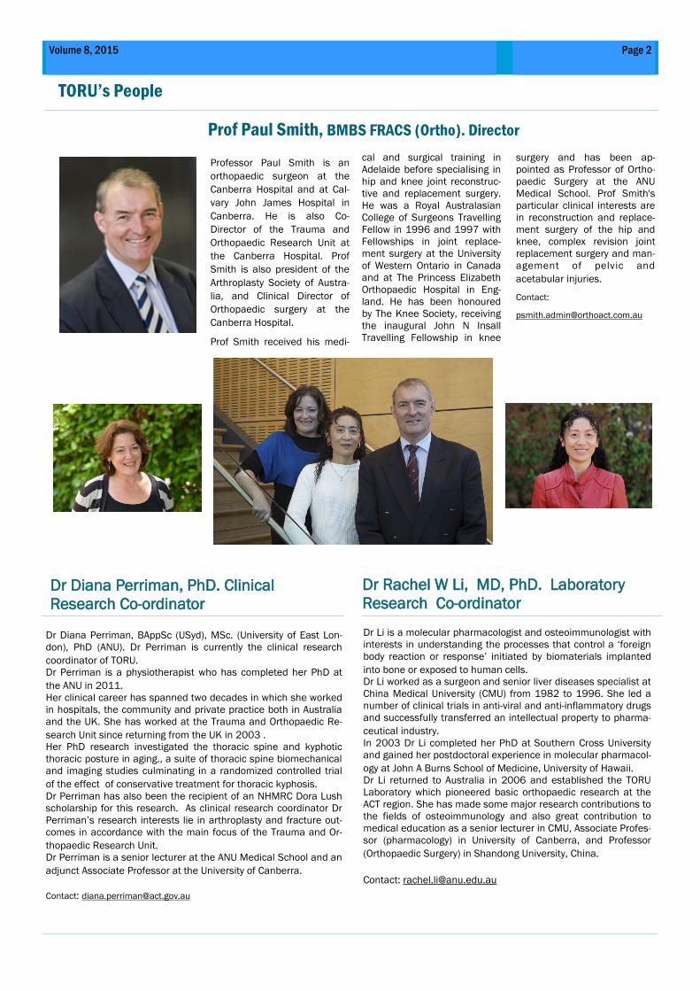
Dr Rachel W Li, MD, PhD. Laboratory
Research Co-ordinator
Professor Paul Smith is an
orthopaedic surgeon at the
Canberra Hospital and at Cal-
vary John James Hospital in
Canberra. He is also Co-
Director of the Trauma and
Orthopaedic Research Unit at
the Canberra Hospital. Prof
Smith is also president of the
Arthroplasty Society of Austra-
lia, and Clinical Director of
Orthopaedic surgery at the
Canberra Hospital.
Prof Smith received his medi-
Dr Diana Perriman, PhD. Clinical
Research Co-ordinator
cal and surgical training in
Adelaide before specialising in
hip and knee joint reconstruc-
tive and replacement surgery.
He was a Royal Australasian
College of Surgeons Travelling
Fellow in 1996 and 1997 with
Fellowships in joint replace-
ment surgery at the University
of Western Ontario in Canada
and at The Princess Elizabeth
Orthopaedic Hospital in Eng-
land. He has been honoured
by The Knee Society, receiving
the inaugural John N Insall
Travelling Fellowship in knee
surgery and has been ap-
pointed as Professor of Ortho-
paedic Surgery at the ANU
Medical School. Prof Smith's
particular clinical interests are
in reconstruction and replace-
ment surgery of the hip and
knee, complex revision joint
replacement surgery and man-
agement of pelvic and
acetabular injuries.
Contact:
Dr Diana Perriman, BAppSc (USyd), MSc. (University of East Lon-
don), PhD (ANU). Dr Perriman is currently the clinical research
coordinator of TORU.
Dr Perriman is a physiotherapist who has completed her PhD at
the ANU in 2011.
Her clinical career has spanned two decades in which she worked
in hospitals, the community and private practice both in Australia
and the UK. She has worked at the Trauma and Orthopaedic Re-
search Unit since returning from the UK in 2003 .
Her PhD research investigated the thoracic spine and kyphotic
thoracic posture in aging., a suite of thoracic spine biomechanical
and imaging studies culminating in a randomized controlled trial
of the effect of conservative treatment for thoracic kyphosis.
Dr Perriman has also been the recipient of an NHMRC Dora Lush
scholarship for this research. As clinical research coordinator Dr
Perriman’s research interests lie in arthroplasty and fracture out-
comes in accordance with the main focus of the Trauma and Or-
thopaedic Research Unit.
Dr Perriman is a senior lecturer at the ANU Medical School and an
adjunct Associate Professor at the University of Canberra.
Contact: [email protected]
Prof Paul Smith, BMBS FRACS (Ortho). Director
Dr Li is a molecular pharmacologist and osteoimmunologist with
interests in understanding the processes that control a ‘foreign
body reaction or response’ initiated by biomaterials implanted
into bone or exposed to human cells.
Dr Li worked as a surgeon and senior liver diseases specialist at
China Medical University (CMU) from 1982 to 1996. She led a
number of clinical trials in anti-viral and anti-inflammatory drugs
and successfully transferred an intellectual property to pharma-
ceutical industry.
In 2003 Dr Li completed her PhD at Southern Cross University
and gained her postdoctoral experience in molecular pharmacol-
ogy at John A Burns School of Medicine, University of Hawaii.
Dr Li returned to Australia in 2006 and established the TORU
Laboratory which pioneered basic orthopaedic research at the
ACT region. She has made some major research contributions to
the fields of osteoimmunology and also great contribution to
medical education as a senior lecturer in CMU, Associate Profes-
sor (pharmacology) in University of Canberra, and Professor
(Orthopaedic Surgery) in Shandong University, China.
Contact: [email protected]
TORU’s People
Page 2 Volume 8, 2015
TORU Staff & Associates
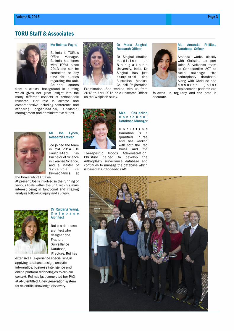
Ms Belinda Payne
Belinda is TORU's
Office Manager,
Belinda has been
with TORU since
2013 and can be
contacted at any
time for queries
regarding the unit.
Belinda comes
from a clinical background in nursing
which gives her great insight into the
many different aspects of orthopaedic
research. Her role is diverse and
comprehensive including conference and
meeting organisation, f inancia l
management and administrative duties.
Mr Joe Lynch,
Research Officer
Joe joined the team
in mid 2014. He
completed his
Bachelor of Science
in Exercise Science,
and a Master of
S c i e n c e i n
Biomechanics at
the University of Ottawa.
At present Joe is involved in the running of
various trials within the unit with his main
interest being in functional and imaging
analysis following injury and surgery.
Dr Ruidang Wang,
D a t a b a s e
Architect
Rui is a database
architect who
designed the
Fracture
Surveillance
Database,
iFracture. Rui has
extensive IT experience specialising in
applying database design, analytic
informatics, business intelligence and
online platform technologies to clinical
context. Rui has just completed her PhD
at ANU entitled A new generation system
for scientific knowledge discovery.
Dr Mona Singhal,
Research Officer
Dr Singhal studied
m e d i c i n e a t
B a n g a l o r e
University, India. Dr
Singhal has just
comple ted th e
Australian Medical
Council Registration
Examination. She worked with us from
2013 to April 2015 as a Research Officer
on the Whiplash study.
Mrs Chr ist ine
H a n r a h a n ,
Database Manager
C h r i s t i n e
Hanrahan is a
qualified nurse
and has worked
with both the Red
Cross and the
Therapeutic Goods Administration.
Christine helped to develop the
Arthroplasty surveillance database and
continues to manage the database which
is based at Orthopaedics ACT.
Ms Amanda Phillips,
Database Officer
Amanda works closely
with Christine as part
Joint Surveillance team
at Orthopaedics ACT to
h elp manage th e
arthroplasty database.
Along with Christine she
e n s u r e s j o i n t
replacement patients are
followed up regularly and the data is
accurate.
Page 3 Volume 8, 2015
Page 4 Trauma and Orthopaedic Research Unit Newsletter
TORU Fellows
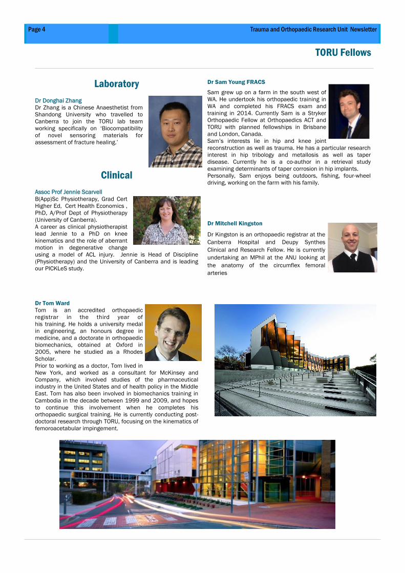
Laboratory
Dr Donghai Zhang
Dr Zhang is a Chinese Anaesthetist from
Shandong University who travelled to
Canberra to join the TORU lab team
working specifically on ‘Biocompatibility
of novel sensoring materials for
assessment of fracture healing.’
Clinical
Assoc Prof Jennie Scarvell
B(App)Sc Physiotherapy, Grad Cert
Higher Ed, Cert Health Economics ,
PhD, A/Prof Dept of Physiotherapy
(University of Canberra).
A career as clinical physiotherapist
lead Jennie to a PhD on knee
kinematics and the role of aberrant
motion in degenerative change
using a model of ACL injury. Jennie is Head of Discipline
(Physiotherapy) and the University of Canberra and is leading
our PICKLeS study.
Dr Tom Ward
Tom is an accredited orthopaedic
registrar in the third year of
his training. He holds a university medal
in engineering, an honours degree in
medicine, and a doctorate in orthopaedic
biomechanics, obtained at Oxford in
2005, where he studied as a Rhodes
Scholar.
Prior to working as a doctor, Tom lived in
New York, and worked as a consultant for McKinsey and
Company, which involved studies of the pharmaceutical
industry in the United States and of health policy in the Middle
East. Tom has also been involved in biomechanics training in
Cambodia in the decade between 1999 and 2009, and hopes
to continue this involvement when he completes his
orthopaedic surgical training. He is currently conducting post-
doctoral research through TORU, focusing on the kinematics of
femoroacetabular impingement.
Dr Sam Young FRACS
Sam grew up on a farm in the south west of
WA. He undertook his orthopaedic training in
WA and completed his FRACS exam and
training in 2014. Currently Sam is a Stryker
Orthopaedic Fellow at Orthopaedics ACT and
TORU with planned fellowships in Brisbane
and London, Canada.
Sam’s interests lie in hip and knee joint
reconstruction as well as trauma. He has a particular research
interest in hip tribology and metallosis as well as taper
disease. Currently he is a co-author in a retrieval study
examining determinants of taper corrosion in hip implants.
Personally, Sam enjoys being outdoors, fishing, four-wheel
driving, working on the farm with his family.
Dr Mitchell Kingston
Dr Kingston is an orthopaedic registrar at the
Canberra Hospital and Deupy Synthes
Clinical and Research Fellow. He is currently
undertaking an MPhil at the ANU looking at
the anatomy of the circumflex femoral
arteries
Page 5 Volume 8, 2015 Page 5 Trauma and Orthopaedic Research Unit Newsletter
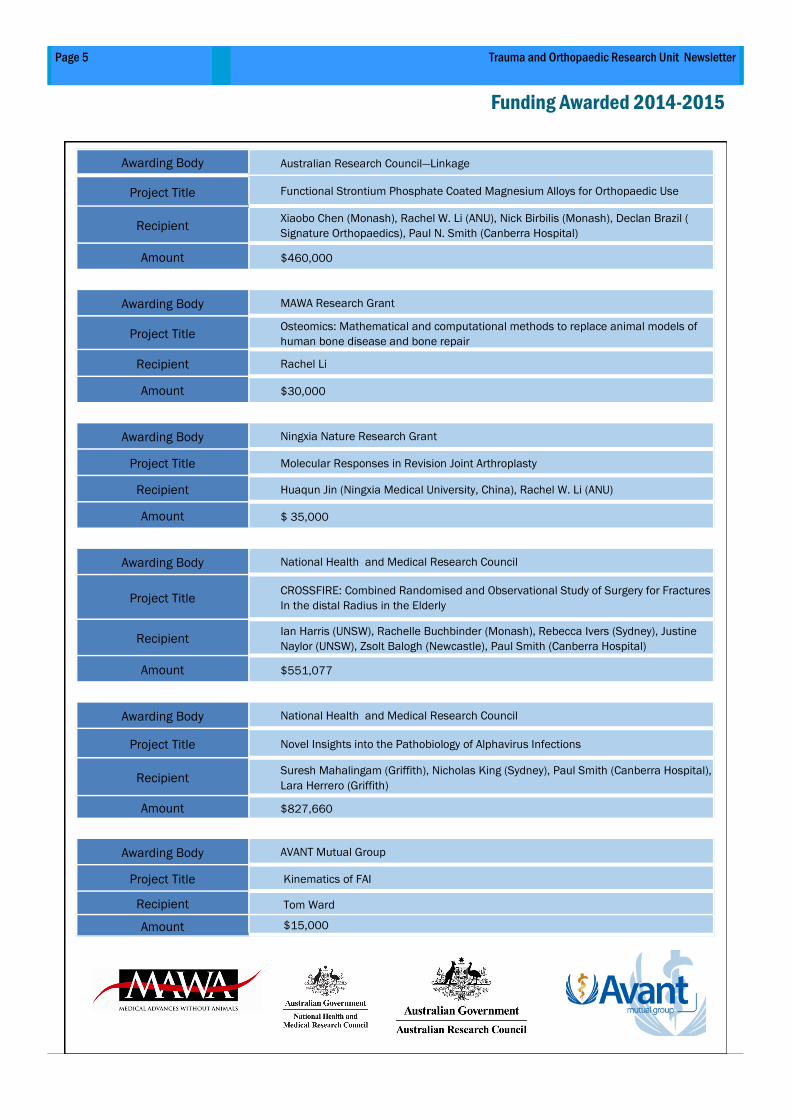
Funding Awarded 2014-2015
Awarding Body Australian Research Council—Linkage
Project Title Functional Strontium Phosphate Coated Magnesium Alloys for Orthopaedic Use
Recipient Xiaobo Chen (Monash), Rachel W. Li (ANU), Nick Birbilis (Monash), Declan Brazil (
Signature Orthopaedics), Paul N. Smith (Canberra Hospital)
Amount $460,000
Awarding Body MAWA Research Grant
Project Title Osteomics: Mathematical and computational methods to replace animal models of
human bone disease and bone repair
Recipient Rachel Li
Amount $30,000
Awarding Body Ningxia Nature Research Grant
Project Title Molecular Responses in Revision Joint Arthroplasty
Recipient Huaqun Jin (Ningxia Medical University, China), Rachel W. Li (ANU)
Amount $ 35,000
Awarding Body National Health and Medical Research Council
Project Title CROSSFIRE: Combined Randomised and Observational Study of Surgery for Fractures
In the distal Radius in the Elderly
Recipient Ian Harris (UNSW), Rachelle Buchbinder (Monash), Rebecca Ivers (Sydney), Justine
Naylor (UNSW), Zsolt Balogh (Newcastle), Paul Smith (Canberra Hospital)
Amount $551,077
Awarding Body National Health and Medical Research Council
Project Title Novel Insights into the Pathobiology of Alphavirus Infections
Recipient Suresh Mahalingam (Griffith), Nicholas King (Sydney), Paul Smith (Canberra Hospital),
Lara Herrero (Griffith)
Amount $827,660
Awarding Body AVANT Mutual Group
Project Title Kinematics of FAI
Recipient Tom Ward
Amount $15,000
Page 6 Trauma and Orthopaedic Research Unit Newsletter
PhD Scholars
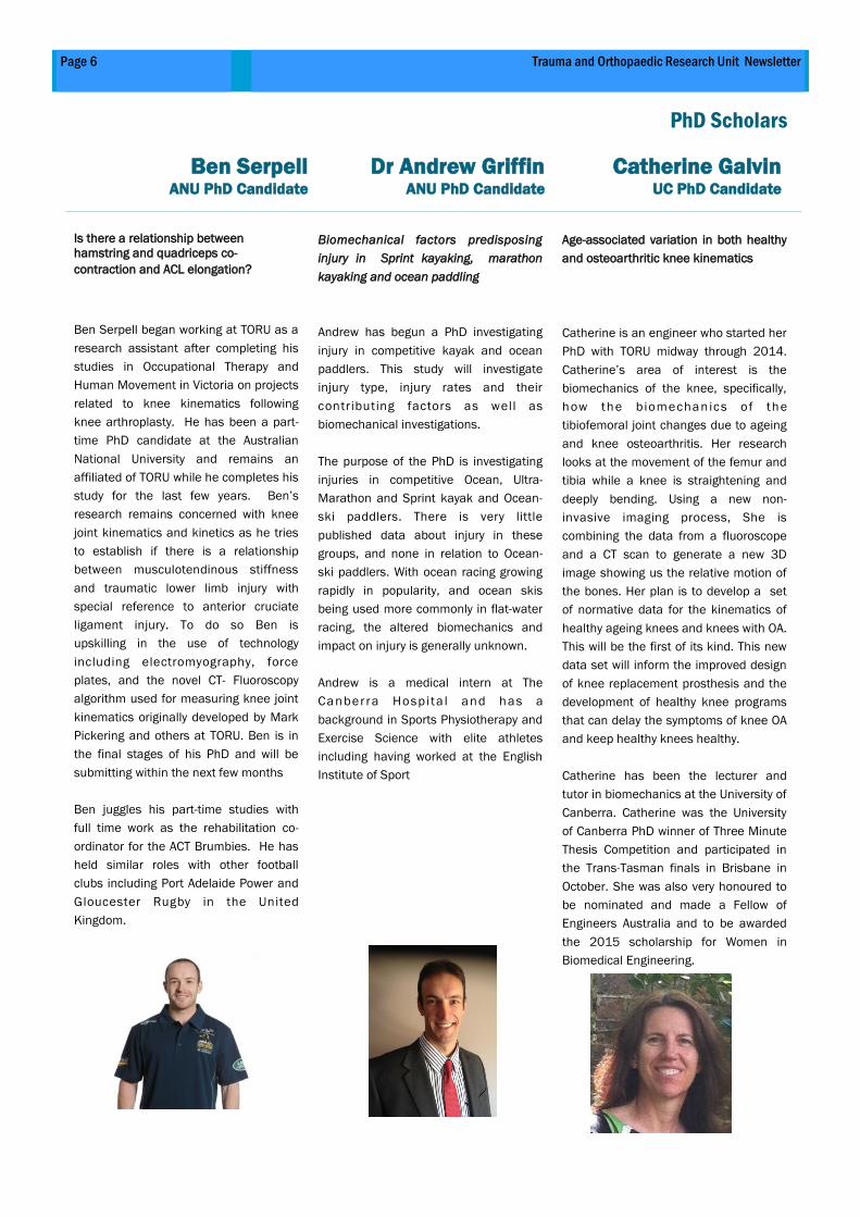
Ben Serpell ANU PhD Candidate
Dr Andrew Griffin ANU PhD Candidate
Catherine Galvin UC PhD Candidate
Is there a relationship between
hamstring and quadriceps co-
contraction and ACL elongation?
Ben Serpell began working at TORU as a
research assistant after completing his
studies in Occupational Therapy and
Human Movement in Victoria on projects
related to knee kinematics following
knee arthroplasty. He has been a part-
time PhD candidate at the Australian
National University and remains an
affiliated of TORU while he completes his
study for the last few years. Ben’s
research remains concerned with knee
joint kinematics and kinetics as he tries
to establish if there is a relationship
between musculotendinous stiffness
and traumatic lower limb injury with
special reference to anterior cruciate
ligament injury. To do so Ben is
upskilling in the use of technology
including electromyography, force
plates, and the novel CT- Fluoroscopy
algorithm used for measuring knee joint
kinematics originally developed by Mark
Pickering and others at TORU. Ben is in
the final stages of his PhD and will be
submitting within the next few months
Ben juggles his part-time studies with
full time work as the rehabilitation co-
ordinator for the ACT Brumbies. He has
held similar roles with other football
clubs including Port Adelaide Power and
Gloucester Rugby in the United
Kingdom.
Biomechanical factors predisposing
injury in Sprint kayaking, marathon
kayaking and ocean paddling
Andrew has begun a PhD investigating
injury in competitive kayak and ocean
paddlers. This study will investigate
injury type, injury rates and their
contributing factors as well as
biomechanical investigations.
The purpose of the PhD is investigating
injuries in competitive Ocean, Ultra-
Marathon and Sprint kayak and Ocean-
ski paddlers. There is very little
published data about injury in these
groups, and none in relation to Ocean-
ski paddlers. With ocean racing growing
rapidly in popularity, and ocean skis
being used more commonly in flat-water
racing, the altered biomechanics and
impact on injury is generally unknown.
Andrew is a medical intern at The
Canberra Hospi ta l and has a
background in Sports Physiotherapy and
Exercise Science with elite athletes
including having worked at the English
Institute of Sport
Age-associated variation in both healthy
and osteoarthritic knee kinematics
Catherine is an engineer who started her
PhD with TORU midway through 2014.
Catherine’s area of interest is the
biomechanics of the knee, specifically,
how the b iomechanics of the
tibiofemoral joint changes due to ageing
and knee osteoarthritis. Her research
looks at the movement of the femur and
tibia while a knee is straightening and
deeply bending. Using a new non-
invasive imaging process, She is
combining the data from a fluoroscope
and a CT scan to generate a new 3D
image showing us the relative motion of
the bones. Her plan is to develop a set
of normative data for the kinematics of
healthy ageing knees and knees with OA.
This will be the first of its kind. This new
data set will inform the improved design
of knee replacement prosthesis and the
development of healthy knee programs
that can delay the symptoms of knee OA
and keep healthy knees healthy.
Catherine has been the lecturer and
tutor in biomechanics at the University of
Canberra. Catherine was the University
of Canberra PhD winner of Three Minute
Thesis Competition and participated in
the Trans-Tasman finals in Brisbane in
October. She was also very honoured to
be nominated and made a Fellow of
Engineers Australia and to be awarded
the 2015 scholarship for Women in
Biomedical Engineering.
Page 7 Trauma and Orthopaedic Research Unit Newsletter
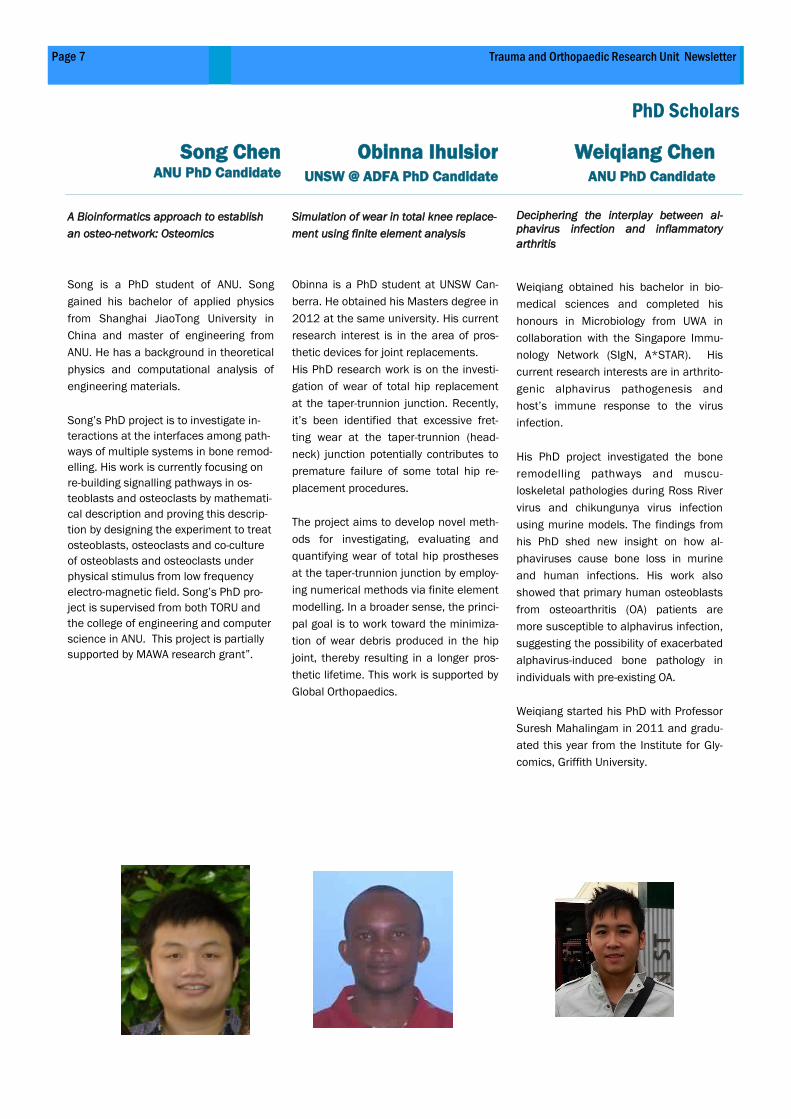
A Bioinformatics approach to establish
an osteo-network: Osteomics
Song is a PhD student of ANU. Song
gained his bachelor of applied physics
from Shanghai JiaoTong University in
China and master of engineering from
ANU. He has a background in theoretical
physics and computational analysis of
engineering materials.
Song’s PhD project is to investigate in-
teractions at the interfaces among path-
ways of multiple systems in bone remod-
elling. His work is currently focusing on
re-building signalling pathways in os-
teoblasts and osteoclasts by mathemati-
cal description and proving this descrip-
tion by designing the experiment to treat
osteoblasts, osteoclasts and co-culture
of osteoblasts and osteoclasts under
physical stimulus from low frequency
electro-magnetic field. Song’s PhD pro-
ject is supervised from both TORU and
the college of engineering and computer
science in ANU. This project is partially
supported by MAWA research grant”.
Simulation of wear in total knee replace-
ment using finite element analysis
Obinna is a PhD student at UNSW Can-
berra. He obtained his Masters degree in
2012 at the same university. His current
research interest is in the area of pros-
thetic devices for joint replacements.
His PhD research work is on the investi-
gation of wear of total hip replacement
at the taper-trunnion junction. Recently,
it’s been identified that excessive fret-
ting wear at the taper-trunnion (head-
neck) junction potentially contributes to
premature failure of some total hip re-
placement procedures.
The project aims to develop novel meth-
ods for investigating, evaluating and
quantifying wear of total hip prostheses
at the taper-trunnion junction by employ-
ing numerical methods via finite element
modelling. In a broader sense, the princi-
pal goal is to work toward the minimiza-
tion of wear debris produced in the hip
joint, thereby resulting in a longer pros-
thetic lifetime. This work is supported by
Global Orthopaedics.
Deciphering the interplay between al-
phavirus infection and inflammatory
arthritis
Weiqiang obtained his bachelor in bio-
medical sciences and completed his
honours in Microbiology from UWA in
collaboration with the Singapore Immu-
nology Network (SIgN, A*STAR). His
current research interests are in arthrito-
genic alphavirus pathogenesis and
host’s immune response to the virus
infection.
His PhD project investigated the bone
remodelling pathways and muscu-
loskeletal pathologies during Ross River
virus and chikungunya virus infection
using murine models. The findings from
his PhD shed new insight on how al-
phaviruses cause bone loss in murine
and human infections. His work also
showed that primary human osteoblasts
from osteoarthritis (OA) patients are
more susceptible to alphavirus infection,
suggesting the possibility of exacerbated
alphavirus-induced bone pathology in
individuals with pre-existing OA.
Weiqiang started his PhD with Professor
Suresh Mahalingam in 2011 and gradu-
ated this year from the Institute for Gly-
comics, Griffith University.
PhD Scholars
Song Chen ANU PhD Candidate
Obinna Ihulsior UNSW @ ADFA PhD Candidate
Weiqiang Chen ANU PhD Candidate
TORU Laboratory Research Report
Page 8 Volume 8, 2015
TORU Laboratory team bridges basic and
clinical sciences and facilitates
communication among TORU’s collaborative
institutes, universities and orthopaedic
industries.
TORU team presently investigates chronic
and complex bone diseases, some of which
cause life-long pain and disability. These
chronic conditions can be rare, such as
revision joint replacement and osteolysis or
can be remarkably common, such as
arthritis, trauma and osteoporotic fractures.
Combined, they afflict millions of
Australians and cause tremendous human
suffering, and cost million dollars in health
care.
The team utilizes a mix of conventional
molecular biology approaches as well as
global methods such as next generation
sequencing to study mRNA expression and
its regulation by non-coding RNA e.g.
microRNAs with an ultimate goal of
identifying novel molecules that regulate
bone resorption, formation, fracture repair
and bone homeostasis.
Key Research Areas
Osteoimmunology, microRNAs’ (miRNA)
regulation and genetic risk factors in
biomaterial related osteolysis in total joint
replacement.
This research has in part supported by AOA
Research Foundation. Building on the
foundation laid by the dendritic cells
involvement in osteolysis, the group is using
off-cut tissues from the cohorts of healthy,
primary and revision subjects of TJR for
characterization of wear particles and
identification of molecular and genetic risk
factors that contribute to the osteolysis. The
ultimate goals are to contribute to the
development of better predictive markers,
treatments, and prevention strategies.
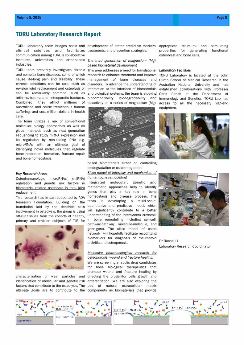
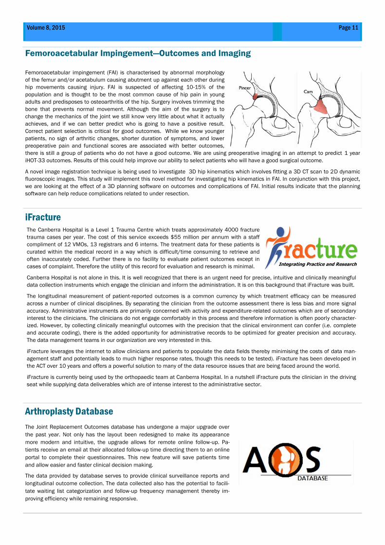
The third generation of magnesium (Mg)-
based biomaterial development
This area addresses a need for translational
research to enhance treatment and improve
management of bone diseases and
disorders. To advance the understanding of
interaction at the interface of biomaterials
and biological systems, the team is studying
biocompatibility, biodegradability and
bioactivity on a series of magnesium (Mg)-
based biomaterials either on controlling
biodegradation or osteointegration.
Silico model of interplay and mechanism of
human bone remodelling
Integrated molecular, genetic and
mathematic approaches help to identify
genes that play a key role in bone
homeostasis and disease process. The
team is developing a multi-scale,
quantitative and predictive model, which
will significantly contribute to a better
understanding of the intersystem crosstalk
in bone remodelling including cell-cell,
pathway-pathway, molecule-molecule, and
gene-gene. The silico model of osteo
network will hopefully facilitate recognizing
biomarkers for diagnosis of rheumatoid
arthritis and osteoporosis.
Molecular pharmacological research for
osteoporosis, wound and fracture healing.
We are screening anabolic drug candidates
for bone biological therapeutics that
promote wound and fracture healing by
directing the progenitor cells growth and
differentiation. We are also exploring the
use of natural extracellular matrix
components as biomaterials that provide
appropriate structural and stimulating
properties for generating functional
osteoblast and bone cells.
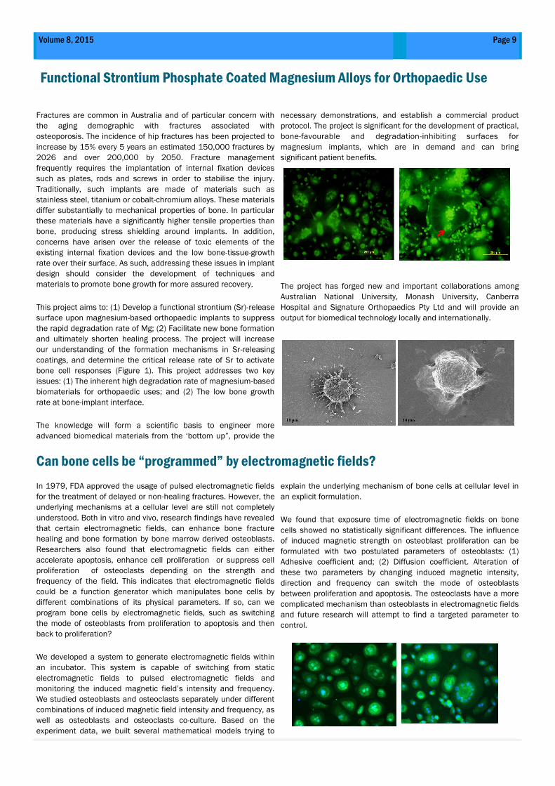
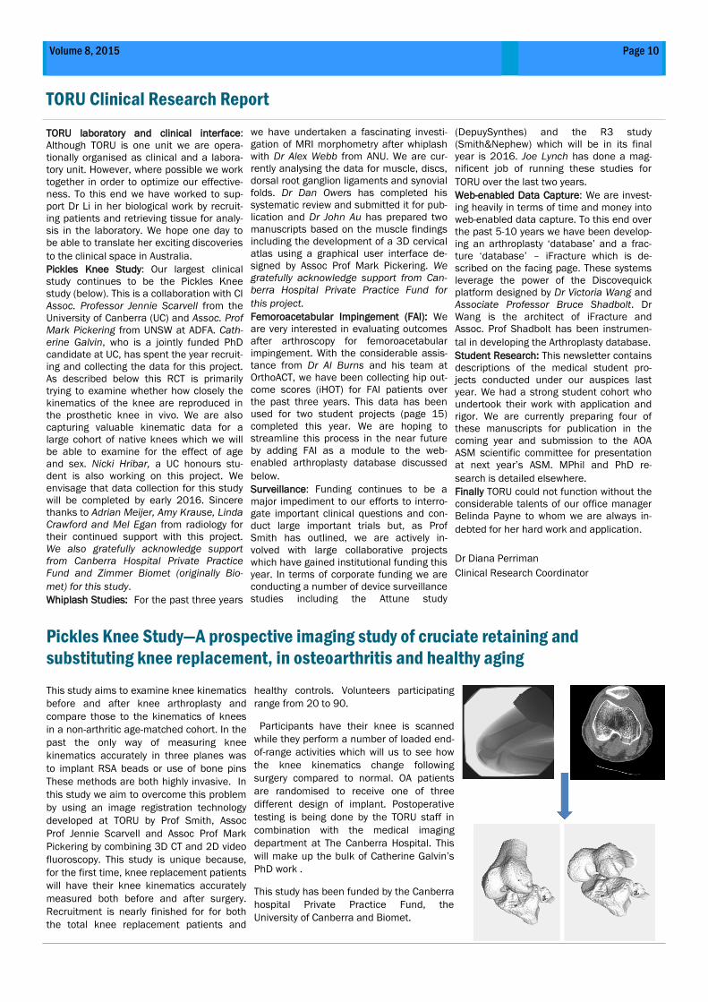
Laboratory Facilities
TORU Laboratory is located at the John
Curtin School of Medical Research in the
Australian National University and has
established collaborations with Professor
Chris Parish at the Department of
Immunology and Genetics. TORU Lab has
access to all the necessary high-end
equipment.
Dr Rachel Li
Laboratory Research Coordinator
Page 9 Volume 8, 2015
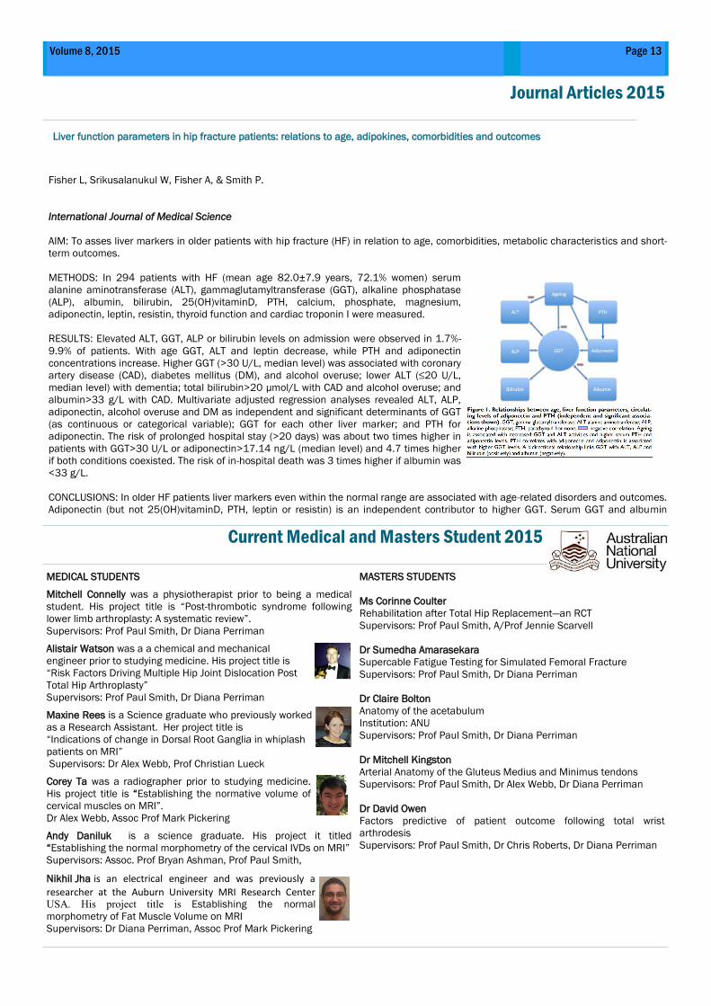
Functional Strontium Phosphate Coated Magnesium Alloys for Orthopaedic Use
Fractures are common in Australia and of particular concern with
the aging demographic with fractures associated with
osteoporosis. The incidence of hip fractures has been projected to
increase by 15% every 5 years an estimated 150,000 fractures by
2026 and over 200,000 by 2050. Fracture management
frequently requires the implantation of internal fixation devices
such as plates, rods and screws in order to stabilise the injury.
Traditionally, such implants are made of materials such as
stainless steel, titanium or cobalt-chromium alloys. These materials
differ substantially to mechanical properties of bone. In particular
these materials have a significantly higher tensile properties than
bone, producing stress shielding around implants. In addition,
concerns have arisen over the release of toxic elements of the
existing internal fixation devices and the low bone-tissue-growth
rate over their surface. As such, addressing these issues in implant
design should consider the development of techniques and
materials to promote bone growth for more assured recovery.
This project aims to: (1) Develop a functional strontium (Sr)-release
surface upon magnesium-based orthopaedic implants to suppress
the rapid degradation rate of Mg; (2) Facilitate new bone formation
and ultimately shorten healing process. The project will increase
our understanding of the formation mechanisms in Sr-releasing
coatings, and determine the critical release rate of Sr to activate
bone cell responses (Figure 1). This project addresses two key
issues: (1) The inherent high degradation rate of magnesium-based
biomaterials for orthopaedic uses; and (2) The low bone growth
rate at bone-implant interface.
The knowledge will form a scientific basis to engineer more
advanced biomedical materials from the ‘bottom up”, provide the
necessary demonstrations, and establish a commercial product
protocol. The project is significant for the development of practical,
bone-favourable and degradation-inhibiting surfaces for
magnesium implants, which are in demand and can bring
significant patient benefits.
The project has forged new and important collaborations among
Australian National University, Monash University, Canberra
Hospital and Signature Orthopaedics Pty Ltd and will provide an
output for biomedical technology locally and internationally.
Can bone cells be “programmed” by electromagnetic fields?
In 1979, FDA approved the usage of pulsed electromagnetic fields
for the treatment of delayed or non-healing fractures. However, the
underlying mechanisms at a cellular level are still not completely
understood. Both in vitro and vivo, research findings have revealed
that certain electromagnetic fields, can enhance bone fracture
healing and bone formation by bone marrow derived osteoblasts.
Researchers also found that electromagnetic fields can either
accelerate apoptosis, enhance cell proliferation or suppress cell
proliferation of osteoclasts depending on the strength and
frequency of the field. This indicates that electromagnetic fields
could be a function generator which manipulates bone cells by
different combinations of its physical parameters. If so, can we
program bone cells by electromagnetic fields, such as switching
the mode of osteoblasts from proliferation to apoptosis and then
back to proliferation?
We developed a system to generate electromagnetic fields within
an incubator. This system is capable of switching from static
electromagnetic fields to pulsed electromagnetic fields and
monitoring the induced magnetic field’s intensity and frequency.
We studied osteoblasts and osteoclasts separately under different
combinations of induced magnetic field intensity and frequency, as
well as osteoblasts and osteoclasts co-culture. Based on the
experiment data, we built several mathematical models trying to
explain the underlying mechanism of bone cells at cellular level in
an explicit formulation.
We found that exposure time of electromagnetic fields on bone
cells showed no statistically significant differences. The influence
of induced magnetic strength on osteoblast proliferation can be
formulated with two postulated parameters of osteoblasts: (1)
Adhesive coefficient and; (2) Diffusion coefficient. Alteration of
these two parameters by changing induced magnetic intensity,
direction and frequency can switch the mode of osteoblasts
between proliferation and apoptosis. The osteoclasts have a more
complicated mechanism than osteoblasts in electromagnetic fields
and future research will attempt to find a targeted parameter to
control.
TORU Clinical Research Report
Page 10 Volume 8, 2015
TORU laboratory and clinical interface:
Although TORU is one unit we are opera-
tionally organised as clinical and a labora-
tory unit. However, where possible we work
together in order to optimize our effective-
ness. To this end we have worked to sup-
port Dr Li in her biological work by recruit-
ing patients and retrieving tissue for analy-
sis in the laboratory. We hope one day to
be able to translate her exciting discoveries
to the clinical space in Australia.
Pickles Knee Study: Our largest clinical
study continues to be the Pickles Knee
study (below). This is a collaboration with CI
Assoc. Professor Jennie Scarvell from the
University of Canberra (UC) and Assoc. Prof
Mark Pickering from UNSW at ADFA. Cath-
erine Galvin, who is a jointly funded PhD
candidate at UC, has spent the year recruit-
ing and collecting the data for this project.
As described below this RCT is primarily
trying to examine whether how closely the
kinematics of the knee are reproduced in
the prosthetic knee in vivo. We are also
capturing valuable kinematic data for a
large cohort of native knees which we will
be able to examine for the effect of age
and sex. Nicki Hribar, a UC honours stu-
dent is also working on this project. We
envisage that data collection for this study
will be completed by early 2016. Sincere
thanks to Adrian Meijer, Amy Krause, Linda
Crawford and Mel Egan from radiology for
their continued support with this project.
We also gratefully acknowledge support
from Canberra Hospital Private Practice
Fund and Zimmer Biomet (originally Bio-
met) for this study.
Whiplash Studies: For the past three years
we have undertaken a fascinating investi-
gation of MRI morphometry after whiplash
with Dr Alex Webb from ANU. We are cur-
rently analysing the data for muscle, discs,
dorsal root ganglion ligaments and synovial
folds. Dr Dan Owers has completed his
systematic review and submitted it for pub-
lication and Dr John Au has prepared two
manuscripts based on the muscle findings
including the development of a 3D cervical
atlas using a graphical user interface de-
signed by Assoc Prof Mark Pickering. We
gratefully acknowledge support from Can-
berra Hospital Private Practice Fund for
this project.
Femoroacetabular Impingement (FAI): We
are very interested in evaluating outcomes
after arthroscopy for femoroacetabular
impingement. With the considerable assis-
tance from Dr Al Burns and his team at
OrthoACT, we have been collecting hip out-
come scores (iHOT) for FAI patients over
the past three years. This data has been
used for two student projects (page 15)
completed this year. We are hoping to
streamline this process in the near future
by adding FAI as a module to the web-
enabled arthroplasty database discussed
below.
Surveillance: Funding continues to be a
major impediment to our efforts to interro-
gate important clinical questions and con-
duct large important trials but, as Prof
Smith has outlined, we are actively in-
volved with large collaborative projects
which have gained institutional funding this
year. In terms of corporate funding we are
conducting a number of device surveillance
studies including the Attune study
(DepuySynthes) and the R3 study
(Smith&Nephew) which will be in its final
year is 2016. Joe Lynch has done a mag-
nificent job of running these studies for
TORU over the last two years.
Web-enabled Data Capture: We are invest-
ing heavily in terms of time and money into
web-enabled data capture. To this end over
the past 5-10 years we have been develop-
ing an arthroplasty ‘database’ and a frac-
ture ‘database’ – iFracture which is de-
scribed on the facing page. These systems
leverage the power of the Discovequick
platform designed by Dr Victoria Wang and
Associate Professor Bruce Shadbolt. Dr
Wang is the architect of iFracture and
Assoc. Prof Shadbolt has been instrumen-
tal in developing the Arthroplasty database.
Student Research: This newsletter contains
descriptions of the medical student pro-
jects conducted under our auspices last
year. We had a strong student cohort who
undertook their work with application and
rigor. We are currently preparing four of
these manuscripts for publication in the
coming year and submission to the AOA
ASM scientific committee for presentation
at next year’s ASM. MPhil and PhD re-
search is detailed elsewhere.
Finally TORU could not function without the
considerable talents of our office manager
Belinda Payne to whom we are always in-
debted for her hard work and application.
Dr Diana Perriman
Clinical Research Coordinator
This study aims to examine knee kinematics
before and after knee arthroplasty and
compare those to the kinematics of knees
in a non-arthritic age-matched cohort. In the
past the only way of measuring knee
kinematics accurately in three planes was
to implant RSA beads or use of bone pins
These methods are both highly invasive. In
this study we aim to overcome this problem
by using an image registration technology
developed at TORU by Prof Smith, Assoc
Prof Jennie Scarvell and Assoc Prof Mark
Pickering by combining 3D CT and 2D video
fluoroscopy. This study is unique because,
for the first time, knee replacement patients
will have their knee kinematics accurately
measured both before and after surgery.
Recruitment is nearly finished for for both
the total knee replacement patients and
healthy controls. Volunteers participating
range from 20 to 90.
Participants have their knee is scanned
while they perform a number of loaded end-
of-range activities which will us to see how
the knee kinematics change following
surgery compared to normal. OA patients
are randomised to receive one of three
different design of implant. Postoperative
testing is being done by the TORU staff in
combination with the medical imaging
department at The Canberra Hospital. This
will make up the bulk of Catherine Galvin’s
PhD work .
This study has been funded by the Canberra
hospital Private Practice Fund, the
University of Canberra and Biomet.
Pickles Knee Study—A prospective imaging study of cruciate retaining and
substituting knee replacement, in osteoarthritis and healthy aging
Page 11 Volume 8, 2015
Femoroacetabular impingement (FAI) is characterised by abnormal morphology
of the femur and/or acetabulum causing abutment up against each other during
hip movements causing injury. FAI is suspected of affecting 10-15% of the
population and is thought to be the most common cause of hip pain in young
adults and predisposes to osteoarthritis of the hip. Surgery involves trimming the
bone that prevents normal movement. Although the aim of the surgery is to
change the mechanics of the joint we still know very little about what it actually
achieves, and if we can better predict who is going to have a positive result.
Correct patient selection is critical for good outcomes. While we know younger
patients, no sign of arthritic changes, shorter duration of symptoms, and lower
preoperative pain and functional scores are associated with better outcomes,
there is still a group of patients who do not have a good outcome. We are using preoperative imaging in an attempt to predict 1 year
iHOT-33 outcomes. Results of this could help improve our ability to select patients who will have a good surgical outcome.
A novel image registration technique is being used to investigate 3D hip kinematics which involves fitting a 3D CT scan to 2D dynamic
fluoroscopic images. This study will implement this novel method for investigating hip kinematics in FAI. In conjunction with this project,
we are looking at the effect of a 3D planning software on outcomes and complications of FAI. Initial results indicate that the planning
software can help reduce complications related to under resection.
Femoroacetabular Impingement—Outcomes and Imaging
iFracture
Arthroplasty Database
The Joint Replacement Outcomes database has undergone a major upgrade over
the past year. Not only has the layout been redesigned to make its appearance
more modern and intuitive, the upgrade allows for remote online follow-up. Pa-
tients receive an email at their allocated follow-up time directing them to an online
portal to complete their questionnaires. This new feature will save patients time
and allow easier and faster clinical decision making.
The data provided by database serves to provide clinical surveillance reports and
longitudinal outcome collection. The data collected also has the potential to facili-
tate waiting list categorization and follow-up frequency management thereby im-
proving efficiency while remaining responsive.
The Canberra Hospital is a Level 1 Trauma Centre which treats approximately 4000 fracture
trauma cases per year. The cost of this service exceeds $55 million per annum with a staff
compliment of 12 VMOs, 13 registrars and 6 interns. The treatment data for these patients is
curated within the medical record in a way which is difficult/time consuming to retrieve and
often inaccurately coded. Further there is no facility to evaluate patient outcomes except in
cases of complaint. Therefore the utility of this record for evaluation and research is minimal.
Canberra Hospital is not alone in this. It is well recognized that there is an urgent need for precise, intuitive and clinically meaningful
data collection instruments which engage the clinician and inform the administration. It is on this background that iFracture was built.
The longitudinal measurement of patient-reported outcomes is a common currency by which treatment efficacy can be measured
across a number of clinical disciplines. By separating the clinician from the outcome assessment there is less bias and more signal
accuracy. Administrative instruments are primarily concerned with activity and expenditure-related outcomes which are of secondary
interest to the clinicians. The clinicians do not engage comfortably in this process and therefore information is often poorly character-
ized. However, by collecting clinically meaningful outcomes with the precision that the clinical environment can confer (i.e. complete
and accurate coding), there is the added opportunity for administrative records to be optimized for greater precision and accuracy.
The data management teams in our organization are very interested in this.
iFracture leverages the internet to allow clinicians and patients to populate the data fields thereby minimising the costs of data man-
agement staff and potentially leads to much higher response rates, though this needs to be tested). iFracture has been developed in
the ACT over 10 years and offers a powerful solution to many of the data resource issues that are being faced around the world.
iFracture is currently being used by the orthopaedic team at Canberra Hospital. In a nutshell iFracture puts the clinician in the driving
seat while supplying data deliverables which are of intense interest to the administrative sector.
Integrating Practice and Research
Medial and lateral hamstrings and quadriceps co-activation
affects knee joint kinematics and ACL elongation: a pilot study
Development and validation of a VISA tendinopathy question-
naire for greater trochanteric pain syndrome, the VISA-G.
Serpell B.G, Scarvell J.M., Pickering M.R., Ball N.B., Newman
P., Perriman D, Warmenhoven J., & Smith P.N.
BMC Musculoskeletal Disorders
Background: Many injury prevention and rehabilitation
programs aim to train hamstring and quadriceps co-activation
to constrain excessive anterior tibial translation and protect
the anterior cruciate ligament (ACL) from injury. However,
despite strong clinical belief in its efficacy, primary evidence
supporting training co-activation of the hamstrings and
quadriceps muscles for ACL injury prevention and
rehabilitation is quite limited. Therefore, the purpose of the
study presented in this paper was to determine if hamstring-
quadriceps co-activation alters knee joint kinematics, and
also establish if it affects ACL elongation.
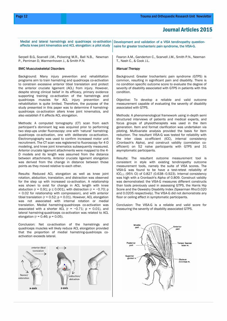
Methods: A computed tomography (CT) scan from each
participant’s dominant leg was acquired prior to performing
two step-ups under fluoroscopy: one with ‘natural’ hamstring-
quadriceps co-activation, one with deliberate co-activation.
Electromyography was used to confirm increased motor unit
recruitment. The CT scan was registered to fluoroscopy for 4-D
modeling, and knee joint kinematics subsequently measured.
Anterior cruciate ligament attachments were mapped to the 4-
D models and its length was assumed from the distance
between attachments. Anterior cruciate ligament elongation
was derived from the change in distance between those
points as they moved relative to each other.
Results: Reduced ACL elongation as well as knee joint
rotation, abduction, translation, and distraction was observed
for the step up with increased co-activation. A relationship
was shown to exist for change in ACL length with knee
abduction (r = 0.91; p ≤ 0.001), with distraction (r = −0.70; p
= 0.02 for relationship with compression), and with anterior
tibial translation (r = 0.52; p = 0.01). However, ACL elongation
was not associated with internal rotation or medial
translation. Medial hamstring-quadriceps co-activation was
associated with a shorter ACL (r = −0.71; p = 0.01), and
lateral hamstring-quadriceps co-activation was related to ACL
elongation (r = 0.46; p = 0.05).
Conclusion: Net co-activation of the hamstrings and
quadriceps muscles will likely reduce ACL elongation provided
that the proportion of medial hamstring-quadriceps co-
activation exceeds lateral.
Fearon A.M., Ganderton C., Scarvell J.M., Smith P.N., Neeman
T., Nash C., & Cook J.L.
Manual Therapy
Background: Greater trochanteric pain syndrome (GTPS) is
common, resulting in significant pain and disability. There is
no condition specific outcome score to evaluate the degree of
severity of disability associated with GTPS in patients with this
condition.
Objective: To develop a reliable and valid outcome
measurement capable of evaluating the severity of disability
associated with GTPS.
Methods: A phenomenological framework using in-depth semi
structured interviews of patients and medical experts, and
focus groups of physiotherapists was used in the item
generation. Item and format clarification was undertaken via
piloting. Multivariate analysis provided the basis for item
reduction. The resultant VISA-G was tested for reliability with
the inter class co-efficient (ICC), internal consistency
(Cronbach's Alpha), and construct validity (correlation co-
efficient) on 52 naïve participants with GTPS and 31
asymptomatic participants.
Results: The resultant outcome measurement tool is
consistent in style with existing tendinopathy outcome
measurement tools, namely the suite of VISA scores. The
VISA-G was found to be have a test-retest reliability of
ICC2,1 (95% CI) of 0.827 (0.638–0.923). Internal consistency
was high with a Cronbach's Alpha of 0.809. Construct validity
was demonstrated: the VISA-G measures different constructs
than tools previously used in assessing GTPS, the Harris Hip
Score and the Oswestry Disability Index (Spearman Rho:0.020
and 0.0205 respectively). The VISA-G did not demonstrate any
floor or ceiling effect in symptomatic participants.
Conclusion: The VISA-G is a reliable and valid score for
measuring the severity of disability associated GTPS.
Page 12 Trauma and Orthopaedic Research Unit Newsletter
Journal Articles 2015
Page 13 Volume 8, 2015
Journal Articles 2015
MEDICAL STUDENTS
Mitchell Connelly was a physiotherapist prior to being a medical
student. His project title is “Post-thrombotic syndrome following
lower limb arthroplasty: A systematic review”.
Supervisors: Prof Paul Smith, Dr Diana Perriman
Alistair Watson was a a chemical and mechanical
engineer prior to studying medicine. His project title is
“Risk Factors Driving Multiple Hip Joint Dislocation Post
Total Hip Arthroplasty”
Supervisors: Prof Paul Smith, Dr Diana Perriman
Maxine Rees is a Science graduate who previously worked
as a Research Assistant. Her project title is
“Indications of change in Dorsal Root Ganglia in whiplash
patients on MRI”
Supervisors: Dr Alex Webb, Prof Christian Lueck
Corey Ta was a radiographer prior to studying medicine.
His project title is “Establishing the normative volume of
cervical muscles on MRI”. Dr Alex Webb, Assoc Prof Mark Pickering
Andy Daniluk is a science graduate. His project it titled
“Establishing the normal morphometry of the cervical IVDs on MRI”
Supervisors: Assoc. Prof Bryan Ashman, Prof Paul Smith,
Nikhil Jha is an electrical engineer and was previously a
researcher at the Auburn University MRI Research Center USA. His project title is Establishing the normal
morphometry of Fat Muscle Volume on MRI
Supervisors: Dr Diana Perriman, Assoc Prof Mark Pickering
MASTERS STUDENTS
Ms Corinne Coulter
Rehabilitation after Total Hip Replacement—an RCT
Supervisors: Prof Paul Smith, A/Prof Jennie Scarvell
Dr Sumedha Amarasekara
Supercable Fatigue Testing for Simulated Femoral Fracture
Supervisors: Prof Paul Smith, Dr Diana Perriman
Dr Claire Bolton
Anatomy of the acetabulum
Institution: ANU
Supervisors: Prof Paul Smith, Dr Diana Perriman
Dr Mitchell Kingston
Arterial Anatomy of the Gluteus Medius and Minimus tendons
Supervisors: Prof Paul Smith, Dr Alex Webb, Dr Diana Perriman
Dr David Owen
Factors predictive of patient outcome following total wrist
arthrodesis
Supervisors: Prof Paul Smith, Dr Chris Roberts, Dr Diana Perriman
Fisher L, Srikusalanukul W, Fisher A, & Smith P.
International Journal of Medical Science
AIM: To asses liver markers in older patients with hip fracture (HF) in relation to age, comorbidities, metabolic characteristics and short-
term outcomes.
METHODS: In 294 patients with HF (mean age 82.0±7.9 years, 72.1% women) serum
alanine aminotransferase (ALT), gammaglutamyltransferase (GGT), alkaline phosphatase
(ALP), albumin, bilirubin, 25(OH)vitaminD, PTH, calcium, phosphate, magnesium,
adiponectin, leptin, resistin, thyroid function and cardiac troponin I were measured.
RESULTS: Elevated ALT, GGT, ALP or bilirubin levels on admission were observed in 1.7%-
9.9% of patients. With age GGT, ALT and leptin decrease, while PTH and adiponectin
concentrations increase. Higher GGT (>30 U/L, median level) was associated with coronary
artery disease (CAD), diabetes mellitus (DM), and alcohol overuse; lower ALT (≤20 U/L,
median level) with dementia; total bilirubin>20 μmol/L with CAD and alcohol overuse; and
albumin>33 g/L with CAD. Multivariate adjusted regression analyses revealed ALT, ALP,
adiponectin, alcohol overuse and DM as independent and significant determinants of GGT
(as continuous or categorical variable); GGT for each other liver marker; and PTH for
adiponectin. The risk of prolonged hospital stay (>20 days) was about two times higher in
patients with GGT>30 U/L or adiponectin>17.14 ng/L (median level) and 4.7 times higher
if both conditions coexisted. The risk of in-hospital death was 3 times higher if albumin was
<33 g/L.
CONCLUSIONS: In older HF patients liver markers even within the normal range are associated with age-related disorders and outcomes.
Adiponectin (but not 25(OH)vitaminD, PTH, leptin or resistin) is an independent contributor to higher GGT. Serum GGT and albumin
Liver function parameters in hip fracture patients: relations to age, adipokines, comorbidities and outcomes
Current Medical and Masters Student 2015
Mike Van Alphen
Patient-reported outcomes
fo l lowing mid-c lavicu lar
fractures: Non-operative vs
Operative Management
Background: Recent studies report that
operative fixation of displaced mid-
clavicular fractures results in fewer
complications in the short-term. The aim
of this study was to assess whether
operative management also results in
superior long term (>5 years) patient-
reported outcomes. A further aim was to
assess which measurements technique
for clavicular displacement best predicts
outcome in non-operatively managed
patients.
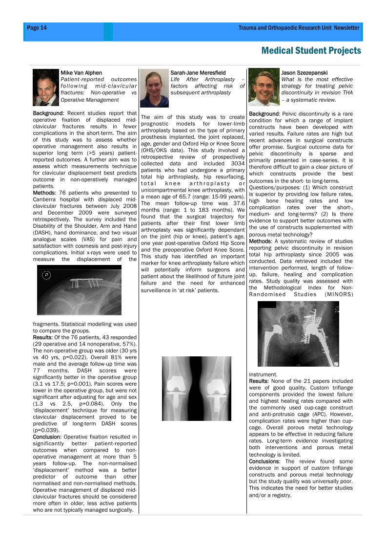
Methods: 76 patients who presented to
Canberra hospital with displaced mid-
clavicular fractures between July 2008
and December 2009 were surveyed
retrospectively. The survey included the
Disability of the Shoulder, Arm and Hand
(DASH), hand dominance, and two visual
analogue scales (VAS) for pain and
satisfaction with cosmesis and post-injury
complications. Initial x-rays were used to
measure the displacement of the
fragments. Statistical modelling was used
to compare the groups.
Results: Of the 76 patients, 43 responded
(29 operative and 14 nonoperative, 57%).
The non-operative group was older (30 yrs
vs 40 yrs, p=0.022). Overall 81% were
male and the average follow-up time was
77 months. DASH scores were
significantly better in the operative group
(3.1 vs 17.5; p=0.001). Pain scores were
lower in the operative group, but were not
significant after adjusting for age and sex
(1.3 vs 2.5, p=0.084). Only the
‘displacement’ technique for measuring
clavicular displacement proved to be
predictive of long-term DASH scores
(p=0.039).
Conclusion: Operative fixation resulted in
significantly better patient-reported
outcomes when compared to non-
operative management at more than 5
years follow-up. The non-normalised
‘displacement’ method was a better
predictor of outcome than other
normalised and non-normalised methods.
Operative management of displaced mid-
clavicular fractures should be considered
more often in older, less active patients
who are not typically managed surgically.
Sarah-Jane Meresfield
Life After Arthroplasty –
factors affecting risk of
subsequent arthroplasty
The aim of this study was to create
prognostic models for lower-limb
arthroplasty based on the type of primary
prosthesis implanted, the joint replaced,
age, gender and Oxford Hip or Knee Score
(OHS/OKS data). This study involved a
retrospective review of prospectively
collected data and included 3034
patients who had undergone a primary
total hip arthroplasty, hip resurfacing,
t o t a l k n e e a r t h r o p l a s t y o r
unicompartmental knee arthroplasty, with
a mean age of 65.7 (range: 15-99 years).
The mean follow-up time was 37.6
months (range: 1 to 183 months). We
found that the surgical trajectory for
patients after their first lower limb
arthroplasty was significantly dependant
on the joint (hip or knee), patient’s age,
one year post-operative Oxford Hip Score
and the preoperative Oxford Knee Score.
This study has identified an important
marker for knee arthroplasty failure which
will potentially inform surgeons and
patient about the likelihood of future joint
failure and the need for enhanced
surveillance in ‘at risk’ patients.
Jason Szezepanski
What is the most effective
strategy for treating pelvic
discontinuity in revision THA
– a systematic review.
Background: Pelvic discontinuity is a rare
condition for which a range of implant
constructs have been developed with
varied results. Failure rates are high but
recent advances in surgical constructs
offer promise. Surgical outcome data for
pelvic discontinuity is sparse and
primarily presented in case-series. It is
therefore difficult to gain a clear picture of
which constructs provide the best
outcomes in the short- to long-terms.
Questions/purposes: (1) Which construct
is superior by providing low failure rates,
high bone healing rates and low
complication rates over the short-,
medium- and long-terms? (2) Is there
evidence to support better outcomes with
the use of constructs supplemented with
porous metal technology?
Methods: A systematic review of studies
reporting pelvic discontinuity in revision
total hip arthroplasty since 2005 was
conducted. Data retrieved included the
intervention performed, length of follow-
up, failure, healing and complication
rates. Study quality was assessed with
the Methodological Index for Non-
Randomised Studies (MINORS)
instrument.
Results: None of the 21 papers included
were of good quality. Custom triflange
components provided the lowest failure
and highest healing rates compared with
the commonly used cup-cage construct
and anti-protrusio cage (APC). However,
complication rates were higher than cup-
cage. Overall porous metal technology
appears to be effective in reducing failure
rates. Long-term evidence investigating
both interventions and porous metal
technology is limited.
Conclusions: The review found some
evidence in support of custom triflange
constructs and porous metal technology
but the study quality was universally poor.
This indicates the need for better studies
and/or a registry.
Page 14 Trauma and Orthopaedic Research Unit Newsletter
Medical Student Projects
Page 15 Volume 8, 2015
Medical Student Projects
Sarah Ellis
The Ischial Spine Sign
Predicts Patients Who
Improve Most After FAI
Surgery
B a ck gr oun d : F em or oa ce t a bu la r
impingement (FAI) is characterised by
abnormal morphology of the femur and/
or acetabulum causing significant hip
pain. The diagnosis of FAI has traditionally
been made on clinical and radiographic
signs. However, diagnosis of FAI does not
guarantee a good post-surgical outcome.
Therefore the aim of this study was to
determine which radiographic signs or
measurements predict improved
outcomes at 12 months following
arthroscopic surgery.
Methods: Radiographs of 42 hips in 40
patients who had undergone arthroscopic
surgery for FAI were reviewed. The
difference between pre-surgical and 1
year International Hip Outcome Scores
(iHOT-33 change) for these patients were
used as an indicator of post-surgical
outcome. Eleven diagnostic radiographic
signs and measurements were
ascertained. A generalised linear model
was used to determine whether there was
an association between any or all of these
signs and measurements and iHOT-33
change.
Results: Of the 11 signs and
measurements examined, the ischial
spine sign was the only FAI diagnostic
sign or measurement that predicted a
significant improvement in iHOT-33 score
following surgery for FAI (p = 0.019).
Discussion: The findings of this study
indicate that the presence of the ischial
spine sign predicts good post-surgical
outcomes suggesting that 1) patients with
a retroverted acetabulum are good
candidates for surgery and 2) the ischial
spine sign is a robust radiological marker
in the clinical environment. The ischial
spine sign should be included in clinical
decision making related to arthroscopic
surgical intervention for FAI.
Stephanie Baddock
Does hip impingement
planning software improve
patient outcomes in
patients who have had
surgery for FAI?
Background: Three-dimensional modelling
software is emerging as a potentially
effective tool for improving patient
outcomes following arthroscopic surgery
for femoroacetabluar FAI. The use of data
acquired from the Dyonics hip
impingement planning software (Smith
and Nephew) could result in improved
patient-reported outcomes and lower post
-op complication rates, in patients who
have had surgery for FAI.
Methods: All patients undergoing hip
arthroscopy for FAI were prospectively
assessed with the iHOT-33 preoperatively
and at, 6 weeks post-op. Femoral and/or
acetabular osteoplasty were undertaken
to address the cause of impingement,
with labral and capsular repair as
required. Two groups were identified: One
included all patients who had preop
Dyonics planning and, the other included
all those who did not. Statistical analysis
was used to compare the two groups
regarding the change in iHOT-33 scores
(iHOT change) from pre-op to 6 weeks
post-op, and complication rates in the
first 12 months post-op.
Results: 151 patients were entered into
this study. Of these, 78 had pre-op
Dyonics planning and 73 did not. There
were no differences between the groups
in terms of demographics or iHOT change
scores (P=0.41). However, there was a
difference between the groups in terms of
complications, with fewer under-
resections in the Dyonics planning group
(P=0.005).
Discussion: Our results indicate that
Dyonics planning significantly reduced the
rate of under-resection in patients
undergoing arthroscopy for FAI. However,
6-week outcomes were not improved by
using the pre-op Dyonics planning.
Reducing the risk of under-resection is of
significant value because incomplete
reshaping from under-resection is the
most frequent indication for revision of
arthroscopic treatment of FAI.
Billy Xian
Assessment of Long-term
Outcomes of Distal Femur
Fractures repaired by IM and
LISS Plates: How Are They
Doing Now?
Introduction: Distal femur fracture is a
rare but severe fracture. Traditional
methods of fixation include intramedullary
nailing (IM), or locking plates using the
LISS technique. Current research gives no
definitive answer to which method is
superior, but some studies have found
LISS to be problematic. In this study, we
aim to assess the long-term outcomes of
both IM and LISS plates.
Methods: An ICD-10 code for distal 1/3
fracture of femur (S72.4) was used to
search the
Canberra Hospital Electronic Medical
Records for years 2008-2011. Patients
with IM and LISS fixations were included
in the study. Exclusion criteria included:
under 18 years of age at time of study,
non-LISS/IM surgeries, and non-
retrograde IM nails. Patients completed
Knee and Osteoarthritis Outcome Score
(KOOS), Visual Analogue Scales (VAS) for
pain and satisfaction, as well as a list of
check-list questionnaires. Patients who
could not be contacted by telephone after
three attempts were considered lost to
follow-up.
Results: Thirteen out of 46 eligible
participants responded to the survey. This
represented a final response rate of 37.1
percent. There were insufficient
participants in the IM group for
comparison between IM and LISS. Falls
were responsible for the majority of
fractures. There was no significant
difference between respondents and non-
respondents in terms of age, sex, and
surgery type. Participants scored
significantly lower on all subcategories of
the KOOS compared to age matched
reference population. Mean VAS scores
were 3.7, 6.7, 6.4, and 5,7 for pain,
satisfaction of surgery, recovery, and
function, respectively.
Conclusion: The large number of
misclassified distal femoral fractures is a
concern for the Canberra Hospital and
needs improvement. Our study showed
that patients with distal femur fracture
have poor long-term outcomes,
suggesting that optimal clinical
intervention is not yet established.
Page 16 Volume 8, 2015
AOA ACT Branch Scientific Meeting, Canberra 2014 Ward T, Burns AW, Pickering MR, et al. Validation of an in vivo three dimensional method for investigating the
kinematics of femoroacetabular impingement.
Richardson A, Perriman D, Ashman B. How when and why do we revise volar plates at TCH?
Moaaz A, Perriman D. Does Tranexamic Acid Help Reduce Transfusion Rates In Hip Frcature Surgery.
Serpell B, Scarvell J, Pickering M, et al. Medial and Lateral Hamstrings and Quadriceps Co-contraction Affects
Knee Joint Kinematics and ACL Elongation.
Kingston M, Webb A, Perriman D, Smith P. Comparison of micro-CT 3-D angiography contrast agents.
Amarasekara S, Smith P, Perriman D. A 12 year study of Periprosthetic Fractures of the Hip.
Lim M, Perriman D, Smith P. POSTER: Validation of an externally placed accelerometer for estimation of pelvic orientation.
AOA National Scientific Meeting 2015 Smith P.N., Wang, V., Perriman D. Establishing a web-bases fracture trauma outcome database in Canberra: triumphs and
pitfalls
Perriman D, Amarasekara S, Smith, P.N and Perriman, D.M,, Shankar K, FIen A, Thabet AM, Smith PN. Supercable Fatigue
Testing for Simulating Femoral Fracture.
Australian Orthopaedic Association Annual Scientific Meeting - South Australia Branch. Adelaide
2015 Smith P.N. Greater Trochanteric Pain Syndrome the scope of the problem and options for treatment
Smith P.N. Management of severe bone loss in revision hip surgery - the acetabular side
Australian Physiotherapy Association 2015 Perriman D., Ellis S., Lynch J., Burns A., Neeman T., Smith P. Femoroacetabular impingement: what
radiographic signs determine who benefits most?
Perriman D, Amarasekara S, Smith, P.N and Perriman, D.M,, Shankar K, FIen A, Thabet AM, Smith
PN. Supercable Fatigue Testing for Simulating Femoral Fracture.
NZOA Hip Society COE Meeting, Auckland 2015 Smith PN. Controversies in Hip Surgery 2015
International Conference of the IEEE Engineering in Medicine and Biology Society of
the IEEE Engineering in Medicine and Biology Society, Milan 2015 Do Q, O'Byrne S, Perriman D, Smith PN. Piezoresistive Nanocomposite as An Embedded Stress Sensor in
Instrumented Knee Prosthesis
Aktar N, Alam MJ, Pickering M, Webb A, Perriman D. Non-Rigid Registration of Cervical Spine MRI Volumes for
the Investigation of Chronic Whiplash Injuries
British Orthopaedic Association Congress, Liverpool UK 2015 Rajagopalan S, Perriman D, Neeman T, Smith P. How reliable is computer assisted THA polyethylene
wear measurement with current radiography practices?
Trauma Society Perriman D, Smith PN, Wang V. iFracture: An Intelligent Fracture Database
16th EFORT Congress. Helsinki, 2015. Rajagopalan S, Perriman D, Neeman T, Smith P. How Reliable Is Computer Assisted THA Polyethylene Wear
Measurement With Current Radiography Practices?
Conference Papers from 2014-2015
Page 17 Trauma and Orthopaedic Research Unit Newsletter
Conference Papers from 2014-2015
17th International Conference on Biomedical Engineering Systems and Technologies. Rome, Italy,
2015. Ihesiulor OK, Shankar K, Smith PN, Fien A. Investigation of wear in orthopaedic hip prosthetic devices
The 2nd Annual World Congress of Orthopaedics 2015 (WCORT-2015). X'ian, China,
2015 Smith PN. Management of Pelvic Discontinuity in Revision Total Hip Arthroplasty
Li R, Patel HR, Perriman D, Lynch J, Quah B, Smith PN. Wear Particles Related Revision Hip Arthroplasty - A View
from Nano Osteoimmunology and Genetics
16th Tetrahedron Symposium Asian Edition, Shanghai, China, 2015. Li R. Workshop discussion – Drug development for Alzheimer’s and Osteoporosis.
Orthopaedic Forum, Beijing, September 24, Beijing, 2015 Li RW, Patel HR, Zhang D, Perriman D, Lynch J, Quah B, Smith PN. Nano Osteoimmunology and Genetics in Revision Hip
Arthroplasty
Canberra Health Annual Research Meeting, 2015 Li RW, Xiaobo C, Zhang D, Chen S, Smith PN, Birbilis N. Innovative SrP Conversion
Coated Magnesium for Future Osteogenic Implants
Coulter C, Neeman T, Scarvell J, Smith PN. Rehabilitation after Elective Total Hip
replacement - A Randomised Controlled Trial.
Perriman D, Smith PN, Wang V. iFracture: An Intelligent Fracture Database
Au J, Webb A, Buirski G, Smith PN, Pickering M, Perriman D. Anatomical Variations of the Levator Scapulae Muscle - a MR
Imaging Study
Lim M, Perriman D, Smith PN. Dynamic Pelvic Tilt: Can it be Measured with Accelerometry?
Griffin A, Perriman D, Neeman T, Smith PN. The Prevalence of Musculoskeletal Injury in Australian Paddle Sports.
Perriman D, Amarasekara S, Smith, P.N and Perriman, D.M,, Shankar K, FIen A, Thabet AM, Smith PN. Supercable Fatigue
Testing for Simulating Femoral Fracture.
Chen S, Li RW, SMith PN, QIn QH. A Framework for Numerical Simulation of Bone Remodelling at Cellular Level.
Au J, Perriman D, Pickering M, Buirski G, Smith PN, Webb A. POSTER: The Development of a MR Atlas for the Cervical Spine
Musculature.
Pickup H, Perriman D, Neeman T, Smith PN. POSTER: Do Volar Plates Lead to the Best Outcome in Elderly Patients with Distal
Radius Fractures?
2014 - 2015 Collaborative Associates
Page 18 Volume 8, 2015
A/Prof Jennie Scarvell Dr Nick Ball Head of Physiotherapy, University of Canberra Head of Sport Science, University of Canberra
A/Prof Mark Pickering, Dr Sean O’Byrne, School of Engineering and Information Technology School of Engineering and Information Technology Dr Krishna Shankar School of Engineering and Information Technology
Prof Chris Parish Dr Alexandra Webb
Department of Immunology, John Curtin School of Medical Re-
search School of Anatomy
Mitali Fadia Prof Jan Provis
Pathology Director, School of Anatomy
Prof Qinghua Qin A/Prof Alex Fisher College of Engineering and Computer Science Geriatric Medicine Dr Teresa Neeman Prof Jane Dahlstrom Statistical Consulting Unit Sub Dean Canberra Hospital Campus; Anatomical Pathology A/Prof Yun Liu Head of the Functional Materials Research Group, Research
School of Chemistry
Dr Bryan Ashman Dr Bruce Shadbolt Director of Surgery and Orthopaedic Surgeon, Canberra Hospital Centre for Clinical Epidemiology, Canberra Hospital Di Lane, Amy Krause Adrian Meyer and Linda Crawford Medical Imaging, Radiography, Canberra Hospital
Page 19 Trauma and Orthopaedic Research Unit Newsletter
Prof Dongsheng Zhou Prof Jiake Xu
Department of Trauma and Orthopaedics, Shandong University,
Jinan, China.
School of Pathology and Laboratory Medicine and Head of mo-
lecular laboratory, UWA
Prof Brett Kirk Dr Jian-Ping Wu Associate Deputy Vice-Chancellor, Research office of Research
and Development, Supervisor of 3D imaging and bioengineering
lab, Department of Mechanical Engineering Deputy supervisor of 3D imaging and bioengineering lab, Depart-
ment of Mechanical Engineering
Dr Alexander Burns Dr Damian Smith Consultant orthopaedic Surgeon Consultant orthopaedic Surgeon
Prof Bill Walsh Prof Ian harris
UNSW Orthopaedic Research Institute UNSW South Western Sydney Clinical School
Whitlanm Orthopaedic Research Centre
Prof Shen Lu, Prof Xiaomin Wang, Department of Pathology, Dalian Medical University, Dalian China Capital Medical University, Beijing, China
Professor Nicholas Birilis Dr Xiaobo Chen Dean of the Dept of Materials Engineering, Monash University Dept of Materials Engineering, Monash University
Prof David Little Prof Qunhua Jin The Kids Research Institute, The Children's Hospital Westmead
Sydney Head of General Hospital, Ningxia Medical University, China
Trauma and Orthopaedic Research Unit
Building 6, level 1
Canberra Hospital
PO Box 11
Woden, ACT 2606, Australia
Phone: 6244 3858
Fax: 6205 2157
E-mail: [email protected]
www.health.act.gov.au/research
TORU is affiliated with the Australian National University and works closely
with UNSW@ADFA and University of Canberra as well. Prospective higher
degree students are encouraged to consider possible research
opportunities at TORU in 2016. Contact TORU or prepare a 1-2 page
research proposal for TORU to consider. TORU is committed to progressing
research in trauma and orthopaedics, and to developing young
researchers.
Interested in Studying with TORU in 2016?