Transthoracic Doppler echocardiography for the...
83
1 Transthoracic Doppler echocardiography for the detection of coronary artery stenoses and microvascular coronary dysfunction. ________________________________________________________ Thesis for the degree of philosophiae doctor 2017 (PhD) ESPEN HOLTE Trondheim Norwegian University of Science and Technology (NTNU) Faculty of Medicine Department of Circulation and Medical Imaging
Transcript of Transthoracic Doppler echocardiography for the...
1
Transthoracic Doppler echocardiography for
the detection of coronary artery stenoses and
microvascular coronary dysfunction. ________________________________________________________
Thesis for the degree of philosophiae doctor 2017 (PhD)
ESPEN HOLTE
Trondheim
Norwegian University of Science and Technology (NTNU)
Faculty of Medicine
Department of Circulation and Medical Imaging

2

3
Norsk sammenfatning:
Bruk av transtorakal ekkokardiografi for påvisning av kransårestenoser og
mikrovaskulær kransåresykdom.
I den vestlige verden, er kransåresykdom en av de hyppigste årsakene til sykdom og død. Ved
symptomer som gir mistanke om kransåresykdom, er det viktig å få avklart om det foreligger
forsnevringer i kransårene som krever en operativ behandling. Det er ikke alle forsnevringer
som trenger det, forsnevringen må ha en fysiologisk betydning som medfører surstoffmangel i
hjertemuskelen i hvile eller i aktivitet. Flere studier har vist at trange kransårer i varierende
grad kan gi surstoffmangel, og dagens retningslinjer anbefaler kun operativ behandling når
forsnevringen har en fysiologisk betydning.
De små blodårene, mikrosirkulasjonen, forsyner hjertemuskelen med surstoff, og avledes fra
de tre kransårene som ligger på utsiden av hjertemuskelen. Ved kransåresykdom kan sykdom
foreligge enten i kransårene eller i mikrosirkulasjonen i de små blodårene, eller på begge
steder, på grunn av forsnevringer relatert til kolesterolavleiringer eller betennelse i
blodåreveggen. En slik tilstand gir en økt risiko for å få angina pectoris (brystsmerter grunnet
surstoffmangel i hjerte) og hjerteinfarkt.
Gjennom 4 delstudier, har jeg i min avhandling sett på hvordan man kan bruke ultralyd til å
vurdere blodgjennomstrømmingen i kransårene når det foreligger mistanke om
kransåresykdom (studie I-III), og til å vurdere mikrosirkulasjonen i hjertet etter gjennomgått
hjerteinfarkt (studie IV). På ulike måter har jeg brukt ultralyd til å måle
blodstrømshastigheter, for å undersøke om man kan påvise forsnevringer i kransårene eller
unormal funksjon i mikrosirkulasjonen. En sentral metode for å undersøke funksjonen til
kransårene er målingen av koronar blodstrømsreserve (coronary flow reserve). Da måler man
blodstrømshastigheten i kransårene når man er i hvile og tilsvarende i aktivitet (hjertet
belastes medikamentelt når man ligger på undersøkelsesbenken). Graden av koronar
blodstrømsreserve sier noe om kransårene sin evne til å øke blodstrømmen i hjertet under en
belastning. Koronar blodstrømsreserve er nedsatt når en anatomisk forsnevring i kransårene er
av fysiologisk betydning, og ved sykdom som påvirker mikrosirkulasjonen (mikrovaskulær
sykdom).

4
Artikkel 1:
Målet var å se om vi kunne identifisere forsnevringer i kransårene ved å måle koronar
blodstrømsreserve hos pasienter med mistenkt eller kjent kransåresykdom. Studien viste at vi
kunne måle koronar blodstrømsreserve i alle tre kransårene og påvise alvorlige forsnevringer
(>76% forsnevring) med stor nøyaktighet.
Artikkel 2:
Målet med studie 2 var å se om karakterisering av perifer blodstrøm i kransårene hos
pasienter med mistenkt eller kjent kransåresykdom, kunne identifisere forsnevringer lenger
oppstrøms i kransårene. Denne metoden kan brukes på 2 av de 3 kransårene, og vi kunne
påvise forsnevringer med stor nøyaktighet i de 2 kransårene på venstre side.
Artikkel 3:
Målet var å se om økt blodstrømshastighet i en mulig forsnevring i kransårene målt ved
ultralyd kunne identifisere kransåreforsnevringer hos pasienter med mistenkt eller kjent
kransåresykdom. En forsnevring i kransårene fører til økt blodstrømshastighet gjennom
forsnevringen. Vi klarte å påvise forsnevringer med høy nøyaktighet i den viktigste kransåren,
men i mindre grad i de 2 andre kransårene.
Artikkel 4
Målet med studien var å se om betennelsesdempende behandling med tocilizumab, i tillegg til
standard behandling ved hjerteinfarkt, påvirket karfunksjonen i kransårene og i
mikrosirkulasjonen målt ved koronar blodstrømsreserve, og om behandlingen i tillegg
påvirket nivået av forskjellige markører på karfunksjon målt i blodet. Tocilizumab hadde
ingen effekt på koronar blodstrømsreserve sammenlignet med placebobehandling, og man så
en liten stigning i en av karmarkørene hos de som fikk tocilizumab. 24 % av pasientene,
uavhengig av behandling, hadde svekket mikrosirkulasjon målt ved koronar
blodstrømsreserve. Ved kontroll etter 6 måneder hadde disse pasientene fortsatt redusert

5
koronar blodstrømsreserve sammenlignet med gruppen som ikke hadde svekket
mikrosirkulasjon under hjerteinfarktet.
Dette arbeidet viser at transtorakal ultralyd både kan brukes som klinisk diagnostisk verktøy
for påvisning og vurdering av kransåresykdom, og som et nyttig hjelpemiddel i forskning. Det
å kunne gjøre funksjonsvurdering av blodstrømmen i kransårene er viktig både i en klinisk og
i en forskningsmessig sammenheng. Pasienter med kransåresykdom kan ha sykdom både i de
mer sentrale kransårene og i mikrosirkulasjonen. Det siste har fått økende oppmerksomhet de
siste årene, da det er en viktig del av sykdomsbildet. Måling av koronar blodstrømsreserve er
en enkel, billig og ufarlig måte å vurdere mikrosirkulasjonen på, som kan anvendes både i den
kliniske hverdagen og innen forskning.
Navn kandidat: Espen Holte
Institutt: Institutt for sirkulasjon og bildediagnostikk, FMH, NTNU
Hovedveileder: Professor Rune Wiseth, Institutt for sirkulasjon og bildediagnostikk, FMH, NTNU
Biveileder: 1. Amanuensis, Torstein Hole, Institutt for sirkulasjon og bildediagnostikk, FMH, NTNU
Ovennevnte avhandling er funnet verdig til å forsvares offentlig
for graden PhD i Klinisk medisin. Disputas finner sted i Auditoriet MTA, Medisinsk teknisk forskningssenter
Fredag 17.11.17 kl 10.15 og 12.15

6
Contents
ACKNOWLEDGEMENTS...................................................................................................................................9
LISTOFPAPERS..............................................................................................................................................11
SELECTEDABBREVATIONS........................................................................................................................13
1 INTRODUCTION......................................................................................................................................151.1 Generalbackground...................................................................................................................................151.2 Pathophysiology..........................................................................................................................................161.3 Coronaryanatomyandphysiology......................................................................................................161.4 TTEofthecoronaryarteries..................................................................................................................181.4.1 EvaluationofthecoronaryarteriesandcoronarystenosesbyTTE...............................191.4.2 ThefunctionalassessmentofanepicardialcoronarystenosisbyTTE..........................191.4.3 Thecoronaryflowprofile...................................................................................................................22
1.5 InflammationinCAD..................................................................................................................................231.5.1 Coronarymicrovascularandendothelialfunctioninacutecoronarysyndromes....231.5.2 Interleukin-6andanti-inflammatorytreatmentofCAD.......................................................24
2 AIMSOFTHETHESIS...........................................................................................................................252.1.1 Generalaims.............................................................................................................................................252.1.2 Specificaims:............................................................................................................................................25
3 MATERIALSANDMETHODS.............................................................................................................273.1 PatientpopulationI(PaperI-III)..........................................................................................................273.2 PatientpopulationII(PaperIV)............................................................................................................283.3 Transthoracicechocardiographyofcoronaryarteries...............................................................293.4 Thefirststudypopulation(PaperI-III).............................................................................................293.4.1 Echocardiographicandangiographicanalyses;PaperI-III.................................................30
3.5 Thesecondstudypopulation(PaperIV)..........................................................................................303.5.1 Analysesoftheechocardiographicfindings;PaperIV...........................................................31
3.6 ExaminationofthecoronaryarteriesbyTTE.................................................................................313.6.1 Thecoronarybloodflow.....................................................................................................................313.6.2 VisualizationofthecoronaryarteriesbyTTE...........................................................................313.6.3 Coronaryflowreservemeasurements.........................................................................................343.6.4 Coronaryflowreservefordetectionofsignificantstenoses...............................................35
7
3.6.5 Coronaryflowprofilesfordetectionofsignificantstenoses..............................................353.6.6 Transstenoticflowvelocitiesfordetectionofsignificantstenoses.................................363.6.7 Effectofinterleukin-6inhibitiononcoronarymicrovascularandendothelial
functioninmyocardialinfarction...................................................................................................................383.7 Coronaryangiography...............................................................................................................................383.7.1 PatientpopulationI...............................................................................................................................383.7.2 PatientpopulationII.............................................................................................................................39
3.8 Statisticalanalysis.......................................................................................................................................393.8.1 PaperI-III...................................................................................................................................................393.8.2 PaperIV:.....................................................................................................................................................40
3.9 Reproducibility.............................................................................................................................................41
4 SUMMARYOFRESULTS......................................................................................................................434.1 PaperI..............................................................................................................................................................434.2 PaperII.............................................................................................................................................................454.3 PaperIII...........................................................................................................................................................464.4 PaperIV...........................................................................................................................................................47
5 DISCUSSION..............................................................................................................................................495.1 Feasibilityandaccuracyofidentifyingsignificantcoronarystenosesbyassessing
coronaryflowvelocityreserveinallthreecoronaryarteries.................................................................505.2 Feasibilityandaccuracyofidentifyingsignificantcoronarystenosesintheleft
coronaryarteryusingpoststenoticcoronaryflowprofiles......................................................................525.3 TransthoracicDopplerfordetectionofstenosesinthethreemaincoronaryarteriesby
useofstenotictoprestenoticvelocityratioandaliasedcoronaryflow..............................................555.4 Coronarymicrovascularandendothelialdysfunctiondiagnosedbytransthoracic
Doppler............................................................................................................................................................................575.5 Limitations.....................................................................................................................................................60
6 MAINCONCLUSIONS............................................................................................................................63
7 CLINICALPERSPECTIVEANDFUTUREDIRECTIONSFORRESEARCH.........................65
REFERENCES.....................................................................................................................................................69

8
9
ACKNOWLEDGEMENTS The present work was part of two clinical studies, the first examining the accuracy of
transthoracic echocardiography (TTE) in identifying coronary stenoses, and the second using
coronary flow reserve assessed by TTE to investigate the effect of tocilizumab on CFR and
the microvascular dysfunction in patients with an acute non-ST segment myocardial
infarction. The first study was carried out at the Section of Cardiology, Medical Department,
Ålesund Hospital (patient inclusion and echocardiographic examination) and Department of
Cardiology, Trondheim University Hospital (coronary angiography) during the years 2006 –
2007. The second study was carried out at the Department of Cardiology, Trondheim
University Hospital in the years 2011-2013. I was employed at Cardiology Section, Medical
Department, Ålesund Hospital until 2008, and thereafter I have been working at Department
of Cardiology, Trondheim University Hospital and Norwegian University of Science and
Technology (NTNU), Trondheim. The work was funded by grants from Sunnmøre/Møre and
Romsdal Health Trust Research Fund and Helse Midt-Norge Regional Health Trust Research
Fund.
I want to express respect and gratitude to all participating patients for personally contributing
to our research and to the search for improving medicine. I will especially thank my dear
friend, mentor and co-investigator Johnny Vegsundvåg. He introduced me to the inspiring
field/world of cardiology and echocardiography during my first job as a medical doctor at
Ålesund hospital. His ideas, great knowledge, clinical skills and contagious enthusiasm were
essential to carry out the first study to the end. The support from him, during all this years,
has been very important. I am especially thankful to my head supervisor Rune Wiseth, whose
constant availability and great and skillful guidance throughout my work with the thesis, have
been crucial for the realization of my work. Furthermore, he encouraged me to participate in
the second study and to continue my research at St. Olavs Hospital, for which I am very
grateful. His feedback always mirrors his great knowledge and understanding of both clinical
and research perspectives. Also, I have been fortunate to cooperate and receive valuable
advice from my co-supervisor Torstein Hole and head of cardiac catheterization laboratory,
St. Olavs Hospital, Knut Hegbom. I especially would like to thank my dear colleague, PhD
student and friend Ola Kleveland, my co-investigator of the second study. I look forward to
continuing our many inspiring discussions about research and its ups and downs and the
“everyday philosophy” in our shared office.

10
I am also grateful for the help and patience of the nurses at the echocardiographic laboratories
at Ålesund Hospital and Trondheim University Hospital. Especially thanks to Unni Stolsmo
and Eli Granviken in Trondheim. I want to thank my employers for granting me part time
leave during preparation of the study manuscripts and thesis. I am also indebted to my dear
colleagues Torstein Holm Morstøl and Hilde Hellebust Haaland at Ålesund Hospital and Knut
Bjørnstad, Terje Skjærpe, Bjørnar Grenne and Håvard Dalen at Trondheim University
Hospital for the support and for managing the extra burden of clinical work in my absence. I
am also very grateful for the excellent collaboration with my coworkers Pål Aukrust (“with an
extremely short response time on e-mails day and night”), Thor Ueland, Jan Kristian Damås,
Svend Aakhus and Lars Gullestad.
I will also thank my three lovely and energetic children, Christiane, Christopher and
Alexandra, for always reminding me of the existing world outside work and research. Finally,
I will thank my dear and beloved wife Ingvild Rønneberg Holte for all the support and
patience during the period of my PhD work. She has in an admirable way been the principle
organizer of the whole family and she always reminds me of the important values in our daily
life. Her clinical way of thinking and questioning my work resulted in many inspiring talks
during my time as a PhD student and preparation of study manuscripts and thesis.

11
LIST OF PAPERS
I. Vegsundvåg J, Holte E, Wiseth R, Hegbom K, Hole T. Coronary flow velocity
reserve in the three main coronary arteries assessed with transthoracic Doppler: a
comparative study with quantitative coronary angiography. J Am Soc
Echocardiogr 2011;24:758-67.doi:10.1016/j.echo.2011.03.010
II. Holte E, Vegsundvåg J, Hegbom K, Hole T, Wiseth R. Transthoracic Doppler
echocardiography for detection of stenoses in the left coronary artery by use of
poststenotic coronary flow profiles: a comparison with quantitative coronary
angiography and coronary flow reserve. J Am Soc Echocardiogr 2013;26:77-85.
doi:10.1016/j.echo.2012.10.001
III. Holte E, Vegsundvåg J, Hegbom K, Hole T, Wiseth R. Transthoracic Doppler for
detection of stenoses in the three main coronary arteries by use of stenotic to
prestenotic velocity ratio and aliased coronary flow. Eur Heart J Cardiovasc
Imaging. 2015; 12:1323-30. doi: 10.1093/ehjci/jev158. Epub 2015 Jun 25.
IV. Holte E, Kleveland O, Ueland T, Kunszt G, Bratlie M, Broch K, Michelsen AE,
Bendz B, Amundsen BH, Aakhus S, Damås JK, Gullestad L, Aukrust P, Wiseth R.
Effect of interleukin-6 inhibition on coronary microvascular and endothelial
function in myocardial infarction. Heart. 2017 Apr 21. pii: heartjnl-2016-310875.
doi: 10.1136/heartjnl-2016-310875.

12

13
SELECTED ABBREVATIONS
ACS = acute coronary syndromes
CAMs = cellular adhesion molecules
CFR = coronary flow reserve
CFVR = ratio of hyperemic to basal coronary blood flow velocities; coronary flow velocity
reserve
CMD = coronary microvascular dysfunction
CRP = C-reactive protein
Cx = left circumflex coronary artery
CxMb = marginal branch from the left circumflex coronary artery
DS = diameter stenosis
DSVR = diastolic to systolic velocity ratio
hsCRP = High-sensitive C-reactive protein
hsTnT = high-sensitive Troponin T
FFR = fractional flow reserve
LAD = left descending coronary artery
LM = left main coronary artery
NPV = negative predictive value
NSTEMI = non-ST-elevation myocardial infarction
pCFVR = ratio of hyperemic to basal peak coronary blood flow velocities.
PDA = posterior descending coronary artery

14
DSVR = peak diastolic to systolic velocity ratio
PCI = percutaneous coronary intervention
PPV = positive predicted value
QCA = quantitative coronary angiography
RCA = right coronary artery
ROC curve = receiver operating characteristic curve
TTE = transthoracic Doppler echocardiography

15
1 INTRODUCTION ‘‘One of the principal tasks of a physician is to estimate the patient’s reserves... Prognosis is
an estimate of the rate at which this reserve may disappear, and therapy is designed to
increase this reserve and to prevent or eliminate stresses that might compromise it’’
Physiologist Carl Honig.(1)
1.1 General background
Coronary artery disease (CAD) is a progressive condition and may stay asymptomatic for a
long time. With further progression, it may lead to symptoms like angina pectoris or acute
events such as acute coronary syndrome (ACS). Temporal trends suggest a decrease in the
annual death rate due to CAD in several countries, including Norway.(2) However, the
prevalence of CAD does not appear to have decreased, indicating improved prognosis of
those having established CAD. Enhanced sensitivity of diagnostic tools may be an additional
factor contributing to the current high prevalence of CAD. (3) However, despite
improvements in both the diagnosis and treatment of coronary artery disease (CAD), this
disease is still a leading cause of morbidity and mortality in both sexes in Europe and USA.(4,
5)
The management of a patient presenting with symptoms that may indicate chronic or acute
CAD raises two essential questions; does the patient have CAD? If yes, is any coronary
stenosis in need of revascularization? Thus, we need diagnostic tools that confirm the
presence of CAD as well as determine the physiological consequence of an anatomical
luminal narrowing in the coronary artery. Traditionally, coronary angiography has been the
gold standard for assessing coronary artery disease, with a significant coronary stenosis
generally defined as luminal diameter reduction ≥ 50%. However, anatomic severity on
coronary angiography may not reflect the physiologic severity that directly determines
ischemia, left ventricular function, and prognosis.(6) This is reflected in the current
guidelines, which primarily recommend revascularization of coronary arteries having stenosis
causing symptoms or ischemia.(3) Assessing the functional significance of a coronary
stenosis has therefore become one of the cornerstones in the evaluation and further
management of CAD. For this purpose, invasive coronary angiography is often preceded or
followed by non-invasive imaging in current clinical practice.

16
1.2 Pathophysiology
The atherosclerotic process is a complex pathophysiological interplay between several
factors, with lipid levels and inflammation as major actors, resulting in plaques within the
vessel walls.(7, 8) Atherosclerotic lesions in epicardial arteries in patients with stable
coronary artery disease (SCAD) typically show a thick fibrous cap with little or no overlying
thrombus. In contrast, culprit lesions of ACS patients typically show the rupture or tear of a
thin fibrous cap, with a necrotic core of mixed materials.(3) In the various age groups,
epicardial coronary disease is more common in men compared to women.(9) While
myocardial infarctions most often are caused by a thrombus, myocardial ischemia is usually
triggered by one or a combination of the following mechanisms: (i) fixed or dynamic stenosis
of an epicardial coronary artery, (ii) microvascular dysfunction, (iii) focal or diffuse spasm in
an epicardial coronary artery.(3) The epidemiological data on coronary endothelial
dysfunction and microvascular dysfunction, however, are insufficient both among patients
with SCAD and ACS.(3)
Figure 1. The coronary artery tree consists of three major
vessels, the right coronary artery (RCA), and the left anterior
descending (LAD) and left circumflex (Cx) coronary arteries
after having separated from the short left main coronary stem
(LM). These arteries have segments and branches, described
by the American Heart Association AHA 16 segment model.
With permission from Springer.
1.3 Coronary anatomy and physiology
The coronary artery tree consists of three major vessels, the right coronary artery (RCA), and
the left anterior descending (LAD) and left circumflex (Cx) coronary arteries after having
separated from the short left main coronary stem (LM). These arteries have segments and
branches, traditionally described by the American Heart Association (AHA) 16 segment
model (Figure 1). Functionally, each coronary artery consists of three segments, with different
functions. The epicardial arteries with a diameter in the range from 1.0-5.0 mm represent the
first functional segment with a capacitance function and offer little resistance to blood flow.
The epicardial arteries dilates during systole and accumulate elastic energy as they increase
their blood content by 25%. During the early diastole this elastic energy transforms into blood
kinetic energy, supporting the reopening of the intramyocardial vessels, which have been
squeezed closed by systole. The second functional segment is the small transmural
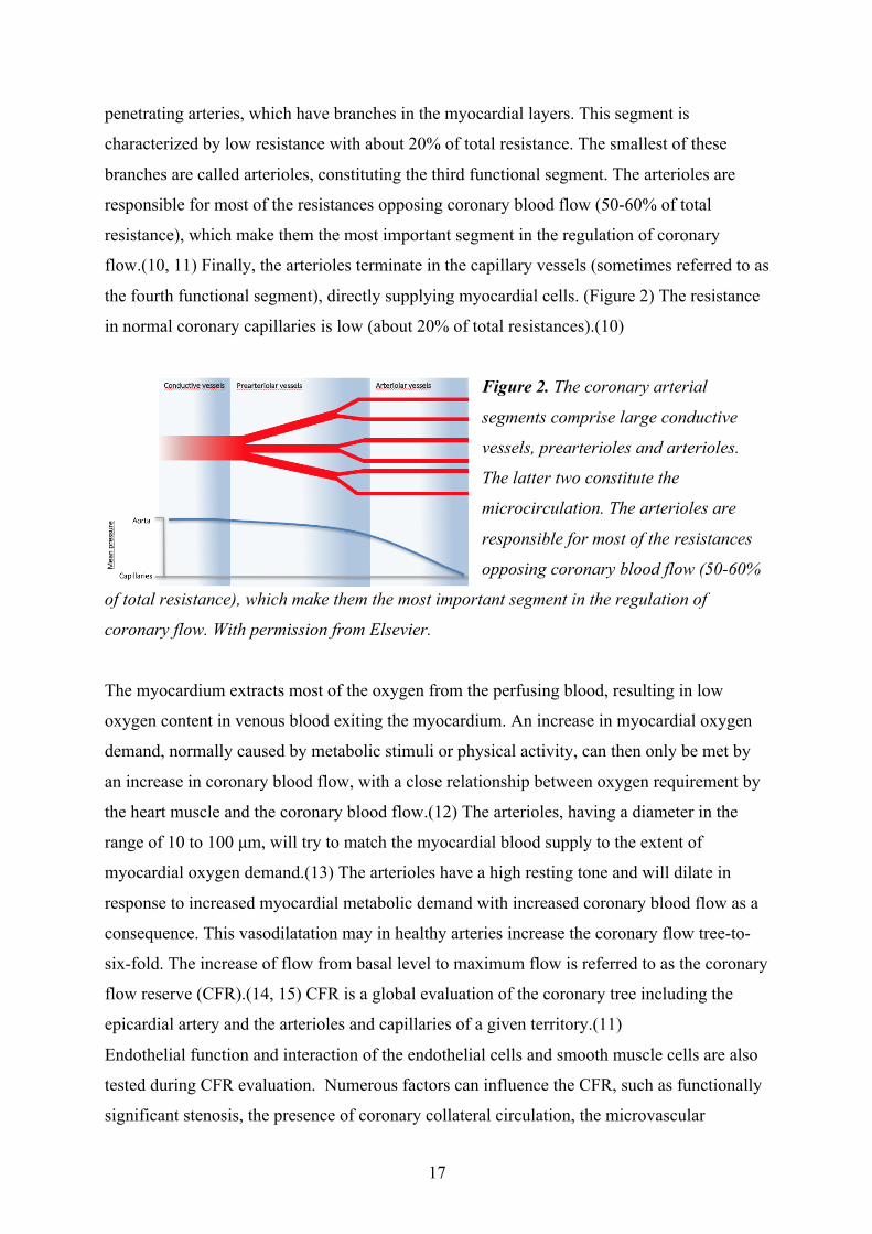
17
penetrating arteries, which have branches in the myocardial layers. This segment is
characterized by low resistance with about 20% of total resistance. The smallest of these
branches are called arterioles, constituting the third functional segment. The arterioles are
responsible for most of the resistances opposing coronary blood flow (50-60% of total
resistance), which make them the most important segment in the regulation of coronary
flow.(10, 11) Finally, the arterioles terminate in the capillary vessels (sometimes referred to as
the fourth functional segment), directly supplying myocardial cells. (Figure 2) The resistance
in normal coronary capillaries is low (about 20% of total resistances).(10)
Figure 2. The coronary arterial
segments comprise large conductive
vessels, prearterioles and arterioles.
The latter two constitute the
microcirculation. The arterioles are
responsible for most of the resistances
opposing coronary blood flow (50-60%
of total resistance), which make them the most important segment in the regulation of
coronary flow. With permission from Elsevier.
The myocardium extracts most of the oxygen from the perfusing blood, resulting in low
oxygen content in venous blood exiting the myocardium. An increase in myocardial oxygen
demand, normally caused by metabolic stimuli or physical activity, can then only be met by
an increase in coronary blood flow, with a close relationship between oxygen requirement by
the heart muscle and the coronary blood flow.(12) The arterioles, having a diameter in the
range of 10 to 100 µm, will try to match the myocardial blood supply to the extent of
myocardial oxygen demand.(13) The arterioles have a high resting tone and will dilate in
response to increased myocardial metabolic demand with increased coronary blood flow as a
consequence. This vasodilatation may in healthy arteries increase the coronary flow tree-to-
six-fold. The increase of flow from basal level to maximum flow is referred to as the coronary
flow reserve (CFR).(14, 15) CFR is a global evaluation of the coronary tree including the
epicardial artery and the arterioles and capillaries of a given territory.(11)
Endothelial function and interaction of the endothelial cells and smooth muscle cells are also
tested during CFR evaluation. Numerous factors can influence the CFR, such as functionally
significant stenosis, the presence of coronary collateral circulation, the microvascular

18
component of coronary resistance, left ventricular hypertrophy, age, the presence of
concomitant anti-ischemic therapy,(16) myocardial infarction,(17) arterial hypertension,
diabetes mellitus, hypertrophic cardiomyopathy,(13) and smoking. A hemodynamic
significant epicardial coronary stenosis creates strong proximal resistances higher than that
opposed from the vasodilating microcirculation, reduce the CFR, and lead to inadequate
myocardial blood perfusion during effort or even at rest.(10) Interestingly, 30 years ago,
Gould and Lipscomb(18) demonstrated in a dog model that resting coronary flow is preserved
until severe narrowing occurs (85% stenosis). However, CFR decreases earlier, already by a
moderate degree of stenosis (40%). This inverse curvilinear relationship between the degree
of coronary artery lumen narrowing and hyperemic capacity has been demonstrated both in
selected patient populations and animal studies (Figure 3).(18, 19)
Figure 3. Relationship between increments of coronary blood flow and degree of coronary
diameter stenosis. Modified from Gould and Lipscomb [Am J Cardiol 1974].
1.4 TTE of the coronary arteries
The coronary arteries have been visualizedby transthoracic echocardiography for many years,
first by two-dimensional (2D) echocardiography. Subsequently, the introduction of Doppler
velocity measurements of the coronary blood flow allowed a functional assessment of the
coronary circulation.(15) Modern high-end echocardiographic equipment permits excellent
imaging of coronary artery blood flow in various coronary segments.(20) Non-invasive

19
imaging of coronary arteries by transthoracic Doppler echocardiography (TTE) is an
emerging diagnostic tool to evaluate coronary flow velocities and flow profiles both at rest
and during hyperemia.(21-27) The coronary blood flow velocities are measured by pulsed-
wave Doppler echocardiography, using a sample volume of 1.5-5 mm placed on the colour
signal in the artery, with an optimized alignment of the Doppler beam and the blood flow.(22)
TTE is non-invasive, widely available, and may offer an opportunity for long-term follow-up
of patients.
1.4.1 Evaluation of the coronary arteries and coronary stenoses by TTE
Several recent reports have documented the feasibility of visualizing most segments of the
main coronary arteries by TTE.(20, 28, 29) Direct visualization of these segments may help to
diagnose significant coronary artery stenoses.(30, 31) In the presence of a significant stenosis,
local blood flow velocities across the stenosis are increased to maintain coronary flow.
Several stenosis factors such as degree of diameter stenosis (DS), length and shape of the
stenosis, as well as possible effects from driving pressure and the coronary artery status
downstream of stenosis will influence the flow velocities.(4, 28, 32, 33) Local flow
acceleration and turbulence at the site of the stenosis are detectable by colour flow
Doppler.(30, 31, 34, 35) Flow velocities at the site of colour flow aliasing can be compared
with the nearest upstream non-accelerated prestenotic flow velocities to further evaluate a
suspected stenosis. A stenotic to prestenotic velocity ratio (SPVR) of ≥2.0, or alternatively
≥2.2, has been proposed as cutoff values for DS of ≥50%, as defined by QCA.(28, 30, 31, 36,
37) However, in some cases, stenotic or prestenotic flow velocities cannot be determined,
either because of an incorrect angle between the coronary blood flow and the ultrasonic beam,
or because of non-measureable flow velocities. In such cases, local mosaic flow at
considerably elevated velocity range (Nyquist limit) may demonstrate significant stenosis.(34)
1.4.2 The functional assessment of an epicardial coronary stenosis by TTE
Coronary atherosclerosis will often cause one or more epicardial stenoses that may cause
symptoms on effort or at rest, leading to invasive coronary angiography. As already
mentioned, anatomic evaluation of a stenosis does not always reflect the functional
significance of the lesion. The potential problems of the functional characterization of an
anatomic coronary lesion may have several causes, such as degree of luminal narrowing,
geometrical characteristics of the stenosis, varying degrees of diffuse atherosclerosis, multiple

20
stenoses, and heterogeneous arterial remodeling.(6) A study showed that 65% of lesions with
diameter stenosis of 50-70% and 20% of lesions with diameter stenosis of 71-90% were
hemodynamically non-significant.(38) The functional aspect of a coronary stenosis is
important, because anatomic significant but functionally non-significant stenoses have a good
prognosis without invasive treatment.(39, 40) Fractional flow reserve (FFR) has been the
preferred reference for the functional evaluation of a coronary stenosis.(41, 42) FFR can be
assessed during cardiac catheterization using an intracoronary pressure wire, with FFR being
the ratio of mean hyperemic poststenotic coronary pressure to mean proximal coronary or
aortic pressure during maximal hyperemia. A FFR < 0.80 (i.e., a value of 0.80 reflects a 20%
reduction in poststenotic compared to prestenotic coronary artery pressure) is considered
hemodynamically significant, implying a functionally significant coronary stenosis with need
of treatment.(14, 38, 43). Invasive measurement of CFR using Doppler flow wire is an
additional gold standard in the functional evaluation of an anatomical stenosis. The
instantaneous wave-free ratio (iFR) is an alternative measure that can be used to assess the
hemodynamic severity of a lesion, and this measure does not require the administration of a
vasodilator but instead relies on the calculation of the translesional pressure gradient during
diastole.(44) Recent trials have concluded that iFR is non-inferior to FFR in evaluation of
coronary artery stenosis.(45) Invasive examinations of FFR and CFR are expensive and are
available only during cardiac catheterization.
CFR evaluated by TTE is a well-established and validated method for evaluating the coronary
blood flow, defined as the ratio of hyperemic to basal peak diastolic coronary flow velocities.
The coronary flow velocities are measured by pulsed-wave Doppler guided by colour
Doppler. Thus, this method directly measures changes in coronary flow velocity, referred to
as coronary flow velocity reserve (CFVR), at the very beginning of the ischaemic cascade,
instead of looking at the consequences of ischaemia on myocardial contraction. The intra- and
interobserver variabilities and the day-to-day variability of CFR measurements are low (2.6–
2.8%, 2.6–8.6% and 6.1–11.4%, respectively), which make CFR suitable for repeated
measurements.(15) CFR is affected by both epicardial stenoses and the microcirculation. In
the absence of stenosis in the epicardial coronary arteries, CFR mainly depicts the reactivity
of the coronary microcirculation. However, in clinical practice there is a lack of specificity for
CFR for the epicardial vessel: an excessively low CFR value can both be related to epicardial
stenosis, to microvascular disease, or to a combination of both.(11) Both adenosine and
dipyridamole is widely used to induce pharmacological vasodilatation in non-invasive

21
diagnosis of coronary heart disease and in scientific studies of the coronary circulation.(46)
Adenosine induces a vasodilatory effect through direct actions on smooth muscle cell
adenosine receptors in extracellular space, with a complex interplay between local vasodilator
mechanisms and systemic homeostasis. Furthermore, the increased blood flow mediated by
this vasodilation induces shear stress on the artery wall, followed by release of substances
from endothelial cells that further dilate the artery.(15) In healthy humans it is demonstrated
that adenosine-induced myocardial hyperemia is partly endothelium dependent mediated
through endogenous nitric oxide (NO).(46) Thus, a decline in myocardial perfusion reserve
may partly be caused by endothelial dysfunction.(46) The short half-life of adenosine and the
rapid regression of the effect enable a practical and safe use of adenosine. Furthermore, it
makes repetitive measurements possible, if necessary.(11) Several studies have documented
strong correlations between invasively measured CFR and CFR (the ratio of hyperemic blood
flow velocity to resting blood flow velocity) measured by TTE, documented for the LAD,(47-
49) the posterior descending coronary artery (PDA),(49, 50) and the Cx.(51, 52) A CFR value
<2.0 measured by TTE during adenosine infusion has been shown to indicate one or more
hemodynamic significant stenoses located upstream in the coronary artery,(21) documented
for all three major coronary arteries.(27, 51, 53)
Coronary occlusion may be detected by demonstrating retrograde flow in the coronary artery
during TTE.(54) Most studies, however, have evaluated CFR (measured by TTE) only in
single coronary arteries in limited patient cohorts and have not included TTE findings of
retrograde coronary artery flow. Furthermore, there is a paucity of studies comparing CFR
obtained by TTE with various degrees of coronary obstruction as defined by invasive
coronary angiography.

22
1.4.3 The coronary flow profile
The coronary artery flow velocity waveform appears as a complex of a systolic wave and a
trapezoid diastolic wave (Figure 4).
Figure 4. The systolic and diastolic component
of the coronary blood flow profile. D, Spectral
Doppler tracings of diastolic coronary blood
flow; S, spectral Doppler tracings of systolic
coronary blood flow.
Normal coronary arteries display a predominant diastolic flow pattern, which is less marked
in the distal right coronary artery (RCA), due to the lower intramyocardial systolic pressure in
the right ventricle.(55) Several studies have shown that in the presence of a significant
coronary stenosis, the ratio between the peak diastolic and systolic coronary blood flow
velocities, diastolic-to-systolic velocity ratio (DSVR), is significantly reduced when
invasively measured downstream to the stenosis.(55-58) This reduction is postulated to be
caused by a combined poststenotic decrease of diastolic flow and an increased systolic flow
from an intramyocardial systolic contraction pump acting on the intramyocardial capacitance
vessels.(59) Recent reports have indicated that findings of reduced DSVR measured by TTE
in the distal LAD may be a simple, non-invasive method for the detection of high-grade
coronary stenoses located upstream in the LAD.(25, 26, 60) This is demonstrated for patients
with or without wall motion abnormalities of the left ventricle.(60) DSVR (DSVR) values <
1.6 to 1.8 are proposed to represent high-grade stenosis, however only validated for LAD.(25,
26, 60) Furthermore, there is a lack of data comparing DSVR measured by TTE with various
degree of DS defined by QCA and a functional parameter like CFR. The normal DSVR in the
RCA is low and probably close to pathologic values, which limits the potential utility of distal
DSVR measurements primarily to LAD and Cx in the search of possible upstream stenoses.

23
1.5 Inflammation in CAD
The understanding of atherosclerosis has evolved beyond the view that these lesions consist
of a lifeless collection of lipid debris. Inflammation is now regarded as a major player in all
phases of the atherosclerotic process. Both cellular and molecular inflammatory events are
pivotal in all stages of atherosclerosis, from endothelial dysfunction and plaque formation to
plaque destabilization and disruption with superimposed thrombosis.(61) Signs of
inflammation occur together with incipient lipid accumulation in the artery wall. In normal
endothelium blood leucocytes poorly adhere. However, inflamed endothelium expresses
cellular adhesion molecules (CAMs), like vascular cell adhesion molecule-1 (VCAM-1), (62)
resulting in adhesion and migration of leukocytes into the intima. Furthermore, the
inflammatory response is a complex interplay between various inflammatory mediators such
as different chemokines and cytokines. Several of these inflammatory markers may act
significantly in developing plaque instability by facilitating vascular inflammation, matrix
degeneration, and thrombus formation.(63) Patients with acute coronary syndromes (ACS) are
characterized by having an elevated inflammatory response and endothelial dysfunction,
which are markers of worse prognosis and increased risk of recurrent cardiovascular
events.(64, 65)
1.5.1 Coronary microvascular and endothelial function in acute coronary
syndromes.
Importantly, endothelial dysfunction is one of the first recognizable signs of atherosclerosis,
and is closely related to its risk factors.(66) Endothelial dysfunction constitutes an
intermediate step on the progression to adverse events throughout the natural history of
coronary artery disease (CAD), often affecting clinical outcomes.(67) In contrast to general
and peripheral endothelial dysfunction, coronary microvascular dysfunction (CMD) following
ACS and percutaneous coronary intervention (PCI) is a more complex issue. CMD in these
patients is a multifactorial phenomenon, which can be composed of several components,
including endothelial-independent factors such as distal embolization of thrombus and
atherosclerotic plaque material, edema, neutrophil plugging and other factors, with all
possibly contributing to the phenomenon of no-reflow/microvascular obstruction.(68) No-
reflow/microvascular obstruction is a marker of poor prognosis.(66) However, endothelial-
dependent CMD and thus genuine coronary endothelial dysfunction also seems to be
prevalent in non-culprit coronary arteries in patients with ACS,(69, 70) with these patients
also characterized by widespread coronary inflammation.(71) The transition from stable
24
coronary artery disease to acute coronary syndromes coincides with elevated levels of
inflammatory markers like C-reactive protein, amyloid A, or interleukin-6.(62, 72)
Furthermore, evidence suggest that there is a link between inflammation and endothelial
dysfunction in these patients.(65) In addition, patients with ACS are characterized by reduced
CFR, which partially reflects coronary endothelial function.(15, 22) Endothelial dysfunction
plays a key role in determining myocardial ischemia in all clinical manifestations of ischemic
heart disease. However, the prognostic implications of endothelial-dependent CMD during
ACS and how it relates to established markers of endothelial activation and dysfunction such
as CAMs and von Willebrand Factor (vWF) are not fully explored.
1.5.2 Interleukin-6 and anti-inflammatory treatment of CAD
Interleukin-6 (IL-6) is a multifunctional cytokine which is a major actor in the inflammatory
arm of CAD(73) and in the acute inflammatory response triggered by ACS.(64) In ACS, an
elevated level of IL-6 is a marker of adverse outcomes and probably reflects the degree of
myocardial damage.(64) Furthermore, IL-6 seems to contribute to ischemia/reperfusion injury
in these patients.(74) IL-6 is also associated with endothelial dysfunction in ACS,(65) and
may have a causal role as it induces increased expression of markers of endothelial
dysfunction such as CAMs(75) and vWF(76) in these patients. Moreover, increasing levels of
CAMs(75) and vWF(76) are related to both mortality and recurrent cardiovascular events in
patients with ACS.
Parallel to the evolving understanding of the underlying mechanisms of the ACS beyond the
concept of a progressive atherosclerosis as a bland lipid storage disease, multiple therapeutic
approaches, i.e. statins and anti-hypertensive drugs, have been shown to modify inflammatory
response in addition to their other effects. (77) Despite this development, the phenomenon of
inflammation in ACS is still eluding specific therapeutic treatment.(77, 78) However,
increased scientific interest for more specific anti-inflammatory treatment of atherosclerosis
has resulted in novel agents directed against specific targets in the inflammatory cascade
being applied in clinical settings. (77) Tocilizumab is a humanized anti-IL-6 receptor
antibody which is effective and well tolerated in autoimmune disorders.(79) Furthermore, it
has been shown that tocilizumab improves endothelial function and aortic stiffness in patients
with rheumatoid arthritis.(80)

25
2 AIMS OF THE THESIS
2.1 General aims
The general aim of this thesis was to evaluate the suitability of transthoracic Doppler
echocardiographic examinations (TTE) of coronary artery blood flow to (1) identify and
characterize coronary stenoses in a patient cohort of suspected or documented coronary
disease prior to coronary angiography and (2) evaluate the coronary microvascular and
endothelial function in an acute NSTEMI population randomized to anti-inflammatory
treatment or placebo.
2.1.1 Specific aims:
1. To assess the feasibility and accuracy of diagnosing high-grade stenoses and
occlusions in all three main coronary arteries using transthoracic Doppler coronary
flow velocity reserve (CFVR, coronary flow reserve (CFR) measured by transthoracic
Doppler) or findings of retrograde coronary flow during the process of visualizing the
individual coronary segment for CFR measurements, with invasive coronary
angiography as the gold standard (Paper I).
2. To assess the feasibility and accuracy of diastolic-to-systolic velocity ratio (DSVR)
measurements as a simple method for diagnosing significant stenoses in the left main,
left anterior descending, and left circumflex coronary arteries, using invasive coronary
angiography and CFR as the anatomic and functional references, respectively (Paper
II).
3. To assess the ability of pulsed wave Doppler and colour Doppler aliasing by
transthoracic Doppler to identify coronary artery stenoses, with invasive coronary
angiography and CFR measured by TTE as the anatomical and functional references,
respectively (Paper III).
4. To examine the effect of tocilizumab on the CFR and circulating markers of
endothelial cell activation after NSTEMI, by assessing CFR during transthoracic
echocardiography and measuring markers of endothelial activation. (Paper IV).

26

27
3 MATERIALS AND METHODS This work was carried out at three different hospitals, Ålesund Hospital, St Olavs
Hospital/Trondheim University Hospital and Oslo University Hospital Rikshospitalet, and
consists of two study populations, referred to as patient population I and II. The same
technique for CFR measurements are used in both patient populations. CFR measured by TTE
is the calculated ratio of hyperemic to basal peak diastolic velocities, often referred to as
coronary flow velocity reserve (CFVR). CFR refers to CFVR if not further specified.
3.1 Patient population I (Paper I-III)
In the period 2006-2007 patients from the local area of Ålesund Hospital was included
prospectively with the following inclusion criteria: (1) already scheduled for coronary
angiography because of documented or suspected stable or unstable coronary disease, (2) age
> 18 years, and (3) met no exclusion criteria. The exclusion criteria were (1) previous
aortocoronary bypass surgery, (2) presumed insufficient acoustic windows because of severe
emphysema or severe overweight, (3) significant valvular disease, (4) atrial fibrillation, and
(5) administrative reasons. The study protocol was approved by the Regional Committee for
Medical and Health Research Ethics and the Norwegian Data Inspectorate. All participants
gave written, informed consent. Six patients did not enter the study, because of insufficient
acoustic windows (n = 3), lack of consent (n = 2), or aortic stenosis (n = 1). We included 115
patients in the study, but 7 patients were later excluded from further analysis because of
protocol violations: aortic stenosis (n = 2), atrial fibrillation (n = 2), patient refusal of
coronary angiography (n = 2), and no indication for coronary angiography (n = 1). The final
cohort of 108 patients were included in studies 1 – 3, with clinical characteristics of the
patient group in these studies presented in Table 1.
28
3.2 Patient population II (Paper IV)
This study was part of a randomized, double-blind, placebo controlled trial designed to
evaluate the effect of a single dose of the anti-IL-6 receptor antibody tocilizumab in patients
with NSTEMI (ClinicalTrials.gov, NCT01491074). The study was performed at Oslo
University Hospital Rikshospitalet, Oslo, and St. Olavs Hospital, Trondheim, Norway. The
study was approved by the Regional Committee for Medical and Health Research Ethics and
the Norwegian Medicines Agency, and was conducted according to the Helsinki Declaration.
Written, informed consent was provided by all patients. Between August 2011 and November
2013, a total of 117 patients (placebo n=59, tocilizumab n=58) were included in the trial.
Follow-up ended in April 2014. The 60 patients that were included at St. Olavs Hospital
were all eligible for inclusion in a pre-defined sub-study, evaluating the effects of tocilizumab
on coronary flow reserve (CFR) during acute hospitalizaton for NSTEMI and at 6 months
follow-up. Markers of endothelial cell activation (VCAM-1, ICAM-1 and vWF) were
measured in all patients (n=117) recruited at both trial centers. Exclusion criteria were (1)
LAD stenosis (n = 10), (2) intolerable side effects of adenosine (n=2), (3) administrative
reasons (n=5), and (4) insufficient acoustic windows (n=1). The final study group consisted of
42 patients (placebo n=20, tocilizumab n=22), with clinical characteristics of the patient
group in these studies presented in Table 2.
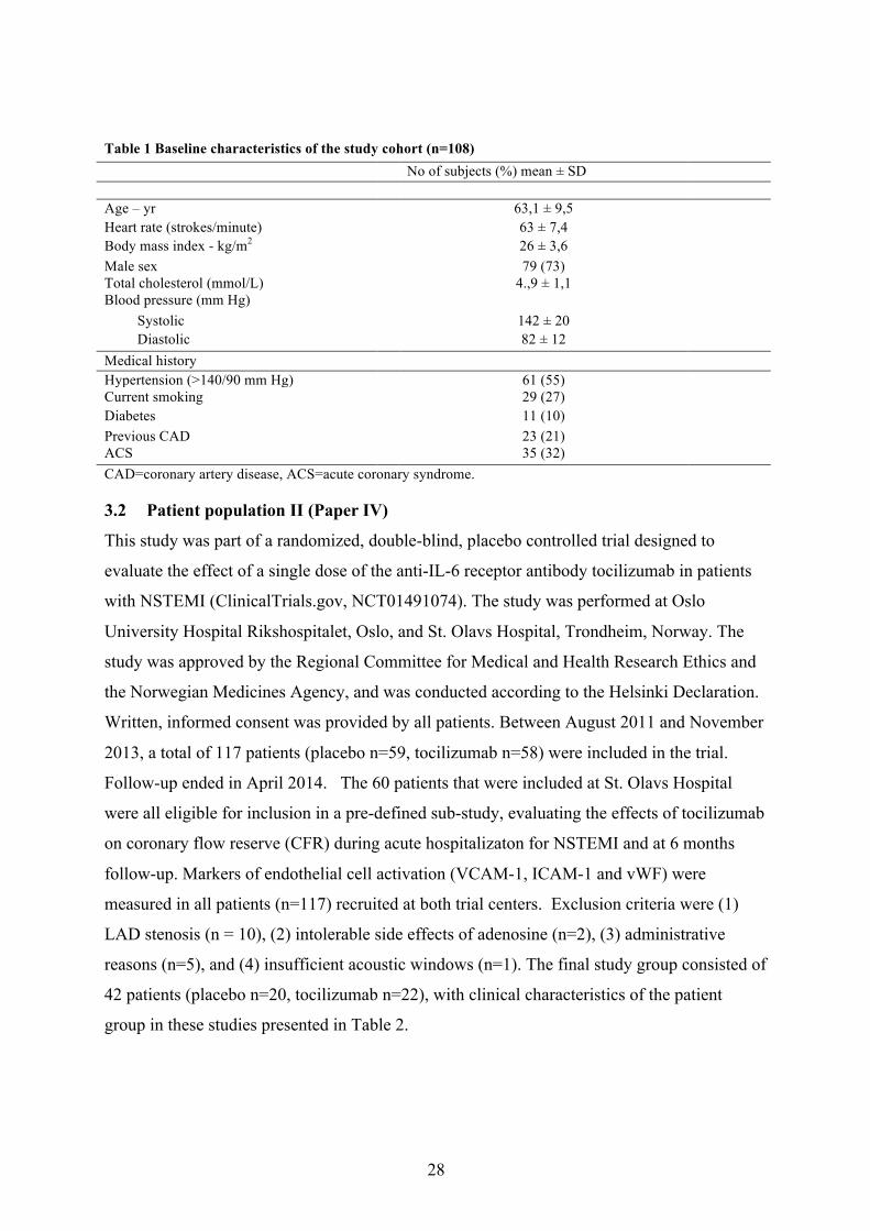
Table 1 Baseline characteristics of the study cohort (n=108) No of subjects (%) mean ± SD Age – yr 63,1 ± 9,5 Heart rate (strokes/minute) 63 ± 7,4 Body mass index - kg/m2 26 ± 3,6 Male sex 79 (73) Total cholesterol (mmol/L) 4.,9 ± 1,1 Blood pressure (mm Hg) Systolic 142 ± 20 Diastolic 82 ± 12 Medical history Hypertension (>140/90 mm Hg) 61 (55) Current smoking 29 (27) Diabetes 11 (10) Previous CAD 23 (21) ACS 35 (32) CAD=coronary artery disease, ACS=acute coronary syndrome.
29
3.3 Transthoracic echocardiography of coronary arteries.
This thesis aimed to evaluate different aspects using colour and pulsed-wave Doppler by
transthoracic echocardiography to assess coronary artery disease and coronary microvascular
and endothelial dysfunction by measuring and evaluating the coronary blood flow velocities.
The two study populations were examined using two different vendors of ultrasound
equipment, as specified below. Colour Doppler was used to investigate the course of the
coronary vessels, using each vendor’s pre-set for coronary application. Pulsed-wave Doppler
was used to measure coronary blood flow velocities. Contrast agent was not used.
3.4 The first study population (Paper I-III)
We included patients with suspected stable angina or ACS. The patients with suspected stable
angina referred for cardiac catheterization were examined by TTE (including CFR
measurements) as close as possible to the date of the invasive coronary angiography. The
TTE examination (including CFR measurements) of the patients hospitalised for ACS was not
performed earlier than the day after hospital admission and only after the patients were
clinically stable. An Acuson Sequoia C512 (Siemens Medical Solutions USA, Inc, Mountain
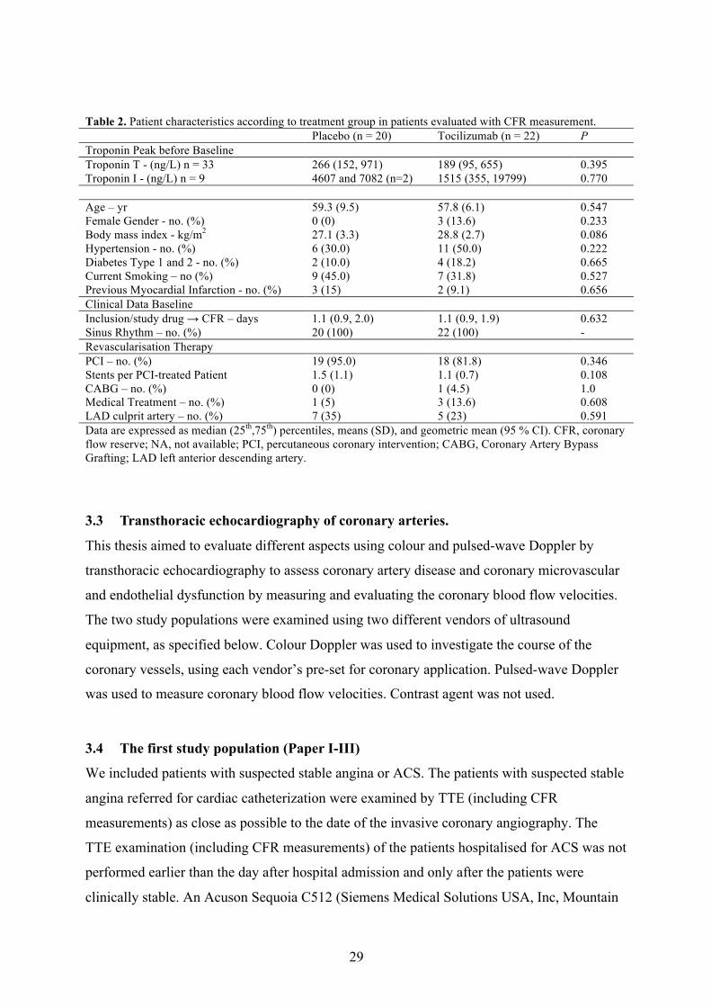
Table 2. Patient characteristics according to treatment group in patients evaluated with CFR measurement. Placebo (n = 20) Tocilizumab (n = 22) P Troponin Peak before Baseline Troponin T - (ng/L) n = 33 266 (152, 971) 189 (95, 655) 0.395 Troponin I - (ng/L) n = 9 4607 and 7082 (n=2) 1515 (355, 19799) 0.770 Age – yr 59.3 (9.5) 57.8 (6.1) 0.547 Female Gender - no. (%) 0 (0) 3 (13.6) 0.233 Body mass index - kg/m2 27.1 (3.3) 28.8 (2.7) 0.086 Hypertension - no. (%) 6 (30.0) 11 (50.0) 0.222 Diabetes Type 1 and 2 - no. (%) 2 (10.0) 4 (18.2) 0.665 Current Smoking – no (%) 9 (45.0) 7 (31.8) 0.527 Previous Myocardial Infarction - no. (%) 3 (15) 2 (9.1) 0.656 Clinical Data Baseline Inclusion/study drug → CFR – days 1.1 (0.9, 2.0) 1.1 (0.9, 1.9) 0.632 Sinus Rhythm – no. (%) 20 (100) 22 (100) - Revascularisation Therapy PCI – no. (%) 19 (95.0) 18 (81.8) 0.346 Stents per PCI-treated Patient 1.5 (1.1) 1.1 (0.7) 0.108 CABG – no. (%) 0 (0) 1 (4.5) 1.0 Medical Treatment – no. (%) 1 (5) 3 (13.6) 0.608 LAD culprit artery – no. (%) 7 (35) 5 (23) 0.591 Data are expressed as median (25th,75th) percentiles, means (SD), and geometric mean (95 % CI). CFR, coronary flow reserve; NA, not available; PCI, percutaneous coronary intervention; CABG, Coronary Artery Bypass Grafting; LAD left anterior descending artery.

30
View, CA) ultrasound system connected to standard 4V1C and 7V3C transthoracic
transducers was used during the study. All patients took their medications on the day of the
echocardiographic study. The Acuson Sequoia C512 has a colour Doppler mapping with data
postprocessing mix function, which makes the colours transparent. The velocity scale of
colour Doppler was set to 0.24 m/s, but was actively changed to provide optimal images.
Coronary blood flow velocities were measured using pulsed-wave Doppler with 1.75-MHz to
3.5-MHz frequencies in a sample volume of 1.5 to 5 mm, with the sample volume positioned
on the colour flow Doppler signal.
3.4.1 Echocardiographic and angiographic analyses; Paper I-III
The case report form (CRF) containing the prespecified measurements was completed after
each echocardiographic examination and sealed in an envelope, which was stored until the
analyses of the coronary angiograms were finished. The echocardiographic examination was
digitally stored, and analyzed by a single experienced echocardiographer (EH), who was
blinded to the findings of angiography.
Coronary angiography was performed using standard techniques. All angiographic studies
were digitally stored with later offline reviewing and measurements, blinded to the findings
by TTE. Two invasive cardiologist (RW and KH) analyzed the angiograms using a 16-
segment model of the coronary arteries.(81) Philips Xcelera was used for the QCA analysis.
Disagreements in interpretation were resolved by consensus.
The dataset was unmasked after all the echocardiographic and angiographic examinations
were analysed separately. Coronary angiography was defined as the gold standard.
3.5 The second study population (Paper IV)
CFR was measured as part of the transthoracic echocardiographic examination at day 2 or 3
during hospitalization and at 6 months follow-up, using a Vivid E9 XDclear (GE Vingmed
Ultrasound AS, Horten, Norway) ultrasound system connected to a M5Sc-D transthoracic
transducer. The pre-set application called “Coronary” for visualisation of the coronary arteries
was used.

31
3.5.1 Analyses of the echocardiographic findings; Paper IV
Each examination was blinded both in terms of treatment group and order of the
echocardiographic examinations (during hospitalization or 6 months follow-up). All the CFR
measurements were analysed by EH, while all other echocardiographic data were analysed by
OK.
3.6 Examination of the coronary arteries by TTE
3.6.1 The coronary blood flow
Pulsed-wave Doppler was used to measure the peak diastolic and systolic coronary blood
flow velocities. Angle correction was used during velocity measurements to keep the angle
between blood flow and Doppler beam as small as possible. In Paper IV we only measured
flow in the distal LAD, which normally shows a satisfying alignment with the Doppler beam,
minimizing the need for angle correction. The spectral Doppler gain was actively adjusted to
best discriminate the flow velocity waveforms from the Doppler background noise. Stop-
motion frames and clips were digitally recorded for offline analysis (Echopac, GE Vingmed
Ultrasound AS, Horten, Norway). We attempted to find at least three optimal profiles of flow
velocities for the measurements, and the results were averaged. Retrograde coronary artery
flow was distinguished from coronary venous flow by finding inverted coronary flow velocity
waveform when colour flow Doppler recordings indicated reversed coronary artery flow.(82,
83) In contrast, the coronary venous flow appears as a prominent systolic flow wave.
3.6.2 Visualization of the coronary arteries by TTE
Colour Doppler was used to identify and follow the course of the main coronary arteries. With
the patient in the supine, left and right lateral decubitus positions, all standard and modified
apical, parasternal, and subcostal views were used to follow the course of the LM, LAD, Cx,
and RCA, from the start of each artery and distally as far as possible. The LM had one
segment and the other main coronary arteries (LAD, Cx, and RCA) had each a proximal,
middle and distal segment. For each segment three different possibilities were defined: (i) the
segment was completely visualised; (ii) the segment was not satisfactorily visualised if any
part of the individual segment was not seen or the segment was not visualised at all; (iii) the
segment was defined with retrograde flow.

32
Left main and left anterior descending coronary arteries
The LM was examined from the left parasternal views focusing on the area adjacent to the left
sinus of Valsalva cranial to the aortic valve (Figure 5).(20) In the same views the proximal
LAD (pLAD) could be seen leaving the LM and turning slightly toward the transducer
(Figure 5).(20) The division between the pLAD and mid LAD (mLAD) was set by the origin
of the first septal branch or approximately halfway to the level of the left ventricular papillary
muscles. The course of the mLAD and distal LAD (dLAD) was imaged from parasternal
modified views focusing on the anterior interventricular sulcus.(20) The level of the left
ventricular papillary muscles marked the division between the mid and distal segments of the
LAD.
Figure 5. Examples of anterograde coronary artery flow in the LM, proximal and middle
segments of LAD, and proximal and middle segments of Cx. (A) In modified parasternal
short-axis view focusing on the area adjacent to the left sinus of Valsalva the proximal left
anterior descending (LAD) and proximal circumflex (pCx) coronary arteries are seen leaving
the left main coronary artery (LM). (B and C) In modified parasternal short-axis views the
proximal and middle segments of the circumflex coronary artery (Cx) are found passing
caudally in the lateral atrioventricular sulcus. (D) From parasternal modified long-axis view
focusing on the anterior interventricular sulcus and the lateral atrioventricular sulcus parts
of the proximal and middle LAD and the middle Cx (mCx) are seen traversing in distal

33
direction. Ao = aortic root/valve; LA = left atrium; LV = left ventricle; mLAD = middle
segment of LAD; PA = pulmonary artery; pLAD = proximal segment of LAD; RV = right
ventricle.
Left circumflex coronary artery
The proximal part of the Cx (pCx) was found using the same views as searching for the LM.
Focusing on the atrioventricular sulcus, the pCx was seen passing in front of the left atrial
appendage. The inferor wall of the left atrial appendage marked the division between the pCx
and middle segment of the Cx (mCx). By modified parasternal views, mCx was visualized as
it passed caudally in the atrioventricular sulcus to the inferior margin of the sulcus (Figure
5).(20) Flow measurements in the main trunk of the Cx are difficult because of cyclic cardiac
motion. Instead, the Cx marginal branches (CxMb) were used for CFR measurements. To
include as much as possible of the main trunk of the Cx the most inferior marginal branch
found was used for measurements. Marginal branches of the Cx leave the artery at various
levels, and from modified apical four-chamber views focusing on different levels of the lateral
wall of the left ventricle, CxMbs could be visualized coursing in the distal direction on the
epicardial surface of the left ventricle toward the transducer.(22)
The right coronary artery
From parasternal and subcostal views, the proximal segment of the RCA (pRCA) was looked
for in the area adjacent to the right sinus of Valsalva, and by focusing on the anterior tricuspid
ring pRCA could be followed down to the inferior margin of the right ventricle. The mid and
distal segments (collectively reported as the mid-RCA) were investigated mostly from
subcostal views focusing on the medial and posterior tricuspid ring. The posterior descending
coronary artery (PDA) was visualized in the posterior interventricular sulcus using apical
views.(20)
As outlined above, the LM had one segment and the other main coronary arteries (LAD, Cx,
and RCA) had each a proximal, middle and distal segment. For each segment three different
possibilities were defined: (i) the segment was completely visualised; (ii) the segment was not
satisfactorily visualised if any part of the individual segment was not seen or the segment was
not visualised at all; (iii) the segment was defined with retrograde flow.

34
3.6.3 Coronary flow reserve measurements
The method used for CFR measurement by TTE was similar for both study populations. In
study population I, CFR was assessed in all three coronary arteries (mid to distal LAD, Cx
marginal branches (CxMb), and PDA) (Figure 6).
Figure 6 Examples of
antegrade coronary flow in the
mid LAD (mLAD), CxMb, and
PDA, with resting and
hyperemic Doppler velocities.
(A1) In modified parasternal
short-axis view, the mLAD is
seen coursing toward the apex
(Ax) in the anterior
interventricular sulcus. (B1) In
modified apical four-chamber
view, a CxMb is seen coursing toward the apex on the epicardial surface of the left ventricle
(LV). (C1) From modified apical two-chamber view, the PDA is seen coursing toward the
apex in the posterior interventricular sulcus. (A2, B2, C2) Baseline spectral Doppler tracings
of blood flow in the mid LAD, CxMb. PDA, respectively. (A3, B3, C3) Spectral Doppler
tracings of blood flow in the mLAD, CxMb. PDA, during hyperemia, respectively. D, Spectral
Doppler tracings of diastolic coronary blood flow; IVS, interventricular septum; LA, left
atrium; MR, mitral ring; MV, mitral valve; RA, right atrium; RV, right ventricle; S, spectral
Doppler tracings of systolic coronary blood flow.
The respective coronary segment was identified by colour Doppler and the sample volume
was positioned distal to any visualized turbulent colour flow Doppler signal, because
turbulent colour flow Doppler signals might represent stenosis. Thereafter, coronary flow
velocities were measured by recording spectral Doppler signals in the mid to distal LAD,
CxMb and PDA at baseline and during hyperemia. Hyperemia was achieved using
intravenous adenosine (0.14 mg/kg/min for maximum 2 minutes). The procedure of assessing
CFR was repeated at least twice, and the best series were used for measurements. We tried to
find at least three consecutive cardiac cycles to average flow velocities, both at baseline and

35
during adenosine infusion. Diastolic flow velocities were measured, and CFR was calculated
as the ratio of hyperemic to basal peak diastolic flow velocities (pCFVR). In study population
II (Paper IV), CFR was only assessed in mid to distal LAD.
CFR is an important functional parameter for understanding the pathophysiology of the
coronary circulation. CFR can be used both to assess epicardial coronary stenoses and to
examine the integrity of endothelial function/microvascular circulation. In the absence of
coronary artery stenosis, the CFR reflects the endothelial function/microvascular circulation.
3.6.4 Coronary flow reserve for detection of significant stenoses
In the first study (Paper I), we aimed to assess the feasibility and accuracy of TTE to
diagnose high-grade stenoses and occlusions in all three main coronary arteries using CFR or
findings of retrograde coronary flow during the process of visualizing the individual coronary
segment for CFR measurements, with QCA as the gold standard. CFR was investigated when
the mid to distal LAD, CxMb, and PDA were visualized with anterograde flow by colour
Doppler (Figure 6). Findings of retrograde flow were interpreted as an upstream occlusion,
and CFR was not measured. A predefined cutoff value of < 2.0 for pCFVR was used for
significant stenosis, in accordance with previous studies.(47, 49, 51)
3.6.5 Coronary flow profiles for detection of significant stenoses
In the second study (Paper II), we aimed to assess the feasibility and accuracy of DSVR
measurements on TTE as a simple method for diagnosing significant stenoses in the left
coronary artery, using QCA and CFR measured by TTE as the anatomic and functional
references, respectively. Anterograde peak diastolic and peak systolic coronary blood flow
velocities were measured in the distal LAD (or distal portion of the mid LAD if the distal
LAD could not be imaged) and CxMb, with the sample volume positioned distally to any
visualized turbulent colour flow Doppler signal. Whenever possible, the most inferior
marginal branch viewed was used for measurements. The same CxMb were used for DSVR
and CFR measurements. The ratio between the peak diastolic and peak systolic velocities was
measured in each cardiac cycle (Figure 7), and the average of these peak velocity ratios
measured in the consecutive cycles was the DSVR.

36
Figure 7. Examples of anterograde
coronary blood flow in the distal left
anterior descending coronary artery (dLAD)
and marginal branch from the left
circumflex coronary artery (CxMb), with
diastolic-to-systolic velocity ratio (DSVR) in
non-stenosed arteries. (A1) In the modified
apical two-chamber view, the dLAD is seen
coursing toward the apex in the anterior interventricular sulcus. (A2) Spectral Doppler
tracings of blood flow in the dLAD, with DSVR of 1.94. (B1) In the modified apical four-
chamber view, a CxMb is seen coursing toward the apex on the epicardial surface of the left
ventricle (LV). (B2) Spectral Doppler tracings of blood flow in the CxMb, with DSVR of 1.87.
D, Spectral Doppler tracings of diastolic coronary blood flow; IVS, interventricular septum;
LA, left atrium; RA, right atrium; RV, right ventricle; S, spectral Doppler tracings of systolic
coronary blood flow.
3.6.6 Transstenotic flow velocities for detection of significant stenoses
In the third study (Paper III), we aimed to assess the feasibility and accuracy of SPVR
measurements or demonstration of local mosaic flow as a simple method for diagnosing DS
50–99% in all three major coronary arteries, with QCA and CFR measured by TTE as the
anatomical and functional references, respectively. Using transthoracic Doppler, the three
main coronary arteries were investigated for local colour Doppler aliasing representing
increased transstenotic flow velocities (Figure 8).
Figure 8 Examples of stenotic-to-prestenotic
velocity ratio (SPVR). (A1) The middle
segment of the left coronary artery (mLAD)
showing local flow colour aliasing suggestive
of stenosis. (A2) Prestenotic blood flow
velocities and (A3) stenotic blood flow
velocities in the mLAD, with a SPVR of 3.8.
QCA demonstrated a DS of 55% in the mLAD.
(B1) The proximal segment of the left
circumflex coronary artery (pCx) with local
mLAD%stenosis%
IVS%
RV%
LV%
D%
S%
D%S
D%S%
DS
Ao%
pLAD%
pCx%stenosis%
LA%
RVOT%Ax%
A1%
A3%
A2%
B3%
B2%
B1%

37
flow acceleration suggestive of stenosis. (B2) Prestenotic blood flow velocities and (B3)
stenotic blood flow velocities in the pCx, with a SPVR of 5.8. QCA demonstrated a DS of 57%
in the pCx. Ao, ascending aorta; D, spectral Doppler tracings of diastolic coronary blood
flow; IVS, interventricular septum; LA, left atrium; LV, left ventricle; pLAD, proximal left
anterior descending coronary artery; RV, right ventricle; RVOT, right ventricular outflow
tract; S, spectral Doppler tracings of systolic coronary blood flow. With permission from
Oxford University Press.
All standard and modified apical, parasternal, and subcostal views were used to follow the
course of the main coronary arteries. Coronary segments found with anterograde flow were
searched for local flow acceleration and turbulence expressed as colour aliasing by colour
flow Doppler and accelerated flow velocities, as indication of possible stenosis. Peak diastolic
blood flow velocities at the site of colour flow aliasing and the nearest upstream non-
accelerated prestenotic flow velocities were measured using pulsed-wave Doppler, with angle
correction used to keep the angle between blood flow and Doppler beam as small as possible.
The severity of the stenosis was expressed as the SPVR, which was calculated as the ratio of
the average peak flow velocities at the site of aliasing and the nearest upstream non-
accelerated flow velocities.
We used a SPVR cutoff value of ≥2.0 as proposed in earlier studies.(31) When SPVR could
not be assessed because of suboptimal angle between coronary blood flow direction and
ultrasound beam or difficulties measuring either peak stenotic or peak prestenotic flow
velocities, the presence of a coronary stenosis was approximated using rescaling of the
Nyquist limit of colour flow Doppler. A persistent colour flow Doppler aliasing by a flow
velocity scale of ≥0.48 m/s was defined as a significant stenosis, based on our experience that
locally persistent colour aliasing by colour flow Doppler at Nyquist limit ≥0.48 m/s usually
indicated significant coronary stenosis (Figure 9).
Figure 9 Example of locally present colour flow
aliasing at Nyquist limit of 0.51 m/s in the middle
left anterior descending coronary artery
(mLAD), suggestive of stenosis. QCA showed a
mLAD DM stenosis of 55%. IVS, interventricular
septum; LV, left ventricle; RV, right ventricle.
mLAD%stenosis%
IVS%
RV%
LV%
D%
S%
D%S
D%S%
DS
Ao%
pLAD%
pCx%stenosis%
LA%
RVOT%Ax%
A1%
A3%
A2%
B3%
B2%
B1%

38
We tried to find at least three cardiac cycles to average peak diastolic coronary flow velocities
at each measuring site.
3.6.7 Effect of interleukin-6 inhibition on coronary microvascular and endothelial
function in myocardial infarction
In the fourth study (Paper IV), a pre-defined sub-study, we sought to examine after NSTEMI
the effect of IL-6 inhibition by tocilizumab on coronary endothelial and microvascular
function, using CFR and measuring circulating markers of endothelial cell activation. All
patients were treated according to the current recommendations for acute NSTEMI. Patients
with LAD stenosis were not included in our study because an epicardial LAD stenosis might
influence CFR. After study drug administration and coronary angiography, six blood samples
were obtained during the first 3 days of hospitalization (day 1: evening; day 2: morning,
afternoon, evening; day 3: morning, afternoon). TTE was performed on day 2 or 3 during
hospitalization. Patients included at St Olavs Hospital (n=60) were eligible for CFR
measurement during TTE. Blood samples were repeated at 3 and 6 months follow-up, while
TTE with CFR was repeated at 6 months. Markers of endothelial cell activation such as
cellular adhesion molecules (CAMs) and von Willebrand factor (vWF) were measured in all
patients (n=117), and CFR was assessed in 42 of these. Vascular cell adhesion molecule-1
(VCAM-1) was measured in 20 control patients with stable CAD. The cutoff value of CFR
for significant CMD was set to < 2.5 due to limited data on CMD and CFR in NSTEMI, and
because we wanted to study the gray-zone CFR range of 2.0 - 2.5.(84, 85)
3.7 Coronary angiography
Coronary angiography was performed using standard techniques. All angiographic studies
were digitally stored with later offline reviewing and analyses, blinded to the findings by
TTE.
3.7.1 Patient population I
All angiograms were classified according to left or right dominance. The severity of coronary
stenoses in the LM and three major coronary arteries was determined by QCA. The
angiograms were analyzed using a 16-segment model of the coronary arteries.(81) Each main
coronary artery could have more than one stenosis, with the most severe lesion defining the
degree of stenosis. In studies I-II, each coronary artery segment was categorized in one of the
39
following three groups: (1) DS 0-49%, (2) DS 50-75% (borderline stenosis), and (3) DS 76-
100% (high-grade stenosis or occlusion). However, in Paper III the coronary artery segments
were categorized in four groups: (1) and (2) equivalent to groups (1) and (2) in study I-II, (3)
DS 76-99% (high-grade stenosis), and (4) DS 100% with focal absence of anterograde flow
(occlusion). Collateral flow to occluded arteries was graded according to the Rentrop
classification (grade 0 = no visible filling of any collateral channel, grade 1 = filling of side
branches of the occluded artery, grade 2 = partial filling of the epicardial vessel, grade 3 =
complete collateral filling of the epicardial vessel).(86) The presence of one-vessel, two-
vessel, or three-vessel disease was defined by DS ≥ 50% in the main course of the coronary
arteries. DS ≥ 50% in the LM was defined as two-vessel disease except in the case of left
dominance, for which it was defined as three-vessel disease.
3.7.2 Patient population II
PCI was performed in 44 out of 60 patients (73%). Patients were further discussed at the
heart-team meeting if the angiogram invited to CABG. Patients were included in a pre-
defined sub-study of coronary endothelial function if no flow-limiting stenosis in the LAD
was present.
3.8 Statistical analysis
3.8.1 Paper I-III
Normally distributed continuous variables are presented as mean ± standard deviation (SD)
and categorical variables as fractions and percentages. Comparisons of mean values were
performed using Student’s t-tests for normally distributed parameters. Logistic regression
analyses were used to explore relationships between the success rate for measurements of
variables and demographic and clinical variables. Linear regression analyses were used to
explore the relationships between variables and baseline characteristics. Receiver operating
characteristic (ROC) curve analysis was used to assess the optimal cutoff value of DSVR in
Paper II and SPVR in Paper III. Sensitivity, specificity, positive predictive value (PPV), and
negative predictive value (NPV) were assessed using standard formulas and were given for
the different tests. Two-sided p values < 0.05 were considered statistically significant. All
analyses were performed with SPSS for Windows version 15.0 – 24.0 (SPSS, Inc., Chicago,
IL).

40
3.8.2 Paper IV:
The primary endpoint for this sub-study was the between-group difference in CFR during
hospitalization. Assessment of CFR was a sub-study, accordingly the total number of patients
included is not based on power estimations regarding between-group differences for markers
of endothelial cell activation or CFR, but for the overall main endpoint which was the
between-group difference in the area under the curve (AUC) for CRP during hospitalization.
However, based on data from previous studies reporting CFR in patients with ACS(22) and
healthy individuals,(87) one could assume that to observe a 33% increase in CFR in the
tocilizumab group compared to the placebo group, with an α of 5% and a power of 80 %, one
would need a total of 32 patients (16 patients in each group). We calculated AUCs for
markers of endothelial cell activation during hospitalization using the trapezoidal rule(88) and
expressed the results as unit/hour. Log AUC was compared between treatment groups using
linear regression adjusting for baseline values (i.e. log ICAM-1 or log VCAM-1). The
changes from baseline were calculated for each time point during hospitalization. Normally
distributed values are expressed as mean ± SD, while non-normally distributed values were
log transformed prior to analysis and are expressed as geometric mean and 95% confidence
interval (CI). Some skewed data were not normalized with log transformation and are
expressed as median and interquartile ranges. For differences between two treatment groups
either unpaired t-tests or Mann-Whitney U tests were used depending on distribution. For
longitudinal data a one-way repeated measures analysis of variance test was used a priori
when > 2 time points were compared, followed by pairwise comparisons of subsequent time
points versus baseline when significant. For categorical data we used the chi-square test for
independence with Yates´ correction for continuity, or the Fisher’s exact test when the
expected value of at least one cell was < 5 in the 2x2 contingency tables. Correlations
between variables were evaluated with the Spearman Rho correlation coefficient. All
statistical analyses were performed as two-sided tests, with p < 0.05 regarded as significant.
For longitudinal analyses, adjustment for multiple testing of baseline values vs subsequent
time points was not performed as this was a pre-specified statistical approach. All statistical
analyses were performed using IBM SPSS Statistics ver. 23.0 (IBM Corp., Armonk, NY).

41
3.9 Reproducibility
Reproducibility of echocardiographic measurements is influenced both by biologic variability
and reading of the recordings. The reproducibility evaluation done in this context is only
evaluation of the strict measurement repeatability without new recordings. Two independent
readers (JV and EH) have performed independent off-line measurements on recordings from
12-13 randomly selected patients. For the intraobserver reproducibility, measurements were
done 2 weeks apart blinded for previous results. As in the clinical studies at least 3 waveforms
were measured and averaged. The reproducibility is evaluated according to Bland and Altman
[73] with evaluation of any systematic differences in mean values (bias), and of measurement
variability expressed as the coefficient of variation, which is the standard deviation of the
individual measurement differences divided by the mean value and expressed in percent.
Paper I: There were no significant differences in mean values between the observers during
off-line repeat analyses of the same recordings. The biases for peak diastolic velocities were
1.4% for intraobserver and 0% for interobserver measurements, respectively. The
intraobserver and interobserver coefficients of variation were 2.9% and 3.6% for peak
diastolic velocities, respectively.
Paper II: There were no significant differences in mean values between the observers. The
biases for peak diastolic and peak systolic coronary flow velocities and DSVR were 1.2%,
0.7%, and 0.7% for intraobserver and 8.4%, 2.1%, and 6.3% for interobserver measurements,
respectively. The intraobserver and interobserver coefficients of variation were 3.2% and
4.4% for peak diastolic coronary flow velocities, 4.7% and 7.9% for peak systolic coronary
flow velocities, and 4.2% and 12.8% for DSVR, respectively.
Paper III: There was no significant difference in mean peak stenotic and peak prestenotic
diastolic velocities between the observers. No biases for peak stenotic and peak prestenotic
velocities were found for intraobserver and for interobserver measurements. The intraobserver
and interobserver coefficients of variation were 2.2% and 2.2% for peak stenotic velocities
and 5.1% and 2.6% for peak prestenotic velocities, respectively.
Paper IV: In this paper identical measurements as in paper I were performed and repeat
assessment of reproducibility of echocardiographic measurements was not performed.

42
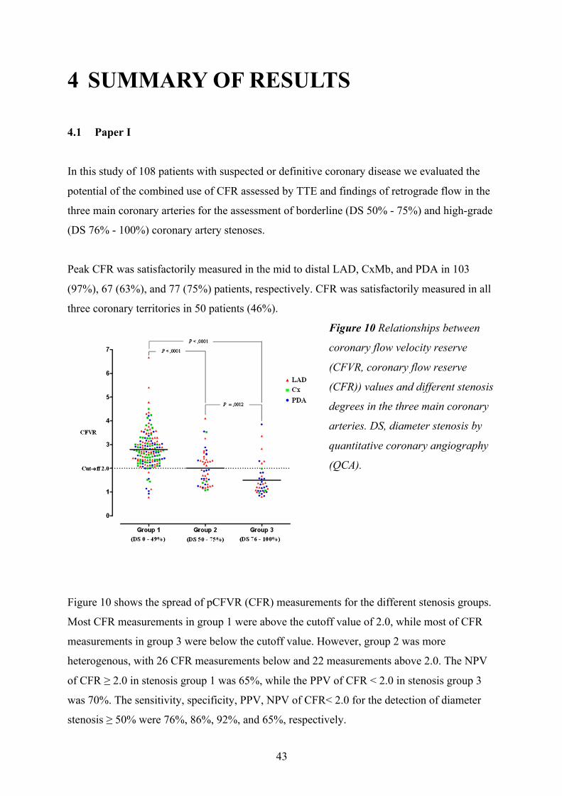
43
4 SUMMARY OF RESULTS
4.1 Paper I
In this study of 108 patients with suspected or definitive coronary disease we evaluated the
potential of the combined use of CFR assessed by TTE and findings of retrograde flow in the
three main coronary arteries for the assessment of borderline (DS 50% - 75%) and high-grade
(DS 76% - 100%) coronary artery stenoses.
Peak CFR was satisfactorily measured in the mid to distal LAD, CxMb, and PDA in 103
(97%), 67 (63%), and 77 (75%) patients, respectively. CFR was satisfactorily measured in all
three coronary territories in 50 patients (46%).
Figure 10 Relationships between
coronary flow velocity reserve
(CFVR, coronary flow reserve
(CFR)) values and different stenosis
degrees in the three main coronary
arteries. DS, diameter stenosis by
quantitative coronary angiography
(QCA).
Figure 10 shows the spread of pCFVR (CFR) measurements for the different stenosis groups.
Most CFR measurements in group 1 were above the cutoff value of 2.0, while most of CFR
measurements in group 3 were below the cutoff value. However, group 2 was more
heterogenous, with 26 CFR measurements below and 22 measurements above 2.0. The NPV
of CFR ≥ 2.0 in stenosis group 1 was 65%, while the PPV of CFR < 2.0 in stenosis group 3
was 70%. The sensitivity, specificity, PPV, NPV of CFR< 2.0 for the detection of diameter
stenosis ≥ 50% were 76%, 86%, 92%, and 65%, respectively.

44
Combined use of CFR < 2.0 or findings of retrograde coronary artery flow correctly identified
42 of 49 patients with stenoses in group 3, with sensitivity, specificity, PPV, and NPV of
86%, 70%, 70%, and 85%, respectively. We correctly identified stenoses in 13 of 14 patients
with three-vessel disease, 18 of 21 patients with two-vessel disease, and 25 of 38 patients with
one-vessel disease. The unidentified coronary stenoses were mostly in stenosis group 2.
Key findings: CFR measurements in the mid to distal LAD were feasible in the great majority
of patients, while corresponding measurements in the CxMb and PDA were feasible in two
thirds and three quarters of patients, respectively. CFR < 2.0 combined with any findings of
retrograde flow in the main coronary arteries had high accuracy for diagnosing severe
coronary stenoses, defined as angiographic diameter stenosis of 76% to 100%. Half of the
coronary arteries with angiographic intermediate stenosis showed no flow-limiting stenosis,
defined as CFR < 2.0.

45
4.2 Paper II
In this study of 108 patients with suspected or definitive coronary disease we tried to
determine whether DSVR assessed by TTE could accurately identify significant stenoses in
the left coronary artery.
Peak diastolic flow velocities showed no statistical difference among the stenosis groups.
Peak systolic flow velocity was significantly lower in group 1 compared with group 2 (0.19 ±
0.06 vs 0.23 ± 0.06 m/s) and group 3 (0.24 ± 0.08 m/s), with no statistical difference between
groups 2 and 3. DSVR was 1.92 ± 0.32 in group 1, significantly higher than in groups 2 (1.53
± 0.18) and 3 (1.43 ± 0.13). The difference between groups 2 and 3 did not reach statistical
difference. ROC curve analysis demonstrated that the optimal DSVR cutoff value of 1.68 had
sensitivity of 90%, specificity of 84%, PPV of 71%, and NPV of 95% for the detection of
diameter stenosis ≥ 50%.
Figure 11 Relationships between peak diastolic-to-
systolic velocity ratio (DSVR) values and different
stenosis degrees in the left anterior descending (LAD)
and left circumflex (Cx) coronary arteries as defined
by quantitative coronary angiography (QCA). CxMb,
Cx marginal branch; DS, Diameter stenosis by QCA.
Figure 11 shows the spread of DSVR measurements for the different stenosis groups. DSVR
was significantly higher in the patient group with CFR ≥ 2.0 (1.86 ± 0.32) compared with
DSVR in the group with CFR < 2.0 (1.53 ± 0.31). ROC curve analysis demonstrated that the
optimal DSVR cutoff value of < 1.68 had sensitivity of 86%, specificity of 74%, PPV of 51%,
and NPV of 94% for the detection of CFR < 2.0.
Key findings: DSVR measurements in the distal to mid LAD were feasible in the majority of
patients, while corresponding measurements in the CxMb were feasible in one third of
patients. DSVR < 1.68 had high accuracy for the detection of diameter stenosis ≥ 50% as
defined by QCA in the LM, LAD, and proximal and mid segments of Cx. DSVR ≥ 1.68 had
high accuracy for excluding functionally significant stenoses, as defined by CFR.

46
4.3 Paper III
In this study of 108 patients with suspected or definitive coronary disease we tried using TTE
to determine whether SPVR or mosaic flow at Nyquist limit ≥ 48 m/s when SPVR could not
be measured could accurately identify significant stenoses in the left coronary artery.
Sixty-five lesions suggestive of stenosis in segments with anterograde flow were found by
TTE. Among these 65 lesions, QCA demonstrated 54 stenoses, of which 35 were borderline
stenoses and 19 were high-grade stenoses. The SPVR was measured in 53 of the 65 lesions.
Using a SPVR cutoff value of ≥ 2.0, eight cases were in the stenosis group 1 (DS 0% – 49%),
31 cases were in the stenosis group 2 (DS 50% – 75%), and 14 cases were in the stenosis
group 3 (DS 76% – 99%), with a mean SPVR of 2.8 + 0.7, 3.7 + 1.2, and 4.9 + 2.5 in stenosis
groups 1, 2, and 3, respectively. Significant differences were only found between mean
SPVRs in stenosis groups 1 and 3. The remaining 12 lesions suggestive of stenosis were
identified by finding local mosaic coronary flow at Nyquist limit ≥ 0.48 m/s. QCA confirmed
borderline and high-grade stenosis in four and five of the lesions, respectively, whereas three
lesions showed a DS in the range of 0–49%.
Combining findings of SPVR ≥ 2.0 and mosaic flow at Nyquist limit ≥ 48 m/s, the sensitivity
and specificity of demonstrating significant stenoses in the LM, LAD, Cx, and RCA were
75% and 98%, 74% and 95%, 40% and 87%, and 34% and 98%, respectively.
Key findings: A SPVR of ≥ 2.0 demonstrated high specificity for detection of significant
stenoses in all coronary arteries. The sensitivity for identifying significant stenoses in the LM
and LAD was high, but the ability to detect lesions in the Cx and RCA was low.
Demonstration of local mosaic flow at Nyquist limit ≥ 0.48 m/s when SPVR could not be
assessed increased the ability to detect coronary stenoses.

47
4.4 Paper IV
In this study, we sought to examine the effect of tocilizumab on coronary microvascular
dysfunction by CFR measured by TTE and circulating markers of endothelial cell activation
after NSTEMI.
Markers of endothelial cell activation were analyzed in a total of 117 patients (placebo n=59,
tocilizumab n=58) and CFR was measured in 42 of these patients (placebo n=20, tocilizumab
n=22). The infarct related artery was LAD in 12 out of 42 patients, with no significant
between-group differences in distribution.
There were no between-group differences in CFR during hospitalization or at 6 months. CFR
increased significantly in both treatment groups after 6 months follow-up with no between-
group difference in changes in CFR from hospitalization. Coronary microvascular
dysfunction defined as CFR < 2.5 was present in 10 of 42 patients (24%) during
hospitalization. Whereas CFR improved significantly at 6 months in patients without initial
coronary microvascular dysfunction, this was not observed in patients with coronary
microvascular dysfunction during hospitalization.
Adjusting for baseline VCAM-1, the AUC for VCAM-1 during hospitalization was
significantly higher in the tocilizumab group than in the placebo group (median 622 vs. 609
ng/ml/hr, tocilizumab and placebo respectively). The patterns for markers of endothelial cell
activation in the subset of patients evaluated for CFR were similar to the patterns observed for
the total population (data not shown). In the placebo group, there was a strong and inverse
correlation between VCAM-1 and CFR during hospitalization. In contrast, there were no
significant correlations between markers of endothelial cell activation and CFR in the
tocilizumab group during hospitalization.
There also was a strong inverse correlation between VCAM-1 and CFR in the placebo-treated
patients at 6 months, and at follow-up we also observed a strong inverse correlation between
vWF and CFR in the placebo group. In the tocilizumab group, however, no significant
correlations between CFR and markers of endothelial cell activation were found at follow-up.
In patients evaluated for CFR, there were positive and similar correlations between high-
sensitive C-reactive protein (hsCRP) and high-sensitive Troponin T (hsTnT) in both treatment

48
groups during hospitalization, but not at follow-up. There were no relevant correlations
between hsCRP or hsTnT and CFR or makers of endothelial cell activation in either treatment
group during hospitalization or at 6 months’ follow-up.
When we evaluated CFR in patients without and with LAD as the infarct related artery
separately, there was still no significant between-group differences in CFR during
hospitalization or in the change from hospitalization to 6 months. However, in the placebo
group, the associations between the different markers of endothelial cell activation and the
association between these markers and CFR were enhanced when we excluded patients with
LAD as the culprit artery from the analyses. Notably, this was not observed among
tocilizumab-treated patients.
Key findings: In the acute phase 24% of the patients had coronary microvascular dysfunction
defined as CFR <2.5. Tocilizumab did not affect CFR during hospitalization or at 6 months.
Moreover, VCAM-1 was significantly increased in the tocilizumab group during the acute
phase of NSTEMI. In the placebo group, but not in the tocilizumab group, there was an
inverse correlation between VCAM-1 and CFR.

49
5 DISCUSSION
This thesis is based upon transthoracic echocardiography investigating the coronary artery
flow in the evaluation of different aspects of ischemic heart disease. The main focus has been
the use of transthoracic Doppler as a non-invasive diagnostic tool for diagnosing epicardial
coronary stenoses (Paper I-III). Furthermore, this non-invasive modality was used in a
randomized clinical trial assessing the effect of tocilizumab on coronary microvascular
dysfunction (Paper IV). Accordingly, the thesis has assessed diagnostic aspects of
transthoracic Doppler of coronary blood flow as well as the potential for this method as a
valuable research tool in clinical trials.
CFR can be used for two purposes; first to evaluate whether epicardial stenoses are flow
limiting or not, and secondly, to assess the coronary microvascular function in the absence of
an upstream significant epicardial stenosis. In Paper I, we assessed CFR in all three coronary
arteries searching for coronary artery stenoses. In Paper II, pulsed-wave Doppler was used to
evaluate the poststenotic coronary flow profile in search of stenoses in the left coronary
artery. In Paper III, pulsed-wave Doppler was used to investigate potential coronary stenoses
detected by colour flow Doppler aliasing and measuring the ratio of stenotic to prestenotic
flow velocities. By measuring CFR and markers of endothelial cell activation, in Paper IV we
investigated the effect of tocilizumab on coronary microvascular dysfunction and circulating
markers of endothelial cell activation after NSTEMI.
The main findings of using transthoracic Doppler as a non-invasive diagnostic tool were the
following:
1. First, CFR < 2.0 or retrograde flow in main coronary arteries show high accuracy
for diagnosing severe coronary stenosis. CFR can further differentiate the
functional significance of a borderline stenosis. (Paper I).
2. Second, DSVR show high accuracy identifying angiographic significant stenoses
in the LAD and Cx (Paper II).
3. Third, a high SPVR or coronary mosaic flow demonstrated high specificity for
detecting significant stenoses in all coronary arteries, and a high sensitivity for
identifying significant stenoses in the LM and LAD (Paper III).

50
4. Forth, tocilizumab after NSTEMI did not affect CFR during hospitalization or at 6
months. Furthermore, 24% of the patients had coronary microvascular dysfunction
during the acute phase, defined as CFR <2.5 (Paper IV).
5.1 Feasibility and accuracy of identifying significant coronary stenoses by
assessing coronary flow velocity reserve in all three coronary arteries
The feasibility of CFR measurements in the different coronary arteries in our study is
comparable to that in other studies.(23, 89-91) In agreement with other studies we found the
highest feasibility of CFR in LAD (97% of the patients), which possibly is explained by the
LAD being a large coronary artery and close to the chest wall. There are few data on CFR
evaluation of the Cx in previous studies.(23, 51) However, we found a feasibility of 63% and
75% of CFR measurements in the Cx and RCA, respectively, partly explained by inherent
anatomical and technical limitations such as reduced resolution of the lateral and inferior
walls of the heart.
In our study, stenoses in group 3 (DS 76% - 100%) typically demonstrated CFR < 2.0
consistent with functionally significant stenoses. Several investigators have demonstrated that
invasive treatment of functionally significant stenoses results in less ischemia and clinical
events compared with optimal medical treatment alone.(92) Only 6 of 37 arteries (16%) in
stenosis group 3 demonstrated normal CFR (Figure 10). An angiographic finding of a severe
coronary stenosis does not always correspond to a flow-limiting stenosis, with a recent study
showing that 20% of lesions with diameter stenosis of 71% to 90% were functionally not
significant.(38) Hence, several of the stenoses in group 3 categorized with false-negative CFR
values could represent angiographic but not hemodynamic significant lesions. Five of seven
arteries found with CFR < 1.0 had more proximally located occlusions or severe stenoses.
The close correlation of CFR < 1.0 to coronary occlusions and severe stenoses has been
demonstrated by other investigators as well and might represent coronary steal
phenomenon.(93)
Although angiographic diameter stenoses > 75% most likely represent flow-limiting stenoses,
the functional significance of a borderline stenosis (DS 50% - 75%) is often uncertain,(38)
with a recent study showing that 65% of lesions with diameter stenosis of 50% to 70% were
functionally not significant.(38) Other investigators have shown that CFR measurements by

51
TTE are able to further differentiate borderline stenoses found by angiography, with CFR <
2.0 indicating hemodynamic significant stenosis.(89, 90) In our study, approximately half of
the stenoses in group 2 (DS 50% – 75%) showed CFR ≥ 2.0, suggesting that stenoses in this
group were a mix between hemodynamic significant and non-significant stenoses (Figure 10).
Furthermore, patients with angiographic intermediate LAD stenoses combined with LAD
CFR > 2.0 measured by TTE have a good prognosis without invasive treatment.(94) Hence,
the clinical consequence of further functional assessment by CFR measurements of borderline
stenoses may be important. For instance, borderline stenoses with CFR > 2.0 may be deferred
from invasive treatment, or repeated angiography in question of restenosis might be avoided
when CFR is > 2.0.
In group 1 (DS 0% – 49%) 149 of 158 coronary arteries (94%) demonstrated normal CFR
(Figure 10), indicating that most diameter stenoses < 50% were without functional
significance. CFR was < 2.0 in nine arteries in this group. The microvascular coronary flow
regulation may be dysfunctional from various predisposing conditions, such as smoking,
aging, diabetes, and left ventricular hypertrophy, causing reduced CFR even without
hemodynamic significant epicardial stenoses.(14, 21) Several of the false-positive CFR values
in stenosis group 1 could have been caused by microvascular coronary dysfunction.
Interestingly, the one patient with angiographic normal coronary arteries and pathologic CFR
in all three arteries (CFR of 0.78-1.44) had developed normal CFR values (CFR of 2.9-3.8),
when reexamined seven months later, having stopped smoking and started with lipid-lowering
treatment with a statin. Other possible explanations for pathologic CFR measurement when
normal or sub-significant findings by angiography in the main artery may be inadequate
recordings of spectral Doppler signals during the hyperemic phase, or QCA underestimation
of the individual stenosis. Serial sub-significant stenoses or changes in the epicardial artery
may together create sufficient coronary pathology to reduce CFR.
The combination of normal CFR findings together with angiographic significant stenosis
could have several possible explanations other than demonstrating a functionally non-
significant stenosis. QCA may overestimate the individual stenosis, giving the impression of
false-normal CFR measurements. Coronary collaterals may significantly mitigate the effects
of severe stenoses or occlusions.(95) Hemodynamic significant stenoses in the main coronary
arteries may elude detection if CFR is measured proximally to the stenosis. Stenoses in the
distal segment of the Cx will probably not be detected using more proximal Cx branches for

52
CFR measurements. Stenoses in distal segments of the main coronary arteries are, however,
usually clinically less important than stenoses in mid or proximal segments, because distal
segments supply smaller amount of myocardium. Finally, CFR can erroneously be measured
in nearby parallel coursing vessels.
Retrograde coronary flow may be found during the process of visualizing the individual
coronary segment for CFR measurements. Such a finding is the functional diagnosis of
upstream coronary obstruction. Interestingly, findings of CFR < 2.0 or retrograde coronary
flow identified most of the patients having coronary diameter stenoses in the range of 75% -
100% or having three-vessel or two-vessel disease, and two thirds of the patients having one-
vessel disease.
5.2 Feasibility and accuracy of identifying significant coronary stenoses in the left
coronary artery using poststenotic coronary flow profiles
The normal coronary arteries display a predominant diastolic flow pattern, which is less
pronounced in the distal right coronary artery. Several studies have shown that in presence of
a significant coronary stenosis, the ratio between the diastolic and systolic coronary peak flow
velocity ratio (DSVR), is significantly reduced when invasively measured distal to the
stenosis.(55) Thus, DSVR is a measure of the phasic blood flow in the coronary arteries and is
determined by the interaction of several parameters influencing coronary artery flow.
Epicardial stenoses are major determinants of DSVR.(59) DSVR is also influenced by the
position at which measurements are performed. DSVR can be reduced by 15% to 20% in
distal coronary locations compared with proximal locations.(55) The normal DSVRs in LAD
and Cx are similar.(55) Because the normal DSVR in the distal RCA is different with lower
values that probably are close to pathological values in the left coronary artery, we chose to
only investigate DSVR in the LAD and Cx in this study. In the presence of a significant
coronary stenosis, the poststenotic difference between the diastolic and systolic blood flow
velocities is reduced, with a lower DSVR.(55) Spaan et al.(59) found, downstream to a
significant stenosis, a reduction of diastolic flow in combination with an increased systolic
flow component. The increased systolic flow was attributed to an intramyocardial systolic
pump acting on the intramyocardial capacitance vessels, with this systolic pump becoming
53
increasingly predominant with progressive upstream stenosis. Several studies, both invasive
and by TTE, have demonstrated that angiographic significant LAD stenoses reduce the
poststenotic DSVR to pathologic values of < 1.6 to 1.8.(25, 55, 57) Furthermore, studies have
shown that LAD DSVR < 1.6 to 1.7 is in agreement with findings of reversible ischemia by
myocardial perfusion imaging.(96)
The high feasibility (85%) of measurements in the distal to mid LAD in our study is
comparable with that in other studies.(96) The feasibility of measurements in the CxMb was
considerably lower (32%). This was mainly caused by failure to visualize the CxMb for
DSVR measurements or inadequate spectral Doppler recordings of coronary flow through
both systole and diastole, with the low-velocity systolic flow component the most difficult to
measure. The latter was partly because cardiac motion hampers the detection of complete
Doppler spectral envelopes, especially the systolic component. Furthermore, the lateral wall
suffers from a poorer resolution. However, this is not the case for measuring DSVR in the
LAD as LAD runs closer to the sternum/chest wall. To the best of our knowledge, this is the
first study using TTE to investigate the feasibility and value of DSVR measurements for
diagnosing stenoses in the Cx.
Findings of DSCVR < 1.68 by TTE showed high accuracy for identifying angiographic
significant stenoses in the LM, LAD, and proximal and mid Cx. This cutoff value corresponds
well with cutoff values proposed by others.(25, 26) However, the definition of significant
stenosis differs significantly among the various studies, with significant stenosis defined as
DS ≥ 50% in our study and > 75% to 90% in other studies using TTE.(25, 26) Our study has
the advantage of exploring the DSVR cutoff value in relation to the traditional definition of
angiographic significant stenosis (DS ≥ 50%). The sensitivity and specificity of identifying
significant stenoses in our study compared favorably with that in other studies using TTE,
which have reported sensitivity and specificity of 77% to 100% and 76% to 82%,
respectively. Interestingly, all group 3 stenoses (DS 76% - 100%) in our study showed
DSVRs below the cutoff value of 1.68 (Figure 11). Crowley and Shapiro(24) demonstrated
that DSVR was significantly reduced when comparing LAD lesions with DS 30% to 70%
with lesions with DS < 30%, and DSVR was further significantly reduced when comparing
the group with DS > 70% with that with DS of 30% to 70%. This suggests a gradual reduction
of DSVR with increasing degree of stenosis, with DSVR reduction starting relatively early in
the stenotic process. Our study confirms that both borderline and high-grade stenoses

54
significantly reduce DSVR compared with arteries with no stenosis, as defined by QCA. In
our study, DSVR was lower in high-grade stenoses compared with borderline stenoses, but
this difference did not reach statistical significance, probably because of the small number of
patients.
DSVR < 1.68 corresponded reasonably well with findings of CFVR < 2.0 measured in the
same artery. However, DSVR ≥ 1.68 had high accuracy for excluding functionally significant
stenoses. These findings support earlier reports of DSVR being able to identify or exclude
functionally significant LAD stenoses.(56, 96) In our study, only four DSVR values were
false-negatives related to functional stenosis (DSVR ≥ 1.68, CFR < 2.0). In these cases, no
stenoses were found by QCA in the respective arteries, and these discrepancies could be
caused by limitations of CFR measurements. Twenty-four arteries with normal CFRs (≥ 2.0)
showed DSVRs below the cutoff value. Nine of these arteries had borderline stenoses (DS
50% - 75%), and two arteries had high-grade stenoses (DS 76% - 100%). Accordingly, these
stenoses were angiographically but not functionally significant. In the remaining 13 arteries,
no stenoses were found by QCA, and these discrepancies could be caused by limitations of
CFR measurements. We suspect that our findings of lower sensitivity and specificity of for
functional evaluation (as defined by CFR) compared with anatomic evaluation (as defined by
QCA) of coronary stenoses are explained mainly by the fact that this index is a velocity ratio
is less related to the functional compared with the anatomic degree of stenosis. Discrepancies
between DSVR and CFR may also be caused by measurements in different locations in the
artery, with a different number of stenoses upstream from the measuring site. However, in our
study, we were meticulous in using the same site for measurements of DSVR and CFR.
Differences between CFR and DSVR might also be because the latter possibly is less
influenced by functional abnormalities of the resistance vessels or because multiple upstream
stenoses could affect DSVR and CFR differently. The same optimal DSVR cutoff value of <
1.68 identified for both angiographic and functionally significant stenoses may be an
incidental finding related to the study cohort. Nevertheless, this finding suggests that the
DSVR cutoff values for angiographic and functionally significant stenoses are close.
The detailed mechanisms of poststenotic regulation of coronary blood flow and changes in
flow profiles seem not fully understood. Several investigators have explained the poststenotic
decrease in DSVR in the presence of a significant epicardial stenosis as caused by a
significant reduction of peak diastolic velocity(24, 25, 55) while others have found increased

55
peak systolic velocity as the main cause of the reduced DSVR.(26, 97) In our study, the
decrease in DSVR in stenosis group 2 and 3 was mainly caused by a significant increase in
peak systolic velocity. We found a minor, non-significant decrease in peak diastolic velocity
when comparing the group without stenosis (group 1) with the group with stenosis > 75%
(group 3), and there was a significant increase in peak systolic velocity between group 1 and
3. Accordingly, our data indicate that the decrease in DSVR with significant coronary stenosis
may be caused by both a decrease in diastolic velocity and an increase in systolic velocity,
supporting the findings of Spaan et al.(59) In our study, both peak systolic and peak diastolic
velocities showed positive correlations with systolic blood pressure, and the lack of a
correlation of DSVR with systolic blood pressure may be caused by the positive correlations
of both peak systolic and peak diastolic velocities to this pressure.
5.3 Transthoracic Doppler for detection of stenoses in the three main coronary
arteries by use of stenotic to prestenotic velocity ratio and aliased coronary flow
The blood flow in a non-stenotic coronary artery is normally laminar, with a decreasing
velocity downstream the coronary artery. A coronary stenosis causes a luminal narrowing,
resulting in increased transstenotic flow velocity attempting to maintain coronary blood
supply. Several stenosis factors such as degree of diameter stenosis (DS), length and shape of
the stenosis, as well as possible effects from driving pressure and the coronary situation
downstream of stenosis influence the flow velocities.(32, 98) Local flow acceleration and
turbulence at the site of the stenosis are detectable by colour flow Doppler.(31) Furthermore,
flow velocities at the site of colour flow aliasing can be compared with the nearest upstream
non-accelerated prestenotic flow velocities. A peak stenotic to prestenotic velocity ratio
(SPVR) of ≥ 2.0 has been proposed as cut-off values for diameter stenosis of ≥ 50%, as
defined by QCA.(31, 36) If SPVR cannot be assessed because of non-measureable flow
velocities, local mosaic flow at considerably elevated velocity range (Nyquist limit) may be
used to demonstrate significant stenosis.(34) The functional significance of a stenosis,
especially in borderline stenoses (DS in the range of 50 – 70%), will often be inaccurately
assessed by QCA. However, the functional significance of a stenosis can be evaluated by CFR
measured by TTE, with a value of <2.0 indicating a hemodynamic significant/flow limiting
stenosis.(21) To the best of our knowledge, the combined use of SPVR and findings of
mosaic flow at substantially elevated velocity range have not been investigated or compared
with functional assessment of the stenosis.

56
We demonstrated a high specificity of combining findings of SPVR ≥2.0 and mosaic flow at
Nyquist limit ≥0.48 m/s for demonstrating significant stenoses in the LM (98%), LAD (95%),
Cx (87%), and RCA (98%). A SPVR of ≥2.0 had high sensitivity of detection of significant
stenoses in the LM and LAD, but showed only moderate ability to detect stenoses in the Cx
and RCA. The superior accuracy of detecting stenoses in the LAD by colour Doppler
compared to RCA and Cx is explained by the fact the LAD is runs closer to the chest wall,
offering better conditions for visualization with colour Doppler. The unsatisfactory accuracy
of detecting stenoses in Cx and RCA- is probably partly caused by inherent anatomical and
technical limitations. This is also reflected by a lower feasibility to visualize the different
segments of Cx and RCA with colour Doppler.(20) Joutsiniemi et al found in their study an
alternative optimal SPVR cutoff value of ≥ 2.2 instead of the more established cutoff of ≥
2.0.(37) A cutoff value of 2.2 in our study only led to minor decrease in the sensitivity for
detection of significant stenoses in the LAD and RCA from 74% to 72% and 34% to 31%,
respectively, with no other changes in diagnostic performance. SPVR was significantly lower
in group 1 stenoses compared with high-grade stenoses, which agrees with other studies.(28,
33) SPVR was higher in borderline stenoses compared with non-significant stenoses, and in
high-grade stenoses compared with borderline stenoses. However, these differences did not
reach statistical significance, probably due to small number of patients.
Demonstrating local coronary flow acceleration and turbulence on TTE may become an
attractive approach for the identification of significant stenoses without pharmacological
provocation, either by comparing stenotic and prestenotic flow velocities, or by finding local
mosaic flow at substantially elevated velocity range (Nyquist limit). From earlier experience,
we hypothesized that locally persistent coronary mosaic flow at Nyquist limit ≥0.48 m/s
showed high accuracy for detection of significant stenoses. A recent study reported high
accuracy in detecting diameter stenoses ≥70% in the proximal LAD and LM when local
mosaic flow was found at an optimal Nyquist limit of ≥0.475 m/s.(99) With the additional
demonstration of local mosaic flow at Nyquist limit ≥0.48 m/s when SPVR could not be
assessed, we were able to diagnose 20% more stenoses compared with only using SPVR
measurements, without reducing specificity. The results from our study are comparable to
those from the study from Krzanowski et al.(30) Importantly, both studies were performed
without preceding knowledge of the location of the coronary stenoses. This is in contrast to

57
other studies reporting higher feasibility and accuracy of TTE SPVR measurements in the
detection of significant coronary stenoses.(28, 36, 37) Investigators in these studies had
preceding anatomical information provided by coronary computed tomography
angiography(37) and previous percutaneous coronary intervention,(28) with reported
sensitivity of 44% – 100% and specificity of 94% – 100% for detecting significant stenoses.
We therefore anticipate that preceding anatomical knowledge of stenoses significantly
influence and increase both the sensitivity and specificity of finding significant stenoses.
Using the alternative proposed SPVR cutoff of ≥ 2.2 no change in diagnostic performance
was found, except for only a minor decrease in sensitivity for diagnosing significant stenosis
in the LAD and RCA. Our study lends support to the proposed cutoff of 2.0. However, as a
clinical tool it may be limited to LAD and LM because of the unsatisfactory sensitivity for
detecting significant stenoses in the Cx and RCA.
5.4 Coronary microvascular and endothelial dysfunction diagnosed by
transthoracic Doppler
In this study, we evaluated the coronary microcirculation and endothelial function by
measuring CFR in the LAD in a patient cohort treated according to the current
recommendations for NSTEMI. As patients with an epicardial LAD stenosis were not
included in the study, the microvascular function and endothelial function was the major
determinants of the CFR. We sought to examine the effect of tocilizumab on coronary
microvascular dysfunction and endothelial function by measuring CFR by TTE as well as
measuring circulating markers of endothelial cell activation after NSTEMI. Furthermore, we
investigated CFR both during the acute phase of the NSTEMI and at 6 months follow-up.
Tocilizumab did not affect CFR during hospitalization or at 6 months. Moreover, VCAM-1
was significantly increased in the tocilizumab group during the acute phase of NSTEMI. In
the placebo group, but not in the tocilizumab group, there was an inverse correlation between
VCAM-1 and CFR. We have recently shown that tocilizumab attenuates systemic
inflammation and TnT release in these patients.(74) The present study suggests that IL-6
inhibition in NSTEMI does not influence coronary endothelial function and may increase
rather than decrease VCAM-1 levels.

58
We found that patients with NSTEMI had reduced CFR during the acute phase, with
significant improvement at 6 months follow-up. CFR partially reflects endothelial function,
thus our findings are in agreement with previous studies showing reduced coronary
endothelial function during the acute phase of NSTEMI.(69, 70) Coronary microvascular
dysfunction defined as CFR < 2.5 was present in 10 of 42 patients (24%) during
hospitalization. The number of patients with coronary microvascular dysfunction did not
differ significantly between the two treatment groups (data not shown). We predefined a
cutoff of 2.5, based on previous studies(100, 101) and limited knowledge of CMD among
patients with NSTEMI. All the 10 patients with CMD had a CFR in the range of 2.0 to 2.5.
The relatively modest reduction of CFR could partly be explained by the relatively small
myocardial damage in the study population, with a maximal TnT of 280 (166,470) and 422
(156,1140) and TnI of 2884 (611,13630) and 46107 (n=1) in the group without and with
CMD, respectively. There is limited data on CMD with a more genuine coronary endothelial
dysfunction, since most studies have evaluated the coronary microvasculature in the setting of
the culprit lesion of a ST-elevation myocardial infarction (STEMI). This phenomenon known
as microvascular obstruction (MVO) or “no-reflow”-phenomenon, is complex with several
components, often with a more pronounced reduction of CFR (< 2.0).(68) In patients with
LAD as the infarct-related artery, CFR could potentially be affected by vascular- and
endothelial-independent factors such as microvascular obstruction.(102) When we evaluated
CFR in patients with LAD and patients without LAD as the infarct-related artery (IRA)
separately, we still observed no between-group difference during hospitalization or at follow-
up. Interestingly, in the placebo group, removing patients with LAD as the infarct-related
artery enhanced the association between the different markers of endothelial cell activation
and the association between these markers and CFR. Notably, these associations were
disrupted in the tocilizumab group suggesting some effects of anti-IL-6-therapy on vascular
inflammation. CFR was not significant reduced in the patients with LAD as IRA, probably
because of the low number of patients with LAD as IRA together with relatively small
infarctions in our study.
Whereas CFR improved significantly at 6 months in patients without coronary microvascular
dysfunction during hospitalization, this was not observed in those with coronary
microvascular dysfunction during hospitalization. In line with this, we observed a more
intense hyperemic response to adenosine also at 6 months in patients without coronary
microvascular dysfunction during hospitalization.

59
This persistently depressed endothelial function could be related to increased risk of future
coronary events.(64, 75) However, IL-6 inhibition with tocilizumab did not seem to influence
these processes. Furthermore, this is in agreement with other studies; Elbaz et al found a
persistent endothelial dysfunction in a non-culprit area of the myocardium at 6 months
follow-up among paitents with NSTEMI.(69) Uren et al. found reduced coronary endothelial
function in the vacular bed of the non-infarctrelated arteries at 6 months follow-up after a
myocardial infarction.(103)
We found no association between hsCRP and markers of endothelial cell activation or CFR in
patients with acute NSTEMI in either treatment group. Whereas CRP and IL-6 could reflect
coronary inflammation and endothelial cell activation in patients with stable CAD,(104, 105)
in acute MI, CRP and IL-6 seem to reflect the degree of myocardial injury(64) rather than the
widespread coronary inflammation also present in ACS.(71) This notion is also supported by
the present study, where hsCRP was strongly and positively correlated to hsTnT in both
treatment groups, but not to markers of endothelial cell activation or CFR. However, at
follow-up there was still no correlation between markers of endothelial cell activation and
CRP. Both CRP and markers of endothelial cell activation are clearly related to inflammation,
but they may at least in part be somewhat differently regulated.
IL-6 has been found to be associated with markers of endothelial cell activation such as
VCAM-1, ICAM-1 and vWF,(65, 75, 76) and tocilizumab seems to improve endothelial
function in patients with rheumatoid arthritis.(80) In the present study, however, a single dose
of tocilizumab did not seem to improve coronary endothelial function during the acute phase
of NSTEMI or at 6 months follow-up. In fact, tocilizumab seemed to contribute to higher
VCAM-1 levels during the acute phase, and high VCAM-1 levels have been associated with
increased risk of future coronary events in ACS.(64, 75) However, whereas VCAM-1
correlated inversely with CFR during the acute phase in placebo treated patients, no such
correlation was found in the tocilizumab group. Our results suggest that the beneficial effects
of tocilizumab in NSTEMI as previously described by our group(74) are not mediated via
pathways reflected by soluble endothelial markers. The reason for the lack of an association
between VCAM-1 and CFR in patients treated with tocilizumab is not clear, but could
indicate that the elevated VCAM-1 levels with tocilizumab are neutralized via other
pathways.

60
The present study provides new insight in coronary endothelial function as reflected by
transthoracic CFR, how it relates to markers of endothelial cell activation, as well as the effect
of IL-6 inhibition by a single dose of tocilizumab on these parameters during NSTEMI. Our
findings suggest that the promising effects of tocilizumab on inflammation and TnT release
during the acute phase of NSTEMI does not involve inhibition of endothelial cell activation.
Additionally, it confirms the presence of CMD in patients with NSTEMI, even with relatively
small infarctions. Furthermore, the depressed coronary microvascular function persists at 6
months follow-up. However, this study was not powered to evaluate the prognostic effect of
CMD. Our findings could, however, encourage forthcoming studies to examine further the
potential effects of tocilizumab during MI and evaluate the clinical impact and the
mechanisms of the enhancing effect of tocilizumab on VCAM-1 levels in these patients and
the impact of CMD in the aftermath of an NSTEMI.
5.5 Limitations
Patient population I
Because of longer distance to the echocardiographic probe, the Cx, CxMbs and RCA/PDA
were more difficult to visualize than the LM and LAD. For the same reason, recording
spectral Doppler signals at baseline and during adenosine-induced hyperemia was more
complicated in the CxMb and PDA, especially if any hyperventilation occurred during the
hyperemic phase. The use of ultrasound contrast agent might have improved the feasibility of
demonstrating flow in main coronary trunks and coronary branches. Because of limited
clinical experience in patients with ACS, we chose not to use ultrasound contrast when
planning the study. We cannot exclude that nearby parallel coursing LAD branches may on
occasion have been mistaken as the main LAD trunk, resulting in CFR and DSVR
measurements in this branch instead of the main LAD. CFR displays hemodynamic
dependency on left ventricular contractility, filling pressure, and hypertrophy, and the lack of
estimation of these parameters may have limited the CFR results. CFR may be influenced by
collateral supply, and we did not take collateral perfusion into account when evaluating CFR.
Furthermore, stenoses may have eluded detection if DSVR and CFR were measured
proximally to the stenoses. The responses in CFR, DSVR, and SPVR are probably not binary
(normal or abnormal) or absolute for the cutoff values, implying that measurements slightly
below or above the cutoff values might give uncertain estimates. Future studies with larger
patient cohorts may possibly refine our proposed DSVR cutoff value. The cutoff value for

61
DSVR was calculated and tested on the same population due to the limited number of patients
with measured DSVR. This is suboptimal and represents a statistical challenge. However, the
cutoff value corresponded well with findings in previous studies. Nevertheless, future studies
with larger patient cohorts is needed to verify and maybe refine our proposed DSVR cutoff
values. This represents a notably limitation to the study. The use of additional functional
stress tests (such as dobutamine stress echocardiography or myocardial perfusion imaging)
might have further clarified the functional significance of angiographic findings. Moreover,
QCA can both overestimate and underestimate stenoses.
We used pulsed-wave Doppler to measure the coronary blood flow velocities. The cosines
angle in the Doppler equation represents a well-known source of error in Doppler
measurements. To minimize the possible source of error caused by angle differences between
the coronary blood flow and the Doppler beam we tried to align the Doppler beam as close as
possible to the direction of the coronary blood flow. The coronary arteries have a torturous
course, however, and this might result in an angle between the coronary blood flow and the
Doppler beam. Nevertheless, as we mostly studied the relationships between two peak
velocities (measurements of CFR, DSVR, and SPVR), any angle difference would mean less.
CFR was measured keeping the probe in the same position at baseline and during the
hyperemia to minimize the angle difference between the measurements.
Finally, we cannot exclude selection bias in our results, because our study cohort included
only patients planned for coronary angiography and excluded those with previous coronary
artery bypass surgery, presumed insufficient acoustic windows, significant valvular disease,
or atrial fibrillation.
Patient population II
The main limitation is the limited number of participants, which probably is the main reason
for the study not meeting its primary endpoint (tocilizumab effecting the CFR). Additionally,
CFR was measured on day 2 or 3 after infusion of the study drug, at a time where the effect of
tocilizumab was not fully present.(74) Patients that developed CMD showed at higher level of
inflammation at inclusion and at the time of CFR measurements and this was especially
marked among patients receiving tocilizumab.

62
We cannot exclude that nearby parallel coursing LAD branches may on occasion have been
mistaken as the main LAD trunk, resulting in CFR measurements in this branch instead of the
main LAD. However, since no significant epicardial stenosis were present a CFR
measurement on a LAD branch would probably not affect the result.

63
6 MAIN CONCLUSIONS
1. CFR measurements in the mid to distal LAD were feasible in almost all patients, while
corresponding measurements in the CxMb and PDA were feasible in two thirds and
three quarters of patients, respectively. A CFR value < 2.0 and findings of retrograde
flow had high accuracy for diagnosing severe coronary stenoses (angiographic
diameter stenosis, 76% – 100%). Half of the coronary arteries with angiographic
intermediate stenosis (diameter stenosis, 50% – 75%) showed no functionally
significant stenosis, defined as CFR < 2.0, underscoring both the current limitation of
angiographic assessment of intermediate severity of stenoses and the importance of
performing functional stress testing to detect myocardial ischemia.
2. DSVR measurements by TTE in the distal to mid LAD were feasible in most patients
and in the CxMb in one third of patients. To the best of our knowledge, this is the first
study using TTE to investigate the value of DSVR measurements in the Cx. A DSVR
value < 1.68 showed high accuracy to detect angiographic significant stenoses in the
LAD, LM, and proximal and mid segments of the Cx. A DSVR value < 1.68 showed
moderate ability to detect functionally significant stenoses, while DSVR ≥ 1.68 had
high accuracy to exclude these stenoses. When feasible, assessing DSVR in the distal
to mid LAD and CxMb provides a simple, noninvasive method to identify and rule out
angiographic and hemodynamic significant stenoses in the LAD, LM, and proximal
and mid segments of Cx.
3. Detection of localized colour flow aliasing and measurement of SPVR are useful in
the noninvasive diagnosis of significant coronary disease in the three main coronary
arteries. A SPVR of ≥ 2.0 demonstrated high specificity for detection of significant
stenoses in all coronary arteries. This methodology showed high sensitivity for
identifying significant stenoses in the LM and LAD, but the ability to detect lesions in
the Cx and RCA was low. Demonstration of local mosaic flow at Nyquist limit ≥ 0.48
m/s when SPVR could not be assessed increased the ability to detect coronary
stenoses.
64
4. The present study provides new insight into coronary endothelial function as reflected
by CFR, how it relates to markers of endothelial cell activation, as well as the effect of
IL-6 inhibition by a single dose of tocilizumab on these parameters in the course of
NSTEMI. Our findings suggest that the promising effects of tocilizumab on
inflammation and TnT release during the acute phase of NSTEMI do not involve
inhibition of endothelial cell activation. Additionally, our study confirms the presence
of CMD in patients with NSTEMI, even with relatively small infarctions.
Furthermore, the depressed coronary microvascular function persists at 6 months
follow-up.

65
7 CLINICAL PERSPECTIVE AND FUTURE DIRECTIONS FOR RESEARCH
This thesis has several implications both for clinical and research purposes. First, TTE can be
used to diagnose coronary artery disease by visualizing coronary stenoses using colour flow
Doppler and further characterize the coronary flow profile in evaluating the stenosis, as
demonstrated in study II and study III using stenotic and poststenotic flow measurements.
Assessing SPVR is feasible for all three coronary arteries, whereas DSVR can be used in
LAD and Cx. However, the methods have the highest feasibility for detecting significant
stenoses in the LM, LAD, and proximal segment of the Cx. These methods may be important
alternative non-invasive methods to detect coronary stenoses. Furthermore, angiographic
borderline stenoses without functional significance are common and can be further clarified
by demonstrating CFR ≥ 2.0 when measured downstream to the stenoses, as demonstrated in
study I. However, in some cases the interpretation of CFR < 2.0 may be confounded by the
presence of microvascular dysfunction.
CFR can be used to monitor effect of lifestyle and pharmacologic interventions in patients
with microvascular dysfunction, as the case referred to in paper I demonstrates.
Despite improved diagnostic and therapeutic options several major paradoxes still remain in
the management of CAD, like the fact that percent diameter stenosis does not reliably relate to
maximum flow capacity or coronary flow reserve, or that revascularization procedures to
improve coronary blood flow do not reduce coronary events more than optimal medical
treatment in randomized trails.(106) As demonstrated in study I, the actual DS by QCA do not
necessarily reflect the physiological consequence of the luminal narrowing. An optimal
coronary flow is pivotal to meet and adapt to the different demands of the myocardium.
Furthermore, compromised coronary flow, regardless of its origin, leads to myocardial
ischemia responsible for both morbidity and mortality. Thus, increased research of the
different aspects and characteristics of the coronary flow and circulation may be very
important in terms of increased knowledge about the pathophysiological chains, better
diagnostic tests and improved treatment options.
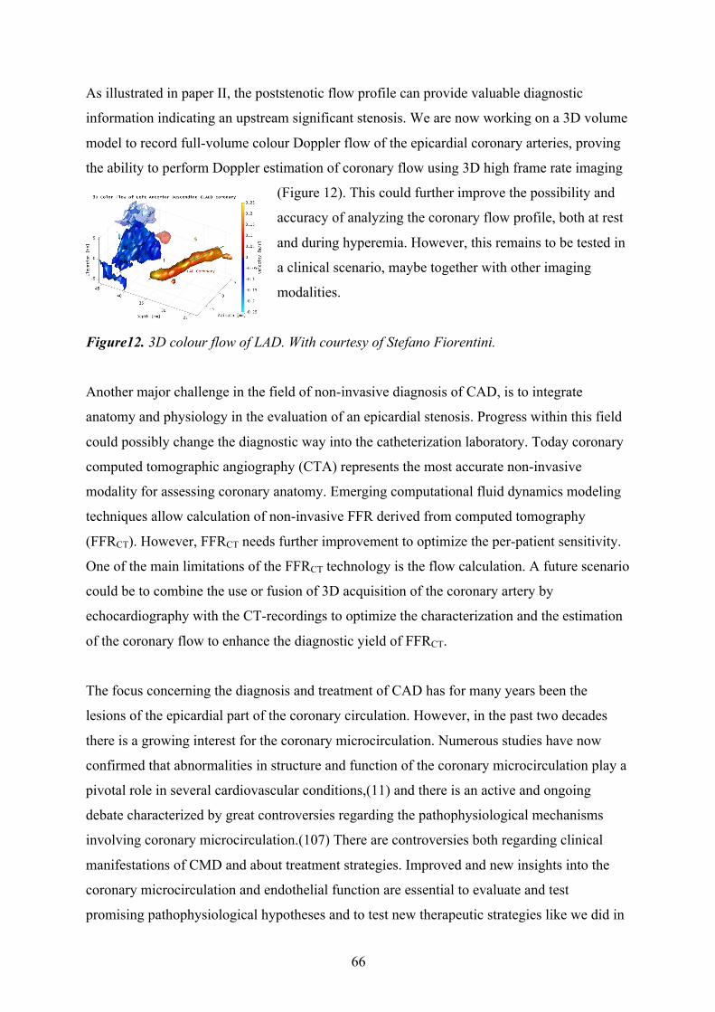
66
As illustrated in paper II, the poststenotic flow profile can provide valuable diagnostic
information indicating an upstream significant stenosis. We are now working on a 3D volume
model to record full-volume colour Doppler flow of the epicardial coronary arteries, proving
the ability to perform Doppler estimation of coronary flow using 3D high frame rate imaging
(Figure 12). This could further improve the possibility and
accuracy of analyzing the coronary flow profile, both at rest
and during hyperemia. However, this remains to be tested in
a clinical scenario, maybe together with other imaging
modalities.
Figure12. 3D colour flow of LAD. With courtesy of Stefano Fiorentini.
Another major challenge in the field of non-invasive diagnosis of CAD, is to integrate
anatomy and physiology in the evaluation of an epicardial stenosis. Progress within this field
could possibly change the diagnostic way into the catheterization laboratory. Today coronary
computed tomographic angiography (CTA) represents the most accurate non-invasive
modality for assessing coronary anatomy. Emerging computational fluid dynamics modeling
techniques allow calculation of non-invasive FFR derived from computed tomography
(FFRCT). However, FFRCT needs further improvement to optimize the per-patient sensitivity.
One of the main limitations of the FFRCT technology is the flow calculation. A future scenario
could be to combine the use or fusion of 3D acquisition of the coronary artery by
echocardiography with the CT-recordings to optimize the characterization and the estimation
of the coronary flow to enhance the diagnostic yield of FFRCT.
The focus concerning the diagnosis and treatment of CAD has for many years been the
lesions of the epicardial part of the coronary circulation. However, in the past two decades
there is a growing interest for the coronary microcirculation. Numerous studies have now
confirmed that abnormalities in structure and function of the coronary microcirculation play a
pivotal role in several cardiovascular conditions,(11) and there is an active and ongoing
debate characterized by great controversies regarding the pathophysiological mechanisms
involving coronary microcirculation.(107) There are controversies both regarding clinical
manifestations of CMD and about treatment strategies. Improved and new insights into the
coronary microcirculation and endothelial function are essential to evaluate and test
promising pathophysiological hypotheses and to test new therapeutic strategies like we did in

67
paper IV. In that study, we demonstrated that tocilizumab attenuated inflammation and TnT
release in NSTEMI, but did not seem to influence coronary endothelial function. This
implicates that larger studies with clinical endpoints as well as further studies on its
mechanisms of action are needed before IL-6 inhibition could be part of the clinical practice
in NSTEMI.
Microvascular dysfunction following a myocardial infarction is affecting both the
myocardium and the prognosis. However, there are gaps in evidence about the extent and
importance of microvascular dysfunction in the NSTEMI populations. Despite relatively
small myocardial damage in study IV, 24% of the study population had indication of
microvascular dysfunction, which persisted at 6 months´ follow-up. This indicates a long-
lasting effect of the coronary microvasculature and endothelial function, maybe representing a
potential therapeutic target. We do not have specific treatment strategies to prevent
microvascular dysfunction. Thus, we need more data from larger NSTEMI populations to
better understand and develop interventions to minimize the potential microvascular damage
caused by myocardial infarctions.
In summary, TTE examinations of coronary flow, especially looking for changes in CFR, may
become an increasingly important clinical research tool in future studies of interventional and
pharmacological treatments of coronary disease. Further developments of echocardiographic
technology may increase the feasibility of visualizing the coronary artery tree and the
coronary microcirculation. Assessment of the latter has been hampered by lack of suitable
methods for clinical use. New flow catheters can now simultaneously measure absolute
coronary blood flow and absolute microvascular resistance. This could be a new window to
the coronary microcirculation. However, this is still an invasive strategy, so in parallel we
need further improvement of a non-invasive method. TTE measurements of CFR may be
technically challenging and are in the same way as dobutamine stress echocardiography
dependent on operator skills. Thus, if this method is to gain clinical use it should be more
strongly implemented in the echocardiographic education. The use of ultrasound contrast
agent may improve both the feasibility of imaging and characterization of the coronary
vessels and the ability and quality of coronary flow measurements.
Microvascular dysfunction following a myocardial infarction is affecting both the
myocardium and the prognosis. However, there are gaps in evidence according of the extent
and importance of microvascular dysfunction in the NSTEMI populations. Despite relatively

68
small myocardial damage in study IV, around aquater of the study population had indication
of microvascular dysfunction, which persisted at 6 months follow-up. This indicates a long-
lasting effect on the coronary microvasculature and endothelial function, maybe representing
a potential therapeutic target. We do not have specific treatment strategies to prevent
microvascular dysfunction, thus we need more data from larger NSTEMI populations to
better understand and develop interventions to minimize the potential microvascular damage
in the line of a myocardial infarction.

69
REFERENCES 1. C.H.Moderncardiovascularphysiology.Moderncardiovascularphysiology:Boston:LittleBrownandCo;1981.p.pp.XVI–XVII.2. AmiriM,JanssenF,KunstAE.ThedeclineinischaemicheartdiseasemortalityinsevenEuropeancountries:explorationoffuturetrends.JEpidemiolCommunityHealth.2011;65(8):676-81.3. MontalescotG,SechtemU,AchenbachS,AndreottiF,ArdenC,BudajA,etal.2013ESCguidelinesonthemanagementofstablecoronaryarterydisease:theTaskForceonthemanagementofstablecoronaryarterydiseaseoftheEuropeanSocietyofCardiology.EurHeartJ.2013;34(38):2949-3003.4. NicholsM,TownsendN,ScarboroughP,RaynerM.Trendsinage-specificcoronaryheartdiseasemortalityintheEuropeanUnionoverthreedecades:1980-2009.EurHeartJ.2013;34(39):3017-27.5. centerNI.Coronaryaterydisease[Availablefrom:http://www.nhs.uk/Conditions/Coronary-heart-disease/Pages/Introduction.aspx.6. GouldKL,JohnsonNP,BatemanTM,BeanlandsRS,BengelFM,BoberR,etal.Anatomicversusphysiologicassessmentofcoronaryarterydisease.Roleofcoronaryflowreserve,fractionalflowreserve,andpositronemissiontomographyimaginginrevascularizationdecision-making.JAmCollCardiol.2013;62(18):1639-53.7. RossR.Thepathogenesisofatherosclerosis:aperspectiveforthe1990s.Nature.1993;362(6423):801-9.8. LibbyP,TherouxP.Pathophysiologyofcoronaryarterydisease.Circulation.2005;111(25):3481-8.9. PrescottE,AbildstromSZ,AzizA,MerzNB,GustafssonI,HalcoxJ,etal.Improvingdiagnosisandtreatmentofwomenwithanginapectorisandmicrovasculardisease:theiPOWERstudydesignandrationale.AmHeartJ.2014;167(4):452-8.10. DimitrowPP,GalderisiM,RigoF.Thenon-invasivedocumentationofcoronarymicrocirculationimpairment:roleoftransthoracicechocardiography.CardiovascUltrasound.2005;3:18.11. CamiciPG,d'AmatiG,RimoldiO.Coronarymicrovasculardysfunction:mechanismsandfunctionalassessment.NatRevCardiol.2015;12(1):48-62.12. KhouriEM,GreggDE,RayfordCR.Effectofexerciseoncardiacoutput,leftcoronaryflowandmyocardialmetabolismintheunanesthetizeddog.CircRes.1965;17(5):427-37.13. HungM-J,CherngW-J.ClinicalApplicationsofTransthoracicDopplerEchocardiographicCoronaryFlowReserveMeasurementsintheLeftAnteriorDescendingCoronaryArtery.JMedUltrasound.2011;19(4):115-21.14. KernMJ,LermanA,BechJW,DeBruyneB,EeckhoutE,FearonWF,etal.Physiologicalassessmentofcoronaryarterydiseaseinthecardiaccatheterizationlaboratory:ascientificstatementfromtheAmericanHeartAssociationCommitteeonDiagnosticandInterventionalCardiacCatheterization,CouncilonClinicalCardiology.Circulation.2006;114(12):1321-41.15. KiviniemiT.Assessmentofcoronarybloodflowandthereactivityofthemicrocirculationnon-invasivelywithtransthoracicechocardiography.ClinPhysiolFunctImaging.2008;28(3):145-55.16. RigoF.Coronaryflowreserveinstress-echolab.Frompathophysiologictoytodiagnostictool.CardiovascUltrasound.2005;3:8.17. HamiraniYS,WongA,KramerCM,SalernoM.EffectofmicrovascularobstructionandintramyocardialhemorrhagebyCMRonLVremodelingandoutcomesaftermyocardialinfarction:asystematicreviewandmeta-analysis.JACCCardiovascImaging.2014;7(9):940-52.18. GouldKL,LipscombK.Effectsofcoronarystenosesoncoronaryflowreserveandresistance.AmJCardiol.1974;34(1):48-55.

70
19. UrenNG,MelinJA,DeBruyneB,WijnsW,BaudhuinT,CamiciPG.Relationbetweenmyocardialbloodflowandtheseverityofcoronary-arterystenosis.NEnglJMed.1994;330(25):1782-8.20. VegsundvagJ,HolteE,WisethR,HegbomK,HoleT.Transthoracicechocardiographyforimagingofthedifferentcoronaryarterysegments:afeasibilitystudy.CardiovascUltrasound.2009;7:58.21. VociP,PizzutoF,RomeoF.Coronaryflow:anewassetfortheecholab?EurHeartJ.2004;25(21):1867-79.22. VegsundvagJ,HolteE,WisethR,HegbomK,HoleT.CoronaryflowvelocityreserveinthethreemaincoronaryarteriesassessedwithtransthoracicDoppler:acomparativestudywithquantitativecoronaryangiography.JAmSocEchocardiogr.2011;24(7):758-67.23. MurataE,HozumiT,MatsumuraY,FujimotoK,SugiokaK,TakemotoY,etal.CoronaryflowvelocityreservemeasurementinthreemajorcoronaryarteriesusingtransthoracicDopplerechocardiography.Echocardiography.2006;23(4):279-86.24. CrowleyJJ,ShapiroLM.Noninvasiveanalysisofcoronaryarterypoststenoticflowcharacteristicsbyusingtransthoracicechocardiography.JAmSocEchocardiogr.1998;11(1):1-9.25. HigashiueS,WatanabeH,YokoiY,TakeuchiK,YoshikawaJ.SimpledetectionofseverecoronarystenosisusingtransthoracicDopplerechocardiographyatrest.AmJCardiol.2001;87(9):1064-8.26. TaniT,TanabeK,KitaiT,YamaneT,KurehaF,KatayamaM,etal.DetectionofseverestenosisandtotalocclusionintheleftanteriordescendingcoronaryarterywithtransthoracicDopplerechocardiographyintheemergencyroom.Echocardiography.2009;26(1):15-20.27. HyodoE,HirataK,HiroseM,SakanoueY,NishidaY,AraiK,etal.DetectionofrestenosisafterpercutaneouscoronaryinterventioninthreemajorcoronaryarteriesbytransthoracicDopplerechocardiography.JAmSocEchocardiogr.2010;23(5):553-9.28. KrzanowskiM,BodzonW,DudekD,HebaG,RzeszutkoM,NizankowskiR,etal.Transthoracic,harmonicmode,contrastenhancedcolorDopplerechocardiographyindetectionofrestenosisafterpercutaneouscoronaryinterventions.Prospectiveevaluationverifiedbycoronaryangiography.EurJEchocardiogr.2004;5(1):51-64.29. KrzanowskiM,BodzonW,DimitrowPP.Imagingofallthreecoronaryarteriesbytransthoracicechocardiography.Anillustratedguide.CardiovascUltrasound.2003;1:16.30. KrzanowskiM,BodzonW,BrzostekT,NizankowskiR,SzczeklikA.Valueoftransthoracicechocardiographyforthedetectionofhigh-gradecoronaryarterystenosis:prospectiveevaluationin50consecutivepatientsscheduledforcoronaryangiography.JAmSocEchocardiogr.2000;13(12):1091-9.31. SarasteM,VesalainenRK,YlitaloA,SarasteA,KoskenvuoJW,ToikkaJO,etal.TransthoracicDopplerechocardiographyasanoninvasivetooltoassesscoronaryarterystenoses--acomparisonwithquantitativecoronaryangiography.JAmSocEchocardiogr.2005;18(6):679-85.32. KernMJ.Coronaryphysiologyrevisited:practicalinsightsfromthecardiaccatheterizationlaboratory.Circulation.2000;101(11):1344-51.33. SarasteM,KoskenvuoJW,MikkolaJ,PelttariL,ToikkaJO,HartialaJJ.Technicalachievement:transthoracicDopplerechocardiographycanbeusedtodetectLADrestenosisaftercoronaryangioplasty.ClinPhysiol.2000;20(6):428-33.34. AnjaneyuluA,RaghuK,ChandramukhiS,SatyajitGM,ArramrajaS,RaghavarajuP,etal.Evaluationofleftmaincoronaryarterystenosisbytransthoracicechocardiography.JAmSocEchocardiogr.2008;21(7):855-60.35. HolteE,VegsundvagJ,WisethR.Directvisualizationofasignificantstenosisoftherightcoronaryarterybytransthoracicechocardiography.Acasereport.CardiovascUltrasound.2007;5:33.36. HozumiT,YoshidaK,AkasakaT,AsamiY,KanzakiY,UedaY,etal.ValueofaccelerationflowandtheprestenotictostenoticcoronaryflowvelocityratiobytransthoraciccolorDopplerechocardiographyinnoninvasivediagnosisofrestenosisafterpercutaneoustransluminalcoronaryangioplasty.JAmCollCardiol.2000;35(1):164-8.

71
37. JoutsiniemiE,SarasteA,PietilaM,UkkonenH,KajanderS,MakiM,etal.Restingcoronaryflowvelocityinthefunctionalevaluationofcoronaryarterystenosis:studyonsequentialuseofcomputedtomographyangiographyandtransthoracicDopplerechocardiography.EurHeartJCardiovascImaging.2012;13(1):79-85.38. ToninoPA,FearonWF,DeBruyneB,OldroydKG,LeesarMA,VerLeePN,etal.AngiographicversusfunctionalseverityofcoronaryarterystenosesintheFAMEstudyfractionalflowreserveversusangiographyinmultivesselevaluation.JAmCollCardiol.2010;55(25):2816-21.39. MeimounP,BenaliT,ElmkiesF,SayahS,Luycx-BoreA,DoutrelanL,etal.Prognosticvalueoftransthoraciccoronaryflowreserveinmedicallytreatedpatientswithproximalleftanteriordescendingarterystenosisofintermediateseverity.EurJEchocardiogr.2009;10(1):127-32.40. PijlsNH,vanSchaardenburghP,ManoharanG,BoersmaE,BechJW,van'tVeerM,etal.Percutaneouscoronaryinterventionoffunctionallynonsignificantstenosis:5-yearfollow-upoftheDEFERStudy.JAmCollCardiol.2007;49(21):2105-11.41. LeesarMA,Abdul-BakiT,AkkusNI,SharmaA,KannanT,BolliR.Useoffractionalflowreserveversusstressperfusionscintigraphyafterunstableangina.Effectondurationofhospitalization,cost,proceduralcharacteristics,andclinicaloutcome.JAmCollCardiol.2003;41(7):1115-21.42. KushnerFG,HandM,SmithSC,Jr.,KingSB,3rd,AndersonJL,AntmanEM,etal.2009focusedupdates:ACC/AHAguidelinesforthemanagementofpatientswithST-elevationmyocardialinfarction(updatingthe2004guidelineand2007focusedupdate)andACC/AHA/SCAIguidelinesonpercutaneouscoronaryintervention(updatingthe2005guidelineand2007focusedupdate)areportoftheAmericanCollegeofCardiologyFoundation/AmericanHeartAssociationTaskForceonPracticeGuidelines.JAmCollCardiol.2009;54(23):2205-41.43. WindeckerS,KolhP,AlfonsoF,ColletJP,CremerJ,FalkV,etal.2014ESC/EACTSGuidelinesonmyocardialrevascularization:TheTaskForceonMyocardialRevascularizationoftheEuropeanSocietyofCardiology(ESC)andtheEuropeanAssociationforCardio-ThoracicSurgery(EACTS)DevelopedwiththespecialcontributionoftheEuropeanAssociationofPercutaneousCardiovascularInterventions(EAPCI).EurHeartJ.2014;35(37):2541-619.44. BhattDL.AssessmentofStableCoronaryLesions.NEnglJMed.2017;376(19):1879-81.45. DaviesJE,SenS,DehbiHM,Al-LameeR,PetracoR,NijjerSS,etal.UseoftheInstantaneousWave-freeRatioorFractionalFlowReserveinPCI.NEnglJMed.2017;376(19):1824-34.46. BuusNH,BottcherM,HermansenF,SanderM,NielsenTT,MulvanyMJ.Influenceofnitricoxidesynthaseandadrenergicinhibitiononadenosine-inducedmyocardialhyperemia.Circulation.2001;104(19):2305-10.47. HozumiT,YoshidaK,AkasakaT,AsamiY,OgataY,TakagiT,etal.NoninvasiveassessmentofcoronaryflowvelocityandcoronaryflowvelocityreserveintheleftanteriordescendingcoronaryarterybyDopplerechocardiography:comparisonwithinvasivetechnique.JAmCollCardiol.1998;32(5):1251-9.48. CaiatiC,MontaldoC,ZeddaN,BinaA,IlicetoS.Newnoninvasivemethodforcoronaryflowreserveassessment:contrast-enhancedtransthoracicsecondharmonicechoDoppler.Circulation.1999;99(6):771-8.49. LethenH,HPT,KerstingS,LambertzH.Validationofnoninvasiveassessmentofcoronaryflowvelocityreserveintherightcoronaryartery.AcomparisonoftransthoracicechocardiographicresultswithintracoronaryDopplerflowwiremeasurements.EurHeartJ.2003;24(17):1567-75.50. UenoY,NakamuraY,TakashimaH,KinoshitaM,SomaA.NoninvasiveassessmentofcoronaryflowvelocityandcoronaryflowvelocityreserveintherightcoronaryarterybytransthoracicDopplerechocardiography:comparisonwithintracoronaryDopplerguidewire.JAmSocEchocardiogr.2002;15(10Pt1):1074-9.51. FujimotoK,WatanabeH,HozumiT,OtsukaR,HirataK,YamagishiH,etal.Newnoninvasivediagnosisofmyocardialischemiaoftheleftcircumflexcoronaryarteryusing

72
coronaryflowreservemeasurementbytransthoracicDopplerechocardiography:comparisonwiththallium-201singlephotonemissioncomputedtomography.JCardiol.2004;43(3):109-16.52. AuritiA,PristipinoC,CianfroccaC,GranatelliA,GuidoV,PellicciaF,etal.DistalleftcircumflexcoronaryarteryflowreserverecordedbytransthoracicDopplerechocardiography:acomparisonwithDoppler-wire.CardiovascUltrasound.2007;5:22.53. LethenH,TriesHP,BrechtkenJ,KerstingS,LambertzH.ComparisonoftransthoracicDopplerechocardiographytointracoronaryDopplerguidewiremeasurementsforassessmentofcoronaryflowreserveintheleftanteriordescendingarteryfordetectionofrestenosisaftercoronaryangioplasty.AmJCardiol.2003;91(4):412-7.54. WatanabeN,AkasakaT,YamauraY,AkiyamaM,KoyamaY,KamiyamaN,etal.NoninvasivedetectionoftotalocclusionoftheleftanteriordescendingcoronaryarterywithtransthoracicDopplerechocardiography.JAmCollCardiol.2001;38(5):1328-32.55. OfiliEO,LabovitzAJ,KernMJ.Coronaryflowvelocitydynamicsinnormalanddiseasedarteries.AmJCardiol.1993;71(14):3D-9D.56. DeychakYA,SegalJ,ReinerJS,RohrbeckSC,ThompsonMA,LunderganCF,etal.Dopplerguidewireflow-velocityindexesmeasureddistaltocoronarystenosesassociatedwithreversiblethalliumperfusiondefects.AmHeartJ.1995;129(2):219-27.57. DonohueTJ,KernMJ,AguirreFV,BachRG,WolfordT,BellCA,etal.Assessingthehemodynamicsignificanceofcoronaryarterystenoses:analysisoftranslesionalpressure-flowvelocityrelationsinpatients.JAmCollCardiol.1993;22(2):449-58.58. SerruysPW,DiMarioC,MeneveauN,deJaegereP,StrikwerdaS,deFeyterPJ,etal.Intracoronarypressureandflowvelocitywithsensor-tipguidewires:anewmethodologicapproachforassessmentofcoronaryhemodynamicsbeforeandaftercoronaryinterventions.AmJCardiol.1993;71(14):41d-53d.59. SpaanJA,BreulsNP,LairdJD.Diastolic-systoliccoronaryflowdifferencesarecausedbyintramyocardialpumpactionintheanesthetizeddog.CircRes.1981;49(3):584-93.60. OkuraH,FuyukiH,KuboT,IwataK,TaguchiH,TodaI,etal.NoninvasivediagnosisofischemicandnonischemiccardiomyopathyusingcoronaryflowvelocitymeasurementsoftheleftanteriordescendingcoronaryarterybytransthoracicDopplerechocardiography.JAmSocEchocardiogr.2006;19(5):552-8.61. HanssonGK.Inflammation,atherosclerosis,andcoronaryarterydisease.NEnglJMed.2005;352(16):1685-95.62. LibbyP.Inflammationinatherosclerosis.Nature.2002;420(6917):868-74.63. HalvorsenB,OtterdalK,DahlTB,SkjellandM,GullestadL,OieE,etal.Atheroscleroticplaquestability--whatdeterminesthefateofaplaque?ProgCardiovascDis.2008;51(3):183-94.64. ZamaniP,SchwartzGG,OlssonAG,RifaiN,BaoW,LibbyP,etal.Inflammatorybiomarkers,death,andrecurrentnonfatalcoronaryeventsafteranacutecoronarysyndromeintheMIRACLstudy.JAmHeartAssoc.2013;2(1):e003103.65. LeeKW,BlannAD,LipGY.Inter-relationshipsofindicesofendothelialdamage/dysfunction[circulatingendothelialcells,vonWillebrandfactorandflow-mediateddilatation]totissuefactorandinterleukin-6inacutecoronarysyndromes.IntJCardiol.2006;111(2):302-8.66. GutierrezE,FlammerAJ,LermanLO,ElizagaJ,LermanA,Fernandez-AvilesF.Endothelialdysfunctionoverthecourseofcoronaryarterydisease.EurHeartJ.2013;34(41):3175-81.67. LermanA,CannanCR,HiganoSH,NishimuraRA,HolmesDR,Jr.Coronaryvascularremodelinginassociationwithendothelialdysfunction.AmJCardiol.1998;81(9):1105-9.68. AmierRP,TeunissenPF,MarquesKM,KnaapenP,vanRoyenN.InvasivemeasurementofcoronarymicrovascularresistanceinpatientswithacutemyocardialinfarctiontreatedbyprimaryPCI.Heart.2014;100(1):13-20.69. ElbazM,CarrieD,BaudeuxJL,ArnalJF,MaupasE,LotterieJA,etal.Highfrequencyofendothelialvasomotordysfunctionafteracutecoronarysyndromesinnon-culpritandangiographicallynormalcoronaryarteries:areversiblephenomenon.Atherosclerosis.2005;181(2):311-9.
73
70. HosokawaS,HiasaY,TomokaneT,OguraR,MiyajimaH,OharaY,etal.Theeffectsofatorvastatinoncoronaryendothelialfunctioninpatientswithrecentmyocardialinfarction.ClinCardiol.2006;29(8):357-62.71. BuffonA,BiasucciLM,LiuzzoG,D'OnofrioG,CreaF,MaseriA.Widespreadcoronaryinflammationinunstableangina.NEnglJMed.2002;347(1):5-12.72. BujaLM,WillersonJT.Roleofinflammationincoronaryplaquedisruption.Circulation.1994;89(1):503-5.73. Theinterleukin-6receptorasatargetforpreventionofcoronaryheartdisease:amendelianrandomisationanalysis.Lancet.2012;379(9822):1214-24.74. KlevelandO,KunsztG,BratlieM,UelandT,BrochK,HolteE,etal.Effectofasingledoseoftheinterleukin-6receptorantagonisttocilizumaboninflammationandtroponinTreleaseinpatientswithnon-ST-elevationmyocardialinfarction:adouble-blind,randomized,placebo-controlledphase2trial.EurHeartJ.2016.75. MulvihillNT,FoleyJB,MurphyRT,CurtinR,CreanPA,WalshM.Riskstratificationinunstableanginaandnon-Qwavemyocardialinfarctionusingsolublecelladhesionmolecules.Heart.2001;85(6):623-7.76. BernardoA,BallC,NolascoL,MoakeJF,DongJF.Effectsofinflammatorycytokinesonthereleaseandcleavageoftheendothelialcell-derivedultralargevonWillebrandfactormultimersunderflow.Blood.2004;104(1):100-6.77. TousoulisD,OikonomouE,EconomouEK,CreaF,KaskiJC.Inflammatorycytokinesinatherosclerosis:currenttherapeuticapproaches.EurHeartJ.2016;37(22):1723-32.78. CamiciPG,CreaF.Coronarymicrovasculardysfunction.NEnglJMed.2007;356(8):830-40.79. SmolenJS,BeaulieuA,Rubbert-RothA,Ramos-RemusC,RovenskyJ,AlecockE,etal.Effectofinterleukin-6receptorinhibitionwithtocilizumabinpatientswithrheumatoidarthritis(OPTIONstudy):adouble-blind,placebo-controlled,randomisedtrial.Lancet.2008;371(9617):987-97.80. ProtogerouAD,ZampeliE,FragiadakiK,StamatelopoulosK,PapamichaelC,SfikakisPP.Apilotstudyofendothelialdysfunctionandaorticstiffnessafterinterleukin-6receptorinhibitioninrheumatoidarthritis.Atherosclerosis.2011;219(2):734-6.81. AustenWG,EdwardsJE,FryeRL,GensiniGG,GottVL,GriffithLS,etal.Areportingsystemonpatientsevaluatedforcoronaryarterydisease.ReportoftheAdHocCommitteeforGradingofCoronaryArteryDisease,CouncilonCardiovascularSurgery,AmericanHeartAssociation.Circulation.1975;51(4Suppl):5-40.82. HirataK,AmudhaK,ElinaR,HozumiT,YoshikawaJ,HommaS,etal.Measurementofcoronaryvasomotorfunction:gettingtotheheartofthematterincardiovascularresearch.ClinSci(Lond).2004;107(5):449-60.83. BoshchenkoAA,VrublevskyAV,KarpovRS.Transthoracicechocardiographyinthedetectionofchronictotalcoronaryarteryocclusion.EurJEchocardiogr.2009;10(1):62-8.84. ReisSE,HolubkovR,ConradSmithAJ,KelseySF,SharafBL,ReichekN,etal.Coronarymicrovasculardysfunctionishighlyprevalentinwomenwithchestpainintheabsenceofcoronaryarterydisease:resultsfromtheNHLBIWISEstudy.AmHeartJ.2001;141(5):735-41.85. MurthyVL,NayaM,TaquetiVR,FosterCR,GaberM,HainerJ,etal.Effectsofsexoncoronarymicrovasculardysfunctionandcardiacoutcomes.Circulation.2014;129(24):2518-27.86. RentropKP,CohenM,BlankeH,PhillipsRA.Changesincollateralchannelfillingimmediatelyaftercontrolledcoronaryarteryocclusionbyanangioplastyballooninhumansubjects.JAmCollCardiol.1985;5(3):587-92.87. KiviniemiTO,SarasteA,ToikkaJO,SarasteM,RaitakariOT,ParkkaJP,etal.Effectsofcognaconcoronaryflowreserveandplasmaantioxidantstatusinhealthyyoungmen.CardiovascUltrasound.2008;6:25.88. WagnerJG,AyresJW.Bioavailabilityassessment:methodstoestimatetotalarea(AUCOtoinfinity)andtotalamountexcreted(Ainfinitye)andimportanceofbloodandurinesamplingschemewithapplicationtodigoxin.JPharmacokinetBiopharm.1977;5(5):533-57.

74
89. MeimounP,BenaliT,SayahS,Luycx-BoreA,BoulangerJ,ZemirH,etal.Evaluationofleftanteriordescendingcoronaryarterystenosisofintermediateseverityusingtransthoraciccoronaryflowreserveanddobutaminestressechocardiography.JAmSocEchocardiogr.2005;18(12):1233-40.90. PizzutoF,VociP,MarianoE,PudduPE,ChiavariPA,RomeoF.NoninvasivecoronaryflowreserveassessedbytransthoraciccoronaryDopplerultrasoundinpatientswithleftanteriordescendingcoronaryarterystents.AmJCardiol.2003;91(5):522-6.91. WinterR,GudmundssonP,WillenheimerR.Feasibilityofnoninvasivetransthoracicechocardiography/Dopplermeasurementofcoronaryflowreserveinleftanteriordescendingcoronaryarteryinpatientswithacutecoronarysyndrome:anewtechniquetestedinclinicalpractice.JAmSocEchocardiogr.2003;16(5):464-8.92. ShawLJ,BermanDS,MaronDJ,ManciniGB,HayesSW,HartiganPM,etal.Optimalmedicaltherapywithorwithoutpercutaneouscoronaryinterventiontoreduceischemicburden:resultsfromtheClinicalOutcomesUtilizingRevascularizationandAggressiveDrugEvaluation(COURAGE)trialnuclearsubstudy.Circulation.2008;117(10):1283-91.93. MatsumuraY,HozumiT,WatanabeH,FujimotoK,SugiokaK,TakemotoY,etal.Cut-offvalueofcoronaryflowvelocityreservebytransthoracicDopplerechocardiographyfordiagnosisofsignificantleftanteriordescendingarterystenosisinpatientswithcoronaryriskfactors.AmJCardiol.2003;92(12):1389-93.94. RigoF,SicariR,GherardiS,Djordjevic-DikicA,CortigianiL,PicanoE.Prognosticvalueofcoronaryflowreserveinmedicallytreatedpatientswithleftanteriordescendingcoronarydiseasewithstenosis51%to75%indiameter.AmJCardiol.2007;100(10):1527-31.95. RigoP,BeckerLC,GriffithLS,AldersonPO,BaileyIK,PittB,etal.Influenceofcoronarycollateralvesselsontheresultsofthallium-201myocardialstressimaging.AmJCardiol.1979;44(3):452-8.96. DaimonM,WatanabeH,YamagishiH,KuwabaraY,HasegawaR,ToyodaT,etal.Physiologicassessmentofcoronaryarterystenosiswithoutstresstests:noninvasiveanalysisofphasicflowcharacteristicsbytransthoracicDopplerechocardiography.JAmSocEchocardiogr.2005;18(9):949-55.97. OkamuraA,ItoH,IwakuraK,KawanoS,InoueK,YamamotoK,etal.Usefulnessofanewgradingsystembasedoncoronaryflowvelocitypatterninpredictingoutcomeinpatientswithacutemyocardialinfarctionhavingpercutaneouscoronaryintervention.AmJCardiol.2005;96(7):927-32.98. W.NW,F.ORM.Thecoronarycirculation.In:W.NW,F.ORM,McDonaldDA,editors.McDonald’sbloodflowinarteriestheoretical,experimentialandclinicalprinciples.London:Arnold;1998.p.317-32.99. NishimuraK,OkayamaH,InoueK,SaitoM,YoshiiT,HiasaG,etal.UsefulnessoftheMOSAIC(measurementofstenosisbyaliasingcoronaryflow)methodusingtransthoraciccolorDopplerechocardiographyinunstableanginapatients.IntJCardiol.2011;151(2):170-4.100. MarroquinOC,KipKE,MulukutlaSR,RidkerPM,PepineCJ,TjandrawanT,etal.Inflammation,endothelialcellactivation,andcoronarymicrovasculardysfunctioninwomenwithchestpainandnoobstructivecoronaryarterydisease.AmHeartJ.2005;150(1):109-15.101. VaccarinoV,KhanD,VotawJ,FaberT,VeledarE,JonesDP,etal.Inflammationisrelatedtocoronaryflowreservedetectedbypositronemissiontomographyinasymptomaticmaletwins.JAmCollCardiol.2011;57(11):1271-9.102. CuculiF,DeMariaGL,MeierP,Dall'ArmellinaE,deCaterinaAR,ChannonKM,etal.ImpactofMicrovascularObstructionontheAssessmentofCoronaryFlowReserve,IndexofMicrocirculatoryResistance,andFractionalFlowReserveAfterST-SegmentElevationMyocardialInfarction.JAmCollCardiol.2014;64(18):1894-904.103. UrenNG,CrakeT,LefroyDC,deSilvaR,DaviesGJ,MaseriA.Reducedcoronaryvasodilatorfunctionininfarctedandnormalmyocardiumaftermyocardialinfarction.NEnglJMed.1994;331(4):222-7.

75
104. HaverkateF,ThompsonSG,PykeSD,GallimoreJR,PepysMB.ProductionofC-reactiveproteinandriskofcoronaryeventsinstableandunstableangina.EuropeanConcertedActiononThrombosisandDisabilitiesAnginaPectorisStudyGroup.Lancet.1997;349(9050):462-6.105. RidkerPM,RifaiN,StampferMJ,HennekensCH.Plasmaconcentrationofinterleukin-6andtheriskoffuturemyocardialinfarctionamongapparentlyhealthymen.Circulation.2000;101(15):1767-72.106. GouldKL.Doescoronaryflowtrumpcoronaryanatomy?JACCCardiovascImaging.2009;2(8):1009-23.107. PriesAR,ReglinB.Coronarymicrocirculatorypathophysiology:canweaffordittoremainablackbox?EurHeartJ.2017;38(7):478-88.

76

77
PAPER I

78

79
PAPER II

80

81
PAPER III

82

83
PAPER IV