Transradial Orbital Atherectomy Facilitated Primary ... · Transradial Orbital Atherectomy...
17
Transradial Orbital Atherectomy Facilitated Primary Angioplasty Dr. Muhammad Imran bin Abdul Hafidz Cardiologist University of Malaya Medical Centre, Malaysia
Transcript of Transradial Orbital Atherectomy Facilitated Primary ... · Transradial Orbital Atherectomy...

Transradial Orbital Atherectomy Facilitated
Primary Angioplasty
Dr. Muhammad Imran bin Abdul Hafidz
Cardiologist
University of Malaya Medical Centre, Malaysia
Case History
• 83 year old male
• Past medical history of hypertension, colorectal cancer,benign prostatic hypertrophy and hypothyroidism
• Central crushing chest pain onset at 10 am
• Arrived hospital 11.20 am
• BP 146/61 mmHg, HR 65/minute, SpO2 96% on room aair
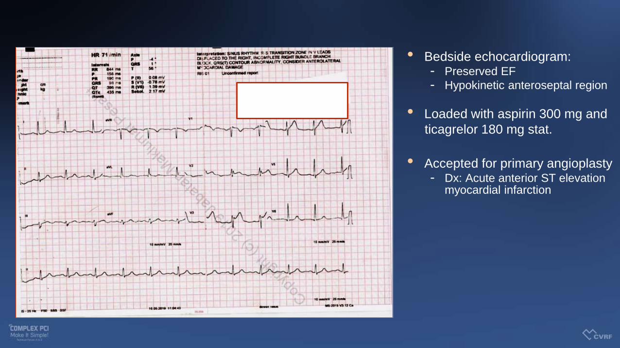
• Bedside echocardiogram:- Preserved EF
- Hypokinetic anteroseptal region
• Loaded with aspirin 300 mg and
ticagrelor 180 mg stat.
• Accepted for primary angioplasty- Dx: Acute anterior ST elevation
myocardial infarction

Standard set up – Radial 6Fr. Culprit lesion noted in mid-LAD

Severe calcification at the target lesion

Severe calcification at the target lesion

Heavy presence of calcium
• Heavy calcification of culprit lesion.
• ?Ulcerated plaque distally.
• Decision to spend time to prepare lesion with non-compliant/ scoring balloons vs up-front preparation for atherectomy.

System set up
•Radial sheath upgraded to a 7 Fr sheath
•7Fr XB 3.5 guiding catheter used to
engage
•Terumo Runthrough Floppy guidewire
down the LAD and this subsequently
exchanged with a Finecross microcatheter
to a Viperwire Advance guidewire

Multiple low speed OA runs from proximal to mid LAD

Slow flow!!!!!

Vessel preparation for stenting

Distal LAD DES placed and deployed
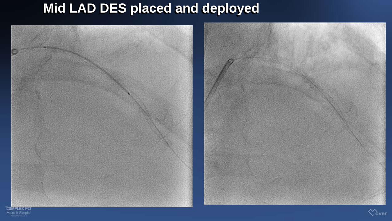
Mid LAD DES placed and deployed

Final shots after non-compliance ballooning post PCI

Pre and Post shots ☺

Discussion Points
• Presence of calcified lesions in acute coronary syndromes is a challenge. (associated with higher rates of stent thrombosis
and urgent revascularisation at 1 year)
• How do we tackle calcified lesions in a high risk situation? e.g.STEMI
• Should I have used sequential NC/ scoring balloons first or
upfront atherectomy acceptable?
• Rotational vs orbital?? Safety and speed?

• Coronary artery calcification is common and is more likely with increased age, diabetes and those
with renal impairment.
• Calcified target lesions are associated with poorer outcomes due to
challenges to optimal stent delivery and expansion.
• Calcified culprit lesions in acute coronary syndromes are associated with increased rates of stent t
hrombosis and target lesion revascularization at 1 year.
• Calcified lesions require adequate preparation prior to stenting and may
require utilization of cutting balloons and atherectomy devices.
• Orbital atherectomy in acute STEMI is unconventional but may be used
to facilitate timely delivery of PCI
Conclusions