Transparencies and slides identification points
24
TRANSPARENCIES AND SLIDES IDENTIFICATION POINTS HISTOPATHOLOGY FOR MIDTERM MBBS 4 th Year 2012 By SIDRA NAWAZ SIRSYED COLLEGE OF MEDICAL SCIENCES FOR GIRLS
-
Upload
sidra-nawaz -
Category
Education
-
view
1.056 -
download
1
Transcript of Transparencies and slides identification points
TRANSPARENCIES AND SLIDES IDENTIFICATION POINTS
HISTOPATHOLOGY FOR
MIDTERM MBBS 4th Year
2012 By
SIDRA NAWAZ SIRSYED COLLEGE OF MEDICAL
SCIENCES FOR GIRLS
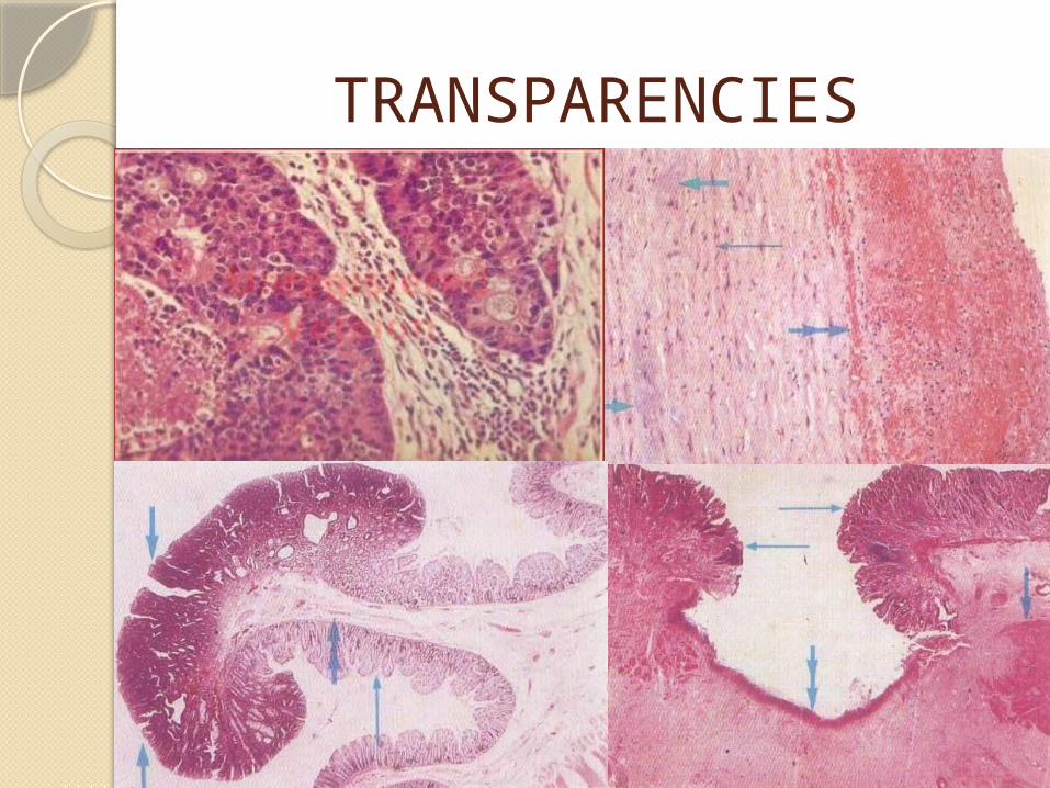
TRANSPARENCIES
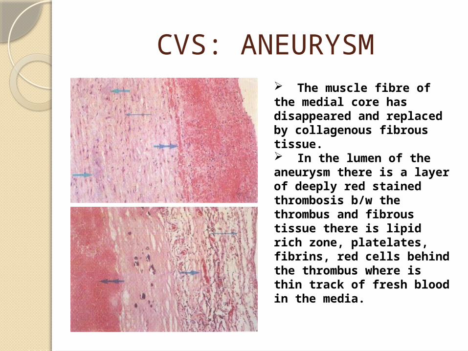
CVS: ANEURYSM The muscle fibre of the medial core has disappeared and replaced by collagenous fibrous tissue. In the lumen of the aneurysm there is a layer of deeply red stained thrombosis b/w the thrombus and fibrous tissue there is lipid rich zone, platelates, fibrins, red cells behind the thrombus where is thin track of fresh blood in the media.
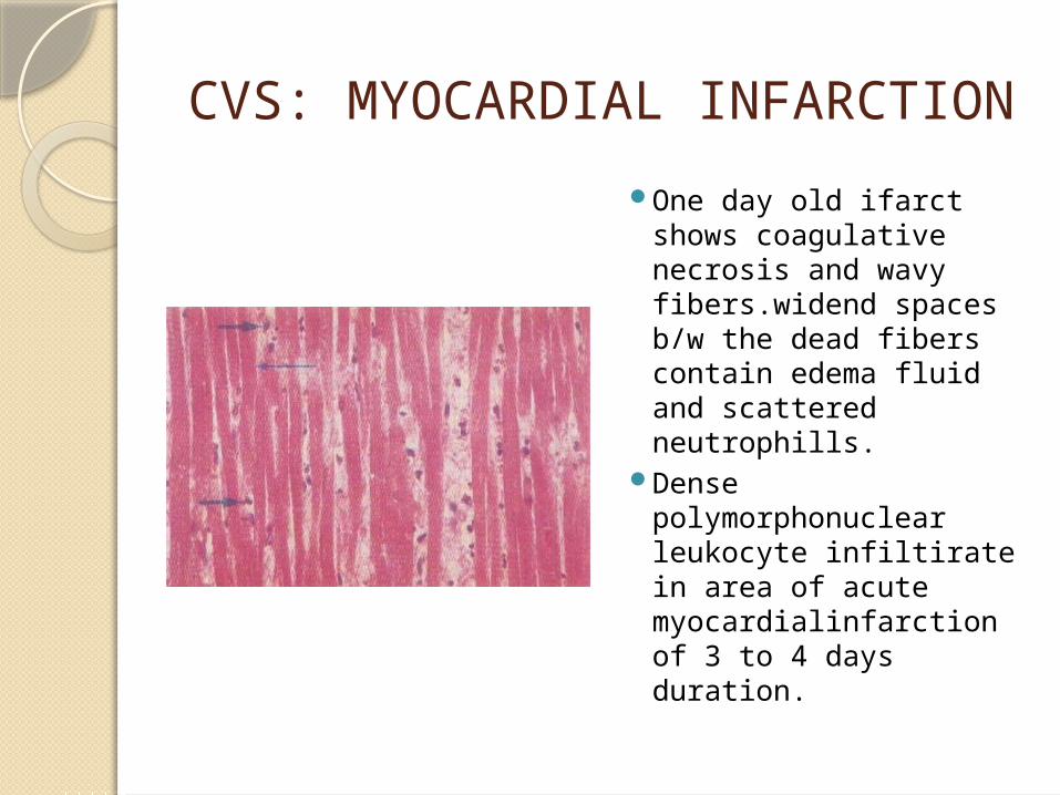
CVS: MYOCARDIAL INFARCTION
One day old ifarct shows coagulative necrosis and wavy fibers.widend spaces b/w the dead fibers contain edema fluid and scattered neutrophills.
Dense polymorphonuclear leukocyte infiltirate in area of acute myocardialinfarction of 3 to 4 days duration.
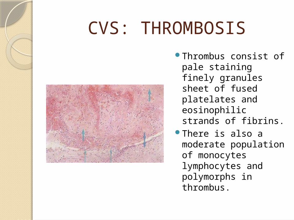
CVS: THROMBOSISThrombus consist of
pale staining finely granules sheet of fused platelates and eosinophilic strands of fibrins.
There is also a moderate population of monocytes lymphocytes and polymorphs in thrombus.
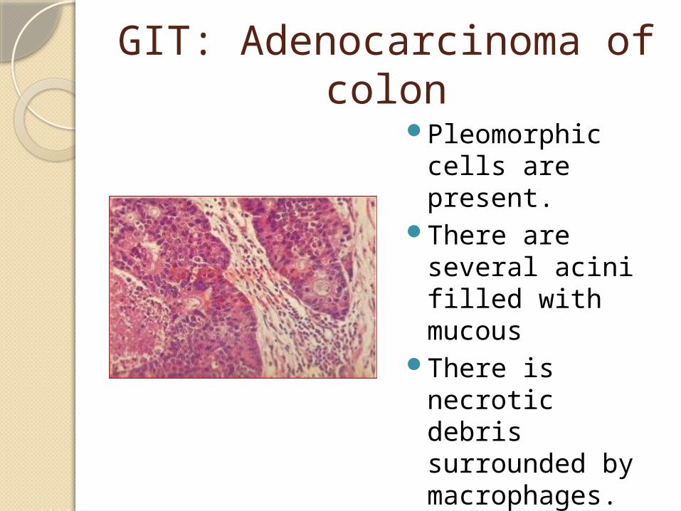
GIT: Adenocarcinoma of colon
Pleomorphic cells are present.
There are several acini filled with mucous
There is necrotic debris surrounded by macrophages.
C.T is filled with plasma cells.
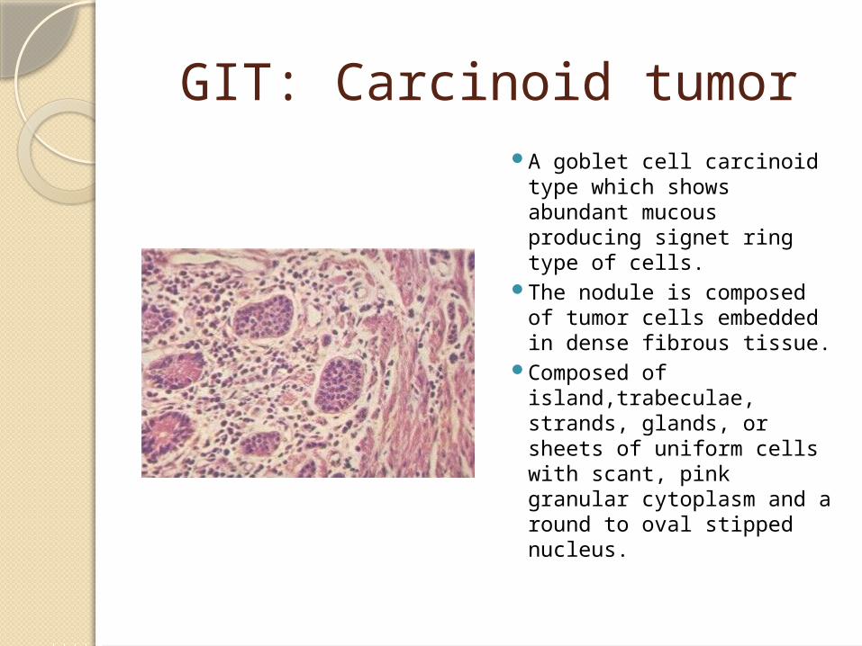
GIT: Carcinoid tumor A goblet cell carcinoid
type which shows abundant mucous producing signet ring type of cells.
The nodule is composed of tumor cells embedded in dense fibrous tissue.
Composed of island,trabeculae, strands, glands, or sheets of uniform cells with scant, pink granular cytoplasm and a round to oval stipped nucleus.
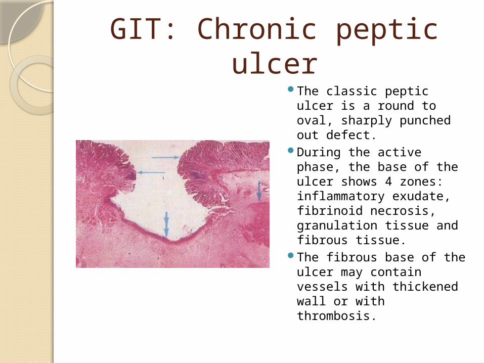
GIT: Chronic peptic ulcer The classic peptic ulcer
is a round to oval, sharply punched out defect.
During the active phase, the base of the ulcer shows 4 zones: inflammatory exudate, fibrinoid necrosis, granulation tissue and fibrous tissue.
The fibrous base of the ulcer may contain vessels with thickened wall or with thrombosis.
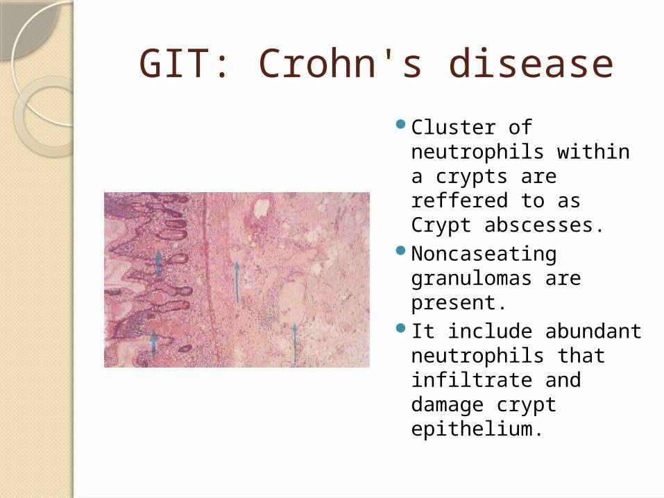
GIT: Crohn's diseaseCluster of
neutrophils within a crypts are reffered to as Crypt abscesses.
Noncaseating granulomas are present.
It include abundant neutrophils that infiltrate and damage crypt epithelium.
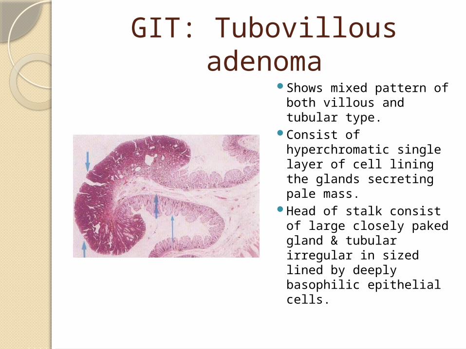
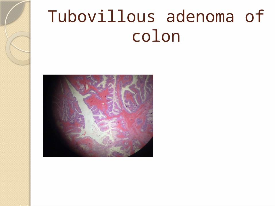
GIT: Tubovillous adenomaShows mixed pattern
of both villous and tubular type.
Consist of hyperchromatic single layer of cell lining the glands secreting pale mass.
Head of stalk consist of large closely paked gland & tubular irregular in sized lined by deeply basophilic epithelial cells.
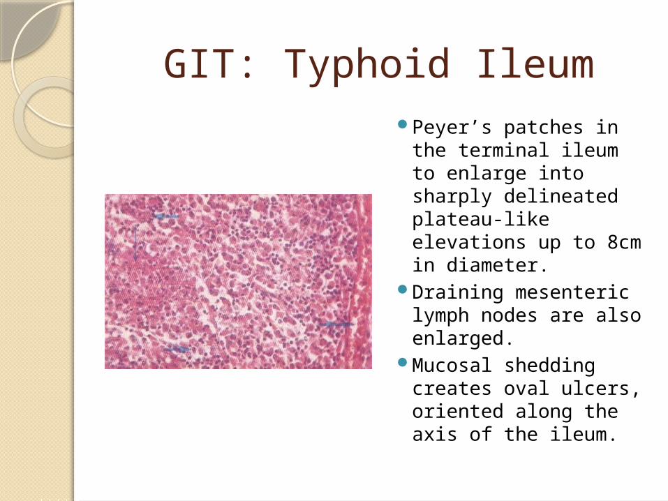
GIT: Typhoid IleumPeyer’s patches in
the terminal ileum to enlarge into sharply delineated plateau-like elevations up to 8cm in diameter.
Draining mesenteric lymph nodes are also enlarged.
Mucosal shedding creates oval ulcers, oriented along the axis of the ileum.
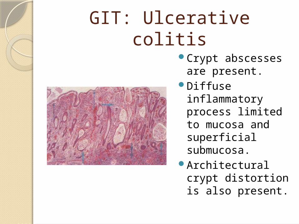
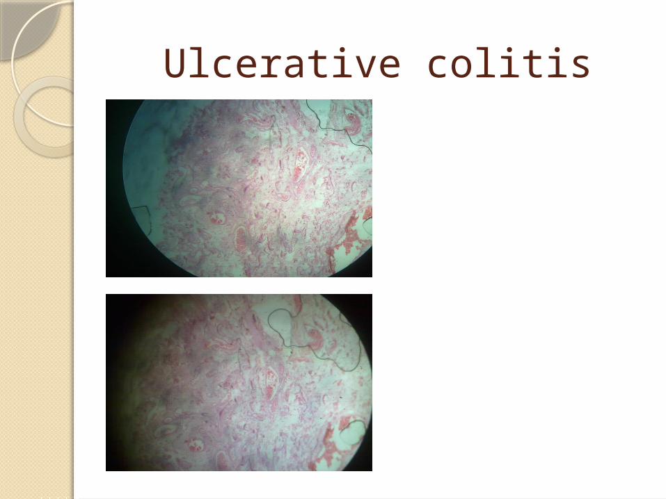
GIT: Ulcerative colitisCrypt abscesses
are present.Diffuse
inflammatory process limited to mucosa and superficial submucosa.
Architectural crypt distortion is also present.
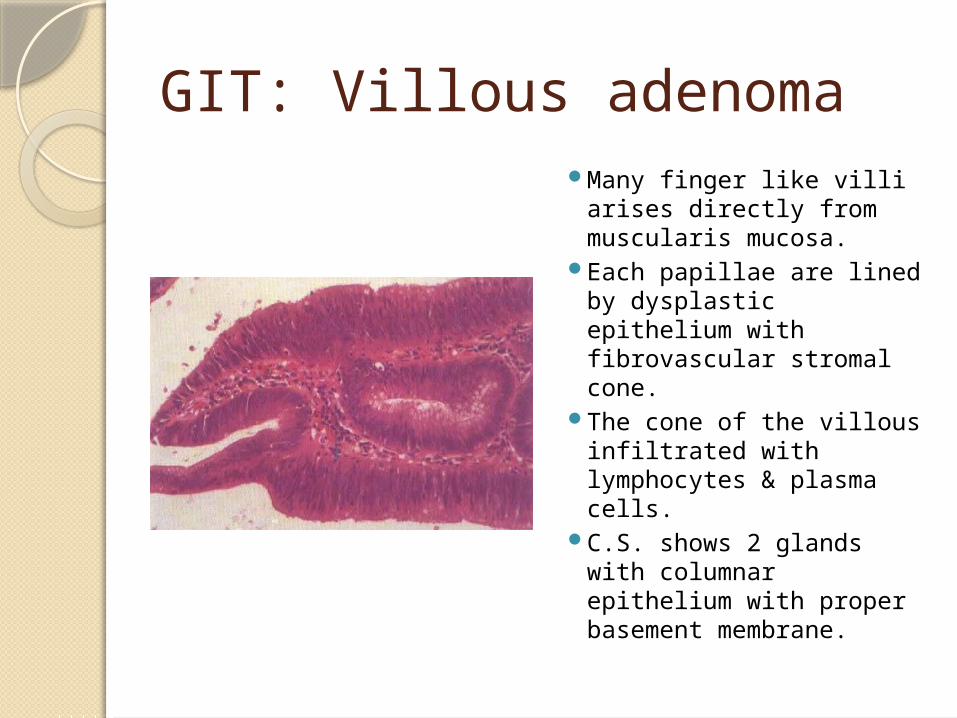
GIT: Villous adenoma Many finger like villi
arises directly from muscularis mucosa.
Each papillae are lined by dysplastic epithelium with fibrovascular stromal cone.
The cone of the villous infiltrated with lymphocytes & plasma cells.
C.S. shows 2 glands with columnar epithelium with proper basement membrane.

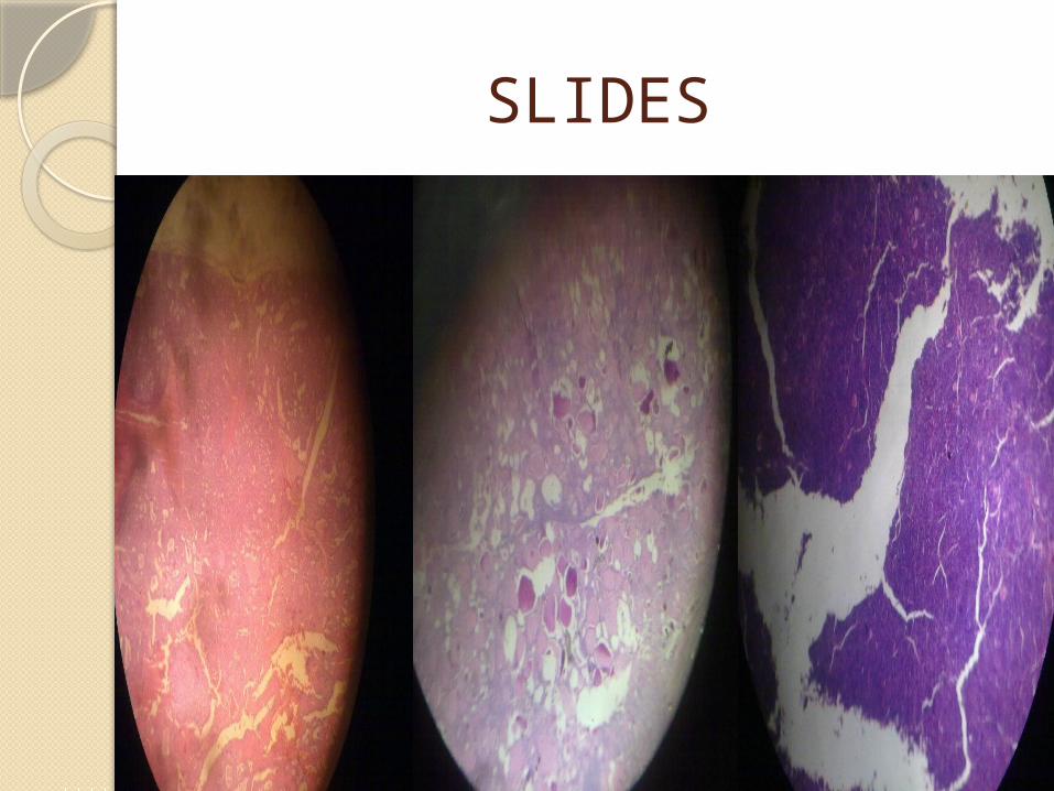
SLIDES
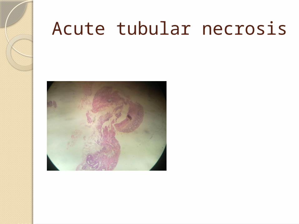
Acute tubular necrosis
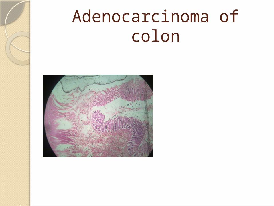
Adenocarcinoma of colon
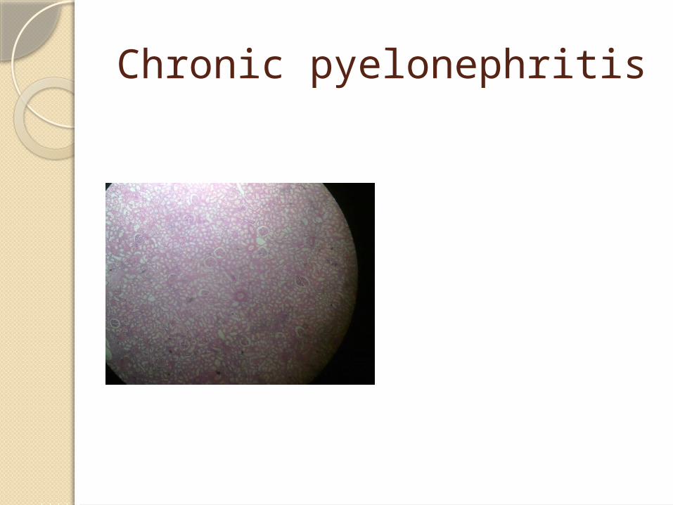
Chronic pyelonephritis
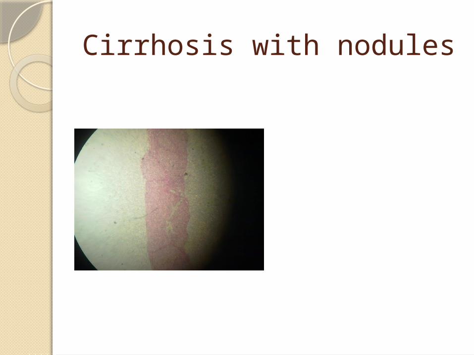
Cirrhosis with nodules
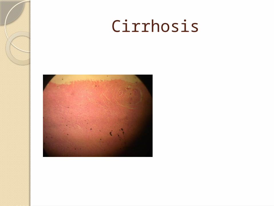
Cirrhosis
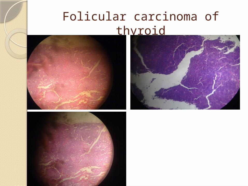
Folicular carcinoma of thyroid
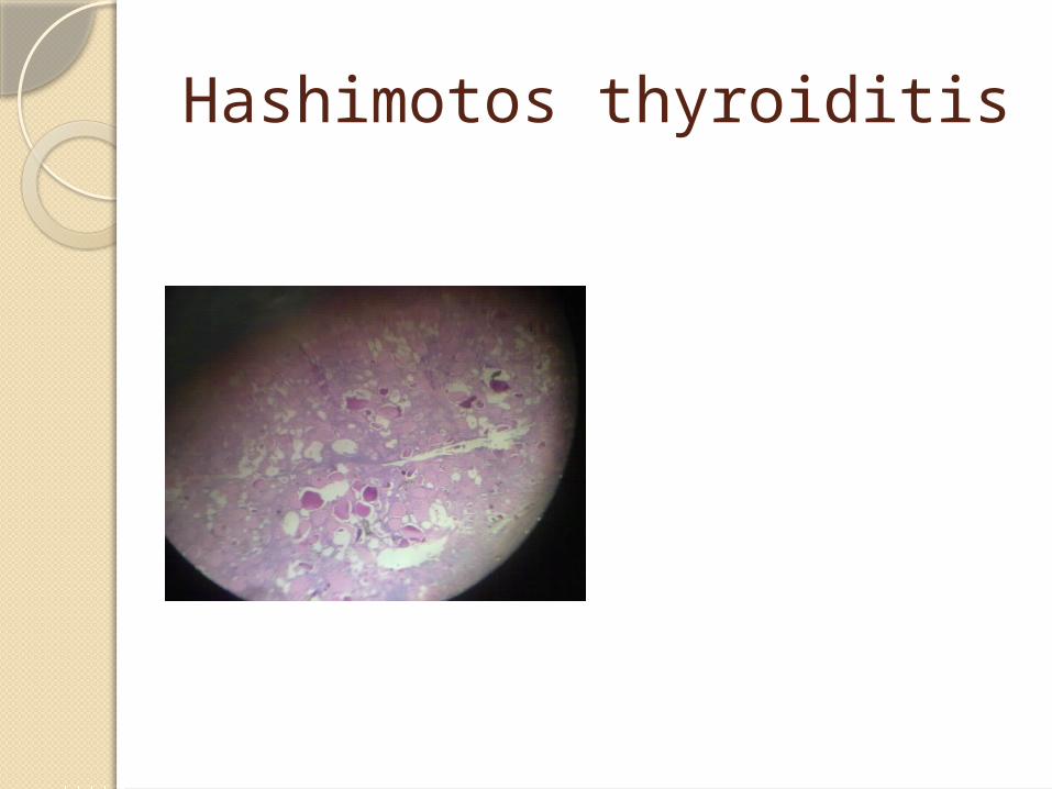
Hashimotos thyroiditis
Tubovillous adenoma of colon
Ulcerative colitis