Transmembrane Protein Structure Predictionkhuri/AUA_2016/Transmembrane/AUA_2016_EIGHT_Trans...©2016...
11
8.1 ©2016 Sami Khuri June 2016 American University of Armenia Introduction to Bioinformatics Transmembrane Protein Structure Prediction Eight Introduction to Bioinformatics Sami Khuri Computer Science San José State University June 2016 Transmembrane Protein Structure Prediction Lipid Bilayer Membrane Protein Bitopic Polytopic TMHMM HMMTOP PROFtmb ©2016 Sami Khuri Figure 10-19 Molecular Biology of the Cell (© Garland Science 2008) Membrane Proteins and Lipid Bilayer Most transmembrane proteins extend across the lipid bilayer as 1: a single alpha helix, 2: multiple alpha helices, 3: rolled-up beta sheets (beta barrel). ©2016 Sami Khuri Types of Membrane Proteins • Membrane proteins can be categorized by their degree of interaction with the membrane. ©2016 Sami Khuri Membrane Proteins • Some are only anchored to one side of the membrane. See A and B. – These follow the general structural rules of proteins. ©2016 Sami Khuri Transmembrane Proteins (I) • Transmembrane or integral proteins have a part that is entirely embedded within the lipid bilayer. polytopic bitopic ©2016 Sami Khuri
Transcript of Transmembrane Protein Structure Predictionkhuri/AUA_2016/Transmembrane/AUA_2016_EIGHT_Trans...©2016...

8.1 ©2016 Sami Khuri
June 2016 American University of Armenia Introduction to Bioinformatics
Transmembrane Protein Structure Prediction
Eight
Introduction to Bioinformatics
Sami Khuri Computer Science
San José State University June 2016
Transmembrane Protein Structure Prediction
v Lipid Bilayer v Membrane Protein v Bitopic v Polytopic v TMHMM v HMMTOP v PROFtmb
©2016 Sami Khuri
Figure 10-19 Molecular Biology of the Cell (© Garland Science 2008)
Membrane Proteins and Lipid Bilayer
Most transmembrane proteins extend across the lipid bilayer as 1: a single alpha helix, 2: multiple alpha helices, 3: rolled-up beta sheets (beta barrel).
©2016 Sami Khuri
Types of Membrane Proteins
• Membrane proteins can be categorized by their degree of interaction with the membrane.
©2016 Sami Khuri
Membrane Proteins
• Some are only anchored to one side of the membrane. See A and B. – These follow the general structural rules of proteins.
©2016 Sami Khuri
Transmembrane Proteins (I)
• Transmembrane or integral proteins have a part that is entirely embedded within the lipid bilayer.
polytopic bitopic
©2016 Sami Khuri

8.2 ©2016 Sami Khuri
June 2016 American University of Armenia Introduction to Bioinformatics
Transmembrane Proteins (II)
• Knowing a membrane protein’s topology can be a significant step toward inferring both its structure and function.
polytopic bitopic
©2016 Sami Khuri
Transmembrane Proteins (III) polytopic bitopic
...
Single-Pass Transmembrane Protein Mainly hydrophobic 15 to 30 residues long Most are alpha helices
Multi-Helix Transmembrane Protein 15 to 30 residues long Most are alpha helices
©2016 Sami Khuri
Single-Pass Transmembrane Proteins (I)
Hydrophobicity scales are used to assign values to individual residues. The values are converted into hybrophobic profiles by using a sliding window to average the values over a number of residues.
©2016 Sami Khuri
Single-Pass Transmembrane Proteins (II) • There are many different hydrophobicity
scales. – They produce different results. – Therefore, one has to use several transmembrane
predictors. • This method works pretty well for single-pass
transmembranes.
©2016 Sami Khuri
Multi-Helix Transmembrane Proteins (I)
Helices contain both hydrophobic and charged residues, forming a structural element that has a different character on each side – an amphipathic helix.
©2016 Sami Khuri
Multi-Helix Transmembrane Proteins (II)
Use of hydrophobic profiles only will not suffice to guarantee good predictions.
The hydrophobic moment is used.
It measures the hybrophobicity of a peptide at different angles of rotation.
©2016 Sami Khuri

8.3 ©2016 Sami Khuri
June 2016 American University of Armenia Introduction to Bioinformatics
Functions of Membrane Proteins
Receptor Tyrosine Kinases are functionally very important as they are the launch sites of many complex signal transduction pathways in the cell.
A seven-transmembrane spanning molecule.
©2016 Sami Khuri
The ErbB Signaling Network Normal cells receive growth-stimulatory signals from their surroundings. These signals are processed and integrated by complex circuits within the cell, which decide whether cell growth and division is appropriate or not.
Biology of Cancer by R. Weingberg ©2016 Sami Khuri
Bovine Rhodopsin Ribbon Diagram of bovine rhodopsin which is a member of the G-protein-coupled receptor (GPCR) family. GPCRs have 7 membrane helices.
Intracellular
Extracellular
©2016 Sami Khuri
GPCR (I)
G-Protein-Coupled Receptors (GPCRs) share a conserved structure composed of seven transmembrane (TM) helices
en.wikipedia.org/wiki/G_protein-coupled_receptor ©2016 Sami Khuri
GPCR (II) • G-protein-coupled receptors (GPCRs) constitute a
large and diverse family of proteins whose primary function is to transduce extracellular stimuli into intracellular signals.
• GPCRs are among the largest and most diverse protein families in mammalian genomes.
• On the basis of homology with rhodopsin, GPCRs are predicted to contain seven membrane-spanning helices, an extracellular N-terminus and an intracellular C-terminus.
• This gives rise to GPCRs other names, the 7-TM receptors or the heptahelical receptors.
www.ibibiobase.com/projects/db-drd4/G_protein.htm ©2016 Sami Khuri
GPCR (III) • GPCRs transduce extracellular stimuli to give
intracellular signals through interaction of their intracellular domains with heterotrimeric G proteins.
• This class of membrane proteins can respond to a wide range of agonists, including photon, amines, hormones, neurotransmitters and proteins.
• Some agonists bind to the extracellular loops of the receptor, others may penetrate into the transmembrane region.
www.ibibiobase.com/projects/db-drd4/G_protein.htm ©2016 Sami Khuri

8.4 ©2016 Sami Khuri
June 2016 American University of Armenia Introduction to Bioinformatics
Cystic Fibrosis
• Cystic Fibrosis is an autosomal recessive disorder that affects the respiratory and digestive systems.
• Cystic Fibrosis is associated with mutations in the CFTR (Cystic Fibrosis Transmembrane Regulator) gene.
• Cystic Fibrosis is fatal and treatment is limited to slowing the progress of the disease.
©2016 Sami Khuri
A vest designed to improve lung function for cystic fibrosis patients
©2016 Sami Khuri
CFTR Protein
• The CFTR protein is found in the membrane of epithelial cells.
• It forms a channel through which chloride ions (Cl-) can pass.
• The channel can be opened or closed. • The flow of ions is necessary for water to be
released into secretions such as mucus in the lungs.
©2016 Sami Khuri
Function of CFTR Gene (I)
• The CFTR gene provides instructions for making a protein called the cystic fibrosis transmembrane conductance regulator. This protein functions as a channel across the membrane of cells that produce mucus, sweat, saliva, tears, and digestive enzymes.
• The channel transports negatively charged particles called chloride ions into and out of cells.
http://ghr.nlm.nih.gov/gene=cftr ©2016 Sami Khuri
Function of CFTR Gene (II)
• The transport of chloride ions helps control the movement of water in tissues, which is necessary for the production of thin, freely flowing mucus.
• Mucus is a slippery substance that lubricates and protects the lining of the airways, digestive system, reproductive system, and other organs and tissues.
http://ghr.nlm.nih.gov/gene=cftr ©2016 Sami Khuri
Schematic diagram showing the structure of the CFTR protein that regulates transport of chloride through cell membranes
©2016 Sami Khuri

8.5 ©2016 Sami Khuri
June 2016 American University of Armenia Introduction to Bioinformatics
CFTR
journals.cambridge.org
Membrane-Spanning Domain
Nucleotide-Binding Domain (Fold)
©2016 Sami Khuri
∆F508 Mutation in CFTR Missing or altered chloride transporter Insufficient secretion of water by epithelial cells Thick lung secretions lead to lung infections Defective pancreatic secretion of digestive enzymes Sterility in males
©2016 Sami Khuri
CFTR Protein’s Fate
With normal CFTR, once the protein is synthesized, it is transported to the endoplasmic reticulum (ER) and Golgi apparatus for additional processing before being integrated into the cell membrane. When a CFTR protein with the delta F508 mutation reaches the ER, the quality-control mechanism of this cellular component recognizes that the protein is folded incorrectly and marks the defective protein for degradation. As a result, delta F508 never reaches the cell membrane.
www.ornl.gov/sci/techresources/Human_Genome/posters/chromosome/cftr.shtml ©2016 Sami Khuri
Location of the ∆F508 Mutation
©2016 Sami Khuri
CFTR Gene and Protein (I)
©2016 Sami Khuri
Selected mutations are shown. The exons, introns, and domains of the protein are not drawn to scale. MSD: Membrane-Spanning Domain NBD: Nucleotide-Binding Domain R-domain: Regulatory Domain.
CFTR Gene and Protein (II)
©2016 Sami Khuri

8.6 ©2016 Sami Khuri
June 2016 American University of Armenia Introduction to Bioinformatics
Cystic Fibrosis (I) • More than 1,000 mutations in the CFTR gene have
been identified in people with cystic fibrosis. • Most of these mutations change single protein
building blocks (amino acids) in the CFTR protein or delete a small amount of DNA from the CFTR gene.
• The most common mutation, called delta F508, is a deletion of one amino acid at position 508 in the CFTR protein.
http://ghr.nlm.nih.gov/gene=cftr ©2016 Sami Khuri
Cystic Fibrosis (II) • The resulting abnormal channel breaks down
shortly after it is made, so it never reaches the cell membrane to transport chloride ions.
• Disease-causing mutations in the CFTR gene alter the production, structure, or stability of the chloride channel.
• All of these changes prevent the channel from functioning properly, which impairs the transport of chloride ions and the movement of water into and out of cells.
http://ghr.nlm.nih.gov/gene=cftr ©2016 Sami Khuri
Cystic Fibrosis (III)
• As a result, cells that line the passageways of the lungs, pancreas, and other organs produce mucus that is abnormally thick and sticky.
• The abnormal mucus obstructs the airways and glands, leading to the characteristic signs and symptoms of cystic fibrosis.
http://ghr.nlm.nih.gov/gene=cftr ©2016 Sami Khuri
CF Mutation Database
©2016 Sami Khuri
Exon 11 of the CFTR Gene
©2016 Sami Khuri
Cystic Fibrosis Carrier Frequency
http://progenity.com/cystic-fibrosis-cf ©2016 Sami Khuri
8.7 ©2016 Sami Khuri
June 2016 American University of Armenia Introduction to Bioinformatics
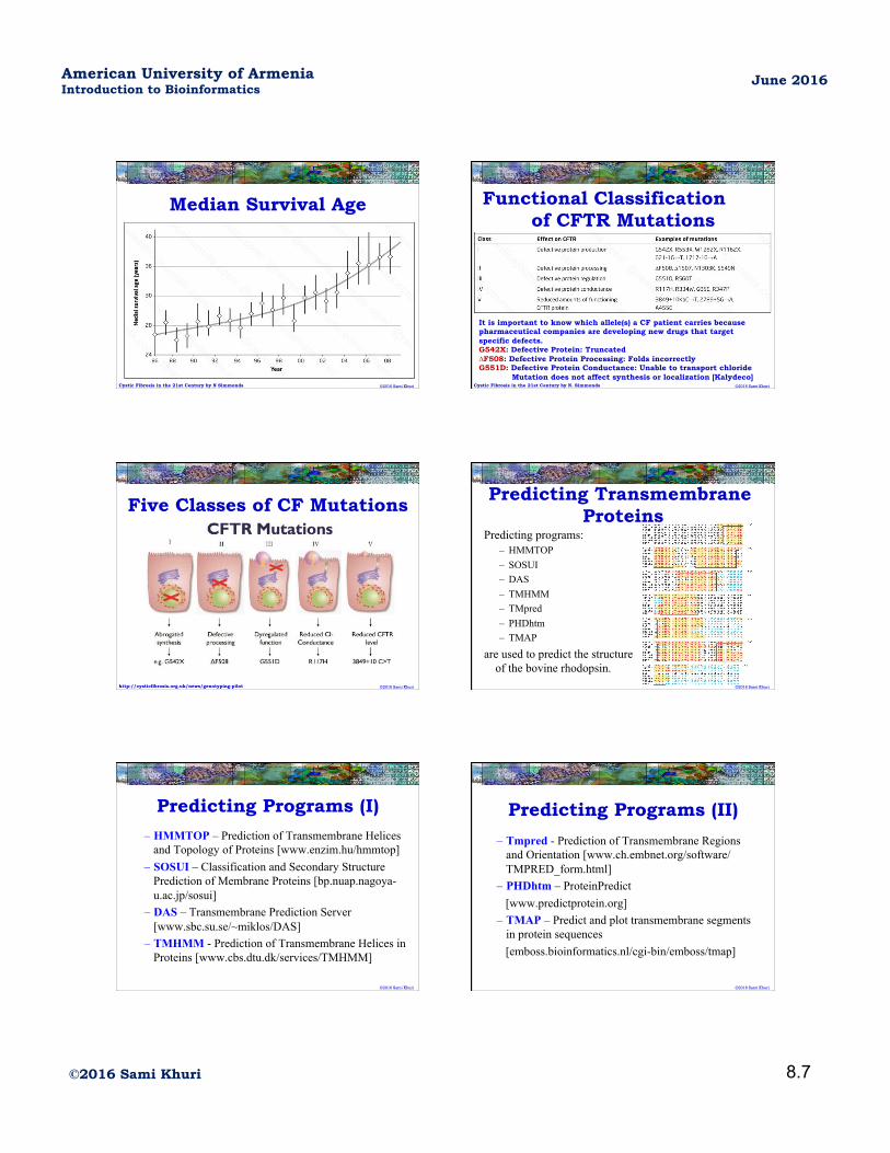
Median Survival Age
Cystic Fibrosis in the 21st Century by N Simmonds ©2016 Sami Khuri
Functional Classification of CFTR Mutations
Cystic Fibrosis in the 21st Century by N. Simmonds
It is important to know which allele(s) a CF patient carries because pharmaceutical companies are developing new drugs that target specific defects. G542X: Defective Protein: Truncated ∆F508: Defective Protein Processing: Folds incorrectly G551D: Defective Protein Conductance: Unable to transport chloride
Mutation does not affect synthesis or localization [Kalydeco] ©2016 Sami Khuri
Five Classes of CF Mutations
http://cysticfibrosis.org.uk/news/genotyping-pilot ©2016 Sami Khuri
Predicting Transmembrane Proteins
Predicting programs: – HMMTOP – SOSUI – DAS – TMHMM – TMpred – PHDhtm – TMAP
are used to predict the structure of the bovine rhodopsin.
©2016 Sami Khuri
Predicting Programs (I)
– HMMTOP – Prediction of Transmembrane Helices and Topology of Proteins [www.enzim.hu/hmmtop]
– SOSUI – Classification and Secondary Structure Prediction of Membrane Proteins [bp.nuap.nagoya-u.ac.jp/sosui]
– DAS – Transmembrane Prediction Server [www.sbc.su.se/~miklos/DAS]
– TMHMM - Prediction of Transmembrane Helices in Proteins [www.cbs.dtu.dk/services/TMHMM]
©2016 Sami Khuri
Predicting Programs (II)
– Tmpred - Prediction of Transmembrane Regions and Orientation [www.ch.embnet.org/software/TMPRED_form.html]
– PHDhtm – ProteinPredict [www.predictprotein.org] – TMAP – Predict and plot transmembrane segments
in protein sequences [emboss.bioinformatics.nl/cgi-bin/emboss/tmap]
©2016 Sami Khuri

8.8 ©2016 Sami Khuri
June 2016 American University of Armenia Introduction to Bioinformatics
Using X-Ray Crystallography • The top row contains the results
obtained from X-Ray crystallography.
• The transmembrane helices are highlighted in yellow.
• Extracellular loops are in black. • Cytoplasmic loops are in blue. • Boxed sequences are predicted
to be transmembrane based on the consensus results of all prediction packages.
©2016 Sami Khuri
Part of the Alignment
• The third (out of 7) membrane (yellow residues) is shown in the box as predicted by the packages.
• Amino acids that are part of the extracellular loop are in black.
• Residues that are part of the cytoplasmic loop are in blue.
©2016 Sami Khuri
Transmembrane Predicting Methods
• There are many transmembrane predicting packages that are based on the following techniques: – Statistical Methods
• Example: TMpred
– Knowledge-based Methods • Example: SOSUI
– Evolutionary-based Methods • Example: TMAP
– Neural Networks • Example: PHDhtm
©2016 Sami Khuri
HMM for Transmembrane Protein Prediction (I)
• HMMs can incorporate: – Hydrophobicity – Charge bias – Helix length – Grammatical constraints
©2016 Sami Khuri
HMMs for Transmembrane Protein Prediction (II)
Simple Model: – Define a set of states: each residue is then
predicted to be in one of the states. – Example:
• A state for inside loops • A state for outside loops • A state for transmembrane segments
©2016 Sami Khuri
The Simple HMM
• Each state has an associated probability distribution over the 20 amino acids that describe the variability of each amino acid in the modeled region.
• States are connected to each other in a biological reasonable manner.
• The HMM is trained to have adequate emission and transition probabilities.
©2016 Sami Khuri

8.9 ©2016 Sami Khuri
June 2016 American University of Armenia Introduction to Bioinformatics
HMMTOP Architecture
Transmembrane Helices: 17-25 aa Inside and Outside: 1-15 aa
Intracellular
Extracellular o
o
O
h
h
i
i
I ©2016 Sami Khuri
Five States of HMMTOP
©2016 Sami Khuri
TMHMM: Finite State Diagram • TMHHM has seven states:
©2016 Sami Khuri
The TMHMM Package • TMHHM has seven states:
– Core transmembrane helix – Helical cap – Helical tail – Loops on cytoplasmic end – Short loops outside the cell – Long loops inside the cell – Golbular-domain-like structures in the middle of
each loop.
©2016 Sami Khuri
TMHMM: Output (I)
©2016 Sami Khuri
TMHMM: Output (II)
©2016 Sami Khuri

8.10 ©2016 Sami Khuri
June 2016 American University of Armenia Introduction to Bioinformatics
VKOR (I) • Warfarin was approved for use as a medication in
the early 1950s and has remained very popular. • Warfarin is the most widely prescribed
anticoagulant drug in North America. • Warfarin decreases blood coagulation by
inhibiting vitamin K epoxide reductase (VKOR).
• The gene encoding the catalytic subunit of VKOR was identified as an integral membrane protein.
©2016 Sami Khuri
VKOR (II)
• These vitamin K-dependent proteins are important as coagulation factors, and are involved in bone metabolism and signal transduction.
• In order to understand structure-function relationship of these proteins, it is important to understand the membrane topology.
• Seven transmembrane prediction packages were used for that purpose.
©2016 Sami Khuri
©2016 Sami Khuri
VKOR (III)
Experiments were performed and it was determined that VKOR has three transmembrane helices.
©2016 Sami Khuri
VKOR (IV)
Two packages predicted the wrong number of helices. Two packages predicted the wrong location of the C
terminus ©2016 Sami Khuri
Other Types of Transmembranes
• Some transmembrane structures contain beta sheets instead of alpha helices.
• Tailored-made predictors were designed to detect them.
• Sometimes 2 or 3 alpha helices intertwine to form coiled-coil structures.
• Coiled-coil structures can be found in transmembrane as well as intracellular proteins.
©2016 Sami Khuri

8.11 ©2016 Sami Khuri
June 2016 American University of Armenia Introduction to Bioinformatics
©2016 Sami Khuri
β-Barrels • Located in mitochondria, chloroplasts, bacteria • Functions include:
– Transport channel – Receptor – Enzyme
©2012 Sami Khuri
PROFtmb for Beta-Barrel Prediction
Predicting transmembrane beta-barrels in proteomes, by Bigelow et al., NAR, 2004
©2016 Sami Khuri