Transjugular Renal Biopsy - Nephrologymedia.nephrology.edu.au/anzsin/2013/Kendal Redmond -...
37
Transjugular Renal Biopsy Kendal Redmond Princess Alexandra Hospital ANZSIN Sept. 2013
Transcript of Transjugular Renal Biopsy - Nephrologymedia.nephrology.edu.au/anzsin/2013/Kendal Redmond -...

Transjugular Renal Biopsy
Kendal RedmondPrincess Alexandra Hospital
ANZSIN Sept. 2013

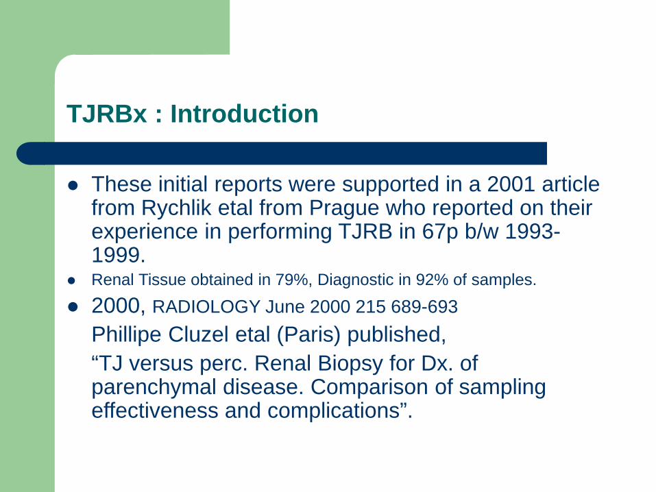
TJRBx : Introduction
Percutaneous renal Bx first described by IVERSEN and BRUN, 1951
Subsequent significant improvements in equipment and image guidance.
(Associated with a recognized clinically apparent complication rate of up to 3.5% and perirenal haematoma b/w 57-85%)
There are recognized group of patients who will benefit from a histological diagnosis but in whom Perc. Renal Bx poses an unacceptably high risk.(estimated at up to 7% in clinical practice)

TJRBx : Introduction
1989; Dr Frederic Mal performed a TJ Liver Bx at Jean Verdier Hospital (France) and received a pathology report from DrPatrice Callard describing a sample of renal tissue.
(Biopsied patient having an “uneventful course”).
Mal and Callard collaborated on a Cadaver study to assess the feasibility of TJRBx.
Performed procedure on tot. 5 cadavers initially with the standard 15g COLAPINTO needle (COOK) which had been safely used for TJLBx since 1964.
They redesigned the needle to limit risk of capsular perforations evident when using the standard COLAPINTO needle. (Limiting needle protrusion from catheter to 13mm).

TJRBx : Introduction
1990;They presented their initial data in a poster at the annual meeting of the American Society of Nephrology.
Initially published in, Kidney International 1990;37:279 as an abstract, Mal F, Meyrier A, Callard P etal. “Transvenous renal biopsy: A preliminary report on 36 patients.”
This same group followed with, Transjugular Renal Biopsy in Lancet 1990 v335, issue 8704, p1512-1513. Discussing TJRBx results from there first 50 patients.Obtaining specimens of renal tissue in 44. (Glomeruli in 38/44) and no major Cx. (Patients all followed with USS within 18hrs)(Minor Cx, incl. Mild Transient R) lumbar pain, Transient macroscopic haematuria, asymptomatic perirenal haematoma).

TJRBx : Introduction

TJRBx : Introduction
1992, Same group followed with “ The Diagnostic Yield of TJRBx. Experience in 200 cases. published in Kidney International 1992 41,445-449.
This summarised data from 195p all with uss f/u within 24hrs. Bx performed with modified Colapinto needle and intended max.3 passes, (4 in
15p). Renal tissue obtained 88% (176/200) Diagnostic tissue in 83% (Av. Glom. 10). (range 1-35) Uneventful outcome 90% (179/200) Clinically silent perirenal haematoma 3% (6/200) Clinically significant haematomas 2% (4/200). 3 (TFused) 1.5% Macroscopic Haematuria 15/200. 7.5%, Transient 14, 1 (TFused) TOTAL Major Complication 2.5% (2% needing transfusion).
(2005 Nephrology Dialysis Transplant article Alan Meyrier, describes Cx from last 200 TJRB at Jean Vernier Hospital performed as “virtually nil”).
TJRBx : Introduction
These initial reports were supported in a 2001 article from Rychlik etal from Prague who reported on their experience in performing TJRB in 67p b/w 1993-1999.
Renal Tissue obtained in 79%, Diagnostic in 92% of samples. 2000, RADIOLOGY June 2000 215 689-693
Phillipe Cluzel etal (Paris) published,“TJ versus perc. Renal Biopsy for Dx. of parenchymal disease. Comparison of sampling effectiveness and complications”.

TJRBx : Introduction
Discussing results and complications of 400 consecutive TJRB performed b/w Nov 1993 and Dec. 1998 (Using modified Colapinto needle, Mal Transjugular renal Bx set. (COOK).
Compared retrospectively with 400 perc. Renal Bx performed during same period.

TJRBx : Introduction
Perc renal Bx. 360 “blind” pre marked, 40 image guidance. Renal tissue obtained 95.5% Mean gl LM 11.2 , IF 6.4 Dx. Adequate specimen 98.2% Major Cx. 3p (0.75%). 2 AVF, haematuria, embolised. 1 inadvertant splenic Bx.
(splenectomy).TJRBx. Biopsy in 397/400. (2 recurrent renal v. 1 thrombosed renal v.) 75.5% 303p with bleeding disorder 13 Left renal Bx. Renal tissue obtained in 95.8% mean gl LM 9.8, IF 4.6(additional Bx, myocardium 14 and liver 35 Dx. Adequate specimen 98.2% Major Cx 4p (1%). 1 IJV haematoma (t/f), 3 perirenal haematomas,(2 emb.1t/f)

TJRBx :Comment on Technique.1. Modified Colapinto Needle, Menghini Aspiration Technique
9fr 62.5cm long catheter 45deg precurved tip advanced from R)IJV to renal vein, wedged into posterolateral interlobar medullary vein. Small contrast injection then advance15g Modified Colapinto needle primed with saline through the catheter and advance forcibly beyond it’s tip then aspirate as the needle is withdrawn. (Catheter and needle Designed to only advance 13mm beyond catheter tip). Menghini 1 second Aspiration Technique.

M
S
COLAPINTO NEEDLE

Alan Meyrier, TJ Renal Biopsy. Update on Hepato-renal needlework. Nephrology Dialysis Transplantation (2005) 20: 1299-1302

TJRBx :Comment on Technique.2. Alternative Biopsy Technique. (Side Cutting quick core Biopsy Needle (2cm throw))
“Blunt tipped” 60cm length 19g (or 18g) core biopsy needle. 2cm throw.
Number of authors have published smaller case series of TJRBperformed using this needle.
Abbot etal. BMC Nephrology 7/2002. 9p, Walter Reed Hospital. Dx tissue 100%, capsular perf. 90%. No Major Cx.
Thompson BC etal. (init. Poster annual meeting of Am. Soc. of Neph. and Abstract CVIR Nov.2002). Then published AJKD April 2004 (v43,issue 4 p651-662)
– TJRB side cutting needle, 25p. Mean 3.5 cores.– Renal tissue 23/25. Dx. 21/23 (91.3%); Mean glom. (LM) 9.9.– Capsular perf. 17/23, (73.9%)– 2 major Cx. (4%). 1 arteriocalyceal fistula, embolised. 1 renal v
thrombosis.

TJRBx :Comment on Technique.2. Alternative Biopsy Technique. (Side Cutting quick core Biopsy Needle (2cm throw))
See T.C, Thompson BC etal (Cambridge), CVIR 2008: TJRB: “Our experience and technical considerations”
Retrospective R/V of 59p using Quickcore Bx needle- Dx. Specimen 56/59 (95%)- Mean Glom. No.(LM)10.3, (IF) 2.6- Isolated capsular perf. 33%- Contained subcaps leak 18%- Major Cx 12.5%. (7p, 5 transfused, 1 Arterio Calyceal Fist. (embol.) 1RV thrombosis.(incr. risk capsular perf. > 6 cores)

TJRBx :Comment on Technique.2. Alternative Biopsy Technique. (Side Cutting quick core Biopsy Needle (2cm throw))
Misra S etal, JVIR Apr. 2004 19(4) 546-51; “Safety and Dx yield using the quick core Bx method in high risk patients.”
Retrospective review at Mayo clinic (Rochester) of 39p with F/U imaging in 25/39.All with pre/post LABS
Tech. success 38/39 (97%) Mean Core No. 1.8. Glom. No (LM) 5, (IF) 2.1 Dx. 92% Major Cx 2.6%, Minor Cx 52%. (mainly perirenal haematomas)
(and small No. CIN). IMAJ V13 July 2011 (Israel) similar findings in report of TJRB in 12p.
(Tech. success 11/12. Dx. 11/11 (100%).Minor Cx. 2/12.

TJRBx :Comment on Technique.Alternative Biopsy Technique. WHY?
Smaller needle allowing a more peripheral sample (but at expense of capsular perf. But usually not clinically signifigant) - (Marchetto etal JVIR 1997. Increased glomeruli/specimen with capsular perforation).
Fewer passes for Dx. Specimen. (Larger tissue specimens with reduced fragmentation.
Technique easier to learn/ Teach. (Reduce risk with technique modification, ie.
Limit the throw to less than 13mm as in original descriptions)

TJRBx :Overall Review
Over all published literature. For TJRBX independent of method.Technical success approx. 92%Dx specimens obtained 94-100%(overall Dx. Success 89-97%)

TJRBx: Indications
(Not an alternative to percutaneous renal Biopsy but to be utilised when percutaneous Biopsy unable to be safely performed)
Bleeding disorder/Coagulopathy is major indication in all literature.
Mechanical ventilation. Unable to lie prone. Respiratory insufficiency Anatomic considerations: Obesity, Solitary Kidney, Horseshoe K, Atrophic K,
Obstructed K. Renal a microaneurysms. Uncontrolled HTN Ascites Combined Bx. Eg Renal and Liver or myocardium. Placement of Dialysis Catheter Hibernating Kidney (pre endovasc. Intervention)

TJRBx. Contraindications
SVC / IVC/ Renal V thrombosis Absent R) IJV. (relative CI) Recurrent course R) Renal V
(? Transfemoral approach) Iodinated Contrast anaphylaxis.

TJRBx: Technique
Informed consent, Lab Ix. (Correct adverse features as able.)
Pre procedure imaging (us. USS or CT). Aseptic technique. Supine on angiographic table, Conscious Sedation. R) IJV access, (R/T USS guidance and M/P technique). (more superior IJV
access unless planning dialysis catheter placement.) 5fr sheath, exchange to 9fr after advancing guidewire to IVC. Select lower pole renal vein (Posterolateral medullary interlobar vein) with MPA
catheter over glidewire then exchange for Amplatz guidewire. We use LABS 200 TJBx kit (COOK)
7fr 50.5cm TJ sheath with 45deg. Preshaped tip.(dilator)60cm 19g quick core biopsy needle (in catheter)

TJRBx: Technique
Advance TJ sheath to medullary vein. Gentle venogram aiming for cortical parenchymal enhancement.
Advance Quick core needle within catheter and Biopsy (limiting excursion of Needle to < 13mm). (Direct postero-laterally to attain region of > cortical depth, reduce risk of large vessel injury or inadvertent bowel injury.
Specimen assessed by cytologist in attendance Usually 2 cores. (? Embolise tract, Gelfoam / coils if see perforation)

TJRBx: Technique

TJRBx: Technique
COOK Micropuncture Set
R) IJV access
Guidewire to IVC


TJRBx: Technique
Catheter selecting lower pole renal v.Small contrast injections to minimiseRisk of CIN
Guidewire exchange 7fr TJ sheath as closeTo wedged as possible

TJRBx: Technique
Cortical ParenchymalEnhancement demonstrates position of capsule
R) lower pole venogram

TJRBx: TechniqueLimit Bx needle“Throw”
CapsulePost Bx Venogram
Core Bx

TJRBx: Technique
Needle tip to capsule
TJ SpecimenX 20
Perc. Spec.x20

PAH Audit TJRBx December 2004 – September 2013
16p, mean age 46y (22y.o -69y.o) M:F (11: 5) Indications/ Major C/I to perc. Bx
-12/16 Bleeding disorder/ Coagulopathy-1/16 Mechanical Ventilation-2/16 Unable to lie prone. 1 massive ascites, 1 severe CCF.
-1/ 16 Combined Liver / Renal Bx (and Dialysis Catheter)

PAH Audit TJRBx December 2004 – September 2013
Diagnostic specimen 14/16. (87.5%) Mean No Glomeruli. LM 9.8 (3-20), IF 3.3 (0-7) Mean Cores; 2.2 Major Cx. 1/16 (6.25%)
Macroscopic Haematuria, (T/F), Arterial embolisation. (Arteriocalyceal fistula).
Minor Cx. 4/16 (25%)2 perinephric haematomas (12.5%)1 Transient Macroscopic Haematuria (6.25%)1 right lumbar pain, –ve CT (pain spont. improved)

TJRBx: COMPLICATIONS
Negative / Non Dx Bx. (overall similar to perc. Bx) MAJOR:
-Bleeding requiring Rx. (1-2.5%)Mostly resusc. And Transfusion. Less frequent embolisation etc.Perirenal Haematoma (self limiting unless arterial aetiol.)
<30% overall for core Bx. Even when capsular perf. Rate reported up to 90%. Perirenal fat tamponades. Partic. In obese. Cf (57-85% perc. Bx)Pelvicalyceal. Haematuria eg arterio-calyceal fistula
Calyceal/ Renal pelvis puncture. Rarely symptomatic but may need ureteric stent.
Adjacent structure injury: Not reported. CERVICAL Haematoma (rare even in coagulopathy Pneumothorax. (1/1000) Contast induced nephropathy

TJRBx: COMPLICATIONS
MINOR:-Pain, Peri-renal haematoma, Transient haematuria, Cervical Haematoma

TJRBx PAH ComplicationsAsymptomatic Peri-renal Haematoma
Pre Bx. Venogram Biopsy Check Venogram post Biopsy. Perirenal Haematoma.
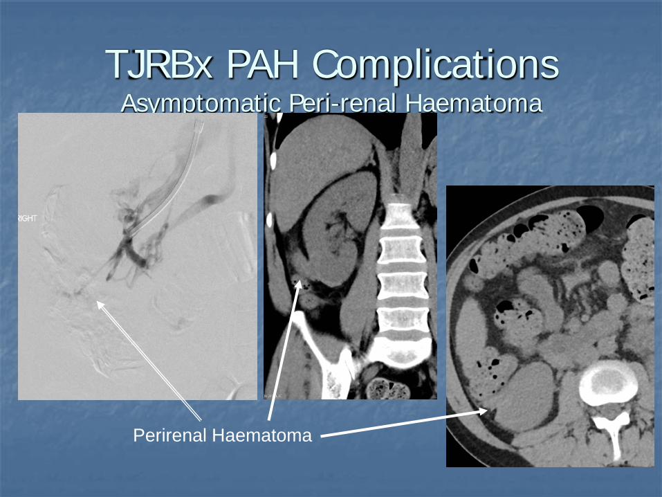
TJRBx PAH ComplicationsAsymptomatic Peri-renal Haematoma
Perirenal Haematoma

TJRBx PAH ComplicationsArterio-calyceal fistula
DSA, embolisation 3/7 post Bx for Macroscopic Haematuria req. Transfusion

TJRBx PAH ComplicationsArterio-calyceal fistula
DSA, embolisation 3/7 post Bx for Macroscopic Haematuria req. Transfusion
2 sites of arterial injury with pseudoaneurysm and AVF.

TJRBx : Conclusion
Not an alternative to percutaneous RBx and not a routine Bx method but useful in armamentarium when Perc. Bx Contraindicated.
Advantages include being able to be relatively safely performed in high risk patients with relatively low complication rate for population being performed in, but
Not risk free, Reduced core size with reduced glomeruli/core.
Time consuming and more expensive than Percutaneous Bx.


![ASN Kidney Week 2016 – Renal Biopsy: Clinical Correlations · ASN Kidney Week 2016 – Renal Biopsy: Clinical Correlations. ... ( ds) DNA, ANCA, SPEP/UPEP ]. ... ASN Kidney Week](https://static.fdocuments.in/doc/165x107/5aec58b47f8b9a90318e2a7d/asn-kidney-week-2016-renal-biopsy-clinical-correlations-kidney-week-2016-.jpg)