Radiofrequency ablation versus repeat hepatectomy in the ...
Upload
balbir-singhCategory
view
213download
1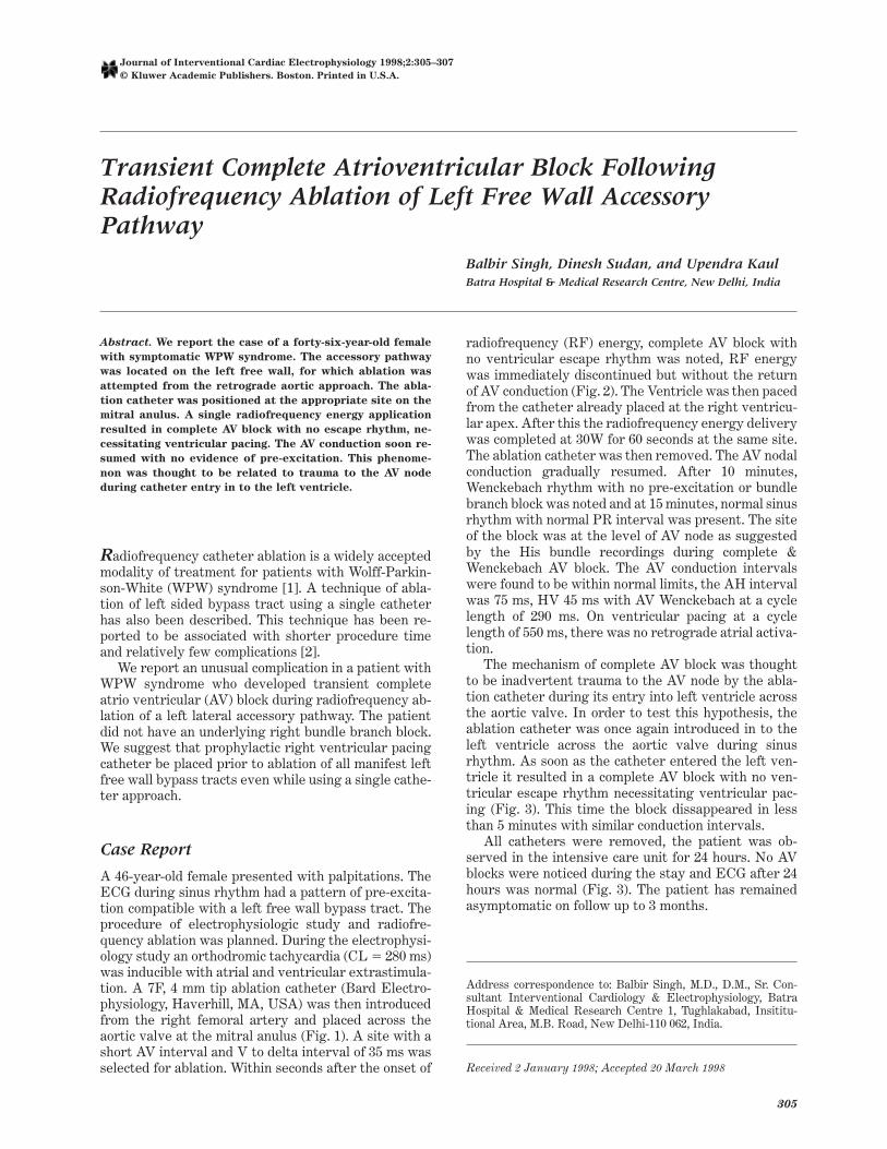
Singh, Sudan and KaulTransient Complete Atrioventricular Block
Transient Complete Atrioventricular Block FollowingRadiofrequency Ablation of Left Free Wall AccessoryPathway
Balbir Singh, Dinesh Sudan, and Upendra KaulBatra Hospital & Medical Research Centre, New Delhi, India
Abstract. We report the case of a forty-six-year-old female
with symptomatic WPW syndrome. The accessory pathway
was located on the left free wall, for which ablation was
attempted from the retrograde aortic approach. The abla-
tion catheter was positioned at the appropriate site on the
mitral anulus. A single radiofrequency energy application
resulted in complete AV block with no escape rhythm, ne-
cessitating ventricular pacing. The AV conduction soon re-
sumed with no evidence of pre-excitation. This phenome-
non was thought to be related to trauma to the AV node
during catheter entry in to the left ventricle.
Radiofrequency catheter ablation is a widely acceptedmodality of treatment for patients with Wolff-Parkin-son-White (WPW) syndrome [1]. A technique of abla-tion of left sided bypass tract using a single catheterhas also been described. This technique has been re-ported to be associated with shorter procedure timeand relatively few complications [2].
We report an unusual complication in a patient withWPW syndrome who developed transient completeatrio ventricular (AV) block during radiofrequency ab-lation of a left lateral accessory pathway. The patientdid not have an underlying right bundle branch block.We suggest that prophylactic right ventricular pacingcatheter be placed prior to ablation of all manifest leftfree wall bypass tracts even while using a single cathe-ter approach.
Case Report
A 46-year-old female presented with palpitations. TheECG during sinus rhythm had a pattern of pre-excita-tion compatible with a left free wall bypass tract. Theprocedure of electrophysiologic study and radiofre-quency ablation was planned. During the electrophysi-ology study an orthodromic tachycardia (CL 5 280 ms)was inducible with atrial and ventricular extrastimula-tion. A 7F, 4 mm tip ablation catheter (Bard Electro-physiology, Haverhill, MA, USA) was then introducedfrom the right femoral artery and placed across theaortic valve at the mitral anulus (Fig. 1). A site with ashort AV interval and V to delta interval of 35 ms wasselected for ablation. Within seconds after the onset of
radiofrequency (RF) energy, complete AV block withno ventricular escape rhythm was noted, RF energywas immediately discontinued but without the returnof AV conduction (Fig. 2). The Ventricle was then pacedfrom the catheter already placed at the right ventricu-lar apex. After this the radiofrequency energy deliverywas completed at 30W for 60 seconds at the same site.The ablation catheter was then removed. The AV nodalconduction gradually resumed. After 10 minutes,Wenckebach rhythm with no pre-excitation or bundlebranch block was noted and at 15 minutes, normal sinusrhythm with normal PR interval was present. The siteof the block was at the level of AV node as suggestedby the His bundle recordings during complete &Wenckebach AV block. The AV conduction intervalswere found to be within normal limits, the AH intervalwas 75 ms, HV 45 ms with AV Wenckebach at a cyclelength of 290 ms. On ventricular pacing at a cyclelength of 550 ms, there was no retrograde atrial activa-tion.
The mechanism of complete AV block was thoughtto be inadvertent trauma to the AV node by the abla-tion catheter during its entry into left ventricle acrossthe aortic valve. In order to test this hypothesis, theablation catheter was once again introduced in to theleft ventricle across the aortic valve during sinusrhythm. As soon as the catheter entered the left ven-tricle it resulted in a complete AV block with no ven-tricular escape rhythm necessitating ventricular pac-ing (Fig. 3). This time the block dissappeared in lessthan 5 minutes with similar conduction intervals.
All catheters were removed, the patient was ob-served in the intensive care unit for 24 hours. No AVblocks were noticed during the stay and ECG after 24hours was normal (Fig. 3). The patient has remainedasymptomatic on follow up to 3 months.
Address correspondence to: Balbir Singh, M.D., D.M., Sr. Con-sultant Interventional Cardiology & Electrophysiology, BatraHospital & Medical Research Centre 1, Tughlakabad, Insititu-tional Area, M.B. Road, New Delhi-110 062, India.
Received 2 January 1998; Accepted 20 March 1998
305
Journal of Interventional Cardiac Electrophysiology 1998;2:305–307
© Kluwer Academic Publishers. Boston. Printed in U.S.A.

Fig. 1. Left anterior oblique (40 degree) and right anterior oblique (30 degree) view showing the ablation catheter (arrow), the othermultipolars catheters placed in the coronary sinus, His bundle and right ventricle.
Fig. 2. Surface and intracardiac electrogram recordings dur-ing the delivery of radiofrequency energy. Complete AV blockwith no ventricular escape rhythm is seen soon after the onsetof radiofrequency energy.
Fig. 3. Surface electrograms and arterial pressure recordingduring re-introduction of ablation catheter in to the left ventri-cle, resulting in complete AV block.
306 Singh, Sudan and Kaul

Discussion
AV nodal blocks have been described during ablation ofAV nodal reentry or septal bypass tracts, however, thiswould be an unusual event in patients undergoing ab-lation of left free-wall bypass tract. In a previous re-port, complete AV nodal block was noticed during theradiofrequency ablation in a patient with lateral acces-sory pathway and RBBB [3].
In the present report, the patient had no evidence ofRBBB as suggested by the electrograms during ortho-dromic tachycardia and after ablation.
The mechanism of transient complete heart block inthis case was thought to be due to trauma to the AVnode as the ablation catheter crossed the aortic valve.The complete heart block thus occured prior to thedelivery of RF energy, however it remained unnoticeddue to the presence of overt pre-excitation. Thismechanism was con~rmed by the introduction of theablation catheter across the aortic valve after ablationduring normal sinus rhythm; this again resulted incomplete heart block with ventricular asystole (Fig. 3).The trauma by the catheter in the AV node on the rightside may have also caused the heart block, however the
induction of an orthodromic tachycardia immediatelyprior to ablation argues against this mechanism.
The outcome in this patient would have been seriousif temporary ventricular pacing was not immediatelyavailable. In the technique of single catheter ablationfor left lateral accessory pathways, immediate tempo-rary ventricular pacing is not available. We suggestthat in all patients with pre-excitation due left sidedpathways, a right ventricular pacing catheter shouldbe inserted prior to ablation.
References
1. Calkins H, Sousa J, Rosenbeck S, et al. Diagnosis and cureof Wolff-Parkinson-White syndrome or paroxysmal supra-ventricular tachycardias during a single electrophysiologiclast. N Engl J Med 1991;324:1612–1618.
2. Kuck KH, Schluter M. Single catheter approach to radiofre-quency current ablation of left sided accessory pathways inpatients with Wolff-Parkinson-White syndrome. Circula-tion 1991;84:2366–2375.
3. Stamato NJ, Eddy SL, Whiting DJ. Transient completeheart block during radiofrequency ablation of left lateralbypass tract with right bundle branch block. PACE1996;19:1351–1354.
Transient Complete Atrioventricular Block 307