Transfusion related acute lung injury
24
Presented By intern Dr Khagendra Acharya TRANSFUSION RELATED ACUTE LUNG INJURY(TRALI)
-
Upload
9857038254 -
Category
Health & Medicine
-
view
50 -
download
1
Transcript of Transfusion related acute lung injury

Presented By intern Dr Khagendra
Acharya
TRANSFUSION RELATED ACUTE
LUNG INJURY(TRALI)

Definition
TRALI is defined as an acute lung injury that is
temporally related to a blood transfusion;
specifically, it occurs within the first six hours
following a transfusion.
It is typically associated with plasma components
such as platelets and Fresh Frozen Plasma,
though cases have been reported with packed
red blood cells since there is some residual
plasma in the packed cells. The blood component
transfused is not part of the case definition.

Transfusion-related acute lung injury (TRALI) is
an uncommon syndrome that is due to the
presence of leukocyte antibodies in transfused
plasma. TRALI is believed to occur in
approximately one in every 5000 transfusions.
Leukoagglutination and pooling of granulocytes in
the recipient's lungs may occur, with release of
the contents of leukocyte granules, and resulting
injury to cellular membranes, endothelial
surfaces, and potentially to lung parenchyma. In
most cases leukoagglutination results in mild
dyspnea and pulmonary infiltrates within about 6
hours of transfusion, and spontaneously resolves.

History Transfusion-related acute lung injury (TRALI) was
first reported in 1951and 1957,and findings from the initial case series were published in 1966. In 1970 and 1971, it was postulated that leukoagglutinins to HLA and non-HLA antigens were etiologic in TRALI reactions; however, it was not until 1985, with the report of a series of 36 patients,that TRALI was recognized as a distinct clinical entity.
With more aggressive transfusion support and increased recognition of this syndrome, TRALI has become a common clinical complication of transfusion. In the past 2 reporting years, it has been named the leading cause of transfusion-related death in the United States.7

Incidence The true incidence of TRALI is unknown because
of the difficulty in making the diagnosis and because of underreporting. It is estimated to occur in 1:1300 to 1:5000 transfusions of plasma-containing products.
The mortality rate from TRALI ranges from 5% to 25%.Most patients recover within 72 hours
Plasma containing blood product such as whole blood, platelet concentrates, fresh frozen plasma(FFP) , packed red cells, granulocytes, cryoprecipitate and intervenous immunoglobulin have all been implicated as a possible cause of TRALI.

Etiology and risk factors
The etiology of TRALI is currently not fully
understood. TRALI is thought to be immune
mediated.
Antibodies directed toward Human Leukocyte
Antigens (HLA) or Human Neutrophil Antigens
(HNA) have been implicated.
1. Multiparous donor- Antibodies develop through
exposure to fetal blood.
2. Blood component – platelet concentration
>FFP>packed red
cell>granulocytes>cryoprecipitate>iv
immunoglobulin.

3. Massive transfusion
4. Stored blood products- inflammatory mediators
like cytokines and lipid soluble substance
accumulate during storage of blood product.
5. Underlying clinical condition- factors such as
trauma, major surgery, sepsis.

Pathogenesis of TRALI The exact pathogenesis of TRALI is not known thus
several theories have been proposed. Two basis
mechanism have been proposed for the pathogenesis
of TRALI for immune competent hosts.
It is hypothesized that TRALI may be precipitated by
the infusion of donor antibodies directed against
recipient leukocytes. The infusion of donor anti-HLA
(human leukocyte antigens) or anti-HNA (human
neutrophil antigens) antibodies is thought to directly
cause complement activation, resulting in the influx of
neutrophils into the lung, followed by neutrophil
activation and release of cytotoxic agents, with
subsequent endothelial damage and capillary leak
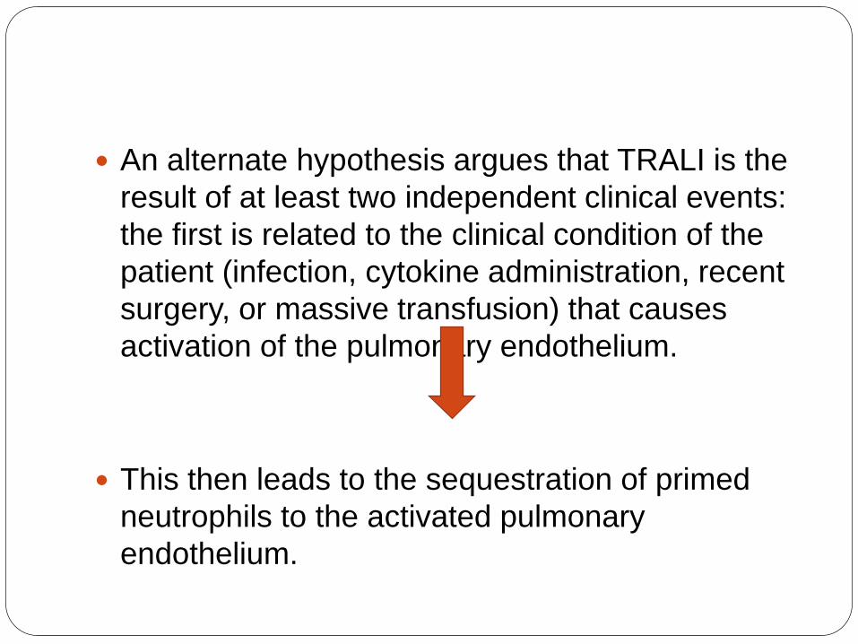
An alternate hypothesis argues that TRALI is the
result of at least two independent clinical events:
the first is related to the clinical condition of the
patient (infection, cytokine administration, recent
surgery, or massive transfusion) that causes
activation of the pulmonary endothelium.
This then leads to the sequestration of primed
neutrophils to the activated pulmonary
endothelium.

The second event is the infusion of donor derived
anti-HLA or anti-HNA antibodies directed against
antigens on the neutrophil surface and/or
biological response modifiers (e.g., lipids) in the
stored blood component that activate these
adherent, functionally hyperactive neutrophils,
causing neutrophil-mediated endothelial damage
and capillary leak.
Third hypothesis suggests that high levels of
donor derived vascular endothelial growth factor
(VEGF) or antibodies to class II HLA antigens
residing on pulmonary vascular endothelium may
directly cause endothelial shape change and
fenestration.


Criteria For TRALI
Acute onset hypoxemia.
Ratio of Pao2/FiO2 <300 or SpO2 <90% at room
temperature.
Occur during or within 6 hours before transfusion.
B/L diffuse pulmonary infiltrates
No evidence of left atrial hypertension(i.e.
circulatory overload).

Clinical feature Dyspnoea/ respiratory distress requiring o2
support-virtually all. Requiring mechanical ventilation-70% Documented hypoxemia- virtually all Cyanosis-very common. Hypotension- majority. Fever-very common Clinical exam reveals respiratory distress
and pulmonary crackles may be present with no signs of congestive heart failure or volume overload.

CXR shows evidence of bilateral
pulmonary edema unassociated
with heart failure (non-cardiogenic
pulmonary edema), with bilateral
patchy infiltrates, which may rapidly
progress to complete "white out"
indistinguishable from Acute
Respiratory Distress Syndrome
(ARDS).

) Chest radiograph at the
time TRALI was recognized,
which demonstrates
extensive bilateral areas of
consolidation in the mid and
upper lobes of the lung
consistent with aspiration or
edema with a normal cardiac
silhouette, new since the
previous examination earlier
on the same day

Diagnostic feature of TRALI
Onset within 1-6 hrs of transfusion.
Acute respiratory distress.
Acute b/l pulmonary oedema (non-cardiogenic).
Severe hypoxemia.
Hypotension.
Fever.
Mild to severe clinical spectrum.

Differential diagnosis
Transfusion-related bacterial sepsis-after transfusion of contaminated peripheral red blood cells (PRBCs) or platelet concentrates manifests as fever, hypotension, and vascular collapse, which may include respiratory distress, and must be considered in patients with pulmonary insufficiency who have undergone transfusion.
anaphylactic transfusion reactions- involve respiratory distress related to bronchospasm manifested by tachypnea, wheezing, cyanosis, and severe hypotension. Facial and truncal erythema and edema are common with urticaria, characteristically involving the head, neck, and trunk. The respiratory distress from anaphylactic transfusion reactions is related to laryngeal and bronchial edema rather than to pulmonary edema, as in TRALI. These reactions occur rapidly during the transfusion of any type of protein-containing blood component and may occur after the transfusion of small volumes of blood.

Transfusion-associated circulatory overload
(TACO) -develops within minutes to hours of
transfusion as respiratory distress with
tachypnea, tachycardia, hypertension, and
cyanosis. All blood components have been
implicated in TACO, and it rapidly responds to
aggressive diuresis and ventilatory support.
Hemolytic transfusion reaction- may overlap with
TRALI, they are easily distinguished by the
presence of hemolysis.

TRALI Vs TACO
TRALI TACO
Fever
Hypotension
Acute dyspnoea
JVP unchanged
Auscultation- rales
X-Ray- diffuse b/linfiltrates
EF- normal
Pulmonary edema fluid-exudate
Response to diuretic-minimal
No fever
Hypertension
Acute dyspnoea
Can be changed
Rales + S3
Diffuse b/l infiltrates
Decreased
Transduate
Significant improvement

Immediate management of
TRALI
Stop the transfusion immediately
Support the patient
If the patient is intubated, obtaine undiluted
edema fluid as soon as possible(within 15 min)
and simultaneous plasma for determination of
total protein.
Obtain CBC with DC and chest radiography
Notify the blood bank of possible trali request a
different unit and quarantine other unit from the
same donar.

1. For mild TRALI cases Supplimental o2 and
supportive care may be sufficient.
2. For the most severe case- IV fluid, mechanical
or non invasive ventilation and invasive
cardiovascular monitoring may be required .
3. Extracorporeal membrane oxygenation has
been used successfully in a severe case of trali.
4. Others less well documented and unproven
therapies(eg. Diuretices , corticosteroids, etc )
have also been used.
5. Antibiotics – prevention of infection

Prevention of TRALI
Avoiding blood from multiparous women- these
women are at risk of producing anti-leucocyte
antibodies during previous pregnancy.
Donors whose blood has resulted in TRALI like
rxn previously.
Blood which has been stored for long duration-
long storage results in production of anti-
leucocyte antibodies.
Not using whole blood.
Leukoreduction can be done .

Leukoreduction
To remove WBCs from RBCs and platelets.
Reduce the risk of human leukocyte antigen
(HLA) alloimminization, febrile nonhaemolytic
transfusion rxn and CMV transmission in a pt who
require these precution.
LR is usually perform by filtration,although some
apheresis collection have sufficiently precise cell
seperation to minimize WBCs content.
It is also called transfusion related
immunomodulation because it prevent TRALI i.e
possible immunosupressive and pro-inflammatory
effect mediated by donor WBCs.

Thank you
![3041576/3041578 DO NOT ADMINISTER … PI...• For patients at risk of thrombosis, ... • Monitor patients for pulmonary adverse reactions (transfusion-related acute lung injury [TRALI]).](https://static.fdocuments.in/doc/165x107/5abc002a7f8b9ab1118daa85/30415763041578-do-not-administer-pi-for-patients-at-risk-of-thrombosis.jpg)