Transdermal Scopolamine Drug Delivery Systems for Motion
23
Transdermal Scopolamine Drug Delivery Systems for Motion Sickness Gloria Fung, Terence Ho, Soyoon Lee, Joseph Munaretto, Christine Tsai BEE 453 May 5 th , 2003 Executive Summary Transdermal drug delivery systems are involved in the continuous administration of drug molecules from the surface of the skin into the circulatory system. Such systems have proved advantageous for delivery of certain drugs, such as scopolamine, nicotine, nitroglycerine, and estradiol. Compared with oral administration, transdermal drug delivery offers better uniformity of drug concentrations in plasma throughout their duration of use. Scopolamine is the active ingredient in motion sickness medication that targets the nerve fibers in the inner ear. The scopolamine patch is effective for about three days, longer than if administered orally which is effective for only several hours. One of the main restraints of this transdermal system is its absorption through the skin, especially through the stratum corneum, its outermost part. This study examines the rate of diffusion of transdermal scopolamine across the skin and into the systemic circulation. Our objective is to optimize the drug delivery by way of a scopolamine patch by minimizing absorption rates, while maintaining its advantage of a long-term effect. A comparative study of the effects in the presence of penetration enhancers were undertaken to show how steady state is approached at different rates. The model we used does offer certain limitations, as diffusivity values specific to human skin and scopolamine are not readily available. Introduction and Design Objectives Motion sickness is a common ailment that many individuals experience in the form of sea sickness, car sickness, or air sickness. Motion sickness occurs when the body is subjected to accelerations of movement in different directions. This movement causes the balance center of the inner ear to send conflicting information to the brain that differ from any visual cues that indicate an apparent stillness. Despite these prevention methods, medication specifically designed to control such a condition is the most effective. Even with prescriptions and over the counter oral medications to prevent or limit symptoms, drugs effective until one hour after they are administered. This is best for individuals who are aware of their tendency to experience motion sickness while on a plane, ship, or car. Oral medication is most effective for short trips or when symptoms occur intermittently. Alternatively, for those interested in a long-term effect (i.e. 3 days), the transdermal drug delivery method is most effective. The commercially available scopolamine patch is Transderm-Scop. It is to be placed on the skin behind the ear approximately four hours before the effects will be needed. The Transderm-Scop is programmed to deliver in-vivo approximately 1.0 mg of scopolamine over three days. As with other transdermal drug systems, scopolamine delivery is hindered by skin permeability,
Transcript of Transdermal Scopolamine Drug Delivery Systems for Motion

Transdermal Scopolamine Drug Delivery Systems for Motion Sickness
Gloria Fung, Terence Ho, Soyoon Lee, Joseph Munaretto, Christine Tsai BEE 453
May 5th, 2003 Executive Summary Transdermal drug delivery systems are involved in the continuous administration of drug molecules from the surface of the skin into the circulatory system. Such systems have proved advantageous for delivery of certain drugs, such as scopolamine, nicotine, nitroglycerine, and estradiol. Compared with oral administration, transdermal drug delivery offers better uniformity of drug concentrations in plasma throughout their duration of use. Scopolamine is the active ingredient in motion sickness medication that targets the nerve fibers in the inner ear. The scopolamine patch is effective for about three days, longer than if administered orally which is effective for only several hours. One of the main restraints of this transdermal system is its absorption through the skin, especially through the stratum corneum, its outermost part. This study examines the rate of diffusion of transdermal scopolamine across the skin and into the systemic circulation. Our objective is to optimize the drug delivery by way of a scopolamine patch by minimizing absorption rates, while maintaining its advantage of a long-term effect. A comparative study of the effects in the presence of penetration enhancers were undertaken to show how steady state is approached at different rates. The model we used does offer certain limitations, as diffusivity values specific to human skin and scopolamine are not readily available. Introduction and Design Objectives Motion sickness is a common ailment that many individuals experience in the form of sea sickness, car sickness, or air sickness. Motion sickness occurs when the body is subjected to accelerations of movement in different directions. This movement causes the balance center of the inner ear to send conflicting information to the brain that differ from any visual cues that indicate an apparent stillness. Despite these prevention methods, medication specifically designed to control such a condition is the most effective. Even with prescriptions and over the counter oral medications to prevent or limit symptoms, drugs effective until one hour after they are administered. This is best for individuals who are aware of their tendency to experience motion sickness while on a plane, ship, or car. Oral medication is most effective for short trips or when symptoms occur intermittently. Alternatively, for those interested in a long-term effect (i.e. 3 days), the transdermal drug delivery method is most effective. The commercially available scopolamine patch is Transderm-Scop. It is to be placed on the skin behind the ear approximately four hours before the effects will be needed. The Transderm-Scop is programmed to deliver in-vivo approximately 1.0 mg of scopolamine over three days. As with other transdermal drug systems, scopolamine delivery is hindered by skin permeability,

including the stratum corneum, which is perhaps the major physical barrier. Once diffusion through the stratum corneum is achieved, the molecules diffuse through the dermis, are absorbed into capillary beds, and then circulate to the targeted area. Our objective is to model the absorption of scopolamine through the skin to reach systemic circulation. Fick’s law of diffusion is implemented to model the delivery of the drug across a membrane, in this case the skin. The different regions of diffusion that we take into account are the epidermis and the dermis. The epidermis is the outermost layer of the skin where hair projects out from the pores of the skin. This layer consists of free sensitive nerve endings as well. Next, the dermis layer consists of sweat glands and sebaceous glands as well as collagen fibers. The first model used was an axi-symmetric model of epidermis and dermis with zero flux at the side boundaries, as shown in the figure below.
Fig 1. Dimensions of H In addition, we will also model the diffusion in the nonanoic acid and oleic acid, and compare how muapplied, a patch alone should take four hours to achtime of action to decrease when accompanied with
x
s
s
0.05cm
m
Dermi
Epidermi
0.01cR=0.892cm
uman Skin Model
presence of “penetration enhancers” such as ch the rate of penetration is increased. Once ieve its desired effect. We expect this initial a penetration enhancer.
Results For our solution, we calculated our first diffusivity value from permeability constant, partition coefficient, and rat skin values from a study by Calpena, A.C. et. al. (See appendix for actual values and calculations) After running the program with these values and the appropriate boundary and initial conditions, our solution showed that steady state was reached at around 40hours, as can be observed in the figure below. Node 212 is shown in Fig B1 and B2 in Appx B. The contour plots at steady state are shown in Fig B5 and B6.
Fig 2. History Plot of Original Model at Node 212 (reaches steady state at 40hrs) However, in a study of scopolamine absorption in humans by Muir, C. and Metcalf, R., steady state was reached after 8 hours of application of the patch. Based on these findings, we decided to use the software to determine (by iteration) the diffusivity value that would lead to such steady state. The following plot was obtained to confirm that steady state was achieved after roughly eight hours. The contour plots are shown in Fig B3 and B4.

Fig 3. History Plot of Model at Node 212 with Diffusivity Values Found by Iteration (reaches steady state at 8hrs)
Compared to our first set of values, the values obtained from the program are off by a magnitude of 10. This may be explained by the errors that may result from obtaining values based on rat skin properties, which will undoubtedly be different from human skin. Skin from the dorsal side of rats might be substantially different from the skin in the post-auricular area of humans. Likewise, the experimental results obtained from the rat study involved the use of Franz diffusion cells, which may also have an effect on the permeability rate constants that were determined (and that we used to calculate diffusivity). It was thought to be realistic to add an extra skin entity to find out how much of the chemical would diffuse outwards, therefore transforming from a 1D to a 2D problem. The diffusivity values used for this case were the ones that gave us 8hrs to steady state. We ran the program again to show a more accurate model that allows diffusion to take place outwards (horizontally) along the side boundaries (see below). We were interested in seeing how the species concentration at different times and regions varies with this new model, as well as how it affects the time to get to reach steady state.

Fig 6. Model of Human Skin with Extra Skin Entity Included
Our solution shows that there is indeed some outwards diffusion, as the following contour plot shows. As we can see in the figure below, our outer boundaries are far enough such that the species concentration side edges are zero. (Note that the set of diffusivity values we used in this analysis is the one that gives steady state concentration in the skin after 8hrs in our original mesh without the skin extensions). The mesh is shown in Fig B7 and B8.
Fig 7. Contour Plot of Human Skin with Extra Skin Entity Included
Patch (same size as previously)
Extra Skin Entity

The following graph is the history plot of species concentration at node 250, which is right at the interface between the epidermis and dermis. Fig 8. History Plot at Node 250 (Human Skin with Extra Skin Entity Included) Mesh Convergence Investigation In order to see whether our mesh definition is appropriate (i.e. we have achieved mesh convergence), making a finer mesh (i.e. incorporating more elements) should not change the solution. The conventional approach is to create a similar model with the same geometry with increased elements, and strive to find a mesh definition that strikes a balance between computation time and quality of solution. However, since our mesh seems to converge already, we decided to create another mesh with much fewer elements to contrast this mesh, as the one below. The overall mesh is shown in Fig B10.

Fig 4. Unrefined Mesh (zoomed in at Node 4) Our results indicate that our initial, finer mesh displays similar results (i.e. steady state after 8 hours) but also takes care of the irregular jump at the beginning of the process, that is shown in the following figure using the mesh with fewer elements.
Fig 5. History Plot of Unrefined Mesh at Node 4

Sensitivity Analysis 1 – Removal of Patch after 30hrs (with extra skin entity) In addition, we wanted to model how long it takes for the drug to diffuse through the skin once the patch is removed (with extra entity still in place). In other words, we were interested in the diffusion profile once the constant flux of 5.56 x 10⁻³ mg/cm2/hr on the top surface is removed. We wanted to know how long it takes for the drug to diffuse out of the skin, and thus, how long after the patch is removed will the drug be circulating in the system. For this purpose, we decided to have a constant flux for 30 hrs, after which the patch would be removed and the flux would then be = 0 mg/cm2. Our results showed that once the patch is removed after hour 30, it takes about 15 hours for the drug to diffuse out of the region, as is revealed in the following plot. Node 437 is shown in Fig 9.
Fig 9. History Plot at Node 437 (Human Skin with Extra Skin Entity Included, but with patch removed after 30hrs)

Sensitivity Analysis 2 – Temperature Enhancement and Penetration Enhancers We also measured sensitivity to diffusivity based on changes in temperature and chemical enhancers. It is found that heat raises diffusion rates, increasing flux to as much as twice its original value. This happens due to increased body fluid circulation, blood vessel wall permeability and drug solubility etc. The results we obtained with a diffusivity value of twice the original value are represented in the following figure:
Fig 10. History Plot at Node 212 (with Temperature Enhancement)
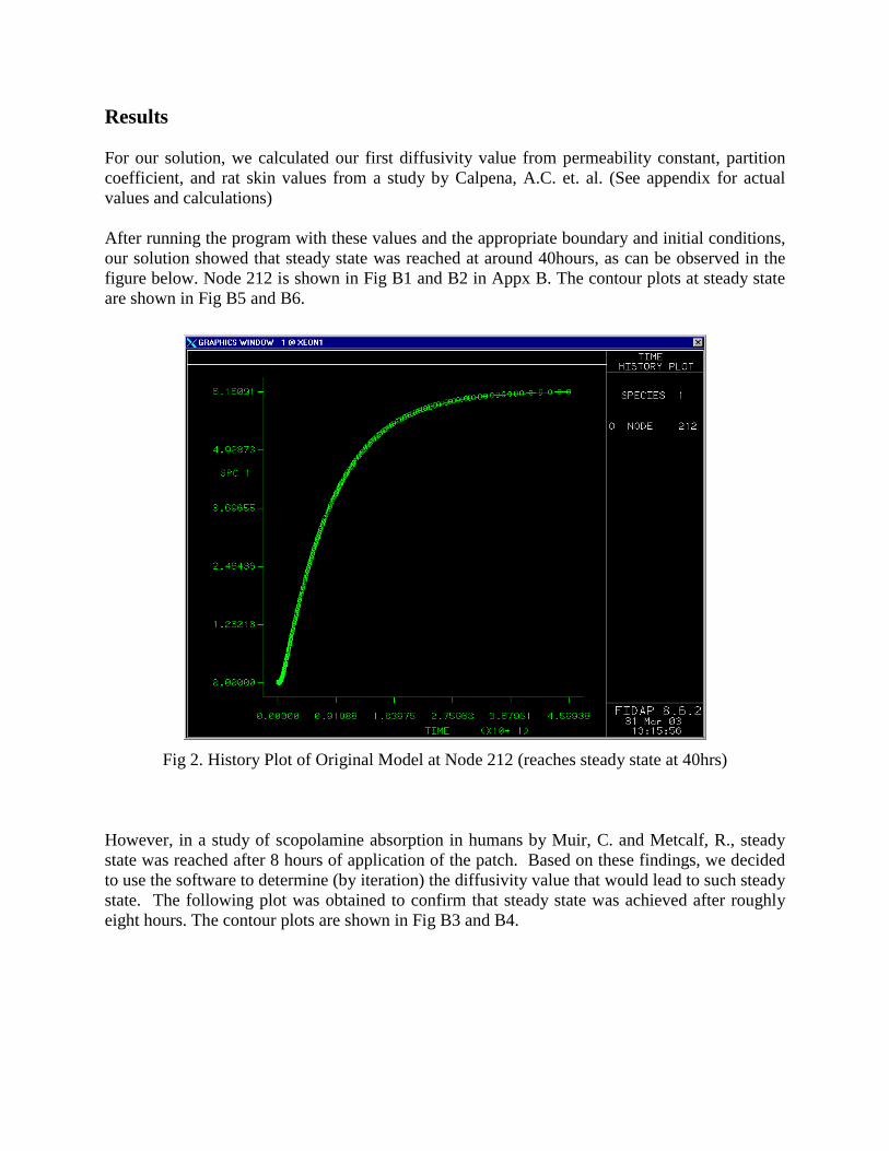
The plot indicates that a twofold increase in diffusivity achieves steady state after approximately six hours. In addition, we tested the effect of two chemical penetration enhancers. An article by Kanikkannan indicates that the chemical nonanoic acid has an enhancement factor of approximately 3.83. Based on this data, we ran our program with the original diffusivity increased by the above factor to obtain the following results:
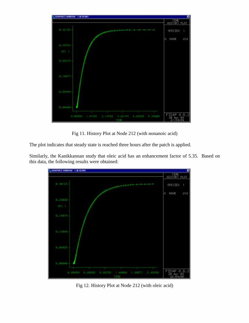
Fig 11. History Plot at Node 212 (with nonanoic acid) The plot indicates that steady state is reached three hours after the patch is applied. Similarly, the Kanikkannan study that oleic acid has an enhancement factor of 5.35. Based on this data, the following results were obtained:
Fig 12. History Plot at Node 212 (with oleic acid)
Discussion and Conclusions The results we obtained correspond to what we expected. Because the patch is designed to have a controlled release rate and to provide relief for up to three days, we were aware that steady state would be reached after a certain time in order to sustain the provision of scopolamine to the systemic circulation at a controlled rate. Likewise, we were also able to model how long it takes for the drug to diffuse out of the skin once the patch is removed. This can be useful since some users may present side effects or skin irritation to prolonged use of the patch and it may be useful to know how long the drug is in the skin (or in the systemic circulation) once the flux is cut off. Also, for those users who are interested in using the patch for less than its full capacity (three days), it is practical to know how much longer the effects of the patch would last once it is removed, to determine when they should remove it. Furthermore, manufacturers can use this data to produce patches with different capacities that suit varying needs. Finally, we were able to study how the patch can be improved by increasing temperatures or incorporating chemical enhancers, which all achieve a steady state process at shorter times. With these variations, the user may not need to wait four hours before achieving the desired effects of the drug (as the current scopolamine patch requires). However, from these studies, we are unable to determine exactly how much shorter this time would be since we did not model the process once the drug reaches systemic circulation. In general, FIDAP served as a useful tool to study transdermal delivery systems by providing with the possibility of changing the model (i.e. changing parameters) and observing the appropriate results. For instance, we were able to see how increasing the diffusivities affect the process quantitatively. Nevertheless, the use of FIDAP presupposes the availability of empirical data, in this case diffusivity values and skin properties. This was a challenge for our analysis since we were unable to find any previous study that had diffusivity values specific for scopolamine in post-auricular skin. For this reason, we had to use data on rat studies, which undoubtedly introduces errors. Likewise, the use of our program to determine diffusivity values assumes that our model is right. However, given that we are lumping the multiple skin variations and properties into just two regions (epidermis and dermis) we are bound to have an oversimplified model. Hence, this leads to less accurate diffusivity values.
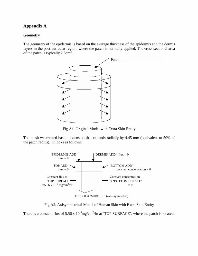
Appendix A Geometry The geometry of the epidermis is based on the average thickness of the epidermis and the dermis layers in the post-auricular region, where the patch is normally applied. The cross sectional area of the patch is typically 2.5cm2.
Fig A1. Original Model with Extra Skin Entity The mesh we created has an extension that expands radially by 4.45 mm (equivalent to 50% of the patch radius). It looks as follows: ‘EPIDERMIS ADD’ ‘DERMIS ADD’: flux = 0 flux = 0
‘TOP ADD’ ‘BOTTOM ADD’ flux = 0 Ic constant concentration = 0
Constant flux at Constant concentration ‘TOP SURFACE’ I II at ‘BOTTOM SUFACE’
=5.56 x 10⁻³ mg/cm2/hr = 0 Flux = 0 at ‘MIDDLE’ (axis-symmetric)
Fig A2. Axisymmetrical Model of Human Skin with Extra Skin Entity There is a constant flux of 5.56 x 10⁻³mg/cm2/hr at ‘TOP SURFACE’, where the patch is located.
Patch

Governing Equation
where CD = concentration of the drug; u = velocity due to convective flux; t = time; x = distance from top; DD = diffusivity constant of scopolamine; and r = generation/metabolism of scopolamine in dermis. In this model, we are dealing with a solid (i.e. no fluid flow), therefore the velocity term is eliminated. Likewise, there is no storage/metabolism of scopolamine in the skin, and therefore, the reaction term is also zero. Boundary Conditions The scopolamine patch is programmed to deliver in-vivo approximately 1.0 mg of scopolamine at an approximately constant rate to the systemic circulation over 3 days. Thus, the controlled release rate is of 0.0139mg/hr. With a patch area of 2.5cm2, a constant flux of 5.56 x 10⁻³ mg/hr/cm2 is achieved at the top boundary (x=0), where the patch is located. For the bottom boundary beneath the dermis, it is assumed that the rate of blood flow transports the scopolamine fast enough such that concentration is zero (there is no building up of scopolamine at the bottom boundary). Thus, the boundary conditions are:
• -D(dC/dx) = 5.56 x 10⁻³ mg/hr/cm2 at ‘TOP SURFACE’
• C = 0 at ‘BOTTOM’ and ‘BOTTOM ADD’
• -D(dC/dx) = 0 mg/hr/cm2 at ‘MIDDLE’, ‘TOP ADD’, ‘EPIDERMIS ADD’, and ‘DERMIS ADD’
Initial Conditions
• C = 0 at t = 0 for all x. Input parameters Post auricular skin properties
Epidermis thickness 0.1 mm (Lee, Y. and K. Hwang, 2002)
Dermis thickness 0.5 mm (Lee, Y. and K. Hwang, 2002)
DD
DDD r
x
cD
x
cu
t
c +∂
∂=∂
∂+∂
∂2
2

Diffusivities A) From formula From the study of transdermal absorption of scopolamine in rats by Calpena, A.C. et. al, we took values of permeability rate constant obtained experimentally for scopolamine, the partition coefficient in octanol-water for scopolamine, and the average rat epidermis thickness to calculate the diffusivity of scopolamine based on the following relationship Kp = D* po/w / d , where Kp is the permeability rate constant, D is the diffusivity, po/w is the partition coefficient, and d is the rat epidermis thickness Diffusivity in epidermis (from
formula) 5.23 x 10⁻⁶ cm2/hr (Calpena, A.C. et. al, 1993)
Diffusivity in dermis (assuming 400 times
diffusivity in epidermis) 0.002 cm2/hr (Dalby, R., 2001)
B) From FIDAP In a study of scopolamine absorption in humans by Muir, C. and Metcalf, R., steady state was reached after 8 hours of application of the patch. Based on these findings, we decided to use the software to determine the diffusivity value that would lead to such steady state. Diffusivity in epidermis (from
FIDAP) 2.0 x 10⁻⁴ cm2/hr (Muir, C. and R. Metcalf, 1983)
Diffusivity in dermis (assuming 400 times
diffusivity in epidermis) 0.008 cm2/hr (Dalby, R., 2001)
C) Enhanced diffusivities using qualitative data
Based on qualitative data obtained in various sources, we enhanced the diffusivity by factors of 2 (diffusivity enhanced by increase in temperature); 5.35 (diffusivity increased with oleic acid as chemical enhancer); and 3.83 (diffusivity increased with nonanoic acid as chemical enhancer).
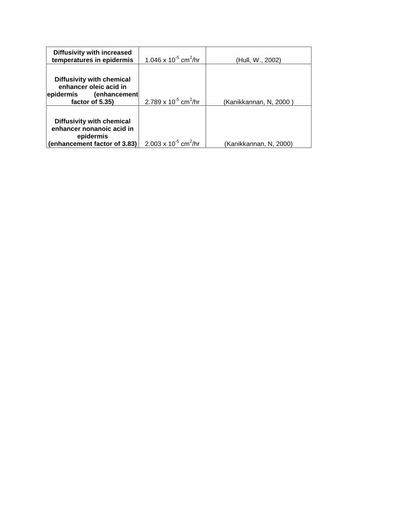
Diffusivity with increased temperatures in epidermis 1.046 x 10⁻⁵ cm2/hr (Hull, W., 2002)
Diffusivity with chemical enhancer oleic acid in
epidermis (enhancement factor of 5.35) 2.789 x 10⁻⁵ cm2/hr (Kanikkannan, N, 2000 )
Diffusivity with chemical enhancer nonanoic acid in
epidermis (enhancement factor of 3.83) 2.003 x 10⁻⁵ cm2/hr (Kanikkannan, N, 2000)

FIDAP input parameters SIMULATION PROBLEM command:
PROB (AXI-, INCO, TRAN, LAMI, LINE, NEWT, NOMO, ISOT, FIXE, NOST, NORE, SING, SPEC = 1.0)
The design of our problem was modeled as axisymmetric in geometry. Substances used for the model are all incompressible and Newtonian. The problem analysis was performed in transient and not in steady state while any fluid flows were assumed to be laminar. Convection was not considered in this problem, therefore resulting in a problem design without momentum analysis. The problem was isothermal due to assumed fixed temperatures, therefore excluding the energy equation. The design was also modeled with fixed surfaces with no structural solver and no remeshing required. The problem was assumed to be a single phase design. Finally, only one species, scopolamine, was considered in this problem. EXECUTION and SOLUTION command: EXEC (NEWJ) SOLU (S.S. = 10, ACCF = 0.000000000000E+00) The newjob parameter in the execution command informs the program that this is a new problem. The Solution command input parameters tell FIDAP to use successive substitutions to solve for each time step with a maximum number of iterations set at 10 for each step. The acceleration of the solver is set at zero. TIMEINTEGRATION command:
TIME (BACK, NSTE = 1000, TSTA = 0.000000000000E+00, TEND = 100.0, DT = 0.100000000000E-05, VARI = 0.100000000000E-03, NOFI = 5, INCMAX = 2.0)
The number of steps is set at 1000 while the starting and ending times of problem are set at 0 and 100, respectively. ENTITY
ENTI (NAME = "Epidermis", SOLI, SPEC = 1.0, MDIF = "scopoepidermis") ENTI (NAME = "Dermis", SOLI, SPEC = 1.0, MDIF = "scopodermis") ENTI (NAME = "Top Surface", PLOT) ENTI (NAME = "Bottom Surface", PLOT) ENTI (NAME = "Middle-Epidermis", PLOT) ENTI (NAME = "Middle-Dermis", PLOT) ENTI (NAME = "End-Epidermis", PLOT) ENTI (NAME = "End-Dermis", PLOT)

This step specifies each of the separate entities created for analysis of the problem. In this case, we have 8 total entities including the epidermal and dermal layers of the skin, which are whole solid entities into which diffusion of the species, scopolamine, diffuses. Additionally, the top and bottom surfaces of the skin, the middle axis of symmetry for the epidermis and dermis and the outer boundary of the epidermis and dermis regions are also specified as the boundaries of the model. PROPERTIES DIFFUSIVITY command:
DIFF (SET = "scopoepidermis", CONS = 0.200000000000E-04) DIFF (SET = "scopodermis", CONS = 0.800000000000E-02)
This command allows us to set the diffusivity property of scopolamine through the epidermal and dermal layers to constant numerical values. BOUNDARY CONDITIONS BCFLUX command:
BCFL (SPEC = 1.0, ENTI = "Top Surface", CONS = 0.556000000000E-02) The set boundary condition for the top surface of the model was set to a constant 5.56E-3 mg/hr/cm2 flux of scopolamine. BCNODE command:
BCNO (SPEC = 1.0, ENTI = "Bottom Surface", ZERO) The boundary condition for the bottom surface of the element was fixed at a constant zero scopolamine value to insure no flux. INITIAL CONDITIONS ICNODE command:
ICNO (SPEC = 1.0, ZERO, ALL)
The initial condition for the model was specified as a constant zero concentration of scopolamine in all of the entities.

Appendix B
Fig B1: Original Mesh of Human Skin (Epidermis and Dermis)
Fig B2: Zoomed in Mesh showing Node 212 (Epidermis and Dermis)
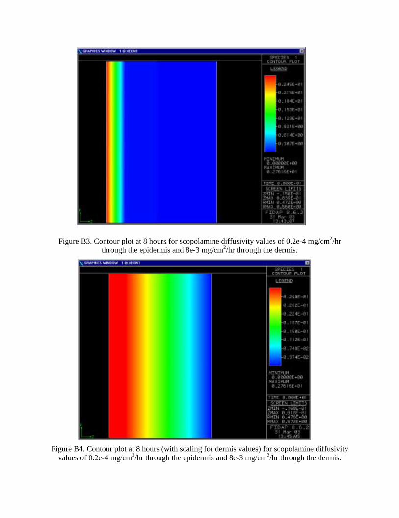
Figure B3. Contour plot at 8 hours for scopolamine diffusivity values of 0.2e-4 mg/cm2/hr through the epidermis and 8e-3 mg/cm2/hr through the dermis.
Figure B4. Contour plot at 8 hours (with scaling for dermis values) for scopolamine diffusivity values of 0.2e-4 mg/cm2/hr through the epidermis and 8e-3 mg/cm2/hr through the dermis.

Figure B5. Contour plot at 40 hours for scopolamine diffusivity values of 5.23e-6 mg/cm2/hr through the epidermis and 2.0e-3 mg/cm2/hr through the dermis.
Figure B6. Contour plot at 40 hours (with scaling for dermis values) for scopolamine diffusivity values of 5.23e-6 mg/cm2/hr through the epidermis and 2.0e-3 mg/cm2/hr through the dermis.

Figure B7: New mesh with extra skin modeled. Node 250 is shown.
Figure 3: Contour Plot at 8 hours
Figure B8: New mesh with extra skin modeled.
No Patch
Patch

Figure B9: Location of Node 437
Figure B10: Unrefined Mesh

References Calpena, A.C. A Comparative in Vitro Study of Transdermal Absorption of Antiemetics. Journal
of Pharmaceutical Sciences. Vol. 83. No. 1, January 1994. Dalby, R. Transdermal Drug Delivery. School of Pharmacy, University of Maryland. Baltimore.
2001. http://www.pharmacy.umaryland.edu/faculty/rdalby/Teaching%20Web%20Pages/Transdermal%20Drug%20Delivery.pdf
Hull, W. Heat-Enhanced Transdermal Drug Delivery: A Survey Paper. The Journal of Applied
Research in Clinical and Experimental Therapeutics. Vol. 2, No. 1, 2002. Kanikkannan, N. Structure-Activity Relationship of Chemical Penetration Enhancers in Transdermal Drug
Delivery. Current Medicinal Chemistry. Issue 7, 593-608. 2000. Lee, Y. and K. Hwang. Skin thickness of Korean adults
http://link.springer-ny.com/link/service/journals/00276/contents/02/00034/s00276-002-0034- 5ch002.html Muir, C. and R. Metcalf. A Comparison of Plasma Levels of Hyoscine After Oral and
Transdermal Administration. Journal of Pharmaceutical Biomedical Analysis. 1: 363-367. 1983
Vollmer, U. et. al. An improved Model for Studies on Transdermal Drug Absorption In-vivo in
Rats. J.Pharm. Pharmacol. 45: 242-245. 1993.


![Transdermal Drug Delivery System [TDDS]](https://static.fdocuments.in/doc/165x107/587c9c9c1a28abfa5e8b7b4f/transdermal-drug-delivery-system-tdds.jpg)