Transcriptomic and Phenotypic Analysis Reveals New Functions … · The Tat system was first...
11
Transcriptomic and Phenotypic Analysis Reveals New Functions for the Tat Pathway in Yersinia pseudotuberculosis Ummehan Avican, a,b Michael Beckstette, c Ann Kathrin Heroven, c Moa Lavander, a * Petra Dersch, c Åke Forsberg a,b Department of Molecular Biology, Umeå Center for Microbial Research (UCMR), Umeå University, Umeå, Sweden a ; Department of Molecular Biology, Laboratory for Molecular Infection Medicine Sweden (MIMS), Umeå University, Umeå, Sweden b ; Department of Molecular Infection Biology, Helmholtz Center for Infection Research, Braunschweig, Germany c ABSTRACT The twin-arginine translocation (Tat) system mediates the secretion of folded proteins that are identified via an N-terminal sig- nal peptide in bacteria, plants, and archaea. Tat systems are associated with virulence in many bacterial pathogens, and our pre- vious studies revealed that Tat-deficient Yersinia pseudotuberculosis was severely attenuated for virulence. Aiming to identify Tat-dependent pathways and phenotypes of relevance for in vivo infection, we analyzed the global transcriptome of parental and tatC mutant strains of Y. pseudotuberculosis during exponential and stationary growth at 26°C and 37°C. The most significant changes in the transcriptome of the tatC mutant were seen at 26°C during stationary-phase growth, and these included the al- tered expression of genes related to virulence, stress responses, and metabolism. Subsequent phenotypic analysis based on these transcriptome changes revealed several novel Tat-dependent phenotypes, including decreased YadA expression, impaired growth under iron-limited and high-copper conditions, as well as acidic pH and SDS. Several functionally related Tat substrates were also verified to contribute to these phenotypes. Interestingly, the phenotypic defects observed in the Tat-deficient strain were generally more pronounced than those in mutants lacking the Tat substrate predicted to contribute to that specific func- tion. Altogether, this provides new insight into the impact of Tat deficiency on in vivo fitness and survival/replication of Y. pseu- dotuberculosis during infection. IMPORTANCE In addition to its established role in mediating the secretion of housekeeping enzymes, the Tat system has been recognized as being involved in infection. In some clinically relevant bacteria, such as Pseudomonas spp., several key virulence determinants can readily be identified among the Tat substrates. In enteropathogens, such as Yersinia spp., there are no obvious virulence de- terminants among the Tat substrates. Tat mutants show no growth defect in vitro but are highly attenuated in in vivo. This makes Tat an attractive target for the development of novel antimicrobials. Therefore, it is important to establish the causes of the attenuation. Here, we show that the attenuation is likely due to synergistic effects of different Tat-dependent phenotypes that each contributes to lowered in vivo fitness. B acteria have evolved several specialized secretion systems for protein export as part of their successful strategies to colonize niches where they encounter various environmental conditions. Gram-negative bacteria possess different mechanisms to trans- port/export proteins, including virulence-related factors from the cytoplasm across the inner membrane and/or the outer mem- brane, either in a single step or acting together with other secretion systems (1). Two pathways, the secretory (Sec) and the twin-argi- nine translocation (Tat) pathways, are involved in the transloca- tion of proteins from the cytoplasm to the periplasm. In Gram- negative bacteria, they can cooperate with other secretion systems to promote the secretion of virulence effectors across the outer membrane to the external environment (2). The Tat system was first discovered in plant chloroplasts and designated the Cp-Tat pathway. Later, homologues of the path- way were found in bacteria and archaea. The Tat translocation complex consists of three inner membrane proteins, TatA, TatB, and TatC. Of these, TatC is the most highly conserved protein among bacteria, plants, and in archaea. One unique hallmark of the Tat pathway is the ability to translocate fully folded proteins across the cytoplasmic membrane. The translocation process re- lies on the proton motive force (PMF). Proteins that are translo- cated via the Tat pathway have an N-terminal signal peptide ([S/T]-R-R-X-F-L-K) with a distinctive “twin-arginine” motif (3, 4). Even though the Tat-dependent proteins are relatively few compared to those secreted by the Sec system, they still have a variety of important functions with impact on bacterial cell phys- iology, such as respiratory energy metabolism, iron acquisition, stress response, and cell division. The Tat pathway also has an important role in the transport of virulence factors in pathogenic bacteria, such as phospholipase toxins, PlcC and PlcH in Pseu- Received 2 May 2016 Accepted 28 July 2016 Accepted manuscript posted online 8 August 2016 Citation Avican U, Beckstette M, Heroven AK, Lavander M, Dersch P, Forsberg Å. 2016. Transcriptomic and phenotypic analysis reveals new functions for the Tat pathway in Yersinia pseudotuberculosis. J Bacteriol 198:2876 –2886. doi:10.1128/JB.00352-16. Editor: P. J. Christie, McGovern Medical School Address correspondence to Åke Forsberg, [email protected]. * Present address: Moa Lavander, Microbiology Division, National Food Agency, Uppsala, Sweden. Supplemental material for this article may be found at http://dx.doi.org/10.1128 /JB.00352-16. Copyright © 2016, American Society for Microbiology. All Rights Reserved. crossmark 2876 jb.asm.org October 2016 Volume 198 Number 20 Journal of Bacteriology on March 15, 2020 by guest http://jb.asm.org/ Downloaded from
Transcript of Transcriptomic and Phenotypic Analysis Reveals New Functions … · The Tat system was first...

Transcriptomic and Phenotypic Analysis Reveals New Functions forthe Tat Pathway in Yersinia pseudotuberculosis
Ummehan Avican,a,b Michael Beckstette,c Ann Kathrin Heroven,c Moa Lavander,a* Petra Dersch,c Åke Forsberga,b
Department of Molecular Biology, Umeå Center for Microbial Research (UCMR), Umeå University, Umeå, Swedena; Department of Molecular Biology, Laboratory forMolecular Infection Medicine Sweden (MIMS), Umeå University, Umeå, Swedenb; Department of Molecular Infection Biology, Helmholtz Center for Infection Research,Braunschweig, Germanyc
ABSTRACT
The twin-arginine translocation (Tat) system mediates the secretion of folded proteins that are identified via an N-terminal sig-nal peptide in bacteria, plants, and archaea. Tat systems are associated with virulence in many bacterial pathogens, and our pre-vious studies revealed that Tat-deficient Yersinia pseudotuberculosis was severely attenuated for virulence. Aiming to identifyTat-dependent pathways and phenotypes of relevance for in vivo infection, we analyzed the global transcriptome of parental and�tatC mutant strains of Y. pseudotuberculosis during exponential and stationary growth at 26°C and 37°C. The most significantchanges in the transcriptome of the �tatC mutant were seen at 26°C during stationary-phase growth, and these included the al-tered expression of genes related to virulence, stress responses, and metabolism. Subsequent phenotypic analysis based on thesetranscriptome changes revealed several novel Tat-dependent phenotypes, including decreased YadA expression, impairedgrowth under iron-limited and high-copper conditions, as well as acidic pH and SDS. Several functionally related Tat substrateswere also verified to contribute to these phenotypes. Interestingly, the phenotypic defects observed in the Tat-deficient strainwere generally more pronounced than those in mutants lacking the Tat substrate predicted to contribute to that specific func-tion. Altogether, this provides new insight into the impact of Tat deficiency on in vivo fitness and survival/replication of Y. pseu-dotuberculosis during infection.
IMPORTANCE
In addition to its established role in mediating the secretion of housekeeping enzymes, the Tat system has been recognized asbeing involved in infection. In some clinically relevant bacteria, such as Pseudomonas spp., several key virulence determinantscan readily be identified among the Tat substrates. In enteropathogens, such as Yersinia spp., there are no obvious virulence de-terminants among the Tat substrates. Tat mutants show no growth defect in vitro but are highly attenuated in in vivo. Thismakes Tat an attractive target for the development of novel antimicrobials. Therefore, it is important to establish the causes ofthe attenuation. Here, we show that the attenuation is likely due to synergistic effects of different Tat-dependent phenotypes thateach contributes to lowered in vivo fitness.
Bacteria have evolved several specialized secretion systems forprotein export as part of their successful strategies to colonize
niches where they encounter various environmental conditions.Gram-negative bacteria possess different mechanisms to trans-port/export proteins, including virulence-related factors from thecytoplasm across the inner membrane and/or the outer mem-brane, either in a single step or acting together with other secretionsystems (1). Two pathways, the secretory (Sec) and the twin-argi-nine translocation (Tat) pathways, are involved in the transloca-tion of proteins from the cytoplasm to the periplasm. In Gram-negative bacteria, they can cooperate with other secretion systemsto promote the secretion of virulence effectors across the outermembrane to the external environment (2).
The Tat system was first discovered in plant chloroplasts anddesignated the Cp-Tat pathway. Later, homologues of the path-way were found in bacteria and archaea. The Tat translocationcomplex consists of three inner membrane proteins, TatA, TatB,and TatC. Of these, TatC is the most highly conserved proteinamong bacteria, plants, and in archaea. One unique hallmark ofthe Tat pathway is the ability to translocate fully folded proteinsacross the cytoplasmic membrane. The translocation process re-lies on the proton motive force (PMF). Proteins that are translo-cated via the Tat pathway have an N-terminal signal peptide
([S/T]-R-R-X-F-L-K) with a distinctive “twin-arginine” motif (3,4). Even though the Tat-dependent proteins are relatively fewcompared to those secreted by the Sec system, they still have avariety of important functions with impact on bacterial cell phys-iology, such as respiratory energy metabolism, iron acquisition,stress response, and cell division. The Tat pathway also has animportant role in the transport of virulence factors in pathogenicbacteria, such as phospholipase toxins, PlcC and PlcH in Pseu-
Received 2 May 2016 Accepted 28 July 2016
Accepted manuscript posted online 8 August 2016
Citation Avican U, Beckstette M, Heroven AK, Lavander M, Dersch P, Forsberg Å.2016. Transcriptomic and phenotypic analysis reveals new functions for the Tatpathway in Yersinia pseudotuberculosis. J Bacteriol 198:2876 –2886.doi:10.1128/JB.00352-16.
Editor: P. J. Christie, McGovern Medical School
Address correspondence to Åke Forsberg, [email protected].
* Present address: Moa Lavander, Microbiology Division, National Food Agency,Uppsala, Sweden.
Supplemental material for this article may be found at http://dx.doi.org/10.1128/JB.00352-16.
Copyright © 2016, American Society for Microbiology. All Rights Reserved.
crossmark
2876 jb.asm.org October 2016 Volume 198 Number 20Journal of Bacteriology
on March 15, 2020 by guest
http://jb.asm.org/
Dow
nloaded from

domonas aeruginosa, and Shiga toxin A1-B1 subunits in Esche-richia coli O157:H7 (5, 6). However, Tat mutants of some otherpathogenic bacteria not known to harbor any Tat-dependent vir-ulence factors show pleiotropic phenotypes, such as reduced orloss of motility, impaired biofilm formation, delayed cell division,and reduced survival under different stress conditions. These phe-notypes linked to Tat functions result in virulence attenuation,reduced in vivo fitness, and survival in the infected host (7–12).
The three human-pathogenic Yersinia species are Yersinia pes-tis, the causative agent of bubonic and pneumonic plague, and theenteric pathogens Y. enterocolitica and Y. pseudotuberculosis. Themost studied secretion system in Yersinia is the Ysc-Yop type threesecretion system (T3SS), which is encoded by a 70-kb virulenceplasmid (pYV). The T3SS enables targeting of virulence effectorproteins denoted Yersinia outer proteins (Yops) to different im-mune cells where they interfere with host immune response topromote infection (13, 14). We have previously shown that a lossof Tat function in Y. pseudotuberculosis results in strong virulenceattenuation. The level of attenuation was similar to that of T3SSknockout mutants without an apparent effect on function or ex-pression of the T3SS (15). In addition, no in silico-predicted Tatsubstrates could be linked directly to virulence (15). To investigatethe reason for the avirulent phenotype of the Y. pseudotuberculosisTat mutant, we compared the global gene expression profiles ofwild-type and �tatC mutant strains of Y. pseudotuberculosis bymicroarray analysis. We found that the transcription of genes in-volved in virulence, as well as stress adaptation and central meta-
bolic pathways, was reprogrammed. This suggested that severalimportant virulence-related functions could contribute to thepleiotropic phenotype of a Tat-deficient strain. Some phenotypeswere confirmed by direct functional assays, and these includeddecreased YadA expression, impaired growth under iron-limitedand high-copper conditions, sensitivity to acidic pH, and SDS.This provides novel information for the function of Tat pathwayon in vivo survival of Y. pseudotuberculosis during infection.
MATERIALS AND METHODSBacterial strains and growth conditions. The bacterial strains and plas-mids used in this study are listed in Table 1. Bacteria were routinely cul-tured in Luria-Bertani (LB) broth, Yersinia selective agar (YSA), or liquidmodified Higuchi’s (TMH) medium (16) at 26°C or 37°C for Y. pseudo-tuberculosis IP32953 and at 37°C only for E. coli with aeration. For cultur-ing of Y. pseudotuberculosis at 37°C, the LB medium was supplementedwith 2.5 mM CaCl2, and strains were pregrown at 26°C for 1 h beforeshifted to 37°C. For the virulence plasmid-cured strains (pYV�), cultureswere grown without CaCl2 supplementation at 37°C. The final antibioticconcentrations used for selection were 25 �g/ml chloramphenicol and100 �g/ml carbenicillin. Isopropyl-�-D-1-thiogalactopyranoside (IPTG)was added to the medium at a final concentration of 0.4 mM to inducegene expression for genes cloned under control of the tac promoter.
Construction of deletion and insertion mutants. In-frame deletionof the tatC and cynT genes was performed as previously described (17).Insertion mutants were constructed by cloning internal fragments of 300to 350 bp of the genes encoding fhuD, amiC, cueO, and sufI into plasmidpNQ705 by the HD In-Fusion kit (Clontech, USA), according to the man-
TABLE 1 Strains and plasmids used in this study
Strain or plasmid Descriptiona Source or reference
StrainsY. pseudotuberculosis
WT IP32953, serotype I E. Carniel, Institut Pasteur, Paris, FranceWTc IP32953, cured of pYV This study�tatC mutant IP32953, tatC in-frame deletion This study�tatCc mutant IP32953, tatC in-frame deletion, cured of pYV This study�fhuD mutant IP32953, fhuD insertion mutation, Cmlr This study�fhuDc mutant IP32953, fhuD insertion mutation, Cmlr, cured of pYV This study�cueO mutant IP32953, cueO insertion mutation, Cmlr This study�cueOc mutant IP32953, cueO insertion mutation, Cmlr, cured of pYV This study�cynT mutant IP32953, cynT in-frame deletion This study�cynTc mutant IP32953, cynT in-frame deletion, cured of pYV This study�sufI mutant IP32953, sufI insertion mutation, Cmlr This study�sufIc mutant IP32953, sufI insertion mutation, Cmlr, cured of pYV This study�amiC mutant IP32953, amiC insertion mutation, Cmlr This study�amiCc mutant IP32953, amiC insertion mutation, Cmlr, cured of pYV This study
E. coli S17-1�pir RP4-2 Tc::Mu-Km::Tn7 (�pir) 69
PlasmidspNQ705 Suicide integration vector, Cmlr 70pUA1 pNQ705 with bases 80–391 of fhuD, Cmlr This studypUA2 pNQ705 with bases 80–391 of cueO, Cmlr This studypUA3 pNQ705 with bases 125–437 of sufI; Cmlr This studypUA4 pNQ705 with bases 100–413 of amiC; Cmlr This studypDM4 Suicide vector, Cmlr 17pDM4tatC �tatC in pDM4 This studypDM4cynT �cynT in pDM4 This studypMMB66HE ptac expression vector, Ampr 71pML31 pMMB66EH with tatC, Ampr 15
a Cmlr, chloramphenicol resistant; Ampr, ampicillin resistant.
Transcriptome of Tat-Deficient Y. pseudotuberculosis
October 2016 Volume 198 Number 20 jb.asm.org 2877Journal of Bacteriology
on March 15, 2020 by guest
http://jb.asm.org/
Dow
nloaded from

ufacturer’s instructions, and then introduced directly into the conjugaldonor strain E. coli S17-1 �pir. The resulting donor strains were thenconjugated with the recipient Y. pseudotuberculosis IP32953 strain to gen-erate insertion mutants, and the correct insertions were confirmed byPCR. For trans complementation of the tatC deletion mutant, plasmidpML31 carrying the tatC gene (15) was transferred into the recipientYersinia mutant strain by conjugation.
RNA isolation and hybridization for microarray. RNA isolation formicroarray analysis was performed as previously described (18). Briefly,overnight cultures of Y. pseudotuberculosis IP32953 (wild type [WT]) andthe �tatC mutant were diluted to an optical density at 600 nm (OD600) of0.1 and grown until mid-exponential phase (OD600, 0.8) and early sta-tionary phase (OD600, 2.0) at both 26°C and 37°C. The sampling wasbased on established growth curves for the conditions used, where expo-nential growth was seen between OD600 of 0.2 and 1.6, and a final OD600
reached at late-stationary phase was 4.0. Sixteen independent cultureswere grown for each of the four growth conditions. One milliliter of twoindependent cultures of each condition and strain were pooled and mixedwith a 0.2 volume of stop solution (5% water-saturated phenol in 95%ethanol) and snap-frozen in liquid nitrogen. The resulting 8 samples foreach strain and growth conditions were thawed on ice, centrifuged (2 minat 14,000 rpm and 4°C), and RNA was isolated using the SV total RNApurification kit (Promega), according to the manufacturer’s instructions.RNA concentration and quality were determined by measurement of A260
and A280, and samples were stored at �80°C.Sequences used for the design of the microarrays (8 � 15 K format;
Agilent), containing three different 60-nucleotide (nt) oligonucleotidesfor all 4,172 chromosomal genes (open reading frames [ORFs], �30codons) of the Y. pseudotuberculosis YPIII genome and six probes for the92 genes of the virulence plasmid pYV of Y. pseudotuberculosis strainIP32953, were obtained from the NCBI GenBank database (accessionnumbers NC_010465 and NC_006153). The ORF-specific oligonucleo-tides were designed using the Web design application eArray from Agi-lent. In total, four RNA replicates were obtained by pooling two replicatesfor each strain and growth condition, and 1 �g of each was used in hy-bridization experiments, as previously described (18). After washing anddrying of the microarray slide, data were scanned using Axon GenePixpersonal 4100A scanner, and array images were captured using the soft-ware package GenePix Pro 6.015.
Quantitative real-time PCR. Total RNA was prepared from three in-dependent cultures of the wild-type and tatC mutant strains grown tologarithmic and stationary phase at 26°C or 37°C using the same protocolas for the microarray analysis. Five hundred nanograms of total RNA wasconverted to cDNA with the Revert-Aid first strand cDNA synthesis kit(Thermo Scientific). The gene-specific primers designed to give a 200- to250-bp PCR product are listed in Table S1 in the supplemental material.Real-time quantitative PCR analysis was performed in triplicate, and di-lutions of cDNA on each run were performed to control PCR efficiency,by using Kapa SYBR Fast Bio-Rad iCycler 2� qPCR master mix (KapaBiosystems) on a Bio-Rad i5 light cycler. The expression levels were nor-malized to levels of the rpoA gene as a reference, and the relative expres-sion levels of each gene were calculated as previously described (19).
Growth conditions and phenotypic assays. Phenotypic assays wereperformed at 26°C and 37°C. For the assays performed at 37°C, the viru-lence plasmid-cured variants (pYV�) of the same strains were used toomit the effect of CaCl2. For growth under iron-limited conditions, theglassware used in the assay was first rinsed with 8% HCl and then withdouble-distilled water. The different strains were grown overnight at 26°Cin TMH medium where FeCl3 was omitted (TMH �Fe). The cultureswere then diluted in TMH �Fe medium supplemented with 50 �M 2-2dipyridyl (VWR, Sweden) or with 10 �M ferrichrome (Sigma) to anOD600 of 0.05, and growth at 26°C or 37°C was followed for 10 h. In orderto monitor the acidification of the medium, overnight cultures were di-luted to an OD600 of 0.1, and 5 �l was spotted on LB agar that was sup-plemented with 0.2% glucose and 0.04 g/liter phenol red and incubated at
26°C. A copper susceptibility assay was carried out with overnight culturesthat were pelleted and resuspended in 2 ml of LB medium to an OD600 of0.05 containing different concentrations of CuCl2 (Merck). Bacterialgrowth at either 26°C or 37°C was determined by the OD600 after 6 h.Percent survival was calculated by the OD600 value of bacteria grown in LBwith copper divided by the OD600 value of bacteria grown in LB andmultiplied with 100. Survival at low pH and susceptibility to the hydro-phobic detergent SDS were determined as described previously (20) butwith some minor modifications. For survival at low pH, overnight cul-tures grown at 26°C were diluted to an OD600 of 0.1 in 5 ml and grown at37°C until they reached mid-exponential phase (OD600, 0.5). The cultureswere centrifuged at 4,000 rpm for 10 min and resuspended both in LBbroth as a control and LB broth with pH adjusted to 3 by the addition of100 mM citrate, followed by incubation at 37°C for 15 min. After thelow-pH exposure, the cultures were serially diluted and plated on Luriaagar (LA) or LA containing 100 �g/ml carbenicillin. CFU were countedafter 48 h. To test the susceptibility to SDS, overnight cultures of pYV�
and pYV strains were diluted to an OD600 of 0.1 and grown for 3 h at37°C and 26°C, respectively. Viable bacterial numbers were enumeratedafter serial dilution and plating onto LA plates with 0.0125% (for 26°C)and 0.00625% (for 37°C) SDS, followed by incubation at 26°C and 37°Cfor 48 h.
Western blotting. To monitor YadA expression, cell extracts wereprepared as described previously (21) from different strains grown to anOD600 of 0.8 in LB medium with 2.5 mM CaCl2 (T3SS noninducing) or 5mM EGTA and 20 mM MgCl2 (T3SS inducing) at 37°C. One milliliter ofthe culture was centrifuged for 1 min at top speed, and the pellet resus-pended in 1� SDS sample buffer. After electrophoresis on SDS–7.5%polyacrylamide gel, proteins were transferred to a polyvinylidene difluo-ride (PVDF) membrane (Millipore) and incubated in Tris-buffered salinewith Tween 20 (TBST) with polyclonal anti-YadA (1:1,000) antibody oranti-DnaK antibody (1:5,000) as a primary antibody and horseradish per-oxidase-conjugated donkey anti-rabbit antibody (1:10,000) (Amersham)in TBST as a secondary antibody. The membrane was developed using achemiluminescence method (Millipore) and analyzed in an LAS4000(Fuji, Inc.) instrument.
Anaerobic growth. Overnight cultures of bacterial strains were di-luted to an OD600 of 0.1 and incubated at 26°C until they reached anOD600 of 1.0. Cultures were then diluted to an OD600 of 0.05 in 3 mlof TMH medium supplemented with 0.4% glucose and 1 �MNa2MoO4·(H2O)2 in the presence of alternative electron acceptors (0.4%dimethyl sulfoxide [DMSO], 50 mM nitrite, 50 mM nitrate, 50 mM for-mate, 50 mM fumarate). Two-milliliter screwcap tubes were filled on topwith bacterial cultures to create a microaerophilic environment. OD600
values were determined after static incubation for 20 h at 26°C.Fumarate assay. Overnight cultures of the different strains were di-
luted to an OD600 of 0.1 and incubated at 26°C until they reached to anOD600 of 2.0. Cultures with equal CFU calculations were centrifuged at4,500 rpm for 15 min, and pellets were resuspended in assay buffer. Bac-teria were lysed by sonication (10-s pulse intervals for 2 min, 30% ampli-tude), and the fumarate concentrations in the cell lysate were quantifiedwith the fumarate assay kit (Sigma), according to the manufacturer’s in-structions.
Accession number. Microarray data analysis was performed as pre-viously described in reference 22 using the Limma package from theR/Bioconductor framework (23). All array data generated in this studywere deposited in the Gene Expression Omnibus database and are avail-able under accession number GSE80532.
RESULTS AND DISCUSSIONTranscriptome of a �tatC mutant shows multiple changes un-der different growth conditions. To investigate the globalchanges and physiological consequences of the loss of Tat func-tion, a microarray study was performed. The transcriptomes ofwild-type and �tatC mutant strains of Y. pseudotuberculosis
Avican et al.
2878 jb.asm.org October 2016 Volume 198 Number 20Journal of Bacteriology
on March 15, 2020 by guest
http://jb.asm.org/
Dow
nloaded from

IP32953 were compared under four different culture conditions:exponential and early stationary-growth phase at environmentaland host temperatures (26°C and 37°C, respectively). We determinedthe genes that were differentially regulated (fold change, �1.8; P 0.05) in the �tatC mutant strain. During growth at 37°C, 41 genes(15 upregulated and 26 downregulated) and 18 genes (14 upregu-lated and 4 downregulated) were differentially expressed in earlystationary-phase and exponential growth, respectively. Duringgrowth at 26°C, 389 genes (200 upregulated and 189 downregu-lated) and 56 genes (23 upregulated and 23 downregulated) weredifferentially expressed in stationary-phase and exponentialgrowth, respectively. This highlights that a loss of Tat functionresults in a reprogramming of transcription under all growth con-ditions, and changes are especially significant at environmentaltemperature during stationary phase. Using PRED-TAT, Y. pseu-dotuberculosis is predicted to encode 27 Tat substrates that func-tion in cell cycle, respiration, iron acquisition, and transport (24).Loss of functional targeting of these substrates in the tatC mutantis likely to have an impact on the physiology and the transcrip-tome of the bacteria, which is also reflected in the large number ofdifferentially expressed genes seen in the tatC mutant.
To clarify which metabolic and/or physiological pathways wereinfluenced by this global change in the transcriptome, we per-formed a functional gene clustering analysis of differentially ex-pressed genes. KEGG database and BLAST searches were used toidentify the functional categories of the affected genes (Fig. 1A; seealso Table S2 in the supplemental material). During stationary-phase growth at 26°C, a large number of genes predicted to beinvolved in virulence, stress adaptation, metabolism, and trans-port were differentially regulated in the absence of TatC (Fig. 1A;see also Table S2). The majority of the differentially regulatedgenes at 37°C at stationary phase encoded proteins that are in-volved in metabolism, and transport. Moreover, during growth atexponential phase at both temperatures, the differentially ex-pressed genes were related to stress responses, metabolism, andvirulence genes (Fig. 1A; see also Table S2). The differential ex-pression of a subset of genes identified in the microarray analysiswas also validated by real-time quantitative PCR (qRT-PCR) (seeFig. S1 in the supplemental material).
In total, 31 differentially expressed genes were common duringexponential and stationary growth at 26°C in the �tatC mutant.These included genes involved in virulence, stress response, me-tabolism, and transport. In contrast, only 6 differentially regulatedgenes in the �tatC mutant were in common at 37°C in the expo-nential and stationary phase, indicating that more pathways are
under Tat control at moderate temperatures (Fig. 1B). Based onthis initial characterization of the changes in global transcription,the TatC-deficient strain would be predicted to exhibit multiplephenotypes associated with altered expression of genes related tocertain virulence, stress response, and metabolic functions. Tolearn more about Tat function and with the view of identifyingnovel Tat substrates, we decided to perform a number of func-tional studies to experimentally verify several of the phenotypesthat could be predicted based on the transcriptome analysis.
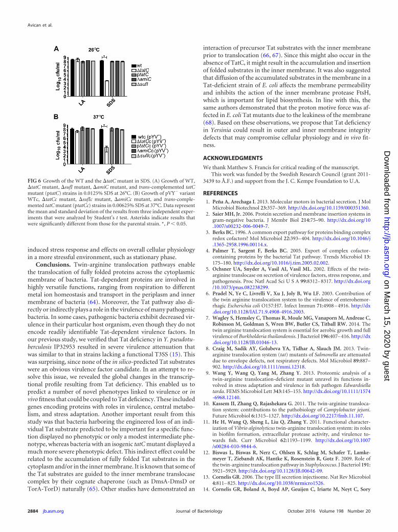
Tat pathway is essential for iron acquisition. Efficient ironacquisition is a prerequisite for pathogenic bacteria to replicate inthe host. Interestingly, several genes encoding iron transport andstorage proteins were differentially expressed in the �tatC mutant.The bfr gene encoding bacterioferritin was upregulated under allconditions tested (see Table S2 in the supplemental material). Therole of bacterioferritin is to store and/or supply iron and reduce itstoxicity (25). As such, bacterioferritin has been shown to be im-portant for the virulence of Mycobacterium tuberculosis (26). Al-though less is known about its function in the Yersinia species, a19-kDa cytoplasmic bacterioferritin-like protein in Y. pestis is sug-gested to be involved in iron storage (27). In addition, expressionlevels of three genes, fbpC, fbpB, and fbpA, in Y. pestis proposed tobe the proteins of an iron uptake system were also upregulated inthe �tatC mutant (see Table S2). Therefore, we decided to inves-tigate growth of the �tatC mutant under iron-limited conditions.A mutant lacking the FhuD protein (�fhuD), a predicted Tat sub-strate, known to be involved in iron-hydroxamate siderophoreuptake in Yersinia (28), was also evaluated for growth under iron-limited conditions. Compared to the wild-type strain, the �tatCmutant showed a severe growth defect in iron-limited media atboth 26°C and at 37°C (pYV� derivatives of the same strains wereused), which could be complemented with a wild-type copy of thetatC gene supplied in trans (Fig. 2A and B). The �fhuD mutantalso showed impaired growth at host temperature, but the defectwas less severe than for the �tatC mutant (Fig. 2B). On the otherhand, at 26°C, the �fhuD mutant showed a growth defect almostas severe as that with the �tatC mutant (Fig. 2A). Hence, FhuDfunction is more important at 26°C, when changes in the �tatCmutant transcriptome are most dramatic. This also suggests thatother iron acquisition systems, such as the Ybt system that isknown to be expressed mainly at host temperature, could have apreferred role in iron uptake at 37°C, and that a functional Tatsystem is required for the function of one or more iron uptakesystems (29). To investigate the involvement of FhuD in thegrowth defect of the tatC mutant at 37°C and to look into the Tat
FIG 1 Functional clustering of differentially regulated genes in the �tatC mutant. (A) Functional clustering was performed by using the KEGG pathwaymapping, KEGG database, and BLAST search. Inf., information. (B) Venn scheme of differentially regulated genes under all conditions tested. Exp., exponentialphase; Stat., stationary phase.
Transcriptome of Tat-Deficient Y. pseudotuberculosis
October 2016 Volume 198 Number 20 jb.asm.org 2879Journal of Bacteriology
on March 15, 2020 by guest
http://jb.asm.org/
Dow
nloaded from

dependency of FhuD export, we also investigated the growth ofthese strains in iron-limited medium containing ferrichrome.FhuD functions in the uptake of ferric-hydroxamate-type fer-richrome, which is an iron-chelating siderophore (28). As a con-trol, we also included a �ybtP mutant, as ybtP is a gene requiredfor another independent iron uptake system. The �fhuD and the�tatC mutant strains showed similar and highly impaired growthphenotypes, arguing that the growth defect observed for the tatCmutant was caused by the failure to translocate FhuD to periplasmand, as a consequence, to take up ferrichrome (Fig. 2C). Thisphenotype was readily complemented by expression of an intacttatC gene in trans. As expected, the addition of ferrichrome didnot influence growth of the ybtP mutant strain, as this mutationaffects another nonrelated iron uptake system.
Overall, our findings show that a functional Tat system is re-quired for iron uptake and that this at least in part is due to a lossof FhuD translocation across the inner membrane. At host tem-perature, the growth defect under iron limitation is more severe inthe �tatC mutant strain than in the fhuD mutant. This indicatesthat the defect cannot be solely due to a loss of FhuD function.Finally, the impaired growth under iron-limited conditions wouldbe expected to significantly impact in vivo fitness and virulence.
Virulence-related genes are differentially regulated in the�tatC mutant. Several of the genes encoding T3SS components,including secreted effectors (yopE, yopK, and yopM), translocatorsand regulators (lcrV, lcrG, and yopD), and the secretome compo-nents (yscN and yscO), were downregulated at 26°C in stationaryphase (see Table S2 in the supplemental material). However, un-der these conditions, the expression of T3SS genes is overall low,and none of the genes encoding T3SS components were differen-tially regulated at 37°C. This finding was in line with our previousstudy that a loss of Tat function did not have any impact on secre-tion and expression of Yops- or YopE-mediated cytotoxicity in anin vitro cell infection model (15).
Y. pseudotuberculosis YPIII harbors four type VI secretion sys-tem clusters (T6SS-1 to -4). Interestingly, we observed that 11 outof 18 genes in a homologous T6SS-4 cluster in the IP32953 strainwere upregulated in the �tatC mutant background at 26°C instationary phase (see Table S2 in the supplemental material). Pre-vious reports have shown the T6SS-4 cluster to be highly ex-pressed at 26°C during stationary phase but downregulated at
37°C (30). The T6SS-4 cluster is important for Y. pseudotubercu-losis survival under acidic conditions, high osmolarity, and expo-sure to the detergents, such as deoxycholate, that impair outermembrane integrity. Consistent with this, T6SS-4 is known to beregulated by RpoS and might function as a part of the generalstress response (31–33). Hence, the observed upregulation in theTatC-deficient strain at 26°C could be a response to alterations inthe outer membrane integrity (see also below) and the consequentupregulation of one or more stress response genes involved in themaintenance and repair of this structure.
Finally, expression of yadA encoding the multifunctional ad-hesin was highly downregulated at 26°C, and in contrast to theother pYV-carried T3SS genes, yadA was also downregulated dur-ing exponential growth at 37°C (see Table S2 in the supplementalmaterial). We could also demonstrate the downregulation ofYadA production significantly in the tatC mutant by immuno-blotting using an anti-YadA antibody, when bacteria were grownin both the presence and absence of Ca2 at 37°C (Fig. 3). YadA isencoded by the common pYV virulence plasmid of human-patho-genic Yersinia spp., and expression is induced by a temperatureshift to 37°C (34). Significantly, YadA has been shown to be crucialfor the virulence of Y. enterocolitica (35). There is no obvious
FIG 2 Comparison of in vitro growth under iron-limited conditions. Growth of parental (wt), �tatC mutant, �fhuD mutant, and trans-complemented tatCmutant (ptatC) strains in iron-depleted TMH medium at 26°C (A) and of their pYV� variants at 37°C (B). (C) Growth of parental (wtc), �tatC mutant, �fhuDmutant, �ybtP mutant, and trans-complemented tatC mutant (ptatC) strains in iron-depleted TMH medium supplemented with 10 �M ferrichrome (Ferchr).The results are representative of three independent experiments, with the error bars representing the standard deviation. OD600, optical density at 600 nm.
FIG 3 YadA expression is downregulated in the �tatC mutant. Parental (wt),�tatC mutant, and trans-complemented tatC mutant (ptatC) strains were in-cubated in LB medium supplemented with or depleted of Ca2 with the addi-tion of 5 mM EGTA. Whole-cell extracts were separated on SDS-polyacryl-amide gel and analyzed by Western blotting using rabbit polyclonal anti-YadAantibody (top). The membrane was also incubated with anti-DnaK antibodyas a loading control (bottom). The signal intensities of each of the YadA bandswere normalized with DnaK signals in each strain. The relative signal levels ofYadA were calculated with comparison to the wild type (set as 100%). Theanalyses were performed with ImageJ. Molecular masses (Spectra multicolorhigh-range protein ladder; Thermo Scientific) are shown on the left.
Avican et al.
2880 jb.asm.org October 2016 Volume 198 Number 20Journal of Bacteriology
on March 15, 2020 by guest
http://jb.asm.org/
Dow
nloaded from

direct link between a functional Tat system and expression ofYadA, suggesting that the observed effect on expression is likely tobe mediated through another unidentified pathway. In a mousemodel, Y. pseudotuberculosis yadA mutants can still cause a sys-temic infection but with significantly lower numbers of bacteria insystemic organs (36). A role for YadA in persistence and systemicdissemination has been shown to occur in cooperation with theAil adhesion (37) and involves an interaction with neutrophils(38). Based on this finding, we speculate that a downregulation ofYadA expression could constitute an impediment during the ini-tial in vivo colonization by the �tatC mutant.
Loss of Tat function has a major impact on carbon metabo-lism. Functional annotation analysis revealed that under all testedconditions, a significant fraction (23%) of the differentially regu-lated genes are involved in metabolism. The most notable effectoccurs during stationary-phase growth at 26°C, where 40% of the91 differentially regulated genes are implicated in carbon metab-olism, while others are involved in amino acid, nucleotide, energy,cofactor, vitamin, and lipid metabolism (Fig. 4A). Upregulation ofgenes encoding enzymes involved in glycolysis, such as pgi, pgk,eno, pykF, pfkA, and gpmA, indicates that the glycolytic pathway isinduced in response to the lack of a functional Tat system. Toconfirm that there was an upregulation of glycolysis, we compared
the growth of the WT and �tatC mutant strains in LB agar con-taining glucose and phenol red. Phenol red is a pH indicator; if thepH is acidic, the medium turns from orange to yellow, or it turnspink if the pH is alkaline. After growth at 26°C, there was a veryclear yellow color around the colony of the �tatC mutant (Fig.4C). The acidification of medium by the �tatC mutant is in linewith the observed upregulation of glycolysis that would result inhigher levels of acidic end products. However, the mediumaround colonies of the WT and the trans-complemented �tatCmutant strain both turned pink, which indicated that they bothfermented the peptones in the medium and secreted alkaline endproducts (Fig. 4C). Both the dld and lld genes that encode lactatehydrogenases were also upregulated in the �tatC mutant, indicat-ing the excess pyruvate had been oxidized to lactic acid, confirm-ing the intense secretion of acidic end products. It is not clear whythe �tatC mutant uses this pathway more actively, since glycolysisyields low energy and that would render a low metabolic state,especially in stationary phase. Interestingly, genes encoding en-zymes involved in succinate biogenesis through the oxidativebranch of the tricarboxylic acid (TCA) cycle (sdhABCD andsucBC) were downregulated, while the expression of the frdABCDoperon that is involved in the reductive branch of the TCA cyclewas upregulated (Fig. 4B; see also Table S2 in the supplemental
FIG 4 (A) Genes involved in different metabolic functions that are differentially regulated in the �tatC mutant at 26°C in stationary phase are shown in the piegraph. Degr., degradation; Met., metabolism. (B) Genes involved in central carbon metabolism differentially regulated in the �tatC mutant at 26°C in stationaryphase. (Upregulated genes are indicated in red; downregulated genes are indicated in green). diP, diphosphate; CoA, coenzyme A. (C) Growth of parental (wt),�tatC mutant, and trans-complemented tatC mutant (ptatC) strains on LB–1.5% agar supplemented with phenol red and 0.2% glucose at 26°C for 24 h. (D)Intracellular concentrations of fumarate in parental (wt), �tatC mutant, and trans-complemented tatC mutant (ptatC) strains. Data represent the mean andstandard deviation of the results from four independent experiments that were analyzed by Student’s t test. Asterisks indicate results that were significantlydifferent from those for the parental strain. ***, 0.0001 � P 0.001.
Transcriptome of Tat-Deficient Y. pseudotuberculosis
October 2016 Volume 198 Number 20 jb.asm.org 2881Journal of Bacteriology
on March 15, 2020 by guest
http://jb.asm.org/
Dow
nloaded from

material). These changes in central carbon metabolism are similarto those previously described for E. coli and Shigella flexneri uponentry into stationary phase (39, 40), and it is noteworthy that thesechanges were far more pronounced in a Tat-deficient strain thanin the wild type. Additionally, the expression of argH encodingargininosuccinate lyase that produces fumarate from aspartateand the downstream urease operon (ureABDCEG) was also up-regulated (Fig. 4B; see also Table S2), which could cause the up-regulation of fumarate reduction. These findings were furthersupported, as they also correlated with increased intracellular fu-marate concentration in the �tatC mutant. The �tatC mutant hadan average of 29.5 ng/�l fumarate, whereas the parental straincontained 18.2 ng/�l (Fig. 4D). The phenotype was partially com-plemented by expressing ptatC in trans with fumarate levels of22.1 ng/�l (Fig. 4D). Furthermore, upregulation of the reductivebranch of the TCA cycle (frdABCD) suggests implication in anaer-obic respiration pathways in response to lower oxygen availability.It is known that bacteria repress aerobic metabolism in late-sta-tionary phase (41), and also there are 6 predicted Tat substrates inY. pseudotuberculosis with possible function in anaerobic respira-tion and that could result in the upregulation of anaerobic-likemetabolism in the �tatC mutant. Importantly, however, we foundthe Tat-deficient strain was not impaired in in vitro growth underanaerobic conditions (data not shown), similar to what was re-ported for Salmonella species (8).
Approximately 26 genes encoding ribosomal proteins and var-ious tRNA synthetases were found to be downregulated at 26°C instationary phase in the �tatC mutant, which is again the hallmarkof bacterial stationary phase (42). Altogether, the changes in cen-tral carbon and energy metabolism in the �tatC mutant are a clearindication that Tat-deficient strains are more stressed in harshenvironments, such as those with nutrient starvation, and thiswould be expected to negatively impact their in vivo fitness.
Tat system is important for copper homeostasis of Y. pseu-dotuberculosis. High copper levels are toxic, and bacteria haveevolved copper resistance and translocation systems that functionto equilibrate the intracellular copper levels. In E. coli, the CopA-CueO and CusCFBA systems are involved in protecting the bac-terium during exposure to moderate and high copper concentra-tions under aerobic and anaerobic conditions, respectively (43).Our transcriptomic analysis showed that the Cop system is differ-entially regulated in the �tatC mutant. At 26°C in stationaryphase, the copA gene encoding a copper resistance protein wasupregulated 2.4-fold, whereas the copCD genes were downregu-lated (see Table S2 in the supplemental material). It is notable thatthe periplasmic multicopper oxidase CueO involved in coppertolerance is an in silico-predicted Tat substrate in Y. pseudotuber-culosis (15). Moreover, CueO homologous proteins of E. coli, Sal-monella enterica serovar Typhimurium, and M. tuberculosis areestablished Tat substrates (8, 44, 45). This prompted us to com-pare the copper tolerance of Y. pseudotuberculosis wild-type,�tatC mutant, and �cueO mutant strains at different tempera-tures. When strains were grown in media with increasing copperconcentrations at either 26°C or 37°C, the �tatC mutant was sig-nificantly more sensitive to copper than the wild-type strain, andthis sensitivity could be complemented with the ptatC strain inmost of the tested concentrations at both temperatures (Fig. 5Aand B). The lowered complementation level at elevated copperconcentrations (�2 mM CuCl2) might be due to growth difficul-ties that the parental strain also encounters. Additionally, a �cueO
mutant showed an intermediate sensitivity, although the putativeTat substrate CueO seemed to be more important for copper tol-erance at elevated temperature. Nevertheless, these results suggestthat the mislocalization of CueO in the absence of a functional Tatpathway is only partly responsible for the marked copper sensitiv-ity of the Y. pseudotuberculosis �tatC mutant. Similar to the find-ing in E. coli, our data support the hypothesis that Tat deficiencycauses a more general effect that cannot be attributed to the mis-localization of any given Tat substrate (46). However, since patho-genic bacteria often must cope with copper stress generated upontheir internalization by phagocytes (47), increased copper sensi-tivity could impair the in vivo fitness of Tat-deficient bacteria.
Stress responses are strongly induced in the absence of afunctional Tat system. Several studies indicate that a functionalTat system is required for many pathogenic bacteria, such as Pseu-domonas syringae pv. tomato DC3000, S. enterica, Campylobacterjejuni, and Ralstonia solanacearum (5, 48–51), to efficiently re-
FIG 5 Survival of the �tatC mutant in CuCl2 and in acidic pH. (A) Survival ofWT, �tatC mutant, �cueO mutant, and trans-complemented tatC mutant(ptatC) strains in increasing CuCl2 concentrations at 26°C. (B) Growth ofpYV� variant WTc, �tatCc mutant, �cueOc mutant, and trans-complementedtatC mutant (ptatCc) strains in increasing CuCl2 concentrations at 37°C. Datarepresent the mean and standard deviation of the results from four indepen-dent experiments that were analyzed by Student’s t test. (C) Survival of pYV�
variant WTc, �tatCc mutant, �cynTc mutant, and trans-complemented tatCmutant (ptatCc) strains in LB medium with pH 3.0 at 37°C. Percent survivalwas calculated from 15 min of growth of each strain in LB medium with pH 7.0at 37°C. Data represent the mean and standard deviation of the results fromfive independent experiments that were analyzed by Student’s t test. Asterisksindicate results that were significantly different from those for the parentalstrain. ****, P 0.0001; ***, 0.0001 � P 0.001; **, 0.001 � P 0.05; *, P 0.05.
Avican et al.
2882 jb.asm.org October 2016 Volume 198 Number 20Journal of Bacteriology
on March 15, 2020 by guest
http://jb.asm.org/
Dow
nloaded from

spond to diverse forms of physicochemical stresses. Indeed, ourtranscriptomic analysis of the Y. pseudotuberculosis �tatC mutantrevealed that a number of prominent stress-responsive genes wereinduced at both temperatures, and this number was higher duringstationary-phase growth. During growth at 26°C in exponentialphase, genes encoding osmotically inducible protein (osmY),osmotic response regulator (ompR), superoxide dismutases(sodCB), sigma 54-modulation protein (yfiA), universal stressprotein (uspA), and peptidase (pepT) were upregulated (see TableS2 in the supplemental material). Strikingly, at 26°C during sta-tionary-phase growth, these genes and several additional genes (28in total) that encode most types of stress responses ranging fromacid, heat shock, and stationary phase survival to cold shock, ox-idative stress, and high osmolarity were found to be upregulated.Significantly, the rpoS gene encoding the sigma factor S, known toregulate responsiveness to high osmolarity, high temperature, andoxidative stress during entry into stationary phase (52), was alsoupregulated 4.1-fold at 26°C during stationary growth (see TableS2). The RpoS regulon in Yersinia spp. has not been investigatedextensively but was shown to be crucial for adaptation to variousstresses as well as biofilm formation and motility in both Y. pseu-dotuberculosis and Y. enterocolitica (33, 53). Hence, our data pro-vide further evidence that the stationary-phase environment ismuch harsher for the �tatC mutant than for the wild type andresults in the induction of many different stress responses.
The acid stress response genes are of particular interest, sincesurvival at low pH is crucial for the enteric route of infection.Interestingly, genes encoding proteins involved in several differ-ent acid tolerance systems were upregulated in the �tatC mutant.These included the urease operon (ureABCDEG) encoding ureaseaccessory proteins and adiA and adiC encoding amino acid decar-boxylase and antiporter systems, respectively. Finally, the hdeBand hdeD genes, encoding the acid-stress-activated periplasmicchaperone HdeB and membrane protein HdeD, respectively, wereupregulated in the �tatC mutant (see Table S2 in the supplemen-tal material). In addition to its role in metabolism, the ureaseoperon is known to be essential for virulence and acid resistance ofenteric pathogens, including Y. enterocolitica (54). It has been sug-gested that the expression of urease genes is regulated by OmpR inY. pseudotuberculosis (55), but there is no direct evidence for a roleof urease in virulence (56). The adiAC genes, encoding argininedecarboxylase and the arginine-agmatine antiporter, respectively,were highly upregulated (fold change, 7.4 and 6.8, respectively;Table S2). This system consumes cytoplasmic protons and pro-motes acid survival in E. coli (57). Bacteria that are exposed to acidchallenge often upregulate the expression of the respiratory chaincomplexes that function to pump out protons, while ATP syn-thases that transport protons to the inside are downregulated (58).We also observed a downregulation of atpHBI encoding the ATPsynthases, and that could be an indication of severe acid stress inthe �tatC mutant, even though we did not observe a correspond-ing upregulation of specific proton-pumping cytochromes. Nev-ertheless, these transcription data corroborate previous resultswhere we showed that Tat deficiency impaired the bacterial sur-vival in acidic pH at moderate temperature (15). An interestingobservation from our previous study was that the putative Tatsubstrate carbonic anhydrase (CynT) may play a role in the sur-vival of Y. pseudotuberculosis at low pH (15). This is also in linewith the expression levels of cynT, which were upregulated at bothtested growth temperatures (see Table S2). For these reasons, we
examined a �cynT mutant in this study and also performed theexperiment at 37°C. We found that the wild-type and �cynTc(pYV� derivative) mutant strains showed similar survival rates,whereas the survival rate of the �tatCc (pYV� derivative) mutantwas much lower in LB medium at pH 3.0 (Fig. 5C). The transcomplementation in the strain expressing tatC in trans was in-complete and most likely due to the reasons discussed above. Thisverifies that the �tatC mutant is indeed sensitive to acidic condi-tions at 37°C but that the putative Tat substrate CynT is actuallydispensable for survival under acidic conditions. The attenuationof the tatC mutant in the oral infection mouse model is likely to becaused by the lower survival under acid conditions encounteredduring the passage through the stomach or upon internalizationby host cells.
Envelope defects result in physiological changes of the �tatCmutants. A pleiotropic defect in the outer membrane relating toTat function was first seen in E. coli, where Tat mutants werecharacterized by sensitivity to SDS and hydrophobic drugs (59).These effects were found to be the result of the mislocalization oftwo Tat-dependent periplasmic amidases, AmiC and AmiA (60).The same was also reported for Salmonella spp., but in this case,the sensitivity in addition involved the mislocalization of theperiplasmic protein SufI. These defects were suggested to cause asevere attenuation of the Tat mutant in a mouse model (8). More-over, a transcriptomic study of E. coli revealed that the rcs envelopestress regulon genes were upregulated in the Tat mutant in re-sponse to pleiotropic defects in the outer membrane (46). How-ever, our transcriptomic analysis revealed no differences in theexpression of the rcs regulon of Yersinia species (61). In addition,no other known genes encoding envelope and periplasmic stressresponse regulators, such as rpoE, bae, or inner membrane stressresponse system psp, were upregulated in the tatC mutant. Basedon the results from E. coli and Salmonella, we targeted the role ofAmiC and SufI especially in envelope stress, since both are pre-dicted to be Tat substrates in Yersinia (15). Especially, we tested a�amiC and a �sufI mutant together with a �tatC mutant and thewild-type strain for SDS sensitivity at 26°C and 37°C. Y. pseudotu-berculosis was found to be highly sensitive to envelope stress at37°C, which necessitated a decrease in the SDS concentration usedcompared to that with assays at 26°C. This indicates that envelopeintegrity and/or permeability are different at host temperaturethan under environmental conditions. The wild-type, �sufI mu-tant, and �amiC mutant all grew equally well on LA plates aloneand with SDS, while the �tatC mutant was highly impaired forgrowth (10,000-fold, compared to wild type) on LA plates con-taining SDS at both growth temperatures (Fig. 6). This clearlyshows that Tat deficiency drastically impairs cell envelope integ-rity, and that this phenotype is not caused solely by the loss ofAmiC and SufI. Additionally, these results indicate that the rela-tion of outer membrane integrity to the Tat pathway in Yersinia isdifferent from that in E. coli and Salmonella. Since AmiA has aclear Sec signal peptide in Yersinia, the mislocalization of AmiCalone in the Tat-deficient strain cannot be directly attributed tothe defect in outer membrane integrity. However, the downregu-lation of msbB that is responsible for lipid A biosynthesis andupregulation of the mechanosensitive channel protein encoded bygenes mscL and mscS that function in sensing and release of turgorpressure during hypo-osmotic shock could be linked to increasedouter membrane permeability changes (62, 63). Therefore, it isthat the changes in the outer membrane permeability explain the
Transcriptome of Tat-Deficient Y. pseudotuberculosis
October 2016 Volume 198 Number 20 jb.asm.org 2883Journal of Bacteriology
on March 15, 2020 by guest
http://jb.asm.org/
Dow
nloaded from
induced stress response and effects on overall cellular physiologyin a more stressful environment, such as stationary phase.
Conclusions. Twin-arginine translocation pathways enablethe translocation of fully folded proteins across the cytoplasmicmembrane of bacteria. Tat-dependent proteins are involved inhighly versatile functions, ranging from respiration to differentmetal ion homeostasis and transport in the periplasm and innermembrane of bacteria (64). Moreover, the Tat pathway also di-rectly or indirectly plays a role in the virulence of many pathogenicbacteria. In some cases, pathogenic bacteria exhibit decreased vir-ulence in their particular host organism, even though they do notencode readily identifiable Tat-dependent virulence factors. Inour previous study, we verified that Tat deficiency in Y. pseudotu-berculosis IP32953 resulted in severe virulence attenuation thatwas similar to that in strains lacking a functional T3SS (15). Thiswas surprising, since none of the in silico-predicted Tat substrateswere an obvious virulence factor candidate. In an attempt to re-solve this issue, we revealed the global changes in the transcrip-tional profile resulting from Tat deficiency. This enabled us topredict a number of novel phenotypes linked to virulence or invivo fitness that could be coupled to Tat deficiency. These includedgenes encoding proteins with roles in virulence, central metabo-lism, and stress adaptation. Another important result from thisstudy was that bacteria harboring the engineered loss of an indi-vidual Tat substrate predicted to be important for a specific func-tion displayed no phenotypic or only a modest intermediate phe-notype, whereas bacteria with an isogenic tatC mutant displayed amuch more severe phenotypic defect. This indirect effect could berelated to the accumulation of fully folded Tat substrates in thecytoplasm and/or in the inner membrane. It is known that some ofthe Tat substrates are guided to the inner membrane translocasecomplex by their cognate chaperone (such as DmsA-DmsD orTorA-TorD) naturally (65). Other studies have demonstrated an
interaction of precursor Tat substrates with the inner membraneprior to translocation (66, 67). Since this might also occur in theabsence of TatC, it might result in the accumulation and insertionof folded substrates in the inner membrane. It was also suggestedthat diffusion of the accumulated substrates in the membrane in aTat-deficient strain of E. coli affects the membrane permeabilityand inhibits the action of the inner membrane protease FtsH,which is important for lipid biosynthesis. In line with this, thesame authors demonstrated that the proton motive force was af-fected in E. coli Tat mutants due to the leakiness of the membrane(68). Based on these observations, we propose that Tat deficiencyin Yersinia could result in outer and inner membrane integritydefects that may compromise cellular physiology and in vivo fit-ness.
ACKNOWLEDGMENTS
We thank Matthew S. Francis for critical reading of the manuscript.This work was funded by the Swedish Research Council (grant 2011-
3439 to Å.F.) and support from the J. C. Kempe Foundation to U.A.
REFERENCES1. Peña A, Arechaga I. 2013. Molecular motors in bacterial secretion. J Mol
Microbiol Biotechnol 23:357–369. http://dx.doi.org/10.1159/000351360.2. Saier MH, Jr. 2006. Protein secretion and membrane insertion systems in
gram-negative bacteria. J Membr Biol 214:75–90. http://dx.doi.org/10.1007/s00232-006-0049-7.
3. Berks BC. 1996. A common export pathway for proteins binding complexredox cofactors? Mol Microbiol 22:393– 404. http://dx.doi.org/10.1046/j.1365-2958.1996.00114.x.
4. Palmer T, Sargent F, Berks BC. 2005. Export of complex cofactor-containing proteins by the bacterial Tat pathway. Trends Microbiol 13:175–180. http://dx.doi.org/10.1016/j.tim.2005.02.002.
5. Ochsner UA, Snyder A, Vasil AI, Vasil ML. 2002. Effects of the twin-arginine translocase on secretion of virulence factors, stress response, andpathogenesis. Proc Natl Acad Sci U S A 99:8312– 8317. http://dx.doi.org/10.1073/pnas.082238299.
6. Pradel N, Ye C, Livrelli V, Xu J, Joly B, Wu LF. 2003. Contribution ofthe twin arginine translocation system to the virulence of enterohemor-rhagic Escherichia coli O157:H7. Infect Immun 71:4908 – 4916. http://dx.doi.org/10.1128/IAI.71.9.4908-4916.2003.
7. Wagley S, Hemsley C, Thomas R, Moule MG, Vanaporn M, Andreae C,Robinson M, Goldman S, Wren BW, Butler CS, Titball RW. 2014. Thetwin arginine translocation system is essential for aerobic growth and fullvirulence of Burkholderia thailandensis. J Bacteriol 196:407– 416. http://dx.doi.org/10.1128/JB.01046-13.
8. Craig M, Sadik AY, Golubeva YA, Tidhar A, Slauch JM. 2013. Twin-arginine translocation system (tat) mutants of Salmonella are attenuateddue to envelope defects, not respiratory defects. Mol Microbiol 89:887–902. http://dx.doi.org/10.1111/mmi.12318.
9. Wang Y, Wang Q, Yang M, Zhang Y. 2013. Proteomic analysis of atwin-arginine translocation-deficient mutant unravel its functions in-volved in stress adaptation and virulence in fish pathogen Edwardsiellatarda. FEMS Microbiol Lett 343:145–155. http://dx.doi.org/10.1111/1574-6968.12140.
10. Kassem II, Zhang Q, Rajashekara G. 2011. The twin-arginine transloca-tion system: contributions to the pathobiology of Campylobacter jejuni.Future Microbiol 6:1315–1327. http://dx.doi.org/10.2217/fmb.11.107.
11. He H, Wang Q, Sheng L, Liu Q, Zhang Y. 2011. Functional character-ization of Vibrio alginolyticus twin-arginine translocation system: its rolesin biofilm formation, extracellular protease activity, and virulence to-wards fish. Curr Microbiol 62:1193–1199. http://dx.doi.org/10.1007/s00284-010-9844-6.
12. Biswas L, Biswas R, Nerz C, Ohlsen K, Schlag M, Schafer T, Lamke-meyer T, Ziebandt AK, Hantke K, Rosenstein R, Gotz F. 2009. Role ofthe twin-arginine translocation pathway in Staphylococcus. J Bacteriol 191:5921–5929. http://dx.doi.org/10.1128/JB.00642-09.
13. Cornelis GR. 2006. The type III secretion injectisome. Nat Rev Microbiol4:811– 825. http://dx.doi.org/10.1038/nrmicro1526.
14. Cornelis GR, Boland A, Boyd AP, Geuijen C, Iriarte M, Neyt C, Sory
FIG 6 Growth of the WT and the �tatC mutant in SDS. (A) Growth of WT,�tatC mutant, �sufI mutant, �amiC mutant, and trans-complemented tatCmutant (ptatC) strains in 0.0125% SDS at 26°C. (B) Growth of pVY� variantWTc, �tatCc mutant, �sufIc mutant, �amiCc mutant, and trans-comple-mented tatC mutant (ptatCc) strains in 0.00625% SDS at 37°C. Data representthe mean and standard deviation of the results from three independent exper-iments that were analyzed by Student’s t test. Asterisks indicate results thatwere significantly different from those for the parental strain. *, P 0.05.
Avican et al.
2884 jb.asm.org October 2016 Volume 198 Number 20Journal of Bacteriology
on March 15, 2020 by guest
http://jb.asm.org/
Dow
nloaded from

MP, Stainier I. 1998. The virulence plasmid of Yersinia, an antihost ge-nome. Microbiol Mol Biol Rev 62:1315–1352.
15. Lavander M, Ericsson SK, Broms JE, Forsberg A. 2006. The twin argi-nine translocation system is essential for virulence of Yersinia pseudotu-berculosis. Infect Immun 74:1768 –1776. http://dx.doi.org/10.1128/IAI.74.3.1768-1776.2006.
16. Straley SC, Bowmer WS. 1986. Virulence genes regulated at the transcrip-tional level by Ca2 in Yersinia pestis include structural genes for outermembrane proteins. Infect Immun 51:445– 454.
17. Milton DL, O’Toole R, Horstedt P, Wolf-Watz H. 1996. Flagellin A isessential for the virulence of Vibrio anguillarum. J Bacteriol 178:1310 –1319.
18. Heroven AK, Sest M, Pisano F, Scheb-Wetzel M, Steinmann R, BohmeK, Klein J, Munch R, Schomburg D, Dersch P. 2012. Crp inducesswitching of the CsrB and CsrC RNAs in Yersinia pseudotuberculosis andlinks nutritional status to virulence. Front Cell Infect Microbiol 2:158.
19. Pfaffl MW. 2001. A new mathematical model for relative quantification inreal-time RT-PCR. Nucleic Acids Res 29:e45. http://dx.doi.org/10.1093/nar/29.9.e45.
20. Obi IR, Nordfelth R, Francis MS. 2011. Varying dependency of periplas-mic peptidylprolyl cis-trans isomerases in promoting Yersinia pseudotu-berculosis stress tolerance and pathogenicity. Biochem J 439:321–332.http://dx.doi.org/10.1042/BJ20110767.
21. Eitel J, Dersch P. 2002. The YadA protein of Yersinia pseudotuberculosismediates high-efficiency uptake into human cells under environmentalconditions in which invasin is repressed. Infect Immun 70:4880 – 4891.http://dx.doi.org/10.1128/IAI.70.9.4880-4891.2002.
22. Avican K, Fahlgren A, Huss M, Heroven AK, Beckstette M, Dersch P,Fallman M. 2015. Reprogramming of Yersinia from virulent to persistentmode revealed by complex in vivo RNA-seq analysis. PLoS Pathog 11:e1004600. http://dx.doi.org/10.1371/journal.ppat.1004600.
23. Gentleman RC, Carey VJ, Bates DM, Bolstad B, Dettling M, Dudoit S,Ellis B, Gautier L, Ge YC, Gentry J, Hornik K, Hothorn T, Huber W,Iacus S, Irizarry R, Leisch F, Li C, Maechler M, Rossini AJ, Sawitzki G,Smith C, Smyth G, Tierney L, Yang JYH, Zhang JH. 2004. Bioconduc-tor: open software development for computational biology and bioinfor-matics. Genome Biol 5:R80. http://dx.doi.org/10.1186/gb-2004-5-10-r80.
24. Bagos PG, Nikolaou EP, Liakopoulos TD, Tsirigos KD. 2010. Combinedprediction of Tat and Sec signal peptides with hidden Markov models.Bioinformatics 26:2811–2817. http://dx.doi.org/10.1093/bioinformatics/btq530.
25. Carrondo MA. 2003. Ferritins, iron uptake and storage from the bacte-rioferritin viewpoint. EMBO J 22:1959 –1968. http://dx.doi.org/10.1093/emboj/cdg215.
26. Reddy PV, Puri RV, Khera A, Tyagi AK. 2012. Iron storage proteins areessential for the survival and pathogenesis of Mycobacterium tuberculosisin THP-1 macrophages and the guinea pig model of infection. J Bacteriol194:567–575. http://dx.doi.org/10.1128/JB.05553-11.
27. Perry RD, Lucier TS, Sikkema DJ, Brubaker RR. 1993. Storage reservoirsof hemin and inorganic iron in Yersinia pestis. Infect Immun 61:32–39.
28. Forman S, Nagiec MJ, Abney J, Perry RD, Fetherston JD. 2007.Analysis of the aerobactin and ferric hydroxamate uptake systems ofYersinia pestis. Microbiology 153:2332–2341. http://dx.doi.org/10.1099/mic.0.2006/004275-0.
29. Chauvaux S, Rosso ML, Frangeul L, Lacroix C, Labarre L, Schiavo A,Marceau M, Dillies MA, Foulon J, Coppee JY, Medigue C, Simonet M,Carniel E. 2007. Transcriptome analysis of Yersinia pestis in human plas-ma: an approach for discovering bacterial genes involved in septicaemicplague. Microbiology 153:3112–3124. http://dx.doi.org/10.1099/mic.0.2007/006213-0.
30. Zhang W, Xu S, Li J, Shen X, Wang Y, Yuan Z. 2011. Modulation of athermoregulated type VI secretion system by AHL-dependent quorumsensing in Yersinia pseudotuberculosis. Arch Microbiol 193:351–363.
31. Gueguen E, Durand E, Zhang XY, d’Amalric Q, Journet L, Cascales E.2013. Expression of a type VI secretion system is responsive to envelopestresses through the OmpR transcriptional activator. PLoS One 8:e66615.http://dx.doi.org/10.1371/journal.pone.0066615.
32. Zhang W, Wang Y, Song Y, Wang T, Xu S, Peng Z, Lin X, Zhang L,Shen X. 2013. A type VI secretion system regulated by OmpR in Yer-sinia pseudotuberculosis functions to maintain intracellular pH homeo-stasis. Environ Microbiol 15:557–569. http://dx.doi.org/10.1111/1462-2920.12005.
33. Guan J, Xiao X, Xu S, Gao F, Wang J, Wang T, Song Y, Pan J, Shen X,
Wang Y. 2015. Roles of RpoS in Yersinia pseudotuberculosis stress survival,motility, biofilm formation and type VI secretion system expression. JMicrobiol 53:633– 642. http://dx.doi.org/10.1007/s12275-015-0099-6.
34. Skurnik M, Toivanen P. 1992. LcrF is the temperature-regulated activa-tor of the yadA gene of Yersinia enterocolitica and Yersinia pseudotubercu-losis. J Bacteriol 174:2047–2051.
35. Roggenkamp A, Schubert S, Jacobi CA, Heesemann J. 1995. Dissectionof the Yersinia enterocolitica virulence plasmid pYVO8 into an operatingunit and virulence gene modules. FEMS Microbiol Lett 134:69 –73. http://dx.doi.org/10.1111/j.1574-6968.1995.tb07916.x.
36. Heise T, Dersch P. 2006. Identification of a domain in Yersinia virulencefactor YadA that is crucial for extracellular matrix-specific cell adhesionand uptake. Proc Natl Acad Sci U S A 103:3375–3380. http://dx.doi.org/10.1073/pnas.0507749103.
37. Paczosa MK, Fisher ML, Maldonado-Arocho FJ, Mecsas J. 2014. Yer-sinia pseudotuberculosis uses Ail and YadA to circumvent neutrophils bydirecting Yop translocation during lung infection. Cell Microbiol 16:247–268. http://dx.doi.org/10.1111/cmi.12219.
38. Durand EA, Maldonado-Arocho FJ, Castillo C, Walsh RL, Mecsas J.2010. The presence of professional phagocytes dictates the number of hostcells targeted for Yop translocation during infection. Cell Microbiol 12:1064 –1082. http://dx.doi.org/10.1111/j.1462-5822.2010.01451.x.
39. Zhu L, Liu XK, Zhao G, Zhi YD, Bu X, Ying TY, Feng EL, Wang J,Zhang XM, Huang PT, Wang HL. 2007. Dynamic proteome changes ofShigella flexneri 2a during transition from exponential growth to station-ary phase. Genomics Proteomics Bioinformatics 5:111–120. http://dx.doi.org/10.1016/S1672-0229(07)60021-7.
40. Nyström T. 1994. The glucose-starvation stimulon of Escherichia coli:induced and repressed synthesis of enzymes of central metabolic pathwaysand role of acetyl phosphate in gene expression and starvation survival.Mol Microbiol 12:833– 843. http://dx.doi.org/10.1111/j.1365-2958.1994.tb01069.x.
41. Chang DE, Smalley DJ, Conway T. 2002. Gene expression profiling ofEscherichia coli growth transitions: an expanded stringent response model.Mol Microbiol 45:289 –306. http://dx.doi.org/10.1046/j.1365-2958.2002.03001.x.
42. Navarro Llorens JM, Tormo A, Martinez-Garcia E. 2010. Stationaryphase in Gram-negative bacteria. FEMS Microbiol Rev 34:476 – 495. http://dx.doi.org/10.1111/j.1574-6976.2010.00213.x.
43. Rademacher C, Masepohl B. 2012. Copper-responsive gene regulation inbacteria. Microbiology 158:2451–2464. http://dx.doi.org/10.1099/mic.0.058487-0.
44. Stanley NR, Palmer T, Berks BC. 2000. The twin arginine consensusmotif of Tat signal peptides is involved in Sec-independent protein target-ing in Escherichia coli. J Biol Chem 275:11591–11596. http://dx.doi.org/10.1074/jbc.275.16.11591.
45. Rowland JL, Niederweis M. 2013. A multicopper oxidase is required forcopper resistance in Mycobacterium tuberculosis. J Bacteriol 195:3724 –3733. http://dx.doi.org/10.1128/JB.00546-13.
46. Ize B, Porcelli I, Lucchini S, Hinton JC, Berks BC, Palmer T. 2004.Novel phenotypes of Escherichia coli tat mutants revealed by global geneexpression and phenotypic analysis. J Biol Chem 279:47543– 47554. http://dx.doi.org/10.1074/jbc.M406910200.
47. Samanovic MI, Ding C, Thiele DJ, Darwin KH. 2012. Copper in micro-bial pathogenesis: meddling with the metal. Cell Host Microbe 11:106 –115. http://dx.doi.org/10.1016/j.chom.2012.01.009.
48. Bronstein PA, Marrichi M, Cartinhour S, Schneider DJ, DeLisa MP.2005. Identification of a twin-arginine translocation system in Pseudomo-nas syringae pv. tomato DC3000 and its contribution to pathogenicity andfitness. J Bacteriol 187:8450 – 8461. http://dx.doi.org/10.1128/JB.187.24.8450-8461.2005.
49. Mickael CS, Lam PK, Berberov EM, Allan B, Potter AA, Koster W.2010. Salmonella enterica serovar Enteritidis tatB and tatC mutants areimpaired in Caco-2 cell invasion in vitro and show reduced systemicspread in chickens. Infect Immun 78:3493–3505. http://dx.doi.org/10.1128/IAI.00090-10.
50. Rajashekara G, Drozd M, Gangaiah D, Jeon B, Liu Z, Zhang Q. 2009.Functional characterization of the twin-arginine translocation system inCampylobacter jejuni. Foodborne Pathog Dis 6:935–945. http://dx.doi.org/10.1089/fpd.2009.0298.
51. González ET, Brown DG, Swanson JK, Allen C. 2007. Using the Ralsto-nia solanacearum Tat secretome to identify bacterial wilt virulence factors.
Transcriptome of Tat-Deficient Y. pseudotuberculosis
October 2016 Volume 198 Number 20 jb.asm.org 2885Journal of Bacteriology
on March 15, 2020 by guest
http://jb.asm.org/
Dow
nloaded from

Appl Environ Microbiol 73:3779 –3786. http://dx.doi.org/10.1128/AEM.02999-06.
52. Battesti A, Majdalani N, Gottesman S. 2011. The RpoS-mediated generalstress response in Escherichia coli. Annu Rev Microbiol 65:189 –213. http://dx.doi.org/10.1146/annurev-micro-090110-102946.
53. Badger JL, Miller VL. 1995. Role of RpoS in survival of Yersinia entero-colitica to a variety of environmental stresses. J Bacteriol 177:5370 –5373.
54. Burne RA, Chen YY. 2000. Bacterial ureases in infectious diseases.Microbes Infect 2:533–542. http://dx.doi.org/10.1016/S1286-4579(00)00312-9.
55. Hu Y, Lu P, Wang Y, Ding L, Atkinson S, Chen S. 2009. OmpRpositively regulates urease expression to enhance acid survival of Yersiniapseudotuberculosis. Microbiology 155:2522–2531. http://dx.doi.org/10.1099/mic.0.028381-0.
56. Riot B, Berche P, Simonet M. 1997. Urease is not involved in the viru-lence of Yersinia pseudotuberculosis in mice. Infect Immun 65:1985–1990.
57. Lin J, Lee IS, Frey J, Slonczewski JL, Foster JW. 1995. Comparativeanalysis of extreme acid survival in Salmonella Typhimurium, Shigellaflexneri, and Escherichia coli. J Bacteriol 177:4097– 4104.
58. Slonczewski JL, Fujisawa M, Dopson M, Krulwich TA. 2009. Cytoplas-mic pH measurement and homeostasis in bacteria and archaea. Adv Mi-crob Physiol 55:1–79, 317.
59. Stanley NR, Findlay K, Berks BC, Palmer T. 2001. Escherichia coli strainsblocked in Tat-dependent protein export exhibit pleiotropic defects in thecell envelope. J Bacteriol 183:139 –144. http://dx.doi.org/10.1128/JB.183.1.139-144.2001.
60. Ize B, Stanley NR, Buchanan G, Palmer T. 2003. Role of the Escherichiacoli Tat pathway in outer membrane integrity. Mol Microbiol 48:1183–1193. http://dx.doi.org/10.1046/j.1365-2958.2003.03504.x.
61. Hinchliffe SJ, Howard SL, Huang YH, Clarke DJ, Wren BW. 2008. Theimportance of the Rcs phosphorelay in the survival and pathogenesis ofthe enteropathogenic yersiniae. Microbiology 154:1117–1131. http://dx.doi.org/10.1099/mic.0.2007/012534-0.
62. Booth IR, Edwards MD, Black S, Schumann U, Bartlett W, Rasmussen
T, Rasmussen A, Miller S. 2007. Physiological analysis of bacterialmechanosensitive channels. Methods Enzymol 428:47– 61. http://dx.doi.org/10.1016/S0076-6879(07)28003-6.
63. Kloser A, Laird M, Deng M, Misra R. 1998. Modulations in lipid A andphospholipid biosynthesis pathways influence outer membrane proteinassembly in Escherichia coli K-12. Mol Microbiol 27:1003–1008. http://dx.doi.org/10.1046/j.1365-2958.1998.00746.x.
64. Palmer T, Berks BC. 2012. The twin-arginine translocation (Tat) proteinexport pathway. Nat Rev Microbiol 10:483– 496.
65. Jack RL, Buchanan G, Dubini A, Hatzixanthis K, Palmer T, Sargent F.2004. Coordinating assembly and export of complex bacterial proteins.EMBO J 23:3962–3972. http://dx.doi.org/10.1038/sj.emboj.7600409.
66. Shanmugham A, Wong Fong Sang HW, Bollen YJ, Lill H. 2006. Mem-brane binding of twin arginine preproteins as an early step in transloca-tion. Biochemistry 45:2243–2249. http://dx.doi.org/10.1021/bi052188a.
67. Bageshwar UK, Whitaker N, Liang FC, Musser SM. 2009. Interconvert-ibility of lipid- and translocon-bound forms of the bacterial Tat precursorpre-SufI. Mol Microbiol 74:209 –226. http://dx.doi.org/10.1111/j.1365-2958.2009.06862.x.
68. Brüser T, Sanders C. 2003. An alternative model of the twin argininetranslocation system. Microbiol Res 158:7–17. http://dx.doi.org/10.1078/0944-5013-00176.
69. de Lorenzo V, Timmis KN. 1994. Analysis and construction of stablephenotypes in Gram-negative bacteria with Tn5- and Tn10-derived mini-transposons. Methods Enzymol 235:386 – 405. http://dx.doi.org/10.1016/0076-6879(94)35157-0.
70. Milton DL, Norqvist A, Wolf-Watz H. 1992. Cloning of a metallopro-tease gene involved in the virulence mechanism of Vibrio anguillarum. JBacteriol 174:7235–7244.
71. Fürste JP, Pansegrau W, Frank R, Blocker H, Scholz P, Bagdasarian M,Lanka E. 1986. Molecular cloning of the plasmid RP4 primase region in amulti-host-range tacP expression vector. Gene 48:119 –131. http://dx.doi.org/10.1016/0378-1119(86)90358-6.
Avican et al.
2886 jb.asm.org October 2016 Volume 198 Number 20Journal of Bacteriology
on March 15, 2020 by guest
http://jb.asm.org/
Dow
nloaded from