Transcription Regulation in Eukaryotes - Marjorie Brand...
223
Transcription Regulation in Eukaryotes
-
Upload
nguyendieu -
Category
Documents
-
view
219 -
download
1
Transcript of Transcription Regulation in Eukaryotes - Marjorie Brand...

Transcription Regulationin Eukaryotes

HFSP Workshop Reports
Senior editor: Jennifer AltmanAssistant editor: Chris Coath
I. Coincidence Detection in the Nervous System, eds A. Konnerth,R. Y. Tsien, K. Mikoshiba and J. Altman (1996)
II. Vision and Movement Mechanisms in the Cerebral Cortex,eds R. Caminiti, K.-P. Hoffmann, F. Laquaniti and J. Altman (1996)
III. Genetic Control of Heart Development, eds R. P. Harvey, E. N. Olson,R. A. Schulz and J. S. Altman (1997)
IV. Central Synapses: Quantal Mechanisms and Plasticity, eds D. S. Faber,H. Korn, S. J. Redman, S. M. Thompson and J. S. Altman (1998)
V. Brain and Mind: Evolutionary Perspectives, eds M. S. Gazzanigaand J. S. Altman (1998)
VI. Cell Surface Proteoglycans in Signalling and Development, eds A. Lander,H. Nakato, S. B. Selleck, J. E. Turnbull and C. Coath (1999)
VII. Transcription Regulation in Eukaryotes, eds P. Chambon, T. Fukasawa,R. Kornberg and C. Coath (1999)
Forthcoming
VIII. Replicon Theory and Cell Division, eds M. Kohiyama, W. Fangman,T. Kishimoto and C. Coath
IX. The Regulation of Sleep, eds A. A. Borbély, O. Hayaishi, T. Sejnowskiand J. S. Altman
X. Axis Formation in the Vertebrate Embryo, eds S. Ang, R. Behringer,H. Sasaki, J. S. Altman and C. Coath
XI. Neuroenergetics: Relevance for Functional Brain Imaging, eds P. J.Magistretti, R. G. Shulman, R. S. J. Frackowiak and J. S. Altman
WORKSHOP VII
Transcription Regulationin Eukaryotes
Copyright © 1999 by the Human Frontier Science Program
Please use the following format for citations:“Transcription Regulation in Eukaryotes”
Eds P. Chambon, T. Fukasawa, R. D. Kornberg and C. Coath. HFSP, Strasbourg, 1999

The HFSP workshops were initiated by Michel Cuénod (Secretary-General,HFSPO) with the support of the Board of Trustees and the Council of Scientists.The workshops, held in English, are organized at the HFSPO office in Strasbourgon specific scientific topics of particular timeliness in the two fields of researchcovered by HFSP: brain functions and biological functions at the molecular level.The guiding principles are general significance and novelty of the topic, treatmentby leading experts, emphasis on discussion, and broad international, inter-continental and interdisciplinary representation. There is no general audience butthe reports, edited by the organizers, present the issues discussed in a formatattractive to specialists, post-doctoral trainees and students. The present report is based on a meeting held in Strasbourg from December 3–5, 1998.
AcknowledgementsThe proposal for the workshop came from Pierre Chambon (Strasbourg), ToshioFukasawa (Tokyo) and Roger Kornberg (Stanford) and the meeting was organizedby them with the help of the HFSP staff.
Our thanks go to the five rapporteurs who wrote the initial reports of the talksgiven at the meeting:Geneviève Almouzni (Paris): Kingston, Hörz, Struhl, EglyStefan Björklund (Umeå): Kornberg, Sakurai, Green, SvejstrupIrwin Davidson (Strasbourg): Roeder, Meisterernst, Tora, SentenacKarl Nightingale (Cambridge): Workman, Wu, Becker, TjianKerstin Weiss (Zürich): Richmond, Travers, Allis, Cairns
These reports were edited and the book produced by Chris Coath (Norwich) andJennifer Altman (London).
We thank Valérie Dupé (London) and Olivia Wendling (Strasbourg) for technicalsupport at the meeting, Stuart Molloy (Resolution Design Ltd, London) for the artwork,Alissa Surges (Ann Arbor) for compiling the bibliography and Solange Metzger(HFSPO, Strasbourg) for her unfailing support in administration and production.
We acknowledge the following publishers for their permission to reproduce copy-righted figures: Academic Press (Fig. 9 from The Journal of Molecular Biology);The American Society for Microbiology (Fig. 62 from Molecular and CellularBiology); Cell Press (Figs 18, 19, 20, 58 and Table 7 from Cell; Figs 21, 36, 37, 38,41, 42, 60, 68, 69, 70 from Molecular Cell); Cold Spring Harbor Laboratory Press(figure in Box 1 from Cold Spring Harbor Symposium on Quantitative Biology; Fig.61 from Genes and Development); The National Academy of Sciences USA (Figs43, 44 from The Proceedings of the National Academy of Sciences USA).
HFSP secretariatSecretary-General: Michel CuénodDeputy Secretary-General: Yoshiki KuriharaDirectors for Research Grants: Martin Reddington, Takashi ShimizuDirector for Fellowships and Workshops: Danuta KrotoskiDirector for Administration and Finance: Olivier LaustriatAdministrative Officer: Takuya OkamotoSecretarial staff: Pascale Beudin, Laurence Lapp, Solange Metzger, Sarah Naett,Marie-Claude Perdiguès, Carine Schmitt, Dominique Tanguy
2 HFSP WORKSHOP 7
HFSP WORKSHOPS

Participants . . . . . . . . . . . . . . . . . . . . . . . . . . . . . . . . . . . . . . . . . . 3
Glossary and Abbreviations . . . . . . . . . . . . . . . . . . . . . . . . . . . . . . 7
INTRODUCTION (Roger D. Kornberg) . . . . . . . . . . . . . . . . . . . . . . 17
PART I - CHROMATIN STRUCTURE IN GENE REGULATION
Introduction (Karl Nightingale) . . . . . . . . . . . . . . . . . . . . . . . . . . 23
The nucleosome core structure at 2.0 Å resolution(Timothy J. Richmond) . . . . . . . . . . . . . . . . . . . . . . . . . . . . . . . . 28
Nucleosomal location of the linker histone and its rolein transcriptional repression (Andrew A. Travers). . . . . . . . . . . . . . 36
Linking histone modifications to gene activation (C. David Allis) . . 42
Function and composition of transcription adaptor histoneacetyltransferase complexes (Jerry L. Workman) . . . . . . . . . . . . . . 50
ATP-dependent chromatin remodelling by NURF andtranscriptional activation (Carl Wu) . . . . . . . . . . . . . . . . . . . . . . . 57
CHRAC, a chromatin remodelling complex driven by theATPase ISWI (Peter B. Becker) . . . . . . . . . . . . . . . . . . . . . . . . . . . 65
New components of the chromatin remodelling complexes RSCand SWI/SNF (Bradley R. Cairns) . . . . . . . . . . . . . . . . . . . . . . . . 72
Regulation of human chromatin remodelling (Robert E. Kingston) . 80
Chromatin remodelling in vivo (Wolfram Hörz) . . . . . . . . . . . . . . . 88
PART II - FACTORS REQUIRED FOR ACTIVATED TRANSCRIPTION
Introduction (Stefan Björklund) . . . . . . . . . . . . . . . . . . . . . . . . . 97
The Mediator complex and in vitro transcriptional activationin yeast (Roger D. Kornberg) . . . . . . . . . . . . . . . . . . . . . . . . . . . 100
Regulation of transcription through general and gene-specificcoactivators (Robert G. Roeder) . . . . . . . . . . . . . . . . . . . . . . . . . 106
Transcriptional activation by Sp1: a biochemical journey(Robert Tjian) . . . . . . . . . . . . . . . . . . . . . . . . . . . . . . . . . . . . . . 122
TRANSCRIPTION REGULATION IN EUKARYOTES 3
CONTENTS

Structure and function of proteins that modulate RNApolymerase II transcription (Michael Meisterernst). . . . . . . . . . . . 130
Functional correlation between Gal11, TFIIE and TFIIHin S. cerevisiae (Hiroshi Sakurai) . . . . . . . . . . . . . . . . . . . . . . . . 138
Functional analysis of the components of yeast TFIID in vivo(Michael R. Green) . . . . . . . . . . . . . . . . . . . . . . . . . . . . . . . . . . 146
Mechanism for in vivo transcriptional activation and repressionin yeast (Kevin Struhl) . . . . . . . . . . . . . . . . . . . . . . . . . . . . . . . . 155
The role of TAFII30-containing complexes in vertebrategene regulation (Làszlò Tora) . . . . . . . . . . . . . . . . . . . . . . . . . . . 162
TFIIH and the transcription-repair syndromes (Jean-Marc Egly) . . 171
Elongator, a component of the elongating RNA polymerase IIholoenzyme (Jesper Q. Svejstrup) . . . . . . . . . . . . . . . . . . . . . . . . 176
Molecular interactions leading to the recruitmentof RNA polymerase III (André Sentenac). . . . . . . . . . . . . . . . . . . 183
CONCLUSION (Toshio Fukasawa) . . . . . . . . . . . . . . . . . . . . . . . . 193
BIBLIOGRAPHY . . . . . . . . . . . . . . . . . . . . . . . . . . . . . . . . . . . . . 200
4 HFSP WORKSHOP 7

TRANSCRIPTION REGULATION IN EUKARYOTES 5
PARTICIPANTS*
C. David ALLISDepartment of Biochemistry
and Molecular GeneticsUniversity of VirginiaCharlottesville, USA
Geneviève ALMOUZNIUMR 144 CNRSInstitut Curie, Section RechercheParis, FRANCE
Jennifer S. ALTMANSenior EditorLondon, UK
Peter B. BECKERAdolf-Butenandt- Institut,
MolekularbiologieLudwig-Maximilians-UniversitätMünchen, GERMANY
Stefan BJÖRKLUNDDepartment of Medical Biochemistry
and BiophysicsUmeå UniversityUmeå, SWEDEN
Bradley R. CAIRNSDepartment of Oncological SciencesHuntsman Cancer InstituteUniversity of UtahSalt Lake City, USA
Pierre CHAMBONInstitut de Génétique et de Biologie
Moléculaire et CellulaireUniversité Louis PasteurIllkirch, FRANCE
Chris COATHEditorNorwich, UK
Michel CUÉNODSecretary-GeneralHuman Frontier Science ProgramStrasbourg, FRANCE
Irwin DAVIDSONInstitut de Génétique et de Biologie
Moléculaire et CellulaireUniversité Louis PasteurIllkirch, FRANCE
Valérie DUPÉDepartment of Developmental
NeurobiologyGuy’s and St Thomas Medical
and Dental SchoolLondon, UK
Jean-Marc EGLYInstitut de Génétique et de Biologie
Moléculaire et CellulaireUniversité Louis PasteurIllkirch, FRANCE
Toshio FUKASAWADepartment of MicrobiologyKeio University School of MedicineTokyo, JAPAN
Michael R. GREENHoward Hughes Medical InstituteUniversity of Massachusets
Medical SchoolWorcester, USA
Wolfram HÖRZInstitut für Physiologische ChemieLudwig-Maximilians-UniversitätMünchen, GERMANY
Robert E. KINGSTONDepartment of Molecular BiologyHarvard UniversityBoston, USA
Roger D. KORNBERGDepartment of Structural BiologyStanford University School of
MedicineStanford, USA
* Collaborators have the same affiliation as the participant presenting the paper unlessshown in a footnote to the relevant article.

6 HFSP WORKSHOP 7
Danuta KROTOSKIDirector of Fellowships
and WorkshopsHuman Frontier Science ProgramStrasbourg, FRANCE
Michael MEISTERERNSTLaboratory for Molecular Biology –
GenzentrumLudwig-Maximilians-UniversitätMünchen, GERMANY
Karl NIGHTINGALEDepartment of BiochemistryUniversity of CambridgeCambridge, UK
Martin REDDINGTONDirector of Research GrantsHuman Frontier Science ProgramStrasbourg, FRANCE
Timothy J. RICHMONDInstitut für Molekularbiologie
und BiophysikETH HönggerbergZürich, SWITZERLAND
Robert G. ROEDERLaboratory of Biochemistry
and Molecular BiologyThe Rockefeller UniversityNew York, USA
Hiroshi SAKURAIFaculty of Medicine,Kanazawa UniversityKanazawa, JAPAN
André SENTENACService de Biochimie et
de Génétique MoléculaireCommissariat à l’Énergie
Atomique/SaclayGif-sur-Yvette, FRANCE
Takashi SHIMIZUDirector of Research GrantsHuman Frontier Science ProgramStrasbourg, FRANCE
Kevin STRUHLDepartment of Biological Chemistry
and Molecular PharmacologyHarvard Medical SchoolBoston, USA
Jesper Q. SVEJSTRUPMechanism of Gene Transcription
LaboratoryImperial Cancer Research FundSouth Mimms, UK
Robert TJIANDepartment of Molecular and Cell
BiologyUniversity of California/HHMIBerkeley, USA
Làszlò TORAInstitut de Génétique et de Biologie
Moléculaire et CellulaireUniversité Louis PasteurIllkirch, FRANCE
Andrew A. TRAVERSDivision of Cell BiologyMRC Laboratory of Molecular
BiologyCambridge, UK
Kerstin WEISSInstitut für Molekularbiologie
und BiophysikETH HönggerbergZürich, SWITZERLAND
Olivia WENDLINGInstitut de Génétique et de Biologie
Moléculaire et CellulaireUniversité Louis PasteurIllkirch, FRANCE
Jerry L. WORKMANDepartment of Biochemistry
and Molecular BiologyPennsylvania State UniversityUniversity Park, USA
Carl WULaboratory of Molecular Cell
BiologyNCI, National Institutes of HealthBethesda, USA

GLOSSARY AND ABBREVIATIONS
Terms shown in italics are explained elsewhere in the Glossary.
DNA structure and chromosome organization
B-form DNA the classic right-handed helical DNA structure, with anaxial repeat of about 34 Å and approximately 10 basepairs per turn
chromatid one half of a replicated chromosome
chromatin the packaged eukaryotic chromosome in which theDNA is highly organized into chromatosomes; seehigher-order structure
chromatosome nucleosome with linker histone H1 (H5 in avian cells)bound to linker DNA
duplex DNA double-stranded DNA
dyad an axis of two-fold rotational symmetry; in the case ofthe core nucleosome particle, the dyad provides thereference point for the major core histone contacts; cf.midpoint
euchromatin diffuse region of interphase chromatin in which genesare actively transcribed; note that it also contains inac-tive regions
heterochromatin a highly compact, transcriptionally inactive portion ofthe chromatin in interphase; see also euchromatin andhigher-order structure
higher-order supranucleosomal organization of the chromatin; DNA, structure organized into nucleosomes joined by linker DNA and
associated histone H1, i.e., chromatosome, is furthercondensed into a fibre of diameter 30 nm, which itselfis folded in some manner
histones small group of highly conserved basic proteins thatbind DNA and form core of a nucleosome
homeobox conserved DNA sequence of 180 nucleotides found incoding region of many genes that encodes the homeo-domain, a helix–turn–helix DNA-binding proteinstructure
TRANSCRIPTION REGULATION IN EUKARYOTES 7

linker DNA DNA connecting nucleosomes
major groove exterior surface of DNA, important in DNA–proteininteractions; major groove is the side with the C4 ofthe pyrimidine and N7 of purine; minor groove withO2 of pyrimidine and N3 of purine
midpoint centre of the DNA region protected by the corehistones and the central globular domain in thechromatosome; cf. dyad
minor groove see major groove
nucleosome basic repeating subunit in eukaryotic chromatincomprising a histone octamer core of two copies eachof H2A, H2B, H3 and H4, and an average of180–200 bp of DNA; see higher-order structure
promoter transcription-controlling domain in a gene; the promotergenerally contains the binding site for RNA polymerase,the TATA box, and sites for the binding of regulatoryproteins, i.e., transcription factors, activators andrepressors
TATA box DNA motif with the 7 bp consensus sequenceTATA(A/T)A(A/T) found in many eukaryoticpromoters; TBP binds the TATA box as the first step inthe formation of the preinitiation complex
UAS upstream activation sequence; in yeast can be used as asynonym for enhancer in other eukaryotes
Transcription
activator protein product of a regulatory gene that inducesexpression of a target gene(s) usually by binding to theactivation sequence of that gene or by interaction withtranscription factors; cf. repressor
adaptor intermediary factor that transduces a signal fromDNA-bound regulatory proteins, e.g., activators,to the basal transcription machinery
basal transcription in in vitro systems consisting of RNA transcription polymerase, the basal transcription factors and naked
DNA template; also used to describe in vivo tran-scription observed in the absence of known activators
cis-acting element a DNA sequence that affects its own activity or the(sequence) activity of a gene (sequence) in the same chromosome
8 HFSP WORKSHOP 7
GLOSSARY AND ABBREVIATIONS

coactivator generally proteins that enhance in vitro or in vivotranscription in some way in addition to the effectof activators
CTD C-terminal repeat domain of RNA polymerase II; aheptapeptide repeated 26–52 times at the C terminusof the largest subunit of RNA polymerase II;the activity of the polymerase is regulated by thephosphorylation of this repeated heptapeptidesequence, which is necessary for transcription
enhancer binding site for a regulatory protein(s) or activator thatdetermines the efficiency of transcription
elongation process by which the RNA transcript chain issequentially lengthened by RNA polymerase usingDNA as the template
helicase an ATP-dependent enzyme that can unwind a nucleicacid duplex
holoenzyme see holo-RNA polymerase II
holopolymerase see holo-RNA polymerase II
holo-RNA core RNA polymerase II associated with the Mediatorpolymerase II complex; note that there is some controversy associated
with this term, as it is sometimes intended to designatethe polymerase associated with basal factors in a high-molecular mass complex whose existence in vivoremains to be established
kinase an enzyme that catalyses phosphorylation, e.g., thekinase in transcription factor TFIIH phosphorylates theCTD of RNA polymerase II
promoter transcription-controlling domain in a gene; thepromoter generally contains the binding site for RNApolymerase, the TATA box, and sites such as enhanceror UAS for the binding of regulatory proteins, i.e.,transcription factors, activators and repressors;synonym for ‘core promoter’, the upstream regioncontaining the TATA box, the initiator or binding sitesfor any protein required for the accurate initiation oftranscription
TRANSCRIPTION REGULATION IN EUKARYOTES 9

promoter opening process by which the region of the gene containing thepromoter is made accessible to regulatory proteins;often refers to the promoter region becoming single-stranded so that RNA polymerase can proceed frominitiation to elongation
repressor protein product of a regulatory gene that preventsexpression of a target gene(s) usually by binding to theoperator sequence of that gene or by interaction withactivators, histones or transcription factors; cf.,activator
RNA polymerases enzymes that catalyse the formation of polymers; ineukaryotes there are three types of RNA polymeraseresponsible for gene transcription: I synthesizes mostrRNA; II synthesizes most mRNA and some snRNA;III synthesizes precursors of 5S rRNA, tRNA and theremaining snRNAs and cytosolic RNAs
topoisomerase enzyme that catalyses the conversion betweentopological isomers of DNA and generally reduces thelevel of positive or negative supercoiling;topoisomerase I cleaves one DNA strand whereastopoisomerase II cleaves both DNA strands
transcriptional negative regulation of transcription; local repressionsilencing of gene expression by a change in chromatin structure,
e.g., in the telomere or another region inheterochromatin
Transcriptional complexes
ACF ATP-dependent chromatin assembly and remodellingfactor
CHRAC chromatin accessibility complex
CRSP cofactor required for Sp1 activation
Mediator general transcription regulatory complex, found first inyeast, with homologues in many eukaryotes, includingCRSP and SMCC; forms holoenzyme with RNApolymerase II
NURF nucleosome remodelling factor
PIC see preinitiation complex
10 HFSP WORKSHOP 7
GLOSSARY AND ABBREVIATIONS

preinitiation complex of general transcription factors, i.e., TFIID, B,complex F and E, and RNA polymerase II assembled at the
promoter sufficient for basal transcription; the complexcan support a low level of transcription withoutactivators
RSC remodels the structure of chromatin
SMCC Srb- and Med-containing coactivator complex
SWI/SNF switching mating types/sucrose non-fermenting
TFTC TBP-free TAF-containing complex
Histone-modifying complexes/proteins
CBP CREB-binding protein; also known as p300
CREB cyclic AMP response element binding factor
HAT histone acetyltransferase
NuA4 nucleosome-associated–histone H4
PCAF p300/CBP-associated factor
SAGA Spt1-Ada-Gcn5-acetyltransferase
Other transcription factors
AAD acidic activation domains; transcriptional activationdomain rich in acidic amino acids found in sometranscription factors
ARC activator-recruited cofactor
BAF Brg1- or Brahma-associated factor
CAF chromatin assembly factor
CAK Cdk-activating kinase
GAGA factor specific eukaryotic transcription factor that binds toGA/CT-rich sites
hTAF, yTAF, human, yeast, Drosophila TAFs. The prefix is used only dTAF where confusion is likely, otherwise it is assumed the
TAF is from the organism discussed in that article
ISWI imitation switch
TRANSCRIPTION REGULATION IN EUKARYOTES 11

PAF PCAF-associated factor
TAF TBP-associated factor; TAFII, TAFs for class II genes,i.e., those transcribed by RNA polymerase II
TBP TATA box binding protein
TF see transcription factor
transcription a protein that participates in gene transcription, oftenfactor by binding to a specific DNA sequence, e.g., TFIID
TTF-1 transcription termination factor 1
USA upstream stimulatory activity
VP16 viral protein 16 from herpes simplex 1, also known asetoposide; an activator
Genetics and cell cycle
Conventions
gene names in italics throughout the text
dominant all capitals (yeast, human); first letter capital(Drosophila, mouse); all lower case (nematodes)
recessive all capitals (human); all lower case (Drosophila, yeast,mouse, nematodes)
proteins unless stated otherwise, the protein coded by a geneuses the same name/abbreviation but in regular typewith first letter always capital and following letterslower case, except for nematodes (all capitals) and inhumans when the protein name is an acronym
alleles variants of a gene found in the normal population; asindividuals carry two copies of each gene, one on eachpair of chromosomes, they may have identical(homozygous) or different (heterozygous) alleles
conditional allele that affects the phenotype only under specificalleles conditions, e.g., a temperature-sensitive mutation
12 HFSP WORKSHOP 7
GLOSSARY AND ABBREVIATIONS

∆ used to indicate a deletion strain; see gene knockout.Usage: ∆ before the deleted gene in bacteria and afterin yeast; mammals do not use this terminology
G1/S phase phase in cell cycle between G1 phase, during whichcells prepare for DNA replication, and S phase, whenDNA is replicated
gene knockout complete loss of function of a gene in vivo, usuallyachieved by making a transgenic organism in which thegene has been made nonfunctional by targeteddisruption or replacement with a nonfunctional copy;also known as a deletion
genetic rescue confirmation of the possible function of a gene throughrestoring function by introducing a cDNA for thenormal gene into an organism in which the gene isdefective
heterozygous organism that has different alleles of the gene understudy, or one allele that carries a mutation or has beendeleted
homozygous organism carrying two identical alleles or mutations ofa gene, or with both alleles deleted (cf. heterozygous)
interphase part of cell cycle made up of G1, S and G2 phases; seeG1/S phase
kinetochore protein complex that attaches to the microtubules andhelps to segregate DNA during mitosis
lacZ reporter a gene is fused to bacterial lacZ, which encodesβ-galactosidase, so that the expression of that gene isdetected by a coloured reaction product
null an allele of the gene that has been deleted, disrupted ormutated, so that it does not produce any trace of theencoded protein
open reading a DNA or RNA sequence between a start signal andframe termination codon that can be translated into a
polypeptide
phenotype physical manifestation of wild type, mutant or deletedgene
TRANSCRIPTION REGULATION IN EUKARYOTES 13

temperature conditional allele that expresses its mutant phenotypesensitive only at a higher (restrictive or nonpermissive)
temperature but shows a wildtype phenotype at alower temperature
transcript RNA or product of transcription of a particular gene
ts see temperature sensitive
wildtype organism with no known mutation in the gene understudy
General terms and abbreviations
acetyl CoA acetyl coenzyme A, an acetyl group carrier
BAH bromo-adjacent homology region/domain
bp base pairs; measure of size of piece of DNA
Cre/Lox a method to direct single-copy site-specific integrationof exogenous DNA into the genome (Sauer andHenderson, 1990); Cre recombinase is a bacteriophageP1 protein that catalyses a site-specific recombinationbetween a pair of specific LoxP sequences without theneed of cofactors
downregulation reduction, not cessation, in the effect being studied,e.g., gene expression
downstream 1. location of a motif or domain in a gene nearer the 3’end of the sequence than a reference site; genesequences are read from the amino (NH2) terminal,also called the 5’ end, to the carboxy (C) terminal or 3’end; 2. later reactions in a biochemical cascade orpathway
epitope tagging method that allows a protein to be identified in the celland to investigate the effects of altering specific aminoacids in that protein; a fusion protein is engineeredcontaining a short peptide, the epitope, that can berecognized by an antibody or purified byimmunological procedures from the cell
FLAG a specific type of epitope tag; see epitope tagging
footprinting technique to identify position of DNA sequences boundby particular proteins
14 HFSP WORKSHOP 7
GLOSSARY AND ABBREVIATIONS

Hsp70 heat-shock protein with Mr ~70K; a family ofstress-induced proteins, some of which are induced inresponse to heat
kb kilobase = 1000 base pairs; measure of size of piece ofDNA
MAPK mitogen- (or messenger-) activated protein kinase
-mer denotes number of units in a molecule; e.g., 12-meroligonucleotide indicates a molecule with 12nucleotides
mitogen an agent that can induce mitosis
Mr relative molecular mass, no units; an Mr of 1000 isrepresented by 1K; Mr is numerically equivalent todaltons (1K = 1kDa)
Pi inorganic phosphate
Sarkosyl an anionic detergent (sodium lauroylsarcosine orsodium N-lauroylsarcosinate) commonly used todisrupt multiprotein complexes; it can preventprotein–protein interactions without disrupting proteincomplexes already formed
snRNA small nuclear RNA
TRAP thyroid hormone receptor-associated proteins
TRRAP transformation/transcription domain-associated protein
upregulation the opposite of downregulation
upstream opposite direction to downstream
VDR vitamin D3 receptor
WD40 repeat a 40 amino-acid repeat usually ending in Trp-Asp, firstproteins described in the guanine nucleotide binding protein
β-subunit, thought to be involved in protein–proteininteractions
TRANSCRIPTION REGULATION IN EUKARYOTES 15

INTRODUCTION
Roger D. Kornberg
Gene regulation in eukaryotes begins with the binding ofsequence-specific activator and repressor proteins to DNA elem-ents termed enhancers, operators and silencers. It was oncethought that such DNA-binding proteins regulated transcriptionby direct interaction with the basal transcription machinery atpromoters. This view was altered by the discovery of global regu-lators, so-called because of their effects on the transcription ofmany promoters, which play an intermediary role and transduceinformation from regulatory DNA elements to promoters. Thecurrent intense interest in these molecules, reflected by theinvolvement of many laboratories and a burgeoning literature,prompted the organization of the Human Frontier Workshopreported in this volume.
Global regulators came to light in genetic screens in yeastand from biochemical studies in yeast, Drosophila and mammals.The SWI, SNF, SPT, SRB* and GAL11 families, comprising over50 members, were identified in genetic screens and, with the bio-chemical isolation of NURF, CHRAC, ACF, SAGA, RSC,*
Mediator and other multiprotein complexes, over 100 globalregulatory molecules are now known. More than half of thesemolecules interact with chromatin, perturbing its structure inways that activate or repress transcription, e.g., by disruptingnucleosome structure. One of the areas generating most excite-ment in the field is the complexes that have enzymaticactivities directed towards chromatin. SAGA and other globalregulatory proteins possess histone acetyltransferase activities,believed to play a role in activation, and repression is broughtabout, at least in part, by deacetylation, e.g., by the Sin3–Rpd3histone deacetylase complex.
TRANSCRIPTION REGULATION IN EUKARYOTES 17
* see Glossary for full names and definitions.

Another target of global regulators is the chief transcriptionenzyme, RNA polymerase II. The Srb proteins, members of theGAL11 family and newly discovered Med proteins combine toform a 20-subunit complex, termed Mediator, which interactswith the C-terminal repeat domain of polymerase II. The C-ter-minal domain is a feature unique to this enzyme and essential fortranscriptional activation in both yeast and mammalian cells invivo. Mediator conveys regulatory information to the polymerasethat modulates the frequency of initiation of transcription.During elongation of the RNA chain, Mediator is replaced on theC-terminal domain by (an)other regulatory complex(es). Thebinding of the TATA-binding protein (TBP) to the TATA elementof polymerase II promoters, an early step in the initiation oftranscription, is regulated by other proteins termed TBP-associ-ated factors (TAFs). The relative contributions of Mediator andTAFs to transcriptional regulation is an important topic cur-rently being investigated.
The proceedings of the workshop have been ably reviewed(Björklund et al., 1999). Here I introduce the main themescovered in this book — the mechanisms of chromatin remodellingand transcriptional activation and their universality — andresults published since the meeting.
Chromatin-remodelling and chromatin-modifying complexes
The X-ray structure of the nucleosome core particle (Richmond),i.e., the basic unit of chromatin, comprising DNA wrappedaround a histone octamer, and the structural basis of theinteraction between linker histone H5 and nucleosome (Travers)place studies on chromatin in context. Particularly interesting isthe lack of a defined conformation of the histone ‘tails’, amino-and carboxy-terminal domains extending from the body of the coreparticle. They make no contribution to its structure but rather serveas sites for interaction with regulatory enzymes and proteins.
One important chromatin-remodelling complex, Drosophila
18 HFSP WORKSHOP 7
INTRODUCTION
NURF, requires the histone tails for its activity (Wu). NURF andanother chromatin-remodelling complex, CHRAC (Becker), alterchromatin structure by catalysis of nucleosome ‘sliding’ and con-sequent exposure of DNA sequences previously covered bynucleosomes (Hamiche et al., 1999; Längst et al., 1999). Themode of action of RSC, and by inference other members of theSWI/SNF family of remodelling complexes, differs from that ofNURF and CHRAC in several regards (Kingston, Cairns,Kornberg): the histone tails are not required but rather nucleo-somal DNA may be may attacked from the ends to form an ‘acti-vated’ state of the nucleosome, characterized by a grossly pertur-bed histone–DNA interaction. These complexes can also catalysehistone octamer transfer to naked DNA, apparently by means ofthe same activated intermediate (Lorch et al., 1999).
The histone tails are also targets of chromatin-modifying com-plexes. Acetylation and deacetylation of histone H3 and H4 tailsare a primary basis for transcriptional control by coactivators andco-repressors. These important regulatory proteins may them-selves possess catalytic activity, such as p300/CBP (see Glossary)which is a histone acetyltransferase, or they may attract catalyticfactors, e.g., the yeast and mammalian co-repressors that bridgebetween DNA-binding proteins and histone deacetylase complexes(Struhl). The major histone acetyltransferase complex, SAGA, isbrought to promoters in vitro by direct interaction with VP16 andGcn4 activation domains, which potentiates both acetylation andtranscription of nucleosomal templates (Workman). Histoneacetyltransferases may also influence RNA chain elongation(Svejstrup). Phosphorylation of histone tails may play an impor-tant transcriptional regulatory role but has been little studied.Discovery of a histone-tail kinase that is a downstream targetof the mitogen-activated protein kinase cascade indicates thathistone phosphorylation may be an endpoint in the nucleus of acellular signalling pathway (Allis).
The most pertinent questions are the consequences of all
TRANSCRIPTION REGULATION IN EUKARYOTES 19
R. D. KORNBERG

histone modifications for chromatin structure and how theyinfluence transcription. A role for the histone tails in higher-order coiling, i.e., structure at the highest level (see Glossary),of chromatin fibres is indicated by the lack of involvement incore particle structure, together with other observations. Thereis evidence for both direct interaction of the tails with adjacentnucleosomes and interaction with other proteins which, in turn,create higher-order structures. In the yeast PHO5 promoter, aninterplay between the configuration of a nucleosomal array andhistone acetylation in vivo has been revealed (Hörz).
Mediator
Disruption of chromatin structure by complexes that remodeland modify chromatin allows promoters to interact with RNApolymerases and general transcription factors. Although a largeand probably complete set of these transcription proteins hasbeen identified in a wide range of organisms, most aspects of thetranscription mechanism remain obscure. However, similaritieshave now been demonstrated between transcription by thedifferent RNA polymerases of eukaryotic cells (Sentenac) andconnections established between transcription, DNA repair andhuman disease (Egly).
As well as influencing the chromatin structure of promoters,transcriptional activator and repressor proteins are believed toaffect the assembly and function of transcription-initiation com-plexes. This dual role of DNA-binding regulatory proteins isreflected in a duality of global regulators. In addition to thecomplexes that remodel and modify chromatin, important fortranscription of many promoters, the necessity for a transcrip-tional mediator may be even more widespread. Mediator activitywas originally identified in yeast extracts that were required fora partially reconstituted RNA polymerase II transcription systemto respond to activator proteins and is now known to reside in a20-subunit complex containing Srb, Gal11 and other previously
20 HFSP WORKSHOP 7
INTRODUCTION

described proteins implicated by genetic evidence in tran-scriptional control (Kornberg). Mediator interacts with both RNApolymerase II and general transcription factors, such as TFIIE andTFIIH (Sakurai); analysis of a yeast mutant shows that Mediatoris required for transcription of almost all promoters in vivo.
One of the revelations of the workshop was the universalityof Mediator in diverse systems. Multiprotein complexes isolatedfrom mouse and human cells by affinity methods or on the basisof a requirement for transcriptional activation all contain a setof related proteins (Kornberg, Roeder, Tjian). Another multi-protein complex, termed TRAP or DRIP, which plays an inter-mediary role in transcriptional regulation by nuclear receptors,was reported after the meeting to be essentially the same ashuman Mediator, also known as SMCC (M. Ito et al., 1999;Roeder, this volume).
Other coactivators that interact with the basal transcriptionmachinery and that may play global regulatory roles as well havebeen isolated. Principal among these are the TAFII complex andthe upstream stimulatory activity fraction. The regulatory rolesof several TAFII-containing complexes that have been isolatedfrom both yeast and human cells (Workman, Tora) are underinvestigation by genetics and other means (Green, Struhl, Tora).The TAFII complex, as well as a negative-acting component ofthe upstream stimulatory activity fraction, NC2 (Meisterernst),interact directly with TBP, modulating its function in an undeter-mined manner.
The current intense interest in global regulators may be only aprelude to even greater activity in the future, as studies of diversetranscriptional regulatory systems converge on the importance ofthese molecules. The emphasis to date has been on discovery and anastonishing number and variety of molecules have been identified.Now the molecules are being classified and their functional signifi-cance determined. This consolidation should soon reveal a large partof the overall picture of eukaryotic gene regulation.
TRANSCRIPTION REGULATION IN EUKARYOTES 21
R. D. KORNBERG

PART I
CHROMATIN STRUCTURE IN GENE REGULATION
Introduction
Karl Nightingale
The packaging of the DNA template in eukaryotic cells raises anintriguing and complex biological question: how does the cell fitthe two-metre-long DNA molecule into the nucleus while ensur-ing that the genome is optimally organized, particularly as thepattern of gene activity in a typical cell is constantly changing?This remarkable feat of biological engineering is performed byorganizing the DNA template into a highly complex and dyna-mic protein scaffold termed chromatin. Yet despite its necessarycomplexity, the basic structure of chromatin is extremely simple,consisting of a regular array of nucleosomes spaced along theDNA at an average, between species, of about 180–200 base-pair intervals. Each nucleosome, the fundamental building blockof chromatin, consists of a compact ball of eight highly foldedcore histone proteins, two each of histones H2A, H2B, H3 andH4, around which usually 146 base pairs of DNA are wrapped inalmost two complete turns. The high-resolution structure of thenucleosome is described by Timothy Richmond. The linker his-tone H1 stabilizes the DNA on the nucleosome core by bindingat the point where the DNA enters and exits the core. H1 boundto the nucleosome forms a unit known as the chromatosome andusually binds an additional 20 base pairs of DNA. The length ofthe linker DNA between chromatosomes varies between speciesand cells but the average is 55 base pairs.
The assembly of DNA into a nucleosome and then a chro-matosome gives a basic level of compaction of the DNA mol-ecule but further condensation of the chromatin fibre is prom-oted by subsequent interactions between nucleosomes (Fig. 1).
TRANSCRIPTION REGULATION IN EUKARYOTES 23

These initially produce a solenoid of about 30 nm in diameter,termed the 30 nm fibre; after further refolding this yields theultimate level of condensation seen in the metaphase chromo-some, known as the higher-order structure. Thus the fundamen-tal structure of chromatin provides an elegant means of packag-ing the genome.
However, the functions of chromatin are not merely restri-cted to packaging DNA; the chromatin framework is also used intranscriptional regulation. This is immediately apparent from thehigh level of protein complexity superimposed upon the under-lying array of nucleosomes in vivo, reflecting the various struc-tural and functional requirements of the different regions ofthe DNA template. The heterogeneity arises not only from the
24 HFSP WORKSHOP 7
PART I
Figure 1. Rendering chromatin more permissive for transcription. Severalproteins or multiprotein complexes modulate transcriptional activity bytheir effects on chromatin structure. These include linker histone H1, whichacts as a transcriptional repressor. In contrast, both acetylation of nucleo-somal histones and chromatin remodelling activities can preclude tran-scriptional activation. See text for details.

histone proteins, with specific variants and modifications, butalso from the non-histone proteins that are associated with chro-matin in specific functional states.
In this section, several apparently discrete but potentiallyinter-related chromatin-mediated mechanisms contributing totranscriptional regulation are considered (reviewed in Workmanand Kingston, 1998). The proteins and multi-subunit complexesdiscussed seem to have direct effects on chromatin structure,which may simplistically be imagined to be through two non-exclusive ways: globally over entire genes or large regions ofchromatin (chromatin domains); and/or locally, at specificnucleosomes associated with precise DNA sequences, such astranscription-factor binding sites.• Linker histones bind nucleosomes together. Histone H1, or therelated protein H5 in chicken, is the general family name for sev-eral abundant and highly related lysine-rich proteins that bind ator near the nucleosome dyad and stabilize the DNA entering andleaving the nucleosome. Andrew Travers discusses recent experi-ments pinpointing the location of histone H5 when bound to thenucleosome and discusses its role in transcriptional repression.• Histone modification. Histones are subject to several post-translational modifications at highly conserved residues in theflexible amino- and carboxy-terminal ‘tails’ (Van Holde, 1989;Table 1). These tails extend from the central histone core andinteract with many of the components of chromatin, includingthe linker DNA, adjacent nucleosomes and non-histone proteins.
TRANSCRIPTION REGULATION IN EUKARYOTES 25
INTRODUCTION
Table 1. Post-translational modifications of histones.
Histone Sites Modification Functional role(s)
H4, H3, H2A, H2B Lysine Acetylation Transcriptional regulationDosage compensationChromatin assembly
H1, H4, H3, H2A, H2B Serine Phosphorylation UnknownH2A, H2B Lysine Ubiquitination UnknownH3, H4 Lysine Methylation UnknownH1, H3, H2B – ADP-ribosylation Unknown

They contribute functionally to both transcriptional activationand repression and are required for the higher-order folding ofthe chromatin fibre. Modification of the histone tails is thereforelikely to have important consequences for both the structure ofchromatin and the process of transcription in chromatin.
Histone acetylation is a highly dynamic modification andincreases in acetylation can affect both chromatin structure andfunction. Histone hyperacetylation reduces the ability of nucleo-some arrays to fold into higher-order structures in vitro, indicat-ing that this chromatin forms an ‘open’ or extended structure;functionally, histone acetylation has been biochemically linked totranscription. David Allis reviews the occurrence of histoneacetyltransferases (HAT) and Jerry Workman summarizes recentadvances on the composition of several HAT complexes andtheir role in transcriptional regulation.
Both active and potentially active genes are assembled intochromatin with increased acetylation of histones, whereas tran-scriptionally inactive heterochromatin contains little acetylation.However, this correlation does not hold in all cases, indicatingthat the acetylation of specific residues on specific histones,rather than global or ‘bulk’ acetylation, may be functionallyimportant. Interest in histone acetylation has increased with therecent discovery that a number of transcriptional coactivatorsand a component of the RNA polymerase II machinery have HATactivity in vitro, indicating that histone acetylation could be tar-geted to promoter regions (Allis, Workman). Conversely, com-plexes containing histone deacetylases are recruited to promoterregions by transcriptional repressors.
Histone phosphorylation also seems to have several roles in thecell. One example, described by Allis, is the specific phosphoryla-tion of histone H3, which has roles both in the pathway leading togene activation in response to mitogens and in the cell cycle.• Chromatin remodelling complexes. As the DNA template isassembled into chromatin, nucleosomes prevent transcriptional
26 HFSP WORKSHOP 7
PART I

activators and basal transcription factors from binding to theircognate sites. Thus chromatin is associated with transcrip-tional repression, which can be overcome by mechanisms that‘remodel’ nucleosomes to allow transcription factors access toDNA. Recently, multi-subunit chromatin remodelling complexeshave been identified that use the energy of ATP hydrolysis toaccomplish this remodelling (Table 2). These complexes havebeen identified either in yeast genetic screens designed to findtranscriptional coactivators, such as SWI/SNF (Bradley Cairnsand Roger Kornberg), or were isolated using biochemical chro-matin disruption assays from Drosophila (NURF, described byCarl Wu; CHRAC by Peter Becker) or RSC in yeast (Cairns andKornberg).
At present the concept of chromatin remodelling is used todescribe several diverse and potentially different observations invarious assays and the detail of the molecular process(es) isunclear. However, the functional picture is beginning to emergeas the component subunits of the chromatin remodelling com-plexes are identified and characterized (Becker, Cairns,Kingston, Wu). Recent biochemical studies on the mechanism ofnucleosome disruption or chromatin remodelling by the CHRAC
Table 2. Chromatin remodelling complexes.
Factor Organism ATPase Size (Mr) Number In vivoof subunits function
SWI/SNF Saccharomyces Swi2/Snf2 2,000K 11 Transcription ofcerevisiae specific genes
Brahma Drosophila Brm 2,000K Unknown Essential forcomplex (Brahma) cell viability
Mammalian Human Brg1 2,000K 9–12* UnknownSWI/SNF hBrm 2,000K 9–12*
RSC S. cerevisiae Sth1 1,000K 15 Essential formitotic growth
NURF Drosophila ISWI 500K 4 UnknownCHRAC Drosophila ISWI 670K 5 UnknownACF Drosophila ISWI 220K 2 Unknown
Mr, relative molecular mass; * subunit heterogeneity.
TRANSCRIPTION REGULATION IN EUKARYOTES 27
INTRODUCTION

complex (Becker) or by the related yeast RSC (Cairns/Kornberg)and human SWI/SNF complexes (Kingston), give some insightinto the process of nucleosome remodelling and subsequenttranscription factor binding. Lastly, Wolfram Hörz describes invivo studies that examine the process of chromatin disruptionassociated with transcriptional activation at a specific promoter,the PHO5 gene in yeast.
The nucleosome core structureat 2.0 Å resolution
Timothy J. Richmondin collaboration with
Karolin Luger, Armin Mäder and Dave Sargent
The nucleosome is the elemental repeating entity of chromatinand is responsible for the most fundamental level of DNA organ-ization in the nuclei of eukaryotic cells. First visualized by elec-tron microscopy as ‘beads-on-a-string’, the overall structure ofthe nucleosome was determined 25 years ago (Kornberg, 1974).The structure of the nucleosome core particle was solved usingX-ray crystallography, first to a molecular resolution of 7 Å(Richmond et al., 1984) and more recently at 2.8 Å (Luger et al.,1997). The nucleosome core has a relative molecular mass of206,000 (Mr 206K) and consists of a protein octamer compris-ing a tetramer of two copies of histones H3 and H4 bound bytwo dimers of histones H2A and H2B and a roughly equal massof DNA. In chromatin the nucleosome cores are linked by shortstretches of DNA bound by the linker histones H1/H5. Using X-ray data obtained at 2.8 Å and 2.0 Å spacings, we describe thestructure of the nucleosome core particle and indicate the import-ance of water molecules and divalent ions in shaping thisstructure.
28 HFSP WORKSHOP 7
PART I

X-ray structure
The X-ray structure of the nucleosome core particle at 2.8 Å res-olution (Fig. 2) shows that 147 bp of DNA are wrapped in 1.65left-handed superhelical turns around the histone octamer. Toaccommodate this path, the DNA double helix has to bend andtwist substantially, making it highly distorted compared tocanonical B-form DNA. The histone protein chains are dividedinto three types of structures: rigid, folded α-helical domains,named the histone fold; histone-fold extensions, which interactwith each other and the histone folds; and flexible histone tails,
TRANSCRIPTION REGULATION IN EUKARYOTES 29
T. J. RICHMOND
Figure 2. The structure of the nucleosome, the universal repeating unit ofDNA packaging in eukaryotic cells. The crystal structure of the core par-ticle at 2.8 Å resolution shows that the DNA double helix, effectively 147bp in two chains (dark green and brown), is wound around a histoneoctamer (two copies each of H2A, orange; H2B, red; H3, blue; and H4,light green) in 1.65 left-handed superhelical turns. The left view is down theDNA superhelix axis. The right view is orthogonal to the DNA superhelixand to the overall molecular pseudo-fold axis. Although the histone-folddomains, their extensions and the DNA can be seen fully in the electrondensity map, only about one-third of the histone tail regions are seen.Modified from Luger et al., 1997.
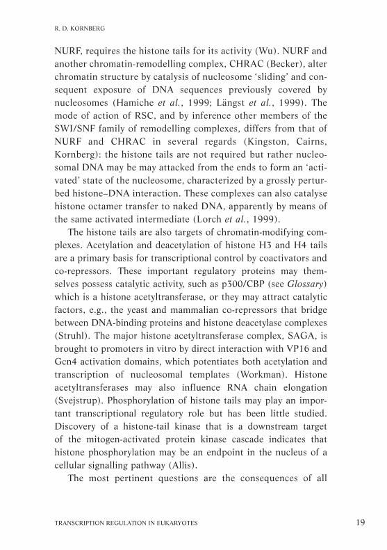
which are the N-terminal regions of the histone proteins. Thestructure refined to 2.0 Å provides almost three times more X-ray data than at 2.8 Å, allowing us to include approximately1,000 solvent water molecules and ions, which make importantcontributions to the protein–DNA and protein–protein inter-actions throughout the complex (Fig. 3).
The histone-fold domains form crescent-shaped hetero-dimers, H3 with H4 and H2A with H2B that provide extensiveinteraction interfaces. The contacts within the dimers are madebetween the first two of the three α-helices that comprise thehistone fold. The C termini of the second and third α-helices ofthe histone folds combine with each other to join the halves ofthe H3/H4 tetramer, through H3–H3 interactions, and add
30 HFSP WORKSHOP 7
PART I
Figure 3. The 2.0 Å X-ray structure of the nucleosome core particle allowsthe addition of nearly 1,000 well-ordered water molecules and salt ions(white and magenta spheres) to the over 12,000 atoms in protein and DNAseen in the structure at 2.8 Å (Fig. 2). The water molecules are important instabilizing histone protein and DNA structure as well as adding many indi-rect protein–DNA interactions. Red and orange, H2A–H2B dimer; blue, H3histone-fold domain; light green, H4 tail. X-ray data were obtained at theEuropean Synchrotron Radiation Facility in Grenoble, France.

the two H2A/H2B dimers to complete the octamer, throughH4–H2B interactions. Networks of ordered water molecules arefound between the histone subunit interfaces of H3 and H2A(Fig. 4) and of H4 and H2B and these further stabilize theoctamer structure. The histone-fold extensions contribute to thenucleosome core particle structure in several ways, from octamerformation to DNA binding. One or more of the extensions arelikely to be involved in internucleosomal contacts in the higher-order structure or in interactions with non-histone factors.
The flexible N-terminal tails of histones reach out betweenand around the gyres of the DNA superhelix to contact neigh-bouring particles in the crystal. About one-third of these flexibletails is apparent in the electron density maps calculated from theX-ray diffraction data. The disordered state of the tails, particu-larly at their sites of post-translational modifications — acetyl-ation, methylation and phosphorylation (see Aliss, Workman,this volume) — indicates that the tails are primarily involved inhigher-order chromatin organization. The transition between themore compact higher-order structure and the relatively opennucleofilament, i.e., beads-on-a-string, may be induced throughmodifications introduced by transcriptional coactivators as partof the overall control of gene expression.
The X-ray structure indicates that these regions of the proteinchains are most likely to make internucleosomal interactions.The contact between the H4 N-terminal tail and an acidic patchcreated by H2A/H2B at the octamer surface of a neighbouringnucleosome core particle is essential for crystal formation. At2.0 Å resolution, a manganese ion is seen to be bound in perfectoctahedral coordination, forming a bridge from the H2A–H2Bdimer of one nucleosome core particle to the H4 tail and H3 his-tone-fold domain of another (Fig. 5). These interactions may berelevant to the higher-order structure, a hypothesis that can betested in vitro by using site-directed mutagenesis of defined seg-ments of higher-order structure.
TRANSCRIPTION REGULATION IN EUKARYOTES 31
T. J. RICHMOND

The four pairs of histone-fold domains in the histone octamerorganize only 121 bp of DNA in the nucleosome core superhelix,not the entire 147 bp. One α-N histone-fold extension, i.e., on themost N-terminal α-helix in the H3 sequence, occurs just beforeeach H3 histone fold and binds the first and last remaining 13 bpof the DNA superhelix. The odd number of base pairs in the DNA
32 HFSP WORKSHOP 7
PART I
Figure 4. A complicated network of water molecules bridges between eachpair of histone H2A (orange) and H3 (blue) molecules. Networks of watermolecules (white) stabilize the interactions of the histones with each otherwithin the nucleosome core. In the region of the histone octamer shown,the hydrogen bonds are in the interface between dimer and tetramer sub-assemblies.

superhelix, with one base pair lying on the molecular dyad axis ofthe whole particle, agrees with the results from high-resolutionmapping of the histone octamer on DNA (Flaus and Richmond,1998). A variety of interactions occur between protein and DNAat their 14 contact sites: three are made by each of the H3/H4 andH2A/H2B histone-fold pairs and two by the H3 α-N extensions.
TRANSCRIPTION REGULATION IN EUKARYOTES 33
T. J. RICHMOND
Figure 5. The divalent manganese ions (35–50 mM; yellow) used in thecrystallization of the nucleosome core particle bind DNA most frequently atthe guanine bases (N7 atoms), although in this example a manganese ionis tightly bound between adjacent core particles. Manganese ions are iden-tified by electron density greater than that for water molecules and theircharacteristic octahedral coordination of ligands.

Many basic side-chain interactions occur with the DNA phos-phate groups and hydrophobic side-chain interactions withdeoxyribose groups. The 2.0 Å crystal structure reveals the loca-tion of several solvent molecules that aid the interaction of theDNA with protein. However, the direct hydrogen bonds from theamide groups on the histone main chain to the phosphate oxygenatoms seem to specify the DNA conformation most rigorously.The character and number of these interactions indicate thatthe path of the DNA superhelix and the structure of the DNAdouble helix are largely independent of DNA sequence.
The energetics of nucleosome stability depend on the differ-ence in energy for DNA in its free state compared to its statebound at the different sites along the nucleosome superhelixpath. Overall, for example, the DNA must change its twist from10.5 bp per turn in solution to 10.3 bp per turn in the nucleo-some and bend through 594°. Certain sequences can accommo-date this better than others. Despite a probable invariance ofDNA structure at each binding site within the nucleosome core,certain DNA sequences crystallize better than others. Thehuman centromeric α-satellite DNA used to determine the crys-tal structure of the nucleosome core particle may possess theadvantage that it has an intrinsic curvature that maintains ahistone-bound state at the ends of the superhelix, the weakestbinding site. The sequence-dependent propensity for a terminusof the DNA superhelix to release from the surface of the histoneoctamer may be important for the access of transcription factorproteins to specific DNA sequences bound in the nucleosome.
Histone octamer sliding models
Examination of the X-ray structure of the nucleosome core par-ticle has enabled us to discriminate between possible mecha-nisms of histone octamer ‘sliding’ along DNA. A simple model isthat the octamer simply turns around the DNA superhelix axisinside the gyres of DNA. However, this would be impossible
34 HFSP WORKSHOP 7
PART I

without the supercoils expanding in diameter as well as in sep-aration, because arginine side chains penetrating the minorgroove and histone tails passing between the gyres in alignedminor grooves would prevent this rotation. A second mecha-nism, which almost certainly occurs, is the DNA ‘peeling’ offfrom one side of the particle while the other side remains bound,then the particle rebinding further along the DNA, causing abubble to form on the surface of the octamer. The bulged DNAcould work its way around the octamer by repeated release ofone end or the other until the octamer is centred on a differentsequence. This mechanism has been invoked to explain howRNA polymerase can transcribe the DNA bound in a nucleosomewithout releasing the histone octamer (Studitsky et al., 1997).The polymerase resides in the bubble on the nucleosome.
A third possibility comes from the difference in DNA structureon opposite sides of the core particle. One half of the DNA super-helix has a distortion that does not appear in the other half, despitethe general twofold symmetry that relates the halves of the part-icle. This distortion comprises overwinding of 12 bp of DNA,stretching the double helix to fit a length spanned by 13 bp in thesymmetrically related region on the opposite side of the DNAsuperhelix. Although the observed distortion is near the centre ofthe DNA superhelix, five double-helical turns from the nearestDNA terminus, its origin probably lies in the stretching of the DNAthat permits close packing of DNA termini between particles in thecrystal. The DNA overwinding then propagates to the observedsite, which better accommodates it. This difference in structure onthe two sides of the core particle indicates that twist diffusion canoccur in the DNA along the surface of the nucleosome (first sug-gested by Van Holde and Yager, 1985). If this is the case, then theDNA double helix resembles a flexible screw with the arginine sidechains and histone tail segments tracking in the minor grooves.This mechanism could also account for the nucleosome slidingactivity of chromatin remodelling factors (see Becker, this volume).
TRANSCRIPTION REGULATION IN EUKARYOTES 35
T. J. RICHMOND

Nucleosomal location of the linker histone andits role in transcriptional repression
Andrew A. Traversin collaboration with
Serge Muyldermans and Daniela Rhodes
Condensed chromatin consists of chromatosomes, each of whichcontains a histone octamer (nucleosome) and a lysine-rich link-er histone H1/H5 bound to 168 bp of DNA. The binding of thelinker histone H1/H5 facilitates the proper folding of a nucleo-some array into this higher-order structure. One facet of the reg-ulation of transcriptional activity could be competition betweenthe linker histone and the activating transcription factors. Herewe describe experiments that locate the position of the centralglobular domain of the linker histone in the chromatosome par-ticle. We show that defined positions adopted by core nucleo-some particles on Xenopus somatic oocyte 5S rRNA genes areimportant determinants of the relative affinities for the linkerhistone and the activating transcription factor, TFIIIA. Thuscore nucleosome positions can directly influence the activationand repression of particular genes, probably through a combin-ation of effects at single nucleosomes, in which the DNA tran-scriptional position is stabilized by histone H1 binding, and innucleosomal arrays, where linker-histone binding promotes thefolding of chromatin into higher-order structures.
Position of the linker histone
The linker histones contain basic C- and N-terminal tails flank-ing a central globular domain that is sufficient for chromato-some formation. Their exact location in the chromatosome iscontentious (for review, see Vignali and Workman, 1998). Theglobular domain consists of a three-helix bundle, helices I–III,homologous to helix–turn–helix DNA-binding proteins. Helix IIIis thought to be a recognition site and to bind in the major
36 HFSP WORKSHOP 7
PART I

groove of the DNA, and a cluster of basic amino-acid residuesopposite the recognition helix may be a secondary binding site(Ramakrishnan et al., 1993; Cerf et al., 1994). This structureindicates a model in which the globular domain binds two dup-lexes, i.e., both strands of DNA in each DNA double-strandedmolecule, thus bridging two adjacent DNA gyres (Fig. 6).
In agreement with these predictions, mutations in the second-ary binding site prevent the globular domain of histone H5 bind-ing to two duplex DNA molecules and forming chromatosomes(Goytisolo et al., 1994). In addition, the binding site of H5 onmixed-sequence chicken DNA chromatosomes has been mappedusing a protein–DNA photo-crosslinking method to crosslink tothe DNA at specific cysteine residues substituted for serine inthe wildtype protein domain (Zhou et al., 1998). In particular,serines in helix I (amino-acid 29), helix II (amino-acid 41) and
TRANSCRIPTION REGULATION IN EUKARYOTES 37
A. A. TRAVERS
Figure 6. Model for the binding of the globular domain of histone H5 to thechromatosome. Helix III binds in a major groove close to one terminus (pur-ple) of chromatosomal DNA whereas the loop between helix I and helix IIcontacts the DNA close to the midpoint of the DNA (near the dyad; red).

the recognition helix III (amino-acid 71) were mutated andfound to be near the nucleosomal DNA. Thus helix III binds inthe major groove of the first helical turn of chromatosomal DNAat the exit or entry point of the nucleosomal DNA. Meanwhilethe secondary DNA binding site on the opposite face of the glob-ular domain of H5 contacts the nucleosomal DNA close to itsmidpoint, or dyad (Fig. 6).
By exploiting the ability of some serine-to-cysteine mutants toself-dimerize, it was inferred that helix I of the globular domainof H5 faces the solvent and helix II faces the nucleosome. Inbulk chromatin, the globular domain of the linker histone thusforms a bridge between one terminus of chromatosomal DNAand the midpoint, bringing the C terminus of the globulardomain to lie on the outside of the particle between one end ofchromatosomal DNA and the central gyre (Fig. 7b; Lambert et
38 HFSP WORKSHOP 7
PART I
Figure 7. Models for the placement of the globular domain of linker histoneon the core particle. a, the globular domain (small sphere) binds centrallyto the nucleosome dyad; b, the globular domain is asymmetrically placed,forming a bridge between one terminus of the chromatosomal DNA and theDNA close to the dyad; c, the globular domain binds only to one duplex,about 2.5 double-helical turns from one DNA terminus.

al., 1991; cf. the simple model in Fig. 7a proposed by Allan etal., 1980). The two contact points of the linker histone with thenucleosomal DNA are separated by one superhelical turn. Thisbridging model does not distinguish between two possible posi-tions of the globular domain, one where the linker histone bindsto the DNA entering the nucleosome, the other where it binds tothe exiting DNA in addition to the nucleosome dyad sequences.However, it excludes the possibility of two globular domainsbinding as a dimer, for which there is no experimental evidence.
Our results differ significantly from the model for the chroma-tosome on the Xenopus borealis 5S RNA gene (Hayes, 1996;Pruss et al., 1996). In this model, the globular domain binds onthe inside of one DNA gyre at a single internal site 65 bp fromthe nucleosomal midpoint or dyad and about two helical turnsfrom the proximal terminus of chromatosomal DNA, so that nocontact is made with the dyad region itself (Fig. 7c; Pruss et al.,1996). This model depends critically on the assumption that the5S RNA chromatosome adopts a single dominant translationalposition, which determines the exact DNA sequences that makecontact with the octamer. However, accurate mapping of chro-matosome dyads on this sequence and that of the closely relatedXenopus laevis somatic 5S RNA genes reveals multiple equiva-lent translational positions both with and without histone H1(Panetta et al., 1998).
Role of histone H1 binding in transcriptional regulation
In Xenopus somatic cells, histone H1 effects the transcriptionalrepression of oocyte-type 5S RNA genes without altering thetranscription of the somatic-type 5S RNA genes. Using a directmethod for mapping nucleosome dyads, site-directed hydroxylradical cleavage of the DNA sequence in contact with an EDTAmoiety conjugated to residue 47 of histone H4 (Flaus et al.,1996), we have mapped multiple nucleosome positions afterin-vitro reconstitution of both the oocyte and somatic 5S genes of
TRANSCRIPTION REGULATION IN EUKARYOTES 39
A. A. TRAVERS

40 HFSP WORKSHOP 7
PART I
X. borealis (Fig. 8). These nucleosome positions determine bind-ing of both transcription factor and H1, allowing the transcriptionfactor TFIIIA to bind more efficiently to nucleosomes on thesomatic 5S RNA gene than to those on the oocyte 5S RNA gene.
A significant observation is that, in a binding competition
Figure 8. Multiple nucleosome positions on the somatic 5S rDNA. Right,the preparative fractionation of free DNA (D, left lane) and nucleosomalcomplexes (right lane) on a 5% polyacrylamide gel. Left, representation ofthe nucleosome dyad positions present in each gel band. The dyads arenumbered relative to the start point of 5S RNA transcription and deducedfrom site-directed hydroxyl-radical cleavage. Red circles every 10 bp of theDNA helix axis indicate positions on the 5S rDNA fragment. The regionbound by the histone octamer is shown in red and the free DNA in blue.The TFIIIA binding site is indicated by a green line where exposed and ablack one where covered by a positioned nucleosome. Green star, positionof the radiolabel. Modified from Panetta et al., 1998.

experiment between TFIIIA and H1, TFIIIA preferentially bindsto the somatic nucleosome whereas H1 preferentially binds tothe oocyte nucleosome and prevents TFIIIA binding (Fig. 9).This is a strong indication that nucleosome positioning is impor-tant in the regulation of transcription of 5S RNA genes, as hasalso been shown for the corresponding genes in X. laevis (Howeand Ausio, 1998; Sera and Wolffe, 1998). It provides a mol-ecular mechanism for the selective repression of the oocyte 5SRNA genes by H1. Studies are required to test whether H1 andTFIIIA binding is similarly regulated when the 5S RNA genesare present in nucleosomal arrays and under conditions wherethe histones are differentially acetylated.
TRANSCRIPTION REGULATION IN EUKARYOTES 41
A. A. TRAVERS
Figure 9. H1 and TFIIIA bind differentially to the somatic and oocytenucleosomes. Somatic (lanes 1–5) and oocyte (lanes 6–10) were incubatedwith TFIIIA (lanes 3 and 8), H1 (lanes 4 and 9) and with both H1 andTFIIIA (lanes 5 and 10). The positions of free 5S rDNA (D) and theTFIIIA–DNA complex (DT) are indicated. Bracket, position of the com-plexes between nucleosomes and TFIIIA or H1. The complexes were frac-tionated on a 5% polyacrylamide gel. Reproduced with permission fromPanetta et al., 1998.

Linking histone modifications to gene activation
C. David Allisin collaboration with
Paolo Sassone-Corsi*, Craig Mizzen, Peter Cheungand Claudia Crosio*
The chromatin fibre has to undergo two fundamentally differenttransitions: it has to increase condensation to yield the chromo-some structure in mitosis and meiosis, and it has to unfold toallow the transcription or replication machinery access to thenucleosomal DNA. A correlation between gene regulation andchromatin structure has been established using biochemical andgenetic studies. In eukaryotes, two highly conserved and possiblylinked mechanisms relieve nucleosomal repression: chromatinremodelling (see Becker, Hörz, Kingston, Wu, this volume) andpost-translational histone modification, in particular histoneacetylation (see Workman, this volume) and phosphorylation(Fig. 10). Covalent modifications of histones seem to be impor-tant in gene regulation and thus have significant implicationsfor eukaryotic biology. As well as allowing regulatory factorsto interact specifically with nucleosomes, differential ‘marking’of the various histone tails with covalent modifications mayinfluence nucleosome–nucleosome interactions, thus modulatingthe higher-order organization of chromatin.
Acetylation of lysines in the N-terminal histone tails causesstructural changes in individual nucleosomes and chromatin,and influences the interaction with the modified nucleosomes ofnon-histone proteins, such as regulatory factors (Mizzen andAllis, 1998). Combinations of post-translational histone modifi-cations, including methylation, ADP-ribosylation, ubiquitinationor glycosylation in addition to acetylation and phosphorylation(Table 1), provide a challenge for determining the participants
42 HFSP WORKSHOP 7
PART I
* Institut de Génétique et de Biologie Moléculaire et Cellulaire, CNRS, Strasbourg, France

and targets in chromatin-mediated eukaryotic gene regulation.Here we review the occurrence of histone acetyl transferases(HAT), enzymes involved in acetylation, and report the first evi-dence that a core histone is physiologically phosphorylated by a
TRANSCRIPTION REGULATION IN EUKARYOTES 43
C. D. ALLIS
Figure 10. Proposed roles of histone acetylation and phosphorylation inmodulating chromatin structure. Left, condensed chromatin, in which DNAtranscription and replication are inactive, is characterized by low levels ofpost-translationally modified histones. Right, acetylation of the N-terminaltails of core histones (Ac) by histone acetyltransferases (HAT) and phos-phorylation of core and linker histone tails (P2–) by protein kinases (PK)attenuate their interactions with DNA or other chromatin components.This leads to decondensation of chromatin fibres and increases accessi-bility to DNA that is recognized by factors regulating chromatin activity.Modified histone tails themselves may also be recognized by regulatory fac-tors. Histone deacetylases (HDAC) remove acetyl groups (Ac) and proteinphosphatases (PPase) remove phosphoryl groups from the histone tails,enhancing their binding to nucleosomal DNA or other chromatin compon-ents and promoting a return to the condensed, quiescent, basal state. Thepositions shown for the globular domain of H1 and modified core histonetails are arbitrary and are not intended to reflect experimental data.

mitogen-activated protein kinase. We also provide new insightsinto a poorly understood mechanism that links histone phospho-rylation to activation by mitogens.
Histone acetylation
Several protein complexes responsible for acetylation have beenidentified and isolated in a range of eukaryotes, from yeast tohumans. Their catalytic subunits, the acetyltransferases, arehighly conserved. The first-known transcription-associated HATwas cloned from the ciliated protozoan Tetrahymena thermoph-ila (Brownell et al., 1996) and is significantly homologous to theyeast S. cerevisiae protein Gcn5. Characterization of Gcn5 func-tion in vitro and in vivo indicated that transcriptional activationis linked to HAT activity (Kuo et al., 1998; L. Wang et al., 1998;see Hörz, this volume). Gcn5 homologues in various mammalsand in Drosophila were identified by the domain containing apreviously described putative acetyl-CoA-binding motif(Neuwald and Landsman, 1997; Fig. 11).
Several other transcriptional coactivators, such as the humanp300/cyclic AMP response element binding factor (CREB)-binding protein (CBP), p300/CBP-associated factor (PCAF) andTAFII250, also have intrinsic HAT activity. Yeast Esa1, a proteinhomologous to the SAS2 and SAS3 proteins that are involved inyeast mating-type transcriptional silencing, and the relatedhuman Tip60, a protein that interacts with the Tat transactivatorof the human immunodeficiency virus (HIV), are members of theMYST family of proteins, named after the ‘founding members’:MOZ, YBF2/SAS3, SAS2 and Tip60 (Fig. 11); these all canacetylate histones in vitro (Kuo and Allis, 1998; Smith et al.,1998a,b).
HATs are the catalytic core of nucleosome-acetylating multi-protein complexes, such as the SAGA or the NuA4 complex(Grant et al., 1997; see Workman, this volume). Recent evidencethat SAGA and PCAF histone-modifying complexes share com-
44 HFSP WORKSHOP 7
PART I

TRANSCRIPTION REGULATION IN EUKARYOTES 45
C. D. ALLIS
Figure 11. Features of recently described members of a, the Gcn5 and b, theMYST families of histone acetyltransferases. The proteins are alignedaccording to homology of a previously described putative acetyl-CoA-bind-ing motif in the catalytic domain (Neuwald and Landsman, 1997). Recentevidence indicates multiple forms of hGcn5-S (asterisk). d, Drosophila; h,human; m, mouse; y, yeast.

mon factor requirements with the polymerase II transcriptionapparatus reinforces our view that chromatin structure and tran-scriptional regulation are interdependent (Grant et al., 1998a;Ogryzko et al., 1998).
Histone phosphorylation
Widespread phosphorylation of histones, particularly H1 andH3, correlates with mitosis in mammalian cells and may functionin mitotic and meiotic chromosome condensation (Hendzel etal., 1997; Wei et al., 1998; 1999). In mammalian cells, a subsetof H3 molecules is rapidly and transiently phosphorylated inresponse to mitogenic and other stimuli that activate expressionof immediate-early genes, such as c-fos and c-jun (Mahadevan etal., 1991). Thus H3 phosphorylation may have a role in decon-densing chromatin to facilitate transcriptional activation, similarto that proposed for histone acetylation and interphase phos-phorylation of linker histones (reviewed in Mizzen et al., 1999).To investigate this possibility, we made a synthetic peptide com-prising part of the amino-acid sequence of the histone H3 N-terminal tail domain found in yeast, human and mouse, andphosphorylated the residue corresponding to serine 10 of thecomplete histone H3 sequence. An antibody raised against thephosphorylated peptide is highly selective and reactive and is aremarkable marker of mitosis in a range of eukaryotic cells (Weiand Allis, 1998). In Tetrahymena, mutation of this residue fromserine to alanine causes the chromosomes in the micronucleus toloosen, compromising mitotic segregation (Wei et al., 1999), asignificant indication that phosphorylation at this site is highlyconserved and functionally important.
During the well-known immediate-early response to mitogens,the phosphorylation at Ser10 is stimulated by treatment withmitogens. The antibody raised against the phosphorylated peptide(anti-PS10) reacts with histone H3 in mouse fibroblasts stim-ulated by epidermal growth factor (EGF) or the phorbolester
46 HFSP WORKSHOP 7
PART I

12-O-tetradecanoylphorbol-13-acetate (TPA) in a fast, transientresponse corresponding to the immediate-early response to mito-gens (Mahadevan et al., 1991). The antibody staining seems to bespecific to euchromatic nuclear domains, i.e., decondensed,actively transcribed chromatin domains, so phosphorylation at Ser10 may, like histone acetylation, have a role in decondensing thechromatin fibre to facilitate gene expression.
TRANSCRIPTION REGULATION IN EUKARYOTES 47
C. D. ALLIS
Figure 12. Principles of the activity gel assay for detecting histone-modify-ing enzymes. Top, the method is based on the detection of modificationusing a radioisotopic cofactor, e.g., γ32P-ATP or 3H-acetyl-CoA; asterisks,radiolabel. Below, the Mr of polypeptides with intrinsic modifying activityis determined by electrophoresis on SDS gels containing histones. Afterdenaturing and renaturing the resolved proteins, gels are incubated withradioisotopic cofactor and processed for autoradiography to detect modifiedhistones surrounding active species in the gels. A schematic example show-ing the detection of the Tetrahymena p55 HAT in crude macronuclearextract is shown on the right. We used this method to demonstrate that Rsk-2 possesses intrinsic H3 kinase activity (see text) and in theory it could beused, with the appropriate radiolabelled cofactor, for other histone-modify-ing enzymes, such as histone methyltransferases.

The kinases responsible for histone-specific phosphorylationhave remained elusive. An ‘activity gel assay’, developed initiallyto identify the histone-modifying activity of p55 HAT, has beenmodified to detect other polypeptides with intrinsic histone phos-phorylation activity (Fig. 12). In a histone H3 in-gel kinase assayusing radiolabelled ATP, nuclear extracts from EGF-stimulatedfibroblasts selectively yielded an intense band corresponding to apolypeptide with Mr ~90K. Antibodies raised against p90rsk, ahuman protein targeted by the mitogen-activated protein kinase(MAPK) signalling pathway, reacted with the 90K polypeptideband. The H3 kinase was thus tentatively identified as Rsk-2. Anactivity test on free and nucleosomal histones revealed that Rsk-2 specifically phosphorylates Ser10, in contrast to other kinases,which are able to phosphorylate all histones.
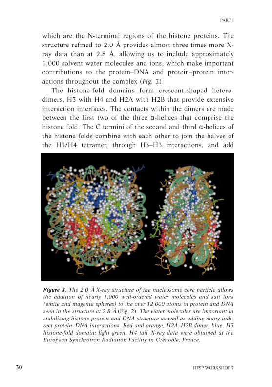
Thus, Rsk-2, a member of the p90rsk family of mitogen-activa-ted kinases implicated in cell proliferation (see Fig. 13), is thekinase responsible for mitogen-stimulated H3 phosphorylation inhuman cells. In agreement with this, in starved embryonic stemcells of rsk-2 knockout mice, the Rsk-2+ phenotype can be rescuedusing human Rsk-2 cDNA. In stimulated cells, selective chromatinimmunoprecipitation with anti-PS10 antiserum promises to be apowerful approach to the identification of DNA sequences forimmediate-early genes that require Rsk-2 for induction.
Mutations in the RSK-2 gene are causally linked to Coffin-Lowry syndrome, an X-linked disorder characterized by mentalretardation and developmental skeletal deformations in humans.Cells derived from individuals with Coffin-Lowry syndrome, i.e.,Rsk-2-deficient, fail to phosphorylate H3 after mitogen stimula-tion (Sassone-Corsi et al., 1999), although during mitosis H3phosphorylation is normal. Two independent kinase pathwaystherefore seem to target histone H3 for phosphorylation duringmitosis and mitogenic stimulation. A chromatin remodelling stepinvolving histone H3 phosphorylation, possibly in concert withhistone acetylation of mitogen-regulated genes (Fig. 13), may
48 HFSP WORKSHOP 7
PART I
TRANSCRIPTION REGULATION IN EUKARYOTES 49
C. D. ALLIS
Figure 13. Possible interactions involved in coordinated recruitment of his-tone kinase and acetyltransferase activities to chromatin. When activatedby MAP kinases, p90rsk proteins are translocated to the nucleus, where Rsk-2, in particular, phosphorylates H3 in the nucleosome in response to mito-genic stimuli. This phosphorylation may be targeted to nucleosomes acety-lated by p300/CBP and/or PCAF because p90rsk proteins can bind to a siteon p300/CBP that is near an interaction site for PCAF. Both these eventsare thought to promote chromatin decondensation and facilitate transcrip-tional activation (see text and Fig. 10). Each of these modifying activitiesmay target other substrates as well as histones. If some of these interactionsare correct, DNA sequences, i.e., mitogen-stimulated target genes, that areclosely associated with modified histones, should be recoverable with chro-matin immunoprecipitation using modification-specific antibodies. Ac,acetyl group; P2–, phosphoryl group.

thus be a critical part of a signalling pathway that, when deregu-lated, is intimately linked to human disease.
Function and composition of transcriptionadaptor histone acetyltransferase complexes
Jerry L. Workman
Reversible acetylation of the N-terminal tails of the histones haslong been linked biochemically with actively transcribed chro-matin. Several multi-subunit HAT complexes have been identi-fied in yeast, including the Spt-Ada-Gcn5-acetyltransferase(SAGA), Ada, nucleosome-acetylated–histone H3 (NuA3) andH4 (NuA4) complexes. These large complexes have different sub-unit compositions and substrate specificities, indicating that theyhave distinct functional roles. Interest in this field increasedrapidly when the yeast transcriptional adaptor protein Gcn5 wasdiscovered to have HAT activity (Brownell et al., 1996), whichis essential for its functional effect. However, despite its potentHAT activity with purified histones (H3 and H4), Gcn5 cannotmodify histones when they are assembled into a nucleosome.
The observation that yeast extracts contain HAT activitiesthat could modify nucleosomal histones prompted studies toidentify these activities. Here I review the progress in identifyingsubunits and biochemical experiments that give insight into howthese acetyltransferases contribute to transcriptional regulation.These studies support the idea that the HAT complexes act astranscriptional adaptors and are recruited by transcriptionalactivators to the promoter through direct interactions withthe cofactor subunits. The subsequent activation of transcriptionis likely to be through several separate mechanisms but theacetylation of adjacent, promoter-bound nucleosomes clearlycontributes to this process.
50 HFSP WORKSHOP 7
PART I

Identification and subunit composition of HAT complexes
Biochemical fractionation of yeast extracts identified four HATcomplexes that can acetylate histones when assembled in nuc-leosomes (Fig. 14; Grant et al., 1997). These had Mrs of 400–1,800K and displayed different histone specificities, indicatingthat they are distinct complexes. This was confirmed by subsequ-ent purification and subunit identification (Table 3).SAGA complex. The SAGA complex is a large HAT with anapparent Mr of 1,800K, that specifically acetylates nucleosomalhistones H3 and H2B. Evidence from immunological and massspectroscopy studies has identified a large number of the com-ponents of this complex, which can be classified into four groups(Fig. 15; Grant et al., 1997; 1998a,b):• GCN5-ADA gene products. Early immunological studies con-firmed that SAGA contained the HAT Gcn5, as well as the Adaproteins, Ada1, -2, -3 and Ada5/Spt20. These had previouslybeen shown to be components of a transcriptional adaptortermed the Ada complex, which functionally interacts with thetranscriptional activator Gcn4 and the VP16 activation domainof the herpes virus (Fig. 16). The SAGA complex cannot form ingcn5∆, ada2∆ or ada3∆ yeast strains, i.e., mutant strains lackinggcn5, ada2 or ada3 genes, confirming that the SAGA complexcontains Ada.
TRANSCRIPTION REGULATION IN EUKARYOTES 51
J. L. WORKMAN
Figure 14. Identification of multiple HAT activities in yeast. The fluoro-gram shows a gel on which histones have been separated from nucleo-somes, all labelled with 3H-acetyl-CoA during incubation and eluted at theindicated fractions from a MonoQ fast-protein liquid chromatography(FPLC) column. Four distinct HAT complexes with different histone speci-ficities were eluted from this column with increasing salt solution, corres-ponding to the Ada, NuA4, NuA3 and SAGA complexes.

Table 3. Properties and known components of the SAGA, Ada, NuA3 andNuA4 HAT complexes.
Complex SAGA Ada NuA3 NuA4
Size (Mr) 1,800K 800K 400K 1,300KSubstrate H3/H2B H3/H2B H3 H4/H2AInteractions Activators ? ? Activators
TBPCatalytic subunit Gcn5 Gcn5 ? Esa1Additional subunits Ada1,-2,-3 Ada2,-3
Spt3,-7,-8Spt20/Ada5TAFII20/17,
-25/23,-60,-61/68,-90
Tra1
• SPT gene products linked to TATA box binding protein (TBP).SAGA contains the SPT gene products Spt3, Spt7 and Spt8,proteins in a class that is thought to affect TBP function. Thisactivity is clearest for Spt3 and Spt8, which contribute to SAGAinteractions with TBP (Roberts and Winston, 1996; Sterner etal., 1999). As ADA5 has previously been shown to be identicalto SPT20, the Ada and Spt proteins may function through acommon physiological pathway.• TBP-associated factors (TAFIIs). SAGA also contains a numberof TAFIIs, including TAFII90, -68/61, -60, -25/23 and 20/17.Inactivation of TAFII-68 in a yeast temperature-sensitive mutantled to an apparent loss in mass to 1,000K–1,200K and a reduc-tion in the amount of TAFII-90 and Spt3 in the complex, estab-lishing that these proteins are an integral component of SAGA.Furthermore, inactivation of TAFII-68 leads to a severe disrup-tion in SAGA nucleosomal HAT activity and in the ability of thecomplex to activate transcription from a chromatin template invitro (Grant et al., 1998a).• Tra1. The largest subunit of the SAGA complex is the 3,744amino-acid protein Tra1 (Grant et al., 1998b; Saleh et al.,1998). Tra1 is the yeast homologue of the human transforma-tion/transcription domain-associated protein TRRAP, a novel
52 HFSP WORKSHOP 7
PART I

member of the family of phosphatidylinositol 3-kinases that aremutated in ataxia telangiectasia (McMahon et al., 1998). Tra1 isa potential target within SAGA for interactions with transcrip-tion activators.
This association of multiple classes of transcriptional cofac-tor proteins, Adas, Spts, TAFIIs and Tra1, is a strong indicationthat SAGA acts as a transcriptional adaptor complex. The largenumber of adaptor protein subunits similarly indicates thatSAGA can respond to a range of stimuli and interact with sev-eral activators, with the potential to regulate a broad range of
TRANSCRIPTION REGULATION IN EUKARYOTES 53
J. L. WORKMAN
Figure 15. The confirmed components of the SAGA transcriptional adaptorHAT complex, which contains multiple transcriptional regulatory proteins,including components implicated in interactions with TBP (Spts and TAFs),transcription activators (Adas and Tra1) and histone acetylation (Gcn5).Ac, acetylated histones; Act., activator protein.

promoters. The role of the Gcn5 HAT activity in this transcrip-tional adaptor complex has been examined biochemically (seebelow).Ada complex. A second HAT, the 800K Ada complex, has a sim-ilar substrate specificity to SAGA, nucleosomal histones H3 andH2B, and also contains the HAT Gcn5 and the Ada proteinsAda2 and Ada3. The relationship between the Ada and SAGAcomplexes is unclear but the Ada complex, which does not con-tain the Spt proteins, may be a sub-complex of the larger SAGAcomplex. An alternative possibility is that the complexes,although having Gcn5 and the Ada proteins as common sub-units, may have distinct nucleosomal HAT activities with differ-ent functions. The exact relationship between these HATs shouldbe resolved when the unknown subunits of each complex and thedegree to which they are common or unique have been identified.NuA3 and NuA4 complexes. In contrast to the SAGA and Adacomplexes, little is known about the subunit composition of the400K NuA3 and 1,300K NuA4 complexes. These HATs have dif-ferent substrate specificities, NuA3 modifying nucleosomal histoneH3 and NuA4 modifying histones H4/H2A. The functional HAT inthe NuA4 complex has been identified as the essential yeast proteinEsa1 (J. Côté, unpublished observations), a homologue of the HATMOF1 in Drosophila, which is involved in X-chromosome dosagecompensation. The HAT protein in NuA3 and the remainingsubunits in both complexes remain to be identified.
Functional properties of HAT complexes
The identification of multiple classes of transcriptional coactiva-tor proteins in Ada and SAGA indicated that these HAT com-plexes were likely to interact with activator proteins. This hasbeen examined by glutathione-S-transferase (GST) pull-downassays, in which the four native yeast HAT complexes were incu-bated with GST fused to the VP16 activation domain of herpesvirus or the yeast transcriptional activator Gcn4 (Utley et al.,
54 HFSP WORKSHOP 7
PART I

1998). Both SAGA (see Fig. 16) and NuA4 can bind both acti-vators but NuA3 and the Ada complex, which contains at leastthree of the same subunits as SAGA, cannot bind either activatorprotein.
Thus these transcriptional activators seem to recruit theSAGA or NuA4 complexes through contacts with their tran-scriptional adaptor subunits, thereby targeting HAT activity toadjacent nucleosomes. This was tested by reconstituting a mono-nucleosome template with a single Gal4-binding site 20 bp fromthe end of the DNA fragment. When this template was incub-ated with increasing amounts of Gal4–VP16 in the presence ofHAT complexes and 3H-acetyl-CoA, the binding of the activatorto the nucleosome increased the level of histone acetylation bySAGA and NuA4 but not by the NuA3 or Ada complexes,consistent with the activator-binding studies.
The recruitment of HAT complexes also has transcriptionaleffects. This was initially analysed using VP16-driven transcrip-tion from a model template containing a minimal E4 promotercontaining five Gal4 sites. When this template was assembledinto a spaced nucleosome array, transcription was highly
TRANSCRIPTION REGULATION IN EUKARYOTES 55
J. L. WORKMAN
Figure 16. Interaction of the SAGA complex with the acidic activationdomain of herpes virus activator VP16 and the yeast activator Gcn4. SAGAfailed to interact with glutathione-S-transferase (GST) alone but interactedwith the GST–VP16 and GST–Gcn4 fusion proteins. GST pull-down exper-iments using purified SAGA were used to assay for SAGA interaction indi-cated by acetylation of histone H3 in nucleosomes. The gel shows 10% ofthe input activity (In), 25% of the pull-down supernatant (S) and 50% ofthe activity recovered on the glutathione beads (B).

repressed. However, in the presence of Gal4–VP16, both SAGAand NuA4 stimulated transcription from this template in anacetyl-CoA-dependent manner (Ikeda et al., 1999; Utley et al.,1998). Complementary in vitro transcription studies on adefined chromatin template containing the HIV-1 promoter,which has a clear correlation between acetylation and transcrip-tional activation in vivo (Steger et al., 1998), showed that allfour HAT complexes, SAGA, Ada, NuA3 and NuA4, can stimu-late transcription from this chromatin template in an acetyl-CoA-dependent manner (Fig. 17) and that the transcriptionalstimulation by NuA4 and Ada results at least in part from theacetylation of histones.
56 HFSP WORKSHOP 7
PART I
Figure 17. Demonstration of transcriptional activation by native yeast HATcomplexes using the primer extension analysis of transcripts from the HIV-1 promoter embedded in a spaced nucleosome array. The Ada (A), NuA4(N4), NuA3 (N3) and SAGA (S) HAT complexes all enhanced transcriptionfrom the HIV-1 template in an acetyl-CoA-dependent reaction (comparelanes 2–5 to lane 1). Addition of more than one HAT complex resulted inan additive stimulatory effect (lanes 6–9). E4, promoter (TATA box andinitiation site) with five Gal4 sites.

ATP-dependent chromatin remodelling by NURFand transcriptional activation
Carl Wuin collaboration with
Toshio Tsukiyama and Gaku Mizuguchi
The assembly of the DNA template into chromatin is associatedwith transcriptional repression. Much of this is repressionof transcriptional initiation because nucleosomes prevent thebinding of transcriptional activators and the basal transcriptionfactors to their cognate sites. Eukaryotic cells have developedseveral ways of overcoming this chromatin-mediated bindinginhibition to enable gene transcription to take place, includingmechanisms that disrupt or ‘remodel’ nucleosomes to permittranscription factors to bind to the underlying DNA sequence.For example, in Drosophila a four-subunit chromatin remodel-ling complex, nucleosome remodelling factor (NURF), facilitatesthe binding of sequence-specific transcription factors to theircognate sites in chromatin and so stimulates transcriptionfrom chromatin templates. We describe here progress in theidentification of the component subunits of NURF and ourbiochemical studies on its role in transcriptional activation fromchromatin templates.
Isolation and properties of NURF
NURF was initially identified using a biochemical assay to exam-ine the mechanism of nucleosome disruption mediated by thebinding of the GAGA factor to sites in the promoter of theDrosophila heat-shock protein gene, hsp70, when this is assem-bled into chromatin (Tsukiyama et al., 1994). We did this usinga chromatin-assembly extract derived from Drosophila embryosto assemble a plasmid containing the intact hsp70 gene (Beckerand Wu, 1992). The extract contains many of the structural andenzymatic activities necessary for chromatin assembly and
TRANSCRIPTION REGULATION IN EUKARYOTES 57

assembles regular, physiologically spaced chromatin on plasmidtemplates.
To isolate NURF from the extract, hsp70 templates wereassembled into chromatin and purified by gel filtration, whichremoves both the extract and ATP (Fig. 18; Tsukiyama and Wu,1995). The purified chromatin was incubated with recombinantGAGA factor and the chromatin digested with micrococcal nucle-ase (MNase), which cleaves chromatin in the linker DNA andgenerates a nucleosomal ladder in regularly spaced chromatin(Fig. 18, lane 1). This assay showed that GAGA factor can bindonly in the presence of ATP (Fig. 18, compare lanes 2 and 4) andthat binding is sensitive to treatment with the detergent Sarkosyl(sodium lauroylsarcosine, 0.05%; Fig. 18, compare lanes 4 and8), indicating that an ATP-dependent cofactor was inactivated orremoved from the chromatin by Sarkosyl treatment. Adding backthe Drosophila embryo extract restores nucleosome disruptionmediated by GAGA-factor binding (Fig. 18, lane 12), so this wasused as an assay to fractionate the extract.
NURF was isolated in a fraction with ATP-dependent activitythat restored the nucleosome disruption mediated by GAGA-factor binding to the hsp70 promoter in Sarkosyl-treated chro-matin templates. Highly purified NURF can facilitate GAGA-factor binding and only about one NURF per 40 nucleosomesis required to catalyse activity. At this low concentration,chromatin is disrupted only in the presence of GAGA factor andonly at the promoter region to which GAGA factor binds (Fig. 19a,top), not at regions within the gene coding region (Fig. 19a, bot-tom). When NURF concentration is close to stoichiometric, i.e., 1per 5 nucleosomes, disruption of the hsp70 promoter increases (Fig.19a). At this concentration there is also a global effect on chromatineven in the absence of GAGA factor, seen as a smearing of thenucleosomal ladder, together with an apparent decrease in nucleo-some spacing indicated by compression of the ladder (Fig. 19b).
Using very pure mononucleosome templates reconstituted
58 HFSP WORKSHOP 7
PART I

TRANSCRIPTION REGULATION IN EUKARYOTES 59
C. WU
Figure 18. Nucleosome disruption at the hsp70 promoter requires GAGAfactor, a Sarkosyl-sensitive factor (NURF) and ATP. Chromatin remodellingis indicated by a smearing of the nucleosomal DNA ladder. In the absenceof GAGA factor, the hsp70 promoter is assembled into a regular nucleo-some array (lane 1); GAGA-factor binding disrupts this nucleosome ladderand produces a smaller fragment corresponding to the protection producedby GAGA factor alone (lane 4, arrow). Bottom, experimental protocol:MNase digestion of Sarkosyl-treated hsp70 chromatin and DNA blothybridization with an hsp70 promoter oligonucleotide. Nucleosome pertur-bation mediated by GAGA-factor binding was visualized by Southern blot-ting and hybridization of this digest using an oligonucleotide correspondingto the GAGA-factor binding site. For further details, see text. Reproducedwith permission from Tsukiyama and Wu, 1995.

from purified histones, we confirmed that NURF does notrequire other components of chromatin for activity. It disruptedthe 10 bp repeat pattern of DNase I cleavage characteristic ofnucleosomal DNA in an ATP-dependent manner, and facilitatedthe binding of GAGA factor to mononucleosome templates, asassessed by DNase I footprinting (Tsukiyama and Wu, 1995).
60 HFSP WORKSHOP 7
PART I
Figure 19. Actions of NURF that are dependent and independent of GAGAfactor. a, GAGA factor, low NURF levels and ATP mediate promoter-spec-ific disruption of nucleosome organization. b, higher NURF levels and ATPin the absence of GAGA factor cause global effects on promoter and codingregions. Chromatin remodelling is indicated by a smearing of the nucleo-somal DNA ladder. See text for details. Reproduced with permission fromTsukiyama and Wu, 1995.

The NURF complex contains a low, constitutive ATPase activitythat is not stimulated either by free DNA or by histones; how-ever, the ATPase activity of the complex was stimulated fivefoldby incubation with reconstituted mononucleosomes (Fig. 20). Asthis was not observed with an unreconstituted mixture of DNAand histones, stimulation of the ATPase activity seems to requiresome aspect of the structure of assembled nucleosomes.
Subunit composition of NURF
NURF contains four main polypeptides, with apparent Mrs of215K, 140K, 55K and 38K, that cosediment in a complex ofabout 500K (Tsukiyama and Wu, 1995). Three of these subunitshave been identified, giving insights into the mechanism ofNURF activity. The 140K subunit (NURF-140), which is respon-sible for ATP hydrolysis, is imitation switch (ISWI; Tsukiyamaet al., 1995), which is also found in two other Drosophilachromatin remodelling complexes, ATP-dependent chromatinassembly and remodelling complex (ACF; T. Ito et al., 1997) and
TRANSCRIPTION REGULATION IN EUKARYOTES 61
C. WU
Figure 20. ATPase activity of NURF is stimulated by nucleosomes but notby free DNA or free histones. Buffer, control. Reproduced with permissionfrom Tsukiyama and Wu, 1995.

chromatin accessibility complex (CHRAC; Varga-Weisz et al.,1997; see Becker, this volume). Purified ISWI alone at high con-centrations is active in several chromatin remodelling assays and inNURF this protein is likely to have a function similar to the key roleit has in CHRAC (Corona et al., 1999; see Becker, this volume).
The 55K subunit is related to RbAp48, a WD, or Trp-Asp,repeat protein that has highly conserved repeating units usuallyending in Trp-Asp. RbAp48, which was originally identified byits in-vitro association with the retinoblastoma protein, is a com-mon component of several chromatin-associated complexes,including chromatin assembly factor (CAF-1) and several mam-malian histone deacetylase complexes (Martínez-Balbás et al.,1998). The related yeast protein Hat2 is a subunit of the cyto-plasmic HAT and yeast MSI1 is a subunit of yeast CAF-1. ThusNURF-55 and its homologues probably have a structural role,perhaps acting as a platform for the assembly of complexesinvolved in chromatin metabolism.
The 38K subunit is homologous to inorganic pyrophos-phatase (Gdula et al., 1998). Both recombinant NURF-38 aloneand the purified NURF complex have inorganic pyrophosphataseactivity but inhibition of this activity with sodium fluoride hasno noticeable effect on the ability of NURF to remodel chro-matin. As the ATPase activity of NURF, which is essential forchromatin remodelling, releases inorganic phosphate rather thanpyrophosphate, the activities of NURF-140 and -38 seem to bebiochemically uncoupled. Hence NURF-38 may have a struc-tural or regulatory role in the NURF complex or NURF may havebeen adapted to deliver pyrophosphatase activity to transcrip-tionally or replicatively active regions of chromatin. These poly-merization processes may be inhibited by the accumulation ofunhydrolysed pyrophosphate during nucleotide incorporation,so NURF pyrophosphatase activity could promote the forwardreaction by the efficient removal of this metabolite.
We have recently cloned the largest subunit, NURF-215,
62 HFSP WORKSHOP 7
PART I

which is encoded by a very large, new open reading frame givinga polypeptide of predicted Mr of 301K. Experiments aimed at theexpression of this gene, with the goal of reconstituting the NURFcomplex from recombinant proteins, are currently underway.
Role of NURF in transcriptional activation
We have found that NURF-mediated chromatin remodellingactivity on chromatin templates can facilitate access of transcrip-tion factors to their sites in the chromatin and so activate tran-scription. A simple model promoter consisting of five tandemlyrepeated Gal4-binding sites upstream of a minimal adenovirus E4core promoter (Mizuguchi et al., 1997) was assembled into chro-matin using a Drosophila embryo extract and separated from theextract and ATP by gel filtration (see Fig. 21a, top). To examinechromatin remodelling, saturating amounts of Gal4 derivativeswere incubated with the chromatin template in the presence ofATP before examining the transcriptional activity of the tem-plates in a Drosophila nuclear extract. The chromatin templatewas highly repressive to transcription in the absence of Gal4 andbecame only weakly active in the presence of Gal4(1–147), whichcontains only the activator DNA-binding domain. In contrast, aderivative containing the Gal4 DNA-binding domain fused to theactivation domain of heat-shock factor (Gal4–HSF) led to tran-scriptional activation (Fig. 21a, bottom, lane 3).
Gal4–HSF-mediated transcriptional activation was sensitiveto Sarkosyl treatment of the chromatin template (Fig. 21a, lane6) but could be rescued by the addition of purified NURF andATP (Fig. 21a, lane 9). Even at sub-stoichiometric concentra-tion, i.e., 1 NURF to 16 nucleosomes, NURF activated tran-scription (Fig. 21a, lane 10). The nucleosomes were remodelledin the promoter region, as smeared nucleosomal ladders wereobserved in the presence of NURF and Gal4(1–147) andGal4–HSF activators in the promoter region (Fig. 21b, top),compared with the lack of remodelling in a distal region of chro-
TRANSCRIPTION REGULATION IN EUKARYOTES 63
C. WU

64 HFSP WORKSHOP 7
PART I
Figure 21. Chromosome remodelling allows transcriptional activation. In areconstituted plasmid chromatin containing five tandem Gal4 sites and theadenovirus E4 core promoter, transcriptional activation follows ATP-dependent nucleosome disruption by Gal4–HSF and NURF. a, experimentalscheme and primer extension analysis of transcription from chromatin tem-plates. b, MNase digestion analysis of the chromatin structure shows thatchromatin remodelling, indicated by a smearing of the nucleosomal DNAladder, occurs only over the promoter region (top) and not over the distalregion (bottom). NE, nuclear extract; NTP, nucleoside triphosphate; PIC,preinitiation complex. See text for details. Reproduced with permissionfrom Mizuguchi et al., 1997.

matin (Fig. 21b, bottom). Purified NURF can restore a largeproportion of the transcriptional activation by Gal4–HSF onuntreated chromatin templates, indicating that the Sarkosyl-sensitive activity required for transcriptional activation can belargely satisfied by NURF. It is of interest that activation waspoor with Gal4(1–147) despite strong remodelling at the pro-moter region, indicating that the activation domain of HSF hasan important role in relieving chromatin-dependent repression.
CHRAC, a chromatin remodelling complexdriven by the ATPase ISWI
Peter B. Beckerin collaboration with
Cedric Clapier, Davide Corona, Gernot Längstand Patrick Varga-Weisz
The chromatin remodelling complex of Drosophila, NURF (seeWu, this volume), was isolated in an assay designed to identifyactivities that enable the GAGA factor, a specific eukaryotic tran-scription factor that binds to GA/CT-rich sites, to access its bind-ing site in a specific promoter sequence. Using this approach,however, we could not distinguish between two possibilities:that the chromatin remodelling activity is actively recruited topromoter regions by the transcription factor or that chromatinremodelling is an untargeted process providing a ‘window ofopportunity’ for proteins to gain access to the DNA. To identifychromatin remodelling activities, an alternative assay, which usesrestriction-enzyme cleavage, was developed for examiningprotein access to multiple sites in chromatin. This has the advan-tage that restriction enzymes, which are prokaryotic proteins, can-not have evolved mechanisms to overcome chromatin-mediatedrepression. Using this assay, a five-subunit complex, the chro-
TRANSCRIPTION REGULATION IN EUKARYOTES 65
C. WU

matin accessibility complex (CHRAC), has been isolated fromDrosophila nuclear extract (Varga-Weisz et al., 1997).
CHRAC is an ATP-dependent chromatin remodelling com-plex that facilitates the access of DNA-binding proteins to sitesin chromatin. It can also realign nucleosomes to generate regu-lar, evenly spaced nucleosome arrays. We review the progressmade in identifying the component subunits of this complex anddiscuss experiments indicating that the ATPase subunit ISWI isthe functional core of CHRAC’s chromatin remodelling activity.
Isolation of CHRAC
The restriction-enzyme assay is based on the assembly of regular,physiologically spaced and highly complex chromatin by extractsfrom Drosophila embryos (Becker and Wu, 1992). Assembledchromatin templates are passed over a gel-filtration column toremove the extract and ATP, and incubated with the restrictionenzyme DraI. Under these conditions, the chromatin largelyresists DraI cleavage, consistent with previous observations thatnucleosome assembly can inhibit cleavage by restriction enzymes.
Cleavage was stimulated when ATP was added back duringincubation with DraI, indicating that an ATP-dependent activityin chromatin facilitated access by the restriction enzyme to sitesin the chromatin template. This activity was inactivated bytreating the chromatin with Sarkosyl to remove ATP-dependentchromatin remodelling activities but reappeared whenDrosophila nuclear extract and ATP were added back. Using thisassay to fractionate Drosophila nuclear extract resulted in theisolation of CHRAC (Varga-Weisz et al., 1997).
Chromatin remodelling activities of CHRAC
Biochemical studies show that CHRAC has two apparentlydifferent actions on chromatin: it increases protein access tochromatin templates and increases the regularity of an irregularnucleosomal array.
66 HFSP WORKSHOP 7
PART I

Increased protein access to chromatin templates. When designingthe assay used to isolate CHRAC, we assumed that increasedcleavage of chromatin templates by DraI was a satisfactory modelfor general access by proteins to sites in chromatin. This proved tobe so for two dissimilar sequence-specific DNA-binding prot-eins:the SV40 replication protein T-antigen and transcription termina-tion factor 1 (TTF-1), a factor required for polymerase I tran-scription and termination on ribosomal genes.• SV40 T-antigen. SV40 replication has an absolute requirementfor the binding of an initiation factor, T-antigen, at the origin ofreplication but replication is inhibited by chromatin assembly.Experiments using chromatin templates assembled usingDrosophila embryo extract showed that T-antigen could gainaccess to its binding site in chromatin in the presence of aSarkosyl-sensitive, ATP-dependent cofactor (Becker and co-workers in collaboration with Claudia Gruss and co-workers,University of Konstanz). This activity could be replaced by puri-fied CHRAC, which can also facilitate T-antigen binding in chro-matin and leads to increased replication on these templates(Alexiadis et al., 1998).• TTF-1. Polymerase I transcription of the mouse ribosomalgenes requires the binding of TTF-1 to a site (To) in the pro-moter region. This protein is chromatin-specific, co-operating withATP-dependent chromatin remodelling activities to position nuc-leosomes within the promoter. This nucleosome remodelling corre-lates with transcription, indicating that a specific chromatin archi-tecture is required for initiation (Längst et al., 1998). Biochemicalstudies showed that purified CHRAC can facilitate TTF-dependentnucleosome remodelling in the ribosomal DNA promoter (Coronaet al., 1999). This assay was performed using chromatin templatesassembled with polyglutamic acid, which contain only histones andDNA, confirming that additional chromatin-bound proteins areunnecessary for CHRAC remodelling activity.
TRANSCRIPTION REGULATION IN EUKARYOTES 67
P. B. BECKER

Increased nucleosome regularity. CHRAC was originally identifiedas a complex that facilitated protein access to sites in chromatin, aprocess that is probably associated with nucleosome removal ordisassembly. However, it can also increase the regularity or order inchromatin. This activity was identified using Sarkosyl-strippedchromatin: the detergent disrupts the regularity of nucleosomespacing, generating a DNA smear when analysed by micrococcalnuclease (MNase) digestion. Addition of ATP and purified CHRACto this chromatin realigns the nucleosomes, generating a regularnucleosome ladder (Fig. 22; Varga-Weisz et al., 1997).
68 HFSP WORKSHOP 7
PART I
Figure 22. CHRAC is a nucleosome spacing factor. Regularly spaced nu-cleosomes are a hallmark of physiological chromatin and are detected bycleavage of chromatin with MNase, which generates a regular ladder ofprotected fragments (arrows). CHRAC can generate regularly spacednucleosome arrays from irregular nucleosomes in the presence of ATP

Two broad models for the mechanism of CHRAC action are con-sistent with these two forms of chromatin remodelling activity:the complex can act either by dissociating nucleosomes into sub-nucleosomal components, thereby enabling access for proteins,and reassembling them elsewhere; or by facilitating nucleosomemovement or sliding along the DNA (Fig. 23).
Identification and role of CHRAC subunits
The CHRAC complex has five subunits, with apparent Mrs of175K, 160K, 130K, 18K and 14K (Fig. 24; Varga-Weisz et al.,1997). Mass spectroscopy of subunit-derived tryptic peptidesand immunological analysis demonstrated that p160 and p130are ATPases: topoisomerase II and ISWI, respectively. Co-immuno-precipitation studies using FLAG-tagged (see Glossary) ISWIconfirm that these reside in a single complex. Subunits p175,p18 and p14 are being identified. ISWI is likely to be the func-tional core of CHRAC and the other subunits probably either
TRANSCRIPTION REGULATION IN EUKARYOTES 69
P. B. BECKER
Figure 23. Two proposals for how CHRAC increases the accessibility ofnucleosomal DNA to transcription factors while improving the regularity ofchromatin. a, model assumes that CHRAC is a nucleosome assembly/dis-assembly factor that influences the equilibrium between nucleosomes andpartially disassembled sub-nucleosomal particles. b, model assumes thatCHRAC increases nucleosome mobility and so introduces a dynamic elementinto chromatin.

perform structural or regulatory roles, or target the complex tospecific chromatin regions or nuclear processes.• ISWI. This subunit is also found in NURF (see Wu, this vol-ume) and the ATP-dependent chromatin assembly and remodel-ling complex (ACF; T. Ito et al., 1997), as well as in CHRAC,indicating that it has a central role in chromatin remodellingactivity. ISWI alone can recognize some aspects of nucleosomestructure and trigger ATP-dependent chromatin remodelling.Comparing the biochemical properties of recombinant ISWIwith purified CHRAC revealed that, surprisingly, ISWI is activein all the CHRAC-associated assays: it has nucleosome-stimulatedATPase activity and can remodel chromatin. The latter wasdemonstrated by its ability to facilitate GAGA-factor bindingand TTF-1-dependent nucleosome rearrangement and to generatenucleosome regularity in irregularly spaced chromatin (Coronaet al., 1999).
70 HFSP WORKSHOP 7
PART I
Figure 24. Purification of CHRAC yields a 670K complex of five subunits(right); topoisomerase II (topo II) is shown as a dimer. Left, purified CHRACwas separated on different SDS gels to reveal the larger (8% gel) and small-er subunits (15% gel). The p160 and p130 subunits have been identified asthe ATPases topoisomerase II and ISWI. M, molecular mass markers.

• Topoisomerase II has roles in a number of cellular processes,including the removal of catenates after replication, chromo-some condensation and decondensation, kinetochore assemblyand the separation of sister chromatids at mitosis. It is not clear,however, why topoisomerase II, an enzyme involved in DNAtopology, is in a chromatin remodelling complex, as its inactiva-tion with the specific inhibitors, VP-16 (also called etoposide)and VM-26, had no effect on CHRAC chromatin remodelling
TRANSCRIPTION REGULATION IN EUKARYOTES 71
P. B. BECKER
Figure 25. Two explanations for the significance of the association inCHRAC of enzymes that modulate both (a) DNA topology and (b) chro-matin structure, given that topoisomerase II has been shown not to bemechanistically involved in CHRAC remodelling activity (see text). TheCHRAC complex is shown on the left.

activity. The protein forms a dimer, so CHRAC probably con-tains two molecules of topoisomerase II, consistent with theincreased strength of the p160 band in purified CHRAC (Fig.24) and the estimated size of the complex, 670K, obtained fromgel-filtration chromatography. The topoisomerase II present inCHRAC is active, because the DNA strand breakage and re-liga-tion activity of topoisomerase II, assessed by ATP-dependentrelaxation of supercoiled plasmid DNA, elutes precisely withCHRAC at the final purification step.
Two models have been proposed: either chromatin remod-elling by ISWI facilitates the binding of topoisomerase II tochromatin, thereby enhancing its function, or topoisomerase IIhelps to target CHRAC to specific chromosomal sites (Fig. 25).Identification of the remaining unknown subunits in CHRACand further biochemical studies on its chromatin remodellingactivity will give a clearer picture of how topoisomerase II con-tributes to CHRAC function.
New components of the chromatin remodellingcomplexes RSC and SWI/SNF
Bradley R. Cairnsin collaboration with
Roger Kornberg, Fred Winston, Paul Tempstand Alisha Schlichter
Two related large protein complexes that utilize ATP for remod-elling the structure of chromatin have been isolated from theyeast S. cerevisiae, the 11-subunit SWI/SNF complex and the15-subunit RSC complex, both with Mr ~1,000K (Fig. 26).SWI/SNF was characterized after copurification of proteinsidentified in genetic screens for mutants defective in mating-type
72 HFSP WORKSHOP 7
PART I

switching (swi) and mutants defective in sucrose fermentation(sucrose non-fermenting, snf; Cairns et al., 1994; Côté et al.,1994). RSC, so-named because of its ability to remodel thestructure of chromatin, was isolated because its subunits havesequence homology with those of the SWI/SNF complex (Cairnset al., 1996). Both complexes contain a DNA-dependent ATPasethat can be stimulated by both free and nucleosomal DNA withcomparable affinities. RSC is 10 times more abundant thanSWI/SNF and most of the genes encoding RSC subunits areessential for mitotic growth.
We report here combined genetic and biochemical approa-ches to the functions of RSC and SWI/SNF and the identifica-tion and characterization of some subunits in each complex:Rsc1 and 2, which are unique to RSC, and the actin-relatedproteins Arp7 and Arp9, which are common to RSC andSWI/SNF.
RSC, SWI/SNF and chromatin remodelling
Detailed information about the functions of the two complexes isonly now emerging. In contrast to SWI/SNF, many components ofRSC are essential for viability, indicating that RSC may have amore general role than SWI/SNF in chromatin remodelling,although its essential targets are unknown. Suppressors of swi/snfmutant phenotypes are found in genes coding for histones or otherchromatin components, indicating a direct interaction betweenchromatin and the remodelling complex. Several homologues ofSWI/SNF and RSC components have been identified in humansand in Drosophila, where they are implicated in cell-cycle control,mitotic growth or nuclear matrix integrity, but their evolutionaryrelationship to the yeast complexes is still poorly understood(Winston and Carlson, 1992; Workman and Kingston, 1998).
Study of RSC function was initially hampered by difficultiesin separating the different forms of RSC–DNA and RSC–nucleo-some complexes. This has been resolved by the demonstration
TRANSCRIPTION REGULATION IN EUKARYOTES 73
B. R. CAIRNS

74 HFSP WORKSHOP 7
PART I
Figure 26. The budding yeast S. cerevisiae contains two related ATP-depen-dent chromatin remodelling machines, SWI/SNF and RSC. SWI/SNF iscomposed of 11 proteins, 9 of which are visible on this gel, and RSC is com-posed of about 15. They both contain the actin-related proteins Arp7 andArp9. A solidus indicates alternative names for the same protein ratherthan two proteins.

that, in the absence of ATP, the affinity constants for RSC–DNAand RSC–nucleosome interactions are similar (Lorch et al.,1998). Whereas the RSC–DNA interaction is ATP-independent,ATP not only stimulates RSC–nucleosome interactions but alsoconverts the complex to a slower-migrating form. As this form ismore readily accessible to restriction endonucleases and DNase I,ATP seems to perturb the structure of the RSC–nucleosome
TRANSCRIPTION REGULATION IN EUKARYOTES 75
B. R. CAIRNS
Figure 27. RSC utilizes ATP to remodel chromatin in vitro. In the presenceof ATP, RSC alters the rotational phase of DNA on the nucleosomal surface.This is the standard assay for in vitro chromatin remodelling and is a cleardemonstration of the activity of RSC. See text for details.

complex, producing what is referred to as the activated form.The persistence of this form is independent of the presence ofATP. Removal of RSC results in an altered nucleosome confor-mation, which can be converted to the starting material by addingback both ATP and RSC. The activated RSC–nucleosome com-plex, rather than the altered form lacking RSC, may be an inter-mediate in the pathway leading to transcriptional activation.
Chromatin remodelling involves the perturbation of nucleo-somes at transcription initiation sites to facilitate the assembly offunctional preinitiation complexes. The increase in accessibility isrevealed by increased sensitivity to DNase I cleavage (Fig. 27).The remodelling requires ATP hydrolysis, which has been attrib-uted to the Swi2/Snf2 component of SWI/SNF and its homologueSth1 in RSC (Fig. 26). Four models of the role of ‘chromatinremodelling machines’ in activation of transcription in chromatinhave been deduced from genetic and biochemical evidence (Fig.28; for review, see Cairns, 1998). Yeast SWI/SNF may utilize allfour models: yeast SWI/SNF interacts with transcriptional acti-vators (Fig. 28a,b), facilitates nucleosome sliding (Fig. 28c), andinteracts with transcriptional repressors Hir1 and Sin1 (Fig.28d). Additional experiments should determine the promoters atwhich these modes are utilized, and how each of these modes isregulated.
Newly identified RSC and SWI/SNF components
Two components unique to RSC have been identified and char-acterized by combined genetic and biochemical approaches:Rsc1 and Rsc2 (Cairns et al., 1999). The two proteins are 50%identical and each contains two bromodomains. The bromod-omain is a 110 amino-acid motif that interacts with acetylatedhistone tails and is present in many proteins associated withtranscriptional control and histone modification, e.g., inp300/CBP, TAFII250, Gcn5, Snf2 and Sth1 (Winston and Allis,1999). Rsc1 and Rsc2 also contain a bromo-adjacent homology
76 HFSP WORKSHOP 7
PART I

TRANSCRIPTION REGULATION IN EUKARYOTES 77
B. R. CAIRNS
Figure 28. Models for how chromatin remodelling machines (CRM) canfacilitate binding of factors to nucleosomal DNA. A hypothetical promoterconsisting of a binding site for a transcriptional activator (ACT) and theTATA-binding protein (TBP) are assembled into a nucleosome array.Nucleosomes perturbed by a CRM remain associated with DNA but disso-ciation could also occur. a, CRM facilitates activator or TBP binding.b, activators bind without CRM involvement, but subsequently recruitCRM to remodel neighbouring nucleosomes. c, CRM action facilitatesnucleosome mobility, uncovering binding sites. d, a DNA-bound tran-scriptional repressor (REP) stabilizes the nucleosome and repressesCRM/ACT action/binding until derepression signals block REP function.Once derepression has occurred, facilitated binding of ACT, TBP or otherfactors by the CRM might occur by any of the mechanisms in models a–c.

motif that is present in the Drosophila transcriptional activatorAsh1, as well as in proteins of unknown function in the nema-tode Caenorhabditis elegans and the yeast Schizosaccharomycespombe (Fig. 29). Assaying growth on caffeine (caffeine sensitiv-ity is a phenotype in many yeast strains with defects in osmoreg-ulation or cAMP signalling) showed that the Ash1-related regionand one of the two bromodomains, BD2, are required for growthon media containing caffeine. Deletion analysis indicates that
78 HFSP WORKSHOP 7
PART I
Figure 29. The bromo-adjacent homology (BAH) domain, a new motiffound in factors involved in chromatin remodelling. Five proteins bearingBAH domains are aligned: Ash1, Drosophila homeotic transcriptional acti-vator; Rsc1, S. cerevisiae member of RSC; polybromo, chicken, possiblemember of human SWI/SNF; CE, C. elegans, unknown function. BLAST(basic local alignment search tool), comparison of amino-acid sequencesagainst a protein sequence database.

the C terminus of either Rsc1 or Rsc2 is sufficient for assemblyinto the RSC complex, whereas neither the bromodomain northe bromo-adjacent homology domain is required for assembly.
Cells with a mutation in either rsc1 or rsc2 (single mutant)exhibit slow mitotic growth and are sensitive to elevated caffeineconcentrations. Although single mutants are viable, the mutations inboth rsc1 and rsc2 are lethal, indicating that the two proteins sharea common essential function. Immunoprecipitation studies indicate
TRANSCRIPTION REGULATION IN EUKARYOTES 79
B. R. CAIRNS
Figure 30. Locations of, and phenotypes conferred by, mutations in ARP7superimposed on the crystal structure of actin. Locations of substitutionsare based on sequence alignments. Red dots, locations of the ARP7 ts-
substitutions, which align with residues important for the structure of actin.Green dots, locations of site-directed replacements performed with theARP7 mutants designed to impair ATP hydrolysis and which conferred nophenotype. Blue dot, location of the divalent ion. Modified from Kabsch etal., 1990.

that there are distinct RSC complexes, one containing Rsc1, theother Rsc2, which explains this synergistic lethality.
SWI/SNF and RSC share two components that cofractionateduring purification. These are the actin-related proteins Arp7,which is known as Rsc11 in RSC and Swp61 in SWI/SNF, andArp9, known as Rsc12 and Swp59 (Fig. 26; Cairns et al., 1998).Both ARP7 and ARP9 are genes essential for growth in the com-mon laboratory yeast strain S288C, whereas cells of W303,another common laboratory strain lacking ARP7 or ARP9, exhibitextremely poor growth. Temperature-sensitive (ts–) mutations ineach gene (Fig. 30) were isolated to study their functions.
Although the structural relationship to actin is supported by thepredicted amino-acid changes, Arp7 and Arp9 seem to differ fromactin in their lack of ATP hydrolytic activity. Arp7 and Arp9 arehomologous to domains 1 and 3 of actin, which bind and hydrolyseATP (actin domains 2 and 4 are involved in protein–protein bind-ing). By analogy to mutants abolishing ATP hydrolysis of actin, sev-eral site-directed mutations in Arp 7 and 9 were predicted to impairATP binding or hydrolysis. However, they did not seem to affect theessential growth function of Arp7 or Arp9. Further evidence of a rolefor these proteins in transcriptional regulation is provided by theobservation that isolated ts– mutants alter the site where transcrip-tion is initiated in vivo.
Regulation of human chromatin remodelling
Robert E. Kingstonin collaboration with
Gavin Schnitzler, Michael Phelan and Steven Brown
The five related complexes, SWI/SNF, NURF, RSC, CHRAC andACF, that facilitate transcription by altering nucleosome struc-ture (for review, see Cairns, 1998; see Becker, Cairns, Wu, this
80 HFSP WORKSHOP 7
PART I

volume) increase either the binding of transcription factors orthe access of restriction enzymes to DNA (for review, seeWorkman and Kingston, 1998). They are all ATP-dependent andare assumed to catalyse the formation of a remodelled nucleo-some, the precise histone configuration of which is unknown.In humans, two separate nucleosome remodelling complexes,referred to collectively as the human SWI/SNF complexes(hSWI/SNF), have been isolated (Kwon et al., 1994; W. Wang etal., 1996a,b).
By incubating the purified hSWI/SNF complex with reconsti-tuted nucleosomes, we have found a nucleosome with prev-iously unknown structure. This form and the standard form arein a dynamic equilibrium that seems to be created by thehSWI/SNF complex. Preferential binding of transcriptional activ-ators to the new form and the displacement of the equilibriumby repressors or activators both indicate a possible mechanismfor the activation and repression of transcription. We also reporthere that the SWI/SNF complex has an active catalytic core offour conserved subunits, which catalyse the remodelling of thenucleosome core. The remaining subunits probably have a regu-latory or targeting function.
A new form of remodelled nucleosome
To purify homogeneous hSWI/SNF complex with high specificactivity, we used a helper-free retrovirus system to establish a cellline that produces an epitope-tagged copy of the smallest subunitof the complex, Ini1, the human homologue of yeast Snf5 protein(Sif et al., 1998). In human cells, Ini1 is the only Snf5 homologuefound in association with hSWI/SNF complexes containing eitherof the closely related ATPases, Brg1 and hBrm. Using an anti-FLAG affinity gel cell lines with the highest level of the FLAG-tagged Ini1 protein were selected and rapidly purified. The iso-lated complex had ATP-dependent remodelling activity and dis-rupted both nucleosome and plasmid chromatin. These activities
TRANSCRIPTION REGULATION IN EUKARYOTES 81
R. E. KINGSTON

were indistinguishable from the previously characterizedhSWI/SNF fractions prepared by conventional chromatography.
Using gel-shift analysis, stable products were detected afterincubation of reconstituted nucleosomes with hSWI/SNF(Schnitzler et al., 1998). Addition of high salt or cold competi-tor DNA after the reaction and before electrophoresis helped toresolve two species, one with the same mobility as the core par-ticle and the other a slower-migrating form (Fig. 31a). The pres-ence of the new form was dependent on both the SWI/SNFcomplex and ATP but did not seem to be bound to SWI/SNF.It also displayed a distinct DNase I footprinting pattern whencompared to a standard nucleosome (Fig. 31b). Protein analysisrevealed that the new form contained the four core histones. Itmigrated faster than a standard mononucleosome on a glycerolgradient and biochemical characterization indicated that it maycorrespond to a dimer. Similar results have been obtained withthe RSC remodelling complex from yeast (Lorch et al., 1998; seeCairns, this volume).
Because a substantial proportion of the input nucleosomeremained in the reaction, the standard nucleosome could be inequilibrium with the new form. To test this, the new form wasconverted back to the standard migrating nucleosome using alow level of hSWI/SNF complex in the presence of ATP (Fig.31a). That the SWI/SNF complex can catalyse a back reactionwas confirmed by comparing the DNase I digestion patterns ofthe gradient-isolated new form to that of standard nucleosome inthe presence and absence of hSWI/SNF. In the absence of eitherthe complex or ATP, the altered pattern with the new band (Fig.31b, lanes 1 and 2) was clearly distinct from that of the standardnucleosome, which showed the pattern repeating at every 10bases typical of a nucleosome in a DNase I analysis (Fig. 31b,lanes 4 and 5). With the hSWI/SNF complex and ATP present,both the new form and the standard nucleosome generated anintermediate pattern (Fig. 31b, cf. lanes 3 and 6), indicating
82 HFSP WORKSHOP 7
PART I
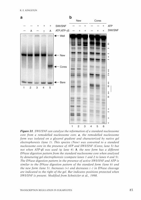
TRANSCRIPTION REGULATION IN EUKARYOTES 83
R. E. KINGSTON
Figure 31. SWI/SNF can catalyse the reformation of a standard nucleosomecore from a remodelled nucleosome core. a, the remodelled nucleosomeform was isolated on a glycerol gradient and characterized by native gelelectrophoresis (lane 1). This species (New) was converted to a standardnucleosome core in the presence of ATP and SWI/SNF (Cores; lane 5) butnot when ATP-γS was used (γ; lane 4). b, the new form has a differentDNase digestion pattern from the standard nucleosome core when analysedby denaturing gel electrophoresis (compare lanes 1 and 2 to lanes 4 and 5).The DNase digestion pattern in the presence of active SWI/SNF and ATP issimilar to the DNase digestion pattern of the standard form (lane 6) andthe new form (lane 3). Increases (+) and decreases (–) in DNase cleavageare indicated to the right of the gel. Bar indicates positions protected whenSWI/SNF is present. Modified from Schnitzler et al., 1998.
that, in the presence of ATP, the hSWI/SNF complex can createa dynamic equilibrium between the standard nucleosome struc-ture and the new form.
To determine whether the new form can facilitate the bindingof a transcriptional activator in vivo, we reconstituted mono-nucleosomes containing a single Gal4-binding site with high orlow affinity, isolated the new form as described above andassessed Gal4 binding by DNase I footprinting. There was pref-erential binding on the new form, irrespective of whether thebinding site was low or high affinity (Schnitzler et al., 1998).A strong implication of these results is that the standardnucleosome can exist in equilibrium with alternative structuresand that a complex involved in gene regulation can affect thetransition between these structures (Fig. 32). In addition, theequilibrium can potentially be driven in either one of two direc-tions to establish states that are either repressed, in the presenceof repressors, or active, in the presence of activators. In this con-text the remodelling machine may be a way simply to make thechromatin more fluid, which can be used in the mechanisms ofboth activation and repression.
Defining the catalytic subunits in the hSWI/SNF complex
The hSWI/SNF complex is a multipolypeptide complex whichcontains from eight up to fifteen subunits. We have identified thefour core catalytic subunits of the complex and reconstructed thecore using recombinant proteins; this recombinant core has sim-ilar activity to that of the intact ‘normal’ holo complex (Fig. 33).Three of the subunits, Brg1- or Brahma-associated factor (BAF)155, BAF170 and Ini1, are highly conserved between yeast andhuman and are always present in both the hSWI/SNF com-plexes. The fourth is the ATPase subunit, which can be eitherBrg1 or the related hBrm protein. Complexes contain either Brg1or hBrm, not both, and the two types of complex are likely tohave different activities and biological roles.
84 HFSP WORKSHOP 7
PART I

TRANSCRIPTION REGULATION IN EUKARYOTES 85
R. E. KINGSTON
Figure 32. A possible model for SWI/SNF action in vivo. SWI/SNF may usethe energy of ATP hydrolysis for continuous interconversion between thestandard and remodelled core structure of the nucleosome. Any protein thatbinds with higher affinity to the remodelled structure, e.g. an activator, willdrive the equilibrium toward the remodelled form, whereas any protein thatbinds with higher affinity to the standard state, e.g. a repressor, will drivethe reaction towards the standard form. Thus SWI/SNF may create a fluidchromatin environment that can be fixed in either a remodelled configur-ation or a standard configuration depending upon the local concentrationsand activities of regulatory proteins.

The ATP-dependent remodelling activity was reconstitutedfrom only the conserved subunits, using recombinant proteinsproduced by a baculovirus expression system. Tagged Ini1 anduntagged BAF155 and BAF170 were coexpressed after a mixedinfection and isolation of the complex was facilitated by thetagged Ini1 protein. Brg1 was produced at high levels by itself asan epitope-tagged (FLAG) version. The four subunits were co-purified from a mixture of extracts from cells infected with thethree subunits and cells infected with Brg1 (Phelan et al., 1999).The remodelling activity of this recombinant core complex wascompared with that of the holo complex using the plasmid super-coiling assay (Fig. 34; Germon et al., 1979). A circular DNA wasreconstituted into chromatin using histones in the presence oftopoisomerase to relax constraint arising from the deposition ofeach nucleosome. After deproteinization, the reconstitutedminichromosomes gave rise to highly supercoiled molecules,which were identified by their migration properties on an
86 HFSP WORKSHOP 7
PART I
Figure 33. The intact human SWI/SNF complex (holo complex, left) con-tains a core of subunits (core, right) that are sufficient for ATP-dependentchromatin remodelling.

agarose gel (Fig. 34, band S). When the reconstituted minichro-mosome was incubated with either the natural SWI/SNF com-plex or the recombinant core in the presence of topoisomeraseand a source of ATP, the changed migration properties of thefinal deproteinized product was easily detected as relaxed topoi-somers (Fig. 34, bands R).
It is not yet clear exactly which changes are promoted in termsof topological constraints at the level of individual remodelledparticles. However, the change in topology detected with both the
TRANSCRIPTION REGULATION IN EUKARYOTES 87
R. E. KINGSTON
Figure 34. Reconstituted hSWI/SNF (rSS) has similar activity to native,intact hSWI/SNF (hSS). Brg1, BAF170, BAF155 and Ini1 were overproducedand purified from baculovirus and reconstituted into a complex, the activityof which was compared to intact SWI/SNF isolated from HeLa cells by meas-uring changes in supercoiling of nucleosomal templates. The reconstitutedcomplex had two- to fourfold less activity than the intact complex. S, super-coiled molecules; N, nicked DNA; R, relaxed topoisomers; L, linear DNA.

holo and core complexes provides a useful assay for defining thecatalytic component(s) of the core subunits. The isolated Brg1subunit was remarkable in that it alone could support remodel-ling in this assay, although at a tenfold lower efficiency than anequimolar concentration of the holo complex. The three subunits,BAF155, BAF170 and Ini1, stimulated this activity. We assumethat the additional subunits in the holo complex (see Fig. 33), arelikely to have regulatory roles. Because Brg1-related polypeptidesare part of other complexes, this family of polypeptides mayrepresent a basic module to create remodelling.
Chromatin remodelling in vivo
Wolfram Hörzin collaboration with
Philip D. Gregory, Martin Münsterkötter and Gudrun Ertinger
The control of chromatin organization with respect to transcrip-tional regulation is being investigated in detail (for review, seeGregory and Hörz, 1998). Genetic studies in yeast have indic-ated that nucleosomes are involved in the repression of tran-scription, e.g., depletion of histone H4 prevented the formationof intact nucleosomes and hence the activation of promoters(Han et al., 1988). Understanding how nucleosomes undergotransition from repressed to active transcriptional states is thusa matter of considerable interest.
The acid phosphatase PHO5 promoter of yeast provides atractable in vivo model of the mechanism of chromatin remodel-ling during gene activation at a regulated promoter (Svaren andHörz, 1997). The repressed gene is packaged in a positionedarray of nucleosomes. When the gene is activated by phosphatestarvation, a transition in chromatin state precisely affects thestructure of four nucleosomes, so that critical parameters of thetransition from closed to open chromatin can be identified. The
88 HFSP WORKSHOP 7
PART I

transcription factor Pho4 binds to the chromatin at an early stepin the chromatin remodelling process. Auxiliary factors, such asATP-dependent remodelling by the SWI/SNF complex (seeCairns, Kingston, this volume) or the enzymatic activity of acomplex with Gcn5 HAT (Brownell et al., 1996; see Allis,Workman, this volume) are not essential for the activation of thegene under normal conditions. As we show here, however, Gcn5HAT activity becomes necessary under conditions that are sub-maximal for inducing gene activation and in its absence a newchromatin pattern appears that may correspond to randomizednucleosomes. Thus, Gcn5, the catalytic subunit of a HAT,directly affects the remodelling of chromatin in vivo.
Remodelling factors at the PHO5 promoter
Starving the yeast S. cerevisiae of phosphate triggers a 50-foldincrease in the production of acid phosphatase, 90% of which isthe product of the PHO5 gene. The gene’s promoter containstwo upstream activator sequence elements (UASp1 and UASp2)that are critical for its activation (Fig. 35). They are bound byPho4, a basic helix–loop–helix (bHLH) transactivator (Svaren etal., 1994), an interaction facilitated by Pho2, a homeobox DNA-binding protein that cooperatively binds with Pho4 at Pho4-binding sites (Barbaric et al., 1998).
Under repressive, i.e., high phosphate, conditions, two nega-tive regulators, Pho80 and Pho85, form a kinase complex thatmaintains Pho4 in a phosphorylated state, preventing its locationin the nucleus (Hirst et al., 1994; O’Neill et al., 1996).Therefore, phosphate starvation or the deletion of the genesencoding these negative regulators can activate the PHO5 pro-moter. Without the negative regulators, the promoter is submax-imally activated, at ~30–50% of the level found in starvationconditions. Activation of the promoter leads to a chromatin tran-sition in which four nucleosomes are disrupted and the entirepromoter becomes accessible (Fig. 35). This is a dynamic
TRANSCRIPTION REGULATION IN EUKARYOTES 89
W. HÖRZ

process that does not require replication (Schmid et al., 1992)or transcription (Fascher et al., 1993).
Two lines of evidence indicated that histone acetylation statescould influence the activation of the PHO5 promoter: the pro-moter is sensitive to the mutation of the potentially acetylatedresidues of histone H4; and strains cannot reach maximal activ-ation or repression after deletion of the gene encoding Rpd3, thehuman homologue of which has a histone deacetylase activity(Taunton et al., 1996). A direct test of the role of histone acetyl-ation in the control of PHO5 promoter states ideally requires themanipulation of a specific enzyme involved in histone acetylation.
The discovery and characterization of a nuclear HAT activityin Tetrahymena thermophila that has striking similarity to theyeast Gcn5 transcriptional coactivator (Brownell et al., 1996)provided the jumping-off point. The effect of deleting GCN5 onthe activation of the PHO5 promoter was examined by creatingyeast strains with the GCN5 gene disrupted and assaying theactivation of the promoter by measuring acid phosphatase activ-ity. PHO5 was induced either by phosphate starvation or the use
90 HFSP WORKSHOP 7
PART I
Figure 35. Chromatin transition at the PHO5 promoter when the gene isactivated by phosphate starvation (–Pi) or disruption of the genes PHO80or PHO85, which encode negative regulators. In the repressed state (+Pi),the promoter is organized in an array of positioned nucleosomes. Fournucleosomes, –1 to –4, are disrupted on activation and the entire promoterbecomes accessible. Upstream activator sequences UASp1 and UASp2 arePho4 binding sites. T, site of TATA box; –360 and –250, distance in bp fromthe coding sequence; shaded circles, nucleosomes that persist when PHO5is activated. Modified from Gaudreau et al., 1997.

of strains with either of the negative regulators, PHO80 orPHO85, disrupted. Under repressive conditions, i.e., high phos-phate (Fig. 36a), Gcn5 was essential for the constitutive phos-phatase activity in strains without either PHO80 or PHO85. Thisis only 30% of the maximal level obtained in the fully inducing
TRANSCRIPTION REGULATION IN EUKARYOTES 91
W. HÖRZ
Figure 36. Disrupting GCN5 diminishes the activity of the PHO5 pro-moter. a, in an otherwise wildtype (WT) strain, activity under repressingconditions (high Pi) drops even further in the absence of Gcn5 (straingcn5∆; dark bars). b, under fully inducing conditions, when no phosphateis present (no Pi), GCN5 disruption has only a small effect. In the absenceof the negative regulators Pho80 (strain pho80∆) and Pho85 (strainpho85∆), PHO5 activity is about 30% of the fully induced level.Reproduced with permission from Gregory et al., 1998.

conditions of phosphate starvation (Fig. 36b), when the depen-dence of the PHO5 promoter on Gcn5 could not be detected.The critical role of Gcn5 for the activation of the PHO5 pro-moter under fully inducing conditions was demonstrated usinganother approach: by weakening the promoter through mutationof either of its UAS elements in lacZ-reporter plasmids driven byPHO5 promoter variants (Barbaric et al., 1998).
92 HFSP WORKSHOP 7
PART I
Figure 37. Chromatin analysis of the PHO5 promoter. a, lanes 1–4, standardwildtype repressed pattern (see Fig. 35 top). Lanes 12–15, pattern of theactive promoter with the four disrupted nucleosomes (see Fig. 35 bottom),here as a consequence of a PHO80 disruption. Lanes 5–8, a new patternappears when Gcn5 is absent with PHO80 disrupted, i.e., in a gcn5∆/pho80∆strain, and the phosphate level is high, the conditions under which activitydrops significantly because of lack of Gcn5 (see Fig. 36a). The new patterndiffers from both the closed and the open pattern and consists of randomlypositioned nucleosomes across the promoter. Lanes 9–11, digests with restric-tion enzymes as markers. b, the chromatin organization of the repressedPHO5 promoter. The open circles are removed under wildtype conditions.T, TATA box. Reproduced with permission from Gregory et al., 1998.

The nucleosomal organization of the PHO5 promoter wasdetermined by DNase I analysis in the presence and absence ofGcn5 (Fig. 37). The wildtype strain produced a standardrepressed pattern with discrete bands, revealing a positionedarray of nucleosomes (Fig. 37a, lanes 1–4). Disruption ofPHO80 resulted in a classic pattern of the active promoter withfour disrupted nucleosomes (Fig. 37a, lanes 12–15). In theabsence of Gcn5 together with PHO80 disruption, a new patternappeared that differed from both the closed and open pattern(Fig. 37a, lanes 5–8); it can be interpreted as a result of ran-domly positioned nucleosomes across the promoter (Fig. 37b).
Under repressive high-phosphate conditions in a strain lack-ing both GCN5 and PHO80, this new chromatin structure hadexceptionally low accessibility to all restriction enzymes tested,an increased protection that is consistent with the loss of nucleo-somal positioning. The discovery of this type of nucleosomalorganization indicates that Gcn5 may be involved in the modu-lation of chromatin in vivo. A similar pattern at the PHO5 pro-moter for transitions between transcriptional states independentof DNA replication has previously been reported (Schmid et al.,1992), so the new pattern may result from a rapid equilibrationbetween the open and the closed state.
Nucleosomal organization requires Gcn5 HAT activity
The effects on chromatin and transcriptional activity observed inthe GCN5-deleted strains are attributable solely to the loss of theHAT activity. Point mutations in Gcn5 that abolish HAT activityhad the same effect on PHO5 promoter activity as loss of theentire protein and were sufficient to generate the new chromatinstructure at the PHO5 promoter (Fig. 38). Thus, the new patternat the PHO5 promoter must result from histones with adecreased acetylation level (L. Wang et al., 1998). This observa-tion supports the connection between HAT activity and the acti-vation of the PHO5 promoter.
TRANSCRIPTION REGULATION IN EUKARYOTES 93
W. HÖRZ

Comparison of other genes in the phosphatase gene family interms of their dependence on Gcn5 will be interesting. PHO10and PHO11, located at the telomere, also code for secreted acid
94 HFSP WORKSHOP 7
PART I
Figure 38. Point mutations in Gcn5 that abolish HAT activity produce thesame pattern of chromatin at the PHO5 promoter as loss of the entire Gcn5protein, implying that this pattern is due to histones with a decreased levelof acetylation. a, diagram of yeast Gcn5, indicating the domains of theprotein and conserved region I within the HAT domain which containsresidues critical for HAT activity. The substitution mutants, named accord-ing to groups of amino acids mutated to alanine, are shown in brackets. b,the HAT activity of each mutant tested is reported as a percentage of wild-type (Gcn5) and the PHO5 promoter activity in these mutants is shown inunits of acid phosphatase activity. Reproduced with permission fromGregory et al., 1998.

phosphatases and are regulated by phosphate depletion in muchthe same way as PHO5. However, the fully activated levels ofexpression of these two genes are considerably lower than thatof PHO5. An alkaline phosphatase gene, PHO8, is also regulatedby Pho4 and undergoes a chromatin transition at the promoter(Barbaric et al., 1992). However, chromatin opening is onlypartial and some nucleosome-mediated protection persists evenwhen the gene is fully activated.
The requirement for Gcn5 HAT activity in the remodelling ofchromatin and transcriptional activation in vivo is well supportedby these studies using the PHO5 promoter. Further work isneeded to analyse whether the effect is mediated through a spec-ific targeting of acetylation within the promoter region. BecauseGcn5 is part of several complexes (see Allis, this volume), it willbe critical to establish whether mutations affecting its partnersin these complexes also result in a similar chromatin phenotypeat the PHO5 promoter. This model yeast promoter promisesmore useful information on the importance of various chromatinremodelling machines in vivo, particularly as genetic manipul-ation is possible.
TRANSCRIPTION REGULATION IN EUKARYOTES 95
W. HÖRZ

96 HFSP WORKSHOP 7

PART II
FACTORS REQUIRED FOR ACTIVATEDTRANSCRIPTION
Introduction
Stefan Björklund
Gene transcription first requires that the DNA template, pack-aged into chromatin, becomes accessible for the binding of tran-scriptional activators and repressors at their cognate sites in thepromoter and for the formation of a preinitiation complex at thetranscription start site (see Part I). The following articles focuson subsequent steps in the process leading to synthesis of amature mRNA: the communication between regulatory proteinsand the general transcription factors, mainly for the transcrip-tion of protein-encoding genes by RNA polymerase II.
Eukaryotic RNA polymerase II by itself is unable to recognizea promoter. However, correct initiation and transcription can bereconstituted in vitro from homogenous RNA polymerase II andfive highly purified factors: TBP, transcription factors IIB(TFIIB), TFIIE, TFIIF and TFIIH. These are highly conservedfrom yeast to mammalian cells. It is generally believed that theyassemble on the promoter to form the preinitiation complex in astepwise mode initiated by the binding of TBP to the conservedTATA sequence that lies 20–30 nucleotides upstream from thetranscription start site of most genes in eukaryotes (see Box 1).
Since the identification of the general transcription factors, aprime interest has been to understand how they interact andwhat their functions in the transcription process are. The firstinsight came from the determination of the atomic structure ofTBP alone and complexed with TFIIB bound to the TATA ele-ment (reviewed in Burley, 1996). Recently published data com-plement and extend this structure (Fu et al., 1999; Poglitsch et
TRANSCRIPTION REGULATION IN EUKARYOTES 97

al., 1999). Combining the known structure of DNA–TBP–TFIIBwith this new structure of RNA polymerase II in a complex withTFIIB shows that the DNA is located in the active centre chan-nel of the polymerase by the interactions between the generaltranscription factors, enabling initiation about 30 bp down-stream from the TATA box.
With the development of in vitro transcription systems reconst-ituted from recombinant or highly purified general transcriptionfactors, it became clear that intermediary factors were needed totransduce signals from DNA-bound transcriptional regulatoryproteins, i.e., activators and repressors, to the basal transcriptionmachinery. These intermediary factors have been given differentnames, such as coactivators, adaptors or mediators. Here they aretermed global transcription regulators to emphasize their generalimportance for regulated transcription of most genes as opposedto transcriptional activators or repressors that exert their effectlocally at certain promoters through sequence-specific interactionwith DNA.
Several global transcription regulators such as TAFIIs,Mediator and upstream stimulatory activity (USA) were origin-ally identified as important components required for thisprocess. Here, Roger Kornberg describes the yeast Mediator andidentifies a specific Mediator domain that is essential for activa-tion by Gal4 and Gcn4. The generality of the Mediator complexhas been questioned because it was first identified only in yeastcells. Mediator-like complexes have now been identified in mam-malian cells as essential for transcriptional regulation by such abroad spectrum of transcriptional regulators as the thyroid hor-mone receptor (TRAP; see Roeder, this volume), Sp1 (cofactorrequired for Sp1 activation (CRSP); see Tjian, thisvolume), the vitamin D receptor (DRIP; Rachez et al., 1999),the adenovirus E1A protein (mammalian Mediator, Boyer et al.,1999; mouse Mediator, see Kornberg, this volume) andSREBP (ARC, see Tjian, this volume). The occurrence and gen-
98 HFSP WORKSHOP 7
PART II

eral importance of Mediator in mammalian cells is reported byKornberg, Robert Tjian and Robert Roeder. Hiroshi Sakuraidescribes the domains in TFIIE and Gal11, a subunit of the yeastMediator, that are essential for their mutual interaction and alsoshows that Gal11 and TFIIE act cooperatively to stimulate phos-phorylation of the RNA polymerase II CTD by the kinase act-ivity of TFIIH.
The USA was originally identified as a partially purified frac-tion from human cells that acts synergistically with TAFIIs inactivated transcription in vitro. Several positive and negativecofactors have subsequently been derived from the USA fractionand Michael Meisterernst summarizes recent results on the pos-itive cofactors PC2, PC4, PC6 and the negative cofactor NC2.
Recent results obtained in vivo have indicated that the TAFIIcomplex does not play as general a role in transcriptional activa-tion as was originally proposed (Moqtaderi et al., 1996a; Walkeret al., 1996). However, some TAFIIs seem to have a rather gen-eral effect whereas others act more specifically on a small set ofpromoters. This might be because the former TAFIIs, in additionto being components of TFIID, are also found as subunits of othertranscription factors with global effects, i.e., PCAF, TBP-free TAF-containing complex (TFTC) and SAGA. Kevin Struhl and MichaelGreen report on studies on the function of TAFIIs at specific pro-moters in vivo, using the microarray technique for whole-genometranscription analyses. Their studies, in combination with resultsobtained by Rick Young and co-workers (Holstege et al., 1998),are starting to reveal this fuller picture of TAFII function in tran-scription. Làszlò Tora reports on the first studies of the in vivofunction of a TAFII subunit in mammalian cells, which reveal thathuman TAFII30 is required for cell-cycle progression and forparietal endodermal differentiation.
The general transcription factor TFIIH comprises nine sub-units and contains the kinase activity responsible for phosphoryl-ation of the C-terminal repeat domain (CTD; see Glossary) of
TRANSCRIPTION REGULATION IN EUKARYOTES 99
INTRODUCTION

the largest subunit of RNA polymerase II (Feaver et al., 1994b;Roy et al., 1994). Mutations in human TFIIH subunits are asso-ciated with the genetic disorders xeroderma pigmentosum,Cockayne’s syndrome and trichothiodystrophy. Jean-Marc Eglydiscusses how the study of these mutations is helping to define arole for TFIIH in transcription, DNA repair and cell-cycleregulation.
Once transcription is initiated, mRNA synthesis proceedsthrough several steps: synthesis of the first phosphodiester bond,promoter clearance, elongation and termination. Elongation isthe only step considered here: Jesper Svejstrup discusses thecomposition of an elongating form of RNA polymerase II andimplicates a chromatin remodelling activity as important for itsfunction. To close, André Sentenac compares RNA polymeraseIII with polymerase II. Recruitment of polymerase III to a pro-moter involves a cascade of protein–protein interactions inwhich TFIIIC acts both as enhancer and promoter-binding factorto overcome nucleosomal repression, perhaps through an intrin-sic HAT activity. The similarity of the polymerase II and III sys-tems is emphasized by the involvement of PC4 and topoiso-merase I, two known polymerase II coactivators, in mammalianTFIIIC function.
The Mediator complex and in vitrotranscriptional activation in yeast
Roger D. Kornbergin collaboration with
Lawrence C. Myers and Claes M. Gustafsson
The view of how transcriptional activator proteins transmit theirsignals to the basal transcription machinery has recently under-
100 HFSP WORKSHOP 7
PART II

gone a dramatic change. Most activators have commonly beenthought to contact specific TAFs in the TFIID complex butseveral experiments have cast doubt on this. First, the Mediatorcomplex has been shown to be necessary and sufficient for regu-lated transcription in the absence of TAFs (Oelgeschläger et al.,1998). Second, in vivo studies indicate that yeast TAFs areimportant for transcription of a very limited set of genes(Moqtaderi et al., 1996a; Walker et al., 1996). Third, largeprotein complexes comprising subunits with high homology tothe yeast Mediator subunits have recently been found in highereukaryotes (see Roeder, Tjian, this volume). Lastly, determina-tion of the structures of the yeast and mouse complexesseparately or in complex with RNA polymerase II reveals strik-ing similarities (Asturias et al., 1999), so they are likely to befunctional counterparts. Thus Mediator-like complexes seem tobe a ubiquitous part of the transcriptional activating mechanismand may take the role, previously ascribed to TAFs, of globalcoactivators.
We describe the identity of the subunits of the purified yeastMediator transcription complex and their various roles, includ-ing interaction with the CTD of RNA polymerase II, and showthat some subunits are essential for normal transcriptional func-tion. We have also identified a mammalian homologue of theyeast complex. We conclude that the Mediator complex is oblig-atory for regulated transcription in vivo and that the individualsubunits determine the responses to different transcriptionalregulatory proteins.
The yeast Mediator complex
Mediator was originally found in a crude protein fraction isol-ated from S. cerevisiae that could relieve activator inhibition oftranscription, known as squelching, in an in vitro transcriptionsystem based on a nuclear extract (Kelleher et al., 1990).A requirement for the Mediator fraction in the activation of
TRANSCRIPTION REGULATION IN EUKARYOTES 101
R. D. KORNBERG

transcription was demonstrated using an in vitro transcriptionsystem composed of semipurified general transcription factors(Flanagan et al., 1991). About the same time, the SRB geneswere identified as suppressors of the cold-sensitive phenotype inyeast mutants that had the CTD of the largest RNA polymeraseII subunit shortened from 26 to 11 heptapeptide repeats; sev-eral of the Srb proteins were subsequently found to associatewith RNA polymerase II in a large multiprotein complex(Thompson et al., 1993). Purification of Mediator to homogene-ity revealed a complex of about 20 polypeptides, including Srbs2 and 4–7 (Kim et al., 1994; see Table 4).
Purified Mediator has three biochemical activities in a fullyreconstituted in vitro transcription system: it stimulates basaltranscription, i.e., transcription in the absence of activators,about tenfold; it enables stimulation by transcriptional activa-tion; and it stimulates phosphorylation of the CTD. That it formsa stable complex with the CTD of RNA polymerase was estab-lished by three independent criteria. First, the Mediator can bepurified to homogeneity either as a separate entity by displace-ment or by using monoclonal antibodies specific for the CTD oras a complex with the polymerase. Second, several polypeptidesin the Mediator were originally identified as CTD-interactingproteins (Srbs) by genetic means. Third, the phosphorylation of
102 HFSP WORKSHOP 7
PART II
Table 4. Mediator subunits divided into three groups according to their pro-posed functions determined from genetic and biochemical experiments.
CTD binding Repression Activation
Srb2 Gal11 Med1Srb4 Rgr1 Med2Srb5 Sin4 Med4Srb6 Pgd1 Med6Srb7 Rox3 Med7
Nut1 Med8Nut2 Med11Cse2
Bold type, yeast proteins for which mammalian homologues have been identified.

the CTD of the holopolymerase by the TFIIH kinase is ~50-foldhigher than that for the core polymerase (see Sakurai, this volume).
All 20 subunits of the Mediator complex have now been iden-tified by a combination of immunoprecipitation, amino-acidsequencing and western blotting (Myers et al., 1998). They havebeen divided into three subgroups based on structural/func-tional studies and physical properties (Table 4). The first is com-posed of the Srb proteins that are thought to interact with theCTD. The second consists of basic proteins encoded by genesthat repress transcription. The third group consists of acidicproteins, now termed Med, encoded by previously unidentifiedgenes. About half the subunits of the yeast Mediator complex areencoded by genes essential for yeast viability. Mammalian hom-ologues of five subunits have been identified (Table 4).
The role of Med proteins in transcriptional activation hasbeen analysed using Mediator complex isolated from yeastmutant strains lacking individual subunits (Y. Li et al., 1995;Myers et al., 1998). The pattern of activation by these differentMediator mutants correlates to their subunit composition.
TRANSCRIPTION REGULATION IN EUKARYOTES 103
R. D. KORNBERG
Figure 39. Structure–function relationship of wildtype and mutantMediator–RNA polymerase II complexes. The subunit organization of theSin4/Rgr1 module of Mediator is based on the results shown in Table 5 andin the text. This model, however, does not preclude the existence of weakinteractions among Med2, Pgd1, Sin4 and other subunits that do not with-stand the rigours of purification. ∆, strains with particular genes deleted.

Mediator from either the med2 or pgd1 deletion strain lacks boththe Med2 and Pgd1 proteins, whereas Mediator isolated from thesin4 deletion strain lacks Sin4, Med2, Pgd1 and also Gal11 (Fig.39; see Sakurai, this volume). Purified Mediator complexes lack-ing the Med2, Pgd1 or Sin4 subunits were unable to supporttranscriptional activation by the acidic activator proteinGal4–VP16 in vitro (Table 5). In contrast, only Mediator lackingSin4 was defective for activation by another acidic tran-scriptional activator, Gcn4.
Because the mutant Mediator complexes from med2 or pgd1deletion strains have no defects in stimulating either the basalactivity or CTD phosphorylation, the separate subdomains thatcompose Mediator must have different functions. The electron-microscopical structure of the Mediator complex purified fromthe sin4 deletion strain supports a modular structure becauseone of the three Mediator subdomains is lacking.
The involvement of the Mediator in transcriptional regulationin vivo has been demonstrated using the med2 deletion strain.This strain is unable to activate a reporter gene driven by a pro-moter containing binding sites for Gal4 under conditions wherethe same reporter construct is fully activated in the wildtypestrain (Table 6). Furthermore, microarray analysis of global geneexpression shows that deletion of med2 affects the expression of
104 HFSP WORKSHOP 7
PART II
Table 5. Functional analysis of wildtype and mutant Mediator complexes ina purified yeast in vitro transcription system. Subunit organization is shownin Fig. 39. Fold activation by transcriptional activators VP16 and Gcn4 isshown as the ratio of full-length transcripts in the presence of activatorfrom a template bearing the appropriate activator-binding sequence, totranscripts in the absence of the activator. This ratio was normalized bydivision by the ratio obtained from a second template lacking the appropri-ate activator-binding sequence.
Activation ratioWildtype med2∆ pgd1∆ sin4∆
VP16 activation 30 1.7 1.8 1.1Gcn4 activation 8.2 6.4 6.9 1.1

more than this one gene: supporting the results obtained in vitro,cells lacking Med2 are impaired in expression of several of theGAL genes required for galactose metabolism. However, SRB4deletions had a much more general effect on gene expression, inline with the observation that SRB4 is an essential gene whereasMED2 is not. This provides further support for the idea that,although the Mediator complex is obligatory for regulated tran-scription in vivo, the individual subunits determine the respon-ses to different transcriptional regulatory proteins.
Table 6. Med2 is required for activation by Gal4–VP16 both in vitro and invivo. Levels of β-galactosidase activity in vivo were assayed in strains witha plasmid containing Gal4–VP16 under control of a promoter and the lacZ-reporter plasmid. The units of activity are normalized to cell absorbance at600 nm and are the means from at least three replicate assays. For detailedresults obtained in vitro, see Table 5.
Fold activationWildtype med2∆
In vitro 30 1.7In vivo 1,156 138
The mammalian Mediator complex
Our identification of a mammalian Mediator complex (Jiang etal., 1998) supports the contention that the Mediator is gener-ally part of the transcriptional activation machinery. The mousecomplex was reveated by chromatographic coelution of themammalian counterparts of the Med7 and Srb7 proteins. Thepurified complex also contained the mammalian Rgr1 and Med6proteins and eight other subunits. During purification, the Med7and Srb7 proteins eluted in different side fractions. Althoughthese fractions have not been purified further because of techni-cal difficulties, their existence indicates there may be severalMediator-like complexes in higher eukaryotes (see also Roeder,Tjian, this volume).
Amino-acid sequencing of the additional subunits did notreveal any other homologues to yeast Mediator subunits, casting
TRANSCRIPTION REGULATION IN EUKARYOTES 105
R. D. KORNBERG

doubt on whether the purified complex is a true functional coun-terpart to the yeast Mediator. The most compelling evidence forthe homology comes from the similar shape and overall size of thetwo complexes at ~40 Å resolution, determined by electronmicroscopy. When bound to the corresponding RNA polymeraseII, both complexes seem to be divided into three subdomains.Moreover, addition of the core RNA polymerase induces a similarconformational change in both complexes, the Mediator adoptinga crescent-like structure that covers much of the polymerase.
Regulation of transcription through generaland gene-specific coactivators
Robert G. Roeder
Biochemical analyses in cell-free systems from mammalian cellshave shown that the regulation of transcription by RNA poly-merase II involves concerted interactions of gene-specific regu-latory factors that bind to distal control elements, general initia-tion factors that act through common core promoter elements toform a preinitiation complex, and a large variety of cofactorsthat mediate functional interactions between these components(summarized in Box 1; see Roeder, 1998). The TAFII class ofcoactivators, which are TBP-associated subunits of TFIID, andthe USA-derived positive cofactors seem indispensable for sig-nificant activation in transcription systems reconstituted withpurified factors. However, TBP alone can fully support bothbasal and activator-mediated transcription when added to aTFIID-depleted HeLa cell nuclear extract (Oelgeschläger et al.,1998), indicating that other coactivators present in the crudeextract, but not in the reconstituted systems, can substitute forTAFII function in activation. I describe here a candidate for allor part of this alternative coactivator activity, a Srb- and Med-
106 HFSP WORKSHOP 7
PART II

containing cofactor complex (SMCC) that contains humanhomologues of a small subset of proteins found in the yeastMediator (Gu et al., 1999). SMCC functions either to repress orto enhance activation in purified reconstituted systems, depend-ing on the TFIIH concentration, and acts synergistically with theUSA-derived positive cofactors.
Studies using the nuclear receptor for thyroid hormone showthat multi-subunit coactivator complexes can also be stably asso-ciated with the activators themselves (Fondell et al., 1996).A high Mr complex of thyroid hormone receptor-associatedproteins (TRAPs) specifically associates (through the TRAP220subunit) with the liganded thyroid hormone receptor in HeLacells. The thyroid hormone receptor–TRAP complex mediatestranscriptional activation from DNA templates in a purifiedreconstituted system with a dependency upon general positivecofactors but not upon TAFIIs (Fondell et al., 1999). On thebasis of these observations, and other reports of nuclear recep-tor coactivators containing histone acetyltransferase activities(Torchia et al., 1998), I propose a multistep model for tran-scriptional activation by nuclear receptors. Moreover, as TRAPand mammalian SMCC complexes have proved to be virtuallyidentical in structure and function (Ito et al., 1999), a pleasingunification of various mammalian and yeast coactivator studiesis emerging.
TAFII-dependent and -independent activation in vitro
Many experiments in metazoan transcription systems reconsti-tuted with highly purified natural and/or recombinant generaltranscription factors and a diverse series of activators haveshown that TAFIIs are absolutely required for efficient activatorfunction, as evidenced by activation with TFIID but not withTBP (Burley and Roeder, 1996). In such systems TAFIIs functionsynergistically with other coactivators present in the partiallypurified USA fraction (Fig. 40; Meisterernst et al., 1991; Chiang
TRANSCRIPTION REGULATION IN EUKARYOTES 107
R. G. ROEDER

et al., 1993). USA-derived positive cofactors that act synergisti-cally with TAFs include the Mr 15K positive cofactor 4 (PC4;Malik et al., 1998) and the Mr 500K PC2 complex (Kretzschmaret al., 1994; see Meisterernst, this volume). In the case of PC4,the coactivator has been shown to interact both with activatorsand with components of the general transcriptional machinery,e.g., TFIIA and RNA polymerase II, thus serving an adaptorfunction (Ge and Roeder, 1994; Malik et al., 1998). The ability of TAFIIs to act as coactivators has been ascribed to their
108 HFSP WORKSHOP 7
PART II
Box 1

specific interactions with the activation domains of many tran-scriptional activators (Verrijzer and Tjian, 1996). However, gen-etic experiments unexpectedly indicated that several TAFIIs werenot generally required for transcriptional activation in yeast(Moqtaderi et al., 1996; Walker et al., 1996), leading to the pro-posal that other factors, such as the Mediator complex (seeKornberg, this volume), could provide necessary and sufficientcoactivator functions in cells, as well as alternative activation pathways.
The above observations prompted a re-evaluation of mam-
TRANSCRIPTION REGULATION IN EUKARYOTES 109
R. G. ROEDER
A comprehensive summary of the stepwise assembly pathway for a preinitiationcomplex (PIC). The scheme shows general transcriptional factors (light grey) on asimple TATA-containing class II core promoter and three classes of cofactors (darkgrey) that variously modulate the functions of DNA-binding regulatory factors(hatched) on corresponding target genes. Interactions between specific basal fac-tors are indicated by short solid bars. The pathway, established using purifiedfactors, begins with recognition of the TATA-element by the TBP component ofTFIID. The assembly and/or function of the PIC may be regulated either by directinteractions between PIC components and DNA-bound regulatory factors; or bydirect interactions between PIC components and certain cofactors, e.g., NC2, act-ing alone; or by direct interactions between PIC components and cofactors thatare recruited to the promoter by interactions with DNA-bound regulatory factors,in which case the cofactors act as adaptors transmitting signals from DNA-boundregulatory factors to general factors. In mammals, the cofactors include a groupassociated with the basal transcriptional machinery: TFIIA, the TAFII componentsof TFIID and the Srb- and Med-containing cofactor complex (SMCC or Mediator)that may associate with RNA polymerase II (Pol II); a group associated with gene-specific regulatory factors, including OCA-B, p300/CBP and TRAPs and othersnot indicated; and a miscellaneous group of potentially more general positive andnegative cofactors (PCs and NCs). The three groups are not necessarily mutuallyexclusive and include both positively and negatively acting cofactors (see this article).These cofactors also include targeted histone acetyltransferases, e.g., p300/CBP,and deacetylases that, along with distinct ATP-dependent factors such as NURFand SWI/SNF complexes, are involved in chromatin remodelling (dashed arrows)before or concomitant with regulatory factor binding and PIC assembly (forreview, see Roeder, 1998). Reprinted with permission from Roeder, 1998.
R.G. Roeder

malian TAFII function in cell-free systems containing a morephysiological complement of nuclear proteins. HeLa cell nuclearextracts were depleted of TFIID using antibodies directedagainst TBP and TAFII100 (Oelgeschläger et al., 1998). Theresulting extracts were almost completely free of TBP and the
110 HFSP WORKSHOP 7
PART II
Figure 40. Optimal activator function in a cell-free system reconstitutedwith purified general initiation factors requires both TAFII components ofTFIID and a positive cofactor(s) in USA. In vitro transcription from anHIV-1 TATA-containing core promoter with five Gal4 sites (pG5HML) andthe adenovirus major late core promoter (pML∆53) was reconstituted withrecombinant or partially purified general initiation factors, including eitheraffinity purified TFIID or recombinant TBP in the presence (+) or absence(–) of Gal4-AH and/or a four-column USA fraction (Ge and Roeder, 1994).Modified from Meisterernst et al., 1991 and Chiang et al., 1993.

TFIID-specific TAFIIs, such as TAFII250, TAFII135 and TAFII55,whereas a large fraction of TAFII31 and TAFII 20/15, which arealso present in the PCAF/Gcn5 and TFTC complexes (see Tora,this volume), remained in the extract (Fig. 41a). The extractslacked detectable transcriptional activity both in the presenceand absence of activators. However, basal transcription and acti-vation by both the acidic activation domain (AAD) of the viralactivator VP16 and the proline-rich activation domain of themammalian activator CTF-1, assayed as Gal4 fusion proteins,could be fully restored not only by addition of immunopurifiedTFIID but also by an equimolar amount of recombinant TBPalone (Fig. 41b; Oelgeschlager et al., 1998). Thus, in a crudeextract with a more natural complement of nuclear factors,TAFII-independent transcription can be observed with at leastsome activators. Interestingly, however, the addition of TBPalone does not restore activation by the glutamine-rich Sp1 acti-vator, indicating that Sp1 has a more rigorous requirement forTAFIIs (M. Guermah and R.G. Roeder, unpublished observations).
Mammalian SMCC modulates transcriptional activation in vitro
If the TAFII components of TFIID are not always required foractivation in nuclear extracts, what are the necessary cofactors?In yeast, the Mediator complex is essential for activation(reviewed in Myer and Young, 1998; see Kornberg, this volume).Mammalian homologues of several yeast Mediator subunits,including Srb7, Srb10 and Srb11, have been identified (seeTable 4). In contrast to immunodepletion of TFIID-specificTAFIIs, partial immunodepletion of Srb7 from HeLa cell extr-acts results in a reduction of activation by the VP16 AAD(Oelgeschläger et al., 1998). The high degree of conservation ofseveral Srb proteins from yeast to mammals, as well as indica-tions of Srb proteins in mammalian holoenzyme preparations(reviewed in Parvin and Young, 1998), indicated the existence ofa Mediator-like complex in mammalian cells. To identify such a
TRANSCRIPTION REGULATION IN EUKARYOTES 111
R. G. ROEDER

112 HFSP WORKSHOP 7
PART II
Figure 41. TAFII-independent transcriptional activation by Gal4-VP16 inHeLa nuclear extract. a, immunoblot analysis of untreated HeLa nuclearextract (HeLa NE) and HeLa nuclear extract depleted of TFIID (NE[∆D])to show that the extracts were almost completely free of the TFIID-specificTAFIIs but contained a large fraction of TAFII31 and TAFII20/15, which arealso present in the PCAF/Gcn5 and TFTC complexes (see Tora, this vol-ume). b, activation of transcription from the Ad2ML, human HSP70 andAd5E4 core promoters, which contain upstream Gal4 sites, by Gal4–VP16.Basal transcription and activation, as observed in untreated HeLa nuclearextract (NE; lanes 1 and 2), can be restored to TFIID-deficient and tran-scriptionally inactive HeLa nuclear extract (NE[∆D]; lanes 3–8) supple-mented with either 6His:TBP (lanes 5 and 6) or epitope-tagged TFIID(f:TFIID; lanes 7 and 8). ND, not detectable. Reprinted with permissionfrom Oelgeschläger et al., 1998.

complex, stably transformed HeLa cell lines expressing epitope-tagged versions of either Srb7, Srb10 or Srb11 were established.All three Srb proteins copurified from these cell lines irrespec-tive of which were tagged, indicating that these proteins exist ina common complex. Gel filtration of cellular extracts showed thepresence of the Srb proteins in either a low Mr complex or a sec-ond much higher Mr complex of around 1,500K (Gu et al., 1999).
Immunoblot analysis showed that the high Mr Srb-containingcomplex, designated SMCC, did not contain basal factors, CBP,BRCA-1 or SWI/SNF proteins, all of which had previously beensuggested to be components of a mammalian RNA polymerase IIholoenzyme. However, SMCC did contain, in addition to thethree SRB proteins, homologues of the yeast regulatory proteinsMed6, Med7, Rgr1, Nut2 and Soh1, as well as a protein of Mr220K (TRAP220) that is also present in the TRAP (see below)and CRSP (see Tjian, this volume) complexes (Gu et al., 1999).Several of the SMCC components, Med6, Med7 and Rgr1, werealso found in a human complex (NAT) that represses activatedtranscription (Sun et al., 1998) and in a murine Mediator com-plex (Jiang et al., 1998).
Homologues of several of the SMCC components were previ-ously identified as components of the yeast Mediator complex.These include components that act either as negative or as posi-tive regulatory factors and in either a gene-specific or a globalfashion, suggesting that similar properties might pertain to thehuman counterparts. SOH1 is not known to be a component ofthe yeast mediator complex but was identified genetically as asuppressor of mutations in HPR1, a component of some prepa-rations of the yeast polymerase II holoenzyme (reviewed in Guet al., 1999). In yeast, SOH1 also interacts genetically with twoRNA polymerase II subunits and with TFIIB, raising the possi-bility that human SOH1 might show similar functions, and thusfacilitate SMCC interactions with RNA polymerase II and TFIIBin the preinitiation complex. Despite these numerous parallels
TRANSCRIPTION REGULATION IN EUKARYOTES 113
R. G. ROEDER
114 HFSP WORKSHOP 7
PART II
between SMCC and yeast Mediator, mammalian RNA poly-merase II was found associated with SMCC only when theimmunopurification was performed at low ionic strength and nodirect SMCC–RNA polymerase II interactions were observedwith the respective purified proteins.
The function of SMCC was tested in an in vitro transcriptionsystem with highly purified/recombinant basal factors, PC4 anda synthetic AAD (Gu et al., 1999). Addition of SMCC at highlevels of TFIIH diminished activated transcription in this system,in a similar way to that observed with the NAT complex (Sun etal., 1998). Activated transcription was also repressed by SMCCwith an RNA polymerase II lacking the CTD of the largest sub-unit. Repression therefore does not involve phosphorylation ofthe CTD by the cyclin/kinase components, Srb10 and Srb11, ofSMCC. However, SMCC does phosphorylate PC4, which inacti-vates its coactivator function and stimulates its single-stranded(ss)DNA-binding activity (see Meisterernst, this volume), indi-cating a potential mechanism by which SMCC represses activ-ated transcription mediated by PC4.
Intrinsic SMCC coactivator function for a synthetic AAD(AH) was observed when transcription assays were performed inthe reconstituted system containing basal factors and eitherintact (form IIA) or CTD-less (form IIB) RNA polymerase II andTFIIH either at limiting concentration or in its absence (Fig.42a). Significantly, there was also a strong synergy betweenSMCC and PC4 under these conditions (Fig. 42b). SMCC-depen-dent activation was seen not only with the synthetic AAD, butalso with the VP16 AAD and the mammalian p53 activationdomain, indicating broader coactivator functions for SMCC (Fig.42c). Moreover, it was possible to show a direct SMCC–p53interaction that was abolished by a double amino-acid substitu-tion that inactivates the p53 activation domain (Gu et al., 1999).This interaction provides a mechanism by which SMCC may berecruited to function at specific promoters. It is not yet clear

whether SMCC can function with TBP in the absence of TAFIIsand therefore whether TAFII and SMCC functions are fullyredundant.
The TRAP complex mediates activation by nuclear receptors
The nuclear receptors for steroid and non-steroid hormones area class of transcriptional activators, the activity of which is mod-ulated by ligand binding. Nuclear receptors are composed ofdistinct functional domains involved in DNA and ligand binding.Several models for transcriptional regulation have emerged fromstudies of the receptors. In the absence of their cognate ligands,the receptors for all-trans retinoic acid and thyroid hormone canrepress transcription. The unliganded retinoic acid and thyroid
TRANSCRIPTION REGULATION IN EUKARYOTES 115
R. G. ROEDER
Figure 42. Coactivator function of SMCC. a, in vitro transcription reactionswere reconstituted with recombinant TFIIA, TFIIB, TFIIE and TFIIF, affinity-purified TFIID, highly purified RNA polymerase IIA (intact) or IIB (CTD-less), the pG5HML (Gal) test template and the pML∆53 (∆53) reference tem-plate. PC4, Gal4–AH (ACT) and SMCC in reactions as indicated. b, coacti-vator assay for SMCC, indicating the synergy between SMCC and PC4. Invitro transcription reactions as in (a) except RNA polymerase IIA was inclu-ded in all reactions and PC4 only in lanes 4–6. SMCC concentration as indi-cated: +, 1 µl; ++, 2.5 µl. c, in vitro transcription reactions as in (a) to showthat SMCC mediates activation by the natural activation domain in p53,assayed as a Gal4-p53 fusion protein, as well as by Gal4-AH. All reactionscontained PC4 and in lanes 4–6 contained 2.5 µl SMCC. Activators as indi-cated. Reprinted with permission from Gu et al., 1999.

hormone receptors interact with co-repressor proteins whichthemselves are part of larger complexes containing histonedeacetylases, leading to the idea that gene repression mayinvolve nuclear receptor-targeted local histone deacetylation andchromatin condensation (Torchia et al., 1998).
On ligand binding, the conformation of the nuclear receptorligand-binding domain changes to create a new surface formed bythe juxtaposition of three α-helices. This creates a hydrophobiccleft, which allows the nuclear receptor ligand-binding domain tointeract with a series of proteins that contain a short amino-acidmotif, Leu-X-X-Leu-Leu (LXXLL), which docks in this cleft.Among the proteins that interact with the nuclear receptors in aligand-dependent manner are the coactivators CBP, PCAF, TIF-2,SRC-1 and ACTR, all of which have HAT activity. These observa-tions have led to the idea that gene activation may involve nuclearreceptor-targeted local histone acetylation and chromatin decon-densation (Torchia et al., 1998).
Most of these nuclear receptor-interacting proteins have beenidentified by two-hybrid screen or direct in vitro binding of prot-eins to immobilized receptors. However, a new class of proteinsinvolved in thyroid hormone receptor function has been identi-fied using a different approach. Because the purified recombi-nant receptor does not activate transcription from DNA tem-plates in vitro under conditions in which many other activatorsare active, a search was made for proteins that may interact withthe receptor and allow it to function in vitro.
Stably transformed HeLa cell lines were established thatexpress epitope-tagged thyroid hormone receptor. When thetagged receptor is purified from these cells after treatment withthyroid hormone, a set of at least 10 major TRAP proteinsranging in Mr from 240 to 80K is found stably associated withthe receptor (Fondell et al., 1996). Subsequent gel filtrationshowed that even in the absence of the receptor, the TRAPcomponents form a complex of high native Mr (Yuan et al.,
116 HFSP WORKSHOP 7
PART II
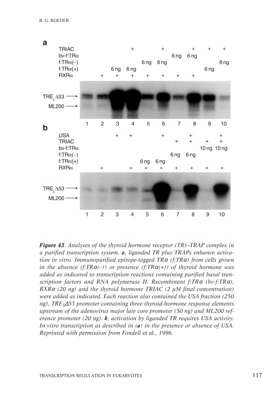
TRANSCRIPTION REGULATION IN EUKARYOTES 117
R. G. ROEDER
Figure 43. Analyses of the thyroid hormone receptor (TR)–TRAP complex ina purified transcription system. a, liganded TR plus TRAPs enhance activa-tion in vitro. Immunopurified epitope-tagged TRα (f:TRα) from cells grownin the absence (f:TRα(–)) or presence (f:TRα(+)) of thyroid hormone wasadded as indicated to transcription reactions containing purified basal tran-scription factors and RNA polymerase II. Recombinant f:TRα (bv-f:TRα),RXRα (20 ng) and the thyroid hormone TRIAC (2 µM final concentration)were added as indicated. Each reaction also contained the USA fraction (250ng), TRE3∆53 promoter containing three thyroid-hormone response elementsupstream of the adenovirus major late core promoter (50 ng) and ML200 ref-erence promoter (20 ng). b, activation by liganded TR requires USA activity.In vitro transcription as described in (a) in the presence or absence of USA.Reprinted with permission from Fondell et al., 1996.
1998). In contrast to recombinant receptor and epitope-taggedreceptor purified from cells in the absence of thyroid hormone,the receptor–TRAP complex markedly activates transcription invitro in a retinoid X receptor (RXR)-dependent manner (Fig.43a). A seemingly identical complex has also been identifiedthrough its ability to bind selectively to the liganded vitamin D3receptor (VDR) in vitro. The complex containing VDR andVDR-interacting proteins (DRIPs) is moderately active in tran-scription in vitro (Rachez et al., 1998).
Most importantly, the TRAP complex was found to be devoidof other known nuclear receptor coactivators, such as p300/CBP,TIF-2 and SRC-1. A further characterization of TRAP compo-nents, including cognate cDNA cloning, revealed many withLXXLL motifs. However, only the TRAP220 component, contain-ing two LXXLL motifs, was found to interact significantly withthyroid hormone receptor in a ligand-dependent manner (Yuan etal., 1998). These results indicate that TRAP220 may mediate theprincipal contact between the thyroid hormone receptor and theTRAP complex. In a further analysis, TRAP220 showed strongligand-dependent or ligand-enhanced interactions with theretinoic acid receptor (RAR), RXR, VDR and peroxisome prolif-erator-activated receptors α and γ, whereas it interacted onlyweakly, albeit specifically, with the oestrogen receptor (Yuan etal., 1998). As with thyroid hormone receptor, these receptorsshowed only very weak interactions with TRAP100, which con-tains many LXXLL motifs, consistent with other studies showingcontext effects of LXXLL motif interactions with nuclear recep-tors. Thus, although the TRAP220 shows some preference inreceptor interactions, the results indicate a broader role forTRAP220 and the TRAP complex in nuclear receptor functions.
Another significant finding is that transcriptional activationby the thyroid hormone receptor (TR)–TRAP complex in a puri-fied reconstituted system absolutely requires other coactivators,showing that the requirement for a specialized coactivator does
118 HFSP WORKSHOP 7
PART II
not obviate the requirement for the general coactivators. Thus,on naked DNA templates, activation by the TR–TRAP complexabsolutely requires the USA fraction (Fig. 43b) or, for optimalactivity, a combination of the derived PC2 and PC4 (Fondell etal., 1996; 1999). However, the activation is TAFII-independent,indicating a functional redundancy between TAFIIs and theTRAP complex (Fondell et al., 1999).
A multistep model for activation by nuclear receptors
Our observations, along with those of other laboratories(reviewed by Torchia et al., 1998 and Fondell et al., 1999), indi-cate that activation by nuclear receptors may be a multistepprocess (Fig. 44). In a first step, ligands interact with promoter-bound nuclear receptors to facilitate dissociation of co-repressorsand associated histone deacetylases. In a second step, ligandedreceptors interact with a series of coactivators that, through associ-ated HAT activators, allow a local modification of chromatin struc-ture. The receptors may then interact with TRAP proteins that inturn act in concert with other general coactivators to enhancepreinitiation complex formation, with RNA polymerase II andgeneral initiation factors, and transcription initiation. The viewthat the TRAPs and the other nuclear receptor interacting factorsperform different functions is strengthened by the fact that CBPenhances activation by nuclear receptors on chromatin templatesbut not on naked DNA templates, whereas the TRAP complex canpromote activation using naked DNA templates.
An interesting but still unanswered question concerns themechanisms by which the HAT-containing coactivators on thepromoter might be displaced by the TRAPs. One possibility isthat HATs and TRAPs both interact reversibly with ligandedreceptors following co-repressor disassociation but that onlyHAT cofactor interactions allow chromatin modifications thatare prerequisites for stable interactions of general initiationfactors at core promoter elements. Concerted synergistic inter-
TRANSCRIPTION REGULATION IN EUKARYOTES 119
R. G. ROEDER

120 HFSP WORKSHOP 7
PART II
Figure 44. Multistep model for gene activation by thyroid hormone receptor(TR), based on studies showing ligand-independent TR-mediated repressionthrough TR-interacting co-repressors (N-COR/SMRT, Sin3) and the assoc-iated histone deacetylase (HDAC), followed by ligand-dependent dissociationof co-repressors and corresponding association of coactivators with HAT activ-ity (p300/CBP, SRC-1-related factors, PCAF). Following ligand-dependentassociation of TRAPs, and with probable displacement of HAT coactivators,the complex acts in conjunction with general coactivators PC2 and PC4 tomediate transcription from DNA templates by RNA polymerase II and gen-eral initiation factors. GTF, general transcription factor; T3, thyroid hormone.See text for details. Reprinted with permission from Fondell et al., 1999.

actions between general initiation factors (including RNA poly-merase II), TRAPs and other coactivators may then shift theequilibrium to favour TRAP displacement of HATs and forma-tion of a stable preinitiation complex.
Convergence of coactivator studies
Intriguingly, TRAP220 was also identified as a component both ofSMCC (Gu et al., 1999) and of CRSP, a complex that is required,with TAFIIs, for Spl activity on DNA templates (see Tijan, this vol-ume) and that seems to be related to the PC2 component of USA(S. Malik and R.G. Roeder, unpublished observations). Theseresults raised the interesting possibility that various componentsmay be shared among various cofactor complexes, just as someTAFIIs are common to TFIID and PCAF/Gcn5 complexes (seeTora, Workman, this volume), or that they may be mixed andmatched to create complexes of varying specificity and function. Itseemed important in this regard to determine the full extent ofcomponents shared between TRAP/DRIP, SMCC, CRSP and PC2complexes, and whether SMCC and CRSP can also interact withnuclear receptors and thereby facilitate crosstalk between differentactivators through TRAP220. Indeed, recent results have estab-lished that the TRAP and SMCC complexes are virtually identicalin structure and function (M. Ito et al., 1999) and that PC2 is madeup of a large subset of the TRAP/SMCC components (S. Malik andR.G. Roeder, unpublished observations). Although somewhat sur-prising, these findings represent a pleasing convergence and unifi-cation of various mammalian and yeast coactivator studies.However, although the TRAP complex, and the related complexessubsequently described by us and others, contain apparent homo-logues of several yeast Mediator components and ostensibly func-tion by similar mechanisms, the vast majority of the componentsubunits have no yeast counterparts and probably reflect meta-zoan-specific functions and mechanisms.
TRANSCRIPTION REGULATION IN EUKARYOTES 121
R. G. ROEDER

Transcriptional activation by Sp1:a biochemical journey
Robert TjianIn collaboration with
Anders Näär and Soojin Ryu
Transcriptional activation by upstream activators is a multistepprocess requiring several classes of cofactor that interface withthe basal transcription machinery. Here we review the evolutionof biochemical studies on the regulation of gene expression bythe human promoter selectivity factor, Sp1, that have revealedthe complexity of this process. Sp1 is a glutamine-rich transcrip-tional activator required for the efficient transcriptional activat-ion of many cellular and viral genes by RNA polymerase II.
Over the past decade a variety of biochemical approacheshave revealed that several factors contribute to Sp1-mediatedtranscriptional activation (Näär et al., 1999), which includethe TAFIIs and a new multi-subunit complex termed cofactorrequired for Sp1 activation (CRSP; Pugh and Tjian, 1990).Other requirements, demonstrated by reconstitution of syner-gistic activation of the native low-density lipoprotein receptorpromoter induced by Sp1 and the cholesterol-regulated tran-scription factor SREBP-1a, include chromatin assembly and aSREBP-binding multiprotein coactivator containing CBP, calledactivator-recruited cofactor (ARC), as well as TFIID.
CRSP has several components in common with other tran-scriptional coactivator complexes, e.g. Mediator, NAT and TRAP(see Kornberg, Roeder, this volume). This sharing of compo-nents seems to be emerging as a common theme in transcrip-tional regulation: e.g., a subset of TAFIIs are found in both theSAGA and TFIID complexes (see Workman, this volume) andseveral distinct chromatin remodelling complexes contain thecommon ATPase ISWI subunit (see Becker, Wu, this volume).Thus transcriptional cofactor complexes such as CRSP may act
122 HFSP WORKSHOP 7
PART II

through a combinatorial mechanism, interacting with a varietyof proteins at multiple promoters.
TFIID and the TAFIIs
Initial biochemical studies used partially purified basal tran-scriptional factors, RNA polymerase II and a simple model tem-plate to identify essential factors needed to reconstitute Sp1-activated transcription in vitro. The template contained threetandem Sp1-binding sites upstream of a minimal adenovirusmajor late promoter (Pugh and Tjian, 1990; Fig. 45). The gen-eral transcription factor TFIID was thought to be the first factorrecruited to the core promoter region and thus likely to be atarget for activators. Biochemical fractionation showed thatpurified TFIID was indeed needed for activated transcriptionand could not be replaced by recombinant TBP alone.
TFIID is a complex consisting of TBP and several TAFIIs (seeGreen, Struhl, Tora, this volume). The function of individualTAFIIs was examined by purifying separate subunits of TFIID
TRANSCRIPTION REGULATION IN EUKARYOTES 123
R. TJIAN
Figure 45. Top, structure of transcriptional activator Sp1, indicating theglutamine-rich transcriptional activation domains and the zinc finger DNA-binding domain. Bottom, the process of transcriptional activation on a sim-ple model template containing three Sp1-binding sites (GC boxes).

(Dynlacht et al., 1991), cloning them and producing recombi-nant proteins. This revealed that distinct classes of activator canbind different TAFIIs; Sp1 directly interacts with DrosophilaTAFII110 (dTAFII110; human homologue TAFII130) through itsglutamine-rich activation domains (Hoey et al., 1993), whereasthe Drosophila transcription factor NTF-1 binds dTAFII150 anddTAFII60 (Chen et al., 1994a).
The contribution of individual TAFs to the function of TFIIDin transcriptional activation was reconstituted biochemicallyusing subsets of recombinant TAFIIs to assemble partial TFIIDcomplexes (Chen et al., 1994a). A minimal TFIID complex con-taining TBP and TAFII250 supported basal transcription in thepurified transcription system but additional TAFIIs wererequired for activated transcription. These varied for differentclasses of activators, e.g., Sp1 activation was supported by apartial TFIID complex containing TBP, TAFII250, TAFII150 andTAFII110 and assembly of different partial complexes showedthat TAFII110, the subunit that contacts Sp1, is necessary. Incontrast, response to NTF-1 required TAFII150 and TAFII60 inpartial complexes. Thus direct contact between activators andthe TAFIIs in TFIID seemed to correlate with transcriptionalactivation. However, partial TFIID complexes could not fullyreconstitute the high level of transcription observed with recon-stituted holo-TFIID, so the TAFIIs that are not directly involvedin contacting a specific activator may also contribute to TFIIDcoactivator function or stability.
The functional roles of some of the other TAFIIs are alsobeing clarified: TAFII150 recognizes and binds to certain corepromoter sequences and may act in promoter specificity bystabilizing TFIID binding at certain promoters (Verrijzer et al.,1994; 1995). TAFII250 may be structural, acting as a core sub-unit contacted by TBP and the other TAFIIs but it could also beengaged in activated transcription by transmitting an activationsignal, possibly through a conformational change. In addition
124 HFSP WORKSHOP 7
PART II

TAFII250 interacts with core promoter elements; contains mult-iple kinase domains; can phosphorylate several proteins; and hasHAT activity; however, the physiological substrates of theseactivities remain unclear.
The TAFII components of TFIID clearly play multiple roles inactivated transcription (Goodrich et al., 1996), including pro-moter selectivity through direct contact with DNA and funct-ioning as a coactivator by making direct contact with diverseclasses of activator that transmit the activating signals to thebasal machinery. The mechanism of this signal transduction isunclear but is likely to involve conformational change within theTFIID complex and/or the modulation of enzymatic activitiesresulting in the formation of a stable and active preinitiationcomplex at the promoter.
Cofactor for Sp1 activation (CRSP)
The development of purer and more active transcription systemspermitted a more refined analysis of the factor requirements forSp1-mediated transcriptional activation. Using a simple modeltemplate and a combination of recombinant and purified factors,we found that an activity present in a crude phosphocellulosefraction of HeLa extracts is required (Fig. 46; Ryu et al., 1999).This was identified as a new, multiprotein complex, which weterm CRSP with Mr ~700K and consisting of at least 7 subunits:p200, p150, p130, p77, p70, p33a and p33b. The presence ofthese subunits in a cofactor complex was confirmed by affinitypurification using anti-p150 antibodies, which immunodepletesa high-salt-resistant complex containing the CRSP subunits froma crude HeLa fraction. The antibody also inhibits CRSP activity,providing evidence that these subunits are responsible for thisactivity.
Several of the CRSP components, subunits 130, 77, 70 and33b, have not previously been identified; others are alreadyknown as components of other complexes involved in transcrip-
TRANSCRIPTION REGULATION IN EUKARYOTES 125
R. TJIAN

tional regulation (Fig. 47). For example, CRSP 150 contains asmall stretch of homology to yeast Rgr1 and CRSP 33a seems tobe the human homologue of Med7. Both are components of theyeast Mediator complex and the negative transcriptional regula-tor NAT complex (Sun et al., 1998). CRSP 130, 77 and 33b haveno homology to known proteins. CRSP also associates with a200K protein homologous to TRIP2/PBP, a component of theTRAP/DRIP coactivator complex (see Roeder, this volume).
We are now using biochemical studies to examine how CRSPstimulates Sp1 activation. The complex does not appear to con-tact the activator directly but does interact with both individual
126 HFSP WORKSHOP 7
PART II
Figure 46. The reconstitution of Sp1 activation requires the basal tran-scriptional factors, RNA polymerase II and TFIID, and is stimulated by theCRSP complex. Transcription from a simple model promoter containingthree Sp1-binding sites (GC box) and a minimal adenovirus E1b promoter(lower band) or from a control template lacking Sp1-binding sites (upperband) is displayed. Triangles, increasing concentrations; r, recombinantfactors TFIIA, B, E and F. Modified from Ryu et al., 1999.

TAFIIs and holo-TFIID (see Fig. 49). CRSP 70, which is en-coded by a newly found gene, exhibits some sequence homologyto the transcriptional elongation protein TFIIS, so we are curiousto see whether CRSP may also act at a step after transcriptionalinitiation.
Synergistic activation by Sp1 and SREBP-1a
Although our studies of Sp1 (see above) have used a single acti-vator on a simple model promoter, in vivo Sp1 often cooperateswith other transcriptional activators on complex promoters. Weare investigating cofactor requirements for Sp1 activation in thecontext of more complex promoters by recapitulating the syner-gistic activation by Sp1 and the sterol-regulated factor SREBP-1aat the promoter of the low-density lipoprotein receptor gene(Näär et al., 1998a, b). In a purified human transcription
TRANSCRIPTION REGULATION IN EUKARYOTES 127
R. TJIAN
Figure 47. Subunit composition of CRSP and its relationship to other tran-scriptional regulatory complexes. Two CRSP subunits, 150 and 33a, sharehomology with Rgr1 and Med7, which are also components of the yeastMediator and NAT complexes, whereas CRSP 200 (TRIP2/PBP) is also pres-ent in the TRAP/DRIP/ARC complex.

128 HFSP WORKSHOP 7
PART II
Figure 48. A coactivator, ARC, is required for synergistic Sp1 and SREBP-1aactivation on the promoter of the low-density lipoprotein receptor gene. a, com-ponents of the purified coactivator. *, nonspecific proteins that do not consis-tently copurify with this activity. b, a large multi-subunit complex (Mr 2,000K,not shown) containing CBP associates selectively with the SREBP-1a activationdomain. HeLa nuclear extract was incubated with affinity resins containing theactivation domains of SREBP-1a (GST–SREBP amino acids 1–50) or Sp1(GST–Sp1A) and the bound fractions analysed by SDS–PAGE. *, two nonspe-cific proteins that bind the GST portion of the fusion proteins. c, purified coac-tivator stimulates transcription from the promoter assembled into chromatin.Transcriptional activity was assessed by primer extension in the absence (–) orpresence (+) of Sp1 and SREBP-1a and either in the absence of coactivator(lanes 1, 2), with a partially purified coactivator fraction (lanes 3, 4) or purifiedcoactivator (lanes 5, 6). d, coactivator does not affect transcription from nakedDNA templates.

system, chromatin, TAFIIs and a new SREBP-binding coactivatoractivity are all required to mediate efficient synergistic activationby Sp1 and SREBP-1a. Purification of the SREBP-binding cofac-tor to near homogeneity (Fig. 48a) identified a large multiprot-ein coactivator we have called activator-recruited cofactor(ARC) that selectively interacts with the SREBP activationdomain (Fig. 48b). This activity contains the transcriptionalcoactivator and the acetyltransferase CBP but the complex can-not be replaced by either recombinant CBP or the CBP-associatedtranscriptional coactivator PCAF.
As the coactivator can mediate high levels of synergistic activa-tion by SREBP-1a and Sp1 on chromatin templates but not on nakedDNA (cf. Fig. 48c and d), we think this large complex may beinvolved in overcoming chromatin-mediated repression. However,its mechanism of action remains unclear but identification of theremaining components of the ARC complex, as indicated in the pro-posed model (Fig. 49), should help to resolve this in the future.
TRANSCRIPTION REGULATION IN EUKARYOTES 129
R. TJIAN
Figure 49. Model of cofactor requirements for Sp1-activated transcriptionand for synergistic activation by Sp1 and SREBP-1a in the low-densitylipoprotein receptor promoter. In HeLa extracts the glutamine-rich domainof Sp1 makes direct contacts with hTAFII130 within TFIID, transmittinginformation to the basal transcriptional machinery and leading toincreased transcription initiation. CRSP contributes to this process throughan unknown mechanism. Chromatin-mediated repression of transcriptioncould be relieved by the recruitment of ARC/CBP coactivator by SREBP,which may facilitate the access of the transcriptional machinery to the tem-plate. The HAT activity of CBP may participate in chromatin remodelling.

Structure and function of proteins thatmodulate RNA polymerase II transcription
Michael Meisterernstin collaboration with
Keiko Ikeda, Jun Xie and Martine Couart
Several proteins beside the general transcription factors andcoactivators such as the TAFIIs and the Med and Srb proteinscan act as transcriptional coactivators in vitro (reviewed inKaiser and Meisterernst, 1996). We have characterized the pos-itive cofactors PC2 and PC4 (see Roeder, this volume) in vitroand the negative regulator NC2 in vitro and in vivo in yeast. Weshow here that PC2 is involved in the formation of the preinit-iation complex and that PC4 contains two partially distinct func-tional domains: an N-terminal domain involved in enhancingactivated transcription through interaction with transcriptionalactivation domains and general transcription factors, and a C-terminal domain that binds single-stranded DNA and repressestranscription. Another coactivator, PC6, seems to be essentialfor activation by the viral acidic activator VP16 in vitro. It maybe a specificity factor for one of the VP16 activation regions ina similar way that CBP is specific for another region, although itsprecise role is uncertain. We also show that NC2 repression isessential in yeast and NC2 may compete with TFIIA for bindingto TBP in vivo as well as in vitro.
The RNA polymerase II coactivator PC2
Transcriptional coactivators required for activator function invitro were originally identified in a crude USA fraction from ionexchange chromatography of HeLa cell nuclear extracts. Furtherpurification showed that the USA fraction contained severalactivities: positively acting coactivators PCs 1–4 and PC7 andnegatively acting factors NC1 and NC2 (Meisterernst et al.,1991). After purification to homogeneity, PC1 was identified as
130 HFSP WORKSHOP 7
PART II

poly-ADP-ribose polymerase and PC3 as topoisomerase I.Others, such as PC2 and PC7, have not been purified to homo-geneity but are likely to correspond to large multiprotein com-plexes (Kaiser and Meisterernst, 1996). Recent work indicatesthat PC2 has subunits homologous to those in Mediator, as well as to human TRAP and DRIP/ARC complexes, indicatingPC2 may be related to yeast Mediator (M. Meisterernst, unpublished observations).
Although PC2 has not been completely purified, biochemicalexperiments have revealed that it is a general coactivator thatwill potentiate activation by transcriptional activators, such asthe glutamine-rich activator Sp1 (see Tjian, this volume), inin vitro systems reconstituted with highly purified endogenousand/or recombinant general transcription factors (Kretzschmaret al., 1994b). PC2 acts during preinitiation complex formationbecause activation does not occur if it is added to the transcrip-tion reaction after the general transcription factors and assemblyof the preinitiation complex. Because activity is diminished butnot fully lost if it is added after TFIID but before the other gen-eral transcription factors, PC2 seems to act, in part, early in theactivation process, possibly on the binding of TFIID.
The requirement for PC2 cannot be eliminated by using pre-melted promoter templates in which the nucleotides around theRNA initiation site are not paired. The use of premelted tem-plates alleviates the requirement for TFIIH and TFIIE and allowstranscription to be performed in a more defined system. Underthese conditions, the synthetic AAD AH-peptide fused to Gal4activator can activate transcription in a PC2-dependent mannerwhen only TBP, TFIIB, polymerase II and TFIIF are included inthe transcription reaction.
Interaction of the VP16 AAD with CBP
PC6, to which we have recently added the term MOVE (formediator of VP16 enhancer), is a coactivator fraction that can be
TRANSCRIPTION REGULATION IN EUKARYOTES 131
M. MEISTERERNST

separated from the other positive cofactors at an early stage ofchromatography. Its activity is required for specific activation ofthe VP16 activator and it must be a distinct cofactor for VP16because it cannot be substituted by PC2, PC4 or PC5.
As PC6-MOVE could not be purified by ion-exchangechromatography, we used affinity chromatography of extracts in acolumn containing the immobilized VP16 AAD. This resulted inthe extract losing the specific ability to support activation byVP16, indicating that PC6-MOVE or a PC6-MOVE-like activityhad been removed and confirming that this activity is essential fortranscriptional activation of region H1 (see below) of VP16 invitro. Immunoblot analysis using antibodies against the knowntranscription factors present in the starting extract showed thatthe VP16 column did not efficiently deplete any of the generaltranscription factors or TAFIIs but did remove the closely relatedp300/CBP proteins. Although CBP binds to another region (H2)of the VP16 AAD, it did not coelute with the PC6-MOVE fractionand is therefore not the PC6-MOVE activity.
132 HFSP WORKSHOP 7
PART II
Figure 50. Summary of the transcription factors that are established asinteracting directly with VP16 or that are required for VP16 activity invitro. Direct interaction between the VP16 acidic activator domain (madeup of regions H1 and H2) and p300/CBP, TAF31, PC4, TBP and TFIIB areindicated. PC6-MOVE is required for the transcriptional activation of theH1 activation subdomain of VP16.

The activation domain of VP16 can be divided into two sub-domains, H1 in the N-terminal region and H2 in the C-terminalregion (Fig. 50). Mutation of a phenylalanine residue in the H1region, which abolishes activation in vitro, did not affectVP16–CBP interactions. In contrast, a triple amino-acid substi-tution in the H2 domain abolished VP16–CBP interaction andthe H2 domain alone was sufficient for binding to CBP, althoughdeletion of this domain did not affect transcriptional activationin vitro in the absence of chromatin.
CBP has HAT activity and can potentiate transcriptional acti-vation in vitro on chromatin templates by activators such as theoestrogen receptor. This indicates that the VP16 AAD has abipartite structure involved in modulating distinct stages of theactivation process. First, the H2 domain may recruit the CBPHAT activity to modify the chromatin structure. The generaltranscription factors and coactivators such as PC6-MOVE arethen recruited to the modified template by interactions with theH1 domain. VP16 also stimulates RNA elongation through inter-actions with TFIIH. The ability of the VP16 AAD to enhancemultiple steps of the activation process may account for the veryhigh activity observed in vivo with this activator.
The positive cofactor PC4
PC4 has been purified to homogeneity and its correspondingcDNA has been cloned. It is a 15K protein comprising 127amino acids that can both stimulate activated transcription and,at higher concentrations, repress basal transcription in vitro(Kretzschmar et al., 1994a; Ge and Roeder, 1994; see Roeder,this volume). PC4 can be divided into a structured C-terminaldomain and a probably unstructured N-terminal domain con-taining many lysine and serine residues. Full homologues of PC4are found in many organisms including C. elegans, although inyeast only the structured C-terminal domain is conserved.
The structure of the PC4 C-terminal domain at the atomic
TRANSCRIPTION REGULATION IN EUKARYOTES 133
M. MEISTERERNST

level (Fig. 51; Brandsen et al., 1997) shows that the domaindimerizes to form two ‘quarter-pipe’ structures, each acting as asingle-stranded (ss) DNA-binding channel. PC4 binds ssDNAwith 100-fold higher affinity than for double-stranded DNA andits C-terminal domain resembles replication protein A, whichbinds strongly to ssDNA (Werten et al., 1998a). One of thefunctions of PC4 may therefore be to bind to melted DNAduplexes in open promoters or during DNA replication, recom-bination or repair.
We used the structural data to design amino-acid substitu-tions in exposed residues of the predicted DNA-binding surfacein order to disrupt ssDNA binding (Werten et al., 1998b). These
134 HFSP WORKSHOP 7
PART II
Figure 51. Ribbon representation of the crystal structure of the PC4 C-ter-minal domain deduced from X-ray crystallography data. The quarter-pipessDNA-binding channels and the principal α-helical and β-sheet regionsare indicated.

mutations do not, however, affect coactivator function, indicat-ing that ssDNA binding is not required for this. Instead, thesemutants affect the ability of PC4 to repress transcription throughbinding to ssDNA. This ability, interestingly, is antagonized byTFIIH, perhaps through an intrinsic helicase, indicating thatTFIIH may be in part an anti-repression factor.
The N-terminal domain of PC4 is required to promote activa-tion because the C-terminal domain alone is inactive. PC4 bindsto several activation domains and to TFIIA and enhances activa-tion by facilitating the binding of TFIID, perhaps through itsinteraction with TFIIA and also by acting at an undefined laterstage of the formation of the preinitiation complex, possiblythrough direct interaction with polymerase II itself (Kaiser et al.,1995). Coactivator activity is, however, abolished upon hyper-phosphorylation of serines in the N-terminal domain by caseinkinase II and perhaps by other more physiologically relevantkinases, such as TFIIH or Srb10/11. The coactivator function ofPC4 is also abolished by mutation of four lysine residues in theN-terminal domain, some of which are targets for acetylation,indicating that PC4 function may be regulated in other ways.
In vivo analysis of human NC2α and NC2β in yeast
Negatively acting factors repress basal transcription in an activ-ator-reversible manner, so enhancing the apparent stimulationby the activator. The negatively acting NC2 factor has two sub-units, NC2α and β, and the heterodimer binds to the TBP–DNAcomplex. This prevents the subsequent association with TFIIAand TFIIB and alters the conformation of the DNA, which blocksformation of the preinitiation complex and transcription (Fig.52). As the α subunit contains a histone-fold motif homologousto that of histone H2A and the β subunit one to that of H2B,NC2 is composed of a histone-like pair (Goppelt et al., 1996).
Homologues of the NC2α and β subunits are found in yeastand the β-subunit homology extends beyond the histone-fold
TRANSCRIPTION REGULATION IN EUKARYOTES 135
M. MEISTERERNST

regions. Null mutations in the yeast NC2 proteins are lethal butmutations in both the α and β subunits can be complementedby their human counterparts. Deletion analysis shows that thehistone-fold region of the α subunit is sufficient for complement-ation, whereas the β subunit requires additional conserved C-ter-minal sequences. Amino-acid substitutions in the histone-folddimerization interface of either subunit result in mutants thatfail to complement, so dimerization through this surface isrequired for function in vivo. Amino-acid substitutions in theβ subunit on the presumed exposed face of the long α2-helix ofthe histone fold are interesting because they also abolish com-plementation but do not affect dimerization. This mutated NC2
136 HFSP WORKSHOP 7
PART II
Figure 52. Model of the mechanism of repression of polymerase II tran-scription by NC2. NC2α and β bind to TBP, blocking the subsequent asso-ciation with TFIIA and TFIIB (crossed arrows) as well as altering the shapeof the DNA in the preinitiation complex (large arrow).

complex does not repress basal transcription in vitro, indicatingthat yeast viability requires the ability of NC2 to act as a tran-scriptional repressor.
Yeast cells grow normally when the human NC2β subunit isexpressed from a high-copy-number plasmid but poorly when itis expressed from a low-copy-number plasmid. This enabled usto use a suppressor-screen technique to identify proteins thatmay be involved in NC2 function. One of these suppressors,which has a cold-sensitive phenotype, contained a Val-to-Phemutation in the yeast TFIIA subunit TOA1 at position 251,which is in the TOA1–TOA2 dimerization interface (Fig. 53).Recombinant TFIIA containing this amino-acid substitution
TRANSCRIPTION REGULATION IN EUKARYOTES 137
M. MEISTERERNST
Figure 53. Ribbon representation of the crystal structure of theTBP–TFIIA–DNA complex showing the location of a single amino-acid sub-stitution, Val-to-Phe at position 251 (V251), in the TOA1 subunit of TFIIA.This form is a suppressor of human NC2β expression in yeast. Blue, N ter-minus of TOA1; pink, C terminus of TOA1; green, TOA2 subunit; red, TBP.

failed to antagonize the binding of NC2 to TBP and did notalleviate transcriptional repression by NC2. So, when limitingamounts of NC2 are present in yeast, transcription is not effi-ciently repressed and growth is compromised. This defect can berescued by mutations in TFIIA which weaken its interaction withTBP and make it a less efficient competitor for NC2.
Functional correlation between Gal11,TFIIE and TFIIH in S. cerevisiae
Hiroshi Sakurai
in collaboration with
Toshio Fukasawa
The GAL11 gene was first identified as a positive regulator forthe expression of genes encoding proteins involved in galactosemetabolism (Nogi and Fukasawa, 1980; Suzuki et al., 1988).Subsequent studies, however, have revealed that GAL11 mayhave more general effects, both positive and negative, on theregulated transcription of several functionally unrelated genes,as well as on the basal transcription machinery (Sakurai andFukasawa, 1997). In line with these results, Gal11 has beenidentified as a component of the yeast RNA polymerase IIholoenzyme (Kim et al., 1994) and more specifically as a subunitof the Mediator complex (see Kornberg, this issue).
Yeast strains lacking GAL11 (gal11∆ strains) are viable buttheir growth on a rich medium is temperature sensitive and theyhave a variety of phenotypes, including slow utilization of galac-tose, sucrose and non-fermentable carbon sources. These pheno-types correlate well with a reduced expression of the genesinvolved in the respective metabolic pathways, e.g., the expressionof GAL1, GAL7 and GAL10 is reduced to 5–20% of wildtype,
138 HFSP WORKSHOP 7
PART II

expression of SUC2, encoding invertase, is reduced to 30%, andexpression of CYC1, encoding cytochrome c, is reduced to 30%.Because transcription of genes involved in histidine synthesis andof genes encoding mating hormones is also affected, gal11∆ strainsare deficient in amino-acid biosynthesis and have sporulation andmating defects (Sakurai and Fukasawa, 1997; Suzuki et al., 1988).
A loss-of-function mutation in GAL11 had the same effect asa mutation in the SPT13 gene, one of a family of genes involvedin transcriptional repression. Cloning the GAL11 and SPT13genes showed that they are identical, so Gal11 could be a nega-tive factor (Fassler and Winston, 1989; see Workman, thisissue). From the in vivo observations (above) and in vitro stud-ies described below, the apparent negative effect of GAL11 maybe an indirect consequence of the loss of Gal11 function. Gal11stimulates basal and activator-induced transcription in yeastnuclear extracts and basal transcription in a fully defined yeasttranscription system reconstituted from highly purified orrecombinant general transcription factors. Gal11 may thus inter-act directly with one or several of the general transcription fac-tors. We discuss Gal11 function in terms of its interactions withthe subunits of the transcription factors TFIIE and TFIIH.
Gal11 and TFIIE interactions and CTD phosphorylation
Affinity chromatography and immunoprecipitation experimentsdemonstrated a specific interaction between Gal11 and TFIIE(Sakurai et al., 1996b). We have mapped the mutual bindingregions in Gal11 and TFIIE (Sakurai et al., 1996b; Sakurai andFukasawa, 1997). The Gal11 protein has two separate domains,A and B, both essential for its function (Fig. 54), which havebeen identified using different GAL11 deletion mutants in vivo.Domain A has also been implicated in the binding of Gal11 tothe RNA polymerase II holoenzyme (Barberis et al., 1995). Thelarge subunit of yeast TFIIE, encoded by TFA1, interacts specif-ically with domain B of Gal11, whereas the small TFIIE subunit,
TRANSCRIPTION REGULATION IN EUKARYOTES 139
H. SAKURAI

encoded by TFA2, interacts with domain A (Fig. 54). The Tfa1subunit of TFIIE contains four characteristic regions (see Fig. 54for details), two of which, ILV and E, are involved in binding todomain B of Gal11. The Tfa2 subunit contains a basic region inits C terminus that forms part of the region binding to domain Aof Gal11.
To study the function of the interaction between TFIIE andGal11 in vivo we constructed yeast with a mutation, TFIIE-∆C,in which TFIIE lacks two of its three Gal11-binding regions, ILV,E and the basic region (Sakurai and Fukasawa, 1997). Thesecells produced versions of both TFIIE subunits truncated at theirC termini, which cannot interact with Gal11, and showed phe-notypes that resembled those of a GAL11 deletion. A yeast straincontaining the TFIIE-∆C mutation and a GAL11 deletion showed
140 HFSP WORKSHOP 7
PART II
Figure 54. Maps of the domains of Gal11 and TFIIE lined up to show theirrespective sites of interaction. Yeast TFIIE is a heterodimer of Tfa1 andTfa2 subunits. Tfa1 has hydrophobic (ILV, rich in isoleucine, leucine andvaline), zinc finger (Zf), glutamic acid-rich (E) and aspartic acid-rich (D)regions. Tfa2 has acidic and basic regions. Domain A of Gal11 binds to thebasic region in Tfa2 and domain B binds to the ILV and E regions in Tfa1.

no additional phenotypes, indicating that Gal11 acts exclusivelythrough interactions with TFIIE. In confirmation, in a reconsti-tuted in vitro transcription system Gal11 stimulated transcrip-tion in the presence of TFIIE but not TFIIE-∆C.
TFIIE and Gal11 act cooperatively to stimulate CTD phos-phorylation (Sakurai and Fukasawa, 1998). The CTD of thelargest subunit of RNA polymerase II is phosphorylated by akinase within TFIIH during the transcription process from initi-ation to elongation (Dahmus, 1996). The RNA polymerase IIholoenzyme is a 50-fold better substrate than the core RNA poly-merase II for CTD phosphorylation by TFIIH (Kim et al., 1994)because it contains Mediator (see Kornberg, this volume). Wefound that TFIIE stimulated CTD phosphorylation of the holo-enzyme a further 10-fold but did not stimulate CTD phosphoryl-ation of the core polymerase or the holoenzyme lacking Gal11;however, addition of recombinant Gal11 to the deficient holo-enzyme restored the high level of phosphorylation mediated byTFIIE.
To identify regions in TFIIE that participate in the stimula-tion of the kinase activity of TFIIH, we repeated these experi-ments using separate TFIIE subunits and found that only theTfa1 subunit was required to stimulate CTD phosphorylation. Inaddition, domain B of the Gal11 protein was essential, so stim-ulation of phosphorylation depends on the interaction betweenTfa1 and domain B. A Gal11 protein lacking domain A producedonly a threefold increase in CTD phosphorylation, a reductionthat cannot be explained by an inability of the Tfa1 subunit tointeract with Gal11; it might, however, result from a less stableinteraction between Gal11 and the RNA holopolymerase.
Core promoter specificity in activated transcription
As expected for a general transcription factor, both subunits ofTFIIE are required for yeast growth (Feaver et al., 1994a). Tostudy the function of TFIIE in vivo, a temperature-sensitive
TRANSCRIPTION REGULATION IN EUKARYOTES 141
H. SAKURAI

mutant of the Tfa1 subunit, tfa1-21, was isolated (Sakurai et al.,1997). When tfa1-21 cells are shifted to the restrictive tempera-ture, synthesis of bulk poly(A)+ RNA and various specificmRNAs ceases, indicating that TFIIE is necessary for mRNA
142 HFSP WORKSHOP 7
PART II
Figure 55. Transcriptional activation of CUP1 is independent of TFIIE andKin28 but SSA4 shows some dependence on TFIIE. a, transcription ofCUP1, encoding copper metallothionein, is induced by copper sulphate intfa1-21 and kin28-ts3 cells but not in rpb1-1 cells at 37 °C. b, transcrip-tional activation of SSA4, encoding Hsp70, by heat shock is inhibited bythe tfa1-21 mutation. By contrast, transcription of SSA4 is activated inkin28-ts3 cells but only at about 30% of that in wildtype cells. Modifiedfrom Sakurai and Fukasawa, 1999.
synthesis from most genes. However, transcription of bothGAL80, a negative regulator of galactose-inducible genes, andHIS3, a histidine-synthesizing enzyme, was maintained after thetemperature shift. Constitutive transcription of GAL80 ismediated by the initiator element and of HIS3 by the non-con-sensus TATA sequence, indicating that TFIIE is dispensable fortranscription from several TATA-lacking promoters. AlthoughGal11 is necessary for full expression of several genes (seeabove), it is not needed for transcription of GAL80 and HIS3(Sakurai et al., 1996a). Thus TFIIE and Gal11 show similar genespecificity relating to the core promoter structure.
These results led us to investigate whether the tfa1-21 muta-tion affected the induction of CUP1 and SSA4 (Sakurai andFukasawa, 1999). CUP1 encodes copper methallothionein andits expression can be induced by addition of copper sulphate tothe growth media. SSA4 encodes Hsp70 and is induced by heatshock. Transcriptional activation of both genes is independent ofthe CTD, the CTD kinase Kin28, which is a subunit of TFIIH,and CTD-associated Mediator components such as Gal11, Srb4and Srb6 (Lee and Lis, 1998; McNeil et al., 1998; Sakurai andFukasawa, unpublished observations).
In cells with a mutation in the largest subunit of RNA poly-merase II (rpb1-1), copper-inducible transcription of CUP1 wasinhibited at the restrictive temperature (37 °C). However, CUP1was induced in tfa1-21 cells and also in cells with a recessivetemperature-sensitive mutation in KIN28 (kin28-ts3 cells), aftershifting to the restrictive temperature. Thus TFIIE and Kin28seem to be dispensable for CUP1 activation (Fig. 55). We con-structed hybrid promoters to determine if the requirements forTFIIE and Kin28 were localized to the core promoter or to anupstream activation sequence (UAS). These promoter constructscontained the UAS and core promoter of the TFIIE/Kin28-dependent GAL7 promoter and the TFIIE/Kin28-independentCUP1 promoter in different combinations. We found that the
TRANSCRIPTION REGULATION IN EUKARYOTES 143
H. SAKURAI

determinant for the TFIIE/Kin28 independence of CUP1 waslocated within its UAS (Fig. 56a). In contrast, the requirementfor induction of SSA4 was slightly different. Like CUP1, SSA4induction was independent of Kin28 (Fig. 55), which was medi-ated by the UAS (Fig. 56b). In the tfa1-21 cells, however, theinduction of SSA4 or SSA4-GAL7-lacZ was almost undetectable(Figs 55, 56b).
144 HFSP WORKSHOP 7
PART II
Figure 56. Upstream activation sequences (UAS) are determinants of therequirement for TFIIE or Kin28. a, transcription of GAL7–lacZ and/orCUP1–lacZ fusion genes was induced by the addition of galactose (UASG)or copper sulphate (UASC) to the culture.. The GAL7 gene is one of thegalactose-inducible genes and its activation is dependent on both TFIIEand Kin28 (line 1). Transcription of the reporter genes containing the UAS(line 4) but not the core promoter (line 3) of CUP1 is induced after inacti-vating TFIIE or Kin28. b, SSA4–GAL7–lacZ fusion gene induced by heatshock (UASH). Kin28 is not essential for activation mediated by the SSA4UAS. Modified from Sakurai and Fukasawa, 1999.
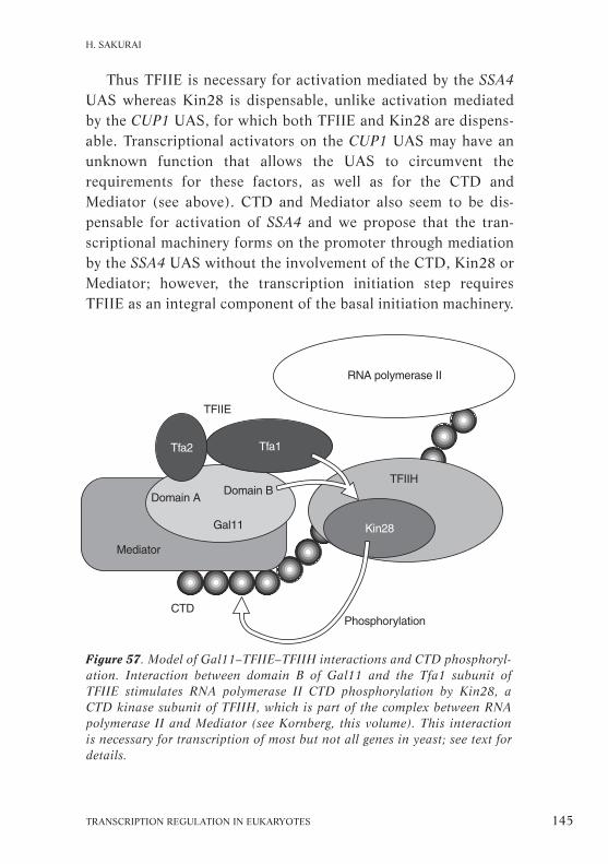
Thus TFIIE is necessary for activation mediated by the SSA4UAS whereas Kin28 is dispensable, unlike activation mediatedby the CUP1 UAS, for which both TFIIE and Kin28 are dispens-able. Transcriptional activators on the CUP1 UAS may have anunknown function that allows the UAS to circumvent therequirements for these factors, as well as for the CTD andMediator (see above). CTD and Mediator also seem to be dis-pensable for activation of SSA4 and we propose that the tran-scriptional machinery forms on the promoter through mediationby the SSA4 UAS without the involvement of the CTD, Kin28 orMediator; however, the transcription initiation step requiresTFIIE as an integral component of the basal initiation machinery.
TRANSCRIPTION REGULATION IN EUKARYOTES 145
H. SAKURAI
Figure 57. Model of Gal11–TFIIE–TFIIH interactions and CTD phosphoryl-ation. Interaction between domain B of Gal11 and the Tfa1 subunit ofTFIIE stimulates RNA polymerase II CTD phosphorylation by Kin28, aCTD kinase subunit of TFIIH, which is part of the complex between RNApolymerase II and Mediator (see Kornberg, this volume). This interactionis necessary for transcription of most but not all genes in yeast; see text fordetails.

Based on these results, we have constructed a model of theinteractions between Gal11, TFIIE, TFIIH and the CTD (Fig.57). In the RNA polymerase II holoenzyme, Gal11 helps theinteraction with TFIIE, which enhances CTD phosphorylationcatalysed by Kin28, the CTD kinase resident in TFIIH. Thisinteraction is necessary for transcription of most but not allyeast genes in vivo. The requirements for TFIIE, the CTD, Kin28and mediator are determined by the activator/UAS interactionand presumably by the structure of the core promoter.Elucidation of the molecular mechanism underlying such gene-specific requirements for components of the transcriptionmachinery should provide further insight into transcriptionalregulation.
Functional analysis of the componentsof yeast TFIID in vivo
Michael R. Green
in collaboration with
Wu-Cheng Shen, Xiao-Yong Li and Lynne M. Apone
TBP helps to initiate transcription in eukaryotes by interactingwith the TATA sequence located about 20–30 nucleotides awayfrom the 5’ side of the transcription start site of most genes.When biochemically fractionated from extracts of human cells,TBP coeluted with a set of associated TAFs with which it formsthe TFIID complex, whereas most TBP in yeast was found as amonomeric protein (Poon and Weil, 1993). As the TAFIIs inhigher eukaryotes are thought to be coactivators essential fortransmitting signals from transcriptional activator proteins tothe basal transcription machinery, it seemed likely that yeast andmammalian cells use different mechanisms to regulate transcrip-
146 HFSP WORKSHOP 7
PART II

tion. However, a fraction of the TBP present in yeast was latershown by affinity purification to be bound to TAFIIs (Poon andWeil, 1993).
With the identification of the genes encoding the human andDrosophila TAFIIs, the sequencing of the entire yeast genomeand the characterization of immunoaffinity-purified yeast TFIID,yeast homologues of most of the TAFIIs in higher eukaryoteshave been identified (Reese et al., 1994; Poon et al., 1995; Box2). Most of the genes encoding TAFIIs are essential for yeast viab-ility indicating that the corresponding protein is involved in anessential and non-redundant function. Some yeast TAFIIs, e.g.,TAFII90, TAFII150 (also known as TSM-1) and TAFII145, affectthe transcription of limited sets of genes, whereas others, suchas TAFII17, have a more general action. Here we compare thefunctions of TAFII145 and TAFII17, which differ not only intheir specificity but also in the location of their determinants.TAFII145 affects particularly genes involved in progressionthrough the cell cycle from G1 to S (see Glossary). We also dis-cuss some recent experiments to resolve the effect of transcrip-tional activators on occupancy of TBP at the TATA box.
Functional studies of yeast TAFII145 in vivo
We studied the function of TAFIIs in vivo by constructing yeastmutants either with a temperature-sensitive mutation in one ofthe genes encoding a TAFII or with the gene under the control ofa conditional allele that can be inactivated by depletion or tem-perature. A specific TAFII subunit can then be analysed at dif-ferent time points after inactivation (Walker et al., 1997). Suchinactivation of individual y TAFIIs surprisingly did not lead todefects in transcription of most genes transcribed by RNA poly-merase II, whereas transcription of most genes rapidly ceasedafter genes encoding general transcription factors or subunits ofthe Mediator complex were inactivated. This result contrastswith several experiments in vitro that implied TAFIIs were essen-
TRANSCRIPTION REGULATION IN EUKARYOTES 147
M. R. GREEN

tial for transcriptional regulation of most, if not all, class IIgenes in yeast (Walker et al., 1996).
The inactivation of one of the subunits, TAFII145, was par-ticularly interesting as it led to a specific arrest of cells in theG1/S phase of the cell cycle. Thus yTAFII145, like its humanhomologue TAFII250, is required for G1/S progression, indicat-
148 HFSP WORKSHOP 7
PART II
Figure 58. Microarray technique showing changes in expression of the yeastopen reading frames after inactivation of RNA polymerase II using a mutantstrain carrying a temperature-sensitive mutation in the largest RNA poly-merase II subunit. Reproduced with permission from Holstege et al., 1998.

ing a functional conservation through evolution. The expressionof CLN1, CLN2, PCL1 and PCL2, which encode cyclin proteinsneeded for progression from G1 to S, was downregulatedrapidly after inactivation of yTAFII145 (Walker et al., 1997).RPS genes, encoding proteins of the small ribosomal subunit, arealso dependent on yTAFII145 function (Shen and Green, 1997).
We are also using the yeast microarray technique for whole-genome transcription analysis to detect the effect of yTAFII145inactivation on gene expression. This technique analyses theexpression from the entire set of about 6,200 yeast open readingframes under different conditions, i.e., growth in different mediaor in a mutant versus the corresponding wildtype strain.Inactivation of RNA polymerase II abolished the transcription ofnearly all these genes, indicating the general importance ofRNA polymerase II in transcription (Fig. 58). In contrast, whenTAFII145 was inactivated, the expression of 1,400 genesincreased or decreased (Fig. 59) and 540 of these showed decaykinetics similar to those observed after inactivation ofRNA polymerase II. Thus TAFII145 is directly required forproper expression of about 10% of the genes in yeast but notrequired for the other 90%. The genes that were most clearlydependent on TAFII145 for expression encoded proteins involvedin cell-cycle control or chromosome metabolism (Table 7).
To identify the determinants for TAFII145 dependence,chimerical promoters were constructed where the UAS for atypical TAFII145-dependent promoter was fused to the corepromoter of a TAFII145-independent gene and vice versa. Theseexperiments showed that the determinant for TAFII145-depen-dence is localized to the core promoter rather than to the UAS,a somewhat surprising result given that several reports suggest arole for the TFIID complex, i.e., TBP and TAFIIs, in contactingupstream-bound transcriptional activator proteins (Burley andRoeder, 1996; Pugh, 1996; Ranish and Hahn, 1996). However, itagrees well with results that indicate the TFIID complex makes
TRANSCRIPTION REGULATION IN EUKARYOTES 149
M. R. GREEN

150 HFSP WORKSHOP 7
PART II
Table 7. Genes that require TAFII145 function.
Gene Description Fold reduction†
Cell cycleDDC1* DNA damage checkpoint protein 10YER066W Similar to CDC4, which degrades G1 cyclins 9SPO1 Possible role in spindle pole body duplication 8LTE1* GDP/GTP exchange factor 8MKK2* Kinase involved in cell wall integrity 8BIM1* Possible role in early spindle pole body assembly 8MDM1* Involved in mitochondrial segregation 7CTR9* Required for normal expression of G1 cyclins 7PAC1* Possible role in spindle pole body orientation 6SCP160* Involved in control of chromosome transmission 6CDC13 Telomere-binding protein 6TOP3* DNA topoisomerase III 5TRX1* Thioredoxin I 5ARD1 N-Acetyltransferase 5SCC2* Required for sister chromatid cohesion 5CLB2* G2/M cyclin 5KIP2* Kinesin-related protein 5MEC1* Cell-cycle checkpoint protein 4RAD9 DNA repair checkpoint protein 4SPC98* Spindle pole body component 4BCK1* Kinase involved in cell wall integrity 4DNA repairRAD3* Involved in nucleotide excision repair 8YHR031C* Possible role in chromosome repair 7RAD5* Involved in DNA repair 6HSM3* Involved in mismatch repair 6RAD50* Involved in recombinational repair 5EXO1* Involved in mismatch repair 5MSH3* Involved in mismatch repair 5YER041W Similar to DNA repair protein Rad2 5REV1 Involved in translesion DNA synthesis 4HDF2 Involved in DNA end-joining repair pathway 4MSH6 Involved in mismatch repair 4DNA synthesisMCM3* Involved in replication initiation, MCM/P1 family 13RLF2 Chromatin assembly complex, subunit 2 9MCM6* Involved in replication initiation, MCM/P1 family 9REV7 DNA polymerase subunit zeta 7MIP1* Mitochondrial DNA-directed DNA polymerase 6CDC47* Involved in replication initiation, MCM/P1 family 6CDC5* Kinase 5CDC46* Involved in replication initiation, MCM/P1 family 5RFC1* DNA replication protein RFC large subunit 5CAC2* Chromatin assembly complex, subunit 1 5
*Gene exhibits equivalent dependence on TAFII145 and Rpb1 for normal expression.†Reduction in expression after TAFII145 inactivation. Reproduced with permissionfrom Holstege et al., 1998.

contact with the core-promoter DNA surrounding the TATA box(Oelgeschlager et al., 1996).
The microarray assays also showed that the expression of alimited number of genes increased after inactivation of TAFII145(Fig. 59). This set of genes had some features in common withgenes that are repressed under normal growth conditions. HXT5
TRANSCRIPTION REGULATION IN EUKARYOTES 151
M. R. GREEN
Figure 59. Microarray technique showing changes in expression of theyeast open reading frames after inactivation of yeast TAFII145 using amutant strain carrying a temperature-sensitive mutation in yeast TAFII145.

encodes an alternative hexose transport protein that is inducedwhen glucose is removed from the medium, ARG3 is normallynot expressed unless cells are grown in the absence of arginine,PRB1 is induced when cells are deprived of nutrients or inducedto enter the stationary phase and the DAN1 gene is expressedunder anaerobic conditions and completely repressed during aer-obic growth. These genes could have been induced as a result ofsecondary effects caused by downregulation of one of severalgenes after TAFII145 inactivation. However, induction is morelikely to be direct because it is as rapid as the effects on theexpression of genes that are downregulated after TAFII145 inac-tivation; and it is specific for TAFII145 and is not seen after inac-tivation of other TAFIIs or in cdc mutants that also arrest in theG1 phase of the cell cycle. Lastly, the same results are obtainedwhen using constructs containing only the core promoter of therespective genes.
Functional studies of yTAFII17 in vivo
Yeast TAFII17 is the homologue of human TAFII32 and Droso-phila TAFII40 and is one of the TAFIIs containing a histone-foldmotif similar to that found in histone H3. The human andDrosophila homologues can make protein–protein interactionswith TFIIB and acidic activators (Klemm et al., 1995) but alsomake DNA contacts with sequences downstream from the TATAbox to the downstream promoter element (Burke and Kadonaga,1996). TAFII17 has also been identified more recently as a sub-unit of the yeast SAGA complex (Grant et al., 1998; seeWorkman, this volume).
We studied the in vivo function of yTAFII17 by generating atemperature-sensitive mutation in yeast TAFII17 combined withthe microarray technique. Inactivating TAFII17 produced atwofold or more downregulation of 72% of all genes, so itseffects are more general than those previously obtained byinactivating yTAFII90 and yTAFII145. Most of these transcripts
152 HFSP WORKSHOP 7
PART II
showed decay kinetics similar to those observed after inactiv-ation of RNA polymerase II. However, no general pattern ofdependency of specific genes on particular TAFIIs has emerged.For example, expression of ADH1, encoding alcohol dehydro-genase, is completely dependent on TAFII17 but not affected byTAFII145 inactivation; RPS5 is dependent on both TAFII17 andTAFII145; PCL1, encoding G1 cyclin, is dependent on TAFII145but not TAFII17; and expression of DED1, encoding a putativeRNA helicase of the DEAD-box family, is independent of bothTAFII17 and TAFII145.
The microarray technique as we used it is limited in that itmonitors the effect of inactivation only on genes that areexpressed under normal growth conditions; it does not showchanges in genes whose expression is induced in response toother stimulation. Studies of various inducible genes showedthat they also differ in their requirement for particular TAFIIs.For example, the heat-shock-inducible SSA4 and the copper-inducible CUP1 genes (see Sakurai, this volume) are indepen-dent of TAFII17, whereas induction of HIS4 by 3-aminotriazolerequires TAFII17. The determinants for TAFII17-dependence,analysed using chimerical promoters, seem to be located withinthe UAS, unlike those for TAFII145 (Apone et al., 1998).
Effects of activators on TFIID binding to the TATA box
These results question the established view that transcriptionalactivators operate by interacting with TAFIIs in the TFIID complexto affect the interaction of TFIID with the TATA box. In this model(see Box 1), the binding of TBP in the presence of TFIID is the keystep in assembling the preinitiation complex, which subsequentlyleads to activation of transcription. We therefore decided to exam-ine the effect of transcriptional activators on occupancy of TBP atthe TATA box. In prokaryotes, activators can work in two ways,either bringing the RNA polymerase to the promoter or acting onRNA polymerases already present on the promoter. In vitro exper-
TRANSCRIPTION REGULATION IN EUKARYOTES 153
M. R. GREEN

iments in eukaryotic systems have indicated that when activatorsare added, they can act on promoters to which the TBP is alreadybound. Footprinting analysis shows that in some cases TBP isbound to the promoters before induction, in other cases it is not(Chen et al., 1994b). Our results show a clear correlation betweentranscription levels and TBP occupancy at the TATA box for a largeset of promoters in combination with mutations in genes encoding:• general transcription factors, i.e., TFIIB;• inhibitors of TBP–TATA interactions, i.e., MOT1;• TAFIIs;• subunits of the mediator complex, i.e., SRB4 (X. Li et al., 1999;see Struhl, this volume).
Discussion: The significance of these results is disputed becausethe experiments were performed under conditions that allowedtranscription to take place, which makes it impossible to deter-mine whether the effect of the transcriptional activators on TBPbinding to the TATA box was direct. It could be an indirect con-sequence of activated levels of transcription induced by path-ways other than those operating directly on TBP. Furthermore,the method used does not detect protein associated with thegene during the activation process but rather proteins present onthe transcribed gene at steady state.
154 HFSP WORKSHOP 7
PART II

Mechanism for in vivo transcriptional activationand repression in yeast
Kevin Struhl
in collaboration with
Zarmik Moqtaderi, David Kadosh, Laurent Kuras andMarie Keaveney
Transcriptional activation involves the binding of a transcrip-tional activator to a cis-acting sequence, termed an enhancer.Most activators have a modular organization, with a DNA-binding domain that targets the factor to a specific enhancersequence and an activation domain that permits activation oftranscription of the promoter under control of the enhancersequence. Activation domains may enhance transcription byrecruiting the polymerase II machinery (Ptashne and Gann,1997), by altering the conformation of components of the poly-merase II machinery (Roberts and Green, 1994) and by modify-ing chromatin structure (reviewed in Workman and Kingston,1998). The relative importance of these three mechanisms hasyet to be estimated.
We discuss here three aspects of the in vivo stimulation oftranscription by transcriptional activators in yeast: recruitmentof the polymerase II transcription apparatus; recruitment of TBP,an important step in the activation process; and the importanceof various TAFs that are required for the response to activators.We also provide evidence for the intimate relationship of tran-scription with some specific chromatin modifications to supporta mechanism of transcription repression through localized his-tone deacetylation.
Recruitment of the RNA polymerase II machinery
To estimate the importance of the polymerase recruitmentmechanism, we transferred activation domains from their normallocation on the enhancer-bound protein (Fig. 60a) to different
TRANSCRIPTION REGULATION IN EUKARYOTES 155

156 HFSP WORKSHOP 7
PART II
Figure 60. Normal organization of transcriptional activators and the poly-merase II machinery at promoters and connected and disconnected con-structs used to examine the polymerase recruitment mechanism. a, thephysiologically relevant organization, in which activator proteins bindenhancer elements (ENH) through specific DNA-binding domains (DBD)and stimulate transcription through functionally autonomous activationdomains (AD). Open arrows, interactions between activation domains andthe TFIID and/or the polymerase II holoenzyme (Pol II holo) complexes;the direct targets within these complexes are not specified. Filled arrowsbetween TFIID and the TATA element and between the polymerase IIholoenzyme and the mRNA initiation site (INR) indicate activatorsincreasing recruitment of the polymerase II machinery to promoters. b, in ➞

components of the RNA polymerase II machinery, either in theTFIID complex or in the polymerase II holoenzyme complex(Fig. 60c). Alternatively, the need for the activator was bypassedby connecting the enhancer-bound protein to a component ofeither TFIID or the polymerase II holoenzyme (Fig. 60b;Keaveney and Struhl, 1998). The TFIID components includeTBP and approximately ten associated factors, among which aretwo TAFs required for cell viability in yeast: TAF17, homologueof Drosophila TAF17; and TAF23, homologue of human TAF30.Two components of the holoenzyme, Srb2 and Gal11 (seeSakurai, this volume) that are not essential for growth werealso used to anchor activation domains. The function of theseconstructs was assayed using either the GAL1 promoter andmeasuring β-galactosidase activity, or the HIS3 promoter andmeasuring growth on selective media.
The polymerase II machinery containing a functionallyautonomous activation domain could neither activate transcrip-tion from core promoter nor synergize with other activators tosupport high levels of transcription in the cases we tested. Thusthe presence of the activation domain within the polymerase IImachinery does not bypass the need for a DNA-binding proteinto interact with enhancer sequences, indicating that the DNA-binding domain can serve as an anchor to recruit the polymeraseII machinery (Keveaney and Struhl, 1998).
TRANSCRIPTION REGULATION IN EUKARYOTES 157
K. STRUHL
the connected construct, transcriptional activation is achieved (+) in thecomplete absence of an activation domain by physically connecting (thickbold line) a component of either TFIID or the polymerase II holoenzyme toan enhancer-bound protein, which artificially recruits the polymerase IImachinery to promoters and bypasses the need for an activation domain. c,in the disconnected construct, the activation domain is transferred from itsnormal location on the enhancer-bound protein to a component of eitherTFIID or the polymerase II holoenzyme; transcriptional activation does notoccur (–). Reproduced with permission from Keaveney and Struhl, 1998.

By measuring TBP occupancy at several promoters under var-ious conditions in vivo with or without activation we establishedthat TBP is generally required for polymerase II transcription. Todo this, we used yeast strains producing a protein tagged withTBP and immunoprecipitated chromatin with antibodies dir-ected against the tag. Under activation conditions, the GAL1 andheat-shock genes displayed high occupancy and quantitat-ive measurement of various genes showed a tight correlationbetween gene activity and TBP occupancy, indicating that TBPoccupancy may be a limiting step in the mechanism of transcrip-tional activation. The CYC1 and COX5a promoters were excep-tions to this rule as they displayed a constitutive level of TBPoccupancy with or without induction and so may have a distinctmode of regulation. Conversely, genes that can be repressed alsoshowed a correlation between TBP occupancy and gene activity.Other issues include the exact target of an activator in vivo;whether one or more targets are involved; and, if more than one,whether they all contribute to the activation. Artificial recruit-ment experiments indicate several targets within the promoter(Gaudreau et al., 1999).
The importance of TAFS specifically found in TFIID, i.e.,TAFs 19, 40, 67 and 130 (see Box 2), was evaluated by remov-ing the TAFs using a ‘copper-induced double shut-off’, whichcauses the repression of TAF transcription, so that pre-existingTAF protein becomes a target for ubiquitin-mediated degrad-ation (Moqtaderi et al., 1996b). Although we did not investigatethe selective effects on transcription of depleting each TAF, theyall affected the HIS3 promoter, which has one weak and onestrong TATA box, by reducing transcription only from the weakone (Moqtaderi et al., 1998). Hence specific features of the corepromoter region can be critical. Because TFIID is the only com-plex known to contain these TAFs, it is probably critical for thefunction of the HIS3 core promoter. Yeast cells with TBPreplaced by a human TBP are interesting because they displayed
158 HFSP WORKSHOP 7
PART II

properties very similar to those of cells with mutations in theTAFs specific to TFIID, further supporting the argument thatTFIID has an important role in the function of core promoter.
In contrast, depletion of TAF17, which is common to theTFIID and SAGA HAT complexes, had a more dramatic effect,causing a decrease in transcription of most, although not all,genes. This may be the result of a combination of defects in boththe TFIID and SAGA complexes and indicates that TAF17 isbroadly required for transcription (Apone et al., 1998; seeGreen, this volume).
Transcriptional repression through targeted deacetylation
Specific sequences with particular properties in terms of theirnucleosomal organization may help regulate transcription, pro-viding a link between chromatin organization and the regulationof transcription. Repetitive sequences, such as the poly-d(A–T)tracks that are the most common element in yeast promoter, areintriguing as they are important in transcription regulation invivo. Such sequences could prevent the formation of stablenucleosomes, so making the sequences more accessible for a pot-ential transcription factor. However, no specific factor bindingto these sequences has been identified; the AT-binding factor 1does not bind to the perfect poly-d(A–T) track.
Recent work has focused on the repression of transcriptioninvolving deacetylation of histones (reviewed in Struhl, 1998).Proteins with the properties of histone deacetylase were identi-fied as homologues of the product encoded by the RPD3 gene inyeast (Taunton et al., 1996). Yeast cells lacking Rpd3 are viablebut show selective defects of gene expression, so a key questionis how the selectivity/specificity is obtained. The mechanismof acetylation could be untargeted, generally targeted to a pro-moter over large sequence regions or tightly targeted to very spe-cific sites within a promoter (Fig. 61).
In yeast, the transcription of one set of genes, which includes
TRANSCRIPTION REGULATION IN EUKARYOTES 159
K. STRUHL
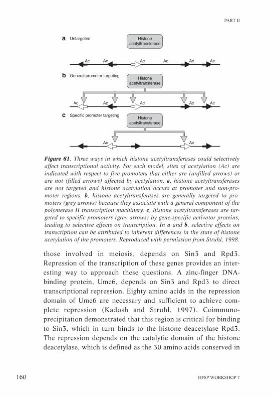
those involved in meiosis, depends on Sin3 and Rpd3.Repression of the transcription of these genes provides an inter-esting way to approach these questions. A zinc-finger DNA-binding protein, Ume6, depends on Sin3 and Rpd3 to directtranscriptional repression. Eighty amino acids in the repressiondomain of Ume6 are necessary and sufficient to achieve com-plete repression (Kadosh and Struhl, 1997). Coimmuno-precipitation demonstrated that this region is critical for bindingto Sin3, which in turn binds to the histone deacetylase Rpd3.The repression depends on the catalytic domain of the histonedeacetylase, which is defined as the 30 amino acids conserved in
160 HFSP WORKSHOP 7
PART II
Figure 61. Three ways in which histone acetyltransferases could selectivelyaffect transcriptional activity. For each model, sites of acetylation (Ac) areindicated with respect to five promoters that either are (unfilled arrows) orare not (filled arrows) affected by acetylation. a, histone acetyltransferasesare not targeted and histone acetylation occurs at promoter and non-pro-moter regions. b, histone acetyltransferases are generally targeted to pro-moters (grey arrows) because they associate with a general component of thepolymerase II transcription machinery. c, histone acetyltransferases are tar-geted to specific promoters (grey arrows) by gene-specific activator proteins,leading to selective effects on transcription. In a and b, selective effects ontranscription can be attributed to inherent differences in the state of histoneacetylation of the promoters. Reproduced with permission from Struhl, 1998.

both Rpd3 homologues and polyamine deacetylases, in which fourhistidines are highly conserved (Kadosh and Struhl, 1998). Thedeacetylase complex containing a mutation in this domain of the cat-alytic subunit was inactive in vitro on a synthetic peptide substrate.
Using chromatin immunoprecipitation, we have examined thenature and extent of the perturbation of chromatin in the vicin-ity of the repressed genes. Transcriptional repression was associ-ated with promoter-specific deacetylation of histones H4 andH3. Precisely mapping the preferentially deacetylated domaindelineated a region of 288 bp with its centre at the URS1 ele-ment, the Ume6-binding site. These data support a model inwhich limited deacetylation is promoted by specific targeting ofthe deacetylase activity through an adaptor, here Sin3, to a spe-cific DNA-binding protein (Fig. 62).
TRANSCRIPTION REGULATION IN EUKARYOTES 161
K. STRUHL
Figure 62. Creation of a repressive chromatin domain by targeted recruit-ment of the Sin3–Rpd3 histone deacetylase complex. The Ume6 repressorbinds URS1, shown in the context of a nucleosomal template, and recruitsthe Sin3–Rpd3 co-repressor complex to the promoter. As a consequence,histones H3 and H4 are deacetylated at lysines 5 and 12 and to a lesserextent lysine 16 over a range of one to two nucleosomes from the site ofrecruitment (indicated by absence of Ac). Arrows, sites of frequent histonedeacetylation. Nucleosomes further downstream and upstream are notspecifically deacetylated (indicated by Ac). The region of local histonedeacetylation is defined with respect to a hypothetical promoter (see text)that includes the UAS element but probably ends upstream of the TATAelements (T). Analogous regions of other Sin3–Rpd3 repressed promoterscould vary in length and position. The TATA elements are indicated in thespacer region for clarity; there is no information on the nucleosomal posi-tion of these TATA elements in vivo. Reproduced with permission fromKadosh and Struhl, 1998.

The role of TAFII30-containing complexesin vertebrate gene regulation
Làszlò Torain collaboration with
Marjorie Brand, Elzbieta Wieczorek and Daniel Metzger
The observation that transcriptional activation in reconstitutedanimal cell-free transcription systems is not supported by TBPbut requires TFIID (Goodrich et al., 1996) has triggered anintensive study of the TAFIIs as essential transcriptional coactiv-ators. In human HeLa cells, several TAFII-containing TFIIDcomplexes with distinct functional properties have beendescribed (Brou et al., 1993a; 1993b; Jacq et al., 1994). Usingantibodies against TAFII30, we found two TFIID populationsthat differ in their subunit composition: both TFIIDα andTFIIDβ contain TAFIIs 250, 135, 80, 55 and 28, whereas TAFIIs30, 20 and 18 are preferentially associated with TFIIDβ (Jacq etal., 1994; Mengus et al., 1995).
We show here that TAFII30 is also present in another complexcontaining a subset of TAFIIs, as well as other components thatinclude the Gcn5-L HAT enzyme, although it lacks TBP. ThisTBP-free, TAF-containing (TFTC) complex is interesting becauseit accurately engages RNA polymerase II to promoters to allowtranscription in vitro. We also demonstrate that TAFII30 isrequired for cell-cycle progression and for parietal endodermaldifferentiation through conditional disruption of the two allelesof the TAFII30 gene in F9 embryonal carcinoma cells. ThusTAFII30 is critical in the regulation of a subset of cell-cycle anddifferentiation genes in vivo.
Complexes containing TAFII30
TAFII30 is present in several distinct complexes (Wieczorek etal., 1998). HeLa cell extracts were immunopurified with a mono-clonal antibody directed against specific TAFII30, the precipitated
162 HFSP WORKSHOP 7
PART II

material eluted with a peptide corresponding to the epitope ofthis antibody and the eluted fraction reprecipitated with an anti-body against TBP. The supernatant fraction surprisingly still con-tained TAFII30, so not all TAFII30 is associated with TBP. Thisfraction, named TFTC, also contained TAFIIs 135, 100, 80, 55, 31and 20 and several unidentified components but TBP, TAFII250and TAFII28 were absent (Fig. 63; Wieczorek et al., 1998).
TFTC binds specifically to promoter sequences, substitutingfor TBP and TFIID in both activated and basal transcriptionfrom TATA-containing and TATA-less promoters in vitro(Wieczorek et al., 1998). This indicates that TBP is not absol-utely required for transcription in vitro. TFTC is also devoid ofa second TBP-like factor that is highly homologous to the coredomain of TBP. We therefore question the long-standing assump-tion that TBP is a universal and indispensable transcription fac-tor (Dantonel et al., 1999).
The non-TAFII components of TFTC include Gcn5-L, an iso-form of the human homologue of the yeast Gcn5 HAT enzyme(Brand et al., 1999). TFTC is therefore similar to the yeastSAGA and human PCAF complexes (Grant et al., 1998; Ogryzkoet al., 1998; Box 2; see Allis, Workman, this volume). There areseveral striking parallels in the composition of the TFIID, TFTC,PCAF and SAGA complexes. All contain a HAT enzyme, a prot-ein with WD40 repeats (see Glossary) and at least five histone-fold motifs (Box 2). The PCAF and SAGA complexes containTAFII31, TAFII30 and TAFII20 or their yeast homologues, aswell as the PCAF subunit or the highly related yeast Gcn5. Inaddition, SAGA contains the yeast homologues of TAFII100 andTAFII80, both of which are found in TFTC. The PCAF complexon the other hand contains PCAF-associated factor (PAF) 65β,which like TAFII100 contains WD40 repeats, and PAF65α,which like TAFII80 contains a histone-fold motif complementaryto that of TAFII31. On the other hand, TFTC could be a mixtureof PCAF and a second uncharacterized complex containing
TRANSCRIPTION REGULATION IN EUKARYOTES 163
L. TORA

164 HFSP WORKSHOP 7
PART II
Comparison of subunit composition of TAFII-containing complexes
Containing TBP Lacking TBP
Protein hTFIID dTFIID yTFIID ySAGA hTFTC hPCAF/ hSTAGAcharacteristics Gcn5
a, TAFIIsHAT and 250 250 145 – – – –
bromo domain (CCG1) (230) (130)Initiator/DNA 150 150 TSM1 – 150 ND –
binding– 135 110 – – 135 – –
WD40 repeats 100 80 90 90 100 100* –Histone H4-like 80 (70) 60 (62) 60 60 80 – –
(PAF65α)– 55 ND 67 – 55* ND ND
Histone H3-like 31 (32) 40 (42) 17 (20) 17 (20) 31 (32) 31 (32) 31 (32)– 30 ND 25 (23) 25 30 30 30– 30
(ANC1)Histone-like 28 30β 40 – – ND –Histone H2B- 20 (15) 30α 68 (61) 68 (61) 20 (15) 20 (15) 20 (15)
like (28/22)Histone H4-like 18 ND 19 – – ND –
(FUN81)TATA box TBP TBP TBP – – – –
binding
b, Spts, Adas, Gcn5 and other componentsHAT and yGcn5 hGcn5 hPCAF/ Gcn5
bromo domain Gcn5-SyAda1 ND ND NDyAda2 – hAda2 NDyAda3 hAda3 hAda3 hAda3ySpt3 hSpt3 hSpt3 hSpt3ySpt7 ND ND NDySpt8 ND ND NDySpt20 ND ND ND(yAda5)
WD 40 repeats ND hPAF65β hPAF65β hPAF65βyTRA1 hTRRAP hTRRAP hTRRAP
Other uncharacterized proteins
The numbers in parenthesis indicate alternative nomenclatures used in the literature.ND, not determined; Ada, alteration deficiency in activation, and Spt, suppressor of Tyinsertion, are proteins found in various yeast genetic screens *, factor is substoichiometric in the respective complex.
Box 2

TRANSCRIPTION REGULATION IN EUKARYOTES 165
L. TORA
Subunit composition of TAFII-containing complexes: a, left, thecomposition of TFIID complexes from human (h), Drosophila (d)and yeast (y); right, a comparison of the TAFIIs present in theSAGA, TFTC, PCAF and hSTAGA (human Spt–TAF–Gcn5–acetyl-transferase) complexes. The TAFIIs in human TFIID are arrangedvertically by their apparent Mr from 250K to 18K; TAFIIs homolo-gous to each are shown on the same line. b, other proteins found inthe complexes.
Some of the characteristic features of each protein are indicatedon the left. The TFIID, TFTC, PCAF and SAGA complexes all con-tain a histone acetyltransferase (HAT) enzyme, either TAFII250,hGcn5-L, PCAF or yGcn5; a WD40 repeat protein (see Glossary),such as hTAFII100, PAF65β or yTAFII90; and five or more histone-fold motifs: in TFIID these are found in hTAFII80, 31, 28, 20 and18; in TFTC, PCAF and STAGA they are in hTAFII80 (or PAF65α),TAFII20 and hSpt3, which contains two complementary histone-foldmotifs analogous to those of hTAFII28 and hTAFII18 of the TFIIDcomplex (Birck et al., 1998); in SAGA, they are in yTAFII68, 60, 17and 3. hSTAGA is found in HeLa cells and acetylates histone H3(Martinez et al., 1998); its role is not yet clear. Some data takenfrom Struhl and Moqtaderi, 1998.
L. Tora, K. Struhl

166 HFSP WORKSHOP 7
PART II
Figure 63. TAFIIs are present in the TBP-free, TAFII-containing (TFTC)complex. Above, TFTC remained in the supernatant after immunoprecipi-tation (lane 1), whereas the TFIIDβ complex bound to the anti-TBP anti-body resin (Res; lane 2), from which it was eluted (lane 3). Mr standardsare shown on the left and the positions of the TAFIIs and TBP on the right.IgGH and IgGL, immunoglobulin heavy and light chains. Below: the sub-unit compositions of the two complexes are shown schematically. Subunitcomposition of the complexes were revealed by silver nitrate staining of anSDS polyacrylamide gel.

TAFII30 along with TAFII135, TAFII100 and TAFII80, whichhave not been reported as PCAF components.
The physiological significance of the conservation of thesestructural motifs is unclear. The histone-fold proteins were orig-inally proposed to form an octamer-like structure (Xie et al.,1996) but at least five histone-fold motifs in each complex arenow known, which is too many to be accommodated in a simpleoctamer-like model. The histone fold does nevertheless providea very tight interaction interface, perhaps facilitating the high-affinity interactions required for the formation of these stablemacromolecular complexes. The similarity among these com-plexes does, however, indicate that they have similar and per-haps even overlapping roles in gene regulation (Fig. 64).
Requirement for TAFII30 in cell-cycle progression
Because genetic experiments in yeast have shown that TAFIIs areessential for viability (Moqtaderi et al., 1996b), the classic knock-out strategy seemed to be inappropriate for testing the role ofTAFIIs in mammalian cells in vivo. Instead, we have used a condi-tional Cre/LoxP-mediated strategy (Sauer and Henderson, 1990;see Glossary) to inactivate TAFII30 in mouse F9 embryonal carci-noma cells. The production of only a very short N-terminal TAFII30peptide was achieved with a targeting vector designed to insert aLoxP site in the intron between exons 1 and 2 in the mouseTAFII30 gene, and a neomycin-resistance gene (Neor) flanked byLoxP sites in the intron between exons 2 and 3. Treatment with theCre recombinase then causes deletion of exon 2. One TAFII30 allelewas modified with this vector by homologous recombination andwhen the gene encoding the Cre recombinase was introduced intocells harbouring the modified allele, the Neor gene was deleted,sometimes with exon 2 as well. These TAFII30+/– heterozygouscells were fully viable, so the second allele also had to be inacti-vated. The second allele was targeted using a similar vector in a cellclone with the Neor gene deleted, leaving exon 2 surrounded by
TRANSCRIPTION REGULATION IN EUKARYOTES 167
L. TORA

LoxP sites. After treatment with the Cre recombinase, no clones ofTAFII30–/– homozygous cells were obtained, indicating that thecells are not viable and that TAFII30 is essential for cell survival(Metzger et al., 1999).
That TAFII30 is required for cell-cycle progression in mam-malian cells was established by creating cells in which the ex-pression of hTAFII30 could be controlled. The hTAFII30 cDNA
168 HFSP WORKSHOP 7
PART II
Figure 64. Possible roles of TAFII-containing complexes in regulating tran-scription. The subunit composition of the different complexes is shownschematically. Dashed arrows indicate that the human PCAF and the yeast(y) SAGA complexes have not yet been shown to function in the initiationof transcription by RNA polymerase II. TLF, TBP-like factor; TLC, hypo-thetical TBP-like factor complex; INR, initiator sequence. Ada, alterationdeficiency in activation, and Spt, suppressor of Ty insertion, are proteinsfound in various yeast genetic screens.

TRANSCRIPTION REGULATION IN EUKARYOTES 169
L. TORA
Figure 65. F9 embryonal carcinoma cells lacking TAFII30 are not viable.Top, TAFII30–/– F9 cells (alleles La and Lb deleted for exon 2) carrying ahTAFII30 cDNA inducible with the tetracycline analogue, doxycyclin, wereconstructed. The reverse tetracycline repressor molecule bound to the pro-moter of the hTAFII30 transgene in the presence of doxycyclin or unboundin the absence of doxycyclin is shown schematically. Transcription of thiscDNA rescued the TAFII30–/– cells. Bottom, cells are viable when doxycy-clin is present (left) but most cells died after 5 days in the absence of doxy-cyclin (right). P, promoter; rtTA, reverse tetracycline (tet) repressor; tetR*,tet repressor; VP16, VP16 acidic activator domain; tetO, tet operator.

was stably introduced under the control of a promoter induciblewith doxycyclin, an analogue of the antibiotic tetracycline, intocells containing two TAFII30 alleles surrounded by LoxP sitesand producing the reverse tetracycline-repressor activatorprotein. Thus the production of the hTAFII30 protein could beinduced by the addition of doxycyclin (Fig. 65). After treatingthese cells with Cre recombinase, we isolated TAFII30–/– cellclones that were viable because they produce the hTAFII30protein. However, production of hTAFII30 protein was shut offin these cells when doxycyclin was removed from the culturemedium and within 2 days hTAFII30 could not be detected. After5 days the TAFII30–/– cells were blocked in the G1/G0 phase ofthe cell cycle and underwent apoptosis (Fig. 65).
We have also shown that TAFII30 is necessary for the activa-tion of a subset of genes required in the differentiation of F9cells into parietal endoderm. Treatment of F9 cells with all-transretinoic acid promotes their differentiation into primitive endo-dermal-like cells, whereas retinoic acid and dibutyryl cyclic AMPtogether promote differentiation into parietal endodermal cells.The differentiation is associated with the activation of many tar-get genes and changes in both cell morphology and cell cycle.After withdrawal of doxycyclin, the TAFII30–/– cells can still,surprisingly, differentiate into primitive endodermal-like cellsbut they cannot differentiate into parietal endoderm (Metzger etal., 1999).
The requirement for TAFII30 both in progression through thecell cycle and in differentiation indicates that it is involved in thecorrect regulation of a subset of cellular promoters. It remains tobe seen whether the defects in the TAFII30–/– F9 cells resultfrom perturbing the function of the TFIID complex or theTFTC/PCAF complex, or a combination of both.
170 HFSP WORKSHOP 7
PART II

TFIIH and the transcription-repair syndromes
Jean-Marc Eglyin collaboration with
Franck Tirode and Frédéric Coin
TFIIH is a large protein complex involved in transcription, DNArepair and cell-cycle regulation (Hoeijmakers et al., 1996;Svejstrup et al., 1996). Mutations in TFIIH subunits, such as thetwo DNA helicases/ATPases xeroderma pigmentosum comple-mentation groups B and D (XPB and XPD), have been associ-ated with the genetically heterogeneous human diseases com-monly referred to as repair syndrome diseases: xerodermapigmentosum, Cockayne’s syndrome and trichothiodystrophy(Bootsma and Hoeijmakers, 1993; reviewed in Semenza, 1998).
A diverse range of clinical defects are observed in thesesyndromes, some of which can be explained by defects innucleotide-excision repair, a highly conserved pathway that islargely involved in the repair of lesions in DNA caused by ultra-violet radiation. For example, characteristics of xerodermapigmentosum include hyperpigmentation of skin exposed to thesun, cutaneous abnormalities and a predisposition to skin can-cer. However, the developmental retardation and neurologicalabnormalities that are also features of this syndrome are lessclearly related to the repair function and may arise from thetranscription function of the TFIIH complex.
To identify the relationship between genotype and clinicalphenotype, we have investigated the enzymatic functions of theTFIIH complex by purifying normal and mutated forms of itssubunits and assaying the properties of the complex. The com-plex was first isolated from cells derived from patients but morerecently we have used a baculovirus expression system to pro-duce either recombinant wildtype or mutant TFIIH to analysethe functions of various subunits. We outline here that the p44subunit is a regulatory factor for the XPD helicase; Cdk7 is
TRANSCRIPTION REGULATION IN EUKARYOTES 171

involved in the phosphorylation of the CTD of RNA polymeraseII; and opening of the promoter to allow reading of the codingstrand depends on XPB.
TFIIH subunits, enzymatic activities and phenotypes
Nine subunits of TFIIH have been cloned and sequenced (Gérardet al., 1991; Fig. 66) and are highly conserved from yeast tohuman (Marinoni et al., 1997). The core TFIIH is a complex thatresists high-salt extraction and contains five subunits: p89/XPB,p62, p52, p44 and p34. The remaining subunits, which dissoci-ate from the complex at high-salt concentrations, are p80/XPDand the ternary kinase complex, consisting of Cdk7 (MO15),cyclin H and MAT1 (for review, see Nigg, 1996), which to-gether are referred to as Cdk7-activating kinase (CAK)/XPD(Drapkin et al., 1996; Rossignol et al., 1997).
We have shown that the enzymatic activities of TFIIH comefrom the Cdk7 kinase, which phosphorylates components of thebasal transcription machinery, and the helicases XPD and XPB;XPD is a 5′-to-3′ helicase, whereas XPB is a weak 3′-to-5′ heli-case (Schaeffer et al., 1993; Coin et al., 1999). RecombinantXPB and XPD were produced in insect cells using baculovirusinfection and immunopurified with antibodies against XPB orXPD. The helicase activity of recombinant XPD increased whenintegrated into the TFIIH complex (Coin et al., 1998a,b) and wehave identified p44 as the subunit that interacts with XPD andregulates its helicase activity. This was achieved by co-infectinginsect cells with two baculoviruses, one producing XPD, theother one of the core subunits of TFIIH. The only recombinantprotein that co-immunoprecipitated with XPD from lysates ofthese cells was p44. Although this had no helicase activity itself,the helicase activity of an immobilized XPD was directly depen-dent on the amount of p44 present.
In patients, mutations in the XPD protein have been mappedto the C-terminal domain; trichothiodystrophy patients also have
172 HFSP WORKSHOP 7
PART II

mutations in this domain (Taylor et al., 1997). Similar mutations inthe baculovirus expression system all had altered interactions withp44, leading to a weaker helicase activity, confirming that they affectthe XPD–p44 interaction. Western-blot analysis of TFIIH purifiedfrom a cell line derived from patients carrying a mutated XPDdemonstrated modified stoichiometry of the complex: this TFIIHlacks part of the CAK complex (Coin et al., 1998a,b).
A similar study of the critical region in XPB was based on themapping of mutations in the XPB gene found in patients withvery severe disease, involving major problems in the nucleotideexcision repair pathway as well as developmental abnormalities(Hwang et al., 1996). We tested the consequences of these
TRANSCRIPTION REGULATION IN EUKARYOTES 173
J.-M. EGLY
Figure 66. TFIIH contains nine subunits ranging in size from XPB with Mr89K to MAT1 with Mr 32K. The subunits resolve into two subcomplexes,the core (medium shading) and the Cdk7-activating kinase (CAK) complex(dark shading), plus the single XPD subunit (light shading).

mutations in enzymatic assays in vitro using TFIIH from celllines derived from patients. The stoichiometry and compositionof the isolated complex was not perturbed and its kinase andDNA-dependent ATPase activities were essentially similar tonormal TFIIH. However, transcription was severely impaired,revealing the crucial role of XPB in this assay; footprinting indi-cated a defect in the opening of the promoter.
A complete reconstitution of the TFIIH complex
We have constructed a complete recombinant TFIIH complexusing the baculovirus expression system (Tirode et al., 1999).The five subunits corresponding to the core TFIIH are absol-utely required to obtain transcription in vitro and the ATPaseand helicase activities in XPB are necessary. The absence of XPB,but not of XPD, completely impairs the initiation of transcrip-tion. Analysis of the promoter opening at the initiation site, –10to +15 from the transcription start point, revealed that XPB iscritical at this step in the transcription process, confirming thefootprinting analysis. The phosphorylation of the CTD of thelargest subunit in RNA polymerase II is independent of thisopening, which indicates that this phosphorylation is not relatedto the initiation of transcription.
We also tested the role of the CAK kinase in transcription. Aholo-TFIIH stimulated transcription equally well from the adeno-virus major late promoter and from the dehydrofolate reductasepromoter. Transcription from the latter depends on phosphoryl-ation of the CTD of the largest subunit in RNA polymerase IIand the CAK complex was required for transcription from thispromoter indicating that CAK is involved in phosphorylating theRNA polymerase II subunit. It is also required for transcriptionfrom the adenovirus promoter indicating that the physicalpresence of the CAK complex is required for optimum tran-scription; in this case CAK may stabilize the formation of thepreinitiation complex.
174 HFSP WORKSHOP 7
PART II

On the basis of these and other experiments, we have beenable to assign properties to the various TFIIH components anddetermine their importance in both transcription and nucleotideexcision repair (Fig. 67). The core TFIIH is involved in tran-scriptional activation because interactions with various activa-tors have been detected, including the acidic activator VP16, thep53 protein, HIV Tat protein, retinoic acid receptor α andoestrogen receptor (Rochette-Egly et al., 1997); CTD phosphoryl-ation is dependent on Cdk7 (Coin et al., 1999); promoter opening
TRANSCRIPTION REGULATION IN EUKARYOTES 175
J.-M. EGLY
Figure 67. TFIIH is probably involved at several levels in transcription, from theformation of the transcription preinitiation complex to the incision of damagedstrands. Enzymes such as XPB and XPD unwind DNA for promoter opening andexcision of a damaged strand. Cdk7 phosphorylates the C-terminal domain(CTD) of RNA polymerase II to activate transcription and probably to regulatesome processing. TFIIH may also be targeted by some regulators of the tran-scription process, e.g., p53, VP16 and nuclear receptors. The action of TFIIH inthe mechanism of transcription-coupled repair is unknown. RARα, retinoic acidreceptor α; DSE, downstream element; ER, oestrogen receptor; CSA, CSB, XPA,XPG, XPF, RPA and ERCC1 are nucleotide excision repair factors.

depends on XPB; and promoter clearance is dependent on Cdk7(Moreland et al., 1999). In nucleotide excision repair XPD andXPB are both required for the opening at the lesion site and XPBis involved in the 5′ incision (Constantinou et al., 1999).
Elongator, a component of the elongatingRNA polymerase II holoenzyme
Jesper Q. Svejstrupin collaboration with
Gabriel Otero, Jane Fellows and Thérèse de Bizemont
Once a preinitiation complex forms (see Box 1), the synthesis ofmRNA requires four steps: synthesis of the first phosphodiesterbond, promoter clearance, elongation and termination. Mostwork has focused on the termination step, including 3’-end pro-cessing of the mRNA transcript (Neugebauer and Roth, 1997),but surprisingly little is known about the factor requirements forthe elongation of transcripts and the mechanisms controllingtranscription through chromatin DNA.
We have identified and characterized a distinct form of RNApolymerase II involved in elongation and established that itforms a complex with elongator proteins. The Elongator com-plex interacts specifically with the phosphorylated form of theCTD of RNA polymerase II and may act by increasing the abili-ty of the polymerase to progress along the DNA, although it doesnot seem directly to affect the rate of transcription.
Identification of the Elongator complex
The CTD is a unique feature of the largest subunit of RNA poly-merase II and is essential for yeast viability. It is thought to beimportant in the transition from initiation to elongation becauseonly the non-phosphorylated form of the polymerase can enter
176 HFSP WORKSHOP 7
PART II

the preinitiation complex, whereas the hyperphosphorylatedform has been found in ternary complexes composed of RNApolymerase II, DNA and RNA (Cadena and Dahmus, 1987;O’Brien et al., 1994; Svejstrup et al., 1997). As most, if not all,general transcription factors remain at the promoter after thetransition from initiation to elongation, phosphorylation of theCTD could induce the partition of the polymerase and the gen-eral transcription factors by introducing a net negative charge atthe CTD. It has not been clear whether the Mediator complex(see Kornberg, this volume) remains bound to the polymeraseduring elongation but RNA polymerase II isolated from the DNAfraction of a whole-cell yeast extract was both hyperphos-phorylated and lacked bound Mediator (Svejstrup et al., 1997).Release of the polymerase from the ternary complex by incuba-tion with the elongation factor TFIIS further indicated that thisis the elongating form of the polymerase (Svejstrup et al., 1997).
The elongating form of the polymerase was characterized bya purification that included adjusting the salt concentration ofthe whole-cell extract to disrupt the DNA binding of mostproteins; RNA polymerases engaged in ternary elongation com-plexes remain stable at higher ionic strengths (Gnatt et al.,1997; Svejstrup et al., 1997). The elongating form of the polymer-ase had an Mr of 500–600K compared with ~400K for the coreRNA polymerase and coeluted with additional polypeptides, atleast three of which seemed to be stoichiometric to the poly-merase subunits. The polypeptides have apparent Mrs of 150K,90K and 60K (Fig. 68) and we have named them elongator prot-eins (Elp) 1–3. Peptide sequencing showed that Elp1 (150K)is encoded by a previously uncharacterized open reading frameon yeast chromosome XII (YLR384C).
A free form of the Elongator complex, without the poly-merase, was purified from the soluble fraction using an antibodyraised against Elp1. The native size of the free Elongator complexis Mr ~600K, indicating that each polypeptide is present in the
TRANSCRIPTION REGULATION IN EUKARYOTES 177
J. Q. SVEJSTRUP

complex as a dimer. Free Elongator was also detected continu-ously throughout the purification from the DNA-bound fraction.Despite using phosphatase inhibitors in all buffers, the CTDseemed to become dephosphorylated at a rate that correlated
178 HFSP WORKSHOP 7
PART II
Figure 68. Purification and separation of the RNA polymerase II elongat-ing holoenzyme and the elongator proteins associated with it in theElongator complex. After separation of the elongating form of the poly-merase from the soluble material by high-speed centrifugation, the RNApolymerase was released from the DNA/RNA by treatment with nucleasesand extraction with ammonium sulphate. The RNA polymerase holoen-zyme was purified over five chromatographic steps ending with a gel filtra-tion (left). The polypeptides designated Elp1, Elp2 and Elp3 (Mrs 150K,90K and 60K) are part of the Elongator complex, which is found both infree form (right) and in the holoenzyme. Rpb1–5, subunits of polymerase.Modified from Otero et al., 1999.

with the dissociation of free Elongator complex. PurifiedElongator interacts directly with core RNA polymerase II to forma complex: when applied separately to a gel-filtration column,Elongator eluted at Mr 650K and core polymerase at 400K butpreincubation of the two complexes resulted in coelution of theproteins at a position equivalent to ~500K (Otero et al., 1999).
Functional studies of the Elongator complex
Many other factors involved in elongation of transcription, i.e.,TFIIS, Spt4 and elongin, are not essential for yeast viability(Archambault et al., 1992; Malone et al., 1993; Aso and Conrad,1997). Likewise, elp1 is not an essential gene in yeast but elp1∆cells, i.e., cells in which elp1 is deleted, did display a pro-nounced delay in adapting to changed growth conditions. Forexample, the elp1∆ strain was much slower to adapt than thewildtype strain when glucose in the medium was substituted bygalactose, sucrose or raffinose, or when the inositol concentra-tion was reduced. However, once they had adapted to the newconditions elp1∆ cells had the same doubling time as the wild-type cells (Otero et al., 1999).
The involvement of the Elps in transcriptional elongation wasdemonstrated in vivo, using the sensitivity of yeast strains withmutations in genes encoding proteins involved in transcriptionelongation to 6-azauracil or mycophenolic acid (Exinger andLacroute, 1992). These drugs deplete the cellular pools ofribonucleotide triphosphate substrates for RNA polymerase II,mainly UTP and GTP, but have no marked effects on wildtypecells (Fig. 69a). However, they clearly affect yeast growth whenan additional constraint on elongation, e.g., a mutation in a geneencoding an elongation factor such as TFIIS, SPT4 or SPT5, isimposed. Growth of a strain (sii∆) that is hypersensitive to highdoses of the drug was only slightly affected by low doses of 6-aza-uracil (Fig. 69b) and an Elongator-deleted strain (elp1∆) showslittle sensitivity to the drug (Fig. 69c). By contrast, sii∆/elp1∆
TRANSCRIPTION REGULATION IN EUKARYOTES 179
J. Q. SVEJSTRUP

180 HFSP WORKSHOP 7
PART II
Figure 69. Genetic evidence of a role for the Elongator complex in elonga-tion in vivo is provided by the in vivo elongation phenotype of yeast cellslacking an Elongator component (Elp3). Yeast cells grown on 6-azauracil(6-AU, 50 µg ml–1) are sensitive to mutations in elongation factors becausethey have a smaller pool of free nucleotides for transcription. a, wildtypecontrol. b, cells of the sii∆ strain are sensitive to 6-AU. c, Elongator-deleted strain (elp3∆) shows little sensitivity to 6-AU. d, sii∆/elp3∆ doubledeletions are hypersensitive to 6-AU. Similar results are seen with elp1∆and elp2∆. Modified from Otero et al., 1999.

double-deletion mutants were hypersensitive to the drug, indicat-ing a drug-induced synthetic lethal phenotype (Fig. 69d; elp3∆,which gives similar results to elp1∆, is shown). Similar resultswere obtained with mycophenolic acid, and the hypersensitivephenotype of the double-deletion strain was rescued by supplyingguanine to the media.
That Elongator is important for normal gene activation wasestablished by determining whether the slow-adaptation pheno-type of elp1∆ correlated with delayed induction of genes encod-ing key enzymes in the pathways that are activated by changes ingrowth conditions. In wildtype cells, expression of the PHO5gene, which is induced when phosphate is limiting in the medium(see Hörz, this volume), became apparent 90 minutes after shift-ing from high- to low-phosphate medium. In the elp1∆ strain,although the PHO5 gene seemed to be induced to the same levelas in the wildtype, the time to reach full induction was signifi-cantly delayed. Similar results were obtained for the GAL1/10gene after shifting from glucose to galactose and for the INO1gene after shifting from high to low concentrations of inositol.
The effect of the Elongator complex in vitro was studiedusing naked terminal transferase-tailed transcripts, where tran-scription by RNA polymerase II can initiate independently ofgeneral transcription factors. The only difference when Elong-ator was present was the appearance of fewer pause sites com-pared to core polymerase alone. However, in similar assays usingnucleosome templates the core polymerase paused at many dis-tinct positions on the template but these pause sites were notdetected when elongating holoenzyme was used.
The identification of the Elongator complex and its specificinteraction with the phosphorylated form of the CTD of RNApolymerase II indicates that the protein interactions of RNApolymerase II form a cycle from initiation through elongation totermination and back (Fig. 70). Most, if not all, polymerases atthe promoter have the Mediator complex bound to their unphos-
TRANSCRIPTION REGULATION IN EUKARYOTES 181
J. Q. SVEJSTRUP

phorylated CTD, which allows them to receive signals from pro-teins that regulate transcription (see Kornberg, this volume).After formation of the preinitiation complex with the generaltranscription factors, interactions between polymerase and theother factors have to be disrupted. Phosphorylation of the CTDby TFIIH probably leads to dissociation of the Mediator and thegeneral transcription factors from the polymerase. TheElongator complex then binds preferentially to the phosphoryl-ated CTD, possibly replacing Mediator, and may protect theCTD from dephosphorylation as long as elongation proceeds.Inhibition of elongation, either at internal pause sites or at thetermination sites downstream from the coding sequence, couldlead to dephosphorylation of the CTD, subsequently releasingfree RNA polymerase II to enter a new round of transcription.
182 HFSP WORKSHOP 7
PART II
Figure 70. Model for a transcription cycle in which the association of poly-merase with the Mediator and Elongator complexes is governed by CTDphosphorylation. Mediator is associated with non-phosphorylated RNApolymerase II during initiation but is displaced at the transition to elong-ation where Elongator associates with hyperphosphorylated RNA poly-merase II. The reassociation of Mediator with the polymerase is madepossible by CTD dephosphorylation mediated by CTD phosphatase. Seetext for details. Modified from Otero et al., 1999.

Molecular interactions leading tothe recruitment of RNA polymerase III
André Sentenacin collaboration with
Christophe Carles, Christine Conesa and Michel Werner
RNA polymerases I, II and III have similarities in their subunitcomposition, with some subunits in common and some that arerelated, although there are others that are specific to one poly-merase (see Fig. 71). In addition, accurate and regulated tran-scription by each polymerase requires a cofactor complexcontaining TBP (Margottin et al., 1991; Hernandez, 1993).Consequently, common principles may govern the molecularmechanism by which each polymerase is recruited to promotersand engaged in an active transcription complex.
RNA polymerase III transcribes transfer (t) RNAs, 5S RNAand a number of small stable RNAs such as the U6 small nuclear(sn) RNA in yeast. It has been purified from both yeast andmammalian cells and yeast polymerase III comprises at least 17subunits (Sentenac, 1985), making it the most complex of thethree nuclear enzymes (Fig. 71).
Combining yeast genetics and biochemistry with mutant poly-merase III enzymes in an in vitro system, we have determinedthe functions of three RNA polymerase III-specific subunits. Wereport here that interactions between the C34 subunit and the70K component of TFIIIB (TFIIIB70) are required for recruit-ment of polymerase III as well as for open complex formation.This contact is not, however, unique because a second specificsubunit, C17, also interacts with TFIIIB70. In contrast, the C11subunit is essential for the intrinsic RNA cleavage activity ofpolymerase III and has a role in elongation and termination.
The RNA polymerase III transcription complex
As with polymerases I and II, accurate and regulated polymerase
TRANSCRIPTION REGULATION IN EUKARYOTES 183

III initiation requires several other factors (Brun et al., 1997).Polymerase III transcription in yeast is remarkable because all27 of the proteins involved are absolutely essential and nullmutations in the corresponding genes are lethal (Table 8). Theprototype zinc-finger protein TFIIIA specifically recognizes an
184 HFSP WORKSHOP 7
PART II
Figure 71. Schematic comparison of the polypeptide components of yeastRNA polymerases I, II and III. Common, related and unique subunits areindicated by differences in shading.

internal element in the 5S RNA gene, whereas TFIIIC is respon-sible for recognition of the intragenic promoter elements in tRNAgenes, named the A and B blocks. The positioning of the B blockcan be variable and is functional even when placed downstreamof the transcription terminator. Once TFIIIC is bound to the Bblock, it also binds to the A block and the intervening DNA willbe ‘looped out’ if the A and B blocks are far apart.
Table 8. The polypeptides required for polymerase III transcription in yeast,determined by genetic analysis. The protein components of polymerase IIIand its auxiliary factors and the corresponding genes are summarized.
Polypeptides Mr (K) Gene
RNA polymerase IIIC160 162 RPO31 (RPC160)C128 129 RET1C82 74 RPC82C53 47 RPC53AC40 38 RPC40C37 32 RPC37C34 36 RPC34C31 28 RPC31ABC27 25 RPB5C25 24 RPC25ABC23 18 RPB6 (RPO26)AC19 16 RPC19C17 18 RPC17ABC14.5 17 RPB8C11 13 RPC11ABC10α 8 RPC10ABC10β 8 RPB10Transcription factorsTFIIIA 50 TFC2TFIIIB
TFIIIB90/B’’ 68 TFC5TFIIIB70/BRF1 67 PCF4 (BRF1, TDS4)TBP 27 SPT15
TFIIICτ138 132 TFC3τ131 120 TFC4 (PCF1)τ95 74 TFC1τ91 75 TFC6τ60 68 TFC8τ55 49 TFC7
TRANSCRIPTION REGULATION IN EUKARYOTES 185
A. SENTENAC

Yeast TFIIIC is a multiprotein complex consisting of six sub-units (Table 8; Fig. 72). TFIIIC binding relieves the repression ofpolymerase III transcription by chromatin (Marsolier et al.,1995) and in mammalian cells, three subunits of TFIIIC exhibitHAT activity with different histone specificities, consistent witha possible role of TFIIIC HAT activity in TFIIIC-dependent chro-matin anti-repression (Kundu et al., 1999).
Binding of TFIIIC allows recruitment of TFIIIB, the equiva-lent of the general transcription factors for polymerase II.In yeast, TFIIIB is not stable. It comprises TBP, TFIIIB90 andTFIIIB70, the homologue of the polymerase II transcription fac-tor TFIIB (see Fig. 72), which are assembled when recruited byinteractions with the τ131 subunit of TFIIIC (Chaussivert et al.,1995; Ruth et al., 1996). The TFIIIB–DNA complex is very sta-ble and can engage polymerase III in multiple rounds of tran-scription; this high efficiency in vitro is due to the rapid recy-cling of polymerase III on the same template after termination(Dieci and Sentenac, 1996). One proposal is that the polymeraseis directly transferred from the termination site to the promot-er–TFIIIB complex and some TFIIIB–polymerase III interactionmay be specifically involved in the efficient recycling of thepolymerase.
Recruitment of polymerase III involves a cascade of prot-ein–protein interactions where, by analogy to the polymerase IIsystem, TFIIIC acts as an enhancer and a promoter-bindingfactor. This analogy can be carried further because PC4 (seeMeisterernst, this volume) and topoisomerase I, two knownpolymerase II coactivators, are also involved in mammalianTFIIIC function (Z. Wang and Roeder, 1998). As in the poly-merase II system (see, for example, Kornberg, Roeder, Tjian, thisvolume), transcription can be observed when TFIIIC or TFIIIBsubunits fused to a DNA-binding domain, e.g., that of Gal4, areartificially recruited to the promoter (Marsolier et al., 1994).
186 HFSP WORKSHOP 7
PART II

RNA polymerase III subunit C34 in polymerase recruitment
Two-hybrid assays and genetic analysis in yeast and directphysical interaction studies have been used to map the prot-ein–protein interactions between the subunits of the polymeraseIII transcription machinery (Fig. 72; Flores et al., 1999). Mostinteractions between polymerase III and the TFIIIC and TFIIIBsubunits involve the polymerase III-specific subunits C34, C17and C53, which is interesting because it indicates how poly-merase-specific recruitment to different genes may be achieved.We have used yeast genetics to provide direct evidence for a roleof C34 in polymerase III recruitment.
TRANSCRIPTION REGULATION IN EUKARYOTES 187
A. SENTENAC
Figure 72. The subunit composition and protein–protein interactions in theyeast polymerase III transcription machinery. Light grey, RNA polymeraseIII; mid grey, TFIIIC; dark grey, TFIIIB. Contacts have been defined eitherby genetic analysis (triangles) or by yeast two–hybrid analysis (filled spots).

Although null mutations in the yeast RPC34 gene, whichencodes the C34 subunit, are lethal, we have generated severalconditional alleles by site-directed mutagenesis, with phenotypesof slow or cryosensitive growth. Most of these alleles result inthe loss of the ability to interact with TFIIIB70 and/or C82 intwo-hybrid assays (Brun et al., 1997). One, rpc34-1124, inwhich two negatively charged residues were changed to alanine,exhibited a cryosensitive phenotype at 16 °C and a selective lossof the C34–TFIIIB70 interaction in two-hybrid assays. Studies ofthis strain have provided evidence that the C34–TFIIIB70 inter-action is required for recruitment of polymerase III (Brun etal., 1997). The purified polymerase III had a normal subunitcomposition and equivalent enzymatic activities to wildtypepolymerase III at saturating concentrations of a poly-d(A–T)template with ATP and UTP substrates. In contrast, when atRNA gene was used as a template, a much higher concentrationof polymerase III was required to give normal levels of correctlyinitiated in vitro transcription. The loss of the C34–TFIIIB70interaction may, therefore, result in defective recruitment, whichcan be compensated for by increased enzyme concentration.
Another allele, rpc34-1109, in which two lysine residues inC34 were replaced by alanine, revealed that the C34–TFIIIB70interaction is also required to influence post-recruitment events,such as open complex formation (Brun et al., 1997). This strainexhibits slow and cryosensitive growth, loss of the TFIIIB70interaction and reduced interaction with the C82 subunit. Thereduced transcription of the tRNA gene in vitro with this poly-merase III could not be corrected by increased enzyme concen-tration, unlike rpc34-1124. Preinitiation complexes made withthe mutant enzyme had a fivefold reduction in open complexformation, where the DNA is melted in the initiation region,compared with that of the wildtype.
Similar conclusions about interactions between TFIIIB andpolymerase III in recruitment and open complex formation have
188 HFSP WORKSHOP 7
PART II

been reached by studying mutations in TFIIIB (Kassavetiset al., 1998). In mammals, three polymerase III-specific sub-units, homologous to the yeast subunits C82, C34 and C31,interact with each other to form a subcomplex designated thecore enzyme. The polymerase III without this subcomplex cantranscribe DNA nonspecifically but, because the missing sub-units normally dictate interactions with components of TFIIIB, itcannot specifically be recruited to polymerase III promoters(Z. Wang and Roeder, 1997).
C17 is another essential subunit in polymerase III that inter-acts with TFIIIB70. Two-hybrid and co-immunoprecipitationexperiments have shown that the N-terminal region of TFIIIB70,which is homologous to TFIIB, is required for interaction withC17 (M.-L. Ferri, G. Peyroche, M. Siaut, O. Lefebvre, C. Carles,C. Conesa and A. Sentenac, unpublished). Polymerase III recruit-ment may, therefore, involve multiple interactions between TFI-IIB70 and distinct polymerase III subunits. Subunits C17 andC34 have no counterpart in polymerase II, although they inter-act with the TFIIB-like part of TFIIIB70.
RNA polymerase III subunit C11 and RNA cleavage
The role of subunit C11 in polymerase III activity differs fromthat of C34. Replacing the gene for C11 in S. cerevisiae by thatof S. pombe results in a temperature-sensitive phenotype. Thepolymerase III enzyme isolated from this strain, designated poly-merase III∆, lacks the C11 subunit. Although the S. pombe genepartially complements the deletion of the S. cerevisiae RPC11gene, the S. pombe C11 protein does not associate with the S.cerevisiae polymerase III as stably as the native protein and so islost during purification (Chédin et al., 1998).
The amino-acid sequence of C11 contains a region withstrong homology to the evolutionarily conserved zinc-ribbonstructure in the polymerase II elongation factor, TFIIS, whichstimulates the backtracking required to promote release of
TRANSCRIPTION REGULATION IN EUKARYOTES 189
A. SENTENAC

190 HFSP WORKSHOP 7
PART II
Figure 73. Comparison of in vitro transcription with wildtype polymeraseIII (Pol III) and polymerase III lacking C11 (Pol III∆) reveals that themutant enzyme fails to excise mispaired nucleotides in newly generatedtranscripts. a, the sequence, up to nucleotide +22, of the RNA-like strandtemplate used for in vitro transcription reactions. XTPs, ATP + UTP + CTP;the occurrence of G residues in the template is shown in bold; +1, start site;+17, arrest site in the absence of GTP. b, transcript cleavage by the haltedwildtype polymerase III after the nucleotides were removed. c, cleavagefails when polymerase III∆ was used. d, purified recombinant (r)C11 res-cued cleavage by polymerase III∆.

paused polymerase II. We have shown that C11 is essential forthe intrinsic polymerase III RNA cleavage activity, similar to theactivity of TFIIS with polymerase II, by analysing the propertiesof the polymerase III∆ enzyme in vitro using a template wheretranscription can proceed in the absence of GTP to position +17(Fig. 73a). Transcription with the wildtype enzyme in theabsence of GTP generated a 17-mer product that could partiallybe degraded to a 15-mer product by the stalled enzyme whenincubated in the absence of nucleotides (Fig. 73b). In contrast,the polymerase III∆ enzyme generated a stable 18-mer product(Fig. 73c), so the mutant enzyme seems to incorporate an addi-tional mispaired nucleotide that it cannot excise. Addition ofrecombinant C11 corrected the defect: the 18-mer product wasrapidly degraded to a 15-mer product (Fig. 73d).
The failure of the polymerase III∆ enzyme to excise the mis-paired nucleotide that is added at the pause results in an inabil-ity to leave the paused state, because the mutant enzyme cannotgenerate full-length transcripts. Transcription in the absence ofGTP allowed initiation and pausing of the polymerase III∆ atposition 18; adding GTP then allowed the synthesis of the full-length transcript. Although the wildtype enzyme resumed tran-scription and generated the full-length transcript, polymeraseIII∆ could not generate the full-length transcript unless recom-binant C11 was present.
Termination of polymerase III transcription occurs at definedtermination sites downstream of the transcribed genes. The poly-merase III∆ enzyme cannot recognize and terminate at thesesites (Fig. 74). Polymerase III termination involves severalrounds of cleavage and synthesis that effectively slow the poly-merase down. The absence of C11 probably results in defectivecleavage and failure of the enzyme to slow down. Support forthis idea comes from the observation that slowing down alsooccurs at limiting nucleotide concentration, allowing correct ter-mination to take place, and reducing the nucleotide concentra-
TRANSCRIPTION REGULATION IN EUKARYOTES 191
A. SENTENAC

tion corrected the termination defect of the polymerase III∆. TheRNA cleavage activity of polymerase III seems to be essential forreleasing the stalled enzyme and removing the kinetic barriers tothe termination process. We propose that C11 allows polymeraseIII to switch between RNA elongation and RNA cleavage modes.
192 HFSP WORKSHOP 7
PART II
Figure 74. Defective termination of transcription by polymerase III∆.Accumulation of run-off (RO) transcript indicates the inability of themutant enzyme to recognize and terminate at the termination site.However, the wildtype enzyme generated a correctly terminated transcript(U6) with little run-off product. In vitro transcriptions using a linearizedtemplate containing the U6 snRNA gene with wildtype polymerase III orpolymerase III∆. The requirement for TFIIIB demonstrates the transcrip-tional specificity of polymerase III∆.

CONCLUSION
Toshio Fukasawa
We have aimed to answer a question that is central to the fieldof eukaryotic transcription: how a gene-specific activator exertsits effect on selected promoter(s) in the highly condensed chro-mosome. The process of regulated transcription in eukaryotescan a priori be divided into at least two stages. First, a gene in arepressed state, because of the packaging of the chromatin,becomes accessible to specific DNA-binding regulatory proteinsand/or the basal transcription machinery; and second, the tran-scription machinery starts to operate under the control of gene-specific regulatory proteins. When this workshop was planned inlate 1997, about half the known global transcription regulatorswere reported to interact with chromatin, indicating they areinvolved in the first stage, whereas the remaining factors werereported to interact with RNA polymerase II indicating involve-ment in the second stage. In practice, the activity of the firstgroup of regulators can only be determined in vivo or in vitrowith nucleosomal templates that mimic chromatin structure,whereas the members of the second group can be studied read-ily in vitro because they are active with naked DNA templates.
The structure of the nucleosome revealed by X-ray diffractiongives an idea of the spatial complexity we are dealing with, aswell as information about the ionic interactions. The crystal struc-ture of the nucleosome core particle analysed at 2.8 Å, publishedonly two years ago (Luger et al., 1997), provided a basis for thepositioning of the nucleosome in the chromosome. The current2.0 Å resolution map reveals fine details of nucleosome structureand indicates that manganese ions may play a role in theformation of higher-order chromatin structure (Richmond).Furthermore, approximately 1,000 water molecules per nucleo-some contribute significantly to DNA–protein interactions.These findings may furnish a basis on which to explain how RNApolymerase can transcribe the DNA bound in a nucleosome with-
TRANSCRIPTION REGULATION IN EUKARYOTES 193

out releasing the histone octamer. Combining this with infor-mation about the role of linker histone HI/H5 in the structure ofthe chromatosome (Travers) provides a hint of the higher-orderstructure and gives insights into how this may regulate theaccess of transcription factors to the DNA.
Many of the global regulators are striking for their size, form-ing large multi-subunit complexes, with Mrs sometimes reportedto be in the order of millions. Rigorous biochemical verificationis required before the existence of a new complex can be estab-lished: purification until the constituent components becomereproducibly constant and each component is present in a stoi-chiometric amount. Several outstanding examples of such wellcharacterized complexes are described here, e.g., Mediator,CHRAC, SAGA and NURF, although not all the known com-plexes could be included.
One remarkable feature of the global regulators is theirmultiplicity. Distinct complexes with subunits in common withsimilar activities have been isolated from the same organism,e.g., SAGA, Ada, NuA3 and NuA4 are all HAT complexesisolated from yeast (Allis, Workman); BAF (Kingston), NURD,NUD and RSF (not discussed in this volume) are ATP-dependentchromatin-remodelling complexes isolated from HeLa cells; andNURF, CHRAC and ACF (Becker, Wu) are chromatin-remodel-ling complexes in Drosophila. The significance of the multiplic-ity remains unknown: it may represent functional differentiation,overlap or redundancy but some of the complexes could merelybe artefacts of isolation.
Another important aspect is the universality of these com-plexes. As functionally and structurally similar complexes havebeen isolated from a range of organisms, they must generally beessential for transcription. The Mediator complex provides agood example as the two complexes isolated from human cells onthe basis of a mediator function, SMCC/TRAP (Roeder) andCRSP (Tjian), contain several subunits homologous with yeast
194 HFSP WORKSHOP 7
CONCLUSION

Mediator and with each other. Many complexes have structuralsimilarities, containing common or homologous subunits, e.g.,SWI/SNF and RSC have Arp7 and Arp9 in common, although theother subunits are exclusive to one or the other (Cairns). Somealso have similar functions, e.g., yeast RSC (Kornberg) andhuman SWI/SNF (Kingston) both alter nucleosome structurewhen incubated with reconstituted mononucleosomes and ATP.
Chromatin remodelling
The regulators that interact with chromatin fall into two classes,one with HAT/deacetylase activity and the other with ATP-dependent chromatin-remodelling activity. Functional studies ofthe HAT complexes show that they enhance transcription acti-vated by Gal4–VP16 in an acetyl CoA-dependent fashion on anucleosomal template that is highly repressed when no HATactivity is available. A prerequisite for this activation is theinteraction between a subunit of the HAT complex and the acti-vator (Workman). One possible model for the function of chro-matin-altering complexes proposes that the complexes targetpromoter regions, whereas another model considers that chro-matin modification is not targeted but sets up an environmentfor proteins to gain access to the DNA. The first model has moresupport. For example, in yeast the depletion of Gcn5, the com-ponent responsible for HAT activity in the SAGA or Ada com-plexes, alters the chromatin structure of the promoter region ofPHO5 in close association with its expression (Hörz); however,it affects expression of only 5% of the genes in the wholegenome (Holstege et al., 1998).
The ATP-dependent chromatin-remodelling complexes cata-lyse the formation of a remodelled nucleosome. For example,Drosophila NURF can render an otherwise completely repressednucleosomal template susceptible to an activator as efficiently asa naked DNA template (Wu). Interestingly, Drosophila CHRAChas the same ATPase subunit as NURF, ISWI, although its
TRANSCRIPTION REGULATION IN EUKARYOTES 195
T. FUKASAWA

functional core seems to be another ATPase, topoisomerase II(Becker). As in the case of the HAT complexes, a similar ques-tion arises of how the chromatin remodelling complex can betargeted to selected genes. As no chromatin-modifying proteinshave been found in SMCC (Roeder) or in Mediator (Kornberg),the remodelling complex does not seem to be recruited to thetarget genes as a combined complex at the same time asMediator. Roeder proposes a multistep model in which histone-modifying complexes are first recruited to the target gene byligand-activated thyroid hormone receptor, facilitating accessof SMCC to the gene. A paper published after the workshop(Cosma et al., 1999) shows that a similar scheme fits the recruit-ment of the ATP-dependent chromatin-remodelling SWI/SNFcomplex to the upstream region of the HO endonuclease pro-moter. Activation occurs at defined stages of the cell cycle, afterbinding of the Swi5 activator, which in turn recruits SAGA. Bothcomplexes facilitate the binding of the second activator, SBF,which triggers the formation of preinitiation complexes at theHO promoter. A question that remains unanswered is how anactivator binds to a gene in the first place. Does a gene-specificfactor with a strong affinity for its cognate element bind to thecognate sequence without altering chromatin structure or is thesequence in a naked DNA region at the outset?
Other steps in transcription, other polymerases
Although most of the studies cited in this volume are devoted toregulatory mechanisms at the initiation step, the elongation stepis also an important target for transcriptional regulation ineukaryotes. An increasing number of factors involved in elonga-tion have been identified, which also often form multi-subunitcomplexes (see Yamaguchi et al., 1999 and references therein).The purification of a complex comprising three subunits froman elongating form of RNA polymerase II fits this picture(Svejstrup). The complex replaces Mediator as the polymerase
196 HFSP WORKSHOP 7
CONCLUSION

passes from initiation to elongation but its functions remain tobe clarified.
We have not described recent progress on the regulation oftranscription by RNA polymerases I and III in this volume,except for the regulatory mechanism of polymerase III(Sentenac). Because the species of RNA polymerase are evolu-tionary related, sharing several subunits with each other, theremust be many analogies among their regulatory mechanisms.The best known is that TBP is an important component of theinitiation complex formed by all of them. Another interestinganalogy between the polymerase II and polymerase III systems,as emphasized by Sentenac, is that PC4 (see Meisterernst,Roeder) and topoisomerase I, two known polymerase II coacti-vators, are involved in the function of the polymerase III initia-tion factor TFIIIC, a complex required for the recruitment andenhancement of polymerase III (Z. Wang and Roeder, 1998).While pursuing analogies among the three RNA polymerases,the question of how the functional differentiation among thesepolymerases is achieved must be answered, i.e., how each RNApolymerase specifically transcribes its target genes. Upstreamactivating factor, a multi-subunit complex apparently corresp-onding to TFIIIC in the polymerase I system (Nomura, 1998),has recently been shown to play an important role in the mech-anism by which the genes encoding ribosomal RNA are exclus-ively transcribed by polymerase I in yeast (Vu et al., 1999) .
Complexity and future perspectives
Most of the component proteins of the transcription complexesdescribed in this volume have been identified and their respec-tive genes sequenced but their exact roles are still poorly under-stood. It is uncertain whether all the subunits are required forfunction or if some merely provide the complex with structuralintegrity. One way to answer this is by reconstituting a complexin its active form in vitro from recombinant components, as Egly
TRANSCRIPTION REGULATION IN EUKARYOTES 197
T. FUKASAWA

reports for the basal transcription factor TFIIH. Studying thisconstruct has not only solved several fundamental questionsabout the role of the subunits in transcription and DNA excisionrepair but also clarified the association between TFIIH andinherited human disorders, such as xeroderma pigmentosum,Cockayne’s syndrome and trichothiodystrophy.
Because most in vitro experiments with the global regulatorsuse artificial activators and promoters, the results need to beconfirmed by experiments in vivo using native activators and/orpromoters. For this purpose, cells must be specifically depletedof the protein in question and the effect on as many genes aspossible determined. Such experiments have now been achievedin yeast for Mediator components (Kornberg, Sakurai) and forTAFs (Green, Struhl) indicating that most, if not all, of the sub-units are vital for normal transcription. However, such experi-ments are not easy in mammalian cells because not only is selec-tive depletion of gene function technically difficult but, moreseriously, many of the global regulators seem to be essential forcell division. Nevertheless, this problem is being resolved byusing techniques, such as the Cre/Lox method, that condition-ally disrupt gene function at a defined stage, enabling Tora todemonstrate that TAFII30 is required both for cell-cycle pro-gression and for parietal endoderm differentiation of mouse F9embryonal carcinoma cells.
Most of the eukaryotic transcription complexes have prob-ably now been identified and any that remain should be discov-ered soon. The factors that are necessary and sufficient for basaltranscription in vitro are defined and the structure of the nucleo-some refined. The big task ahead is to understand how thesefactors act and when and where they regulate target genes dur-ing the life of an organism. One of the main themes that emergedfrom the Workshop is the universality and similarity betweencomplexes in the same and different organisms. It is even poss-ible that the differences between eukaryote and prokaryote
198 HFSP WORKSHOP 7
CONCLUSION

transcriptional mechanisms are not as great as currentlyassumed. This convergence will facilitate learning more aboutthe regulation of transcription, as insights gained from oneorganism can be tested in and extended to others. With so manypowerful techniques at our disposal, the prospects for the nextfew years are exciting indeed.
TRANSCRIPTION REGULATION IN EUKARYOTES 199
T. FUKASAWA

BIBLIOGRAPHY
V. Alexiadis, P. D. Varga-Weisz, E. Bonte, P. B. Becker and C. Gruss (1998) In vitro chromatin remodelling by chromatin accessibility complex (CHRAC) at the SV40 origin of DNA replication. EMBO J 17,3428-3438.
J. Allan, P. G. Hartman, C. Crane-Robinson and F. X. Aviles (1980) The structure of histone H1 and its location in chromatin. Nature 288,675-679.
L. M. Apone, C. A. Virbasius, F. C. Holstege, J. Wang, R. A. Young andM. R. Green (1998) Broad, but not universal, transcriptional require-ment for yTAFII17, a histone H3-like TAFII present in TFIID and SAGA. Mol Cell 2, 653-661.
J. Archambault, F. Lacroute, A. Ruet and J. D. Friesen (1992) Genetic inter-action between transcription elongation factor TFIIS and RNA poly-merase II. Mol Cell Biol 12, 4142-4152.
T. Aso and M. N. Conrad (1997) Molecular cloning of DNAs encoding the regulatory subunits of elongin from Saccharomyces cerevisiae and Drosophila melanogaster. Biochem Biophys Res Commun 241, 334-340.
F. J. Asturias, Y. W. Jiang, L. C. Myers, C. M. Gustafsson andR. D. Kornberg (1999) Conserved structures of mediator and RNApolymerase II holoenzyme. Science 283, 985-987.
S. Barbaric, K. D. Fascher and W. Hörz (1992) Activation of the weakly regulated PHO8 promoter in S. cerevisiae: chromatin transition and binding sites for the positive regulatory protein PHO4. Nucleic Acids Res 20, 1031-1038.
S. Barbaric, M. Munsterkotter, C. Goding and W. Hörz (1998) Cooperative Pho2-Pho4 interactions at the PHO5 promoter are critical for binding of Pho4 to UASp1 and for efficient transactivation by Pho4 at UASp2. Mol Cell Biol 18, 2629-2639.
A. Barberis, J. Pearlberg, N. Simkovich, S. Farrell, P. Reinagel, C. Bamdad, G. Sigal et al. (1995) Contact with a component of the polymerase II holoenzyme suffices for gene activation. Cell 81, 359-368.
200 HFSP WORKSHOP 7

P. B. Becker and C. Wu (1992) Cell-free system for assembly of transcrip-tionally repressed chromatin from Drosophila embryos. Mol Cell Biol 12,2241-2249.
C. Birck, O. Poch, C. Romier, M. Ruff, G. Mengus, A. C. Lavigne,I. Davidson et al. (1998) Human TAF(II)28 and TAF(II)18 interact through a histone fold encoded by atypical evolutionary conserved motifs also found in the SPT3 family. Cell 94, 239-249.
S. Björklund, G. Almouzni, I. Davidson, K. P. Nightingale and K. Weiss (1999) Global transcription regulators of eukaryotes. Cell 96, 759-767.
D. Bootsma and J. H. Hoeijmakers (1993) DNA repair: Engagement with transcription. Nature 363, 114-115.
T. G. Boyer, M. E. Martin, E. Lees, R. P. Ricciardi and A. J. Berk (1999) Mammalian Srb/Mediator complex is targeted by adenovirus E1Aprotein. Nature 399, 276-279.
M. Brand, K. Yamamoto, A. Staub and L. Tora (1999) Identification of TATA-binding protein-free TAFII-containing complex subunits suggests a role in nucleosome acetylation and signal transduction. J Biol Chem274, 18285-18289.
J. Brandsen, S. Werten, P. C. van der Vliet, M. Meisterernst, J. Kroon andP. Gros (1997) C-terminal domain of transcription cofactor PC4 reveals dimeric ssDNA binding site. Nature Struct Biol 4, 900-903.
C. Brou, S. Chaudhary, I. Davidson, Y. Lutz, J. Wu, J. M. Egly, L. Tora et al.(1993a) Distinct TFIID complexes mediate the effect of differenttranscriptional activators. EMBO J 12, 489-499.
C. Brou, J. Wu, S. Ali, E. Scheer, C. Lang, I. Davidson, P. Chambon et al.(1993b) Different TBP-associated factors are required for mediating the stimulation of transcription in vitro by the acidic transactivatorGAL-VP16 and the two nonacidic activation functions of the estrogen receptor. Nucleic Acids Res 21, 5-12.
J. E. Brownell, J. Zhou, T. Ranalli, R. Kobayashi, D. G. Edmondson, S. Y. Roth and C. D. Allis (1996) Tetrahymena histone acetyltransferaseA: a homolog to yeast Gcn5p linking histone acetylation to geneactivation. Cell 84, 843-851.
TRANSCRIPTION REGULATION IN EUKARYOTES 201

I. Brun, A. Sentenac and M. Werner (1997) Dual role of the C34 subunitof RNA polymerase III in transcription initiation. EMBO J 16,5730-5741.
T. W. Burke and J. T. Kadonaga (1996) Drosophila TFIID binds to aconserved downstream basal promoter element that is present in many TATA-box-deficient promoters. Genes Dev 10, 711-724.
S. K. Burley (1996) The TATA box binding protein. Curr Opin Struct Biol6, 69-75.
S. K. Burley and R. G. Roeder (1996) Biochemistry and structural biology of transcription factor IID (TFIID). Annu Rev Biochem 65, 769-799.
D. L. Cadena and M. E. Dahmus (1987) Messenger RNA synthesis inmammalian cells is catalyzed by the phosphorylated form of RNApolymerase II. J Biol Chem 262, 12468-12474.
B. R. Cairns (1998) Chromatin remodeling machines: similar motors,ulterior motives. Trends Biochem Sci 23, 20-25.
B. R. Cairns, Y. J. Kim, M. H. Sayre, B. C. Laurent and R. D. Kornberg (1994) A multisubunit complex containing the SWI1/ADR6, SWI2/SNF2, SWI3, SNF5, and SNF6 gene products isolated from yeast. Proc Natl Acad Sci USA 91, 1950-1954.
B. R. Cairns, Y. Lorch, Y. Li, M. Zhang, L. Lacomis, H. Erdjument-Bromage, P. Tempst et al. (1996) RSC, an essential, abundantchromatin-remodeling complex. Cell 87, 1249-1260.
B. R. Cairns, H. Erdjument-Bromage, P. Tempst, F. Winston and R. D. Kornberg (1998) Two actin-related proteins are shared functionalcomponents of the chromatin-remodeling complexes RSC and SWI/SNF. Mol Cell 2, 639-651.
B. R. Cairns, A. Schlichter, H. Erdjument-Bromage, P. Tempst, R. D.Kornberg and F. Winston (1999) Two functionally distinct forms of the RSC nucleosome-remodeling complex, containing essential AT-hook, BAH, and bromodomains. Mol. Cell 4, 715-723.
C. Cerf, G. Lippens, V. Ramakrishnan, S. Muyldermans, A. Segers, L. Wyns, S. J. Wodak et al. (1994) Homo- and heteronuclear two-dimensional NMR studies of the globular domain of histone H1: full assignment,tertiary structure, and comparison with the globular domain of histone H5. Biochemistry 33, 11079-11086.
202 HFSP WORKSHOP 7
BIBLIOGRAPHY

N. Chaussivert, C. Conesa, S. Shaaban and A. Sentenac (1995) Complex interactions between yeast TFIIIB and TFIIIC. J Biol Chem 270, 15353-15358.
S. Chédin, M. Riva, P. Schultz, A. Sentenac and C. Carles (1998) The RNA cleavage activity of RNA polymerase III is mediated by an essential TFIIS-like subunit and is important for transcription termination. Genes Dev 12, 3857-3871.
J. L. Chen, L. D. Attardi, C. P. Verrijzer, K. Yokomori and R. Tjian (1994a) Assembly of recombinant TFIID reveals differential coactivatorrequirements for distinct transcriptional activators. Cell 79, 93-105.
J. Chen, M. Ding and D. S. Pederson (1994b) Binding of TFIID to the CYC1 TATA boxes in yeast occurs independently of upstream activating sequences. Proc Natl Acad Sci USA 91, 11909-11913.
C. M. Chiang, H. Ge, Z. Wang, A. Hoffmann and R. G. Roeder (1993) Unique TATA-binding protein-containing complexes and cofactors involved in transcription by RNA polymerases II and III. EMBO J 12,2749-2762.
F. Coin, J. C. Marinoni and J. M. Egly (1998a) Mutations in XPD helicase prevent its interaction and regulation by p44, another subunit of TFIIH, resulting in Xeroderma pigmentosum (XP) and trichothiodystrophy (TTD) phenotypes. Pathol Biol 46, 679-680.
F. Coin, J. C. Marinoni, C. Rodolfo, S. Fribourg, A. M. Pedrini andJ. M. Egly (1998b) Mutations in the XPD helicase gene result in XP and TTD phenotypes, preventing interaction between XPD and the p44 subunit of TFIIH. Nat Genet 20, 184-188.
F. Coin, E. Bergmann, A. Tremeau-Bravard and J. M. Egly (1999) Mutations in XPB and XPD helicases found in xeroderma pigmentosum patients impair the transcription function of TFIIH. EMBO J 18, 1357-1366.
A. Constantinou, D. Gunz, E. Evans, P. Lalle, P. A. Bates, R. D. Wood and S. G. Clarkson (1999) Conserved residues of human XPG protein important for nuclease activity and function in nucleotide excision repair. J Biol Chem 274, 5637-5648.
D. F. Corona, G. Längst, C. R. Clapier, E. J. Bonte, S. Ferrari, J. W. Tamkun and P. B. Becker (1999) ISWI is an ATP-dependent nucleosomeremodeling factor. Mol Cell 3, 239-245.
TRANSCRIPTION REGULATION IN EUKARYOTES 203

M. P. Cosma, T. Tanaka and K. Nasmyth (1999) Ordered recruitment of transcription and chromatin remodeling factors to a cell cycle anddevelopmentally regulated promoter. Cell 97, 299-311.
J. Côté, J. Quinn, J. L. Workman and C. L. Peterson (1994) Stimulationof GAL4 derivative binding to nucleosomal DNA by the yeast SWI/SNF complex. Science 265, 53-60.
M. E. Dahmus (1996) Reversible phosphorylation of the C-terminal domain of RNA polymerase II. J Biol Chem 271, 19009-19012.
J.-C. Dantonel, J.-M. Wurtz, O. Poch, D. Moras and L. Tora (1999) The TBP-like factor: an alternative transcription factor in Metazoa? Trends Biochem Sci 24, 335-339.
G. Dieci and A. Sentenac (1996) Facilitated recycling pathway for RNA polymerase III. Cell 84, 245-252.
R. Drapkin, G. Le Roy, H. Cho, S. Akoulitchev and D. Reinberg (1996) Human cyclin-dependent kinase-activating kinase exists in three distinct complexes. Proc Natl Acad Sci USA 93, 6488-6493.
B. D. Dynlacht, T. Hoey and R. Tjian (1991) Isolation of coactivatorsassociated with the TATA-binding protein that mediate transcriptional activation. Cell 66, 563-576.
F. Exinger and F. Lacroute (1992) 6-Azauracil inhibition of GTP biosynthesis in Saccharomyces cerevisiae. Curr Genet 22, 9-11.
K. D. Fascher, J. Schmitz and W. Hörz (1993) Structural and functional requirements for the chromatin transition at the PHO5 promoter in Saccharomyces cerevisiae upon PHO5 activation. J Mol Biol 231, 658-667.
J. S. Fassler and F. Winston (1989) The Saccharomyces cerevisiae SPT13/GAL11 gene has both positive and negative regulatory roles in transcription. Mol Cell Biol 9, 5602-5609.
W. J. Feaver, N. L. Henry, D. A. Bushnell, M. H. Sayre, J. H. Brickner,O. Gileadi and R. D. Kornberg (1994a) Yeast TFIIE. Cloning, expression, and homology to vertebrate proteins. J Biol Chem 269, 27549-27553.
W. J. Feaver, J. Q. Svejstrup, N. L. Henry and R. D. Kornberg (1994b) Relationship of CDK-activating kinase and RNA polymerase II CTD kinase TFIIH/TFIIK. Cell 79, 1103-1109.
204 HFSP WORKSHOP 7
BIBLIOGRAPHY

P. M. Flanagan, R. J. Kelleher, M. H. Sayre, H. Tschochner and R. D. Kornberg (1991) A mediator required for activation of RNA polymerase II transcription in vitro. Nature 350, 436-438.
A. Flaus and T. J. Richmond (1998) Positioning and stability of nucleosomes on MMTV 3’LTR sequences. J Mol Biol 275, 427-441.
A. Flaus, K. Luger, S. Tan and T. J. Richmond (1996) Mapping nucleosome position at single base-pair resolution by using site-directed hydroxyl radicals. Proc Natl Acad Sci USA 93, 1370-1375.
A. Flores, J. F. Briand, O. Gadal, J. C. Andrau, L. Rubbi, V. Van Mullem,C. Boschiero et al. (1999) A protein-protein interaction map of yeast RNA polymerase III. Proc Natl Acad Sci USA 96, 7815-7820.
J. D. Fondell, H. Ge and R. G. Roeder (1996) Ligand induction of atranscriptionally active thyroid hormone receptor coactivator complex. Proc Natl Acad Sci USA 93, 8329-8333.
J. D. Fondell, M. Guermah, S. Malik and R. G. Roeder (1999) Thyroidhormone receptor-associated proteins and general positive cofactors mediate thyroid hormone receptor function in the absence of the TATA box-binding protein-associated factors of TFIID. Proc Natl Acad Sci USA 96, 1959-1964
J. Fu, A. L. Gnatt, D. A. Bushnell, G. J. Jensen, N. E. Thompson, R. R.Burgess, P. R. David and R. D. Kornberg (1999) Yeast RNA polymerase II at 5 Å resolution. Cell 98, 799-810.
L. Gaudreau, A. Schmid, D. Blaschke, M. Ptashne and W. Hörz (1997) RNA polymerase II holoenzyme recruitment is sufficient to remodel chromatin at the yeast PHO5 promoter. Cell 89, 55–62.
L. Gaudreau, M. Keaveney, J. Nevado, Z. Zaman, G. O. Bryant, K. Struhl and M. Ptashne (1999) Transcriptional activation by artificialrecruitment in yeast is influenced by promoter architecture anddownstream sequences. Proc Natl Acad Sci USA 96, 2668-2673.
D. A. Gdula, R. Sandaltzopoulos, T. Tsukiyama, V. Ossipow and C. Wu (1998) Inorganic pyrophosphatase is a component of the Drosophilanucleosome remodeling factor complex. Genes Dev 12, 3206-3216.
H. Ge and R. G. Roeder (1994) Purification, cloning, and characterization of a human coactivator, PC4, that mediates transcriptional activationof class II genes. Cell 78, 513-523.
TRANSCRIPTION REGULATION IN EUKARYOTES 205

M. Gérard, L. Fischer, V. Moncollin, J. M. Chipoulet, P. Chambon andJ. M. Egly (1991) Purification and interaction properties of the human RNA polymerase B(II) general transcription factor BTF2. J Biol Chem266, 20940-20945.
J. E. Germon, J. Rouviere-Yaniv, M. Yaniv and D. Brutlag (1979) Nicking-closing enzyme assembles nucleosome-like structures in vitro. Proc Natl Acad Sci USA 76, 3779-3783.
A. Gnatt, J. Fu and R. D. Kornberg (1997) Formation and crystallization of yeast RNA polymerase II elongation complexes. J Biol Chem 272,30799-30805.
J. A. Goodrich, G. Cutler and R. Tjian (1996) Contacts in context:promoter specificity and macromolecular interactions in transcription. Cell 84, 825-830.
A. Goppelt, G. Stelzer, F. Lottspeich and M. Meisterernst (1996) Amechanism for repression of class II gene transcription through specific binding of NC2 to TBP-promoter complexes via heterodimeric histone fold domains. EMBO J 15, 3105-3116.
F. A. Goytisolo, S. E. Gerchman, X. Yu, C. Rees, V. Graziano,V. Ramakrishnan and J. O. Thomas (1996) Identification of two DNA-binding sites on the globular domain of histone H5. EMBO J 15, 3421-3429.
P. A. Grant, L. Duggan, J. Cote, S. M. Roberts, J. E. Brownell, R. Candau, R. Ohba et al. (1997) Yeast Gcn5 functions in two multisubunitcomplexes to acetylate nucleosomal histones: characterization of an Ada complex and the SAGA (Spt/Ada) complex. Genes Dev 11, 1640-1650.
P. A. Grant, D. Schieltz, M. G. Pray-Grant, D. J. Steger, J. C. Reese,J. R. Yates and J. L. Workman (1998a) A subset of TAF(II)s are integral components of the SAGA complex required for nucleosome acetylation and transcriptional stimulation. Cell 94, 45-53.
P. A. Grant, D. Schieltz, M. G. Pray-Grant, J. R. Yates and J. L. Workman (1998b) The ATM-related cofactor Tra1 is a component of the purified SAGA complex. Mol Cell 2, 863-867.
P. D. Gregory and W. Hörz (1998) Life with nucleosomes: chromatin remodelling in gene regulation. Curr Opin Cell Biol 10, 339-345.
206 HFSP WORKSHOP 7
BIBLIOGRAPHY

P. D. Gregory, A. Schmid, M. Zavari, L. Lui, S. L. Berger and W Hörz (1998) Absence of Gcn5 HAT activity defines a novel state in theopening of chromatin at the PHO5 promoter in yeast. Mol Cell 1,495–505.
W. Gu, S. Malik, M. Ito, C. X. Yuan, J. D. Fondell, X. Zhang, E. Martinez et al. (1999) A novel human SRB/MED-containing cofactor complex, SMCC, involved in transcription regulation. Mol Cell 3, 97-108.
A. Hamiche, R. Sandaltzopoulos, D. A. Gdula and C. Wu (1999) ATP-dependent histone octamer sliding mediated by the chromatinremodeling complex NURF. Cell 97, 833-842.
M. Han, U. J. Kim, P. Kayne and M. Grunstein (1988) Depletion of histoneH4 and nucleosomes activates the PHO5 gene in Saccharomycescerevisiae. EMBO J 7, 2221-2228.
J. J. Hayes (1996) Site-directed cleavage of DNA by a linker histone–Fe(II) EDTA conjugate: localization of a globular domain binding site within a nucleosome. Biochemistry 35, 11931-11937.
M. J. Hendzel, Y. Wei, M. A. Mancini, A. Van Hooser, T. Ranalli,B. R. Brinkley, D. P. Bazett-Jones et al. (1997) Mitosis-specificphosphorylation of histone H3 initiates primarily within pericentromeric heterochromatin during G2 and spreads in an ordered fashioncoincident with mitotic chromosome condensation. Chromosoma 106,348-360.
N. Hernandez (1993) TBP, a universal eukaryotic transcription factor? Genes Dev 7, 1291-1308.
K. Hirst, F. Fisher, P. C. McAndrew and C. R. Goding (1994) Thetranscription factor, the Cdk, its cyclin and their regulator: directingthe transcriptional response to a nutritional signal. EMBO J 13,5410-5420.
J. H. Hoeijmakers, J. M. Egly and W. Vermeulen (1996) TFIIH: a keycomponent in multiple DNA transactions. Curr Opin Genet Dev 6,26-33.
T. Hoey, R. O. Weinzierl, G. Gill, J. L. Chen, B. D. Dynlacht and R. Tjian (1993) Molecular cloning and functional analysis of Drosophila TAF110 reveal properties expected of coactivators. Cell 72, 247-260.
TRANSCRIPTION REGULATION IN EUKARYOTES 207

F. C. P. Holstege, E. G. Jennings, J. J. Wyrick, T. I. Lee, C. J. Hengartner,M. R. Green, T. R. Golub et al. (1988) Dissecting the regulatorycircuitry of a eukaryotic genome. Cell 95, 717-728.
L. Howe and J. Ausio (1998) Nucleosome translational position, not histone acetylation, determines TFIIIA binding to nucleosomal Xenopus laevis 5S rRNA genes. Mol Cell Biol 18, 1156-1162.
J. R. Hwang, V. Moncollin, W. Vermeulen, T. Seroz, H. van Vuuren, J. H. J. Hoeijmakers and J. M. Egly (1996) A 3’➞5’ XPB helicase defect in repair/ transcription factor TFIIH of xeroderma pigmentosum group B affects both DNA repair and transcription. J Biol Chem 271, 15898-15904.
K. Ikeda, D. J. Steger, A. Eberharter and J. L. Workman (1999) Activation domain-specific and general transcription stimulation by native histone acetyltransferase complexes. Mol Cell Biol 19, 855-863.
M. Ito, C. X. Yuan, S. Malik, W. Gu, J. D. Fondell, S. Yamamura, Z. Y. Fu et al. (1999) Identity between TRAP and SMCC complexes indicates novel pathways for the function of nuclear receptors and diversemammalian activators. Mol Cell 3, 361-370.
T. Ito, M. Bulger, M. J. Pazin, R. Kobayashi and J. T. Kadonaga (1997) ACF, an ISWI-containing and ATP-utilizing chromatin assembly and remodeling factor. Cell 90, 145-155.
X. Jacq, C. Brou, Y. Lutz, I. Davidson, P. Chambon and L. Tora (1994) Human TAFII30 is present in a distinct TFIID complex and is required for transcriptional activation by the estrogen receptor. Cell 79, 107-117.
Y. W. Jiang, P. Veschambre, H. Erdjument-Bromage, P. Tempst,J. W. Conaway, R. C. Conaway and R. D. Kornberg (1998) Mammalian mediator of transcriptional regulation and its possible role as anend-point of signal transduction pathways. Proc Natl Acad Sci USA 95,8538-8543.
W. Kabsch, H. G. Mannherz, D. Suck, E. F. Pai and K. C. Holmes (1990) Atomic structure of the actin:DNase I complex. Nature 347, 37-44.
D. Kadosh and K. Struhl (1997) Repression by Ume6 involves recruitment of a complex containing Sin3 corepressor and Rpd3 histone deacetylase to target promoters. Cell 89, 365-371.
D. Kadosh and K. Struhl (1998) Targeted recruitment of the Sin3-Rpd3histone deacetylase complex generates a highly localized domain of repressed chromatin in vivo. Mol Cell Biol 18, 5121-5127.
208 HFSP WORKSHOP 7
BIBLIOGRAPHY

K. Kaiser and M. Meisterernst (1996) The human general co-factors. Trends Biochem Sci 21, 342-345.
K. Kaiser, G. Stelzer and M. Meisterernst (1995) The coactivator p15 (PC4) initiates transcriptional activation during TFIIA-TFIID-promotercomplex formation. EMBO J 14, 3520-3527.
G. A. Kassavetis, A. Kumar, G. A. Letts and E. P. Geiduschek (1998)A post-recruitment function for the RNA polymerase III transcription- initiation factor IIIB. Proc Natl Acad Sci USA 95, 9196-9201.
M. Keaveney and K. Struhl (1998) Activator-mediated recruitment of the RNA polymerase II machinery is the predominant mechanism fortranscriptional activation in yeast. Mol Cell 1, 917-924.
R. J. D. Kelleher, P. M. Flanagan and R. D. Kornberg (1990) A novelmediator between activator proteins and the RNA polymerase IItranscription apparatus. Cell 61, 1209-1215.
Y. J. Kim, S. Björklund, Y. Li, M. H. Sayre and R. D. Kornberg (1994)A multiprotein mediator of transcriptional activation and its interaction with the C-terminal repeat domain of RNA polymerase II. Cell 77,599-608.
R. D. Klemm, J. A. Goodrich, S. Zhou and R. Tjian (1995) Molecular cloning and expression of the 32-kDa subunit of human TFIID reveals interactions with VP16 and TFIIB that mediate transcriptionalactivation. Proc Natl Acad Sci USA 92, 5788-5792.
R. D. Kornberg (1974) Chromatin structure: a repeating unit of histones and DNA. Science 184, 868-871.
M. Kretzschmar, K. Kaiser, F. Lottspeich and M. Meisterernst (1994a)A novel mediator of class II gene transcription with homology to viral immediate-early transcriptional regulators. Cell 78, 525-534.
M. Kretzschmar, G. Stelzer, R. G. Roeder and M. Meisterernst (1994b) RNA polymerase II cofactor PC2 facilitates activation of transcriptionby GAL4-AH in vitro. Mol Cell Biol 14, 3927-3937.
T. K. Kundu, Z. Wang and R. G. Roeder (1999) Human TFIIIC relieves chromatin-mediated repression of RNA polymerase III transcription and contains an intrinsic histone acetyltransferase activity. Mol Cell Biol 19,1605-1615.
TRANSCRIPTION REGULATION IN EUKARYOTES 209

210 HFSP WORKSHOP 7
BIBLIOGRAPHY
M. H. Kuo and C. D. Allis (1998) Roles of histone acetyltransferases and deacetylases in gene regulation. Bioessays 20, 615-626.
M. H. Kuo, J. Zhou, P. Jambeck, M. E. Churchill and C. D. Allis (1998) Histone acetyltransferase activity of yeast Gcn5p is required for theactivation of target genes in vivo. Genes Dev 12, 627-639.
H. Kwon, A. N. Imbalzano, P. A. Khavari, R. E. Kingston and M. R. Green (1994) Nucleosome disruption and enhancement of activator binding by a human SWI/SNF complex. Nature 370, 477-481.
S. Lambert, S. Muyldermans, J. Baldwin, J. Kilner, K. Ibel and L. Wijns (1991) Neutron scattering studies of chromatosomes. Biochem Biophys Res Commun 179, 810-816.
G. Längst, P. B. Becker and I. Grummt (1998) TTF-I determines thechromatin architecture of the active rDNA promoter. EMBO J 17,3135-3145.
G. Längst, E. J. Bonte, D. F. Corona and P. B. Becker (1999) Nucleosome movement by CHRAC and ISWI without disruption or trans-displacement of the histone octamer. Cell 97, 843-852.
D. Lee and J. T. Lis (1998) Transcriptional activation independent of TFIIH kinase and the RNA polymerase II mediator in vivo. Nature 393,389-392
X. Y. Li, A. Virbasius, X. Zhu and M. R. Green (1999) Enhancement of TBP binding by activators and general transcription factors. Nature 399,605-609.
Y. Li, S. Björklund, Y. W. Jiang, Y. J. Kim, W. S. Lane, D. J. Stillman andR. D. Kornberg (1995) Yeast global transcriptional regulators Sin4 and Rgr1 are components of mediator complex/RNA polymerase IIholoenzyme. Proc Natl Acad Sci USA 92, 10864-10868.
Y. Lorch, B. R. Cairns, M. Zhang and R. D. Kornberg (1998) Activated RSC-nucleosome complex and persistently altered form of thenucleosome. Cell 94, 29-34.
Y. Lorch, M. Zhang and R. D. Kornberg (1999) Histone octamer transfer bya chromatin-remodeling complex. Cell 96, 389-392.

K. Luger, A. W. Mader, R. K. Richmond, D. F. Sargent and T. J. Richmond (1997) Crystal structure of the nucleosome core particle at 2.8 Åresolution. Nature 389, 251-260.
L. C. Mahadevan, A. C. Willis and M. J. Barratt (1991) Rapid histone H3 phosphorylation in response to growth factors, phorbol esters, okadaic acid, and protein synthesis inhibitors. Cell 65, 775-783.
S. Malik, M. Guermah and R. G. Roeder (1998) A dynamic model for PC4 coactivator function in RNA polymerase II transcription. Proc Natl Acad Sci USA 95, 2192-2197.
E. A. Malone, J. S. Fassler and F. Winston (1993) Molecular and genetic characterization of SPT4, a gene important for transcription initiation in Saccharomyces cerevisiae. Mol Gen Genet 237, 449-459.
F. Margottin, G. Dujardin, M. Gerard, J. M. Egly, J. Huet and A. Sentenac (1991) Participation of the TATA factor in transcription of the yeast U6 gene by RNA polymerase C. Science 251, 424-426.
J. C. Marinoni, R. Roy, W. Vermeulen, P. Miniou, Y. Lutz, G. Weeda,T. Seroz et al. (1997) Cloning and characterization of p52, the fifthsubunit of the core of the transcription/DNA repair factor TFIIH. EMBO J 16, 1093-1102.
M. C. Marsolier, N. Chaussivert, O. Lefebvre, C. Conesa, M. Werner andA. Sentenac (1994) Directing transcription of an RNA polymerase III gene via GAL4 site. Proc Natl Acad Sci USA 91, 11938-11942.
M. C. Marsolier, S. Tanaka, M. Livingstone-Zatchej, M. Grunstein,F. Thoma and A. Sentenac (1995) Reciprocal interferences between nucleosomal organization and transcriptional activity of the yeast SNR6gene. Genes Dev 9, 410-422.
E. Martinez, T. K. Kundu, J. Fu and R. G. Roeder (1998) A human SPT3-TAFII31-GCN5-L acetylase complex distinct from transcriptionfactor IID. J Biol Chem 273, 23781-23785.
M. A. Martínez-Balbás, T. Tsukiyama, D. Gdula and C. Wu (1998) Drosophila NURF-55, a WD repeat protein involved in histone meta-bolism. Proc Natl Acad Sci USA 95, 132-137.
TRANSCRIPTION REGULATION IN EUKARYOTES 211
S. B. McMahon, H. A. Van Buskirk, K. A. Dugan, T. D. Copeland andM. D. Cole (1998) The novel ATM-related protein TRRAP is anessential cofactor for the c-Myc and E2F oncoproteins. Cell 94,363-374.
J. B. McNeil, H. Agah and D. Bentley (1998) Activated transcriptionindependent of the RNA polymerase II holoenzyme in budding yeast. Genes Dev 12, 2510-2521.
M. Meisterernst, A. L. Roy, H. M. Lieu and R. G. Roeder (1991) Activation of class II gene transcription by regulatory factors is potentiated bya novel activity. Cell 66, 981-993.
G. Mengus, M. May, X. Jacq, A. Staub, L. Tora, P. Chambon and I. Davidson (1995) Cloning and characterization of hTAFII18, hTAFII20 and hTAFII28: three subunits of the human transcription factor TFIID. EMBO J 14, 1520-1531.
D. Metzger, E. Scheer, A. Soldatov and L. Tora (1999) Mammalian TAF(II)30 is required for cell cycle progression and specific cellulardifferentiation programmes. EMBO J 18, 4823-4834.
G. Mizuguchi, T. Tsukiyama, J. Wisniewski and C. Wu (1997) Role ofnucleosome remodeling factor NURF in transcriptional activation of chromatin. Mol Cell 1, 141-150.
C. A. Mizzen and C. D. Allis (1998) Linking histone acetylation totranscriptional regulation. Cell Mol Life Sci 54, 6-20.
C. Mizzen, M. H. Kuo, E. Smith, J. Brownell, J. Zhou, R. Ohba, Y. Weiet al. (1998) Signaling to chromatin through histone modifications:how clear is the signal? Cold Spring Harb Symp Quant Biol 63, 469-481.
Z. Moqtaderi, Y. Bai, D. Poon, P. A. Weil and K. Struhl (1996a)TBP-associated factors are not generally required for transcriptional activation in yeast. Nature 383, 188-191.
Z. Moqtaderi, J. D. Yale, K. Struhl and S. Buratowski (1996b) Yeasthomologues of higher eukaryotic TFIID subunits. Proc Natl Acad SciUSA 93, 14654-14658.
Z. Moqtaderi, M. Keaveney and K. Struhl (1998) The histone H3-like TAF is broadly required for transcription in yeast. Mol Cell 2, 675-682.
212 HFSP WORKSHOP 7
BIBLIOGRAPHY

R. J. Moreland, F. Tirode, Q. Yan, J. W. Conaway, J.-M. Egly and R. C.Conaway (1999) A role for the TFIIH XPB DNA helicase in promoter escape by RNA polymerase II. J. Biol Chem 274, 22127-22130.
V. E. Myer and R. A. Young (1998) RNA polymerase II holoenzymes and subcomplexes. J Biol Chem 273, 27757-27760.
L. C. Myers, C. M. Gustafsson, D. A. Bushnell, M. Lui, H. Erdjument-Bromage, P. Tempst and R. D. Kornberg (1998) The Med proteins of yeast and their function through the RNA polymerase II carboxy-terminal domain. Genes Dev 12, 45-54.
A. M. Näär, P. A. Beaurang, K. M. Robinson, J. D. Oliner, D. Avizonis,S. Scheek, J. Zwicker et al. (1998a) Chromatin, TAFs, and a novelmultiprotein coactivator are required for synergistic activation by Sp1 and SREBP-1a in vitro. Genes Dev 12, 3020-3031.
A. M. Näär, S. Ryu and R. Tjian (1998b) Cofactor requirements fortranscriptional activation by Sp1. Cold Spring Harb Symp Quant Biol63, 189-199.
A. M. Näär, P. A. Beaurang, S. Zhou, S. Abraham, W. Solomon and R. Tjian (1999) Composite co-activator ARC mediates chromatin-directedtranscriptional activation. Nature 398, 828-832.
K. M. Neugebauer and M. B. Roth (1997) Transcription units as RNA Processing units. Genes Dev 11, 3279-3285.
A. F. Neuwald and D. Landsman (1997) GCN5-related histone N-acetyl-transferases belong to a diverse superfamily that includes the yeast SPT10 protein. Trends Biochem Sci 22, 154-155.
E. A. Nigg (1996) Cyclin-dependent kinase 7: at the cross-roads oftranscription, DNA repair and cell cycle control? Curr Opin Cell Biol 8,312-317.
Y. Nogi, and T. Fukasawa (1980) A novel mutation that affects utilization of galactose in Saccharomyces cerevisiae. Curr Genet 2, 115-120.
M. Nomura (1998) Transcription factors used by Saccharomyces cerevisiaeRNA polymerase I and the mechanism of initiation. In: Transcription of Ribosomal RNA Genes by Eukaryotic RNA Polymerase I, ed. M. R. Paule, pp 155-172. New York: Springer-Verlag.
T. O’Brien, S. Hardin, A. Greenleaf and J. T. Lis (1994) Phosphorylationof RNA polymerase II C-terminal domain and transcriptional elongation. Nature 370, 75-77.
TRANSCRIPTION REGULATION IN EUKARYOTES 213

E. M. O’Neill, A. Kaffman, E. R. Jolly and E. K. O’Shea (1996) Regulation of PHO4 nuclear localization by the PHO80-PHO85 cyclin-CDKcomplex. Science 271, 209-212.
T. Oelgeschläger, C. M. Chiang and R. G. Roeder (1996) Topology andreorganization of a human TFIID-promoter complex. Nature 382,735-738.
T. Oelgeschläger, Y. Tao, Y. K. Kang and R. G. Roeder (1998) Transcription activation via enhanced preinitiation complex assembly in a humancell-free system lacking TAFIIs. Mol Cell 1, 925-931.
V. V. Ogryzko, T. Kotani, X. Zhang, R. L. Schlitz, T. Howard, X. J. Yang,B. H. Howard et al. (1998) Histone-like TAFs within the PCAF histone acetylase complex. Cell 94, 35-44.
G. Otero, J. Fellows, Y. Li, T. de Bizemont, A. M. Dirac, C. M. Gustafsson, H. Erdjument-Bromage et al. (1999) Elongator, a multisubunitcomponent of a novel RNA polymerase II holoenzyme for transcriptional elongation. Mol Cell 3, 109-118.
G. Panetta, M. Buttinelli, A. Flaus, T. J. Richmond and D. Rhodes (1998) Differential nucleosome positioning on Xenopus oocyte and somatic 5S RNA genes determines both TFIIIA and H1 binding: a mechanism for selective H1 repression. J Mol Biol 282, 683-697.
J. D. Parvin and R. A. Young (1998) Regulatory targets in the RNApolymerase II holoenzyme. Curr Opin Genet Dev 8, 565-570.
M. L. Phelan, S. Sif, G. J. Narlikar and R. E. Kingston (1999) Reconstitution of a core chromatin remodeling complex from SWI/SNF subunits. Mol Cell 3, 247-253.
C. L. Poglitsch, G. D. Meredith, A. L. Gnatt, G. J. Jensen, W.-H. Chang,J. Fu and R. D. Kornberg (1999) Electron crystal structure of an RNA polymerase II transcription elongation complex. Cell 98, 791-798.
D. Poon and P. A. Weil (1993) Immunopurification of yeast TATA-binding protein and associated factors. Presence of transcription factor IIIBtranscriptional activity. J Biol Chem 268, 15325-15328.
D. Poon, Y. Bai, A. M. Campbell, S. Björklund, Y. J. Kim, S. Zhou,R. D. Kornberg et al. (1995) Identification and characterization of a TFIID-like multiprotein complex from Saccharomyces cerevisiae.Proc Natl Acad Sci USA 92, 8224-8228.
214 HFSP WORKSHOP 7
BIBLIOGRAPHY

D. Pruss, B. Bartholomew, J. Persinger, J. Hayes, G. Arents, E. N. Moudrianakis and A. P. Wolffe (1996) An asymmetric model for the nucleosome: a binding site for linker histones inside the DNA gyres. Science 274, 614-617.
M. Ptashne and A. Gann (1997) Transcriptional activation by recruitment. Nature 386, 569-577.
B. F. Pugh (1996) Mechanisms of transcription complex assembly.Curr Opin Cell Biol 8, 303-311.
B. F. Pugh and R. Tjian (1990) Mechanism of transcriptional activation by Sp1: evidence for coactivators. Cell 61, 1187-1197.
C. Rachez, Z. Suldan, J. Ward, C. P. Chang, D. Burakov, H. Erdjument-Bromage, P. Tempst et al. (1998) A novel protein complex that interacts with the vitamin D3 receptor in a ligand-dependent manner and enhances VDR transactivation in a cell-free system. Genes Dev 12,1787-1800.
C. Rachez, B. D. Lemon, Z. Suldan, V. Bromleigh, M. Gamble, A. M. Näär, H. Erdjument-Bromage et al. (1999) Ligand-dependent transcription activation by nuclear receptors requires the DRIP complex. Nature 398,824-828
V. Ramakrishnan, J. T. Finch, V. Graziano, P. L. Lee and R. M. Sweet (1993) Crystal structure of globular domain of histone H5 and its implications for nucleosome binding. Nature 362, 219-223.
J. A. Ranish and S. Hahn (1996) Transcription: Basal factors and activation. Curr Opin Genet Dev 6, 151-158.
J. C. Reese, L. Apone, S. S. Walker, L. A. Griffin and M. R. Green (1994) Yeast TAFIIS in a multisubunit complex required for activatedtranscription. Nature 371, 523-527.
T. J. Richmond, J. T. Finch, B. Rushton, D. Rhodes and A. Klug (1984) Structure of the nucleosome core particle at 7 Å resolution. Nature 311,532-537.
S. G. Roberts and M. R. Green (1994) Activator-induced conformational change in general transcription factor TFIIB. Nature 371, 717-720.
S. M. Roberts and F. Winston (1996) SPT20/ADA5 encodes a novel protein functionally related to the TATA-binding protein and important for transcription in Saccharomyces cerevisiae. Mol Cell Biol 16, 3206-3213.
TRANSCRIPTION REGULATION IN EUKARYOTES 215

C. Rochette-Egly, S. Adam, M. Rossignol, J.-M. Egly and P. Chambon (1997) Stimulation of RAR alpha activation function AF-1 through binding to the general transcription factor TFIIH and phosphorylation by CDK7. Cell 90, 97-107.
R. G. Roeder (1998) Role of general and gene-specific cofactors in theregulation of eukaryotic transcription. Cold Spring Harb Symp Quant Biol 63, 201-218.
M. Rossignol, I. Kolb-Cheynel and J. M. Egly (1997) Substrate specificity of the cdk-activating kinase (CAK) is altered upon association with TFIIH. EMBO J 16, 1628-1637.
R. Roy, J. P. Adamczewski, T. Seroz, W. Vermeulen, J. P. Tassan,L. Schaeffer, E. A. Nigg et al. (1994) The MO15 cell cycle kinase isassociated with the TFIIH transcription-DNA repair factor. Cell 79,1093-1101.
J. Ruth, C. Conesa, G. Dieci, O. Lefebvre, A. Dusterhoft, S. Ottonello and A. Sentenac (1996) A suppressor of mutations in the class III transcription system encodes a component of yeast TFIIIB. EMBO J 15, 1941-1949.
S. Ryu, S. Zhou, A. G. Ladurner and R. Tjian (1999) The transcriptional cofactor complex CRSP is required for activity of the enhancer-binding protein Sp1. Nature 397, 446-450.
H. Sakurai and T. Fukasawa (1997) Yeast Gal11 and transcription factor IIE function through a common pathway in transcriptional regulation.J Biol Chem 272, 32663-32669.
H. Sakurai and T. Fukasawa (1998) Functional correlation among Gal11, transcription factor (TF) IIE, and TFIIH in Saccharomyces cerevisiae. Gal11 and TFIIE cooperatively enhance TFIIH-mediated phosphorylation of RNA polymerase II carboxyl-terminal domain sequences. J Biol Chem273, 9534-9538.
H. Sakurai and T. Fukasawa (1999) Activator-specific requirement for the general transcription factor IIE in yeast. Biochem Biophys Res Commun261, 734-739.
H. Sakurai, Y. J. Kim, T. Ohishi, R. D. Kornberg and T. Fukasawa (1996a) The yeast GAL11 protein binds to the transcription factor IIE through GAL11 regions essential for its in vivo function. Proc Natl Acad Sci USA93, 9488-9492.
216 HFSP WORKSHOP 7
BIBLIOGRAPHY

H. Sakurai, T. Ohishi and T. Fukasawa (1996b) Core promoter elements are essential as selective determinants for function of the yeast transcription factor GAL11. FEBS Lett 398, 113-119.
H. Sakurai, T. Ohishi and T. Fukasawa (1997) Promoter structure-dependent functioning of the general transcription factor IIE in Saccharomyces cerevisiae. J Biol Chem 272, 15936-15942.
A. Saleh, D. Schieltz, N. Ting, S. B. McMahon, D. W. Litchfield, J. R. Yates, S. P. Lees-Miller et al. (1998) Tra1p is a component of the yeast Ada.Spt transcriptional regulatory complexes. J Biol Chem 273, 26559-26565.
P. Sassone-Corsi, C. M. Mizzen, P. Cheung, C. Crosio, M Monaco,S. Jacquot, A. Hanauer and C. D. Allis (1999) Coupling signaling to chromatin modifications: Requirement of Rsk-2 for mitogen-activated phosphorylation of histone H3. Science 285, 886-891.
B. Sauer and N. Henderson (1990) Targeted insertion of exogenous DNA into the eukaryotic genome by the Cre recombinase. New Biol 2, 441-449.
L. Schaeffer, R. Roy, S. Humbert, V. Moncollin, W. Vermeulen,J. H. Hoeijmakers, P. Chambon et al. (1993) DNA repair helicase:a component of BTF2 (TFIIH) basic transcription factor. Science 260,58-63.
A. Schmid, K. D. Fascher and W. Hörz (1992) Nucleosome disruption at the yeast PHO5 promoter upon PHO5 induction occurs in the absence of DNA replication. Cell 71, 853-864.
G. Schnitzler, S. Sif and R. E. Kingston (1998) Human SWI/SNFinterconverts a nucleosome between its base state and a stableremodeled state. Cell 94, 17-27.
G. L. Semenza (1998) Transcription Factors and Human Disease. Oxford: Oxford University Press.
A. Sentenac (1985) Eukaryotic RNA polymerases. CRC Crit Rev Biochem18, 31-90.
T. Sera and A. P. Wolffe (1998) Role of histone H1 as an architectural determinant of chromatin structure and as a specific repressor oftranscription on Xenopus oocyte 5S rRNA genes. Mol Cell Biol 18,3668-3680.
TRANSCRIPTION REGULATION IN EUKARYOTES 217

W. C. Shen and M. R. Green (1997) Yeast TAF(II)145 functions as a core promoter selectivity factor, not a general coactivator. Cell 90, 615-624.
S. Sif, P. T. Stukenberg, M. W. Kirschner and R. E. Kingston (1998) Mitotic inactivation of a human SWI/SNF chromatin remodeling complex. Genes Dev 12, 2842-2851.
E. R. Smith, J. M. Belote, R. L. Schiltz, X. J. Yang, P. A. Moore, S. L. Berger, Y. Nakatani et al. (1998a) Cloning of Drosophila GCN5: conservedfeatures among metazoan GCN5 family members. Nucleic Acids Res 26,2948-2954.
E. R. Smith, A. Eisen, W. Gu, M. Sattah, A. Pannuti, J. Zhou, R. G. Cook et al. (1998b) ESA1 is a histone acetyltransferase that is essential for growth in yeast. Proc Natl Acad Sci USA 95, 3561-3565.
D. J. Steger, A. Eberharter, S. John, P. A. Grant and J. L. Workman (1998) Purified histone acetyltransferase complexes stimulate HIV-1 tran-scription from preassembled nucleosomal arrays. Proc Natl Acad Sci USA 95, 12924-12929.
D. E. Sterner, P. A. Grant, S. M. Roberts, L. J. Duggan, R. Belotserkovskaya, L. A. Pacella, F. Winston et al. (1999) Functional organization of the yeast SAGA complex: distinct components involved in structuralintegrity, nucleosome acetylation, and TATA-binding proteininteraction. Mol Cell Biol 19, 86-98.
K. Struhl (1998) Histone acetylation and transcriptional regulatorymechanisms. Genes Dev 12, 599-606.
V. M. Studitsky, G. A. Kassavetis, E. P. Geiduschek and G. Felsenfeld (1997) Mechanism of transcription through the nucleosome byeukaryotic RNA polymerase. Science 278, 1960-1963.
X. Sun, Y. Zhang, H. Cho, P. Rickert, E. Lees, W. Lane and D. Reinberg (1998) NAT, a human complex containing Srb polypeptides thatfunctions as a negative regulator of activated transcription. Mol Cell 2,213-222.
Y. Suzuki, Y. Nogi, A. Abe and T. Fukasawa (1988) GAL11 protein, anauxiliary transcription activator for genes encoding galactose-metabolizing enzymes in Saccharomyces cerevisiae. Mol Cell Biol 8, 4991-4999.
218 HFSP WORKSHOP 7
BIBLIOGRAPHY

J. Svaren and W. Hörz (1997) Transcription factors vs nucleosomes:regulation of the PHO5 promoter in yeast. Trends Biochem Sci 22, 93-97.
J. Svaren, J. Schmitz and W. Hörz (1994) The transactivation domain of Pho4 is required for nucleosome disruption at the PHO5 promoter. EMBO J 13, 4856-4862.
J. Q. Svejstrup, P. Vichi and J. M. Egly (1996) The multiple roles oftranscription/repair factor TFIIH. Trends Biochem Sci 21, 346-350.
J. Q. Svejstrup, Y. Li, J. Fellows, A. Gnatt, S. Bjorklund and R. D. Kornberg (1997) Evidence for a mediator cycle at the initiation of transcription. Proc Natl Acad Sci USA 94, 6075-6078.
J. Taunton, C. A. Hassig and S. L. Schreiber (1996) A mammalian histone deacetylase related to the yeast transcriptional regulator Rpd3p. Science272, 408-411.
E. M. Taylor, B. C. Broughton, E. Botta, M. Stefanini, A. Sarasin, N. G. Jaspers, H. Fawcett et al. (1997) Xeroderma pigmentosum and tricho-thiodystrophy are associated with different mutations in the XPD (ERCC2) repair/transcription gene. Proc Natl Acad Sci USA 94, 8658-8663.
C. M. Thompson, A. J. Koleske, D. M. Chao and R. A. Young (1993)A multisubunit complex associated with the RNA polymerase II CTD and TATA-binding protein in yeast. Cell 73, 1361-1375.
F. Tirode, D. Busso, F. Coin and J. M. Egly (1999) Reconstitution of the transcription factor TFIIH: assignment of functions for the threeenzymatic subunits, XPB, XPD, and cdk7. Mol Cell 3, 87-95.
J. Torchia, C. Glass and M. G. Rosenfeld (1998) Co-activators andco-repressors in the integration of transcriptional responses. Curr Opin Cell Biol 10, 373-383.
T. Tsukiyama and C. Wu (1995) Purification and properties of an ATP-dependent nucleosome remodeling factor. Cell 83, 1011-1020.
T. Tsukiyama, P. B. Becker and C. Wu (1994) ATP-dependent nucleosome disruption at a heat-shock promoter mediated by binding of GAGAtranscription factor. Nature 367, 525-532.
T. Tsukiyama, C. Daniel, J. Tamkun and C. Wu (1995) ISWI, a member of the SWI2/SNF2 ATPase family, encodes the 140 kDa subunit of the nucleosome remodeling factor. Cell 83, 1021-1026.
TRANSCRIPTION REGULATION IN EUKARYOTES 219

R. T. Utley, K. Ikeda, P. A. Grant, J. Cote, D. J. Steger, A. Eberharter,S. John et al. (1998) Transcriptional activators direct histone acetyl-transferase complexes to nucleosomes. Nature 394, 498-502.
K. E. Van Holde (1989) Chromatin. New York: Springer-Verlag.
K. E. Van Holde and T. D. Yager (1985) Nucleosome motion: evidence and models. In: Structure and Function of the Genetic Apparatus, edsC. Nicolini and P. O. P. Ts’o, pp 35-53. New York: Plenum Press.
P. D. Varga-Weisz, M. Wilm, E. Bonte, K. Dumas, M. Mann and P. B. Becker (1997) Chromatin-remodelling factor CHRAC contains the ATPases ISWI and topoisomerase II. Nature 388, 598-602.
C. P. Verrijzer and R. Tjian (1996) TAFs mediate transcriptional activation and promoter selectivity. Trends Biochem Sci 21, 338-342.
C. P. Verrijzer, K. Yokomori, J. L. Chen and R. Tjian (1994) Drosophila TAFII150: similarity to yeast gene TSM-1 and specific binding to core promoter DNA. Science 264, 933-941.
C. P. Verrijzer, J. L. Chen, K. Yokomori and R. Tjian (1995) Binding of TAFs to core elements directs promoter selectivity by RNA polymerase II.Cell 81, 1115-1125.
M. Vignali and J. L. Workman (1998) Location and function of linkerhistones. Nature Struct Biol 5, 1025-1028.
L. Vu, I. Siddiqi, B.-S. Lee, C. A. Josaitis and M. Nomura (1999) RNApolymerase switch in transcription of yeast rDNA: role of transcription factor UAF (upstream activation factor) in silencing rDNA transcription by RNA polymerase II. Proc Natl Acad Sci USA 96, 4390-4395
S. S. Walker, J. C. Reese, L. M. Apone and M. R. Green (1996) Transcription activation in cells lacking TAFIIS. Nature 383, 185-188.
S. S. Walker, W. C. Shen, J. C. Reese, L. M. Apone and M. R. Green (1997) Yeast TAF(II)145 required for transcription of G1/S cyclin genes and regulated by the cellular growth state. Cell 90, 607-614.
L. Wang, L. Liu and S. L. Berger (1998) Critical residues for histoneacetylation by Gcn5, functioning in Ada and SAGA complexes, are also required for transcriptional function in vivo. Genes Dev 12, 640-653.
W. Wang, J. Cote, Y. Xue, S. Zhou, P. A. Khavari, S. R. Biggar, C. Muchardt et al. (1996a) Purification and biochemical heterogeneity of the mam-malian SWI-SNF complex. EMBO J 15, 5370-5382.
220 HFSP WORKSHOP 7
BIBLIOGRAPHY

W. Wang, Y. Xue, S. Zhou, A. Kuo, B. R. Cairns and G. R. Crabtree (1996b) Diversity and specialization of mammalian SWI/SNF complexes. Genes Dev 10, 2117-2130.
Z. Wang and R. G. Roeder (1997) Three human RNA polymeraseIII-specific subunits form a subcomplex with a selective function inspecific transcription initiation. Genes Dev 11, 1315-1326.
Z. Wang and R. G. Roeder (1998) DNA topoisomerase I and PC4 caninteract with human TFIIIC to promote both accurate termination and transcription reinitiation by RNA polymerase III. Mol Cell 1, 749-757.
Y. Wei and C. D. Allis (1998) A new marker for mitosis. Trends Cell Biol 8,266.
Y. Wei, C. A. Mizzen, R. G. Cook, M. A. Gorovsky and C. D. Allis (1998) Phosphorylation of histone H3 at serine 10 is correlated withchromosome condensation during mitosis and meiosis in Tetrahymena. Proc Natl Acad Sci USA 95, 7480-7484.
Y. Wei, L. Yu, J. Bowen, M. A. Gorovsky and C. D. Allis (1999) Phosphorylation of histone H3 is required for proper chromosomecondensation and segregation. Cell 97, 99-109.
S. Werten, F. W. Langen, R. van Schaik, H. T. Timmers, M. Meisterernst and P. C. van der Vliet (1998a) High-affinity DNA binding by theC-terminal domain of the transcriptional coactivator PC4 requires simultaneous interaction with two opposing unpaired strands and results in helix destabilization. J Mol Biol 276, 367-377.
S. Werten, G. Stelzer, A. Goppelt, F. M. Langen, P. Gros, H. T. Timmers,P. C. van der Vliet et al. (1998b) Interaction of PC4 with melted DNA inhibits transcription. EMBO J 17, 5103-5111.
E. Wieczorek, M. Brand, X. Jacq and L. Tora (1998) Function of TAF(II)-containing complex without TBP in transcription by RNA polymerase II. Nature 393, 187-191.
F. Winston and C. D. Allis (1999) The bromodomain: a chromatin-targeting module? Nature Struct Biol 6, 601-604.
F. Winston and M. Carlson (1992) Yeast SNF/SWI transcriptional activa-tors and the SPT/SIN chromatin connection. Trends Genet 8, 387-391.
TRANSCRIPTION REGULATION IN EUKARYOTES 221

J. L. Workman and R. E. Kingston (1998) Alteration of nucleosomestructure as a mechanism of transcriptional regulation. Annu Rev Biochem 67, 545-579.
X. Xie, T. Kokubo, S. L. Cohen, U. A. Mirza, A. Hoffmann, B. T. Chait,R. G. Roeder et al. (1996) Structural similarity between TAFs andthe heterotetrameric core of the histone octamer. Nature 380, 316-322.
Y. Yamaguchi, T. Takagi, T. Wada, K. Yano, A. Furuya, S. Sugimoto,J. Hasegawa and H. Handa (1999) NELF, a multisubunit complexcontaining RD, co-operates with DSIF to repress RNA polymerase II elongation. Cell 97, 41-51.
C. X. Yuan, M. Ito, J. D. Fondell, Z. Y. Fu and R. G. Roeder (1998) The TRAP220 component of a thyroid hormone receptor-associated protein (TRAP) coactivator complex interacts directly with nuclear receptors in a ligand-dependent fashion. Proc Natl Acad Sci USA 95, 7939-7944.
Y. B. Zhou, S. E. Gerchman, V. Ramakrishnan, A. Travers and S. Muyldermans (1998) Position and orientation of the globular domainof linker histone H5 on the nucleosome. Nature 395, 402-405.
222 HFSP WORKSHOP 7
BIBLIOGRAPHY
© Edition de Human Frontier Science ProgramISBN : 92-9164-006-9
ISSN : 1026-9819Achever d’imprimer en décembre 1999
sur les presses de l’imprimerie Valblor à Strasbourg

Hum
an F
ront
ier
Wor
ksho
p on
Glo
bal T
rans
crip
tion
Reg
ulat
ors
of E
ukar
yote
s, S
tras
bour
g, D
ecem
ber
3-5,
199
8Fr
om le
ft t
o ri
ght,
bac
k ro
w:
A.Se
nten
ac, W
. Hör
z, V
.Dup
é, I
.Dav
idso
n, K
.Nig
htin
gale
, R.K
ings
ton,
M.M
eist
erer
nst,
R.T
jian,
T.R
ichm
ond,
J.S
vejs
trup
, J.W
orkm
anM
iddl
e ro
w:
R.K
ornb
erg,
M.G
reen
, S.B
jörk
lund
, R.R
oede
r, H
.Sak
urai
, K.W
eiss
, G.A
lmou
zni,
D.A
llis,
A.T
rave
rs, J
.-M
. Egl
y, L
.Tor
aFr
ont
row
:O
.Wen
dlin
g, B
.Cai
rns,
T.F
ukas
awa,
P.C
ham
bon,
K.S
truh
l, P.
Bec
ker,
C.W
u, C
.Coa
th
HUMAN FRONTIER SCIENCE PROGRAM
The aim of the Human Frontier Science Program is to promote, throughinternational cooperation, basic research into the complex mechanisms ofliving organisms, including humans, and to make the results of theresearch available worldwide. The areas of research supported by theProgram cover brain functions and biological functions at the molecularlevel. In addition to its international, particularly intercontinental char-acter, a second emphasis of the HFSP is to encourage, early in theircareers, researchers who are expected to play an important role in origi-nating and pursuing creative research. The third emphasis of the HFSP isto stimulate interdisciplinary research, because significant new ideas,techniques and discoveries often arise at the boundaries between disci-plines. In order to meet these aims, a program of research grants, fellow-ships and workshops was inaugurated in 1989 at the initiative of theJapanese government to support research that transcends national bound-aries. Research grants and long-term fellowships are awarded annually;short-term fellowships and workshops are awarded throughout the year.Detailed information can be obtained from the Secretariat.
HFSP Board of Trustees
CanadaDr. Sadiq HasnainDr. Francis Rolleston
FranceProf. Philippe JeanteurMr. Yves Saint-Geours
GermanyDr. Ulrich SchlüterDr. Christoph Schneider
ItalyProf. Piergiorgio StrataProf. Glauco Tocchini-Valentini
JapanDr. Kozo Iizuka (Chairman)Mr. Hiroyoshi Kurihara
SwitzerlandDr. Isabella BerettaMr. Patrick Piffaretti
United KingdomMrs. Jane LeeDr. Doug Yarrow
United StatesDr. Wendy BaldwinDr. Mary Clutter
European UnionMr. Bruno HansenProf. George Metakides
HFSP Council of Scientists
CanadaDr. Harold AtwoodDr. Joël Weiner
FranceProf. François CuzinProf. Marc Jeannerod
GermanyProf. Carmen BirchmeierProf. Dr. Ulf Eysel
ItalyProf. Antonino CattaneoProf. Arturo Falaschi (Chairman)
JapanProf. Keisuke Toyama Dr. Mitsuhiro Yanagida
SwitzerlandProf. Walter GehringProf. Pierre Magistretti
Honorary memberProf. Masao Ito, Japan
United KingdomProf. Richard PerhamProf. David Porteous
United StatesDr. Mina BissellDr. Susan Hockfield
European UnionProf. Bertil DaneholtProf. Dr. Stan Gielen