Transcription induces gyration ofthe DNAtemplatein ... fileCommunicatedbyAlfred G. Redfield,...
5
Proc. Natl. Acad. Sci. USA Vol. 85, pp. 9416-9420, December 1988 Biochemistry Transcription induces gyration of the DNA template in Escherichia coli (supercoiling/topoisomerases/transcriptional swivel/gene regulation) NARA FIGUEROA* AND LIONELLO Bossit Sclavo Research Center, via Fiorentina 1, 53100 Siena, Italy Communicated by Alfred G. Redfield, September 1, 1988 (received for review March 31, 1988) ABSTRACT We show that transcription modulation of a plasmid sequence in exponentially growing Escherichia coli cells leads to a rapid change in the linking number of plasmid DNA. Activation of transcription is accompanied by an in- grease in the plasmid's level of negative supercoiling. The added superhelical turns, whose number is proportional to the strength of the promoter and to the length of the transcript, are promptly removed when transcription is turned off. The transcriptign-induced increase of template supercoiling can still be detected in the presence of an inhibitor of ATP- dependent DNA gyrase [DNA topoisomerase (ATP-hydro- lyzing), EC 5.99.1.3]. Altogether, our results indicate that, in addition to being under a general control, DNA superhelicity can be modulated locally in response to the topological per- turbations associated with DNA tracking processes. We discuss a model in which supercoiling changes are produced by differential swiveling activities on the opposite sides of a transcriptional flow during transcriptional modulation. The majority of DNA purified from both prokaryotic and eukaryotic sources is found negatively supercoiled (refs. 1 and 2; reviewed in ref. 3). Whereas the superhelicity of eukaryotic DNA can be ascribed to its native state of coiling in nucleosomes, a measurable fraction of DNA extracted from Escherichia coli is thought to be unrestrained by protein interaction in vivo and to exist as an underwound, torsionally stressed helix (4, 5). Torsional strain is generated by the action of DNA gyrase (EC 5.99.1.3), a topoisomerase that introduces negative supercoils in an ATP-dependent reaction (6). Various enzymes can carry out the topological relaxation of negatively supercoiled DNA (reviewed in refs. 7-9). The most important of these activities in E. coli is elicited by DNA topoisomerase I (EC 5.99.1.2), initially known as the w protein (10). According to a widely accepted model, the average level of DNA negative supercoiling in E. coli and Salmonella typhimurium is regulated by the dynamic com- petition between the actions of DNA gyrase and topoisom- erase I (11-13). The point of kinetic balancing is set by homeostatic mechanisms acting at the level of topoisomerase gene expression and, possibly, of enzyme activity. Transcrip- tion of the two DNA gyrase subunit genes (gyrA and gyrB) is stimulated by DNA relaxation (14); in contrast, topA, the gene encoding topoisomerase I, is activated by increased negative superhelicity (15). The potential for yet another level of homeostatic modulation is suggested by the inverse de- pendence of the activities of either enzyme on the topological state of their DNA substrates. Topoisomerase I activity declines as the DNA becomes increasingly more relaxed (10), whereas DNA gyrase's highest affinity is for relaxed DNA (16). It is becoming clear that the DNA supercoiling control process is sensitive to environmental stimuli. Changes in the linking number values of reporter plasmids were observed during transitions from aerobic to anaerobic growth (17), in osmotic regulation (18), and as a result of other changes in the environment including temperature shifts (19, 20). The phys- iological significance of such topological adjustments is not obvious. They could be important in regulating genes whose expression is modulated by DNA superhelicity (17, 18). Alternatively, linking number changes may not reflect a supercoiling change but might be produced by a mechanism that maintains constant helical parameters as intracellular DNA is exposed to different physical environments (19). Most of the studies of DNA supercoiling regulation have focused on cases in which DNA superhelicity changes as a result of external factors or because of mutations in topo- isomerase genes. Remarkably little information is available concerning how DNA supercoiling is affected by processes such as DNA replication and transcription, which are ex- pected to cause local perturbations of DNA topology. Re- cently, this problem has received increasing attention. Pruss and Drlica (21) first observed that transcription of the tetracycline resistance gene of pBR322 is responsible for the unusually high levels of negative supercoiling of plasmid isolated from topoisomerase I mutants. More recently, these results were reinterpreted by Liu and Wang (22) and inte- grated into a topological model in which transcription is viewed as a force generating both positive and negative supercoils as RNA polymerase tracks along the DNA. In this paper, we describe the effects of transcription on plasmid DNA supercoiling. The plasmids we used share the same basic gene organization of pBR322 except that the tetracycline resistance gene is substituted by a chloramphen- icol resistance determinant transcribed from promoters whose activities can be controlled experimentally. Unlike previous studies, carried out under conditions of transcrip- tional steady-state and in which topoisomerase activities were perturbed by mutations or drugs (21-23), we sought to assess the changes in DNA linking number resulting from transcriptional modulation in wild-type cells. Reported here are our findings of supercoiling transitions of unforeseen magnitude and rapidity. We discuss these results in the framework of Liu and Wang's model and their general implications for genome dynamics. MATERIALS AND METHODS Plasmids and Bacterial Strains. All relevant features of the plasmids used in this work are described in the legend to Fig. Abbreviations: CAT, chloramphenicol acetyltransferase; IPTG, iso- propyl P3-D-thiogalactoside. *Present address: Institut de Biologie Physico-Chimique, 13 rue Pierre et Marie Curie, 75005 Paris, France. tPresent address: Centre de Genetique Moleculaire, Centre National de la Recherche Scientifique, Batiment 142, 91191 Gif-sur-Yvette, France. The publication costs of this article were defrayed in part by page charge payment. This article must therefore be hereby marked "advertisement" in accordance with 18 U.S.C. §1734 solely to indicate this fact. 9416
Transcript of Transcription induces gyration ofthe DNAtemplatein ... fileCommunicatedbyAlfred G. Redfield,...
Proc. Natl. Acad. Sci. USAVol. 85, pp. 9416-9420, December 1988Biochemistry
Transcription induces gyration of the DNA template inEscherichia coli
(supercoiling/topoisomerases/transcriptional swivel/gene regulation)
NARA FIGUEROA* AND LIONELLO BossitSclavo Research Center, via Fiorentina 1, 53100 Siena, Italy
Communicated by Alfred G. Redfield, September 1, 1988 (received for review March 31, 1988)
ABSTRACT We show that transcription modulation of aplasmid sequence in exponentially growing Escherichia colicells leads to a rapid change in the linking number of plasmidDNA. Activation of transcription is accompanied by an in-grease in the plasmid's level of negative supercoiling. Theadded superhelical turns, whose number is proportional to thestrength of the promoter and to the length of the transcript, arepromptly removed when transcription is turned off. Thetranscriptign-induced increase of template supercoiling canstill be detected in the presence of an inhibitor of ATP-dependent DNA gyrase [DNA topoisomerase (ATP-hydro-lyzing), EC 5.99.1.3]. Altogether, our results indicate that, inaddition to being under a general control, DNA superhelicitycan be modulated locally in response to the topological per-turbations associated with DNA tracking processes. We discussa model in which supercoiling changes are produced bydifferential swiveling activities on the opposite sides of atranscriptional flow during transcriptional modulation.
The majority of DNA purified from both prokaryotic andeukaryotic sources is found negatively supercoiled (refs. 1and 2; reviewed in ref. 3). Whereas the superhelicity ofeukaryotic DNA can be ascribed to its native state of coilingin nucleosomes, a measurable fraction of DNA extractedfrom Escherichia coli is thought to be unrestrained by proteininteraction in vivo and to exist as an underwound, torsionallystressed helix (4, 5). Torsional strain is generated by theaction of DNA gyrase (EC 5.99.1.3), a topoisomerase thatintroduces negative supercoils in an ATP-dependent reaction(6). Various enzymes can carry out the topological relaxationof negatively supercoiled DNA (reviewed in refs. 7-9). Themost important ofthese activities in E. coli is elicited by DNAtopoisomerase I (EC 5.99.1.2), initially known as the wprotein (10). According to a widely accepted model, theaverage level of DNA negative supercoiling in E. coli andSalmonella typhimurium is regulated by the dynamic com-petition between the actions of DNA gyrase and topoisom-erase I (11-13). The point of kinetic balancing is set byhomeostatic mechanisms acting at the level of topoisomerasegene expression and, possibly, ofenzyme activity. Transcrip-tion of the two DNA gyrase subunit genes (gyrA and gyrB) isstimulated by DNA relaxation (14); in contrast, topA, thegene encoding topoisomerase I, is activated by increasednegative superhelicity (15). The potential for yet another levelof homeostatic modulation is suggested by the inverse de-pendence of the activities of either enzyme on the topologicalstate of their DNA substrates. Topoisomerase I activitydeclines as the DNA becomes increasingly more relaxed (10),whereas DNA gyrase's highest affinity is for relaxed DNA(16).
It is becoming clear that the DNA supercoiling controlprocess is sensitive to environmental stimuli. Changes in thelinking number values of reporter plasmids were observedduring transitions from aerobic to anaerobic growth (17), inosmotic regulation (18), and as a result of other changes in theenvironment including temperature shifts (19, 20). The phys-iological significance of such topological adjustments is notobvious. They could be important in regulating genes whoseexpression is modulated by DNA superhelicity (17, 18).Alternatively, linking number changes may not reflect asupercoiling change but might be produced by a mechanismthat maintains constant helical parameters as intracellularDNA is exposed to different physical environments (19).Most of the studies of DNA supercoiling regulation have
focused on cases in which DNA superhelicity changes as aresult of external factors or because of mutations in topo-isomerase genes. Remarkably little information is availableconcerning how DNA supercoiling is affected by processessuch as DNA replication and transcription, which are ex-pected to cause local perturbations of DNA topology. Re-cently, this problem has received increasing attention. Prussand Drlica (21) first observed that transcription of thetetracycline resistance gene of pBR322 is responsible for theunusually high levels of negative supercoiling of plasmidisolated from topoisomerase I mutants. More recently, theseresults were reinterpreted by Liu and Wang (22) and inte-grated into a topological model in which transcription isviewed as a force generating both positive and negativesupercoils as RNA polymerase tracks along the DNA.
In this paper, we describe the effects of transcription onplasmid DNA supercoiling. The plasmids we used share thesame basic gene organization of pBR322 except that thetetracycline resistance gene is substituted by a chloramphen-icol resistance determinant transcribed from promoterswhose activities can be controlled experimentally. Unlikeprevious studies, carried out under conditions of transcrip-tional steady-state and in which topoisomerase activitieswere perturbed by mutations or drugs (21-23), we sought toassess the changes in DNA linking number resulting fromtranscriptional modulation in wild-type cells. Reported hereare our findings of supercoiling transitions of unforeseenmagnitude and rapidity. We discuss these results in theframework of Liu and Wang's model and their generalimplications for genome dynamics.
MATERIALS AND METHODSPlasmids and Bacterial Strains. All relevant features of the
plasmids used in this work are described in the legend to Fig.
Abbreviations: CAT, chloramphenicol acetyltransferase; IPTG, iso-propyl P3-D-thiogalactoside.*Present address: Institut de Biologie Physico-Chimique, 13 ruePierre et Marie Curie, 75005 Paris, France.
tPresent address: Centre de Genetique Moleculaire, Centre Nationalde la Recherche Scientifique, Batiment 142, 91191 Gif-sur-Yvette,France.
The publication costs of this article were defrayed in part by page chargepayment. This article must therefore be hereby marked "advertisement"in accordance with 18 U.S.C. §1734 solely to indicate this fact.
9416
Proc. Natl. Acad. Sci. USA 85 (1988) 9417
1. Constructions were checked by restriction enzyme anal-ysis or by DNA sequencing when necessary. Two E. colistrains were used throughout this work. Strain 71-18 is a lacrepressor overproducer (24). Strain SU1675 (pro- lac- thi-recA56; obtained from Bob Weiss, Howard Hughes MedicalInstitute, University of Utah) contains a lac operon deletionthat extends into the lac repressor gene.
Supercoiling Experiments. Cells harboring the various plas-mid constructions were grown in M9/glucose medium sup-plemented with vitamin B1 and all amino acids exceptglutamine, leucine, isoleucine, and valine. At an A650 = 0.350,cell cultures were exposed to the various stimuli described inthe text. Effector concentrations in the cell cultures were 1mM isopropyl f3-D-thiogalactoside (IPTG), 0.5 mg of valineper ml, 0.5 mg of isoleucine per ml, and 0.3 mg of rifampicinper ml. Coumermycin A1 (Sigma) was added to a finalconcentration of 100 ,g/ml from a 10 mg/ml stock solution
100 bp
PtacI WA/ I
Ptac ThisRI mu I m
uss PhisR
\!PhisR1I
PhisL =
pKB26
pKA5
pKH38
pKH 10
pKS1
pKK 232-8
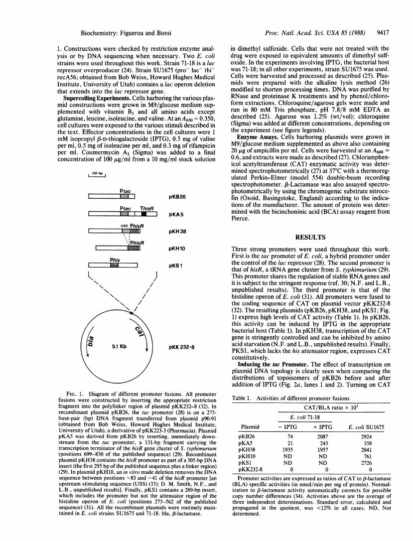
FIG. 1. Diagram of different promoter fusions. All promoterfusions were constructed by inserting the appropriate restrictionfragment into the polylinker region of plasmid pKK232-8 (32). Inrecombinant plasmid pKB26, the tac promoter (28) is on a 275-base-pair (bp) DNA fragment transferred from plasmid p90-91(obtained from Bob Weiss, Howard Hughes Medical Institute,University of Utah), a derivative of pKK223-3 (Pharmacia). PlasmidpKA5 was derived from pKB26 by inserting, immediately down-stream from the tac promoter, a 131-bp fragment carrying thetranscription terminator of the hisR gene cluster of S. typhimurium(positions 699-830 of the published sequence) (29). Recombinantplasmid pKH38 contains the hisR promoter as part of a 305-bp DNAinsert (the first 295 bp of the published sequence plus a linker region)(29). In plasmid pKH10, an in vitro made deletion removes the DNAsequence between positions -83 and -41 of the hisR promoter [anupstream stimulating sequence (USS) (33); D. M. Smith, N.F., andL.B., unpublished results]. Finally, pKS1 contains a 289-bp insert,which includes the promoter but not the attenuator region of thehistidine operon of E. coli (positions 273-562 of the publishedsequence) (31). All the recombinant plasmids were routinely main-tained in E. coli strains SU1675 and 71-18. bla, f3-lactamase.
in dimethyl sulfoxide. Cells that were not treated with thedrug were exposed to equivalent amounts of dimethyl sulf-oxide. In the experiments involving IPTG, the bacterial hostwas 71-18; in all other experiments, strain SU1675 was used.Cells were harvested and processed as described (25). Plas-mids were prepared with the alkaline lysis method (26)modified to shorten processing times. DNA was purified byRNase and proteinase K treatments and by phenol/chloro-form extractions. Chloroquine/agarose gels were made andrun in 80 mM Tris phosphate, pH 7.8/8 mM EDTA asdescribed (25). Agarose was 1.2% (wt/vol); chloroquine(Sigma) was added at different concentrations, depending onthe experiment (see figure legends).Enzyme Assays. Cells harboring plasmids were grown in
M9/glucose medium supplemented as above also containing20 ,ug of ampicillin per ml. Cells were harvested at an A60 =0.6, and extracts were made as described (27). Chloramphen-icol acetyltransferase (CAT) enzymatic activity was deter-mined spectrophotometrically (27) at 37°C with a thermoreg-ulated Perkin-Elmer (model 554) double-beam recordingspectrophotometer. ,B-Lactamase was also assayed spectro-photometrically by using the chromogenic substrate nitroce-fin (Oxoid, Basingstoke, England) according to the indica-tions of the manufacturer. The amount of protein was deter-mined with the bicinchoninic acid (BCA) assay reagent fromPierce.
RESULTS
Three strong promoters were used throughout this work.First is the tac promoter of E. coli, a hybrid promoter underthe control of the lac repressor (28). The second promoter isthat of hisR, a tRNA gene cluster from S. typhimurium (29).This promoter shares the regulation of stable RNA genes andit is subject to the stringent response (ref. 30; N.F. and L.B.,unpublished results). The third promoter is that of thehistidine operon of E. coli (31). All promoters were fused tothe coding sequence of CAT on plasmid vector pKK232-8(32). The resulting plasmids (pKB26, pKH38, and pKS1; Fig.1) express high levels of CAT activity (Table 1). In pKB26,this activity can be induced by IPTG in the appropriatebacterial host (Table 1). In pKH38, transcription of the CATgene is stringently controlled and can be inhibited by aminoacid starvation (N.F. and L.B., unpublished results). Finally,PKS1, which lacks the his attenuator region, expresses CATconstitutively.
Inducing the tac Promoter. The effect of transcription onplasmid DNA topology is clearly seen when comparing thedistributions of topoisomers of pKB26 before and afteraddition of IPTG (Fig. 2a, lanes 1 and 2). Turning on CAT
Table 1. Activities of different promoter fusions
CAT/BLA ratio x 103E. coli 71-18
Plasmid - IPTG + IPTG E. coli SU1675
pKB26 74 2087 2924pKA5 21 243 358pKH38 1935 1957 2041pKH10 ND ND 761pKS1 ND ND 2726pKK232-8 0 0 0
Promoter activities are expressed as ratios of CAT to ,B-lactamase(BLA) specific activities (in nmol/min per mg of protein). Normal-ization to 83-lactamase activity automatically corrects for possiblecopy number differences (34). Activities above are the average ofthree independent determinations. Standard error, calculated andpropagated in the quotient, was <12% in all cases. ND, Notdetermined.
Biochemistry: Figueroa and Bossi

9418 Biochemistry: Figueroa and Bossi
a b
1 2 3 4 5 6
C
2 3
pKB26 pKH38X X __
'ad
;! I I ;W ;N XWX~i WCO >N 0)
Co
FIG. 2. Effect of transcription on plasmid supercoiling. Plasmids extracted from exponentially growing cells were subject to electrophoresisin chloroquine (22 Ag/ml)/1.2% agarose gels. (a) Inducing the tac promoter. Lanes: 1, plasmid pKB26 before IPTG addition; 2, pKB26 afterIPTG addition (3 min); 3, pKB26 after consecutive additions ofIPTG (3 min) and rifampicin (3 min). (b) Triggering the stringent response. Lanes:1, plasmid pKH38 before valine addition; 2, pKH38 after valine addition (5 min); 3, pKH38 after consecutive additions of valine (5 min) andisoleucine (5 min); 4, pKH38 after consecutive additions of valine (5 min), isoleucine (5 min), and rifampicin (3 min); 5, plasmid pKS1 beforevaline addition; 6, pKS1 after valine addition (5 min). (c) Lowering initiation or increasing attenuation. Lanes: 1, plasmid pKK232-8; 2, plasmidpKH10; 3, plasmid pKH38; 4, plasmid pKB26 before IPTG addition; 5, pKB26 after IPTG addition (3 min); 6, plasmid pKA5 before IPTGaddition; 7, pKA5 after IPTG addition (3 min).
transcription induces a rapid and dramatic increase in thenegative supercoiling level of plasmid DNA (8 ± 1 newsuperhelical turns). The topological transition is completedwithin the first 3 min of IPTG addition. No further changesare observed thereafter (data not shown). As fast as they areintroduced, the additional supercoils are removed if tran-scription initiation is blocked with rifampicin (Fig. 2a, lane 3).IPTG does not have any effect on the topoisomer profile ofplasmids, such as pKH38 and pKS1, where CAT expressionis not under the control of the lac repressor (data not shown).Modulating the hisR Promoter. Fig. 2b shows that linking
number variations also accompany the changes in the activityof the hisR promoter. Supercoiling of plasmid pKH38 dimin-ishes (5 ± 1 superhelical turns) following the onset of thestringent response (Fig. 2b, lanes 1 and 2). The latter wasinduced by addition of valine, which in E. coli K12 producesisoleucine starvation (30). The topological relaxation isquickly reversed upon relieving stringency (i.e., supplyingisoleucine; Fig. 2b, lane 3). Under the same conditions,plasmid pKS1 does not exhibit detectable topologicalchanges (Fig. 2b, lanes 5 and 6). Thus, the relaxation ofpKH38 is not a generalized effect of amino acid starvation,but it does reflect the specific inhibition of the hisR promoteractivity.
Affecting Promoter Strength. The efficiency of the hisRpromoter is dependent upon the integrity of an upstreamstimulating element (33). Removal ofthis element by deletionresults in a substantial drop of transcriptional activity (Fig. 1and Table 1). The experiment in Fig. 2c shows that a plasmidcarrying the deleted version of the hisR promoter (pKH10;Fig. 2c, lane 2) is significantly more relaxed than the one withthe intact promoter (pKH38; Fig. 2c, lane 3). Thus, thedegree of plasmid negative supercoiling is directly related tothe strength of the promoter.
Attenuating Transcription. In plasmid pKA5, a transcrip-tion terminator was inserted between the tac promoter andthe beginning of CAT coding sequence. As a result, CATtranscription is dramatically attenuated although it remainsunder the control of the lac repressor (see Table 1). In thisarrangement, induction of the tac promoter with IPTG
produces only a modest change of plasmid linking number(compare lanes 4 and 5 to lanes 6 and 7 in Fig. 2c). Therefore,as previously inferred (21), the transcription-associated su-percoiling increase appears to be a function of the length ofthe transcription unit.
Inhibiting Gyrase. The supercoiling activity ofDNA gyraseis inhibited by a class of antibiotics that interfere with thebinding of ATP to the B subunit of the enzyme (35).Coumermycin Al was shown to block the introduction ofsupercoils into relaxed DNA in vitro (35) and to causerelaxation of supercoiled DNA in vitro (35) and in vivo (36).In the experiment described in Fig. 3a, the tac promoter onplasmid pKB26 was induced after cells had been exposed tocoumermycin for 1 min. Results show that over the back-ground of a progressive plasmid DNA relaxation, the distri-bution of the induced sample contains a set of DNA topo-isomers that are more negatively supercoiled than any ofthose present in the uninduced control (Fig. 3a, lanes 3 and4). In a separate experiment (Fig. 3b), the kinetics ofcoumermycin-induced DNA relaxation was evaluated beforeand after IPTG addition. Whereas in the nontranscribedcontrol the relaxation rate is maximal between 40 and 70 secof coumermycin addition (Fig. 3b, lanes 2 and 3), activatingtranscription at the 40-sec time point has the apparent effectof antagonizing DNA relaxation (compare lanes 2 and 5 inFig. 3b).
DISCUSSIONThe main point of the present study is that in vivo transcrip-tion modifies the susceptibility of the template to the actionof topoisomerases. The linking number of plasmid DNAdecreases concomitantly with transcription activation; thenegative linking difference is maintained for the duration ofthe transcription process and is rapidly reversed when thisprocess is turned off. The magnitude of the supercoilingchanges appears to correlate with the number of elongatingpolymerases. Lowering the strength of the promoter orshortening the transcript have similar effects in delimiting therange of supercoiling variations.
Proc. Natl. Acad Sci. USA 85 (1988)
pribl

Proc. Natl. Acad. Sci. USA 85 (1988) 9419
a b
FIG. 3. Effect of transcription on plasmid supercoiling in thepresence of coumermycin. The experiment was performed with cellsharboring plasmid pKB26. (a) Lanes 1-5 are from a chloroquine (8Ag/ml)/1.2% agarose gel. Lanes 6 and 7 are from a chloroquine (22,ug/ml)/1.2% agarose gel. Lanes: 1, before IPTG; 2, after IPTG (2min); 3, after exposure to coumermycin for 3 min; 4, after consec-utive additions of coumermycin (1 min) and IPTG (2 min); 5, afterconsecutive additions of IPTG (2 min) and coumermycin (3 min).Lanes 6 and 7 are the same samples as in lanes 3 and 4, respectively.[The fact that the topoisomers' profiles in lanes 3 and 4 move up inthe gel when the chloroquine concentration is increased from 8 Ag/mlto 22 A&g/ml (lanes 6 and 7) is taken as evidence that topoisomers arenegatively supercoiled.] (b) DNA relaxation kinetics. Lanes 1-6 arefrom a chloroquine (13 I&g/ml)/1.2% agarose gel. Plasmid pKB26was isolated at the following times (in seconds) after coumermycinaddition: 0 (lane 1), 40 (lane 2), 70 (lane 3), and 100 (lane 4). Inparallel, IPTG was added 40 sec after coumermycin, and plasmid wasisolated after 30 sec (lane 5) and 60 sec (lane 6) of IPTG exposure.
Prior to our work, Pruss and Drlica (21) found thatsteady-state transcription of the tetracycline resistance geneof pBR322 is responsible for the higher levels of negativesupercoiling of plasmid isolated from topoisomerase I mu-tants. These authors speculated that helical unwinding duringtranscription, in causing an apparent relaxation, could favorthe action of DNA gyrase (21). Remaining elusive in thisinterpretation was the role played by the topA mutation.Recently, an alternative interpretation was indicated by Liuand Wang (22) in a model that elaborates on the idea thatduring translocation along the DNA template, an advancingpolymerase will tend to overwind the helix ahead, leavingunderwound the helix behind. Rotational diffusion of theperturbation is probably restrained in a number of instancessuch as in DNA segments flanked by oppositely orientedtranscription units (22). Hence, in plasmid pBR322, wherethe f8-lactamase and tetracycline resistance genes have op-posite polarities, transcription is expected to partition apositively supercoiled domain in front of the transcripts anda negatively supercoiled domain behind them (22). Beingintrinsically symmetrical, the topological separation shouldnot result in an overall supercoiling change unless the twodomains are subject to differential topoisomerase action.Indeed, Liu and Wang interpreted the results of Pruss andDrlica (above) as indicating that topoisomerase I is respon-sible for the relaxation of the negatively supercoiled domain.They proceeded to suggest that DNA gyrase is involved inremoving the positive supercoils ahead of transcription (22).This hypothesis is supported by the finding that if transcribedpBR322 becomes positively supercoiled when cells are ex-posed for some time to a gyrase inhibitor (23, 37).
In contrast to the above studies, in which topoisomeraseactivities were perturbed by mutations or drugs, the super-coiling modulation in our work could be detected under strict
physiological conditions where all topoisomerases are fullyfunctional. As to the mechanism involved, one can speculatethat either an increase ofDNA gyrase activity or a slowdownof topoisomerase I (or a combination of the two) would besimilarly effective in producing a negative linking change(however, see below discussion on gyrase involvement). Aless obvious but particularly attractive model stems fromintegrating polymerase binding into the scenario set by Liuand Wang's elaboration.
In the twin-domain model, the focus is entirely on theprocess ofDNA tracking by RNA polymerase. The topolog-ical relaxation caused by the enzyme binding to the template(38-41) is presumed to have already taken place and neverenters the picture. However, interesting implications resultfrom considering a situation in which, while one polymeraseis traveling along the DNA, a second molecule binds to thepromoter. Binding of the second enzyme is expected to relaxsome of the negative supercoils that form behind the firstpolymerase, whereas there will be no effect on the positivesupercoils ahead of it. Basically we are proposing that whentranscription is activated, during the time it takes for the firstpolymerase to reach the end of the gene, there will be anasymmetry in the partitioning of the twin supercoiling do-mains as more polymerases enter the transcriptional flow.This will unbalance topoisomerase activities resulting inmore positive supercoils to be removed ahead of transcrip-tion than negative supercoils behind. The outcome is anegative linking difference. The extent ofthe asymmetry (andof resulting linking difference) will depend on the number ofinitiation events before the first round of elongation iscompleted. As soon as the latter happens, there will be nofurther linking change as transcription initiation and termi-nation parallel each other. An opposite topological unbalanceis created by the arrest of transcription initiation. Whereasthe positively supercoiled domain will remain unaffected,increased topoisomerase activity will be needed to relax theexcess negative supercoils produced by the movement of thelast elongating complex. The net result is now a linkingnumber increment. Thus, this model explains all of thesupercoiling transitions described here and their triggering bychanging transcriptional states.As shown in Results, a negative supercoiling effect of
transcription could still be detected in the presence of theDNA gyrase inhibitor coumermycin. Although these resultsare not conclusive (one can always argue that a subpopula-tion of gyrase molecules is not subject to the drug inhibition),they strongly suggest that the ATP-dependent activity ofDNA gyrase is not required for the negative supercoilingincrease. [Coumermycin interferes with ATP binding togyrase B subunits, thereby preventing energy-dependentsupercoiling activity (35). The enzyme maintains the ability torelax supercoiled DNA (42, 43)]. This possibility reinforcesthe notion, indicated by our model, that the supercoilingchanges result from a differential swiveling ahead and behindthe transcriptional flow during transcriptional modulation.Swiveling could be driven by the torsional torque of DNAtracking and not rely on ATP hydrolysis. DNA gyrase, orperhaps a gyrase-related activity (44, 45), may be capable ofrelaxing the positive supercoils ahead of transcription bysuch an ATP-independent mechanism.During the early phase of gyrase inhibition, the transcrip-
tion-induced changes ofDNA supercoiling are superimposedon a progressive DNA relaxation. This is probably why in ourexperiments with coumermycin transcription has the appar-ent effect of antagonizing the relaxation process. In thisrespect, our results would seem to be at odds with those ofWu et al. (23) who found that transcription in the presence ofnovobiocin (a coumermycin-like inhibitor) resulted in accu-mulation of positively supercoiled pBR322 topoisomers. Thediscrepancy is likely to originate from important differences
Biochemistry: Figueroa and Bossi

9420 Biochemistry: Figueroa and Bossi
in experimental conditions. Our approach involved activatingtranscription shortly after coumermycin addition and study-ing the changes in plasmid topology for no longer than 3 minaltogether; in contrast, in the experiment of Wu et al. (23),transcription was at steady-state and its effects were exam-ined after a 30-min exposure to novobiocin.
In conclusion, the present study indicates that transcrip-tion contributes to the topological linking difference in DNAof bacterial origin. The possibility of this being a generalproperty of biological systems is suggested by the recentreport of similar findings in the transcription of a eukaryoticgene (46).
We thank Bob Weiss, Ray Gesteland, Kinsey Maundrell, and LullaMelli for comments on the manuscript. We are also grateful to anunidentified referee for constructive criticism. Part of this work wascarried out while one of us (N.F.) was on leave from the Institut deBiologie Physico-Chimique, 13 rue Pierre et Marie Curie, 75005Paris, France.
1. Vinograd, J., Lebowitz, J., Radloff, R., Watson, R. & Laipis,P. (1965) Proc. Natl. Acad. Sci. USA 53, 1104-1111.
2. Worcel, A. & Burgi, E. (1972) J. Mol. Biol. 71, 127-147.3. Bauer, W. R. (1978) Annu. Rev. Biophys. Bioeng. 7, 287-313.4. Sinden, R. R., Carlson, J. 0. & Pettijohn, D. E. (1980) Cell 21,
773-783.5. Bliska, J. B. & Cozzarelli, N. R. (1987) J. Mol. Biol. 194, 205-
218.6. Gellert, M., Mizuuchi, K., O'Dea, M. H. & Nash, H. A. (1976)
Proc. Natl. Acad. Sci. USA 73, 3872-3876.7. Drlica, K. (1984) Microbiol. Rev. 48, 273-289.8. Maxwell, A. & Gellert, M. (1986) Adv. Protein Chem. 38, 69-
107.9. Wang, J. C. (1987) Biochim. Biophys. Acta 909, 1-9.
10. Wang, J. C. (1971) J. Mol. Biol. 55, 523-533.11. Pruss, G. J., Manes, S. H. & Drlica, K. (1982) Cell 31, 35-42.12. DiNardo, S., Voelkel, K. A., Sternglanz, R., Reynolds, A. E.
& Wright, A. (1982) Cell 31, 43-51.13. Richardson, S. M. H., Higgins, C. F. & Lilley, D. M. J. (1984)
EMBO J. 3, 1745-1752.14. Menzel, R. & Gellert, M. (1983) Cell 34, 105-113.15. Tse-Dinh, Y.-C. (1985) Nucleic Acids Res. 13, 4751-4763.16. Morrison, A., Higgins, N. P. & Cozzarelli, N. R. (1980) J. Biol.
Chem. 255, 2211-2219.17. Dorman, C. J., Barr, G. C., Ni Bhriain, N. & Higgins, C. F.
(1988) J. Bacteriol. 170, 2816-2826.18. Higgins, C. F., Dorman, C. J., Stirling, D. A., Waddell, L.,
Booth, I. R., May, G. & Bremer, E. (1988) Cell 52, 569-584.19. Goldstein, E. & Drlica, K. (1984) Proc. Natl. Acad. Sci. USA
81, 4046-4050.20. Balke, V. L. & Gralla, J. D. (1987) J. Bacteriol. 169, 4499-
4506.21. Pruss, G. J. & Drlica, K. (1986) Proc. Natl. Acad. Sci. USA 83,
8952-8956.22. Liu, L. F. & Wang, J. C. (1987) Proc. Natl. Acad. Sci. USA 84,
7024-7027.23. Wu, H.-Y., Shyy, S., Wang, J. C. & Liu, L. F. (1988) Cell 53,
433-440.24. Messing, J., Gronenborg, B., Muller-Hill, B. & Hofchneider,
P. H. (1977) Proc. Natl. Acad. Sci. USA 74, 3642-3646.25. Pruss, G. J. (1985) J. Mol. Biol. 185, 51-63.26. Birnboim, H. C. & Doly, J. (1979) Nucleic Acids Res. 7, 1513-
1523.27. Lupski, J. R., Ruiz, A. A. & Godson, G. N. (1984) Mol. Gen.
Genet. 195, 391-401.28. De Boer, H. A., Comstock, L. J. & Vasser, M. (1983) Proc.
Natl. Acad. Sci. USA 80, 21-25.29. Bossi, L. (1983) Mol. Gen. Genet. 192, 163-170.30. Cashel, M. & Rudd, K. E. (1987) in Escherichia coli and
Salmonella typhimurium: Cellular and Molecular Biology, ed.Neidhart, F. C. (Am. Soc. Microbiol., Washington, DC), Vol.2, pp. 1410-1438.
31. Verde, P., Frunzio, R., Di Nocera, P. P., Blasi, F. & Bruni,C. B. (1981) Nucleic Acids Res. 9, 2075-2086.
32. Brosius, J. (1984) Gene 26, 151-160.33. Bossi, L. & Smith, D. M. (1984) Cell 39, 643-652.34. Klotsky, R. & Schwartz, I. (1987) Gene 55, 141-146.35. Gellert, M., O'Dea, M. H., Itoh, T. & Tomizawa, J. (1976)
Proc. Natl. Acad. Sci. USA 73, 4474-4478.36. Drlica, K. & Snyder, M. (1978) J. Mol. Biol. 120, 145-154.37. Lockshon, D. & Morris, D. R. (1983) Nucleic Acids Res. 11,
2999-3017.38. Wang, J. C., Jacobsen, J. H. & Saucier, J.-M. (1977) Nucleic
Acids Res. 4, 1225-1241.39. Menlikova, A. F., Beabealashvilli, R. & Mirzabekov, A. D.
(1978) Eur. J. Biochem. 84, 301-309.40. Gamper, H. B. & Hearst, J. E. (1982) Cell 29, 81-90.41. Amouyal, M. & Buc, H. (1987) J. Mol. Biol. 195, 795-808.42. Sugino, A., Peebles, C. L., Kreuzer, K. N. & Cozzarelli, N. R.
(1977) Proc. Natl. Acad. Sci. USA 74, 4767-4771.43. Gellert, M., Mizuuchi, K., O'Dea, M. H., Itoh, T. & Tomi-
zawa, J. (1977) Proc. Natl. Acad. Sci. USA 74, 4772-4776.44. Brown, P. O., Peebles, C. L. & Cozzarelli, N. R. (1979) Proc.
Natl. Acad. Sci. USA 76, 6110-6114.45. Gellert, M., Fisher, L. M. & O'Dea, M. H. (1979) Proc. Natl.
Acad. Sci. USA 76, 6289-6293.46. Osborne, B. I. & Guarente, L. (1988) Genes Dev. 2, 766-772.
Proc. Natl. Acad. Sci. USA 85 (1988)