Specific inhibition of transcription by triple helix- forming ...
Upload
trinhxuyenCategory
view
223download
0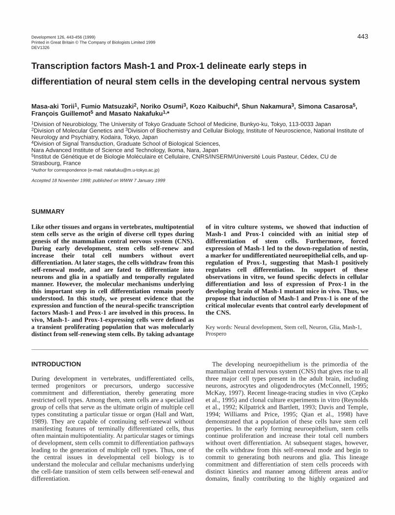
443Development 126, 443-456 (1999)Printed in Great Britain © The Company of Biologists Limited 1999DEV1326
Transcription factors Mash-1 and Prox-1 delineate early steps in
differentiation of neural stem cells in the developing central nervous system
Masa-aki Torii 1, Fumio Matsuzaki 2, Noriko Osumi 3, Kozo Kaibuchi 4, Shun Nakamura 3, Simona Casarosa 5,François Guillemot 5 and Masato Nakafuku 1,*1Division of Neurobiology, The University of Tokyo Graduate School of Medicine, Bunkyo-ku, Tokyo, 113-0033 Japan2Division of Molecular Genetics and 3Division of Biochemistry and Cellular Biology, Institute of Neuroscience, National Institute ofNeurology and Psychiatry, Kodaira, Tokyo, Japan4Division of Signal Transduction, Graduate School of Biological Sciences,Nara Advanced Institute of Science and Technology, Ikoma, Nara, Japan5Institut de Génétique et de Biologie Moléculaire et Cellulaire, CNRS/INSERM/Université Louis Pasteur, Cédex, CU deStrasbourg, France*Author for correspondence (e-mail: [email protected])
Accepted 18 November 1998; published on WWW 7 January 1999
Like other tissues and organs in vertebrates, multipotentialstem cells serve as the origin of diverse cell types duringgenesis of the mammalian central nervous system (CNS).During early development, stem cells self-renew andincrease their total cell numbers without overtdifferentiation. At later stages, the cells withdraw from thisself-renewal mode, and are fated to differentiate intoneurons and glia in a spatially and temporally regulatedmanner. However, the molecular mechanisms underlyingthis important step in cell differentiation remain poorlyunderstood. In this study, we present evidence that theexpression and function of the neural-specific transcriptionfactors Mash-1 and Prox-1 are involved in this process. Invivo, Mash-1- and Prox-1-expressing cells were defined asa transient proliferating population that was molecularlydistinct from self-renewing stem cells. By taking advantage
of in vitro culture systems, we showed that induction ofMash-1 and Prox-1 coincided with an initial step ofdifferentiation of stem cells. Furthermore, forcedexpression of Mash-1 led to the down-regulation of nestin,a marker for undifferentiated neuroepithelial cells, and up-regulation of Prox-1, suggesting that Mash-1 positivelyregulates cell differentiation. In support of theseobservations in vitro, we found specific defects in cellulardifferentiation and loss of expression of Prox-1 in thedeveloping brain of Mash-1 mutant mice in vivo. Thus, wepropose that induction of Mash-1 and Prox-1 is one of thecritical molecular events that control early development ofthe CNS.
Key words: Neural development, Stem cell, Neuron, Glia, Mash-1,Prospero
SUMMARY
allg5;os,eellssr,oehor
INTRODUCTION
During development in vertebrates, undifferentiated celtermed progenitors or precursors, undergo successcommitment and differentiation, thereby generating morestricted cell types. Among them, stem cells are a specialigroup of cells that serve as the ultimate origin of multiple ctypes constituting a particular tissue or organ (Hall and Wa1989). They are capable of continuing self-renewal withomanifesting features of terminally differentiated cells, thuoften maintain multipotentiality. At particular stages or timingof development, stem cells commit to differentiation pathwaleading to the generation of multiple cell types. Thus, onethe central issues in developmental cell biology is understand the molecular and cellular mechanisms underlythe cell-fate transition of stem cells between self-renewal adifferentiation.
ls,iverezedelltt,utssys oftoingnd
The developing neuroepithelium is the primordia of themammalian central nervous system (CNS) that gives rise to three major cell types present in the adult brain, includinneurons, astrocytes and oligodendrocytes (McConnell, 199McKay, 1997). Recent lineage-tracing studies in vivo (Cepket al., 1995) and clonal culture experiments in vitro (Reynoldet al., 1992; Kilpatrick and Bartlett, 1993; Davis and Temple1994; Williams and Price, 1995; Qian et al., 1998) havdemonstrated that a population of these cells have stem cproperties. In the early forming neuroepithelium, stem cellcontinue proliferation and increase their total cell numberwithout overt differentiation. At subsequent stages, howevethe cells withdraw from this self-renewal mode and begin tcommit to generating both neurons and glia. This lineagcommitment and differentiation of stem cells proceeds witdistinct kinetics and manner among different areas and/domains, finally contributing to the highly organized and

444
inginsre
rex-1eds.eded
ithinin
rlyg
.sly
x-tic
iddy
heingrat
urs ofdd.ofy).edUryies.oftedaledtneneree,
themlyed2-
d7).
M. Torii and others
complex morphogenesis of the brain. Thus, cell-fadetermination of neural stem cells and subsequent successteps of differentiation should be under strict control at tmolecular and cellular levels; details of this, however, rempoorly understood.
An apparent complexity of mammalian neural developmeis that the processes of neurogenesis and gliogenesis invmultiple types of intermediate precursors, suggested by recstudies of other experimental organisms. In Drosophila, forexample, neuroectodermal stem cells, termed neuroblasts, rise to secondary precursor cells called ganglion mother c(GMCs), which further divide into postmitotic neurons and/glia (Jan and Jan, 1993). The presence of transient precupopulations has also been demonstrated during developmethe peripheral nervous system (PNS) (Lo and Anderson, 19Gordon et al., 1995). Consistently, recent studies have revethat the developing neuroepithelium is also composed heterogeneous cell types (Kilpatrick et al., 1995; Temple aQian, 1996; Lillien, 1998; Qian et al., 1998). Furthermormany types of epigenetic signals have been shown to influelineage commitment and differentiation of various CNS ctypes (see McKay, 1997 for a detailed review). However, itnot yet precisely determined what cell types are primary targfor these signaling factors, mainly because of the complexof lineage relationships among heterogeneous precursorsorder to solve this cellular complexity and to clarify thunderlying molecular mechanisms, it is particularly importato study intrinsic regulatory molecules that function in distinprecursor cells. We have therefore searched for molecmarkers that can be used to identify and characterize transprecursor cell types during neurogenesis of the CNS.
In this study, we have focused upon the two genes Masand Prox-1, expressed in the early developing brain. Mash-1isa mammalian homolog of the proneural genes of tDrosophila achaete-scutecomplex (AS-C), which encodes basic helix-loop-helix (bHLH)-type transcription facto(Johnson et al., 1990). The fly AS-C genes are expresseneuroblasts as well as in the secondary precursor GMCs,play key roles in multiple steps of neurogenesis (Jan and 1993). Likewise, Mash-1has been found to be expressed in tdeveloping PNS (Lo et al., 1991), and plays an essential for the development of autonomic neurons (Guillemot et a1993; Sommer et al., 1995). Although Mash-1is also widelyexpressed in the developing CNS (Lo et al., 1991; Guillemand Joyner, 1993), no obvious histological anomalies hbeen detected in the brain of Mash-1knock-out mice, exceptfor a subset of olfactory neurons and noradrenergic neuronthe brainstem (Guillemot et al., 1993; Cau et al., 1997; Hirset al., 1998). Thus, it is not yet understood how Mash-1 isinvolved in neurogenesis in the CNS. Prox-1, the other maranalyzed in this study, encodes a homeobox protein thastructurally homologous to Drosophila prospero(Oliver et al.,1993). In fly neurogenesis, expression of prosperodepends onbHLH genes of the AS-C in neuroblasts and GMCs, and iinvolved in subsequent specification of neuronal progeny (Dand Technau, 1993; Hirata et al., 1995). Although a previostudy reported a dynamic expression profile of Prox-1 in thedeveloping brain (Oliver et al., 1993), its regulation anfunction still remain unknown.
Here we have examined spatial and temporal expresspatterns of Mash-1 and Prox-1 proteins in the CNS w
tesiveheain
ntolveent
giveellsorrsornt of95;aledofnde,nce
ell isetsity. Inentctularient
h-1
heard in
andJan,herolel.,
otave
s inch
kert is
t isoeus
d
ionith
particular emphasis upon their correlation with early steps the differentiation of neural stem cells. In the developinforebrain and spinal cord, the cells expressing those protetransiently appeared as proliferating precursors that wedistinct from self-renewing stem cells. We used in vitro cultusystems to demonstrate that induction of Mash-1 and Prospecifically coincided with the time at which stem cells ceasself-renewal and began to differentiate into neuronFurthermore, by using a stem cell-derived cell line, we showthat forced expression of Mash-1 down-regulated thundifferentiated neuroepithelial cell marker nestin anconcomitantly induced expression of Prox-1. Consistent wthese studies in vitro, we found specific defects differentiation of stem cells and loss of Prox-1 expression the brain of Mash-1mutant mice in vivo. Taken together, ourresults demonstrate that Mash-1 and Prox-1 delineate easteps in differentiation of neural stem cells in the developincentral nervous system.
MATERIALS AND METHODS
Immunostaining and in situ hybridization studiesThe antibodies against Mash-1 and Dlx-1 (kind gifts from Drs DAnderson and J. Rubenstein, respectively) were described previou(Lo et al., 1991; Porteus et al., 1994). The affinity-purified anti-Pro1 rabbit antibody was prepared by immunization with a syntheoligopeptide NH2-Lys-Ser-Pro-Asp-Cys-Leu-Gln-Leu-Leu-His-Glu-COOH, corresponding to the predicted C-terminal amino acsequence of murine Prox-1 (Oliver et al., 1993). Our preliminary stuconfirmed that the cDNA for rat Prox-1 encodes exactly the samesequence at its C terminus (T. M. and M. N., unpublished). Texpression of Mash-1, Prox-1 and other markers in the developCNS was examined by immunocytochemistry. Sprague-Dawley embryos were fixed by immersion in freshly prepared 4%paraformaldehyde in phosphate-buffered saline (PBS) for 2-4 hoat 4°C, depending on their ages. To label proliferating cells, 50 mg5-bromo-2′deoxyuridine (BrdU) dissolved in sterile PBS was injecteintraperitoneally into pregnant rats 3 hours before they were killeImmunoperoxidase staining was performed with a combination biotinylated anti-mouse IgG antibody or anti-rabbit IgG antibod(Chemicon) and avidin-biotinylated peroxidase complex (VectorStaining was visualized with an ImmunoPure cobalt/nickel-enhancdiaminobenzidine Substrate Kit (Pierce). For double-staining of Brdwith other antigens, sections were first reacted with primaantibodies and alkaline phosphate (AP)-conjugated antibod(Vector), and visualized with an AP Substrate Kit IV (Vector)Subsequently, the slides were treated with a 2 M solution hydrochloric acid for 20 minutes at room temperature, and subjecto staining with peroxidase-conjugated anti-BrdU monoclonantibody (Boehringer). To count cell numbers, cell nuclei were stainwith 2 µg/ml bis-benzimide (Molecular Probe) for 5 minutes jusbefore mounting. Identification of different histological zones withithe developing neuroepithelium followed the definitions by thBoulder Committee (1970) and Bhide (1996). The ventricular zowas identified as the region containing bipolar cell bodies that weradially aligned relative to the ventricular surface of the neural tubwhich was revealed by nestin staining. On the other hand, subventricular zone cells were more densely packed and randooriented than those in the VZ, many of which were heavily labelwith BrdU. The mantle zone was defined as the region where MAPpositive neurons resided.
Nonradioactive whole-mount in situ hybridization to dissectebrain samples was performed according to Osumi et al. (199

445Cell fate control of neural stem cells
oftive
byllsithts
veor
kitre
andotsere
at
dassissal)
sh-ithndig.
ion1,e
ere
).ely-alT
tohetheionhen.redntheanheesesesr,
Digoxigenin-labeled antisense probes for rat Mash-1, Pax-6 and D1 were described in Nakagawa et al. (1996). The staining wvisualized with alkaline phosphatase-conjugated anti-digoxigenin Ffragment (Boehringer) with its color-indicating substrate.
Wild-type and Mash-1mutant mouse embryos were obtained fromintercrosses of Mash-1+/−mice (Guillemot et al., 1993), andsubjected to immunohistochemical studies as described above.
Indirect immunocytochemical analyses of cultured cells weperformed as described previously (Nakafuku and Nakamura, 19Fluorescein isothiocyanate (FITC)- and Texas Red (TR)-conjugasecondary antibodies (Amersham) were used for double stainingprevious studies, we confirmed that the R24 anti-GD3 gangliosantibody specifically stained oligodendrocyte precursors, but nestin+ or GFAP+ cells under our culture conditions.
Primary culture of neuroepithelial cellsNeuroepithelia of the forebrain of E11.5 and E14.5 rat embryos wdissected out (E0.5 was defined as the day on which the copulaplug was found), and the cells were subsequently cultured under eof the following two conditions as described (Nakagawa et al., 199In monolayer culture, the cells were seeded onto poly-D-lysine (1µg/ml)-coated chambers (Nunc) at a density of 2.5×10⁴ cells/cm2 ina standard medium [1:1 mixture of a Dulbecco’s modified Eaglmedium (DMEM) and F-12 medium (Sanko Junyaku) with 10units/ml penicillin and 100 µg/ml streptomycin] containing the B-27culture supplement (Gibco). The second culture condition is termaggregation culture, in which the cells were maintained in suspensat a density of 1×10⁵ cells/ml in the above standard mediumsupplemented with 10% fetal bovine serum (FBS; Sanko Junya5% horse serum (HS; Gibco), 20 ng/ml basic fibroblast growth fac(bFGF; Boehringer) and 5 ng/ml epithelial growth factor (EGSigma). Culture dishes were coated with poly(2-hydroxy-ethmethacrylate) (polyHEMA; Sigma, 1.6 mg/cm2) to prevent cellattachment. Under these conditions, the cells were allowed to foaggregates, herein termed neurospheres. To continue culture for mthan 4 days, a half volume of the medium was refreshed every 3 dAt subsequent steps, the aggregates were either re-seeded ontoD-lysine-coated dishes and maintained in monolayer described abor were dissociated into single cells by trypsinization and subjecto additional cycles of aggregation cultures. Under the aboconditions, numbers of viable cells were determined by staining wTrypan Blue (Sigma).
Culture and manipulation of MNS-70 cellsThe neural stem cell line MNS-70 was established and maintainedescribed previously (Nakagawa et al., 1996). The cell line wimmortalized by the c-mycer gene, which encodes a fusion product c-Myc and the ligand-binding domain of the estrogen receptor. Wthis system, it is possible to conditionally activate the c-Myc proteby adding estrogen (e.g. β-estradiol) to the culture medium (Nakafukuand Nakamura, 1995). To induce differentiation, MNS-70 cells wesubjected to aggregation culture for 3 days in the standard medcontaining 20 ng/ml bFGF and 1 µM β-estrogen. Cell aggregates werthen re-seeded onto poly-D-lysine-coated dishes, and cultureddifferentiation medium (standard medium supplemented with 10FBS) for 3 days (differentiation culture). Sublines of MNS-70 cethat constitutively expressed Mash-1were established by transfectionwith a Mash-1-expression plasmid by the standard calciuprecipitation method. The plasmid harbored the 2.3 kb rat Mash-1cDNA (a kind gift from Dr Kageyama, Kyoto University) under thcontrol of the Molony sarcoma virus long-terminal repeat and thygromycin-resistant gene. Transfected cells were cultured inmedium containing 0.3 mg/ml of hygromycin B (Sigma), and wecloned during the following 4-6 weeks. Each single clone windependently expanded and subjected to further analysHygromycin B was included in the medium to maintain the clonesmonolayer, but was omitted when the cells were induced
lx-asab
re95).ted. Inidenot
eretoryither6).00
e’s0
edion
ku),torF;yl
rmore
ays. poly-ove,tedveith
d asasofithin
reiume in%
lls
m
ehe areases. into
differentiate. We observed essentially identical expression patternsMash-1, Prox-1 and other protein markers in those clones irrespecof the presence or absence of Hygromycin B.
Western blot analysisExpression patterns of Mash-1, Prox-1 and nestin were analyzedwestern blotting as described (Nakagawa et al., 1996). The cecultured under different conditions were harvested, washed twice wPBS, and lysed directly in Laemmli’s sample buffer. Equal amounof total proteins (approximately 10 µg in each lane) from differentpreparations were blotted with specific antibodies. Immunoreactibands were visualized with peroxidase-conjugated anti-mouse rabbit IgG antibody and an Enhanced Chemiluminescence (Amersham). Relative expression levels of the proteins wequantified by densitometric scanning analysis.
RESULTS
Region-specific expression of Mash-1 and Prox-1 inthe developing CNSPrevious studies reported expression patterns of Mash-1 Prox-1 during development in mouse (Lo et al., 1991; Guillmand Joyner, 1993; Oliver et al., 1993). We extended thestudies and examined their spatio-temporal regulation in modetail. Whole-mount in situ hybridization studies revealed thin the developing rat forebrain at E13.5, the Mash-1+ domaincovered the ventral thalamus (VT), hypothalamus (HT) anganglionic eminence (GE; Fig. 1A). Strong expression walso detected in the dorsal midbrain, where neurogeneproceeds earlier than in the forebrain. In contrast, the dorthalamus (DT) and the primordia of the cerebral cortex (CCwere devoid of expression of Mash-1 at this stage. Thus, Ma1-positive and -negative domains were found adjacent wsharp boundaries at the position between the DT and VT abetween the CC and GE (arrowheads in Fig. 1A; also see F2I). Immunocytochemical studies showed that the expressdomains of Prox-1 followed the discrete patterns of Mash-which also demarcated sharp boundaries (Fig. 1D,E). Wnoticed that these characteristic expression patterns wreminiscent of the two brain-specific homeobox genes, Dlx-1and Pax-6 (Bulfone et al., 1993; Stoykova and Gruss, 1994Thus, the expression domains of Mash-1 and Prox-1 closoverlapped with that of Dlx-1 in the ventral forebrain (Fig. 1DF,G-I), and were rather reciprocal to that of Pax-6 in the dorsregion, except that all of them overlapped each other in the V(Fig. 1B,C). It is notable that these domains are known correspond to important neuromeric compartments of tembryonic forebrain (Puelles and Rubenstein, 1993). Thus, above results suggest that the region-specific expresspatterns of Mash-1 and Prox-1 are closely correlated with testablishment of regional specificity of the developing braiFurthermore, differential onsets of their expressions appeato reflect distinct kinetics of neurogenesis among differedomains of the CNS. The above results showing that texpression of Mash-1 and Prox-1 occurs earlier in the VT thin the DT are consistent with the fact that generation of tmajor neuronal population in the former generally precedthat in the latter (Angevine, 1970). Postmitotic neurons in thGE and HT are also known to be produced earlier than thoin the cortical plate, except for the early-born Cajar-Retziucells located in the pial surface of the CC (Altman and Baye

446
,
d,
r
M. Torii and others
ins of Mash-1 in the developing CNS (A) were compared with those ofole-mount in situ hybridization. Side views of the dissected E13.5he arrowheads indicate the positions of sharp expression boundaries Expression patterns of Mash-1 (D,G), Prox-1 (E,H,J-L) and Dxl-1 (F,I) in examined by immunohistochemistry on parasagittal (D-I) and coronal (J- E14.5 (D-I), E17.5 (J) and E20.5 (K,L). G-I are higher magnification-F, respectively, in which the distribution of Mash-1+ and Prox-1+ cellsx-1+ cells. The arrowheads indicate the positions of sharp expressioned Prox-1+ cells in the ventricular zone of the cerebral cortex boxed in Ktion (see text for details). CC, cerebral cortex; CP, cortical plate; DT,
nic eminence; HT, hypothalamus; MB, midbrain; PT, pretectum; VT,lar zone. Bars, 1 mm (A-F); 200 µm (G-I,L); 5 mm (J,K).
1995) (see Fig. 2K). Consistently, strong expression of Prowas seen to be coincident with the later onsets of neurodifferentiation in the DT and CC at E 17.5 and E20.5 (Fig. 1L). These observations suggest that the spatio-temporegulation of Mash-1 and Prox-1 is tightly associated with tmechanisms controlling the onset of neuronal differentiatio
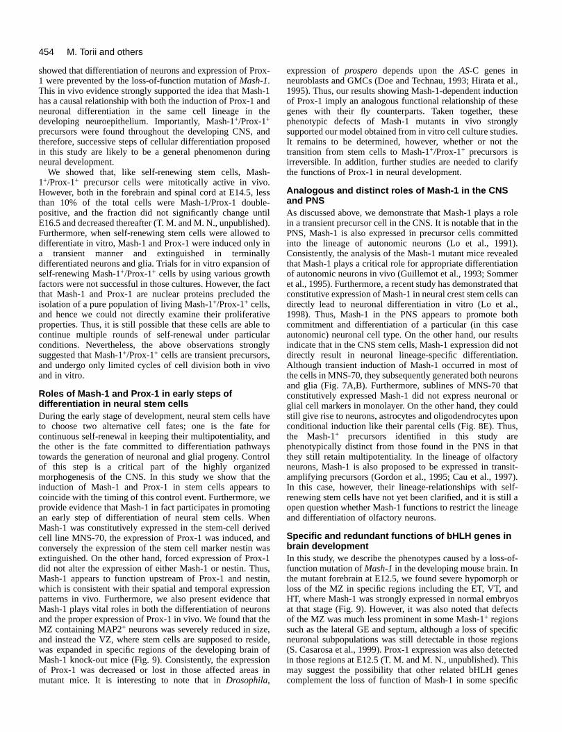
Properties of Mash-1- and Prox-1-expressing cells inthe developing neuroepitheliumNext we compared the expression of Mash-1 and Prox-1 wthose of two other markers for specific neural cell lineagescharacterize the properties of the expressing cells in developing forebrain. Nestin is aclass of intermediate filamentproteins expressed inundifferentiated neuroepithelialcells in the developing CNS(Lendahl et al., 1990). We andothers have shown that CNSstem cells abundantly expressnestin (Redies et al., 1991;Nakafuku and Nakamura, 1995;Reynolds and Weiss, 1996).MAP2 is one of the earliestmarkers specific for postmitoticneurons. In rat embryos, most ofthe cells in the forebrain remainmitotically active until E11.5(Frederiksen and McKay, 1988).At this stage, nestin was stronglyexpressed throughout theneuroepithelium (Fig. 2D),whereas only a few MAP2-positive (MAP2+) cells weredetectable (Fig. 2C, arrows). Inthe same region, Mash-1 andProx-1 were localized in nucleiof some clusters of cells (Fig.2A,B, arrows). A highermagnification clearly showedthat these cells emerged earlierthan MAP2+ neurons andconstituted a minor populationamong nestin+ neuroepithelialcells (Fig. 2E-H). Asdevelopment proceeded toE14.5, a large number ofneurons differentiated in themantle zone (MZ) in the basalforebrain (Fig. 2K,O). Nestin+
cells were found mainly in theventricular zone (VZ) and alsosome in the subventricular zone(SVZ), where proliferatingprecursors resided (Fig. 2L,P).At this stage, many Mash-1+
cells emerged in the VZ andSVZ, which appeared to overlapwith the nestin+ area (Fig.2M,P). At higher magnification,a small scattered population was
Fig. 1. (A-C) Expression domaDlx-1 (B) and Pax-6 (C) by whembryonic brain are shown. Tshared by these genes. (D-L)the developing rat brain wereL) sections of the forebrain atviews of the areas boxed in Dwas compared with that of Dlboundaries. In L, late-generatare shown at higher magnificadorsal thalamus; GE, ganglioventral thalamus; VZ, ventricu
x-1nalJ-ral
hen.
ith tothe
also detected in the inner region of the MZ in the GE (Fig. 2Marrow). On the other hand, Prox-1+ cells were mainly detectedin the SVZ, where they appeared to overlap with Mash-1+ cells(Fig. 2N). Furthermore, some Prox-1+ cells spread further intothe MZ, where its expression partly overlapped with MAP2.
Next we examined whether Mash-1+ cells are mitoticallyactive. Proliferating cells were labeled with BrdU and detecteby double-staining (Fig. 2Q,R). When focused on the GE47±2% (n=4) of the cells located in the VZ and SVZincorporated BrdU after a 3-hour incorporation period. Undethese conditions, 36±3% (n=4) of Mash-1+ cells were labeledwith BrdU, whereas 56±5% (n=4) of BrdU+ cells expressed

447Cell fate control of neural stem cells
wofherrtti--
Fig. 2. Expression patterns ofMash-1 and Prox-1 in theforebrain neuroepithelium.Distributions of cells thatexpressed Mash-1 (A,E,I,M),Prox-1 (B,F,J,N), MAP2(C,G,K,O) and nestin(D,H,L,P) were compared onadjacent coronal sections ofthe forebrain of E11.5 (A-H)and E14.5 (I-R) rat embryos.E-H, M-P and R are highermagnification views of theareas boxed in A-D, I-L andQ, respectively. The arrows inA and B indicate the areaswith scattered Mash-1+ andProx-1+ cells. In C, the arrowsindicate a few MAP2+
neurons present in the dorsaland ventral neural tube. Thetwo small arrows in I indicatesharp boundaries betweenMash-1-positive and negativedomains in the telencephalonand diencephalon. In the DTand CC shown in K, MAP2staining revealed early bornneurons located at the pialsurface of the neural tube.However, note that themajority of neurons, whichlater contribute to theformation of the thalamicnuclei and cortical plate, didnot yet differentiate at thisstage (Angevine, 1970;Altman and Bayer, 1995; seetext for details). The threeshort lines in M-Q indicatethe boundaries between threehistological zones, the VZ,SVZ and MZ. The arrows inM and N indicate the areawhere staining for Mash-1and Prox-1 overlapped withthat of MAP2. In P, thedistribution of nestin+ cells inthe VZ and SVZ are shown,although scattered radial glialfibers in the MZ were alsostained by anti-nestinantibody. In Q and R, co-localization of Mash-1 (blue) in BrdU+ cells (brown) was visualized by double-staining (indicated by arrows). E, eye; MZ, mantle zone; SVZ,subventricular zone; VZ, ventricular zone (all defined in Materials and methods). All other abbreviations are the same as in Fig. 1. Bars, 200µm (A-D); 100 µm (E-H,M-Q); 400 µm (I-L); 20 µm (R).
Mash-1. Considering the 3-hour labeling period and cell cykinetics of proliferating cell populations in the GE (Bhide1996), the majority of the Mash-1+ cells in the VZ and SVZwere mitotically active at this particular stage.
Similar expression profiles of Mash-1 and Prox-1 were aobserved in the developing spinal cord. The domain of Ma1+ cells was located at the ventricular side and reciprocalthat of MAP2+ cells (Fig. 3A,C). The expression of Prox-1
cle,
lsosh- to
overlapped with both of these domains, constituting a narroband between the VZ and MZ. The sequential distributions the above three markers were more clearly seen at higmagnification (Fig. 3E-H). Consistent with a previous repo(Saito et al., 1996), we detected cells labeled by both anMash-1 and anti-BrdU (Fig. 3H,I) in the VZ, where doublelabeled cells corresponded to 24±2% (n=4) of the total Mash-1+ cells. Moreover, Prox-1+/BrdU+ cells were also detectable

448
nnt,nirisof at
is1insistmallater atliaovence
ofgeisineolin
ed.nsle
M. Torii and others
Table 1. Properties of Mash-1- and Prox-1-expressing cells% of Co-expression of other markers
positive cells (% fraction in total positive cells)in total
Marker cell number Mash-1 Prox-1 Nestin MAP2
Mash-1 17.8±3.5 NA 48.9±4.8 58.7±3.1 1.1±0.7Prox-1 9.8±1.2 87.1±3.4 NA 0.3±0.6 13.6±2.9Nestin 38.5±5.4 31.2±4.5 <0.2 NA <0.2MAP2 48.7±4.9 0.4±0.3 1.8±1.3 <0.2 NA
Primary culture was established from the neuroepithelium of the ganglioeminence of E14.5 rat embryos. After seeding dissociated single cells onglass chambers coated with poly-D-lysine (100 µg/ml), doubleimmunostaining was performed with various combinations of specificantibodies for Mash-1, Prox-1, nestin and MAP2. The percentages of poscells for each marker in the total cell number are shown, together with thefractions of the cells that co-expressed other markers, as percentages in ttotal number of positive cells.
Values are the means ± s.d. (n=3-5), in which at least 20 non-overlappinvisual fields and 1000 independent cells were scored.
NA, not applicable; <0.2, less than 0.2%.
Prox-1 in the developing spinal cord. Localizations of cells that expressed MAP2 (C, G) were compared on adjacent transverse sections of theE-H are higher magnification views of the areas boxed in A-D. In D, H andand BrdU antigen (brown) was visualized (arrows in I). Expression ofbeled cells (blue) was shown in J (arrows). In E-H, relative distributionse brackets. MN, motor neurons; MZ, mantle zone; P, pial surface; RP, roofone. Bars, 200 µm (A-D); 50 µm (E-H); 20 µm (I,J).
(Fig. 3J, arrows). These double-positive cells wepredominantly found at the ventricular side within the Prox-+
domain, where its expression overlapped with that of Mas(Fig. 3E,F). Besides the forebrain and spinal cord, Mash-1+ andProx-1+ cells with similar properties were found throughout thCNS (see Fig. 1).
To further explore molecular properties of the Mash-1- aProx-1-expressing cells, we examined isolated cells at single-cell level. Primary cultures were established from tGE of the forebrain at E14.5. Staining with anti-nestin and anMAP2 antibodies indicated that 48.7% and 38.5% of the tocells were postmitotic neurons and undifferentiateneuroepithelial cells, respectively (Table 1). We did nobserved any cells thatexpressed nestin andMAP2 simultaneously orcells expressing glial cellmarkers. In these cultures,Mash-1+ cells constituted17.8% of the total cells, andabout a half of them werenestin+. 48.9% of theMash-1+ cells co-expressedProx-1. Since Prox-1 didnot overlap with nestin,these double-positive cellswere likely to be negativefor nestin. 87.1% of theProx-1+ cells co-expressedMash-1, and a small butsignificant population wereMAP2+, supporting theidea that Prox-1 isexpressed both inprecursors and in immatureneurons.
Taken together, theabove results allowed us topredict a sequentialexpression pattern of
Fig. 3. Expression of Mash-1 and Mash-1 (A,E,I) and Prox-1 (B,F,J),spinal cord of E14.5 rat embryos. I, co-localization of Mash-1 (blue) Prox-1 (brown) in nuclei of BrdU-laof stained cells are indicated by thplate; V, ventricle; VZ, ventricular z
re1h-1
e
ndtheheti-tald
ot
Mash-1 and Prox-1 during development of the CNS. As showin Fig. 4A, we speculate that at early stages of developmeneither Mash-1 nor Prox-1 was expressed in nesti+
undifferentiated neuroepithelial cells. At the onset of thedifferentiation, Mash-1 expression is induced first, and followed by the expression of Prox-1 and down-regulation nestin. Nestin and Prox-1 appear to be mutually exclusivethe single cell level. Importantly, Mash-1+/Prox-1+ cells aremitotically active, although to what extent they can divide unknown. With further progression of differentiation, Mash-expression is extinguished, but expression of Prox-1 remato some extent in newly forming neurons. According to thmodel, Mash-1+/Prox-1+ cells are defined as transienproliferative precursors that are molecularly distinct from stecells and subsequently differentiate into neurons, which we csecondary precursors. Since gliogenesis proceeds only at lstages of neural development, it remains undeterminedpresent whether this particular cell population gives rise to gas well as to neurons. It should be also noted that the abresults do not provide direct evidence that the same sequeindeed occurs within a single cell lineage in vivo.
Correlation of Mash-1 and Prox-1 with initialdifferentiation of neural stem cellsAlthough the above studies revealed the unique natureMash-1- and Prox-1- expressing cells, their precise linearelationship to stem cells was still unknown. To address thissue, we took advantage of an in vitro culture system, which self-renewal and differentiation of stem cells can bmanipulated. Fig. 5A illustrates a detail of the culture protocused in this study. The neuroepithelium of the E11.5 forebrawas dissected out, and primary cultures were establishWhen the cells were maintained under monolayer conditioin a serum-free defined medium (M1), total numbers of viab
nicto
itive
he
g

449Cell fate control of neural stem cells
, as
dand3;ity
n
cells remained relatively constant (Fig. 5B, filled squarealthough BrdU-labeling studies revealed the presence ominor population of dividing cells for the initial 2-3 days (noshown). Under these conditions, MAP2+ neurons becamedetectable at day 3, and thereafter their numbers graduincreased in the following 3 days (Fig. 6B). Gliadifferentiation required an additional 5-10 days (not showIn contrast, when the same population was cultured suspension in the presence of EGF and bFGF (A1 in Fig. 5
urenderles,erse
thes
edSeys.
wogg
ndllstentig.ofly2erre
rstheltser
rex-).
f-ells
al a
the
sedoll
Fig. 4. Model for the expression and function of Mash-1 and Prox-during development of the CNS. (A) Sequential expression patternof Mash-1 and Prox-1 along the histogenesis of the CNS. Nestin,Mash-1, Prox-1 and MAP2 are sequentially expressed in this ordealong with the ventricular-pial axis. In this scheme, the relativepercentages of stained cells in the GE and spinal cord at E14.5 ardepicted proportionally, and include the results of Table 1 wheremutually overlapping and/or exclusive expression patterns amongthese antigens were detected. (B) Proposed model for the expresand functions of Mash-1 and Prox-1. Multipotential neural stem cegive rise to neurons and glia through successive stages ofcommitment and differentiation. During early development, nestin+
stem cells do not express Mash-1 or Prox-1 while they undergocontinuous self-renewal. When their fate is switched from self-renewal to differentiation, stem cells generate a transient secondaprecursor cell type defined by the expression of Mash-1 and ProxSuch cells are still mitotically active, and do not exhibit phenotypeof differentiated neurons or glia. Induction of Mash-1 leads to dowregulation of nestin and concomitant induction of Prox-1, therebypromoting this early step of differentiation. However, furtherprogression of these precursors to terminally differentiated neuronor glia appears to require other regulatory genes. It remains unknwhether the nestin−/Mash-1+/Prox-1+ precursor cells have the abilityof self-renewal, or whether differentiation of neurons and glia furthinvolves other types of precursor cells.
s),f at
allyl
n).in
A),
a small fraction of proliferating cells (1 cell out of 430±45n=5) could be recovered. These cells, even when culturedindividual single cells, continued cell division (calculateddoubling time was 16.2±0.7 hours, n=5; Fig. 5B, filledcircles), and formed large cell aggregates termeneurospheres, as first described by Reynolds et al. (1992) subsequently characterized in detail (Vescovi et al., 199Reynolds and Weiss, 1996; Gross et al., 1996). The majorof cells in these aggregates were nestin+, indicating that theyretained properties of undifferentiated cells (Fig. 5C,D). Whea single neurosphere was transferred to monolayer cult(M2), the cells stopped growing (Fig. 5B, open squares), aspread out on the culture surface (Fig. 5E). Importantly, undappropriate conditions we could detect generation of multipcell types from a single neurosphere, including neuronastrocytes and oligodendrocytes (Fig. 5F-H). On the othhand, when single-cell suspensions were made from thespheres and re-plated under aggregation conditions (A2), cells continued proliferation, again forming neurosphere(Fig. 5B, open circles). In this sense, the cells first recoveras neurospheres fulfilled the criteria of multipotential CNstem cells in that they remained undifferentiated whilundergoing multiple rounds of self-renewal, whereas thecould give rise to neurons and glia under particular conditionThus, this defined culture system allowed us to manipulate tdistinct cell fates of stem cells. One is the fate for continuinself-renewal, and the other is the fate for undergoindifferentiation into neurons and glia.
Using these cultures, the expression patterns of Mash-1 aProx-1 were examined by western blotting. When primary cewere directly placed in monolayer culture (M1), they did noexpress either of these proteins at day 1. However, transiinduction of Mash-1 and Prox-1 was detected at day 3 (F6A). Immunostaining of single cells detected the induction Mash-1+ cells at days 1 and 2 more sensitively, and cleardemonstrated that it preceded the emergence of MAP+
neurons (Fig. 6B). On the other hand, expression of neithMash-1 nor Prox-1 was detected when stem cells wemaintained in aggregation culture (Fig. 6A,A1). Both markeremained negative even when the cells were subjected to second cycle of aggregation culture (Fig. 6A,A2). These resuindicated that self-renewing stem cells do not express eithMash-1 or Prox-1. However, when self-renewing cells wetransferred to monolayer culture conditions, Mash-1 and Pro1 were again induced at day 1 through day 3 (Fig. 6A,M2These results strongly suggest that Mash-1+ and Prox-1+ cellsshow the direct precursor-product relationship with selrenewing stem cells. Furthermore, the emergence of these cclosely coincided with an initial step of differentiation ofneural stem cells.
Functional roles of Mash-1 and Prox-1 in neuralstem cellsA further important question is whether the sequentidifferentiation process proposed in Fig. 4B indeed occurs insingle cell lineage. To address this issue, we used immortalized clonal cell line MNS-70. As describedpreviously (Nakagawa et al., 1996), this cell line exhibitmany of the properties of CNS stem cells, and expressnestin while they were maintained in monolayer (Fig. 7A). Tinduce differentiation, the cells were allowed to form ce
1s
r
e
sionlls
ry-1.sn-
sown
er

450
eic-
y
seasas
M. Torii and others
anipulation of neural stem cells. Primary cultures were establishedin neuroepithelium at E11.5, and cultured under various conditions.
se culture procedures used in this study are illustrated. The first-roundaggregation cultures are named M1 and A1, respectively, whereasultures that are derived from neurospheres of the A1 culture are termedaterials and methods for details). (B) Changes of total numbers of
e scored in each preparation of culture: filled squares, M1; filled circles,es, M2; open circles, A2. (C-H), Immunostaining of cells cultured underns. (C,E) Phase-contrast pictures of single neurospheres cultured underf A1 and M2, respectively. (D) Fluorescent picture of the neurosphere was stained for nestin. (F-H) Fluorescent pictures of cells in M2gs were for MAP2 (F), GFAP (G) and GalC (H). Bars, 200 µm (C-E);
Cel
l num
ber
(×1
0-4 )
aggregates reminiscent of the above mentioned neurosph(for details, see Nakafuku and Nakamura, 1995). Under thconditions, the majority of the cells still remaineundifferentiated, although the fraction of nestin+ cellsdecreased to about 34%, and the intensity of nestin stainbecame much weaker in individual cells. Subsequently, cells were subjected to differentiation culture, in whichmarked induction of neurons and glia was observed (Fig. 7Western blot analyses using the above cultures detectesignificant expression of Mash-1 or Prox-1in monolayer cells (Fig. 7C). In aggregationculture, however, their levels were markedlyelevated. On proceeding to differentiationculture, the levels of Mash-1 and Prox-1became about 20-fold less than those inaggregation culture. Immunocytochemicalstudies also revealed the transientaccumulation of Mash-1+ and Prox-1+ cells(Fig. 7B). In aggregation culture, most ofthe cells became positive for Mash-1(85.6±6.9%, n=5) and Prox-1 (78.5±4.1%,n=5). On the other hand, the percentages oflabeled cells rather decreased indifferentiation culture. This transient patternwas in clear contrast to the induction ofneuronal and glial cell markers, the levels ofwhich were highest in differentiation culture(Fig. 7A). It is important to note here thatthe majority of the cells in aggregationculture expressed Mash-1 and Prox-1,whereas the most of the cells indifferentiation culture expressed eitherneuronal or glial cell makers. Theseproperties of the clonal cell line, which wassupposed to be derived from a single stemcell, strongly suggested that the sequentialinduction events, i.e. early generation ofMash-1+/Prox-1+ cells from nestin+ stemcells and subsequent differentiation intoneurons and glia, indeed occur within asingle cell lineage.
By taking advantage of the abovecharacteristics, we next established sublinesin which Mash-1 or Prox-1 wasconstitutively expressed. Fig. 8A shows thesublines which expressed high levels ofMash-1 proteins in monolayer culture.Under these conditions, expression ofMash-1 was below detectable levels in theparental cells and control sublines. In therepresentative M1-12 and M1-13 cells, highlevels of Mash-1 proteins were maintainedin aggregation culture, which weresubsequently extinguished in the cells indifferentiation culture (Fig. 8B), despite thefact that constitutive high levels of Mash-1mRNA from the transgene were detected inthose cells (data not shown). The reasons forthese phenomena are not clear at present,but expression of Mash-1 proteins may beregulated at the post-transcriptional level.
Fig. 5. In vitro mfrom the forebra(A) The step-wimonolayer and second-round cA2 or M2 (see Mviable cells werA1; open squarvarious conditiothe conditions oshown in C thatcultures. Stainin50 µm (E-H).
eresesed
ingthe aA).
d no
On examining these sublines, we found that constitutivexpression of Mash-1 caused characteristic phenotypchanges of MNS-70 cells. As described above, downregulation of nestin and induction of Prox-1 were closelassociated with Mash-1, both in vivo and in vitro.Consistently, it was found that the expression of nestin waextinguished in the Mash-1-expressing sublines. In threpresentative subline M1-12, the expression of nestin wdetectable under none of the culture conditions, whereas it w

451Cell fate control of neural stem cells
ig.e
ere, the).ity3
Asa1
.tetgofcy
Fig. 6. Transient induction of Mash-1 and Prox-1 in cultures ofneural stem cells. Primary cells of the forebrain neuroepitheliumwere cultured under the conditions described in Fig. 5. (A) Westernblot analysis of Mash-1 and Prox-1. Preparations of cultured cellsdesignated M1, M2, A1 and A2 are described in Fig. 5B. Thecultures of A2 and M2 were derived from the cells that weremaintained for 7 days in the A1 culture (indicated by arrows).(B) The percentages of Mash-1+ (filled squares) and MAP2+ (opensquares) cells that emerged in M1 cultures were plotted against theculture time, in which at least 20 non-overlapping visual fields and1000 independent cells were scored in each case.
Fig. 7. Conditional induction of Mash-1 and Prox-1 in MNS-70 cells.MNS-70 cells were cultured under various conditions as described inthe text. M, A and D indicate monolayer, aggregation anddifferentiation culture conditions, respectively (see text for details).(A,B) The cells were subjected to immunostaining by specificantibodies, and the percentages of stained cells in total cell numbers(determined by nuclear staining with bis-benzimide) were scored asdescribed in Fig. 6B. (A) Nestin, open squares; MAP2, filledsquares; GFAP, open circles; GD3-ganglioside, filled circles.(B) Mash-1, open squares; Prox-1, filled squares. Values are means ±s.d. (n=4-5). (C) Expression levels of Mash-1 (left) and Prox-1(right) proteins were examined by western blot analysis, and areindicated in each panel by the arrow on the right; the positions ofmolecular size markers (Mr×103) are shown on the left. The bandswith molecular masses of 45-50×103 in the Mash-1 panel and thoseof 70-90×103 in the Prox-1 panel were due to nonspecific reaction ofthe antibodies.
successively down-regulated in the control subline H1-1 (F8C). Furthermore, the expression of Prox-1 becamconstitutive in the sublines. In M1-12 and M1-13 cells, thlevels of Prox-1 proteins remained high in aggregation cultuand were subsequently decreased in differentiation culture,pattern of which paralleled that of Mash-1 (Fig. 8DImmunostaining of single cells demonstrated that the majorof the cells in monolayer cultures of M1-12 and M1-1expressed Prox-1 but not nestin, demonstrating that reciprocal regulation of these proteins by Mash-simultaneously occurred in the same cells (data not showIn separate experiments, we also established sublines in whProx-1 was constitutively expressed. None of these sublinshowed altered expression of nestin or Mash-1 (T. M. and N., unpublished). Thus, we conclude that Mash-1 functioneither directly or indirectly, as an upstream regulator for texpression of nestin and Prox-1, thereby contributing to tearly steps of differentiation in neural stem cells.
We next asked whether the Mash-1-expressing cemaintain multipotentiality or are already committed tparticular cell lineages (Fig. 8E). In monolayer, M1-12 celdid not expressed either MAP2, GFAP or GD3-ganglioside atdetectable levels, indicating that forced expression of Mashin MNS-70 did not directly induce terminal differentiation oneuronal or glial cell lineage. We found, however, M1-12 cecould give rise to both neurons and glia when they we
the1n).iches
M.s,
hehe
llsols
-1fllsre
conditionally induced to differentiate like their parental cells.The five other Mash-1-expressing sublines shown in Fig. 8also exhibited similar multipotential properties. These resultsuggested that terminal differentiation of neurons and gliinvolves additional steps other than the induction of Mash-and Prox-1 in MNS-70 cells.
Defects in neurogenesis in Mash-1 knock-out miceTo further explore functional roles of Mash-1 in vivo, wenext examined the mutant mouse strain in which the Mash-1 locus was genetically disrupted (Guillemot et al., 1993)Although developmental defects in the PNS of the mutanhave been characterized (Guillemot et al., 1993; Sommer al., 1995; Cau et al., 1997), phenotypes in the developinCNS has not yet been examined in detail. As in the case rat embryos, Mash-1 was strongly expressed in specifisubdomains in the mouse forebrain at E12.5 (roughl

452
a
nsd
M. Torii and others
expression of Mash-1 in MNS-70 cells. In A-D the positions ofs (×103) are shown on the left. (A) Western blot analysis of Mash-1nd its sublines. Whole cell lysates from cells in monolayer were blotteddy. The left three lanes show the parental MNS-70 and its control. The six lanes on the right including M1-12 to M2-13 are the Mash-1
e bands of Mash-1 proteins are indicated by the arrow on the right.0, M1-12 and M1-13 cells were cultured under the three conditions the levels of Mash-1 proteins were compared. (C) MNS-70, M1-12 anded to western blot analysis of nestin. The levels of nestin were comparedrepared tissue sample of the E11.5 forebrain and midbrain F/M). Several discrete bands and smears with apparent molecular massesd by a bracket on the right) were detected by the antibody, whichto the full-length nestin protein and its degradation products. Prox-1 proteins were examined in the parental MNS-70 and its Mash-1described in B. Abbreviations are the same as in Fig. 7. (E) M1-12 cellstiate by the standard three-step culture protocol. The percentages ofGFAP+ (open circles) and GD3-ganglioside+ (filled circles) cells werein Fig. 7A. D3 and D7 indicate the cells cultured under differentiationays, respectively. Values are means ± s.d. (n=4-5).
corresponds to E14.5 in rats; see Figs 1, 2) including dorsal part of the epithalamus (ET), VT and the ventral floregion of the HT (Fig. 9C). We found that the shape of tneural tube was severely deformed in these three regionthe mutant embryos (Fig. 9A,B). Higher magnification viewrevealed that the neuroepitheliumwas expanded in the ET and VT(about 1.5- to 2-fold thicker thanthose in the wild-type embryos;Fig. 9D,E), and in some cases aprotrusion toward the ventricle wasevident (arrow in Fig. 9B,E). Asimilar but less prominentexpansion was also evident in theventral aspect of the HT (Fig. 9F).On the other hand, MAP2 staining,which identified postmitoticneurons in the MZ of theneuroepithelium, revealed that theMZ was largely reduced in size oralmost missing in the above threeregions (Fig. 9D-F). We alsonoticed a similar hypomorphicphenotype of the MZ in the medianpart of the GE and the dorsal aspectof the neural tube throughout themidbrain to the spinal cord (S.Casarosa et al., 1999). Thus, thefact that the neuroepithelium itselfwas enlarged but the MZ wasshrunk indicated that proliferativecell populations in the VZ wereexpanded in the mutant brain. Itwas also noted that Prox-1+ cellswere also reduced in number oralmost completely lost in thoseaffected domains (Fig. 9B,G),which supported the idea thatMash-1 is involved in regulatingProx-1 in vivo. The lack of Prox-1expression in the cells thataccumulated in the mutantneuroepithelium further suggestedthat they maintained properties ofself-renewing stem cells.Consistently, the majority of themwere stained by anti-nestinantibody (data not shown). Thesephenotypes were in clear contrastto the apparent normal histogenesisin the adjacent domains, i.e. the DTand the dorsal portion of the HT,where the formation of the MZremained intact. Expression ofProx-1 was also detectable in thoseareas. Thus, defects in the mutantforebrain were highly specific tothe domains where Mash-1 wasstrongly expressed in the wild-typeembryos at this stage. The evidencethat the VZ was expanded at the
Fig. 8. Effects of forced molecular sized markerexpressed in MNS-70 awith anti-Mash-1 antibosublines, H1-1 and H1-4expressing sublines. Th(B) The parental MNS-7described in Fig. 7, andH1-1 cells were subjectwith that in the freshly pneuroepithelium (E11.5of 120-200×103 (indicateprobably corresponded (D) Expression levels ofexpressing sublines as were induced to differenMAP2+ (filled squares), quantified as described conditions for 3 and 7 d
theorhes ofs
expense of the MZ in the mutant brain supported the idethat stem cells cannot appropriately differentiate intoneurons or express Prox-1 in the absence of normal functioof Mash-1. Thus, the abnormalities observed here provide

453Cell fate control of neural stem cells
f.das1,t
oftlyurmrsof
ted inde
e forebrain of the Mash-1mutant mice. The expression domains ofB,B′,G,G′) in the developing forebrain at E12.5 were compared betweenpe (A′-G′) mice. The expression of Mash-1 in the wild-type embryos at
he arrowheads in each panel indicate the boundaries of discrete neuromericrebrain. The arrows in B and E indicate a protrusion of thef the VT. The arrowheads in F,F′,G,G′ mark the junction between the
where the formation of the MZ and expression of Prox-1 were missing ind intact in the latter. Abbreviations are the same as in Figs 1 and 2. Bars,-G′).
strong evidence that Mash-1 plays an essential role differentiation of stem cells in vivo.
DISCUSSION
Mash-1 and Prox-1 as molecular markers fortransient precursors in the CNSDuring development in vertebrates, multipotential stem cegive rise to diverse cell types present in the adult CNS (Temand Qian, 1996; McKay, 1997). Recent studies hademonstrated that stem cells generate neurons and glia throa multi-step process involving many types of intermediaprecursors. These precursors are defined as cells that have restricted self-renewing capacities and/or differentiatiopotentials than stem cells (Kilpatrick, et al., 1995; Temple aQian, 1996; Lillien, 1998). However, many of them artransient cell types, and therefore their lineage relationshstill remain poorlyunderstood. Such issues ofidentities and lineagerelationships amongdiverse precursors is oneof the fundamentalquestions indevelopmental biology(Hall and Watt, 1989).
The aim of this studywas to identify transientprecursors present in thedeveloping CNS by usingdefined molecularmarkers. Here we presentevidence that the bHLH-type transcription factorMash-1 and thehomeodomain-containingprotein Prox-1 define atransient precursor that ismolecularly distinct fromthe CNS stem cells. Theexpression patterns ofthose markers both in vivo(Figs 1-3) and in primaryculture of neuroepithelialcells (Figs 5, 6) suggestedthe model that Mash-1+
and Prox-1+ cells originatefrom nestin+ stem cells,and subsequently give riseto terminally differentiatedneurons and glia (see Fig.4B). Recent studies haveshown, however, thatnestin is expressed indifferent types of cells inimmature stages, andhence is not a definitivemarker for stem cells(Zimmerman et al., 1994).In this study, therefore, we
Fig. 9. Defective neurogenesis in thMAP2 (A,A′,D-F,D′-F′) and Prox-1 (the Mash-1mutant (A-G) and wild-tythe same stage was shown in C. Tcompartments of the developing foneuroepithelium at the dorsal part oventral and dorsal parts of the HT, the former, whereas those remaine400 µm (A-C,A′,B′); 200 µm (D-G,D′
for
llspleveughtemoren
ndeips
defined the properties of neural stem cells by their ability oself-renewal and their differentiation potential (Figs 5, 6)Under our culture conditions, self-renewing stem cells diindeed express nestin, and down-regulation of nestin wclosely coupled with the emergence of Mash-1 and Prox-which further led to the generation of neurons and glia. Apresent, however, the transient nature of the expressionMash-1 and Prox-1 precludes direct proof that the apparensequential events of cellular differentiation do indeed occwithin a single cell lineage. Nevertheless, analysis of the stecell-derived clonal cell line MNS-70 strongly supported oumodel (Figs 7, 8). Thus, undifferentiated MNS-70 cellexpressed nestin but not Mash-1 or Prox-1. Upon induction differentiation, however, the majority of them transientlyexpressed Mash-1 and Prox-1, and subsequently generaneurons and glia. Furthermore, forced expression of Mash-1nestin+ MNS-70 cells caused the induction of Prox-1 andown-regulation of nestin in each single cell. In addition, w
454
l.,n
ee
s.e
y
olee
ed).ed
rhatan
theltsot.fnstorldon,
t
sit-).f- age
of-
or
osts
ificnsted
issc
M. Torii and others
showed that differentiation of neurons and expression of Pr1 were prevented by the loss-of-function mutation of Mash-1.This in vivo evidence strongly supported the idea that Mashhas a causal relationship with both the induction of Prox-1 aneuronal differentiation in the same cell lineage in thdeveloping neuroepithelium. Importantly, Mash-1+/Prox-1+
precursors were found throughout the developing CNS, atherefore, successive steps of cellular differentiation propoin this study are likely to be a general phenomenon durneural development.
We showed that, like self-renewing stem cells, Mas1+/Prox-1+ precursor cells were mitotically active in vivoHowever, both in the forebrain and spinal cord at E14.5, lethan 10% of the total cells were Mash-1/Prox-1 doublpositive, and the fraction did not significantly change unE16.5 and decreased thereafter (T. M. and M. N., unpublisheFurthermore, when self-renewing stem cells were alloweddifferentiate in vitro, Mash-1 and Prox-1 were induced only a transient manner and extinguished in terminadifferentiated neurons and glia. Trials for in vitro expansion self-renewing Mash-1+/Prox-1+ cells by using various growthfactors were not successful in those cultures. However, the that Mash-1 and Prox-1 are nuclear proteins precluded isolation of a pure population of living Mash-1+/Prox-1+ cells,and hence we could not directly examine their proliferatiproperties. Thus, it is still possible that these cells are ablecontinue multiple rounds of self-renewal under particulconditions. Nevertheless, the above observations stronsuggested that Mash-1+/Prox-1+ cells are transient precursorsand undergo only limited cycles of cell division both in vivand in vitro.
Roles of Mash-1 and Prox-1 in early steps ofdifferentiation in neural stem cellsDuring the early stage of development, neural stem cells hto choose two alternative cell fates; one is the fate continuous self-renewal in keeping their multipotentiality, anthe other is the fate committed to differentiation pathwatowards the generation of neuronal and glial progeny. Conof this step is a critical part of the highly organizemorphogenesis of the CNS. In this study we show that induction of Mash-1 and Prox-1 in stem cells appears coincide with the timing of this control event. Furthermore, wprovide evidence that Mash-1 in fact participates in promotian early step of differentiation of neural stem cells. WhMash-1 was constitutively expressed in the stem-cell derivcell line MNS-70, the expression of Prox-1 was induced, aconversely the expression of the stem cell marker nestin wextinguished. On the other hand, forced expression of Prodid not alter the expression of either Mash-1 or nestin. ThMash-1 appears to function upstream of Prox-1 and neswhich is consistent with their spatial and temporal expresspatterns in vivo. Furthermore, we also present evidence tMash-1 plays vital roles in both the differentiation of neuroand the proper expression of Prox-1 in vivo. We found that MZ containing MAP2+ neurons was severely reduced in sizand instead the VZ, where stem cells are supposed to reswas expanded in specific regions of the developing brainMash-1 knock-out mice (Fig. 9). Consistently, the expressiof Prox-1 was decreased or lost in those affected areasmutant mice. It is interesting to note that in Drosophila,
ox-
-1nde
ndseding
h-.ss
e-tild).
toinllyof
factthe
ve to
argly,o
aveford
ystroldthetoe
ngenedndas
x-1us,tin,ionhatnsthee,ide,
ofon in
expression of prosperodepends upon the AS-C genes inneuroblasts and GMCs (Doe and Technau, 1993; Hirata et a1995). Thus, our results showing Mash-1-dependent inductioof Prox-1 imply an analogous functional relationship of thesgenes with their fly counterparts. Taken together, thesphenotypic defects of Mash-1 mutants in vivo stronglysupported our model obtained from in vitro cell culture studieIt remains to be determined, however, whether or not thtransition from stem cells to Mash-1+/Prox-1+ precursors isirreversible. In addition, further studies are needed to clarifthe functions of Prox-1 in neural development.
Analogous and distinct roles of Mash-1 in the CNSand PNSAs discussed above, we demonstrate that Mash-1 plays a rin a transient precursor cell in the CNS. It is notable that in thPNS, Mash-1 is also expressed in precursor cells committinto the lineage of autonomic neurons (Lo et al., 1991Consistently, the analysis of the Mash-1 mutant mice revealthat Mash-1 plays a critical role for appropriate differentiationof autonomic neurons in vivo (Guillemot et al., 1993; Sommeet al., 1995). Furthermore, a recent study has demonstrated tconstitutive expression of Mash-1 in neural crest stem cells cdirectly lead to neuronal differentiation in vitro (Lo et al.,1998). Thus, Mash-1 in the PNS appears to promote bocommitment and differentiation of a particular (in this casautonomic) neuronal cell type. On the other hand, our resuindicate that in the CNS stem cells, Mash-1 expression did ndirectly result in neuronal lineage-specific differentiationAlthough transient induction of Mash-1 occurred in most othe cells in MNS-70, they subsequently generated both neuroand glia (Fig. 7A,B). Furthermore, sublines of MNS-70 thaconstitutively expressed Mash-1 did not express neuronal glial cell markers in monolayer. On the other hand, they coustill give rise to neurons, astrocytes and oligodendrocytes upconditional induction like their parental cells (Fig. 8E). Thusthe Mash-1+ precursors identified in this study arephenotypically distinct from those found in the PNS in thathey still retain multipotentiality. In the lineage of olfactoryneurons, Mash-1 is also proposed to be expressed in tranamplifying precursors (Gordon et al., 1995; Cau et al., 1997In this case, however, their lineage-relationships with selrenewing stem cells have not yet been clarified, and it is stillopen question whether Mash-1 functions to restrict the lineaand differentiation of olfactory neurons.
Specific and redundant functions of bHLH genes inbrain developmentIn this study, we describe the phenotypes caused by a loss-function mutation of Mash-1in the developing mouse brain. Inthe mutant forebrain at E12.5, we found severe hypomorph loss of the MZ in specific regions including the ET, VT, andHT, where Mash-1 was strongly expressed in normal embryat that stage (Fig. 9). However, it was also noted that defecof the MZ was much less prominent in some Mash-1+ regionssuch as the lateral GE and septum, although a loss of specneuronal subpopulations was still detectable in those regio(S. Casarosa et al., 1999). Prox-1 expression was also detecin those regions at E12.5 (T. M. and M. N., unpublished). Thmay suggest the possibility that other related bHLH genecomplement the loss of function of Mash-1 in some specifi

455Cell fate control of neural stem cells
s,atsg
cerh-
ird
ak
rforarted
:
in
.
l
ls.
subdomains of the forebrain. In fact, an increasing numberbHLH-type transcription factors has been identified, and soof them show overlapping expression patterns with Mash-1the developing CNS (Gradwohl et al., 1996; Lee, 1997). MNS-70 cells, multiple bHLH factors other than Mash-1including Math-1 and NeuroD, were expressed at detectalevels. Unlike Mash-1, however, overexpression of NeuroD dnot resulted in down-regulation of nestin or induction of Pro1 (M. T. and M. N., unpublished). Thus, at least in thparticular cell line, Mash-1 exhibited specific functions.
Significance of multiple precursors and multiplemolecular regulatorsThe present study has left open the issue of the biologsignificance of multiple precursor cell types durinneurogenesis of the CNS. One tenable idea is that distprecursors expressing different sets of regulatory genes targets for distinct environmental signals. In fact, recent studhave demonstrated that commitment and differentiation neurons and glia are controlled by multiple signaling facto(see McKay, 1997, for a review). In particular, platelet-derivgrowth factor has been proposed to promote neurodifferentiation, whereas bone morphogenetic protein 2 (BMPand ciliary neurotrophic factor specifically stimulate astroglidifferentiation (Johe et al., 1996; Gross et al., 1996; Williamet al., 1997). In the PNS, BMP2 was shown to induce texpression of Mash-1 in neural crest stem cells, and promodifferentiation of autonomic neurons (Shah et al., 199However, in our culture system of CNS stem cells, none of above factors could directly induce the expression of Masor Prox-1 (T. M. and M. N., unpublished). On the other hanbFGF and EGF, which acted as potent mitogens for srenewing stem cells, appeared to inhibit the induction of Ma1 and Prox-1 (see Fig. 5). Thus, it still remains unknown whenvironmental factor(s) is responsible for determining tonset of the expression of Mash-1 in self-renewing stem ceAlternatively, induction of Mash-1 may be under regulation bcell-cell interactions through the Notch-signaling pathway demonstrated in other bHLH genes in Xenopusand Drosophila(reviewed in Kageyama and Nakanishi, 1997). Consistent wthis idea, a recent report has described that targeted disrupof Notch1 and its downstream signaling component RBP-Jkmice resulted in aberrant expression of Mash-1 in the CNS la Pompa et al., 1997).
Spatio-temporal regulation of differentiation ofneural stem cellsBased on the above evidence, the present study explains the time at which the expression of Mash-1 and Prox-1 is fiturned on is highly variable in distinct regions of thdeveloping CNS (Lo et al., 1991; Guillemot and Joyner, 199Oliver et al., 1993). These distinct times of onset are likelyreflect distinct onsets of differentiation of stem cells. This idis supported by our observations that their spatio-tempoexpression patterns are correlated with the onset and kineof neuronal differentiation in various regions (Figs 1, 2Furthermore, we showed that the expression domains of Ma1 and Prox-1 closely overlapped that of Dlx-1 and wereciprocal to that of Pax-6 in the rat forebrain at E13.5 (F1). It is striking that these genes shared important neuromboundaries in the developing forebrain (Puelles a
ofme inIn,bleidx-is
icalginctareiesofrs
ednal2)als
heted
6).theh-1d,
elf-sh-at
hells.y
as
ithtion in(de
whyrste3;
toearaltics).sh-reig.ericnd
Rubenstein, 1993; Bulfone et al., 1993; Stoykova and Grus1994). These observations raise the intriguing possibility ththe mechanisms controlling differentiation of neural stem cellare closely coupled with those underlying the precedinregional specification of the developing neuroepithelium. Infact, a recent study has reported that several distinct enhanelements are responsible for regulating the expression of Mas1 in different regions of the developing CNS (Verma-Kurvaret al., 1998). Further studies to elucidate the moleculamechanisms that coordinate the regional specification andifferentiation of neural stem cells will facilitate betterunderstanding of early development of the vertebrate CNS.
We are grateful to Drs D. Anderson, J. Rubenstein, R. Kageyamand Y. Ihara for providing us with cDNAs and antibodies. We thanT. Suzuki for contributing to the initial phase of this study. We alsothank Drs Y. Ihara, Y. Kaziro, S. Kohsaka and Y. Nabeshima foencouragement and support, and H. Okano and K. Shimamura valuable comments and discussion. This work was supported in pby grants-in-aid from the Ministry of Education, Science and Culturand from CREST (Core research for Evolutional Science anTechnology) of Japan Science and technology Cooperation.
REFERENCES
Altman, J. and Bayer, S. A. (1995). Atlas of the prenatal rat braindevelopment. CRC Press Inc, Ltd., Boca Raton.
Angevine, J. B. Jr. (1970). Time of neuron origin in the diencephalon of themouse. An autoradiographic study. J. Com. Neurol. 139, 129-187.
Bhide, P. G. (1996). Cell cycle kinetics in the embryonic mouse corpusstriatum. J. Comp. Neurol.374, 506-522.
Boulder Committee (1970). Embryonic vertebrate central nervous systemrevised terminology. Anat. Rec.166, 257-261.
Bulfone, A., Puelles, L., Porteus, M. H., Frohman, M. A., Martin, G. R.and Rubenstein, J. L. (1993). Spatially restricted expression of Dlx-1, Dlx-2 (Tes-1), Gbx-2 and Wnt-3 in the embryonic day 12.5 mouse forebradefines potential transverse and longitudinal segmental boundaries. J.Neurosci. 13, 3155-3172.
Casarosa, S., Fode, C. and Guillemot, F. (1999). Mash-1 regulatesneurogenesis in the ventral telencephalon. Development 126, 525-534.
Cau, E., Gradwohl, G., Fode, C. and Guillemot, F. (1997). Mash1 activatesa cascade of bHLH regulators in olfactory neuron progenitors. Development124, 1611-1621.
Cepko, C., Ryder, E. F., Austin, C. P., Walsh, C. and Fekete, D. M. (1995).Lineage analysis using retrovirus vectors. Methods Enzymol.254, 387-419.
Davis, A. A. and Temple, S. (1994). A self-renewing multipotential stem cellin embryonic rat cerebral cortex. Nature372, 263-266.
de la Pompa, J. L., Wakeham, A., Correia, K. M., Samper, E., Brown, S.,Aguilera, R. J., Nakano, T., Honjo, T., Mak, T. W., Rossant, J. andConlon, R. A. (1997). Conservation of the Notch signaling pathway inmammalian neurogenesis. Development124, 1139-1148.
Doe, C. Q. and Technau, G. M. (1993). Identification and cell lineage ofindividual neural precursors in the Drosophila CNS. Trends Neurosci.16,510-514.
Frederiksen, K. and McKay, R. D. (1988). Proliferation and differentiationof rat neuroepithelial precursor cells in vivo. J. Neurosci.8, 1144-1151.
Gordon, M. K., Mumm, J. S., Davis, R. A., Holcomb, J. D. and Calof, A.L. (1995). Dynamics of MASH1 expression in vitro and in vivo suggest anon-stem cell site of MASH1 action in the olfactory receptor neuron lineageMol. Cell. Neurosci.6, 363-379.
Gradwohl, G., Fode, C. and Guillemot, F. (1996). Restricted expression ofa novel murine atonal-related bHLH protein in undifferentiated neuraprecursors. Dev. Biol.180, 227-241.
Gross, R. E., Mehler, M. F., Mabie, P. C., Zang, Z., Santschi, L. andKessler, J. A. (1996). Bone morphogenetic proteins promote astroglialineage commitment by mammalian subventricular zone progenitor cellNeuron17, 595-606.
Guillemot, F. and Joyner, A. L. (1993). Dynamic expression of the murine

456
fic
sed
l
in
S
e
nd
sor
rs.
d
nt
ne
ural
M. Torii and others
Achaete-Scute homologue Mash-1 in the developing nervous system. Mech.Dev.42, 171-185.
Guillemot, F., Lo, L. C., Johnson, J. E., Auerbach, A., Anderson, D. J. andJoyner, A. L. (1993). Mammalian achaete-scute homolog 1 is required the early development of olfactory and autonomic neurons. Cell 75, 463-476.
Hall, P. A. and Watt, F. M. (1989). Stem cells: the generation andmaintenance of cellular diversity. Development106, 619-633.
Hirata, J., Nakagoshi, H., Nabeshima, Y. and Matsuzaki, F. (1995).Asymmetric segregation of the homeodomain protein Prospero durDrosophila development. Nature377, 627-630.
Hirsch, M. R., Tiveron, M. C., Guillemot, F., Brunet, J. F. and Goridis, C.(1998). Control of noradrenergic differentiation and Phox2a expressionMASH1 in the central and peripheral nervous system. DevelopmentSuppl.125, 599-608.
Jan, Y. N. and Jan, L. Y. (1993). HLH proteins, fly neurogenesis andvertebrate myogenesis. Cell 75, 827-830.
Johe, K. K., Hazel, T. G., Muller, T., Dugich-Djordjevic, M. M. andMcKay, R. D. (1996). Single factors direct the differentiation of stem celfrom the fetal and adult central nervous system. Genes Dev.10, 3129-3140.
Johnson, J. E., Birren, S. J. and Anderson, D. J. (1990). Two rat homologuesof Drosophila achaete-scute specifically expressed in neuronal precursNature346, 858-861.
Kageyama, R. and Nakanishi, S. (1997). Helix-loop-helix factors in growthand differentiation of the vertebrate nervous system. Curr. Opin. Genet. Dev.7, 659-665.
Kilpatrick, T. J. and Bartlett, P. F. (1993). Cloning and growth ofmultipotential neural precursors: requirements for proliferation adifferentiation. Neuron10, 255-265.
Kilpatrick, T. J., Richards, L. J. and Bartlett, P. F. (1995). The regulationof neural precursor cells within the mammalian brain. Mol. Cell. Neurosci.6, 2-15.
Lee, J. E. (1997). Basic helix-loop-helix genes in neural development. Curr.Opin. Neurobiol.7, 13-20.
Lendahl, U., Zimmerman, L. B. and McKay, R. D. (1990). CNS stem cellsexpress a new class of intermediate filament protein. Cell 60, 585-595.
Lillien, L. (1998). Neural progenitors and stem cells: mechanisms progenitor heterogeneity. Curr. Opin. Neurobiol.8, 37-44.
Lo, L. and Anderson, D. J. (1995). Postmigratory neural crest cellsexpressing c-RET display restricted developmental and proliferatcapacities. Neuron15, 517-539.
Lo, L. C., Johnson, J. E., Wuenschell, C. W., Saito, T. and Anderson, D.J. (1991). Mammalian achaete-scute homolog 1 is transiently expressespatially restricted subsets of early neuroepithelial and neural crest cGenes Dev.5, 1524-1537.
Lo, L., Tiveron, M. C. and Anderson, D. J. (1998). MASH1 activatesexpression of the paired homeodomain transcription factor Phox2a couples pan-neuronal and subtype-specific components of autononeuronal identity. Development125, 609-620.
McConnell, S. K. (1995). Strategies for the generation of neuronal diversin the developing central nervous system. J. Neurosci. 15, 6987-6998.
McKay, R. (1997). Stem cells in the central nervous system. Science276, 66-71.
Nakafuku, M. and Nakamura, S. (1995). Establishment and characterizatioof a multipotential neural cell line that can conditionally generate neuroastrocytes and oligodendrocytes in vitro. J. Neurosci. Res. 41, 153-168.
Nakagawa, Y., Kaneko, T., Ogura, T., Suzuki, T., Torii, M., Kaibuchi, K.,Arai, K., Nakamura, S. and Nakafuku, M. (1996). Roles of cell-
for
ing
by
ls
ors.
nd
of
ive
d byells.
andmic
ity
nns,
autonomous mechanisms for differential expression of region-specitranscription factors in neuroepithelial cells. Development122, 2449-2464.
Oliver, G., Sosa-Pineda, B., Geisendorf, S., Spana, E. P., Doe, C. Q. andGruss, P. (1993). Prox 1, a prospero-related homeobox gene expresduring mouse development. Mech. Dev. 44, 3-16.
Osumi, N., Hirota, A., Ohuchi, H., Nakafuku, M., Iimura, T., Kuratani,S., Fujiwara, M., Noji, S. and Eto, K. (1997). Pax-6 is involved in thespecification of hindbrain motor neuron subtype. Development124, 2961-2972.
Porteus, M. H., Bulfone, A., Liu, J. K., Puelles, L., Lo, L. C. andRubenstein, J. L. (1994). DLX-2, MASH-1 and MAP-2 expression andbromodeoxyuridine incorporation define molecularly distinct celpopulations in the embryonic mouse forebrain. J. Neurosci.14, 6370-6383.
Puelles, L. and Rubenstein, J. L. (1993). Expression patterns of homeoboxand other putative regulatory genes in the embryonic mouse forebrasuggest a neuromeric organization. Trends Neurosci.16, 472-479.
Qian, X., Goderie, S. K., Shen, Q., Stern, J. H. and Temple, S. (1998).Intrinsic programs of patterned cell lineages in isolated vertebrate CNventricular zone cells. Development125, 3143-3152.
Redies, C., Lendahl, U. and McKay, R. D. (1991). Differentiation andheterogeneity in T-antigen immortalized precursor cell lines from mouscerebellum. J. Neurosci. Res.30, 601-615.
Reynolds, B. A., Tetzlaff, W. and Weiss, S. (1992). A multipotent EGF-responsive striatal embryonic progenitor cell produces neurons aastrocytes. J. Neurosci.12, 4565-4574.
Reynolds, B. A. and Weiss, S. (1996). Clonal and population analysesdemonstrate that an EGF-responsive mammalian embryonic CNS precuris a stem cell. Dev. Biol.175, 1-13.
Saito, T., Lo, L., Anderson, D. J. and Mikoshiba, K. (1996). Identificationof novel paired homeodomain protein related to C. elegansunc-4 as apotential downstream target of MASH1. Dev. Biol.25, 143-155.
Shah, N. M., Groves, A. K. and Anderson, D. J. (1996). Alternative neuralcrest cell fates are instructively promoted by TGFbeta superfamily membeCell 85, 331-343.
Sommer, L., Shah, N., Rao, M. and Anderson, D. J. (1995). The cellularfunction of MASH1 in autonomic neurogenesis. Neuron15, 1245-1258.
Stoykova, A. and Gruss, P. (1994). Roles of Pax-genes in developing anadult brain as suggested by expression patterns. J. Neurosci.14, 1395-1412.
Temple, S. and Qian, X. (1996). Vertebrate neural progenitor cells: subtypesand regulation. Curr. Opin. Neurobiol.6, 11-17.
Verma-Kurvari, S., Savage, T., Smith, D. and Johnson, J. E. (1998).Multiple elements regulate Mash1 expression in the developing CNS. Dev.Biol. 197, 106-116.
Vescovi, A. L., Reynolds, B. A., Fraser, D. D. and Weiss, S. (1993). bFGFregulates the proliferative fate of unipotent (neuronal) and bipote(neuronal/astroglial) EGF-generated CNS progenitor cells. Neuron11, 951-966.
Williams, B. P. and Price, J. (1995). Evidence for multiple precursor celltypes in the embryonic rat cerebral cortex. Neuron14, 1181-1188.
Williams, B. P., Park, J. K., Alberta, J. A., Muhlebach, S. G., Hwang, G.Y., Roberts, T. M. and Stiles, C. D. (1997). A PDGF-regulated immediateearly gene response initiates neuronal differentiation in ventricular zoprogenitor cells. Neuron18, 553-562.
Zimmerman, L., Parr, B., Lendahl, U., Cunningham, M., McKay, R.,Gavin, B., Mann, J., Vassileva, G. and McMahon, A. (1994). Independentregulatory elements in the nestin gene direct transgene expression to nestem cells or muscle precursors. Neuron12, 11-24.


![Tomato R2R3-MYB Proteins SlANT1 and SlAN2: Same Protein ...€¦ · R2R3-MYB family, including P.hybridaAN2(PhAN2) [5],twodifferentbasichelix-loop-helix (bHLH) proteins,P.hybridaAN1(PhAN1)[6]and](https://static.fdocuments.in/doc/165x107/601257a21c17c501452fed45/tomato-r2r3-myb-proteins-slant1-and-slan2-same-protein-r2r3-myb-family-including.jpg)


