Transcription factors and target genes of pre-TCR signaling
21
Transcription factors and target genes of pre-TCR signaling Cristina López-Rodríguez Email [email protected] Jose Aramburu Rosa Berga-Bolaños Immunology Unit, Department of Experimental and Health Sciences, Universitat Pompeu Fabra, Barcelona, Spain Present Address: Immune Cells and Inflammation Section, National Institute on Aging, National Institutes of Health, Baltimore, MD, USA Barcelona Biomedical Research Park, C/Doctor Aiguader Nº88, 08003 Barcelona, Spain Abstract Almost 30 years ago pioneering work by the laboratories of Harald von Boehmer and Susumo Tonegawa provided the first indications that developing thymocytes could assemble a functional TCRβ chain-containing receptor complex, the pre-TCR, before TCRα expression. The discovery and study of the pre-TCR complex revealed paradigms of signaling pathways in control of cell survival and proliferation, and culminated in the recognition of the multifunctional nature of this receptor. As a receptor integrated in a dynamic developmental process, the pre-TCR must be viewed not only in the light of the biological outcomes it promotes, but also in context with those molecular processes that drive its expression in thymocytes. This review article focuses on transcription factors and target genes activated by the pre-TCR to drive its different outcomes. Keywords Thymocyte development Pre-TCR expression Pre-TCR signaling Transcription factor Gene expression NF-κB NFATc NFAT5 ETS1 E2A/HEB proteins Bcl2A1 p53 Intrinsic apoptotic pathway We apologize to those authors whose work was not cited due to space limitations or our oversight. The thymus as the place for T cell development: short overview of thymocyte development The thymus is the organ that supports the differentiation and selection of T cells. The thymic stroma promotes T cell development by producing growth factors and ligands for receptors expressed in thymocytes. During mouse embryogenesis, the thymic rudiment differentiates into the cortical and medullary thymic epithelial cell subsets (cTECs and mTECs), the major constituents of thymic stroma [ 1 ]. Only a small subset of hematopoietic precursors with T-lineage potential is able to enter the thymus [ 2 – 4 ]. Early lymphoid progenitors (ELPs) enter the thymus at the cortico-medullary junction [ 5 ] and begin their development into T cells through several phenotypically distinct and sequential stages, known as double-negative (DN), double-positive (DP) and single- positive (SP) based on the surface expression of the co-receptors CD4 and CD8. DN cells are further divided into four consecutive stages on the basis of CD44 and CD25 expression: DN1 (CD44 CD25 ), DN2 (CD44 CD25 ), DN3 (CD44 CD25 ) and DN4 (CD44 CD25 ) cells [ 6 ]. Initial thymocyte development from DN1 to DN3 is promoted by molecules produced by cTECs, mainly interleukin-7 (IL-7) and Notch ligands [ 7 , 8 ]. ELPs that home into the thymus retain the expression of the surface receptor c-kit [ 9 ], indeed a small subset of early thymocyte progenitors (EPs) defined within DN1 prothymocytes still has high expression of c-kit [ 10 ]. DN thymocytes move to the subcapsular region of the thymic cortex and express the pre-T-cell receptor (pre-TCR) complex [ 11 , 12 ]. The pre-TCR is a unique molecular complex with an invariant pre-TCRα (pTα) chain associated with a recombined TCRβ chain and the signaling 1,3,* 1 1,2 1 2 3 + − + + − + − − Página 1 de 21
Transcript of Transcription factors and target genes of pre-TCR signaling
Transcription factors and target genes of pre-TCR signaling
Cristina López-Rodríguez
Email [email protected]
Jose Aramburu
Rosa Berga-Bolaños
Immunology Unit, Department of Experimental and Health Sciences, Universitat Pompeu Fabra, Barcelona, Spain
Present Address: Immune Cells and Inflammation Section, National Institute on Aging, National Institutes of
Health, Baltimore, MD, USA
Barcelona Biomedical Research Park, C/Doctor Aiguader Nº88, 08003 Barcelona, Spain
Abstract
Almost 30 years ago pioneering work by the laboratories of Harald von Boehmer and Susumo Tonegawa provided the first
indications that developing thymocytes could assemble a functional TCRβ chain-containing receptor complex, the pre-TCR,
before TCRα expression. The discovery and study of the pre-TCR complex revealed paradigms of signaling pathways in control
of cell survival and proliferation, and culminated in the recognition of the multifunctional nature of this receptor. As a receptor
integrated in a dynamic developmental process, the pre-TCR must be viewed not only in the light of the biological outcomes it
promotes, but also in context with those molecular processes that drive its expression in thymocytes. This review article focuses
on transcription factors and target genes activated by the pre-TCR to drive its different outcomes.
Keywords
Thymocyte development
Pre-TCR expression
Pre-TCR signaling
Transcription factor
Gene expression
NF-κB
NFATc
NFAT5
ETS1
E2A/HEB proteins
Bcl2A1
p53
Intrinsic apoptotic pathway
We apologize to those authors whose work was not cited due to space limitations or our oversight.
The thymus as the place for T cell development: short overview of thymocyte developmentThe thymus is the organ that supports the differentiation and selection of T cells. The thymic stroma promotes T cell development
by producing growth factors and ligands for receptors expressed in thymocytes. During mouse embryogenesis, the thymic rudiment
differentiates into the cortical and medullary thymic epithelial cell subsets (cTECs and mTECs), the major constituents of thymic
stroma [ 1 ]. Only a small subset of hematopoietic precursors with T-lineage potential is able to enter the thymus [ 2 – 4 ]. Early
lymphoid progenitors (ELPs) enter the thymus at the cortico-medullary junction [ 5 ] and begin their development into T cells
through several phenotypically distinct and sequential stages, known as double-negative (DN), double-positive (DP) and single-
positive (SP) based on the surface expression of the co-receptors CD4 and CD8. DN cells are further divided into four consecutive
stages on the basis of CD44 and CD25 expression: DN1 (CD44 CD25 ), DN2 (CD44 CD25 ), DN3 (CD44 CD25 ) and DN4
(CD44 CD25 ) cells [ 6 ].
Initial thymocyte development from DN1 to DN3 is promoted by molecules produced by cTECs, mainly interleukin-7 (IL-7) and
Notch ligands [ 7 , 8 ]. ELPs that home into the thymus retain the expression of the surface receptor c-kit [ 9 ], indeed a small subset
of early thymocyte progenitors (EPs) defined within DN1 prothymocytes still has high expression of c-kit [ 10 ]. DN thymocytes
move to the subcapsular region of the thymic cortex and express the pre-T-cell receptor (pre-TCR) complex [ 11 , 12 ]. The pre-TCR
is a unique molecular complex with an invariant pre-TCRα (pTα) chain associated with a recombined TCRβ chain and the signaling
1,3,*
1
1,2
1
2
3
+ − + + − +
− −
Página 1 de 21

molecules CD3γ, CD3δ, CD3ε and CD3ζ, which also form part of the mature TCR [ 12 , 13 ]. The pre-TCR, along with the Delta-
Notch interaction [ 14 ], initiates the signals for further development to DP thymocytes that express αβTCR antigen receptors.
The TCR of cortical DP cells interacts with peptide–MHC complexes in stromal cells (cTECs and dendritic cells (DCs)) and the
outcome of these interactions is the positive and negative selection of thymocytes. This results in the selection and survival of
thymocytes that are potentially reactive to foreign antigens but tolerant to self-antigens, and therefore prevents autoimmune
responses. Positively selected DP thymocytes then start relocating to the medulla and differentiate into T-helper or central
regulatory CD4 SP thymocytes that have the ability to recognize peptides presented by MHC class II molecules, or T-cytotoxic
CD8 SP thymocytes that can interact with MHC class I molecules.
SP thymocytes spend approximately 12 days in the medulla before being exported from the thymus. During this period, they
undergo a maturation process to become mature and naïve SP thymocytes (CD62L CD69 ). SP thymocytes are exported to the
circulation through the perivascular space, which is channeled to post-capillary venules, arterioles and lymphatic vessels.
Thymocyte development, and more specifically their crossing through the pre-TCR-induced β-selection stage, is regulated by
numerous transcriptions factors whose target gene products in turn control thymocyte maturation and also balance cell death and
survival. In this article we review the nature, expression and function of different transcription regulators and their target genes that
act to establish T-cell lineage commitment and the pre-TCR function.
Transcription factors in control of T-cell identity during developmentAcquisition of T-cell-lineage identity includes both specification, which confers T-cell-specific functions, and commitment or loss
of the ability to adopt alternative developmental fates. T-cell identity genes, as well as the transcription factors that regulate their
expression, are turned on in a precise order [ 15 ] (Table 1 ). The composition of functional transcription factors shifts with each
developmental transition of thymocytes, and the same transcription factor can have different roles at different stages. Transcription
factors that are important for T cell development include T-cell identity factors, such as Notch, and non-lineage-specific regulators
that play essential roles in T cell development [ 16 ].
Transcription factors regulating T lymphocyte commitment Transcription factors that induce their gene expression
Notch E proteins
TCF-1 Notch, TCF-1
GATA-3 Notch, TCF-1, Bcl11b, E proteins
Bcl11b Notch, TCF-1, GATA-3, Runx1
Id2/Id3 TCR signaling, NFATc, Egr1
Ets1 Notch
Table 1
.
AQ1
T-cell lineage-restricted transcription factors regulating T cell specification
GATA-3
The zinc-finger transcription factor GATA-3 is highly expressed in mature T cells and natural killer (NK) cells. During T cell
development, GATA-3 is a key lineage specification and commitment factor whose expression gradually increases from DN1 to
DN3 stages, then diminishes at the DN4 stage, and is increased again at the DP stage, becoming upregulated in the CD4 SP
population, but not in CD8 SP cells [ 17 ]. Gata3 expression is regulated by Notch, TCF-1, and Bcl11b [ 18 ]. E proteins have
different roles in regulating GATA-3. The E2A protein E47 induces its expression [ 19 , 20 ], facilitating Notch signaling that turns
on Gata3 and protects it from PU.1 inhibition [ 21 ]. In addition, E2A proteins oppose IL-7R signaling and limit the expression of
GATA-3 to restrain self-renewal and to prevent an arrest in differentiation [ 22 ].AQ2
GATA-3 has crucial dose-dependent roles in early T cell survival, growth, specification and commitment [ 23 ]. While its
overexpression in T-lineage precursors converts them to mast cells [ 24 ], lack of GATA-3 affects the expression of T-lineage
commitment genes such as Bcl11b or Cd3e [ 25 ]. Moreover, GATA-3 directly regulates the expression of proteins that control T cell
development, such as recombination activating gene (RAG) [ 26 ] and Th-POZ-Krüppel-like factor (Th-POK), a CD4-cell specifying
transcription factor [ 27 ].
TCF-1, a Wnt signaling pathway transcription factor
+
+
hi low
+
+
Página 2 de 21

Wnt factors are soluble glycoproteins secreted by thymic epithelial cells. In its canonical pathway, Wnt-mediated signaling is
initiated when Wnt binds to Frizzled receptors and the low-density lipoprotein receptor-related protein (LRP)-5 and LRP-6 on the
cell surface of thymocytes [ 28 ]. The signaling cascade, involving blockade of GSK-3β, stabilizes cytoplasmic β-catenin, which
translocates into the nucleus and displaces the co-repressor Groucho from T-cell factor 1 (TCF-1, encoded by Tcf7) and lymphoid
enhancer factor 1 (LEF1) high mobility group (HMG) box transcription factors. LEF1 is partially redundant with TCF-1 [ 29 ]. The
expression of TCF-1 is restricted to T cells and precedes the expression of intracellular CD3ε [ 30 ]. TCF-1 is directly activated by
Notch during lymphocyte development and reaches its highest expression across the β-selection checkpoint and immature single-
positive CD8 cells [ 31 – 33 ].
In T cell development, the Wnt signaling pathway promotes survival, expansion and differentiation of thymocytes [ 28 ]. TCF-1
targets the key T-cell lineage transcription factor genes Bcl11b, Gata3 and Tcf7 [ 32 ]. TCF-1 deficiency affects the highly
proliferative stages DN2 and DN4 [ 34 ], and conditional deletion of β-catenin inhibits T cell development at the β-selection
checkpoint in DN3 cells [ 35 ]. By contrast, activation of the pathway by in vivo stabilization of β-catenin results in thymocyte
development to DP cells in the absence of pre-TCR signaling and TCR selection [ 36 ]. In addition to mediating Wnt-dependent
signals, β-catenin functions downstream of other pathways and for instance, it is stabilized by signaling from both the pre-TCR and
the mature TCR [ 37 ].
Bcl11b
Bcl11b is a C2H2-type zinc-finger transcription factor highly restricted to T-lineage cells [ 38 ]. Bcl11b is induced in DN2 cells,
coinciding with the commitment to the T-cell fate [ 39 ]. Its expression is blocked by IL-7 and induced by TCF-1 and Notch
signaling [ 40 ]. Bcl11b facilitates the expansion of mature CD8 cells and the differentiation of regulatory T cells [ 41 , 42 ]. In
thymocyte development, Bcl11b is required for αβ T-cell survival [ 43 , 44 ] and its absence causes defective commitment of T-cell
precursors, which retain myeloid and NK cell precursor properties (expression of Il2rb and Nfil3), and also express stem/progenitor-
associated regulatory genes, such as Flt3 [ 40 , 45 , 46 ]. Bcl11b induction marks the downregulation of Kit, the strong upregulation
of the genes encoding the CD3 cluster, Thy1 and Rag1 genes, and the rise in Ptcra expression that peaks before β-selection [ 47 ,
48 ].
Regulation of T cell commitment by transcription factors not restricted to the T-cell lineage
E proteins and Id repressors
Members of the E protein family of basic helix-loop-helix transcription factors dimerize to bind the DNA sequence CANNTG (in
which N denotes any nucleotide) and control a variety of developmental processes in vertebrates. In T cell development, E2A (E47
and E12) and HEB (HEBAlt and HEBCan) proteins function mainly as heterodimers. Besides regulating Gata3 expression and IL-
7R signaling, E2A and HEB are crucial during early T cell development, as they regulate the expression of Rag, Ptcra and Notch1,
therefore enforcing the β-selection checkpoint [ 49 ]. These proteins also implement a proliferative checkpoint in thymocyte
development, and lack of E2A or HEB induces premature hyperproliferation of DN3 cells [ 50 , 51 ]. Also in β-selection, E47 is
required for efficient allelic exclusion of the TCRβ chain [ 52 ]. Once thymocytes reach the DP stage, E2A and HEB proteins are
upregulated and block further development until a positive-selection signal is received [ 53 ].
Id factors are helix-loop-helix proteins that antagonize E protein function during T cell development by dimerizing with them. There
are four mammalian Id factors, Id1, Id2, Id3 and Id4 [ 54 ]. Id1-deficient thymocytes present a severe blockade at the DN1 stage of T
cell development [ 55 ]. When activated physiologically by the pre-TCR, Id3 suppresses E2A activity transiently [ 54 , 56 ], but its
forced overexpression promotes NK cell development at the expense of T cells [ 57 ]. During positive selection, inhibition of E
proteins by Id factors is critical for thymocytes to proceed to the SP stage.
ETS family
All ETS (E-twenty-six) family members share a helix-turn-helix DNA-binding domain that binds to DNA sites with a central GGA
sequence. Some members of this family have their expression and function regulated during T cell development. For instance, ETS1
is upregulated at the DN3 stage and promotes allelic exclusion of the TCRβ gene [ 58 , 59 ], SPIB is induced only at the DN3 stage
to arrest the cell cycle during the β-selection checkpoint [ 60 ], but PU.1, which is needed for the generation of precursors with the
ability to migrate to the thymus, has to be completely downmodulated at the DN3 stage to promote T cell development as it plays a
dominant role to direct myeloid fates. PU.1 specifically inhibits ETS1 and SPIB expression in thymocytes and, consequently, their
induction depends on the downregulation of PU.1 [ 61 ].
Runx factors
The three mammalian Runt-related transcription factors (Runx), although not T cell-specific in their expression or function [ 62 ],
are expressed in thymocytes and function throughout T cell development. Runx proteins promote DN thymocyte differentiation to
DP cells [ 63 ]. Runx protein complexes include the non DNA-binding partner core-binding factor β (Cbfβ). A hypomorphic
mutation of Cbfβ caused the blockade of different consecutive steps within the DN population of thymocytes [ 64 ], and deficiency
of Runx1 (also known as AML1) in bone marrow progenitors causes a blockade of thymocyte development at the DN2 to DN3
transition [ 65 , 66 ]. Runx proteins also control expression of CD8 in the DN-to-DP transition, as well as the selection and
maturation of CD8 SP cells [ 63 , 67 ].+
Página 3 de 21

Ikaros family
Ikaros is the founding member of a family of zinc fingers transcription factors required for the normal development of lymphocytes
and other lineages of blood cells. Ikaros itself is essential for the development of B cell and fetal T cells, and its target genes include
many of the pre-TCR components (Cd3d, Ptcra), the Rag locus, Cd8a and the transcription factor Myc [ 68 ]. Lack of Ikaros causes
thymocytes to transit to the DP stage in a pre-TCR-independent manner, but also gives rise to transformed cells that do require pre-
TCR or TCR signaling [ 69 ].
c-Myc
The basic region/helix-loop-helix/leucine zipper transcription factor c-Myc has been described to have different non-redundant
functions in thymocyte development. c-Myc expression is induced by pre-TCR signaling, and cellular proliferation and growth
during this checkpoint is regulated by c-Myc [ 70 , 71 ]. c-Myc provides a paradigm of the pleiotropic nature of pre-TCR signals,
since c-Myc deficient thymocytes not only undergo TCR rearrangements, but β-selected c-Myc-deficient cells have increased
survival [ 71 ].
NF-κB proteins
Transcription factors with a Rel-like domain comprise the nuclear factor kappa-light-chain-enhancer of activated B cells (NF-κB)
and nuclear factor of activated T cells (NFAT) proteins. NF-κB transcription factors act as homo or heterodimer combinations from
five distinct gene products (p50 and its precursor p105, p52 and its precursor p100, c-Rel, RelA or p65, and RelB) [ 72 , 73 ]. NF-κB
activation by the canonical pathway requires the proteolytic degradation of inhibitory proteins (inhibitors of NF-κB, IκB) that
sequester NF-κB molecules in the cytosol. In mature T cells, TCR engagement induces the pathway through the canonical IκB
kinase (IKK) complex (made of IKKα, β and γ proteins) [ 72 ]. The activated IKK complex phosphorylates IκB, which is
subsequently ubiquinitated and degraded by the proteasome, thus releasing NF-κB to translocate to the nucleus and induce genes
that promote differentiation, expansion and survival of T cells. In thymocytes, nuclear NF-κB activity is markedly increased in DN3
cells and then downregulated in their transition to DP thymocytes [ 74 ]. Pre-TCR-induced NF-κB activation promotes thymocyte
survival, proliferation and differentiation [ 74 – 76 ]. Regulation of the expression on the Bcl-2 homolog A1 (Bcl2a1) by NF-κB
mediates the crucial prosurvival role of the canonical NF-κB pathway downstream the pre-TCR [ 75 , 77 ]. NF-κB also plays cell-
intrinsic and cell-extrinsic roles during positive and negative selection of DP thymocytes, in the differentiation of central regulatory
T cells within the thymus, and in γδ T cell differentiation [ 73 ].
NFAT proteins
Nuclear factor of activated T cells (NFAT) proteins include four NFATc members (NFATc1-4) and NFAT5. NFATc transcription
factors are regulated by the calcium and calmodulin-dependent serine/threonine phosphatase calcineurin, which is activated by an
increase of the cytosolic concentration of calcium [ 78 , 79 ]. Activated calcineurin directly binds to and dephosphorylates NFATc
transcription factors located in the cytoplasm, inducing their translocation into the nucleus for binding and inducing their target
genes. NFATc proteins perform different roles in T lymphocyte development, differentiation, proliferation and survival [ 78 , 79 ]
and for instance NFATc1/αA is the only NFATc isoform supporting antigen-mediated proliferation and protecting lymphocytes
against rapid activation-induced cell death [ 80 ]. Calcineurin and some NFATc proteins are activated downstream both pre-TCR and
TCRαβ signaling pathways [ 75 , 79 , 81 , 82 ]. As described later in more detail, lack of calcineurin Aβ or B1 subunits [ 83 , 84 ]
and lack of NFATc1 (also named NFAT2) or NFATc3 (also named NFAT4) [ 85 – 89 ] causes defects in early thymocyte
development, with a partial blockade at the DN3 stage. Remarkable, while the experimental approaches for addressing the role of
NFATc3 use the viable Nfatc3-deficient mice as well as conditional early thymocyte (Lck-CRE) induced deletion of this gene [ 86 ,
89 ], work with NFATc1 was done using the complementation of RAG blastocyst chimeras with Nfatc1-deficient embryonic stem
cells [ 85 , 87 ] or by retroviral transduction in a model cell line [ 89 ]. The calcineurin/NFATc pathway also regulates positive
selection. Deletion of calcineurin Aβ or B1 subunits, or overexpression of an active form of calcineurin A, revealed that this
phosphatase promotes positive selection of DP thymocytes [ 83 , 84 , 90 ]. Similarly, chimeric mice deficient for NFATc3 in
lymphocytes, the NFATc family member that is predominantly expressed in the thymus [ 91 ], have increased apoptosis of DP
thymocytes and reduced expression of Bcl2 [ 86 ]. Quite strikingly, early deletion of Nfatc1 in thymocyte development revealed that
NFATc1 also plays a calcineurin-independent and prosurvival role downstream IL-7R activation [ 92 ].
NFAT5 is a calcineurin-independent Rel-like protein initially described as an osmoresponsive transcription factor [ 93 , 94 ]. NFAT5
also regulates different osmostress-independent functions such as the transcription response of macrophages to Toll-like receptor
activation by pathogens [ 95 ]. In the thymus, NFAT5 expression is upregulated downstream the pre-TCR in an IKKβ-dependent
manner [ 96 ]. Conditional deletion of NFAT5 in early thymocyte development showed that NFAT5 facilitates thymocyte survival
and β-chain allelic exclusion downstream the pre-TCR, but is not required for the positive or negative selection of DP thymocytes
induced by the mature TCR [ 96 ]. NFAT5 facilitates the transition from DN3 thymocytes to DP cells in the absence of a detectable
osmostress response, an effect that could be associated with enhanced expression of Bcl-2 and A1 (Bcl2a1) and attenuated
p53/Noxa axis.
Other transcription factors not restricted to the T-cell lineage
Besides the factors described above, the factors retinoic acid-related orphan receptor gamma t (RORγt) and myeloblastosis viral
oncogene homolog (Myb), which have been more extensively characterized in mature T lymphocytes and other cells, also regulate
thymocyte development. RORγt is the thymus-specific isoform of RORγ that is induced specifically during β-selection [ 97 ]. Mice
Página 4 de 21

deficient for this factor have a defective DN-to-DP transition in thymocyte development [ 98 ]. Conditional deletion of Myb causes a
developmental blockade at the DN3 stage, with reduced V(D)J recombination in the TCRβ locus [ 99 , 100 ]. Rag and Ptcra are c-
Myb target genes that reflect the relevance of this transcription factor for the commitment of thymocytes to the αβ lineage [ 101 ,
102 ].
Apoptosis in T cell developmentThe regulation of cell survival and death during T cell development contributes significantly to the generation of functional mature
T cells. There are many checkpoints along T cell development that rely on the fine-tuning of cell death and survival to control
progression through the next differentiation step. Thymocytes have to be able to respond to cytokines released from thymic stromal
cells in order to avoid cell death and, in addition, they have to express a functional pre-TCR or TCR to survive and be properly
selected. It is clear that during these processes, the anti-apoptotic and pro-apoptotic signals are of great importance. For instance,
DP thymocytes undergo positive or negative selection after rearranging its TCRα chain [ 103 , 104 ]. Cells bearing a rearranged
mature TCR of high affinity for self-antigens are subjected to negative selection to prevent the existence of autoreactive T
lymphocytes, and those bearing a TCR with intermediate affinity are positively selected. But nevertheless, the majority of DP
thymocytes does not express a successfully rearranged mature TCR and undergoes death by neglect [ 103 , 104 ].
There are two major apoptotic pathways, the extrinsic pathway controlled by death receptors of the tumor necrosis factor receptor
superfamily, and the intrinsic pathway mainly controlled by pro-apoptotic Bcl-2 family members. Although both pathways have
been shown to act during thymocyte development, the intrinsic pathway has been found to be more relevant in many steps of
thymocyte differentiation [ 105 ].
The intrinsic apoptotic pathway and its effect in thymocyte development
The intrinsic apoptotic pathway is activated by stimuli as genomic toxicity and cytokine withdrawal. Intrinsic death signals
converge at the outer membrane of the mitochondria to disrupt its integrity and then activate downstream apoptotic pathways. Bcl-2
family members are the major mediators of the intrinsic apoptotic pathway. This family comprises both death agonists and
antagonists whose activity or expression is tightly controlled at different steps of thymocyte development [ 105 ]. Various examples
of transcription factors that control thymocyte survival by regulating the expression of different Bcl-2 proteins have been mentioned
in the previous section. Bcl-2 family proteins share structural homology within α-helical segments denoted as Bcl-2 homology (BH)
domains numbered BH1-4 [ 106 – 108 ].
There are three groups of Bcl-2 proteins defined by their structural motifs. Anti-apoptotic family members, Bcl-2, Bcl-x , Mcl-1 and
Bcl-2A1 (A1) possess all 4 BH domains. They all inhibit the pro-apoptotic Bcl-2 family members through BH domain interactions.
Bcl-2 has a prominent prosurvival role during early thymocyte development downstream the IL-7 receptor [ 109 , 110 ]. IL-7
signaling also upregulates the expression of Mcl-1, which is then required for DN thymocyte survival [ 111 ]. The expression of Bcl-
x , the main isoform of Bcl-x, is tightly regulated during thymocyte development at the double-positive stage [ 112 , 113 ] and plays
a non-redundant role in DP cell survival [ 113 , 114 ]. Bcl-2A1 (A1) is a transcriptional target of the pre-TCR expressed in an NF-κB
- and NFAT5-dependent manner to facilitate DN3 thymocyte survival [ 76 , 96 ].
The multi-domain pro-apoptotic members Bax and Bak possess the BH1-3 domains. They are the main executers of the intrinsic pro
-apoptotic pathway [ 115 – 117 ]. Their function is explained by their capacity to form supermolecular openings in the outer
membrane of the mitochondria, leading to the loss of mitochondrial integrity with the release of pro-apoptotic factors as cytochrome
C. Cytosolic cysteine proteases called caspases are subsequently activated to induce the downstream steps of cell death. The
transcriptional repressor growth factor-independent 1 (Gfi-1) inhibits the expression of Bax and, in addition to its role facilitating
the DN2 to DN3 transition [ 118 , 119 ], the ratio of Gfi-1 to its target Pim-1 determines that pre-T-cells pass the ‘DN3a’ to ‘DN3b’
transition correctly during β-selection [ 120 , 121 ].
A subset of pro-apoptotic proteins that includes Bid, Bad, Bim, Bik, Bmf, Noxa and Puma, possesses only the BH3 domain. Instead
of being direct executioners of cell death, they act as facilitators of the function of Bax and Bak, or by blocking the anti-apoptotic
effect of prosurvival Bcl-2 family members [ 122 , 123 ]. Different cell death stimuli activate or induce the expression of different
BH-3 only proteins. p53-regulated apoptosis is activated during V(D)J recombination (as it involves double-strand DNA breaks) and
is suppressed when a correct TCRβ rearrangement and a functional pre-TCR signaling are achieved [ 124 – 127 ]. In the absence of
survival signals, p53 induces death by activation of pro-apoptotic molecules such as Noxa, Puma and Bid [ 128 – 131 ]. As discussed
in a previous section, the pre-TCR can promote survival of DN thymocytes through transcriptional induction of anti-apoptotic Bcl2
proteins and attenuation of the p53/Noxa axis [ 75 , 77 , 96 ].
IL-7 and Notch signaling drive early thymocyte development before pre-TCR expressionInterleukin-7 signaling is necessary for early T cell development by promoting cellular expansion and survival [ 8 ]. IL-7 induces the
expression of B cell leukemia/lymphoma 2 (Bcl-2) and myeloid cell leukemia sequence 1 (Mcl-1), which are essential prosurvival
factors [ 109 – 111 ]. During T cell development, the IL-7 receptor (IL-7R) is expressed on DN2 cells, pre-β-selected DN3 cells and
also in SP cells [ 132 , 133 ]. The IL-7R signals via the phosphatidylinositol-3-kinase (PI3K)/Akt pathway, and also via the Janus
kinases (JAK) JAK1 and JAK3 and their target signal transducer and activator of transcription 5 (STAT5) [ 134 ]. STAT5 cooperates
L
L
Página 5 de 21
with NFATc1, which is activated by an alternative JAK3-dependent pathway to guide thymocyte survival and development
downstream the IL-7R [ 92 ]. Subsequent steps in thymocyte development marked by pre-TCR function are favored by the
downregulation of IL-7R signaling and cell-cycle-activating genes by E proteins (E2A and HEB) [ 20 , 51 ].
Notch signaling is also critical for T cell development and represents one of the best-studied T-cell lineage positive regulators.
Notch is a transmembrane receptor involved in direct cell–cell signaling by interacting with a transmembrane ligand of the
Delta/Jagged family in neighboring cells. Mammals possess four Notch receptors (Notch 1–4) and five ligands: two Serrate-like
ligands called Jagged-1 and Jagged-2, expressed by the bone marrow stroma, and three Delta-like (DL) ligands called DL1, DL3
and DL4. In accordance with the importance of Notch signaling in early stages of T cell development [ 135 ], DL1 and DL4 are
expressed by cTECs, being DL4 the predominant Notch ligand in the thymus [ 136 , 137 ].
The extracellular portion of the Notch receptor consists of ligand-binding epidermal growth factor (EGF)-like repeats and
Notch/LIN-12 repeats that prevent ligand-independent signaling [ 138 ]. Receptor–ligand interaction leads to the proteolytic
cleavage of Notch that releases its intracellular domain. The intracellular Notch domain (ICN) translocates to the nucleus to bind the
transcription factor recombination signal binding protein for immunoglobulin Jκ (RBPJκ), displacing the Groucho co-repressor and
recruiting co-activators such as Mastermind-like proteins (MAML). Several genes are directly activated by the Notch/RBPJκ
complex during thymocyte development: Ptcra (encoding the pTα) [ 102 ], the transcription factor hairy, enhancer of split 1 (Hes1),
Deltex1 [ 135 ] and Tcf7 [ 32 ], as well as a number of receptors and transcription regulators, including Cd25, Il7r, Tcf12, Runx1,
Gfi1, Ets1, Gata3, Rag1.
Among all four Notch receptors, Notch1 plays an indispensable role during T cell development, particularly in the T versus B
lineage choice [ 139 ]. Mice deficient in Notch1 in hematopoietic stem cells (HSCs) display an arrest at the DN1 stage of T cell
development and generate B cells intrathymically [ 7 ].
Notch also influences the pre-TCR, since it is necessary for TCRβ rearrangements [ 140 ] and also for the transcription of the pre-
TCR α-chain (pTα) [ 102 ]. Notch facilitates the αβ lineage choice at the point of emergence and divergence between the αβ and γδ
lineages in DN3 cells [ 141 ]. Transition through the β-selection checkpoint requires cooperative signaling of Notch and the pre-
TCR, whereby Notch ensures survival by regulating the metabolism of glucose, an essential nutrient required for cellular energy and
biosynthesis [ 14 ].
Thymocytes become independent from Notch at the immature single-positive (ISP) stage, in the DN-to-DP transition, and Notch
signaling is dispensable in subsequent stages of thymocyte differentiation [ 142 ]. Notch1 is poorly expressed in DP cells and
conditional inactivation of Notch1 after β-selection does not result in any developmental defects [ 143 , 144 ].
Early lineage plasticity in T cell developmentThe DN1 population has the capacity to generate multiple lineages (B cells, NK cells, DCs and myeloid cells) and already
expresses, albeit at low levels, certain T cell receptors (CD44, CD24, Thy-1). Other receptors, such as IL-7Rα, CD25, CD3 chains,
and the TCR signaling components ZAP70, LAT and Lck, are induced when thymocytes differentiate into DN2 cells [ 61 ]. Based on
the expression of Lck and c-kit, DN2 cells can be further subdivided in two subpopulations with different lineage potential: DN2a
(Lck c-kit CD25 ) that can give rise to myeloid, NK cells and DCs; and DN2b (Lck c-kit CD25 ) that become T-lineage restricted
[ 39 ]. Development from early T-cell lineage DN1 progenitors to DN3 cells is independent of the TCR. DN1 and DN2 cells
proliferate extensively while acquiring their first T cell characteristics. Finally, by the DN3 stage, T-cell identity is set, c-kit and
CD44 are downmodulated, cells stop proliferating, and the TCRβ gene is rearranged and expressed as part of the pre-TCR. DN3
thymocytes that succeed in making in-frame TCRβ gene rearrangements become larger cells (referred to as DN3b, and also known
as late DN3 cells, DN3L) expressing low levels of CD27, which distinguishes them from CD27 DN3 cells that were not yet
selected (referred to as DN3a, also known as early DN3 cells, DN3E) [ 145 , 146 ].
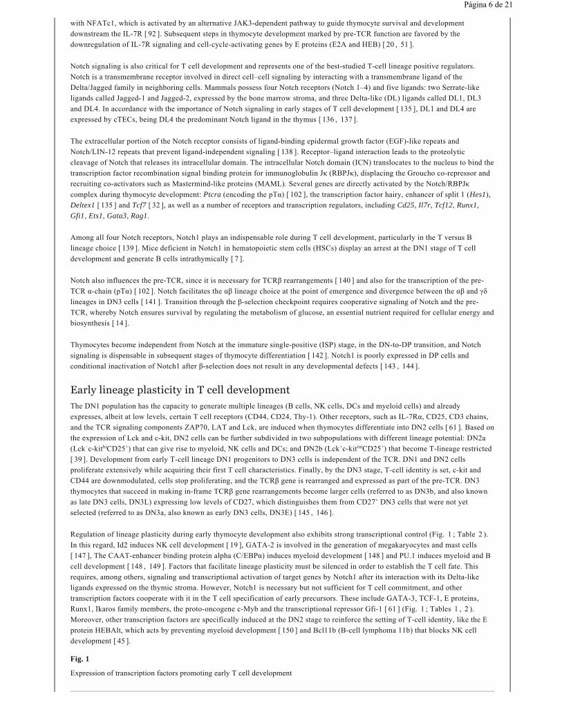
Regulation of lineage plasticity during early thymocyte development also exhibits strong transcriptional control (Fig. 1 ; Table 2 ).
In this regard, Id2 induces NK cell development [ 19 ], GATA-2 is involved in the generation of megakaryocytes and mast cells
[ 147 ], The CAAT-enhancer binding protein alpha (C/EBPα) induces myeloid development [ 148 ] and PU.1 induces myeloid and B
cell development [ 148 , 149 ]. Factors that facilitate lineage plasticity must be silenced in order to establish the T cell fate. This
requires, among others, signaling and transcriptional activation of target genes by Notch1 after its interaction with its Delta-like
ligands expressed on the thymic stroma. However, Notch1 is necessary but not sufficient for T cell commitment, and other
transcription factors cooperate with it in the T cell specification of early precursors. These include GATA-3, TCF-1, E proteins,
Runx1, Ikaros family members, the proto-oncogene c-Myb and the transcriptional repressor Gfi-1 [ 61 ] (Fig. 1 ; Tables 1 , 2 ).
Moreover, other transcription factors are specifically induced at the DN2 stage to reinforce the setting of T-cell identity, like the E
protein HEBAlt, which acts by preventing myeloid development [ 150 ] and Bcl11b (B-cell lymphoma 11b) that blocks NK cell
development [ 45 ].
Fig. 1
Expression of transcription factors promoting early T cell development
− hi + + int +
+
Página 6 de 21

Relevant events before pre-TCR expression Signaling pathways/proteins
Cell-cycle arrest E proteins, SPIB, p27 , p21 , RORγt
TCRβ rearrangements Rag recombinases, Notch, c-Myb
Survival Notch, IL-7R
Inhibition of non-T fatesDownregulation of PU.1, Id2, GATA-2, CEBPα
Upregulation of Bcl11b, HEBalt, Notch
Expression of RAG E proteins, GATA-3, c-Myb, Ikaros, C/EBPα, Egr
Table 2
.
Rearrangement of the TCRβ geneThe accomplishment of DNA rearrangements that generate functional TCR proteins is indispensable for the survival and
progression of T-cell precursors. The genomic locus of each TCR contains many copies of V, D, J segments whose combination
generates the great diversity of TCR specificities that can be found in one individual organism and is largely responsible for the
ability of the immune system to respond to different antigens [ 151 ].
The joining of rearranged V, D, and J segments is imprecise, and two-thirds of the fused DNA segments fail to maintain the
translational reading frame, generating nonfunctional TCR-β proteins [ 152 ]. As a result, almost half of the DN thymocytes
attempting to rearrange their TCR-β genes fail on both alleles. Accumulation of these dead-end cells is prevented by β-selection.
Therefore, implicit in the process of TCRβ V(D)J recombination is the existence of a developmental checkpoint, the so-called “β-
selection checkpoint” [ 145 ], to ensure that only those cells that have productively rearranged their TCRβ loci can undergo further
differentiation, whereas those that fail, suffer cell death [ 153 ].
V(D)J recombination for the TCRβ loci is initiated by the RAG-1 and RAG-2 endonucleases, which cleave gene segments
producing double-strand breaks at specific recognition sequences. Broken ends are then processed and joined by factors generally
involved in repair of non-homologous end joining DNA breaks [ 154 ]. Expression of Rag genes is induced by transcriptional
regulators including GATA-3, Ikaros, C/EBPα, HEB/E2A and c-Myb [ 101 ] (Table 2 ). Once DN3 cells receive a pre-TCR signal,
Rag genes are downregulated, due in part to the upregulation of Id3, which blocks HEB/E2A activity [ 56 ].
The assembly and expression of an antigen receptor chain from one allele inhibits further V(D)J recombination on the other allele.
This is termed “allelic exclusion”, and is defined as the surface expression of BCR or TCR chains from a single allelic copy, and is
maintained by feedback negative regulation [ 155 ]. Allelic exclusion determines that each T cell will only express one TCR
specificity, an important point for facilitating central tolerance of self-reactive lymphocytes and avoiding autoimmunity.
Nonetheless, a small percentage (1–3 %) of TCRβ loci escape allelic exclusion, which results in some lymphocytes exhibiting poly-
specific antigen recognition due to two different TCR being expressed in the same cell [ 155 ]. As discussed below, the mechanisms
that control allelic exclusion in the TCRβ locus are poorly characterized but some transcription factors are known to be involved.
Thymocyte β-selection through different pre-TCR-induced outcomesPre-TCR signaling has the ability to regulate diverse biological outcomes, each one of them appearing to be, for the most part,
functionally unrelated to the rest [ 77 , 156 – 158 ]. Signaling initiation and functional outcomes distinguish the pre-TCR from the
Kip1 waf1
Página 7 de 21
mature γδTCR and αβTCR [ 159 , 160 ]. The pre-TCR rescues cells from apoptosis, restarts the cell cycle, upregulates CD4 and CD8
expression, downregulates CD25 expression, ends TCRβ rearrangements by imposing allelic exclusion, and initiates TCRα
expression. These unique functions of the pre-TCR control the β-selection checkpoint in thymocyte development ensuring that only
thymocytes with productive in-frame TCRβ rearrangements will go into the processes of positive and negative selection, leading to
the TCR repertoire and defining antigen specificity.
The discovery of the pre-TCR was driven by the realization that the rearrangement of the gene encoding TCRβ takes place before
the TCRα gene is rearranged [ 11 , 161 ]. Indeed, TCR-β chains drive the differentiation of DN to DP thymocytes in the absence of
TCR-α chains [ 162 – 165 ]. The pre-TCR is a multisubunit complex that includes a recombined TCRβ chain bound to the invariant
pre-TCRα chain (pTα) and CD3 signaling molecules [ 12 , 13 ]. The pre-TCR is clustered in glycolipid-enriched microdomains of
the plasma membrane [ 159 ] and transduces weak signals that favor the commitment of the immature DN cells towards the αβ
lineage and not the γδ one [ 153 , 166 , 167 ]. The pre-TCR presents lower levels of surface expression than the mature TCR, and
this was proposed to be due, at least in part, to its nature of disulfide-linked heterodimeric proteins, which are very often degraded
in the endoplasmic reticulum [ 157 ]. Another explanation proposed for its low level of surface expression is the fact that its
constitutive signaling would induce the rapid internalization and degradation of the pre-TCR in lysosomes in a proteasome- and
ubiquitin-ligase-dependent manner [ 168 ].
Expression of the Ptcra gene encoding the pre-TCR-specific alpha chain is upregulated at the DN2 stage by the transcription
regulators c-Myb, E proteins (E2A-HEB heterodimers), and the Notch-dependent transcription factor RBPJκ [ 102 , 169 ] (Table 3 ).
The mRNA of Ptcra increases along the differentiation of DN cells, and is fully extinguished in DP cells after the rearrangement of
the TCRα gene and the membrane expression of a mature TCRαβ [ 170 ].
Pre-TCR complex molecules Transcription factors that induce pre-TCR (complex) gene expression
TCRβ promoters and enhancers Ets1, Runx1, Notch
pTa Notch, E proteins, c-Myb, Ikaros, Egr1
CD3 molecules GATA-3, TCF-1, Ikaros
Table 3
.
Signaling by the pre-TCR is ligand-independent and does not require recognition of MHC-presented antigens, in clear contrast to
the mature TCR [ 159 , 171 ]. The signaling ability of the pre-TCR is controlled by both its extracellular immunoglobulin-like
domain, which facilitates the oligomerization of the pre-TCR complex [ 172 ], and also its cytoplasmic tail, which is required for
downstream signal transduction [ 173 ]. Signals from the pre-TCR synergize with signals that activate the Notch pathway in DN3
thymocytes to induce the transition of DN thymocytes to DP cells [ 174 ]. In addition, the chemokine receptor CXCR4 was described
to associate with the pre-TCR acting as a costimulatory molecule to control survival during β-selection in a PI3K-dependent manner
[ 175 , 176 ]. The Wnt signaling pathway contributes to the expansion and differentiation of thymocytes beyond the β-selection
checkpoint. LEF1-deficient mice and mice expressing mutant forms of TCF-1 present an early arrest in T cell development [ 177 ,
178 ]. β-catenin plays a crucial role in activating these transcription factors as pre-TCR-defective DN cells overexpressing β-catenin
proceed to the DP stage of T cell development [ 36 ]. Pre-TCR signals stabilize β-catenin in DN thymocytes during β-selection
through Erk activity, which in turn mediates sustained expression of Egr genes [ 37 ].
Despite its lack of conservation between humans and mice, the cytoplasmic tail of the pre-TCR, and particularly a proline-rich motif
in the tail, is required for its function [ 173 , 179 ]. Targeting of proximal components of the pre-TCR signaling cascade have shown
that the SRC-family kinase p56 (Lck), ζ-associated protein of 70 kDa (ZAP70), linker for activating T cells (LAT), and SH2
domain-containing leukocyte-specific phosphoprotein of 76 kDa (SLP-76) are early signaling components engaged by the pre-TCR
[ 158 , 180 ]. Pre-TCR signals (Fig. 2 ) are transduced through the immunoreceptor tyrosine-based activation motifs (ITAMs)
located in the cytoplasmic tails of CD3 chains, which become phosphorylated by the Src-family protein kinases Lck and Fyn [ 181 ,
182 ]. ZAP-70 and Syk are then recruited to the CD3 tail of the pre-TCR complex [ 183 – 185 ] and activate adaptor proteins as LAT,
SLP-76 and Vav that initiate three signaling pathways through the activation of PI3K, PLCγ and Ras [ 77 , 156 , 157 ] (Fig. 2 ).
Apart from the signaling pathways activated by the pre-TCR, this complex also blocks death signals. These survival signals block
the DNA damage-responsive p53 pathway and function in a manner independent of the adaptor FADD (Fas-associated death domain
protein) [ 77 , 156 ].
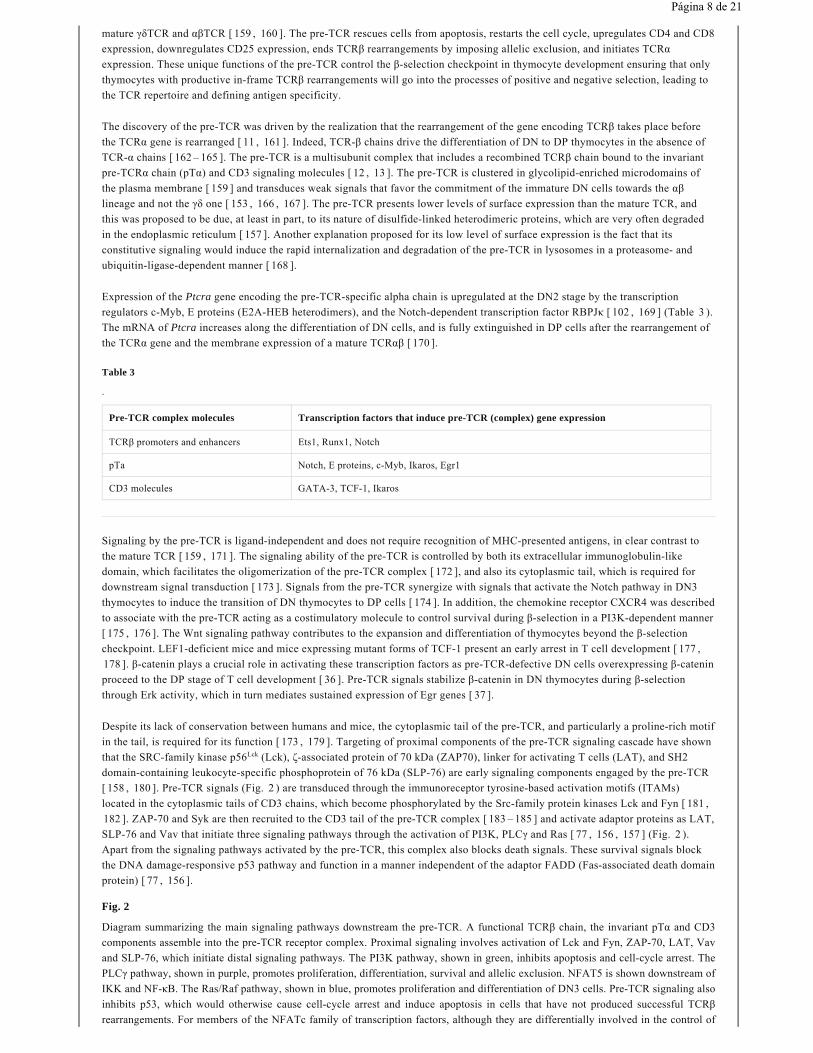
Fig. 2
Diagram summarizing the main signaling pathways downstream the pre-TCR. A functional TCRβ chain, the invariant pTα and CD3
components assemble into the pre-TCR receptor complex. Proximal signaling involves activation of Lck and Fyn, ZAP-70, LAT, Vav
and SLP-76, which initiate distal signaling pathways. The PI3K pathway, shown in green, inhibits apoptosis and cell-cycle arrest. The
PLCγ pathway, shown in purple, promotes proliferation, differentiation, survival and allelic exclusion. NFAT5 is shown downstream of
IKK and NF-κB. The Ras/Raf pathway, shown in blue, promotes proliferation and differentiation of DN3 cells. Pre-TCR signaling also
inhibits p53, which would otherwise cause cell-cycle arrest and induce apoptosis in cells that have not produced successful TCRβ
rearrangements. For members of the NFATc family of transcription factors, although they are differentially involved in the control of
Lck
Página 8 de 21

cellular proliferation and survival, this diagram reflects the reported function for particular NFATc proteins (NFATc1 and NFATc3)
facilitating pre-TCR-induced thymocyte differentiation
While the hierarchy of molecules that assemble the different signaling cascades of the pre-TCR is quite established, knowledge on
the target genes that respond to these inputs and the specific mechanisms that regulate their expression is more limited (Fig. 2 ;
Table 4 ). Completing the puzzle of transcription factors that lay beneath the different signaling branches of the pre-TCR and
learning which genes they regulate would provide a complete picture of the way this receptor controls the commitment of αβ-
lineage thymocytes. The next sections summarize what is known about different signaling branches of the pre-TCR and their
specialization in activating different transcription factors to drive its multiple functions. As discussed previously, transcription
factors known to be involved in pre-TCR expression or function can also work in other, pre-TCR-independent, roles in thymocyte
development or mature T lymphocytes.
Pre-TCR outcomes and signaling pathways
Proteins activated downstream
Survival
Pathways that cooperate or synergize with the pre-TCR: Notch, CXCR4, Wnt
Transcription factors downstream pre-TCR: NK-κB, ETS1, NFAT5
Prosurvival outcomes promoted by the pre-TCR: expression of anti-apoptotic A1, suppression of p53, inhibition of FOXO
Proliferation TCF-1, NF-κB, c-Myb, Id3, Egr3, Cyclin D3. Negative regulators of proliferation include ROR γt and E proteins
Allelic exclusion Ets1, E2A (E47), NFAT5
Differentiation NK-κB, NFATc (NFATc1 and NFATc3), Runx1, TCF-1, Egr
PI3K pathway Inhibits FOXO, Bim, p27
PLCg/DAG/PKC pathway NF-κB, NFAT5, A1
PLCg/IP3 pathway Calcineurin/NFATc pathway
Ras/MAPK pathway ETS1, Egr proteins, Id3, inhibition of RORγt
Table 4
.
Kip1
Página 9 de 21

PI3K pathway: activation of Akt-induced survival
Upon activation of phosphatidylinositol-4,5-bisphosphate 3-kinase (PI3K), various 3-phosphorylated phosphoinositides (PtdIns3P,
PtdIns(3,4)P2, PtdIns(3,5)P2, and PtdIns(3,4,5)P3) are produced in the membrane [ 186 ]. The different phosphoinositides allow the
recruitment to the cellular membrane of various signaling proteins containing phosphoinositide-binding domains, such as PX
domains, pleckstrin homology domains (PH domains), and FYVE domains. Protein kinases PDK1 (phosphoinositide-dependent
kinase-1) and AKT contain a PH domain specific for PtdIns(3,4,5)P3 and PtdIns(3,4)P2, which causes them to translocate to the
plasma membrane upon PI3K activation. Colocalization of activated PDK1 and AKT enables PDK1 to phosphorylate AKT, leading
to its partial activation. AKT activation is of central relevance in pre-TCR signaling since it maintains survival of DN3 cells through
phosphorylation-dependent inhibition of Forkhead box O (FOXO) transcription factors, which promote cell-cycle arrest and
apoptosis [ 187 , 188 ]. The increased susceptibility of AKT-deficient immature thymocytes to undergo apoptosis is probably due to
both dysregulation of the expression of Bcl-2 family members as Bim but also impaired glucose metabolism [ 131 , 187 ]. Notably,
during β-selection, Notch also acts via AKT promoting survival independently of pre-TCR signals [ 14 ]. Apart from AKT, PDK1
also phosphorylates the ribosomal subunit kinases S6K and RSK, which lack a PH domain and are required downstream of Notch
for optimal pre-T cell growth and proliferative expansion [ 186 , 189 ].
PLCγ pathway: activation of NFAT, NF-κB and the MAPK pathways to induce differentiation, survival and proliferation
Phosphorylation of phospholipase Cγ (PLCγ) activates it to yield diacylglycerol (DAG) and inositol 1,4,5-trisphosphate (IP3) from
the hydrolysis of PIP (PtdIns(4,5)P2). Different signaling pathways derive from these two molecules.
IP3 causes an efflux of cytosolic calcium (Ca ) from endoplasmic reticulum stores, and the elevated levels of intracellular Ca
result in calmodulin saturation and the direct activation of the phosphatase calcineurin [ 190 ]. Calcineurin is composed of a catalytic
A subunit and a regulatory B subunit. Activated calcineurin directly binds and dephosphorylates cytosolic NFATc transcription
factors, which then translocate into the nucleus to transactivate their target genes [ 78 , 79 ]. Mice lacking the major calcineurin
catalytic isoform expressed in lymphocytes, CnAβ, or removal of CnB1 function specifically in T cells, present a partial block at the
DN3 stage of thymocyte development [ 83 , 84 ]. Accordingly, it was reported that NFATc proteins promote differentiation of DN3
cells, since lack of either NFATc1 or c3 in thymocytes causes a partial block at the DN3 stage followed by reduced survival of DP
thymocytes [ 36 , 85 – 87 ]. In support of the notion that the calcineurin/NFATc pathway regulates pre-TCR-induced thymocyte
differentiation to DP cells, enforced expression of NFATc1 partially bypasses the need of pre-TCR signaling to induce thymocyte
passage through the β-selection checkpoint and revealed that Id3 is an NFATc target gene [ 88 , 89 ].
DAG activates different forms of the protein kinase C (PKC) family. Particularly relevant for T cells is PKCθ [ 191 , 192 ], which
activates the IKK complex and NF-κB in response to TCR signaling [ 72 ]. Indeed, PKCθ acts as a target of Notch3 signaling to
mediate pre-TCR-induced NF-κB activation [ 193 ], which is consistent with the described role of PKC regulating allelic exclusion
and differentiation downstream the pre-TCR [ 194 ]. As mentioned earlier, NF-κB is an essential regulator of different pre-TCR
functions as it promotes differentiation, intensive cellular expansion, and survival of thymocytes that transit from the β-selection
checkpoint to DP cells [ 74 – 76 ]. The prosurvival Bcl-2 family protein A1 is expressed in an NF-κB-dependent manner downstream
the pre-TCR. Interestingly, the conventional IKKβ/NF-κB pathway regulates the expression of the transcription factor NFAT5,
which in turn, not only binds the promoter region of the A1 gene (Bcl2a1) but also binds the Bcl2 promoter to facilitate their
expression in DN cells and support thymocyte survival [ 96 ]. Although the pre-TCR marks a window of thymocyte development
with reduced Bcl-2 expression, the NFAT5-regulated Bcl-2 expression during the transit of β-selected thymocytes to DP cells can
be interpreted as a means to support a threshold of activity for this prosurvival factor downstream the pre-TCR.
The RAS-mitogen-activated protein kinase (MAPK) pathway: proliferation, differentiation and allelic exclusion
The Ras-Raf-MEK1/2-Erk1/2 MAP kinase (MAPK) cascade, which is directly activated through SLP-76 and Grb2-SOS is a
relevant player downstream the pre-TCR [ 195 , 196 ] to fuel the proliferation and differentiation of β-selected DN3 thymocytes
[ 77 ]. Extracellular signal-regulated kinase (Erk) controls the activation of ETS1 and early growth response (Egr) transcription
factors [ 197 ]. ETS1 is required for thymocyte survival and allelic exclusion downstream of the pre-TCR [ 59 ]. The expression of
Egr family members Egr1, 2, and 3 correlates with pre-TCR expression and thymocyte development beyond the β-selection
checkpoint, and enforced expression of different Egr factors bypasses the block in development associated with defective pre-TCR
function [ 197 , 198 ]. However, different Egr family members may have somewhat distinct roles in promoting thymocyte
development since there are differences in genes that, such as Rag1, Rag2, Ptcra and Tcra, are modulated by enforced expression of
particular Egr factors [ 197 ]. Egr1 was shown to be upregulated in β-selected cells promoting their development to the immature
CD8 single-positive stage [ 199 ]. This transcription factor acts together with NFATc1 to induce development beyond the β-selection
checkpoint, as they synergistically induce expression of Id3 [ 88 , 200 , 201 ], a transcriptional repressor that antagonizes the activity
of E proteins E12, E47 and HEB and therefore promotes proliferation and progression to the DP stage [ 56 ]. Egr3-induced Id protein
expression also results in a transient loss of E protein-dependent RORγt expression and directly affects RORγt function blocking the
expression of anti-proliferative genes, such as cytoplasmic polyadenylation element binding protein 4 (mCPEB4) [ 202 ]. These
works lend support to the view that control of proliferation by pre-TCR signals is highly dependent on the activities of the
transcription factors RORγt, Egr3, E12, and E47. Regarding the activity of E47 downstream pre-TCR, although it is clear that it
must be antagonized to facilitate proliferation, it is also required to control TCRβ allelic exclusion [ 52 ].
2
2+ 2+
Página 10 de 21

Blockade of p53-dependent pro-apoptotic and anti-proliferative functions by the pre-TCR
The response of thymocytes to pre-TCR signaling includes gene rearrangements and proliferation, two processes that are
incompatible and must occur sequentially. V(D)J recombination requires exit from the cell cycle [ 203 ]. Small DN3a cells
undergoing TCRβ rearrangements are quiescent cells, and large DN3b cells represent thymocytes that enter cell cycle after the
rearrangements are completed and the pre-TCR is functional. Among other factors, cell-cycle arrest of DN3a cells is regulated by
the tumor suppressor p53. p53 induces p21 which, in addition to its function as a modulator of cell death and its oncogenic p53-
independent role in thymic lymphomas [ 204 ], acts together with the FOXO-regulated p27 to arrest the cell cycle [ 205 , 206 ]. Re-
entry into the cell cycle occurs in DN3b cells, promoted by functional pre-TCR signaling. Pre-TCR signaling has also been linked to
the inhibition of DNA damage-dependent p53-mediated apoptosis. In the absence of survival signals, an accumulation of p53
induces cell death by activating pro-apoptotic molecules such as Noxa, Puma and Bid, which in turn will activate Bax to translocate
to mitochondria and initiate the cascade of caspases that leads to cell death [ 131 ]. The apoptotic function of p53 is activated during
V(D)J recombination of the TCRβ locus (as it involves DNA double-strand breaks) and is suppressed if a correct TCRβ
rearrangement is produced [ 125 , 126 ]. Indeed, blockade of thymocytes at the DN3 stage observed in mice defective for pre-TCR
signaling is prevented when p53 is deleted [ 124 , 126 , 207 ]. Although it is known that the guanine nucleotide binding
protein/GTPase Rho suppresses p53-mediated apoptosis downstream of the pre-TCR [ 127 ], the signaling steps that link the pre-
TCR to inhibition of p53 are not fully elucidated. Recent evidence shows that the transcription factor Miz-1 induces the expression
of the gene encoding ribosomal protein L22 (RPL22), a negative regulator of p53 translation [ 208 ], which is consistent with the
role of Miz-1 and RPL22 as prosurvival molecules that antagonize p53 during the β-selection checkpoint of thymocytes [ 209 –
211 ].
Besides the regulators discussed in previous sections, there are some pre-TCR induced proteins that regulate thymocyte function,
but whose connection with signaling components of the pre-TCR complex is poorly characterized. For instance, cyclin D3 is
upregulated after β-selection to promote cell-cycle entry, and thymocytes lacking it show defective expansion and are more
susceptible to oncogenic transformation [ 212 ]. On the anti-proliferative side, the ETS transcription factor SPIB is highly expressed
in DN3 cells and restrains their proliferation, but its expression is turned off after β-selection [ 60 ].
Future perspectives
The pre-TCR controls the β-selection checkpoint in thymocyte development through a balanced combination of processes the
determine cell survival, death, proliferation, and cell-cycle arrest. Transcription plays a main role in these processes, and specific
transcription regulators coordinate their function to facilitate the transit of thymocytes through the β-selection checkpoint.
Despite knowledge accumulated during the last two decades on the identity of the transcription factors that sense the pre-TCR
signaling and induce its distinct functional outcomes, the identification of the specific genes they bind and induce is still limited.
Resolving gene expression profiles during pre-TCR-regulated thymocyte transitions, and the changes in chromatin architecture and
transcription factor dynamics involved, poses a major challenge due to the difficulty of isolating cell subsets or differentiation
stages represented in very small numbers. Fortunately, advances in single cell RNA sequencing and novel technologies, such as the
recently developed iChIP assay [ 213 , 214 ], a variation of the chromatin immunoprecipitation sequencing, are greatly facilitating
the analysis of gene regulation events in limited amounts of cells during development transitions. These techniques, in combination
with gene knockouts and genetic tagging for lineage tracking, will likely expand our understanding on how the pre-TCR converts
the activation of its different signaling branches into distinct gene expression programs.
Acknowledgments
Work in CL-R and JA laboratory has been supported by the Ramón y Cajal and I3 Researchers Programs (CL-R), research grants
from the Spanish Government (SAF2009-08066, SAF2012-36535 to CL-R; and BFU2008-01070, SAF2011-24268 to JA), Fundació
la Marató TV3 (080730, 122530 to CL-R and JA), the Marie Curie International Reintegration Program of the European Union
(MCIRG516308 to CL-R), the Spanish Ministry of Health (ISCIII-RETIC RD06/0009-FEDER), and Generalitat de Catalunya
(2009SGR601, 2014SGR1153). CL-R is a recipient of the ICREA Acadèmia Award (Generalitat de Catalunya).
References
1. Rossi SW, Jenkinson WE, Anderson G, Jenkinson EJ (2006) Clonal analysis reveals a common progenitor for thymic cortical
and medullary epithelium. Nature 441:988–991
2. Yang Q, Jeremiah Bell J, Bhandoola A (2010) T-cell lineage determination. Immunol Rev 238:12–22
3. Schwarz BA, Bhandoola A (2004) Circulating hematopoietic progenitors with T lineage potential. Nat Immunol 5:953–960
4. Serwold T, Ehrlich LIR, Weissman IL (2009) Reductive isolation from bone marrow and blood implicates common lymphoid
progenitors as the major source of thymopoiesis. Blood 113:807–815
5. Lind EF, Prockop SE, Porritt HE, Petrie HT (2001) Mapping precursor movement through the postnatal thymus reveals
specific microenvironments supporting defined stages of early lymphoid development. J Exp Med 194:127–134
waf1
kip
Página 11 de 21

6. Godfrey DI, Kennedy J, Suda T, Zlotnik A (1993) A developmental pathway involving four phenotypically and functionally
distinct subsets of CD3-CD4-CD8- triple-negative adult mouse thymocytes defined by CD44 and CD25 expression. J Immunol
150:4244–4252
7. Radtke F, Wilson A, Stark G et al (1999) Deficient T cell fate specification in mice with an induced inactivation of Notch1.
Immunity 10:547–558
8. Von Freeden-Jeffry U, Vieira P, Lucian LA et al (1995) Lymphopenia in interleukin (IL)-7 gene-deleted mice identifies IL-7
as a nonredundant cytokine. J Exp Med 181:1519–1526
9. Ceredig R, Rolink T (2002) A positive look at double-negative thymocytes. Nat Rev Immunol 2:888–897
10. Porritt HE, Rumfelt LL, Tabrizifard S et al (2004) Heterogeneity among DN1 prothymocytes reveals multiple progenitors
with different capacities to generate T cell and non-T cell lineages. Immunity 20:735–745
11. Raulet DH, Garman RD, Saito H, Tonegawa S (1985) Developmental regulation of T-cell receptor gene expression. Nature
314:103–107
12. Von Boehmer H, Fehling HJ (1997) Structure and function of the pre-T cell receptor. Annu Rev Immunol 15:433–452
13. Malissen B, Ardouin L, Lin SY et al (1999) Function of the CD3 subunits of the pre-TCR and TCR complexes during T cell
development. Adv Immunol 72:103–148
14. Ciofani M, Zuniga-Pflucker JC (2005) Notch promotes survival of pre-T cells at the beta-selection checkpoint by regulating
cellular metabolism. Nat Immunol 6:881–888
15. Yui MA, Rothenberg EV (2014) Developmental gene networks: a triathlon on the course to T cell identity. Nat Rev
Immunol 14:529–545
16. Rothenberg EV, Taghon T (2005) Molecular genetics of T cell development. Annu Rev Immunol 23:601–649
17. Ho IC, Tai TS, Pai SY (2009) GATA3 and the T-cell lineage: essential functions before and after T-helper-2-cell
differentiation. Nat Rev Immunol 9:125–135
18. Rothenberg EV (2014) Transcriptional control of early T and B cell developmental choices. Annu Rev Immunol 32:283–321
19. Ikawa T, Fujimoto S, Kawamoto H et al (2001) Commitment to natural killer cells requires the helix-loop-helix inhibitor
Id2. Proc Natl Acad Sci USA 98:5164–5169
20. Schwartz R, Engel I, Fallahi-Sichani M et al (2006) Gene expression patterns define novel roles for E47 in cell cycle
progression, cytokine-mediated signaling, and T lineage development. Proc Natl Acad Sci USA 103:9976–9981
21. Del Real MM, Rothenberg EV (2013) Architecture of a lymphomyeloid developmental switch controlled by PU.1, Notch
and Gata3. Development 140:1207–1219
22. Xu W, Carr T, Ramirez K et al (2013) E2A transcription factors limit expression of Gata3 to facilitate T lymphocyte lineage
commitment. Blood 121:1534–1542
23. Hosoya T, Maillard I, Engel JD (2010) From the cradle to the grave: activities of GATA-3 throughout T-cell development
and differentiation. Immunol Rev 238:110–125
24. Taghon T, Yui MA, Rothenberg EV (2007) Mast cell lineage diversion of T lineage precursors by the essential T cell
transcription factor GATA-3. Nat Immunol 8:845–855
25. García-Ojeda ME, Wolterink RGJK, Lemâitre F et al (2013) GATA-3 promotes T-cell specification by repressing B-cell
potential in pro-T cells in mice. Blood 121:1749–1759
26. Kishi H, Wei XC, Jin ZX et al (2000) Lineage-specific regulation of the murine RAG-2 promoter: GATA-3 in T cells and
Pax-5 in B cells. Blood 95:3845–3852
27. Wang L, Wildt KF, Zhu J et al (2008) Distinct functions for the transcription factors GATA-3 and ThPOK during
intrathymic differentiation of CD4(+) T cells. Nat Immunol 9:1122–1130
28. Staal FJ, Clevers HC (2005) WNT signalling and haematopoiesis: a WNT-WNT situation. Nat Rev Immunol 5:21–30
Página 12 de 21

29. Okamura RM, Sigvardsson M, Galceran J et al (1998) Redundant regulation of T cell differentiation and TCRalpha gene
expression by the transcription factors LEF-1 and TCF-1. Immunity 8:11–20
30. Hattori N, Kawamoto H, Fujimoto S et al (1996) Involvement of transcription factors TCF-1 and GATA-3 in the initiation
of the earliest step of T cell development in the thymus. J Exp Med 184:1137–1147
31. Germar K, Dose M, Konstantinou T et al (2011) T-cell factor 1 is a gatekeeper for T-cell specification in response to Notch
signaling. Proc Natl Acad Sci 108:20060–20065
32. Weber BN, Chi AW, Chavez A et al (2011) A critical role for TCF-1 in T-lineage specification and differentiation. Nature
476:63–68
33. Verbeek S, Izon D, Hofhuis F et al (1995) An HMG-box-containing T-cell factor required for thymocyte differentiation.
Nature 374:70–74
34. Schilham MW, Wilson A, Moerer P et al (1998) Critical involvement of Tcf-1 in expansion of thymocytes. J Immunol
161:3984–3991
35. Xu Y, Banerjee D, Huelsken J et al (2003) Deletion of beta-catenin impairs T cell development. Nat Immunol 4:1177–1182
36. Gounari F, Aifantis I, Khazaie K et al (2001) Somatic activation of beta-catenin bypasses pre-TCR signaling and TCR
selection in thymocyte development. Nat Immunol 2:863–869
37. Xu M, Sharma A, Wiest DL, Sen JM (2009) Pre-TCR-induced beta-catenin facilitates traversal through beta-selection. J
Immunol 182:751–758
38. Liu P, Li P, Burke S (2010) Critical roles of Bcl11b in T-cell development and maintenance of T-cell identity. Immunol Rev
238:138–149
39. Yui MA, Feng N, Rothenberg EV (2010) Fine-scale staging of T cell lineage commitment in adult mouse thymus. J
Immunol 185:284–293
40. Ikawa T, Hirose S, Masuda K et al (2010) An essential developmental checkpoint for production of the T cell lineage.
Science 329:93–96
41. Zhang S, Rozell M, Verma RK et al (2010) Antigen-specific clonal expansion and cytolytic effector function of CD8 T
lymphocytes depend on the transcription factor Bcl11b. J Exp Med 207:1687–1699
42. Vanvalkenburgh J, Albu DI, Bapanpally C et al (2011) Critical role of Bcl11b in suppressor function of T regulatory cells
and prevention of inflammatory bowel disease. J Exp Med 208:2069–2081
43. Wakabayashi Y, Watanabe H, Inoue J et al (2003) Bcl11b is required for differentiation and survival of alphabeta T
lymphocytes. Nat Immunol 4:533–539
44. Albu DI, Feng D, Bhattacharya D et al (2007) BCL11B is required for positive selection and survival of double-positive
thymocytes. J Exp Med 204:3003–3015
45. Li P, Burke S, Wang J et al (2010) Reprogramming of T cells to natural killer-like cells upon Bcl11b deletion. Science
329:85–89
46. Li L, Leid M, Rothenberg EV (2010) An early T cell lineage commitment checkpoint dependent on the transcription factor
Bcl11b. Science 329:89–93
47. Zhang JA, Mortazavi A, Williams BA et al (2012) Dynamic transformations of genome-wide epigenetic marking and
transcriptional control establish T cell identity. Cell 149:467–482
48. Mingueneau M, Kreslavsky T, Gray D et al (2013) The transcriptional landscape of αβ T cell differentiation. Nat Immunol
14:619–632
49. Yashiro-Ohtani Y, He Y, Ohtani T et al (2009) Pre-TCR signaling inactivates Notch1 transcription by antagonizing E2A.
Genes Dev 23:1665–1676
50. Engel I, Murre C (2004) E2A proteins enforce a proliferation checkpoint in developing thymocytes. EMBO J 23:202–211
51. Wojciechowski J, Lai A, Kondo M, Zhuang Y (2007) E2A and HEB are required to block thymocyte proliferation prior to
pre-TCR expression. J Immunol 178:5717–5726
+
Página 13 de 21

52. Agata Y, Tamaki N, Sakamoto S et al (2007) Regulation of T cell receptor β gene rearrangements and allelic exclusion by
the Helix-Loop-Helix protein, E47. Immunity 27:871–884
53. Jones ME, Zhuang Y (2009) Regulation of V(D)J recombination by E-protein transcription factors. Adv Exp Med Biol
650:148–156
54. Kee BL (2009) E and ID proteins branch out. Nat Rev Immunol 9:175–184
55. Kim D, Peng XC, Sun XH (1999) Massive apoptosis of thymocytes in T-cell-deficient Id1 transgenic mice. Mol Cell Biol
19:8240–8253
56. Engel I, Johns C, Bain G et al (2001) Early thymocyte development is regulated by modulation of E2A protein activity. J
Exp Med 194:733–745
57. Heemskerk MH, Blom B, Nolan G et al (1997) Inhibition of T cell and promotion of natural killer cell development by the
dominant negative helix loop helix factor Id3. J Exp Med 186:1597–1602
58. Anderson MK, Hernandez-Hoyos G, Diamond RA, Rothenberg EV (1999) Precise developmental regulation of Ets family
transcription factors during specification and commitment to the T cell lineage. Development 126:3131–3148
59. Eyquem S, Chemin K, Fasseu M, Bories JC (2004) The Ets-1 transcription factor is required for complete pre-T cell
receptor function and allelic exclusion at the T cell receptor beta locus. Proc Natl Acad Sci USA 101:15712–15717
60. Lefebvre JM, Haks MC, Carleton MO et al (2005) Enforced expression of Spi-B reverses T lineage commitment and blocks
beta-selection. J Immunol 174:6184–6194
61. Rothenberg EV, Moore JE, Yui MA (2008) Launching the T-cell-lineage developmental programme. Nat Rev Immunol 8:9–
21
62. Levanon D, Groner Y (2004) Structure and regulated expression of mammalian RUNX genes. Oncogene 23:4211–4219
63. Egawa T, Tillman RE, Naoe Y et al (2007) The role of the Runx transcription factors in thymocyte differentiation and in
homeostasis of naive T cells. J Exp Med 204:1945–1957
64. Talebian L, Li Z, Guo Y et al (2007) T-lymphoid, megakaryocyte, and granulocyte development are sensitive to decreases in
CBFβ dosage. Blood 109:11–21
65. Ichikawa M, Asai T, Saito T et al (2004) AML-1 is required for megakaryocytic maturation and lymphocytic differentiation,
but not for maintenance of hematopoietic stem cells in adult hematopoiesis. Nat Med 10:299–304
66. Growney JD, Shigematsu H, Li Z et al (2005) Loss of Runx1 perturbs adult hematopoiesis and is associated with a
myeloproliferative phenotype. Blood 106:494–504
67. Woolf E, Xiao C, Fainaru O et al (2003) Runx3 and Runx1 are required for CD8 T cell development during thymopoiesis.
Proc Natl Acad Sci USA 100:7731–7736
68. Merkenschlager M (2010) Ikaros in immune receptor signaling, lymphocyte differentiation, and function. FEBS Lett
584:4910–4914
69. Winandy S, Wu L, Wang JH, Georgopoulos K (1999) Pre-T cell receptor (TCR) and TCR-controlled checkpoints in T cell
differentiation are set by Ikaros. J Exp Med 190:1039–1048
70. Dose M, Khan I, Guo Z et al (2006) c-Myc mediates pre-TCR-induced proliferation but not developmental progression.
Blood 108:2669–2677
71. Douglas NC, Jacobs H, Bothwell AL, Hayday AC (2001) Defining the specific physiological requirements for c-Myc in T
cell development. Nat Immunol 2:307–315
72. Hayden MS, West AP, Ghosh S (2006) NF-kappaB and the immune response. Oncogene 25:6758–6780
73. Gerondakis S, Fulford TS, Messina NL, Grumont RJ (2014) NF-κB control of T cell development. Nat Immunol 15:15–25
74. Voll RE, Jimi E, Phillips RJ et al (2000) NF-kappa B activation by the pre-T cell receptor serves as a selective survival
signal in T lymphocyte development. Immunity 13:677–689
Página 14 de 21

76. Mandal M, Borowski C, Palomero T et al (2005) The BCL2A1 gene as a pre-T cell receptor-induced regulator of thymocyte
survival. J Exp Med 201:603–614
77. Aifantis I, Mandal M, Sawai K et al (2006) Regulation of T-cell progenitor survival and cell-cycle entry by the pre-T-cell
receptor. Immunol Rev 209:159–169
78. Kiani A, Rao A, Aramburu J (2000) Manipulating immune responses with immunosuppressive agents that target NFAT.
Immunity 12:359–372
79. Macián F (2005) NFAT proteins: key regulators of T-cell development and function. Nat Rev Immunol 5:472–484
80. Serfling E, Avots A, Klein-Hessling S et al (2012) NFATc1/αA: the other face of NFAT factors in lymphocytes. Cell
Commun Signal 10:16
81. Crabtree GR (1999) Generic signals and specific outcomes: signaling through Ca , calcineurin, and NF-AT. Cell 96:611–
614
82. Hogan PG, Chen L, Nardone J, Rao A (2003) Transcriptional regulation by calcium, calcineurin, and NFAT. Genes Dev
17:2205–2232
83. Bueno OF, Brandt EB, Rothenberg ME, Molkentin JD (2002) Defective T cell development and function in calcineurin A
beta -deficient mice. Proc Natl Acad Sci USA 99:9398–9403
84. Neilson JR, Winslow MM, Hur EM, Crabtree GR (2004) Calcineurin B1 is essential for positive but not negative selection
during thymocyte development. Immunity 20:255–266
85. Ranger AM, Hodge MR, Gravallese EM et al (1998) Delayed lymphoid repopulation with defects in IL-4-driven responses
produced by inactivation of NF-ATc. Immunity 8:125–134
86. Oukka M, Ho IC, de la Brousse FC et al (1998) The transcription factor NFAT4 is involved in the generation and survival of
T cells. Immunity 9:295–304
87. Yoshida H, Nishina H, Takimoto H et al (1998) The transcription factor NF-ATc1 regulates lymphocyte proliferation and
Th2 cytokine production. Immunity 8:115–124
88. Koltsova EK, Ciofani M, Benezra R et al (2007) Early growth response 1 and NF-ATc1 act in concert to promote thymocyte
development beyond the beta-selection checkpoint. J Immunol 179:4694–4703
89. Canté-Barrett K, Winslow MM, Crabtree GR (2007) Selective role of NFATc3 in positive selection of thymocytes. J
Immunol 179:103–110
90. Hayden-Martinez K, Kane LP, Hedrick SM (2000) Effects of a constitutively active form of calcineurin on T cell activation
and thymic selection. J Immunol 165:3713–3721
91. Amasaki Y, Masuda ES, Imamura R et al (1998) Distinct NFAT family proteins are involved in the nuclear NFAT-DNA
binding complexes from human thymocyte subsets. J Immunol 160:2324–2333
92. Patra AK, Avots A, Zahedi RP et al (2013) An alternative NFAT-activation pathway mediated by IL-7 is critical for early
thymocyte development. Nat Immunol 14:127–135
93. Lopez-Rodriguez C, Aramburu J, Rakeman AS, Rao A (1999) NFAT5, a constitutively nuclear NFAT protein that does not
cooperate with Fos and Jun. Proc Natl Acad Sci USA 96:7214–7219
94. Aramburu J, Drews-Elger K, Estrada-Gelonch A et al (2006) Regulation of the hypertonic stress response and other cellular
functions by the Rel-like transcription factor NFAT5. Biochem Pharmacol 72:1597–1604
95. Buxadé M, Lunazzi G, Minguillón J et al (2012) Gene expression induced by Toll-like receptors in macrophages requires
the transcription factor NFAT5. J Exp Med 209:379–393
96. Berga-Bolaños R, Alberdi M, Buxadé M et al (2013) NFAT5 induction by the pre-T-cell receptor serves as a selective
survival signal in T-lymphocyte development. Proc Natl Acad Sci USA 110:16091–16096
2+
2+
Página 15 de 21
75. Aifantis I, Gounari F, Scorrano L et al (2001) Constitutive pre-TCR signaling promotes differentiation through Ca mobilization and activation of NF-κB and NFAT. Nat Immunol 2:403–409

97. Villey I, De Chasseval R, De Villartay JP (1999) RORγT, a thymus-specific isoform of the orphan nuclear receptor
RORγ/TOR, is up-regulated by signaling through the pre-T cell receptor and binds to the TEA promoter. Eur J Immunol 29:4072
–4080
98. Sun Z, Unutmaz D, Zou YR et al (2000) Requirement for RORgamma in thymocyte survival and lymphoid organ
development. Science 288:2369–2373
99. Lieu YK, Kumar A, Pajerowski AG et al (2004) Requirement of c-myb in T cell development and in mature T cell function.
Proc Natl Acad Sci USA 101:14853–14858
100. Bender TP, Kremer CS, Kraus M et al (2004) Critical functions for c-Myb at three checkpoints during thymocyte
development. Nat Immunol 5:721–729
101. Anderson MK (2006) At the crossroads: diverse roles of early thymocyte transcriptional regulators. Immunol Rev 209:191
–211
102. Reizis B, Leder P (2002) Direct induction of T lymphocyte-specific gene expression by the mammalian Notch signaling
pathway. Genes Dev 16:295–300
103. Goldrath AW, Bevan MJ (1999) Selecting and maintaining a diverse T-cell repertoire. Nature 402:255–262
104. Starr TK, Jameson SC, Hogquist KA (2003) Positive and negative selection of T cells. Annu Rev Immunol 21:139–176
105. Zhang N, Hartig H, Dzhagalov I et al (2005) The role of apoptosis in the development and function of T lymphocytes. Cell
Res 15:749–769
106. Chao DT, Korsmeyer SJ (1998) BCL-2 family: regulators of cell death. Annu Rev Immunol 16:395–419
107. Vander Heiden MG, Thompson CB (1999) Bcl-2 proteins: regulators of apoptosis or of mitochondrial homeostasis? Nat
Cell Biol 1:E209–E216
108. Gross A, McDonnell JM, Korsmeyer SJ (1999) BCL-2 family members and the mitochondria in apoptosis. Genes Dev
13:1899–1911
109. Akashi K, Kondo M, von Freeden-Jeffry U et al (1997) Bcl-2 rescues T lymphopoiesis in interleukin-7 receptor-deficient
mice. Cell 89:1033–1041
110. Maraskovsky E, O’Reilly LA, Teepe M et al (1997) Bcl-2 can rescue T lymphocyte development in interleukin-7 receptor-
deficient mice but not in mutant rag-1 mice. Cell 89:1011–1019
111. Opferman JT, Letai A, Beard C et al (2003) Development and maintenance of B and T lymphocytes requires antiapoptotic
MCL-1. Nature 426:671–676
112. Grillot DA, Merino R, Nunez G (1995) Bcl-XL displays restricted distribution during T cell development and inhibits
multiple forms of apoptosis but not clonal deletion in transgenic mice. J Exp Med 182:1973–1983
113. Ma A, Pena JC, Chang B et al (1995) Bclx regulates the survival of double-positive thymocytes. Proc Natl Acad Sci USA
92:4763–4767
114. Motoyama N, Wang F, Roth KA et al (1995) Massive cell death of immature hematopoietic cells and neurons in Bcl-x-
deficient mice. Science 267:1506–1510
115. Rathmell JC, Lindsten T, Zong W-X et al (2002) Deficiency in Bak and Bax perturbs thymic selection and lymphoid
homeostasis. Nat Immunol 3:932–939
116. Lindsten T, Ross AJ, King A et al (2000) The combined functions of proapoptotic Bcl-2 family members Bak and Bax are
essential for normal development of multiple tissues. Mol Cell 6:1389–1399
117. Wei MC, Zong WX, Cheng EH et al (2001) Proapoptotic BAX and BAK: a requisite gateway to mitochondrial dysfunction
and death. Science 292:727–730
118. Yücel R, Karsunky H, Klein-Hitpass L, Möröy T (2003) The transcriptional repressor Gfi1 affects development of early,
uncommitted c-Kit + T cell progenitors and CD4/CD8 lineage decision in the thymus. J Exp Med 197:831–844
119. Phelan JD, Saba I, Zeng H et al (2013) Growth factor independent-1 maintains Notch1-dependent transcriptional
programming of lymphoid precursors. PLoS Genet 9:e1003713
−/−
Página 16 de 21

120. Grimes HL, Gilks CB, Chan TO et al (1996) The Gfi-1 protooncoprotein represses Bax expression and inhibits T-cell
death. Proc Natl Acad Sci USA 93:14569–14573
121. Schmidt T, Karsunky H, Rödel B et al (1998) Evidence implicating Gfi-1 and Pim-1 in pre-T-cell differentiation steps
associated with β-selection. EMBO J 17:5349–5359
122. Huang DC, Strasser A (2000) BH3-Only proteins-essential initiators of apoptotic cell death. Cell 103:839–842
123. Marsden VS, Strasser A (2003) Control of apoptosis in the immune system: Bcl-2, BH3-only proteins and more. Annu Rev
Immunol 21:71–105
124. Haks MC, Krimpenfort P, van den Brakel JH, Kruisbeek AM (1999) Pre-TCR signaling and inactivation of p53 induces
crucial cell survival pathways in pre-T cells. Immunity 11:91–101
125. Bogue MA, Zhu C, Aguilar-Cordova E et al (1996) p53 is required for both radiation-induced differentiation and rescue of
V(D)J rearrangement in scid mouse thymocytes. Genes Dev 10:553–565
126. Guidos CJ, Williams CJ, Grandal I et al (1996) V(D)J recombination activates a p53-dependent DNA damage checkpoint
in scid lymphocyte precursors. Genes Dev 10:2038–2054
127. Costello PS, Cleverley SC, Galandrini R et al (2000) The GTPase rho controls a p53-dependent survival checkpoint during
thymopoiesis. J Exp Med 192:77–85
128. Oda E, Ohki R, Murasawa H et al (2000) Noxa, a BH3-only member of the Bcl-2 family and candidate mediator of p53-
induced apoptosis. Science 288:1053–1058
129. Nakano K, Vousden KH (2001) PUMA, a novel proapoptotic gene, is induced by p53. Mol Cell 7:683–694
130. Yu J, Zhang L, Hwang PM et al (2001) PUMA induces the rapid apoptosis of colorectal cancer cells. Mol Cell 7:673–682
131. Mandal M, Crusio KM, Meng F et al (2008) Regulation of lymphocyte progenitor survival by the proapoptotic activities of
Bim and Bid. Proc Natl Acad Sci USA 105:20840–20845
132. Peschon JJ, Morrissey PJ, Grabstein KH et al (1994) Early lymphocyte expansion is severely impaired in interleukin 7
receptor-deficient mice. J Exp Med 180:1955–1960
133. Hare KJ, Jenkinson EJ, Anderson G (2000) An essential role for the IL-7 receptor during intrathymic expansion of the
positively selected neonatal T cell repertoire. J Immunol 165:2410–2414
134. Pallard C, Stegmann AP, van Kleffens T et al (1999) Distinct roles of the phosphatidylinositol 3-kinase and STAT5
pathways in IL-7-mediated development of human thymocyte precursors. Immunity 10:525–535
135. Maillard I, Adler SH, Pear WS (2003) Notch and the immune system. Immunity 19:781–791
136. Koch U, Fiorini E, Benedito R et al (2008) Delta-like 4 is the essential, nonredundant ligand for Notch1 during thymic T
cell lineage commitment. J Exp Med 205:2515–2523
137. Thompson PK, Zúñiga-Pflücker JC (2011) On becoming a T cell, a convergence of factors kick it up a Notch along the
way. Semin Immunol 23:350–359
138. Kopan R, Ilagan MX (2009) The canonical Notch signaling pathway: unfolding the activation mechanism. Cell 137:216–
233
139. Pui JC, Allman D, Xu L et al (1999) Notch1 expression in early lymphopoiesis influences B versus T lineage
determination. Immunity 11:299–308
140. Wolfer A, Wilson A, Nemir M et al (2002) Inactivation of Notch1 impairs VDJbeta rearrangement and allows pre-TCR-
independent survival of early alpha beta Lineage Thymocytes. Immunity 16:869–879
141. Ciofani M, Knowles GC, Wiest DL et al (2006) Stage-specific and differential Notch dependency at the αβ and γδ T
lineage bifurcation. Immunity 25:105–116
142. Xiong J, Armato MA, Yankee TM (2011) Immature single-positive CD8 thymocytes represent the transition from Notch-
dependent to Notch-independent T-cell development. Int Immunol 23:55–64
143. Laky K, Fowlkes BJ (2008) Notch signaling in CD4 and CD8 T cell development. Curr Opin Immunol 20:197–202
+
Página 17 de 21

145. Dudley EC, Petrie HT, Shah LM et al (1994) T cell receptor beta chain gene rearrangement and selection during thymocyte
development in adult mice. Immunity 1:83–93
146. Taghon T, Yui MA, Pant R et al (2006) Developmental and molecular characterization of emerging beta- and gammadelta-
selected pre-T cells in the adult mouse thymus. Immunity 24:53–64
147. Tsai FY, Orkin SH (1997) Transcription factor GATA-2 is required for proliferation/survival of early hematopoietic cells
and mast cell formation, but not for erythroid and myeloid terminal differentiation. Blood 89:3636–3643
148. Laiosa CV, Stadtfeld M, Xie H et al (2006) Reprogramming of committed T cell progenitors to macrophages and dendritic
cells by C/EBP alpha and PU.1 transcription factors. Immunity 25:731–744
149. DeKoter RP, Singh H (2000) Regulation of B lymphocyte and macrophage development by graded expression of PU.1.
Science 288:1439–1441
150. Braunstein M, Rajkumar P, Claus CL et al (2010) HEBAlt enhances the T-cell potential of fetal myeloid-biased precursors.
Int Immunol 22:963–972
151. Grawunder U, West RB, Lieber MR (1998) Antigen receptor gene rearrangement. Curr Opin Immunol 10:172–180
152. Mallick CA, Dudley EC, Viney JL et al (1993) Rearrangement and diversity of T cell receptor beta chain genes in
thymocytes: a critical role for the beta chain in development. Cell 73:513–519
153. Michie AM, Zuniga-Pflucker JC (2002) Regulation of thymocyte differentiation: pre-TCR signals and beta-selection.
Semin Immunol 14:311–323
154. Gellert M (2002) V(D)J recombination: RAG proteins, repair factors, and regulation. Annu Rev Biochem 71:101–132
155. Brady BL, Steinel NC, Bassing CH (2010) Antigen receptor allelic exclusion: an update and reappraisal. J Immunol
185:3801–3808
156. Kruisbeek AM, Haks MC, Carleton M et al (2000) Branching out to gain control: how the pre-TCR is linked to multiple
functions. Immunol Today 21:637–644
157. Von Boehmer H (2005) Unique features of the pre-T-cell receptor alpha-chain: not just a surrogate. Nat Rev Immunol
5:571–577
158. Von Boehmer H, Aifantis I, Azogui O et al (1999) The impact of pre-T-cell receptor signals on gene expression in
developing T cells. Cold Spring Harb Symp Quant Biol 64:283–289
159. Saint-Ruf C, Panigada M, Azogui O et al (2000) Different initiation of pre-TCR and gammadeltaTCR signalling. Nature
406:524–527
160. Aifantis I, Azogui O, Feinberg J et al (1998) On the role of the pre-T cell receptor in alphabeta versus gammadelta T
lineage commitment. Immunity 9:649–655
161. Snodgrass HR, Dembić Z, Steinmetz M, von Boehmer H (1985) Expression of T-cell antigen receptor genes during fetal
development in the thymus. Nature 315:232–233
162. Von Boehmer H (1990) Developmental biology of T cells in T cell-receptor transgenic mice. Annu Rev Immunol 8:531–
556
163. Kishi H, Borgulya P, Scott B et al (1991) Surface expression of the beta T cell receptor (TCR) chain in the absence of other
TCR or CD3 proteins on immature T cells. EMBO J 10:93–100
164. Shinkai Y, Koyasu S, Nakayama K et al (1993) Restoration of T cell development in RAG-2-deficient mice by functional
TCR transgenes. Science 259:822–825
165. Mombaerts P, Clarke AR, Rudnicki MA et al (1992) Mutations in T-cell antigen receptor genes alpha and beta block
thymocyte development at different stages. Nature 360:225–231
166. Haks MC, Lefebvre JM, Lauritsen JPH et al (2005) Attenuation of gammadeltaTCR signaling efficiently diverts
thymocytes to the alphabeta lineage. Immunity 22:595–606
Página 18 de 21
144. Yuan JS, Kousis PC, Suliman S et al (2010) Functions of notch signaling in the immune system: consensus and controversies. Annu Rev Immunol 28:343–365

168. Panigada M, Porcellini S, Barbier E et al (2002) Constitutive endocytosis and degradation of the pre-T cell receptor. J Exp
Med 195:1585–1597
169. Tremblay M, Herblot S, Lecuyer E, Hoang T (2003) Regulation of pT alpha gene expression by a dosage of E2A, HEB,
and SCL. J Biol Chem 278:12680–12687
170. Trop S, Rhodes M, Wiest DL et al (2000) Competitive displacement of pT alpha by TCR-alpha during TCR assembly
prevents surface coexpression of pre-TCR and alpha beta TCR. J Immunol 165:5566–5572
171. Yamasaki S, Saito T (2007) Molecular basis for pre-TCR-mediated autonomous signaling. Trends Immunol 28:39–43
172. Yamasaki S, Ishikawa E, Sakuma M et al (2006) Mechanistic basis of pre-T cell receptor-mediated autonomous signaling
critical for thymocyte development. Nat Immunol 7:67–75
173. Aifantis I, Borowski C, Gounari F et al (2002) A critical role for the cytoplasmic tail of pTalpha in T lymphocyte
development. Nat Immunol 3:483–488
174. Ciofani M, Schmitt TM, Ciofani A et al (2004) Obligatory role for cooperative signaling by pre-TCR and Notch during
thymocyte differentiation. J Immunol 172:5230–5239
175. Trampont PC, Tosello-Trampont AC, Shen Y et al (2010) CXCR4 acts as a costimulator during thymic beta-selection. Nat
Immunol 11:162–170
176. Janas ML, Varano G, Gudmundsson K et al (2010) Thymic development beyond beta-selection requires
phosphatidylinositol 3-kinase activation by CXCR4. J Exp Med 207:247–261
177. Staal FJ, Clevers HC (2003) Wnt signaling in the thymus. Curr Opin Immunol 15:204–208
178. Goux D, Coudert JD, Maurice D et al (2005) Cooperating pre-T-cell receptor and TCF-1-dependent signals ensure
thymocyte survival. Blood 106:1726–1733
179. Borowski C, Li X, Aifantis I et al (2004) Pre-TCRalpha and TCRalpha are not interchangeable partners of TCRbeta during
T lymphocyte development. J Exp Med 199:607–615
180. Von Boehmer H, Aifantis I, Feinberg J et al (1999) Pleiotropic changes controlled by the pre-T-cell receptor. Curr Opin
Immunol 11:135–142
181. Groves T, Smiley P, Cooke MP et al (1996) Fyn can partially substitute for Lck in T lymphocyte development. Immunity
5:417–428
182. Palacios EH, Weiss A (2004) Function of the Src-family kinases, Lck and Fyn, in T-cell development and activation.
Oncogene 23:7990–8000
183. Cheng AM, Negishi I, Anderson SJ et al (1997) The Syk and ZAP-70 SH2-containing tyrosine kinases are implicated in
pre-T cell receptor signaling. Proc Natl Acad Sci USA 94:9797–9801
184. Pivniouk V, Tsitsikov E, Swinton P et al (1998) Impaired viability and profound block in thymocyte development in mice
lacking the adaptor protein SLP-76. Cell 94:229–238
185. Palacios EH, Weiss A (2007) Distinct roles for Syk and ZAP-70 during early thymocyte development. J Exp Med 204:1703
–1715
186. Fayard E, Moncayo G, Hemmings BA, Hollander GA (2010) Phosphatidylinositol 3-kinase signaling in thymocytes: the
need for stringent control. Sci Signal 3:5
187. Juntilla MM, Wofford JA, Birnbaum MJ et al (2007) Akt1 and Akt2 are required for alphabeta thymocyte survival and
differentiation. Proc Natl Acad Sci USA 104:12105–12110
188. Brunet A, Bonni A, Zigmond MJ et al (1999) Akt promotes cell survival by phosphorylating and inhibiting a Forkhead
transcription factor. Cell 96:857–868
189. Kelly AP, Finlay DK, Hinton HJ et al (2007) Notch-induced T cell development requires phosphoinositide-dependent
kinase 1. EMBO J 26:3441–3450
Página 19 de 21
167. Hayes SM, Li L, Love PE (2005) TCR signal strength influences αβ/γδ lineage fate. Immunity 22:583–593

191. Tan S-L, Parker PJ (2003) Emerging and diverse roles of protein kinase C in immune cell signalling. Biochem J 376:545–
552
192. Hayashi K, Altman A (2007) Protein kinase C theta (PKCtheta): a key player in T cell life and death. Pharmacol Res
55:537–544
193. Felli MP, Vacca A, Calce A et al (2005) PKC theta mediates pre-TCR signaling and contributes to Notch3-induced T-cell
leukemia. Oncogene 24:992–1000
194. Michie AM, Soh JW, Hawley RG et al (2001) Allelic exclusion and differentiation by protein kinase C-mediated signals in
immature thymocytes. Proc Natl Acad Sci USA 98:609–614
195. Rincon M, Flavell RA, Davis RJ (2001) Signal transduction by MAP kinases in T lymphocytes. Oncogene 20:2490–2497
196. Iritani BM, Alberola-Lla J, Forbush KA, Perlmutter RM (1999) Distinct signals mediate maturation and allelic exclusion in
lymphocyte progenitors. Immunity 10:713–722
197. Carleton M, Haks MC, Smeele SA et al (2002) Early growth response transcription factors are required for development of
CD4(−)CD8(−) thymocytes to the CD4(+)CD8(+) stage. J Immunol 168:1649–1658
198. Xi H, Kersh GJ (2004) Sustained early growth response gene 3 expression inhibits the survival of CD4/CD8 double-
positive thymocytes. J Immunol 173:340–348
199. Miyazaki T (1997) Two distinct steps during thymocyte maturation from CD4− CD8− to CD4+ CD8+ distinguished in the
early growth response (Egr)-1 transgenic mice with a recombinase-activating gene-deficient background. J Exp Med 186:877–
885
200. Bain G, Cravatt CB, Loomans C et al (2001) Regulation of the helix-loop-helix proteins, E2A and Id3, by the Ras-ERK
MAPK cascade. Nat Immunol 2:165–171
201. Engel I, Murre C (2002) Disruption of pre-TCR expression accelerates lymphomagenesis in E2A-deficient mice. Proc Natl
Acad Sci USA 99:11322–11327
202. Xi H, Schwartz R, Engel I et al (2006) Interplay between RORgammat, Egr3, and E proteins controls proliferation in
response to pre-TCR signals. Immunity 24:813–826
203. Lin WC, Desiderio S (1994) Cell cycle regulation of V(D)J recombination-activating protein RAG-2. Proc Natl Acad Sci
USA 91:2733–2737
204. De la Cueva E, García-Cao I, Herranz M et al (2006) Tumorigenic activity of p21Waf1/Cip1 in thymic lymphoma.
Oncogene 25:4128–4132
205. El-Deiry WS, Tokino T, Velculescu VE et al (1993) WAF1, a potential mediator of p53 tumor suppression. Cell 75:817–
825
206. Stahl M, Dijkers PF, Kops GJ et al (2002) The forkhead transcription factor FoxO regulates transcription of p27Kip1 and
Bim in response to IL-2. J Immunol 168:5024–5031
207. Mombaerts P, Terhorst C, Jacks T et al (1995) Characterization of immature thymocyte lines derived from T-cell receptor
or recombination activating gene 1 and p53 double mutant mice. Proc Natl Acad Sci USA 92:7420–7424
208. Rashkovan M, Vadnais C, Ross J et al (2014) Miz-1 regulates translation of Trp53 via ribosomal protein L22 in cells
undergoing V(D)J recombination. Proc Natl Acad Sci USA 111:E5411–E5419
209. Anderson SJ, Lauritsen JPH, Hartman MG et al (2007) Ablation of ribosomal protein L22 selectively impairs alphabeta T
cell development by activation of a p53-dependent checkpoint. Immunity 26:759–772
210. Stadanlick JE, Zhang Z, Lee S-Y et al (2011) Developmental arrest of T cells in Rpl22-deficient mice is dependent upon
multiple p53 effectors. J Immunol 187:664–675
211. Saba I, Kosan C, Vassen L et al (2011) Miz-1 is required to coordinate the expression of TCRbeta and p53 effector genes
at the pre-TCR “beta-selection” checkpoint. J Immunol 187:2982–2992
Página 20 de 21
190. Aramburu J, Rao A, Klee CB (2000) Calcineurin: from structure to function. Curr Top Cell Regul 36:237–295

212. Sicinska E, Aifantis I, Le Cam L et al (2003) Requirement for cyclin D3 in lymphocyte development and T cell leukemias.
Cancer Cell 4:451–461
213. Lara-Astiaso D, Weiner A, Lorenzo-Vivas E et al (2014) Chromatin state dynamics during blood formation. Science
345:943–949
214. Buenrostro JD, Giresi PG, Zaba LC et al (2013) Transposition of native chromatin for fast and sensitive epigenomic
profiling of open chromatin, DNA-binding proteins and nucleosome position. Nat Methods 10:1213–1218
Página 21 de 21