Pituitary-Specific Transcription Factor (Pit-1) Binding Site in the ...
Upload
nguyenthienCategory
view
246download
3
Subtypes of associated protein–DNA (TranscriptionFactor-Transcription Factor Binding Site) patternsTak-Ming Chan1,*, Kwong-Sak Leung1, Kin-Hong Lee1, Man-Hon Wong1,
Terrence Chi-Kong Lau2,* and Stephen Kwok-Wing Tsui3,4
1Department of Computer Science & Engineering, The Chinese University of Hong Kong, Shatin, N. T.,2Department of Biology and Chemistry, The City University of Hong Kong, Kowloon, 3School of BiomedicalSciences, The Chinese University of Hong Kong, Shatin, N. T. and 4Hong Kong Bioinformatics Centre, Shatin,N. T., Hong Kong
Received January 3, 2012; Revised July 6, 2012; Accepted July 16, 2012
ABSTRACT
In protein–DNA interactions, particularly transcrip-tion factor (TF) and transcription factor bindingsite (TFBS) bindings, associated residue variationsform patterns denoted as subtypes. Subtypes maylead to changed binding preferences, distinguishconserved from flexible binding residues and revealnovel binding mechanisms. However, subtypes mustbe studied in the context of core bindings. Whilesolving 3D structures would require huge experi-mental efforts, recent sequence-based associatedTF-TFBS pattern discovery has shown to bepromising, upon which a large-scale subtype studyis possible and desirable. In this article, we investi-gate residue-varying subtypes based on associatedTF-TFBS patterns. By re-categorizing the patternswith respect to varying TF amino acids, statisticallysignificant (P values� 0.005) subtypes leading tovarying TFBS patterns are discovered withoutusing TF family or domain annotations. Resultantsubtypes have various biological meanings. Thesubtypes reflect familial and functional propertiesand exhibit changed binding preferences supportedby 3D structures. Conserved residues critical formaintaining TF-TFBS bindings are revealed byanalyzing the subtypes. In-depth analysis on thesubtype pair PKVVIL-CACGTG versus PKVEIL-CAGCTG shows the V/E variation is indicative for dis-tinguishing Myc from MRF families. Discovered fromsequences only, the TF-TFBS subtypes are inform-ative and promising for more biological findings,complementing and extending recent one-sidedsubtype and familial studies with comprehensiveevidence.
INTRODUCTION
Protein–DNA interactions play a central role in geneticactivities (1,2). In particular, transcription factor (TF,the protein side) and transcription factor binding site(TFBS, the DNA side) bindings are critical and primaryprotein–DNA interactions to be deciphered for gene regu-lation. So TF-TFBS bindings will be our focus throughoutthe article. Despite the great variations shown among dif-ferent whole-length TF and TFBS sequences, part of themare conserved as TF binding domains (tens to hundreds ofresidues) and TFBS motifs (usually several to 20 residues),respectively. Within the distance of forming hydrogenbonds, short TF and TFBS subsequences show moreconserved patterns. These associated short binding subse-quences (within 10 residues; 6–8 in our experiments) ofboth TFs and TFBSs surrounding the interacting bondsare denoted as binding cores. However, predicting theseshort binding cores on both the TF and TFBS sides fromsequences only is very challenging.
Amino acid residue variations in the TF binding coresmay lead to intriguing different corresponding TFBSsub-patterns. For example, [A/P]KV[E/V]IL-CA[C/G][C/G]TG may be found to be TF-TFBS binding cores.Specifically, PKVEIL may bind to CAG[C/G]TG,whereas PKVVIL to CACGTG. We denote suchassociated TF-TFBS residue variations as subtypes,which should be studied in the context of associatedTF-TFBS core bindings. ‘PKVEIL-CAG[C/G]TG versusPKVVIL-CACGTG (3rd column)’ and ‘PKVEIL-CAG[C/G]TG versus PKVVIL-CACGTG (4th column)’are two related (column-specific) subtype pairs. Subtypescan reflect familial specificities, exhibit changed bindingpreferences, distinguish conserved residues from flexibleones and reveal novel binding mechanisms. Althoughsuch high-resolution details are usually extracted from3D structures with huge experimental efforts, abundantlow-resolution binding sequence data can be exploited to
*To whom correspondence should be addressed. Tel: +852 39434259; Fax: +852 39435024; Email: [email protected] may also be addressed to Terrence Chi-Kong Lau. Tel: +852 34429327; Fax: +852 34420522; Email: [email protected]
9392–9403 Nucleic Acids Research, 2012, Vol. 40, No. 19 Published online 16 August 2012doi:10.1093/nar/gks749
� The Author(s) 2012. Published by Oxford University Press.This is an Open Access article distributed under the terms of the Creative Commons Attribution Non-Commercial License (http://creativecommons.org/licenses/by-nc/3.0), which permits unrestricted non-commercial use, distribution, and reproduction in any medium, provided the original work is properly cited.
Downloaded from https://academic.oup.com/nar/article-abstract/40/19/9392/2414893by gueston 12 February 2018
predict both testable TF-TFBS binding cores andsubtypes.
In this article, we for the first time introduce and studyresidue varying subtypes based on our sequence-basedassociated TF-TFBS pattern discovery (3). The briefreview is first given in the following sub-sections.TF-TFBS subtype discovery methods are detailed in‘Materials and Methods’ section. Experimental resultsand verifications are reported in ‘Results and Analysis’section, before the final ‘Discussion and Conclusion’section.
TF-TFBS bindings in gene regulation
Because of functional importance of TF-TFBS bindingson regulation, core interaction subsequences from boundTFs and TFBSs are less likely to mutate and exhibit rec-ognizable patterns (i.e. being conserved) from similarTF-TFBS bindings. The short conserved subsequencepatterns on either TF or TFBS side are called motifs.TFs have relatively long conserved regions calleddomains of up to hundreds of amino acids (AA), but thecore interaction subsequences interacting with TFBSs areshown to be highly specific (3,4). TFBS motifs are usuallyshort [within 10 base pairs (bp)], and long motifs (up to 20bp) are usually composites of short patterns separated bynon-conserved gaps.
Existing data
Experiments to determine TF-TFBS bindings at thesequence level include the traditional DNA footprinting,gel electrophoresis and the new chromatin immunopre-cipitation (ChIP) followed by chip (-chip) or sequencing(-seq) technology (5,6). For the resultant TF-TFBSbinding sequence data, the resolution for a whole TF ishundreds of AA without knowing the binding domains,and the resolution for TFBSs is tens to hundreds of bpdepending on experiment techniques. Although sequencelevel data contain noises and do not describe core protein–DNA interactions directly, they serve as the most widelyavailable information for discovering elaborate bindingpatterns (motifs) based on sequence conservation.
TRANSFAC (7) is one of the largest and most repre-sentative databases for sequence-level binding data inregulation, including TFs, TFBSs and nucleotide distribu-tion matrices of the TFBSs (TFBS motifs). The data areannotated and curated from peer-reviewed and experi-mentally proved publications. ChIP-Seq technologyprovides high-throughput and precise TF-TFBS bindingsequence data in vivo for discovering TFBS motifs (8).High-quality ChIP-Seq motifs can serve as independentand indirect verification for our subtypes.
It is much more expensive and laborious to extracthigh-resolution 3D protein–DNA interaction (TF-TFBSbinding) structures with X-ray crystallography ornuclear magnetic resonance (NMR) spectroscopicanalysis. The experiments provide binding data at atomlevel and clearly show interacting residues on both theprotein (TF) and DNA (TFBS) sides. The Protein DataBank (PDB) (9) is the most representative repository forsuch data. However, the available 3D structures are very
limited compared with sequence data. On the other hand,PDB data serve as valuable verification sources forputative associated interaction patterns.
Existing methods to study protein–DNA interactions
On 3D structure data, ‘one-to-one binding codes’ betweensingle amino acids and nucleotides (1,10) for protein–DNA interactions have been sought, followed by‘training-based methods’ predicting protein bindingresidues (11,12). These studies are mainly constrained bythe limited amount of 3D structures and only predictbindings of individual residues.On sequence data, the early computational attempt has
been ‘motif discovery’ of either TFs or TFBSs, with theinspiring subtype discovery (13) focusing on the subtlevariations within single TFBS motifs. Recent TF-TFBS‘associated pattern discovery’ from large-scale bindingsequence data demonstrates a very promising direction,with 3D structure verification and novel predictions. Afew novel studies on ‘TF-TFBS binding co-evolution/variation’ have also been proposed. They are brieflyreviewed as follows:
Motif discoveryBy exploiting the similar subsequences (i.e. conservedpatterns) of TF/TFBS, motif discovery has long beenstudied with certain success. Motifs are usually repre-sented as consensus strings or position weight matrices(PWMs) of the amino acid/nucleotide distributions (14).Recently, TFBS motif discovery has been extended tosubtype discovery as groups of nucleotide variants(subtypes) contribute to distinct modes of regulation(13). Besides the existing challenges (15,16), a significantlimitation of TF or TFBS motif discovery is the lack oflinkage between to directly reveal the binding counterpart(TF-TFBS) relationship. On the other hand, this articleconsiders both TFs and TFBSs beyond one-sided motifdiscovery and investigates binding subtypes on large-scaleexperiment-verified binding data (i.e. TRANSFAC) toprovide insights into motifs and detailed binding mechan-isms. Various TFBS motif (PWM) comparison methodshave been developed (17–19), some of which are readilyemployed (e.g. Euclidean distance and Pearson chi-squaretest) while others can be explored in our future study.
Associated pattern discoveryBeing the most widely available data, TF-TFBS bindingsequences are better exploited on both TF and TFBSsides, than on only one side, for associated patterns toreveal intriguing binding mechanisms (20). Recent associ-ation rule mining (4) from TRANSFAC discovers exactTF-TFBS patterns verified on both literature and PDB3D structures. More recently, approximate associatedTF-TFBS pattern discovery (3) employing the probabilis-tic model significantly expands the verifiable patternsand outperforms traditional motif discovery methods, ifthey are recruited in the TF part of the task. AssociatedTF-TFBS patterns provide more general information thanindividual TF-TFBS binding records. As multiple bindingrecords are included in one associated patterns, we
Nucleic Acids Research, 2012, Vol. 40, No. 19 9393
Downloaded from https://academic.oup.com/nar/article-abstract/40/19/9392/2414893by gueston 12 February 2018
alleviate the limitation imposed by insufficient data infamily-based and individual record-based studies.
Binding co-evolution/variationThere are also two novel studies addressing correlatedevolution/co-variations of TF-TFBS bindings fromsequences (21,22). Positive results are reported, and theyare related to our proposed work. Our TF-TFBS bindingsubtype discovery distinguishes itself from the studiesfrom several aspects:
. The previous studies focus on a small number (3–4) ofspecific TF families. Our study is general and largescale on the whole TRANSFAC database (across allpossible families) and produces more biologically inter-esting case studies and novel results.
. More importantly, although the previous studies relyon known TF domain information, which may belimited [mainly around 10–20 samples per dataset in(22)], we work on computationally discovered approxi-mate associated TF-TFBS patterns without requiringsuch annotations. The associated patterns are moreconserved, facilitating more convenient analysis thandegenerate aligned patterns.
. The previous major results are about statistical signifi-cances (21,22), and case studies are established uponliterature support only (21). We show not only statis-tically significances but also evaluations extensively onPDB 3D structures. The previous case studies (21,22)
have been fully covered and more interesting andnovel examples are presented in this study (summaryin Supplementary Data).
Although the previous studies are not directly compar-able with our work here, our study complements andenriches them with much more comprehensive evidenceand in-depth 3D analysis.
MATERIALS AND METHODS
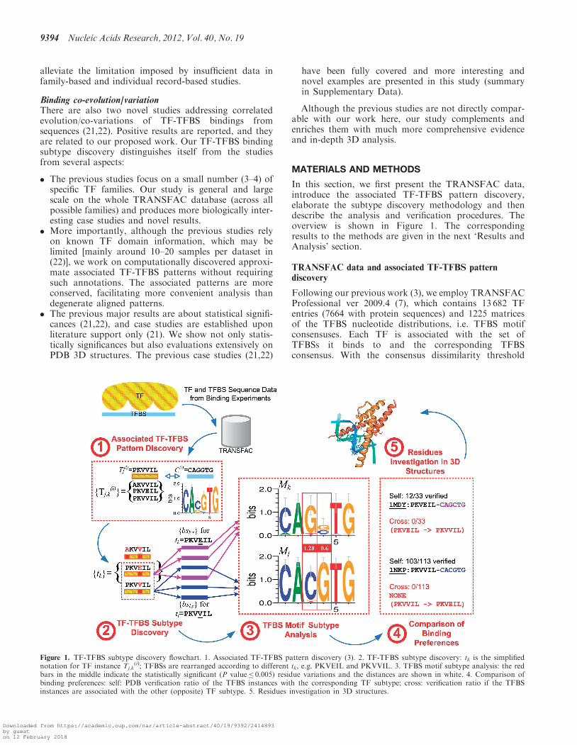
In this section, we first present the TRANSFAC data,introduce the associated TF-TFBS pattern discovery,elaborate the subtype discovery methodology and thendescribe the analysis and verification procedures. Theoverview is shown in Figure 1. The correspondingresults to the methods are given in the next ‘Results andAnalysis’ section.
TRANSFAC data and associated TF-TFBS patterndiscovery
Following our previous work (3), we employ TRANSFACProfessional ver 2009.4 (7), which contains 13 682 TFentries (7664 with protein sequences) and 1225 matricesof the TFBS nucleotide distributions, i.e. TFBS motifconsensuses. Each TF is associated with the set ofTFBSs it binds to and the corresponding TFBSconsensus. With the consensus dissimilarity threshold
Figure 1. TF-TFBS subtype discovery flowchart. 1. Associated TF-TFBS pattern discovery (3). 2. TF-TFBS subtype discovery: tk is the simplifiednotation for TF instance Tj,k
(i); TFBSs are rearranged according to different tk, e.g. PKVEIL and PKVVIL. 3. TFBS motif subtype analysis: the redbars in the middle indicate the statistically significant (P value� 0.005) residue variations and the distances are shown in white. 4. Comparison ofbinding preferences: self: PDB verification ratio of the TFBS instances with the corresponding TF subtype; cross: verification ratio if the TFBSinstances are associated with the other (opposite) TF subtype. 5. Residues investigation in 3D structures.
9394 Nucleic Acids Research, 2012, Vol. 40, No. 19
Downloaded from https://academic.oup.com/nar/article-abstract/40/19/9392/2414893by gueston 12 February 2018
set at TY=0.3 to encourage more diverse groups (3), theTF entries are grouped into 506 datasets each representedby the consensus identifier Ci. The approximate associatedTF-TFBS pattern discovery is applied on all the data sets.The major parameters in the example are set as TF/TFBSwidth W=6,8 and the maximal substitution errorE=1,2. The resultant patterns are in the form of Tj
(i)�
C(i), where T and C represent the jth TF and the ith TFBScore patterns, i.e. PKVVIL and CAGGTG, respectively,in Figure 1, Part 1. In that running example, three differ-ent approximate instances belong to Tj
(i) with mismatches/errors�E: AKVVIL, PKVEIL and PKVVIL, denoted ast1, t2 and t3, respectively, when there is no ambiguity abouti and j. They collectively form a set ftk}.
The discovered associated patterns can be evaluated andverified using TF-TFBS 3D structures [1948 entries as in(3)] from PDB (9). We focus on the verification ratios, RTF
on the TF side and RTF-TFBS on both the TF-TFBS sides.For example, in Figure 1, Part 1, we have a patternPKVVIL-CAGGTG. If under the pattern we have oneAKVVIL, four PKVEIL and five PKVVIL TF instances,and both PKVEIL and PKVVIL can be found in corebinding pairs in PDB, then RTF= (4+5)/(1+4+5)=0.9for this associated pattern. If only PKVVIL-CACGTG(five instances) match the binding pairs in PDB on bothsides while PKVEIL-CACGTG (four instances) doesnot, RTF-TFBS=5/10=0.5. Summarizing all associatedpatterns discovered, we can have the averaged AVGRTF-TFBS to measure the overall performance.
More technical details and the corresponding resultscan be found in the Supplementary Data.
TF-TFBS subtype discovery
On the basis of the successful discovery of approximateprotein–DNA (TF-TFBS) associated patterns, we canfurther study the detailed binding variations contributedby residues which otherwise cannot be accommodatedusing the current limited amount of 3D structure data.By investigating the approximate (i.e. varying) TF in-stances in the associated patterns and extending theconsensuses to the corresponding TFBS instances, weextract TF-TFBS residue variations (column-specificsubtypes) and calculate the distances within a subtypepair. The details are elaborated as follows.
In the TF (core) motif discovery, data are subject toredundancy removal to avoid spurious motifs due toover-sampling (3). In residue variation subtype analysis,however, it is desirable to retrieve as many instances aspossible, to ensure that the subtype discrimination doesnot happen due to insufficient data samples. Furthermore,examining samples beyond where the TF motifs are dis-covered can extensively verify the pattern generality.Therefore, distinct TF instances under a TF motif areretrieved from the whole TRANSFAC together with thecorresponding TFBSs. As illustrated in Figure 1, Part 2,for each distinct instance tk belonging to a TF motif, wesearch the whole TRANSFAC for the TF sequences con-taining it and retrieve all the corresponding bound TFBSinstances, denoted by a set fbsk,x} where x is the index fora particular TFBS. Therefore, for two pairs tk � fbsk,x}
(PKVEIL-purple TFBSs) and tl � fbsl,y} (PKVVIL-blueTFBSs), tk 6¼ tl, under the same associated pattern(PKVVIL-CAGGTG), we can investigate how theTFBSs fbsk,x} and fbsl,y} are different (i.e. TFBSsubtypes) with respect to the TFBS consensus C(i).As raw TFBSs have different widths, they are aligned to
the consensus and truncated at the same width W. In par-ticular, the W-conserved core of C(i) is chosen, i.e. theW subsequence of C(i) with the most conserved nucleo-tides. To measure nucleotide conservation in the consen-sus, we assign conservation score 1 for each conservednucleotide A, C, G or T, 0.5 for mixed di-nucleotide R,Y, M, K, W or S, 0.33 for mixed tri-nucleotide B, D, H orV and 0.25 for the degenerated N. Then each TFBS (aswell as the reverse complement) is aligned to theW-conserved core and the best aligned subsequence(substitution only) is extracted, resulting in the alignedcore TFBS sets fbsk,x} and fbsl,y} with the same width Wfor two different TF instances tk and tl. Note thatTRANSFAC contains noises and we discard TFBSsthat can only poorly aligned with the consensus(mismatches� 0.5 *W). PWMs Mk for fbsk,x} (purple)and Ml for fbsl,y} (blue) are then generated accordinglyas shown in Figure 1, Part 3.
TFBS subtype comparisons
To characterize and shortlist meaningful subtypes, com-parisons are to be made between PWMs Mk (purple) andMl (blue) corresponding to the two distinct TF motifinstances tk and tl, respectively. In particular, column-wiseEuclidean distance and the P values for Pearsonchi-square test are employed. Although there are variouscomparison methods, Euclidean distance frequently showsbest performance (17,19), and Pearson chi-square test isalso among the top ones when sample size is �20 (19),which is exactly our case. Another reason to employchi-square test is to easily obtain comparable statisticsand intuitive P value thresholds to shortlist subtypesaccommodating different sample sizes in different datasets. Other comparison methods such as PearsonCorrelation Coefficient (PCC) and average log-likelihoodratio (ALLR) (17,19) produce consistent subtypes, mostlycovered by our results with appropriate thresholds(see Supplementary Data for the comparisons). As thefocused results are consistent, we employ P values andEuclidean distance in our analysis. Other advancedmethods such as mutual information (MI) (21,22) couldbe employed in the future study.The column-wise distance dcol can be calculated as
follows:
dcolðMk;Ml; qÞ ¼ ðX
b
jMkðb; qÞ �Mlðb; qÞj2Þ
1=2ð1Þ
where b 2 fA,C,G,T} is the nucleotide, q is the qth columnin the PWM and L-2 norm (Euclidean distance) isemployed in our experiments.To investigate into residue varying subtypes, the
column-wise distance dcol is focused on because thesubtypes are largely similar while only small portionsdiverge. The positional discrimination is critical to
Nucleic Acids Research, 2012, Vol. 40, No. 19 9395
Downloaded from https://academic.oup.com/nar/article-abstract/40/19/9392/2414893by gueston 12 February 2018
explain the binding differences for the highly conservedTF motif instances with only few mismatches.Comparing the Euclidean distance (dcol) only may not
be precise enough to evaluate subtypes, as there aresample size variations and biases in TRANSFACleading to distorted distances. Therefore, statistical signifi-cance is employed to first compare and shortlist the TFBSPWM columns for two different TF instances tk and tl.Because we focus on residue varying subtypes, the statis-tical analysis is performed on the nucleotide level (PWMcolumns) rather than the motif level.In particular, Pearson chi-square test of independence
for two samples/outcomes (k and l) is employed. The nullhypothesis is that the nucleotide frequency in the PWMcolumn p (the occurrence of the outcomes) is statisticallyindependent. The expected frequency for r 2 fk,l} and b 2fA,C,G,T} is
Er;b ¼ð fk;b þ fl;bÞð
Pc2fA;C;G;T g fr;cÞ
N; ð2Þ
where in general f*,c denotes the frequency count of nu-cleotide c in PWM column M*(c,q) for * 2 fk,l}, and N isthe total count of nucleotide frequencies of the twosamples. The �2 statistic is
�2ðMk;Ml; qÞ ¼X
r2fk;l g
X
b2fA;C;G;T g
ð fr;b � Er;bÞ2
Er;bð3Þ
and the degree of freedom is (2–1)(4–1)=3. The P valuecan be calculated at the resolutions of 0.001,0.002, 0.005, 0.01, etc. The statistically significantsubtypes are selected for further investigation based onthe threshold P value� 0.005. In other words, we onlyfocus on those statistically significant subtype pairs inthe text below. Corresponding results are in the later‘TF-TFBS Subtype Results’ section.
Subtype investigation and analysis
There are various potential biological meanings and im-plications for the shortlisted TF-TFBS subtypes. Theymay reflect binding properties of the TF residues, exhibitchanged binding preferences, differ in conservation due tocontacting chemical bonds or carry other specific func-tions in regulatory mechanisms. The possibilities areinvestigated and verified with the following proceduresfrom several aspects. The whole subtype discovery proced-ure is illustrated in Figure 1.
OverviewThe subtypes imply different kinds of mechanisms andinformation with biological meanings:
. The subtypes may be directly involved in coreTF-TFBS bindings. The varying residues may showflexibilities which are tolerated in the bindings onone hand and also exhibit changed binding preferenceson the other. They are analyzed in sub-section‘Subtype Comparison of Binding Preferences’ using3D structures.
. The invariant (conserved) TF residues and/or TFBSnucleotides are likely to be critical contactingresidues whose changes may significantly affect thechemical bonds. The information to discriminateconserved and flexible residues is useful for bettermodeling the error distributions of TF and/or TFBSmotifs and improving existing motif representations,e.g. PWMs. They are elaborated through sub-section:‘Subtype Residues Investigation in 3D structures’.
. The subtypes may not be directly involved in protein–DNA contacting chemical bonds. However, the statis-tically significant variations are not likely to happen bychance. They may imply regulatory mechanisms and/or partners beyond direct protein–DNA interactions,for example, co-activator binding and dimerization.An interesting case that may lead to novel discoverieswill be addressed in sub-section: ‘In-depth BindingAnalysis’.
Subtype comparison of binding preferencesTo compare the binding preferences within the statisticallysignificant subtype pairs, we make use of the procedureused in verifying approximate associated TF-TFBSpatterns (3). The same binding protein–DNA (P-D)pairs from PDB are employed. To evaluate a TFsubtype tk (a distinct TF motif instance in width W)associated with a column from the corresponding set ofaligned TFBSs (fbsk,q} in width W), we should evaluate tkwith each instance bsk,q through enumerating all tk � bsk,qpairs. Each tk � bsk,q is verified if both the TF and TFBSsubsequences are contained in certain PDB P-D pairs. Werecord the verified count for the tk � bsk,q pairs. Note thatthe verification is more stringent than our previous study(3), which only requires a TFBS consensus to be approxi-mately matched for verification.
The next step is to investigate whether the residue-varying subtypes exhibit specific binding preferences bycomparing them with artificial controls (TFBS subtypesassociated with wrong TF instances). To check thebinding preference of a pair of two TF subtypes tk and tland their corresponding distinct TFBS patterns bsk,q andbsk,r, we introduce cross-verification comparisons. Besidesthe previous PDB verification count on tk with its actualcorresponding TFBSs (bsk,q), denoted as ‘Self’ verification,we perform the same PDB verification on the artificialcontrol by associating the wrong tl with bsk,q, which isdenoted as ‘Cross’ verification (the control). Note thatthe comparison is only performed when both TF instancestk, tl are verified on PDB, to avoid bias on TF instanceswith PDB evidence over those without. For the subtypesleft out, we may resort to literature search and detailed 3Dstructure analysis.
Intuitively, ‘Self’> ‘Cross’ indicates there are more PDBstructure evidence verifying the correctly associatedTF-TFBS subtype than the artificial control and thussupports existence of the binding preferences. All the‘Self’> ‘Cross’ percentages of the TF-TFBS subtypes arethen reported. Besides ‘Self’> ‘Cross’ verification per-formed on each ‘individual’ TF subtype with the bindingpreference ratio denoted as ‘Individual Subtype Ratios’,
9396 Nucleic Acids Research, 2012, Vol. 40, No. 19
Downloaded from https://academic.oup.com/nar/article-abstract/40/19/9392/2414893by gueston 12 February 2018
binding preference ratios for subtype pairs are alsointroduced. If both TF subtypes in pair satisfy the ‘indi-vidual’ verification tests, they are said to satisfy the ‘pair’verification test. The corresponding ratio is denoted as‘Pair Subtype Ratios’. One illustrative example is inFigure 1. Because both individual ‘Self’ > ‘Cross’ verifica-tion tests are satisfied for PKVEIL-CAGCTG (12/33versus 0/33) and PKVVIL-CACGTG (103/113 versus 0/113), ‘PKVEIL-CAGCTG versus PKVVIL-CACGTG’satisfies the ‘pair’ verification test. Corresponding resultsare in the ‘Comparison Results of Binding Preferences onPDB’ section.
Subtype residues investigation in 3D structuresAs the previous comparisons are indirect and have the riskof sample bias in PDB, we further investigate into thedetailed binding (interaction) properties of the varyingand conserved residues on the PDB 3D structures. Togenerate a concise map from the many (redundant)subtypes, the statistically significant subtypes can be clus-tered according to the associated TF-TFBS patterns onboth sides according to the maximal error E. As TFBSsare more flexible and less conserved, we focus on theresidues (amino acids) on the TF side. They arecategorized into two types in each cluster, the varyingand the invariant (conserved) ones. It is then interestingto analyze the biochemical mechanisms of the stronglyconserved residues (e.g. forming hydrogen bonds withthe TFBS nucleotides) and the varying residues (e.g.showing specificities to different target TFBSs) byinvestigating the 3D structures one by one. Correspondingresults are in the ‘Results of Residues Investigation in 3DStructures’ section.
In-depth binding analysisFinally, for statistically significant subtypes withoutobvious evidence of direct interactions, we select interest-ing examples to perform literature search for reasonableinterpretation. These cases are potential novel discoveriesleading to co-factor bindings and revealing moreintriguing regulatory mechanisms. Corresponding resultsare in the ‘In-depth Analysis for Potential Co-factors’section.
RESULTS AND ANALYSIS
In this section, the detailed TF-TFBS subtype results andstatistics are reported, followed by detailed variationanalysis and comprehensive verification.
TF-TFBS subtype results
Based on the approximate associated TF-TFBS patternsdiscovered, subtype discovery was performed with widthW=6,8 and maximal error E=1,2. All pairs of TFsubtypes (i.e. different TF instances tk and tl within anassociated TF-TFBS pattern) and their correspondingaligned TFBSs (PWMs Mk, Ml displayed as sequencelogos in Figure 1) were analyzed. If there were any nucleo-tide differences leading to P value P� 0.005, the PWMcolumn q associated with the TF subtypes are recorded
and denoted as a subtype ‘pair’. Meanwhile subtypepairs can be shortlisted by a column-wise distance thresh-old Cdis� dcol. dcol is in the range of ½0;
ffiffiffi2p�. As a proof of
concept, we focus on the associated patterns withinstance-level RTF-TFBS� 0.8, and the same analysis canbe applied on other patterns which are verifiable withmore PDB records in the future (3). The statistics of theTF-TFBS subtypes are presented in Tables 1 and 2.
Subtype reflection of biological properties
We investigate into the TF subtype charge properties,which are summarized in Table 3 (details in theSupplementary Data). Residue variations are mainlywithin the same positive (Pos) or neutral (Neu) chargeproperty groups, whereas variations with chargeproperties changed and variations within the negative(Neg) are rare. The observation is consistent with thechemical properties of protein–DNA interactions. As thebackbone of DNA (TFBS) is negatively charged, positiveand neutral residues being hydrophilic are more likely tobe exposed on TF surfaces than negative ones. The DNAprefers positive/neutral amino acids to negative ones.Variations among different charge groups may signifi-cantly affect the bindings and thus are not preferred.
Table 1. The statistics of the TF-TFBS subtype pairs
Setting Pair no. Pattern no. C(i) no. dcol
W6E1 5108 643 145 0.22W6E2 6343 463 158 0.26W8E1 1250 182 69 0.28W8E2 2262 175 67 0.29
Pair no. indicates the number of significant TF-TFBS subtype pairswith P� 0.005. Pattern no. indicates how many associated TF-TFBSpatterns with RTF-TFBS� 0.8 are included for the pairs. C(i) no. indi-cates the corresponding number of consensus groups (labeled byTRANSFAC PWM IDs). dcol is the average column-wise Euclideandistance.
Table 2. The accumulated TF-TFBS subtype counts with different
Cdis values
Cdis� 0.8 0.7 0.6 0.3 0.1
W6E1 5 (3) 39 (9) 88 (18) 947 (121) 4785 (145)W6E2 41 (15) 102 (22) 214 (43) 1759 (140) 6057 (158)W8E1 10 (7) 23 (8) 72 (13) 420 (50) 1239 (69)W8E2 98 (8) 29 (13) 98 (22) 822 (57) 2237 (67)
The numbers of TRANSFAC TFBS consensus groups C(i) involved areshown in brackets.
Table 3. Summary of TF residue variation charge properties
Variation charge Percentage Chemical
Pos-Pos/Neu-Neu High Hydrophilic, preferredChanged/Neg-Neg Low Not preferred
Nucleic Acids Research, 2012, Vol. 40, No. 19 9397
Downloaded from https://academic.oup.com/nar/article-abstract/40/19/9392/2414893by gueston 12 February 2018
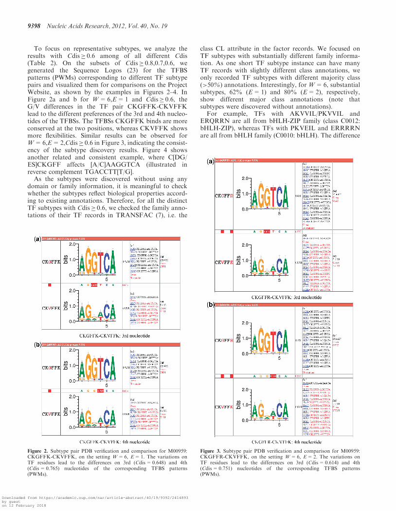
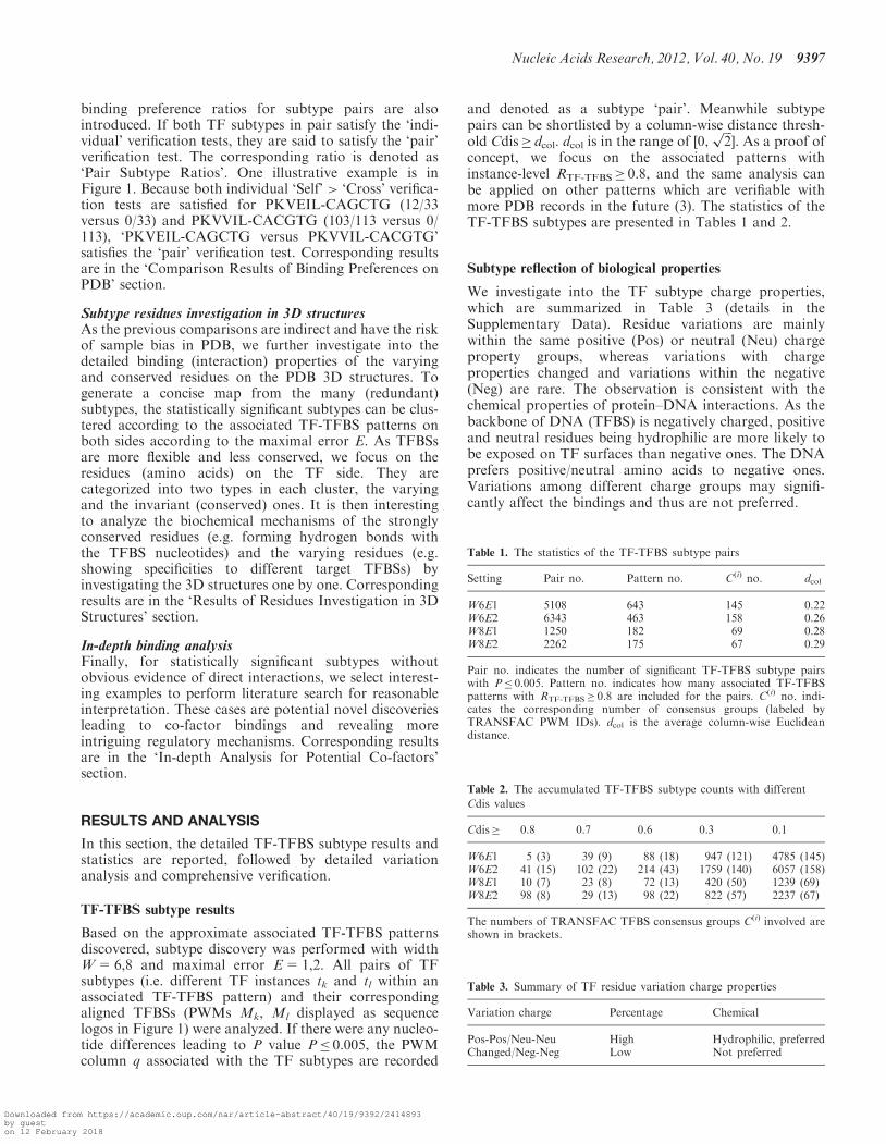
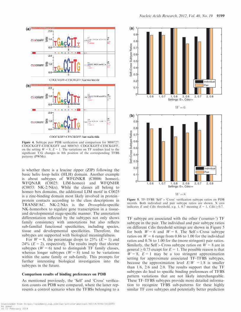
To focus on representative subtypes, we analyze theresults with Cdis� 0.6 among of all different Cdis(Table 2). On the subsets of Cdis� 0.8,0.7,0.6, wegenerated the Sequence Logos (23) for the TFBSpatterns (PWMs) corresponding to different TF subtypepairs and visualized them for comparisons on the ProjectWebsite, as shown by the examples in Figures 2–4. InFigure 2a and b for W=6,E=1 and Cdis� 0.6, theG/V differences in the TF pair CKGFFK-CKVFFKlead to the different preferences of the 3rd and 4th nucleo-tides of the TFBSs. The TFBSs CKGFFK binds are moreconserved at the two positions, whereas CKVFFK showsmore flexibilities. Similar results can be observed forW=6,E=2,Cdis� 0.6 in Figure 3, indicating the consist-ency of the subtype discovery results. Figure 4 showsanother related and consistent example, where C[DG/ES]CKGFF affects [A/C]AAGGTCA (illustrated inreverse complement TGACCTT[T/G].As the subtypes were discovered without using any
domain or family information, it is meaningful to checkwhether the subtypes reflect biological properties accord-ing to existing annotations. Therefore, for all the distinctTF subtypes with Cdis� 0.6, we checked the family anno-tations of their TF records in TRANSFAC (7), i.e. the
class CL attribute in the factor records. We focused onTF subtypes with substantially different family informa-tion. As one short TF subtype instance can have manyTF records with slightly different class annotations, weonly recorded TF subtypes with different majority class(>50%) annotations. Interestingly, for W=6, substantialsubtypes, 62% (E=1) and 80% (E=2), respectively,show different major class annotations (note thatsubtypes were discovered without annotations).
For example, TFs with AKVVIL/PKVVIL andERQRRN are all from bHLH-ZIP family (class C0012:bHLH-ZIP), whereas TFs with PKVEIL and ERRRRNare all from bHLH family (C0010: bHLH). The difference
Figure 3. Subtype pair PDB verification and comparison for M00959:CKGFFR-CKVFFK, on the setting W=6, E=2. The variations onTF residues lead to the differences on 3rd (Cdis=0.614) and 4th(Cdis=0.751) nucleotides of the corresponding TFBS patterns(PWMs).
Figure 2. Subtype pair PDB verification and comparison for M00959:CKGFFK-CKVFFK, on the setting W=6, E=1. The variations onTF residues lead to the differences on 3rd (Cdis=0.648) and 4th(Cdis=0.765) nucleotides of the corresponding TFBS patterns(PWMs).
9398 Nucleic Acids Research, 2012, Vol. 40, No. 19
Downloaded from https://academic.oup.com/nar/article-abstract/40/19/9392/2414893by gueston 12 February 2018
is whether there is a leucine zipper (ZIP) following thebasic helix–loop–helix (HLH) domain. Another exampleis about subtypes of WFGNKR (C0006: homeo),WFQNAR (C0025: LIM-homeo) and WFQNHR(C0053: NK-2/Nkx). While the classes all belong tohomeo box domains, the additional LIM motif in C0025is a zinc-binding domain most likely involved in protein–protein contacts according to the class descriptions inTRANSFAC. NK-2/Nkx is the Drosophila-specificNK-homeobox to regulate gene transcription in a tissue-and developmental stage-specific manner. The annotationdifferentiation reflected by the subtypes not only showsfamily consistency with annotations but also reflectssub-familial functional specificities, including species,tissue and developmental specificities. Therefore, thesubtypes are supported with biological meaningfulness.
For W=8, the percentage drops to 23% (E=1) and24% (E=2), respectively. The results imply that shortersubtypes (W=6) tend to distinguish TF family classes,whereas longer subtypes (W=8) tend to be variationswithin the same family or sub-family. This prompts forfurther interesting biological investigation into thesubtypes in the future.
Comparison results of binding preferences on PDB
As mentioned previously, the ‘Self’ and ‘Cross’ verifica-tion counts on PDB were compared, where the latter rep-resents a control scenario when the TFBSs belonging to a
TF subtype are associated with the other (‘counter-’) TFsubtype in the pair. The individual and pair subtype ratioson different Cdis threshold settings are shown in Figure 5for both W=6 and W=8. The Self>Cross subtyperatios on W=6 range from 0.86 to 1.00 for the individualratios and 0.76 to 1.00 for the (more stringent) pair ratios.Similarly, the Self>Cross subtype ratios on W=8 are ingeneral� 0.73 except for E=1. The possible reason is thatW=8, E=1 may be a too stringent approximationsetting for approximate associated TF-TFBS subtypes,because the approximation level E/W =1/8 is smallerthan 1/6, 2/6 and 2/8. The results support that the TFsubtypes do lead to specific binding preferences of TFBSpattern variations that are not likely interchangeable.These TF-TFBS subtypes provide more detailed informa-tion to recognize TFBS sub-patterns for these highlysimilar TF core subtypes and potentially better prediction
Figure 5. TF-TFBS ‘Self’> ‘Cross’ verification subtype ratios on PDBrecords. Both individual and pair subtype ratios are shown. X axisindicates E and Cdis threshold, e.g. 1, 0.7 meaning E=1, Cdis� 0.7.
Figure 4. Subtype pair PDB verification and comparison for M00727:CDGCKGFF-CESCKGFF and M00763: CDGCKGFF-CESCKGFF,on the setting W=8, E=1. The variations on TF residues lead to thesignificant T/G changes in 8th position of the corresponding TFBSpatterns (PWMs).
Nucleic Acids Research, 2012, Vol. 40, No. 19 9399
Downloaded from https://academic.oup.com/nar/article-abstract/40/19/9392/2414893by gueston 12 February 2018
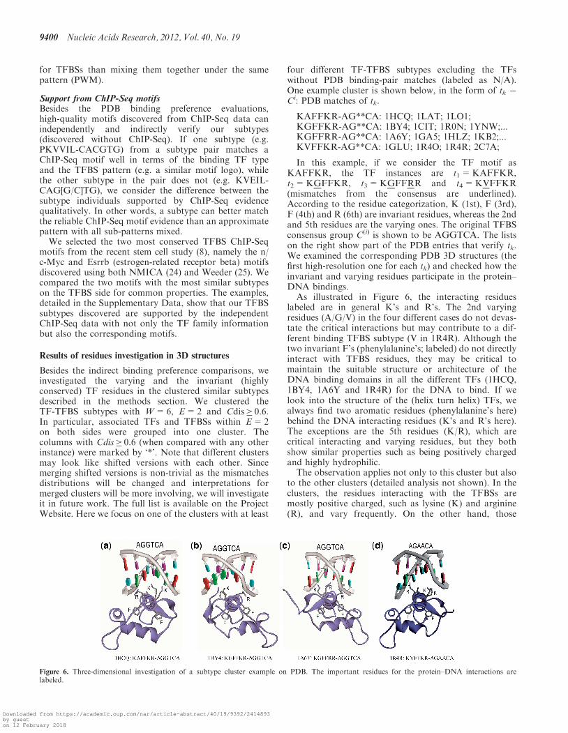
for TFBSs than mixing them together under the samepattern (PWM).
Support from ChIP-Seq motifsBesides the PDB binding preference evaluations,high-quality motifs discovered from ChIP-Seq data canindependently and indirectly verify our subtypes(discovered without ChIP-Seq). If one subtype (e.g.PKVVIL-CACGTG) from a subtype pair matches aChIP-Seq motif well in terms of the binding TF typeand the TFBS pattern (e.g. a similar motif logo), whilethe other subtype in the pair does not (e.g. KVEIL-CAG[G/C]TG), we consider the difference between thesubtype individuals supported by ChIP-Seq evidencequalitatively. In other words, a subtype can better matchthe reliable ChIP-Seq motif evidence than an approximatepattern with all sub-patterns mixed.We selected the two most conserved TFBS ChIP-Seq
motifs from the recent stem cell study (8), namely the n/c-Myc and Esrrb (estrogen-related receptor beta) motifsdiscovered using both NMICA (24) and Weeder (25). Wecompared the two motifs with the most similar subtypeson the TFBS side for common properties. The examples,detailed in the Supplementary Data, show that our TFBSsubtypes discovered are supported by the independentChIP-Seq data with not only the TF family informationbut also the corresponding motifs.
Results of residues investigation in 3D structures
Besides the indirect binding preference comparisons, weinvestigated the varying and the invariant (highlyconserved) TF residues in the clustered similar subtypesdescribed in the methods section. We clustered theTF-TFBS subtypes with W=6, E=2 and Cdis� 0.6.In particular, associated TFs and TFBSs within E=2on both sides were grouped into one cluster. Thecolumns with Cdis� 0.6 (when compared with any otherinstance) were marked by ‘*’. Note that different clustersmay look like shifted versions with each other. Sincemerging shifted versions is non-trivial as the mismatchesdistributions will be changed and interpretations formerged clusters will be more involving, we will investigateit in future work. The full list is available on the ProjectWebsite. Here we focus on one of the clusters with at least
four different TF-TFBS subtypes excluding the TFswithout PDB binding-pair matches (labeled as N/A).One example cluster is shown below, in the form of tk �Ci: PDB matches of tk.
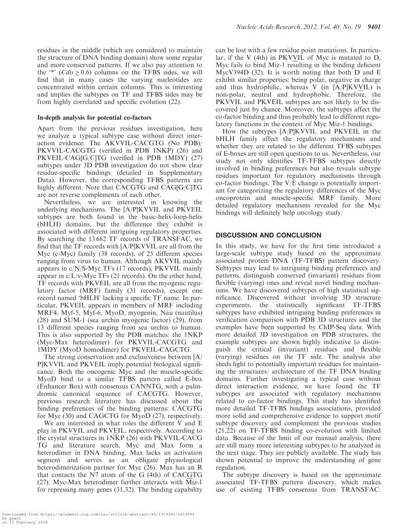
KAFFKR-AG**CA: 1HCQ; 1LAT; 1LO1;KGFFKR-AG**CA: 1BY4; 1CIT; 1R0N; 1YNW;...KGFFRR-AG**CA: 1A6Y; 1GA5; 1HLZ; 1KB2;...KVFFKR-AG**CA: 1GLU; 1R4O; 1R4R; 2C7A;
In this example, if we consider the TF motif asKAFFKR, the TF instances are t1=KAFFKR,t2=KGFFKR, t3=KGFFRR and t4=KVFFKR(mismatches from the consensus are underlined).According to the residue categorization, K (1st), F (3rd),F (4th) and R (6th) are invariant residues, whereas the 2ndand 5th residues are the varying ones. The original TFBSconsensus group C(i) is shown to be AGGTCA. The listson the right show part of the PDB entries that verify tk.We examined the corresponding PDB 3D structures (thefirst high-resolution one for each tk) and checked how theinvariant and varying residues participate in the protein–DNA bindings.
As illustrated in Figure 6, the interacting residueslabeled are in general K’s and R’s. The 2nd varyingresidues (A/G/V) in the four different cases do not devas-tate the critical interactions but may contribute to a dif-ferent binding TFBS subtype (V in 1R4R). Although thetwo invariant F’s (phenylalanine’s; labeled) do not directlyinteract with TFBS residues, they may be critical tomaintain the suitable structure or architecture of theDNA binding domains in all the different TFs (1HCQ,1BY4, 1A6Y and 1R4R) for the DNA to bind. If welook into the structure of the (helix turn helix) TFs, wealways find two aromatic residues (phenylalanine’s here)behind the DNA interacting residues (K’s and R’s here).The exceptions are the 5th residues (K/R), which arecritical interacting and varying residues, but they bothshow similar properties such as being positively chargedand highly hydrophilic.
The observation applies not only to this cluster but alsoto the other clusters (detailed analysis not shown). In theclusters, the residues interacting with the TFBSs aremostly positive charged, such as lysine (K) and arginine(R), and vary frequently. On the other hand, those
Figure 6. Three-dimensional investigation of a subtype cluster example on PDB. The important residues for the protein–DNA interactions arelabeled.
9400 Nucleic Acids Research, 2012, Vol. 40, No. 19
Downloaded from https://academic.oup.com/nar/article-abstract/40/19/9392/2414893by gueston 12 February 2018
residues in the middle (which are considered to maintainthe structure of DNA binding domain) show some regularand more conserved patterns. If we also pay attention tothe ‘*’ (Cdis� 0.6) columns on the TFBS sides, we willfind that in many cases the varying nucleotides areconcentrated within certain columns. This is interestingand implies the subtypes on TF and TFBS sides may befrom highly correlated and specific evolution (22).
In-depth analysis for potential co-factors
Apart from the previous residues investigation, herewe analyze a typical subtype case without direct inter-action evidence. The AKVVIL-CACGTG (No PDB)/PKVVIL-CACGTG (verified in PDB 1NKP) (26) andPKVEIL-CAG[G/C]TG (verified in PDB 1MDY) (27)subtypes under 3D PDB investigation do not show clearresidue-specific bindings (detailed in SupplementaryData). However, the corresponding TFBS patterns arehighly different. Note that CACGTG and CAG[G/C]TGare not reverse complements of each other.
Nevertheless, we are interested in knowing theunderlying mechanisms. The [A/P]KVVIL and PKVEILsubtypes are both found in the basic-helix-loop-helix(bHLH) domains, but the difference they exhibit isassociated with different intriguing regulatory properties.By searching the 13 682 TF records of TRANSFAC, wefind that the TF records with [A/P]KVVIL are all from theMyc (c-Myc) family (38 records), of 25 different speciesranging from virus to human. Although AKVVIL mainlyappears in c/N/S-Myc TFs (17 records), PKVVIL mainlyappear in c/L/v-Myc TFs (21 records). On the other hand,TF records with PKVEIL are all from the myogenic regu-latory factor (MRF) family (31 records), except onerecord named ‘bHLH’ lacking a specific TF name. In par-ticular, PKVEIL appears in members of MRF includingMRF4, Myf-5, Myf-6, MyoD, myogenin, Nau (nautilus)(28) and SUM-1 (sea urchin myogenic factor) (29), from13 different species ranging from sea urchin to human.This is also supported by the PDB matches: the 1NKP(Myc-Max heterodimer) for PKVVIL-CACGTG and1MDY (MyoD homodimer) for PKVEIL-CAGCTG.
The strong conservation and exclusiveness between [A/P]KVVIL and PKVEIL imply potential biological signifi-cance. Both the oncogenic Myc and the muscle-specificMyoD bind to a similar TFBS pattern called E-box(Enhancer Box) with consensus CANNTG, with a palin-dromic canonical sequence of CACGTG. However,previous research literature has discussed about thebinding preferences of the binding patterns: CACGTGfor Myc (30) and CAGCTG for MyoD (27), respectively.
We are interested in what roles the different V and Eplay in PKVVIL and PKVEIL, respectively. According tothe crystal structures in 1NKP (26) with PKVVIL-CACGTG and literature search, Myc and Max form aheterodimer in DNA binding. Max lacks an activationsegment and serves as an obligate physiologicalheterodimerization partner for Myc (26). Max has an Rthat contacts the N7 atom of the G (4th) of CACGTG(27). Myc-Max heterodimer further interacts with Miz-1for repressing many genes (31,32). The binding capability
can be lost with a few residue point mutations. In particu-lar, if the V (4th) in PKVVIL of Myc is mutated to D,Myc fails to bind Miz-1 resulting in the binding deficientMycV394D (32). It is worth noting that both D and Eexhibit similar properties: being polar, negative in chargeand thus hydrophilic, whereas V (in [A/P]KVVIL) isnon-polar, neutral and hydrophobic. Therefore, thePKVVIL and PKVEIL subtypes are not likely to be dis-covered just by chance. Moreover, the subtypes affect theco-factor binding and thus probably lead to different regu-latory functions in the context of Myc Miz-1 bindings.How the subtypes [A/P]KVVIL and PKVEIL in the
bHLH family affect the regulatory mechanisms andwhether they are related to the different TFBS subtypesof E-boxes are still open questions to us. Nevertheless, ourstudy not only identifies TF-TFBS subtypes directlyinvolved in binding preferences but also reveals subtyperesidues important for regulatory mechanisms throughco-factor bindings. The V/E change is potentially import-ant for categorizing the regulatory differences of the Myconcoprotein and muscle-specific MRF family. Moredetailed regulatory mechanisms revealed for the Mycbindings will definitely help oncology study.
DISCUSSION AND CONCLUSION
In this study, we have for the first time introduced alarge-scale subtype study based on the approximateassociated protein–DNA (TF-TFBS) pattern discovery.Subtypes may lead to intriguing binding preferences andpatterns, distinguish conserved (invariant) residues fromflexible (varying) ones and reveal novel binding mechan-isms. We have discovered subtypes of high statistical sig-nificance. Discovered without involving 3D structureexperiments, the statistically significant TF-TFBSsubtypes have exhibited intriguing binding preferences inverification comparison with PDB 3D structures and theexamples have been supported by ChIP-Seq data. Withmore detailed 3D investigation on PDB structures, theexample subtypes are shown highly indicative to distin-guish the critical (invariant) residues and flexible(varying) residues on the TF side. The analysis alsosheds light to potentially important residues for maintain-ing the structures/ architecture of the TF DNA bindingdomains. Further investigating a typical case withoutdirect interaction evidence, we have found the TFsubtypes are associated with regulatory mechanismsrelated to co-factor bindings. This study has identifiedmore detailed TF-TFBS bindings associations, providedmore solid and comprehensive evidence to support motifsubtype discovery and complement the previous studies(21,22) on TF-TFBS binding co-evolution with limiteddata. Because of the limit of our manual analysis, thereare still many more interesting subtypes to be analyzed inthe next stage. They are publicly available. The study hasshown potential to improve the understanding of generegulation.The subtype discovery is based on the approximate
associated TF-TFBS pattern discovery, which makesuse of existing TFBS consensus from TRANSFAC.
Nucleic Acids Research, 2012, Vol. 40, No. 19 9401
Downloaded from https://academic.oup.com/nar/article-abstract/40/19/9392/2414893by gueston 12 February 2018
Further generalization on the modeling and search ofassociated TF-TFBS patterns would provide more inform-ative and sound patterns to facilitate more powerfulsubtype discovery. One interesting direction is to set upa model that accommodates associated subtypes directlyin TF-TFBS pattern discovery. The associated TF-TFBSsubtype study with respect to various species is our nexttarget. Further applications of the TF-TFBS subtypesinclude better prediction of TFBSs given the TF informa-tion based on a formal model and/or classifier andlarge-scale study on 3D structure data with the associatedsubtypes mined for more informative bindingmechanisms.The subtype discovery and analysis have broad poten-
tial biological applications. Subtypes can help to spotinteresting variations that contribute to binding prefer-ences, familial specificities and interacting mechanisms,as our comprehensive analysis has shown. Hierarchicalannotation by introducing subtype besides site anddomain is one possible extension (7,33). Follow-up phylo-genetic studies on subtypes can further shed light on evo-lution and regulatory mechanisms. Subtypes can beutilized for more accurate motif discovery. Current motifmatching suffers from false positives (34). As theassociated TF-TFBS patterns and subtypes are furthergeneralized, knowledge-driven motif matching can be de-veloped as our next target to discriminate subtle variationsto distinguish true TFBSs from false hits. With more andmore data, subtypes can be further applied to analyze themechanisms of alterations in the viral upstream regulatoryregion (URR) (35) to fight against diseases. The ultimateunderstanding TF-TFBS bindings will lead to artificialdesign of gene regulation circuits (36).
AVAILABILITY
Project Website: www.cse.cuhk.edu.hk/%7Etmchan/subtypes/.
SUPPLEMENTARY DATA
Supplementary Data are available at NAR Online:Supplementary Text, Supplementary Tables 1–4 andSupplementary Figures 1–5.
ACKNOWLEDGEMENTS
The authors are grateful to the anonymous reviewers fortheir valuable comments.
FUNDING
Funding for open access change: The Focused InvestmentScheme D on Hong Kong Bioinformatics Centre [ProjectNo. 1904014]; Direct Grant [Project No. 2050500];Chinese University of Hong Kong; GRF grant [ProjectNo. 310111] from the Research Grants Council of HongKong SAR, China.
Conflict of interest statement. None declared.
REFERENCES
1. Luscombe,N.M. and Thornton,J.M. (2002) Protein-DNAinteractions: amino acid conservation and the effects of mutationson binding specificity. J. Mol. Biol., 320, 991–1009.
2. Luscombe,N.M., Austin,S.E., Berman,H.M. and Thornton,J.M.(2000) An overview of the structures of protein-DNA complexes.Genome Biol., 1, REVIEWS001.
3. Chan,T.-M., Wong,K.-C., Lee,K.-H., Wong,M.-H., Lau,C.-K.,Tsui,S.K. and Leung,K.-S. (2011) Discovering approximateassociated sequence patterns for protein-DNA interactions.Bioinformatics, 27, 471–478.
4. Leung,K.-S., Wong,K.-C., Chan,T.-M., Wong,M.-H., Lee,K.-H.,Lau,C.-K. and Tsui,S.K.W. (2010) Discovering protein-DNAbinding sequence patterns using association rule mining. NucleicAcids Res., 38, 6324–6337.
5. Smith,A.D., Sumazin,P., Das,D. and Zhang,M.Q. (2005) MiningChIP-chip data for transcription factor and cofactor binding sites.Bioinformatics, 21(Suppl. 1), i403–i412.
6. MacIsaac,K.D. and Fraenkel,E. (2006) Practical strategies fordiscovering regulatory DNA sequence motifs. PLoS Comput.Biol., 2, e36.
7. Matys,V., Kel-Margoulis,O.V., Fricke,E., Liebich,I., Land,S.,Barre-Dirrie,A., Reuter,I., Chekmenev,D., Krull,M.,Hornischer,K. et al. (2006) TRANSFAC and its moduleTRANSCompel: transcriptional gene regulation in eukaryotes.Nucleic Acids Res., 34, 108–110.
8. Chen,X., Xu,H., Yuan,P., Fang,F., Huss,M., Vega,V.B., Wong,E.,Orlov,Y.L., Zhang,W., Jiang,J. et al. (2008) Integration ofexternal signaling pathways with the core transcriptional networkin embryonic stem cells. Cell, 133, 1106–1117.
9. Berman,H.M., Westbrook,J., Feng,Z., Gilliland,G., Bhat,T.N.,Weissig,H., Shindyalov,I.N. and Bourne,P.E. (2000) The ProteinData Bank. Nucleic Acids Res., 28, 235–242.
10. Luscombe,N.M., Laskowski,R.A. and Thornton,J.M. (July, 2001)Amino acid-base interactions: a three-dimensional analysis ofprotein-DNA interactions at an atomic level. Nucleic Acids Res.,29, 2860–2874.
11. Ahmad,S., Gromiha,M.M. and Sarai,A. (2004) Analysis andprediction of DNA-binding proteins and their binding residuesbased on composition, sequence and structural information.Bioinformatics, 20, 477–486.
12. Ofran,Y., Mysore,V. and Rost,B. (2007) Prediction ofDNA-binding residues from sequence. Bioinformatics, 23,i347–i353.
13. Bais,A.S.S., Kaminski,N. and Benos,P.V. (2011) Finding subtypesof transcription factor motif pairs with distinct regulatory roles.Nucleic Acids Res., 39, e76.
14. Li,M., Ma,B. and Wang,L. (2002) Finding similar regions inmany sequences. J. Comput. Syst. Sci., 65, 73–96.
15. Chan,T.-M., Leung,K.-S. and Lee,K.-H. (2008) TFBSidentification based on genetic algorithm with combinedrepresentations and adaptive post-processing. Bioinformatics, 24,341–349.
16. Chan,T.M., Li,G., Leung,K.S. and Lee,K.H. (2009) Discoveringmultiple realistic TFBS motifs based on a generalized model.BMC Bioinformatics, 10, 22.
17. Mahony,S., Auron,P.E. and Benos,P.V. (2007) DNA familialbinding profiles made easy: comparison of various motif alignmentand clustering strategies. PLoS Comput. Biol., 17, 578–591.
18. Habib,N., Kaplan,T., Margalit,H. and Friedman,N. (2008) ANovel Bayesian DNA motif comparison method for clusteringand retrieval. PLoS Comput. Biol., 4, e1000010.
19. Gupta,S., Stamatoyannopoulos,J., Bailey,T. and Noble,W. (2007)Quantifying similarity between motifs. Genome Biol., 8, R24.
20. Sarai,A. and Kono,H. (2005) Protein-DNA recognition patternsand predictions. Annu. Rev. Biophys. Biomol. Struct., 34, 379–398.
21. Mahony,S., Auron,P.E. and Benos,P.V. (2007) Inferringprotein-DNA dependencies using motif alignments and mutualinformation. Bioinformatics, 23, i297–i304.
22. Yang,S., Yalamanchili,H.K., Li,X., Yao,K.-M., Sham,P.C.,Zhang,M.Q. and Wang,J. (2011) Correlated evolution oftranscription factors and their binding sites. Bioinformatics, 27,2972–2978.
9402 Nucleic Acids Research, 2012, Vol. 40, No. 19
Downloaded from https://academic.oup.com/nar/article-abstract/40/19/9392/2414893by gueston 12 February 2018
23. Crooks,G.E., Hon,G., Chandonia,J.-M. and Brenner,S.E. (2004)WebLogo: a sequence logo generator. Genome Res., 14, 1188–1190.
24. Dogruel,M., Down,T.A. and Hubbard,T.J.J. (2008) NestedMICA asan ab initio protein motif discovery tool. BMC Bioinformatics, 9, 12.
25. Pavesi,G., Mereghetti,P., Mauri,G. and Pesole,G. (2004) Weederweb: discovery of transcription factor binding sites in a set ofsequences from co-regulated genes. Nucleic Acids Res., 32,W199–W203.
26. Nair,S.K. and Burley,S.K. (2003) X-ray structures of Myc-Maxand Mad-Max recognizing DNA. Molecular bases of regulationby proto-oncogenic transcription factors. Cell, 112, 193–205.
27. Ma,P.C., Rould,M.A., Weintraub,H. and Pabo,C.O. (1994)Crystal structure of MyoD bHLH domain-DNA complex:perspectives on DNA recognition and implications fortranscriptional activation. Cell, 77, 451–459.
28. Michelson,A.M., Abmayr,S.M., Bate,M., Arias,A.M. andManiatis,T. (1990) Expression of a MyoD family memberprefigures muscle pattern in Drosophila embryos. Genes Dev., 4,2086–2097.
29. Venuti,J.M., Goldberg,L., Chakraborty,T., Olson,E.N. andKlein,W.H. (1991) A myogenic factor from sea urchin embryoscapable of programming muscle differentiation in mammaliancells. Proc. Natl Acad. Sci., 88, 6219–6223.
30. Blackwell,T.K., Kretzner,L., Blackwood,E.M., Eisenman,R.N. andWeintraub,H. (1990) Sequence-specific DNA binding by thec-Myc protein. Science, 250, 1149–1151.
31. Wanzel,M., Herold,S. and Eilers,M. (2003) Transcriptionalrepression by Myc. Trends Cell Biol., 13, 146–150.
32. Si,J., Yu,X., Zhang,Y. and DeWille,J. (2010) Myc interacts withMax and Miz1 to repress C/EBP� promoter activity and geneexpression. Mol. Cancer, 9, 1–5.
33. Bateman,A., Coin,L., Durbin,R., Finn,R.D., Hollich,V.,GrifRths-Jones,S., Khanna,A., Marshall,M., Moxon,S.,Sonnhammer,E.L.L. et al. (2004) The Pfam protein familiesdatabase. Nucleic Acids Res., 32, D138–D141.
34. Kel,A.E., Goessling,E., Reuter,I., Cheremushkin,E.,Kel-Margoulis,O.V. and Wingender,E. (2003) MATCH: a toolfor searching transcription factor binding sites in DNA sequences.Nucleic Acids Res., 31, 3576–3579.
35. Hubert,W.G. (2005) Variant upstream regulatory region sequencesdifferentially regulate human papillomavirus type 16 DNAreplication throughout the viral life cycle. J. Virol., 79,5914–5922.
36. Zhan,S., Miller,J.F. and Tyrrell,A.M. (2009) An evolutionarysystem using development and artificial Genetic RegulatoryNetworks for electronic circuit design. Biosystems, 98, 176–192.
Nucleic Acids Research, 2012, Vol. 40, No. 19 9403
Downloaded from https://academic.oup.com/nar/article-abstract/40/19/9392/2414893by gueston 12 February 2018