Transactivation and expression patterns of Jun and Fos/AP-1 super-family proteins in human oral...
11
Transactivation and expression patterns of Jun and Fos/AP-1 super-family proteins in human oral cancer Alok Mishra 1,2 , Alok C. Bharti 2 , Daman Saluja 3 and Bhudev C. Das 2,3 1 Laboratory of Molecular Biology, National Cancer Institute, NIH, Bethesda, MD 20892 2 Division of Molecular Oncology, Institute of Cytology and Preventive Oncology, (ICMR), I-7, Sector-39, Noida 201301, India 3 Departments of Medical Biotechnology and Molecular Oncology, Ambedkar Centre for Biomedical Research (ACBR), University of Delhi, Delhi 110 007, India Transcription factor activator protein-1 (AP-1) super-family is known to modulate expression of array of genes during development of many cancers and considered as an important target for modern therapeutics. But the role of AP-1 during development of human oral cancers is still poorly understood. Because oral cancer is one of the most common cancers in India and south-east Asia, we studied the activation and expression pattern of AP-1 family of proteins and mRNA in different stages of oral carcinogenesis. Gel-shift assay, western blotting, immunohistochemistry and northern blotting have been used to assess the binding activity and expression pattern of AP-1 family (c-Jun, JunB, JunD, c-Fos, FosB, Fra-1 and Fra-2) proteins and mRNA transcripts in a total of 100 fresh oral tissue specimens comprising precancer (n 5 40), cancer (n 5 50) and healthy control (n 5 10). Constitutive activation of AP-1 with concomitant upregulated expression of majority of AP-1 family of proteins and mRNA was observed in cancer cases. Interestingly, almost all precancerous cases showed JunD homodimers, whereas c-Fos/JunD was the most prevalent complex found in cancer tissues. The overexpression of EGFR mRNA, p50:p50/NF- jB homodimer formation, together with overexpression of pERK and c-Fos proteins in this study suggests an interesting cross talk between AP-1 and NF-jB pathways in oral cancers. Thus, this study demonstrates differential expression and activation of AP-1 super-family proteins in relation to severity of lesion and their crucial role in human oral carcinogenesis. Oral squamous cell carcinoma (OSCC) is the sixth most common cancer and accounts for 5% of all malignant tumors worldwide. 1 In India and South East Asia, it is the most common malignancy accounting to 50% of all malig- nant tumors. Most of the OSCC is attributed to smoking and alcohol consumption, whereas a proportion of oral cancers have been demonstrated to contain anogenital HPV infections. 2,3 The activator protein-1 (AP-1) super-family of transcrip- tion factor is a dimeric protein complex of structurally and functionally related members of Jun, Fos, ATF (activating transcription factors) and MAF (musculoaponeurotic sar- coma) protein families. The homodimerization of Jun pro- teins (c-Jun, JunB and JunD) or hetrodimerization of Jun and Fos proteins (c-Fos, FosB, Fra-1 and Fra-2) generates a transcriptionally active complex interacting through basic ‘‘leucine zipper’’ motif. AP-1 dimers regulate downstream tar- get gene through interaction with DNA backbone of selective 8 base pair conserved sequence 5 0 TGAGCTCA 3 0 recognized as the TPA (12-O-tetradecanoyl phorbol 13-acetate) response element (TRE) of the regulatory sequences of the wide arrays of different cellular and viral genes. 4,5 In addition to tumor promoters, the DNA binding of the AP-1 complex to the TRE sequence is rapidly induced by several growth factors, cytokines and oncoproteins, implicated in the proliferation, survival, differentiation and transformation of cells. 4,6 Because DNA binding is a necessary prerequisite of transacti- vation, the expression of different proteins of the Jun and Fos family is crucial for the activation of downstream genes regulated by AP-1. AP-1 is also known to control the expres- sion of several target genes that regulate cell cycle (cyclin D1, p16), differentiation (myogenin and involucrin), cell survival (Bcl-2, Bcl-xL and FasL), growth factors (VEGF), cell adhe- sion (VCAM and ECAM-1) and angiogenesis/invasion (MMPs, uPA, osteopontin and CD44). As each of the AP-1 family protein is differentially expressed resulting in subtly different functions and in view of heterogeneity in AP-1 complex composition, it is interesting to investigate the over- all expression and transactivation pattern of AP-1 proteins in oral carcinogenesis. Dysregulated activation and aberrant expression pattern of AP-1 proteins have been observed in several human cancers including head and neck cancer 7,8 and oral cancer 9–11 mainly on cell lines and paraffin sections, but, Key words: oral cancer, human biopsies, carcinogenesis, AP-1, constitutive activation, differential expression Abbreviations: EMSA: electrophoretic mobility shift assay; HNSCC: head and neck SSC; OCL: oral cancer lesion; OSSC: oral SSC; PCL: precancer lesion; SCC: squamous cell carcinoma Grant sponsor: Indian Council of Medical Research (ICMR) DOI: 10.1002/ijc.24807 History: Received 14 Apr 2009; Accepted 23 Jul 2009; Online 3 Aug 2009 Correspondence to: Bhudev C. Das, Ambedkar Centre for Biomedical Research (ACBR), University of Delhi, Delhi 110 007, India, Fax: þ91-11-27-66-6248, E-mail: [email protected] Carcinogenesis Int. J. Cancer: 126, 819–829 (2010) V C 2009 UICC International Journal of Cancer IJC
-
Upload
alok-mishra -
Category
Documents
-
view
213 -
download
1
Transcript of Transactivation and expression patterns of Jun and Fos/AP-1 super-family proteins in human oral...

Transactivation and expression patterns of Jun and Fos/AP-1super-family proteins in human oral cancer
Alok Mishra1,2, Alok C. Bharti2, Daman Saluja3 and Bhudev C. Das2,3
1 Laboratory of Molecular Biology, National Cancer Institute, NIH, Bethesda, MD 208922 Division of Molecular Oncology, Institute of Cytology and Preventive Oncology, (ICMR), I-7, Sector-39, Noida 201301, India3 Departments of Medical Biotechnology and Molecular Oncology, Ambedkar Centre for Biomedical Research (ACBR), University of Delhi,
Delhi 110 007, India
Transcription factor activator protein-1 (AP-1) super-family is known to modulate expression of array of genes during
development of many cancers and considered as an important target for modern therapeutics. But the role of AP-1 during
development of human oral cancers is still poorly understood. Because oral cancer is one of the most common cancers in
India and south-east Asia, we studied the activation and expression pattern of AP-1 family of proteins and mRNA in different
stages of oral carcinogenesis. Gel-shift assay, western blotting, immunohistochemistry and northern blotting have been used
to assess the binding activity and expression pattern of AP-1 family (c-Jun, JunB, JunD, c-Fos, FosB, Fra-1 and Fra-2) proteins
and mRNA transcripts in a total of 100 fresh oral tissue specimens comprising precancer (n 5 40), cancer (n 5 50) and
healthy control (n 5 10). Constitutive activation of AP-1 with concomitant upregulated expression of majority of AP-1 family of
proteins and mRNA was observed in cancer cases. Interestingly, almost all precancerous cases showed JunD homodimers,
whereas c-Fos/JunD was the most prevalent complex found in cancer tissues. The overexpression of EGFR mRNA, p50:p50/NF-
jB homodimer formation, together with overexpression of pERK and c-Fos proteins in this study suggests an interesting cross
talk between AP-1 and NF-jB pathways in oral cancers. Thus, this study demonstrates differential expression and activation of
AP-1 super-family proteins in relation to severity of lesion and their crucial role in human oral carcinogenesis.
Oral squamous cell carcinoma (OSCC) is the sixth mostcommon cancer and accounts for �5% of all malignanttumors worldwide.1 In India and South East Asia, it is themost common malignancy accounting to 50% of all malig-nant tumors. Most of the OSCC is attributed to smoking andalcohol consumption, whereas a proportion of oral cancershave been demonstrated to contain anogenital HPVinfections.2,3
The activator protein-1 (AP-1) super-family of transcrip-tion factor is a dimeric protein complex of structurally andfunctionally related members of Jun, Fos, ATF (activatingtranscription factors) and MAF (musculoaponeurotic sar-coma) protein families. The homodimerization of Jun pro-teins (c-Jun, JunB and JunD) or hetrodimerization of Junand Fos proteins (c-Fos, FosB, Fra-1 and Fra-2) generates a
transcriptionally active complex interacting through basic‘‘leucine zipper’’ motif. AP-1 dimers regulate downstream tar-get gene through interaction with DNA backbone of selective8 base pair conserved sequence 50TGAGCTCA 30 recognizedas the TPA (12-O-tetradecanoyl phorbol 13-acetate) responseelement (TRE) of the regulatory sequences of the wide arraysof different cellular and viral genes.4,5 In addition to tumorpromoters, the DNA binding of the AP-1 complex to theTRE sequence is rapidly induced by several growth factors,cytokines and oncoproteins, implicated in the proliferation,survival, differentiation and transformation of cells.4,6
Because DNA binding is a necessary prerequisite of transacti-vation, the expression of different proteins of the Jun andFos family is crucial for the activation of downstream genesregulated by AP-1. AP-1 is also known to control the expres-sion of several target genes that regulate cell cycle (cyclin D1,p16), differentiation (myogenin and involucrin), cell survival(Bcl-2, Bcl-xL and FasL), growth factors (VEGF), cell adhe-sion (VCAM and ECAM-1) and angiogenesis/invasion(MMPs, uPA, osteopontin and CD44). As each of the AP-1family protein is differentially expressed resulting in subtlydifferent functions and in view of heterogeneity in AP-1complex composition, it is interesting to investigate the over-all expression and transactivation pattern of AP-1 proteins inoral carcinogenesis. Dysregulated activation and aberrantexpression pattern of AP-1 proteins have been observed inseveral human cancers including head and neck cancer7,8 andoral cancer9–11 mainly on cell lines and paraffin sections, but,
Key words: oral cancer, human biopsies, carcinogenesis, AP-1,
constitutive activation, differential expression
Abbreviations: EMSA: electrophoretic mobility shift assay; HNSCC:
head and neck SSC; OCL: oral cancer lesion; OSSC: oral SSC; PCL:
precancer lesion; SCC: squamous cell carcinoma
Grant sponsor: Indian Council of Medical Research (ICMR)
DOI: 10.1002/ijc.24807
History: Received 14 Apr 2009; Accepted 23 Jul 2009; Online 3 Aug
2009
Correspondence to: Bhudev C. Das, Ambedkar Centre for
Biomedical Research (ACBR), University of Delhi, Delhi 110 007,
India, Fax: þ91-11-27-66-6248, E-mail: [email protected]
Carcinog
enesis
Int. J. Cancer: 126, 819–829 (2010) VC 2009 UICC
International Journal of Cancer
IJC

to the best of our knowledge, this study is the first compre-hensive and detailed analysis defining the role of AP-1 super-family proteins based on fresh clinical tissue specimens fromoral precancerous and cancer patients.
Therefore, in this study, we have analyzed the expressionpattern and DNA binding activity of Jun and Fos membersof AP-1 family proteins during human oral carcinogenesisusing tissue biopsies of different histopathological grades.
Material and MethodsTissue specimens
A total 100 fresh oral tissue biopsies were collected compris-ing 50 malignant, 40 premalignant and 10 normal (control)oral tissues from the Department of ENT surgery, LNJP hos-pital, New Delhi, after informed consent from subjects priorto any chemo/radio therapy. The clinico-epidemiologicalcharacteristics are presented in Table 1. Half portion of biop-sies collected in cold 1X phosphate buffer saline (PBS) wasimmediately processed for molecular biological works, andthe other half was sent for histo-pathological diagnosis in for-malin solution.
Preparation of protein extract
Protein extracts from biopsies were prepared by the methodof Dignam12 with certain modifications.13 Briefly, the methodinvolved fine mincing of either fresh tissue or frozen tissuebiopsies stored at �80�C, in cold 1X PBS with surgical bladein petridish on ice. The minced tissue material was later cen-trifuged at 4,000 rpm at 4�C to wash off 1XPBS solution.The pellet was resuspended in ice-cold buffer A [20 mMHEPES pH ¼ 7.6, 20% (v/v) glycerol, 10 mM NaCl, 1.5 mMMgCl2, 0.2 mM EDTA, 1mM DTT, 1 mM PMSF, 2 mg/mlleupeptin and 10 mg/ml aprotinin] and incubated on ice for10 min with frequent vortexing. Lysate was further centri-fuged at 4,000 rpm for 10 min at 4�C to obtain supernatantas cytoplasmic extract. The remaining pellet containing iso-lated nuclei was resuspended in buffer B [20 mM HEPES pH7.6, 25% (v/v) Glycerol, 500 mM NaCl, 1.5 mM MgCl2, 0.2mM EDTA, 1 mM DTT, 1 mM PMSF, 2 mg/ml leupeptinand 10 mg/ml aprotinin] and centrifuged after incubation for1 hr with repeated vortexing on ice at 14,000 rpm for 25 minat 4�C to obtain supernatant designated as nuclear extract.The concentration of protein extracts was determined byspectrophotometic method, and the extracts were stored inaliquots at �80�C till further use.
Electrophoretic mobility-shift assay
Consensus oligonucleotides of AP-1: 50CGCTTGATGACTCAGCCGGAA-30, Oct-1: 50-TGTCGAATGCAAATCACTAGAA-30 and NF-jB: 50AGTTGAGGGGACTTTCCCAGGCC-30 syn-thesized by Applied Biosystems, and annealed oligonucleotidewere labeled with [c-32P] ATP (3000 Ci/mmol, Jonaki, India)with T4 polynucleotide kinase. The binding reaction and com-petition assays were performed to determine the specificity of
DNA probes as described earlier.14 For monitoring compositionof AP-1, NF-jB and Oct-1, following antibodies of Santa CruzBiotechnology were used: c-Jun (sc-45), JunB (sc-73), JunD(sc-74), c-Fos (sc-253), FosB (sc-48), Fra-1 (sc-605), Fra-2(sc-171), p50 (sc-114), p65 (sc-109), p52 (sc-298), c-Rel (sc-70),RelB (sc-226) and Bcl-3 (sc-185). The quantitative densitometryanalysis was performed using Alpha Ease FC version 4.1.0(Alpha Innotech Corporation, IL).
Hybridization probes and Northern blot hybridization
Plasmid harboring cDNAs for c-fos, junD and egfr geneswere kindly provided by Peter Angel (DKFZ, Germany), forthe fra-1 gene by M. Seiki, (Cancer Research Institute, Japan)and for b-actin by L. Kedes (Medical Center, Palo Alto, CA).Probes were labeled according to manufacturer’s protocol(Bangalore Genei, India). Total RNA was extracted by TRIReagent as per instruction manual (Sigma, USA). Northernblotting was carried out by resolving �15 lg of RNA on 1%agarose-MOPS-formaldehyde gel. Capillary blotted membranewas washed in 6� SSC, air dried, exposed in phosphorimager(Fujifilm FLA-5100) after prehybridization and hybridizationin Perfect HYB-PLUS (Sigma) solution as suggested by man-ufacturer’s protocol.
Quantification of signals of mRNA in Northern blots wasperformed by utilizing ImageJ software (Version 1.41, NIH,USA) for standard densitometric analysis. The intensity ofsignals from transcripts was expressed as the mean 6 standarddeviation (SD).
Immunoblotting
Protein extracts (50 lg/lane) were separated in 8–12% poly-acrylamide gel and electrotransferred on PVDF membranes(Millipore, Bedford, MA, USA). The membrane was blockedwith 5% milk and incubated overnight in PBS with 5% milk,0.05% Tween 20 and primary antibody at 4�C. These blots
Table 1. Clinico-pathological characteristics of tissue biopsies fromsubjects recruited for studies
Characteristics NM PCL OCL
Number of biopsies 10 40 50
Mean age (years) 44.4 6 12.5 46.2 6 6.3 50.6 6 7.6
Male: female ratio 4:1 7:1 6.5:1
Tumour sites
Tongue 4 18 28
Mandibular gingiva 1 5 5
Maxillary gingiva 0 3 4
Buccal mucosa 3 7 7
Palate 0 2 2
Lips 2 5 4
The numbers indicate total cases in each category. Abbreviations: PCL,precancerous lesions (hyperplastic and dyplastic lesions includingleukoplakia); OCL, oral cancer lesions; NM, normal mucosa.
Carcinog
enesis
820 AP-1 protein in human oral carcinogenesis
Int. J. Cancer: 126, 819–829 (2010) VC 2009 UICC

Table
2.Exp
ressionofAP-1
familyproteinsin
norm
aloralmucosa
andprecancerousandcancerouslesions1
asobservedin
western
blotting
Proteins
Norm
al(n
510)
Precancerouslesions(n
540)
Cancerouslesions(n
550)
pvalue
Nil(2
)Weak(1
)Medium
(11)
Strong
(1111)
Nil(2
)Weak(1
)Medium
(11)
Strong
(1111)
Nil(2
)Weak(1
)Medium
(11)
Strong
(1111)
C-Jun
–3(30.0)
7(70.0)
–2(5.0)
4(10.0)
28(70.0)
6(15.0)
–2(4.0)
10(20.0)
38(76.0)
0.362
0.033
0.134
JunB
–8(80.0)
2(20.0)
–1(2.5)
1(2.5)
30(75.0)
8(20.0)
––
5(10.0)
45(90.0)
<0.00012
<0.00013
0.194
JunD
1(10.0)
6(60.0)
2(20.0)
1(10.0)
1(2.5)
3(7.5)
30(75.0)
6(15.0)
––
10(20.0)
40(80.0)
0.00032
<0.00013
0.044
c-Fos
6(60.0)
4(40.0)
––
–2(5.0)
28(70.0)
10(25.0)
––
5(10.0)
45(90.0)
<0.00012
<0.00013
0.194
Fra-1
––
4(40.0)
6(60.0)
8(20.0)
22(55.0)
10(25.0)
–43(86.0)
7(14.0)
––
<0.00012
<0.00012
<0.00012
Fra-2
2(20.0)
3(30.0)
4(40.0)
1(10.0)
2(5.0)
9(22.5)
25(62.5)
4(10.0)
2(4.0)
5(10.0)
18(36.0)
25(50.0)
0.262
0.023
0.124
FosB
1(10.0)
4(40.0)
3(30.0)
2(20.0)
8(20.0)
10(25.0)
12(30.0)
10(25.0)
10(20.0)
9(18.0)
15(30.0)
16(32.0)
1.0
2
0.503
0.534
1Arbitrary
levelofexp
ressionin
immunoblotting:strong¼
þþþþ
;medium
¼þþ
;weak¼
þ;nil/notdetectable.pvalue,probabilityfrom
Fischer’sexact
test
(usingtheapproximationofWoolf)
comparingtheexp
ressionofproteins(Nilþ
low
vs.moderate
þstrong)among.2precancerversuscontrols.3cancerversuscontrols.4cancerversusprecancer.Bold
typerefers
tostatistically
significantresu
lts.
Carcinog
enesis
Mishra et al. 821
Int. J. Cancer: 126, 819–829 (2010) VC 2009 UICC

Table
3.Exp
ressionofAP-1
super-familymembers
indifferentgradesoforaltissuebiopsiesasvisu
alizedbyim
munohistoch
emistry
Proteins
Norm
alN5
10(%
)PrecancerouslesionsN5
20(%
)CancerouslesionsN5
20(%
)
pvalue
Nil(2
)Weak(1
)Medium
(11)
Strong
(1111)
Nil(2
)Weak(1
)Medium
(11)
Strong
(1111)
Nil(2
)Weak(1
)Medium
(11)
Strong
(1111)
C-Jun
7(70.0)
2(20.0)
1(10.0)
–3(15.0)
4(20.0)
11(55.0)
2(10.0)
1(5.0)
4(20.0)
4(20.0)
11(55.0)
0.0062
0.0013
0.734
JunB
8(80.0)
1(10.0)
1(10.0)
–2(10.0)
2(10.0)
11(55.0)
5(25.0)
–2(10.0)
6(30.0)
12(60.0)
<0.00041,2
<0.00013
0.664
JunD
7(70.0)
2(20.0)
1(10.0)
–1(5.0)
4(20.0)
9(45.0)
6(30.0)
1(5.0)
3(15.0)
4(20.0)
12(60.0)
0.0012
0.00043
1.0
4
c-Fos
8(80.0)
2(20.0)
––
–1(5.0)
16(80.0)
3(15.0)
–1(5.0)
–19(95.0)
<0.00011,2
<0.00013
1.514
Fra-1
–1(10.0)
2(20.0)
7(70.0)
2(10.0)
2(10.0)
10(50.0)
6(30.0)
15(75.0)
4(20.0)
1(5.0)
–0.642
<0.00012
<0.00012
Fra-2
5(50.0)
2(20.0)
2(20.0)
1(10.0)
2(10.0)
4(20.0)
10(50.0)
4(20.0)
3(15.0)
2(10.0)
5(25.0)
10(50.0)
0.062
0.043
1.0
4
1Arbitrary
levelofexp
ressionin
immunoblotting:strong¼
þþþþ
;medium
¼þþ
;weak¼
þ;nil/notdetectable.pvalue,probabilityfrom
Fischer’sexact
test
(usingtheapproximationofWoolf)
comparingtheexp
ressionofproteins(nilþ
low
vs.Moderate
þstrong)among.2precancerversuscontrols.3cancerversuscontrols.4cancerversusprecancer.Bold
typerefers
tostatistically
significantresu
lts.
Carcinog
enesis
822 AP-1 protein in human oral carcinogenesis
Int. J. Cancer: 126, 819–829 (2010) VC 2009 UICC

were washed, incubated with HRP-anti-rabbit IgG secondaryantibodies and visualized by Luminol detection kit (SantaCruz Biotech). Membrane was probed for b-actin expressionas control. The expression level of proteins was quantitatedon an arbitrary scale where strong ¼ þþþþ; medium ¼þþ; weak ¼ þ and nil/not detectable (Table 2).
Immunohistochemistry
The immunohistochemical staining was performed as follow-ing: after deparaffinization and rehydration, heat-induced epi-tope retrieval was done in the 10 mM citrate buffer (pH 6.0).Nonspecific binding site was blocked using 1.5% blocking se-rum and after incubating overnight in primary antibody, im-munoreactivity was visualized according to manufacturerprotocol (ABC staining kit, Santa Cruz Biotech). Intensityscoring of expression (Table 3) was performed using an arbi-trary semiquantitative scale: none (�); low (þ); moderate(þþ) and high (þþþþ).
Statistical analysis
The data analysis was performed using the computer softwareGraph Pad Instat (version 4.0). Fisher’s exact test (for smallernumbers on subgroup analysis) was used to compare the
expression of proteins among different histopathologicalgrades of tissue biopsies. p values (2-tailed) of <0.05 wereconsidered statistically significant.
ResultsThe DNA-binding activity of transcription factor AP-1dimers and the expression profile of its subunits c-Jun, JunB,JunD, c-Fos, FosB, Fra-1 and Fra-2 were analyzed in all spec-trums of fresh oral tissues from different sites of oral cavityas indicated in Table 1. Mean age (6SD) and male: femalegender ratio varied from 44.4 6 12.5 years and 4:1 in controlsubjects, 46.2 6 6.3 years and 7:1 in precancer and 50.6 67.6 years and 6.5:1 in cancer cases, respectively.
Constitutive activation of AP-1 in oral cancer
The relative DNA binding activity of AP-1 complex wascompared in nuclear extracts from tissue biopsies of normal,precancerous (PCL) and cancerous lesions (OCL) using[c-32P] ATP-labeled probe harboring AP-1 consensussequence by EMSA. Malignant oral tissues showed a promi-nent DNA binding activity of AP-1, whereas moderate bind-ing was also observed in all the precancerous lesions (PCLs).In contrast, nuclear extract from normal tissues did onlyshow very weak detectable level of AP-1 activity (Fig. 1a).
Figure 1. Constitutive AP-1 activation in malignant oral biopsies. (a) EMSA of the different grades of oral biopsies with [c-32P] ATP-labeled
AP-1 oligonucleotide. Cancerous lesion (OCL) shows highest AP-1-binding activity. PCL, precancerous lesion. (b) EMSA with labeled Oct-1
probe showing uniform binding in different grades of biopsies. (c) Binding of AP-1 to DNA probe is sequence specific. Binding specificity
was evidenced in nuclear extracts of malignant tissues incubated with unlabelled 100 molar excess of specific competitor (AP-1) probe in
comparison with competition experiment using nonspecific competitor (Oct-1) and then checked for specific AP-1 binding by EMSA. Band
intensities shown were quantified as described in text.
Carcinog
enesis
Mishra et al. 823
Int. J. Cancer: 126, 819–829 (2010) VC 2009 UICC

Thus, the DNA-binding activity of AP-1 showed a gradualincrease with the increasing severity of oral lesions. But nodifference in the binding activity was observed between nor-mal, PCL and oral cancer lesion (OCL) when Oct-1 was usedas a probe (Fig. 1b), indicating that the modulation of bind-ing activity was AP-1 specific in oral cancers. The bindingspecificity of AP-1 protein complex to its cognate DNA wasalso confirmed by competition assay using a 100-fold molarexcess of cold, specific competitor probe of AP-1 and non-specific competitor, a heterologous probe of transcription fac-tor Oct-1 as described earlier13 (Fig. 1c).
Alteration in composition of AP-1 complex during oral
cancer progression
To understand the role of elevated binding of AP-1 transcrip-tion factor in premalignant (Fig. 2a) and malignant lesions(Figs. 2b and 2c), AP-1 complexes were further dissected toidentify participating AP-1 members using elelectrophoreticmobility supershift assay. The supershift analysis in majorityof the premalignant cases (n ¼ 37 of 40, 92.5%) has shownthe presence of JunD/JunD homodimers in the AP-1 com-plex. But to our surprise, no other Jun or Fos members wereobserved to be involved in DNA binding in any preneoplasticlesions. Interestingly, in majority of cancer cases (31 of 50;62%) a preferential heterodimerization between c-Fos andJunD (Fig. 2b) instead of canonical dimerization of c-Junwith c-Fos, was observed. Among some of tumor samples(n ¼ 19 of 50, 38%), JunB member has also participated incomplex formation as a minor binding partner of c-Fos (Fig.2c). We observed that in all malignant tissues analyzed show-ing c-Fos/JunB/JunD complex (Fig. 2c), more than 70% ofsupershifted band was formed by c-Fos, while only 50% of
Figure 2. Alteration in composition of DNA binding AP-1 complex during neoplastic progression of oral tissue. (a) Precancerous lesions to
(b and c) cancerous lesions. Supershift analysis using nuclear extracts (10 lg) from PCL (a) and OCL (b, c) with specific antibodies (2 lg
each) either against Jun and Fos members.
Figure 3. Immunoblotting showing differential expression pattern of
AP-1 members in different spectrum of oral lesions. Increased
expression of c-Jun, JunD, JunB, c-Fos and decreased expression of
Fra-1 in oral cancer lesions. Fifty micrograms protein extracts each
from normal, precancerous PCL and OCL cases were resolved on
an 8–10% SDS–PAGE, electrotransferred on PVDF membrane and
probed with antibodies described in text. Equal protein loading,
confirmed by reprobing membrane with b-actin expression.
Carcinog
enesis
824 AP-1 protein in human oral carcinogenesis
Int. J. Cancer: 126, 819–829 (2010) VC 2009 UICC
the supershifted band of JunD/c-Fos complex was constitutedby c-Fos member only (Fig. 2b) indicating strongly the pri-mary role of c-Fos protein in generation of AP-1 complex inoral cancer tissues.
AP-1 family proteins show differential expression pattern
during oral carcinogenesis
Western blotting (Fig. 3 and Table 2) and immunohisto-chemistry experiments (Fig. 4 and Table 3) were performed
to analyze the level of expression of AP-1 super-family pro-teins. Western-blotting analysis had shown that among AP-1members, c-Fos, JunD and JunB proteins were found to besignificantly overexpressed (p < 0.0001) in malignant tissueswhen compared with those of controls. A distinct gradual up-regulated expression was observed, as the lesions progressedtowards malignant changes. Interestingly, a complete oppositetrend was noticed for Fra-1 protein, which showed a veryhigh expression in all normal oral tissues but gradually
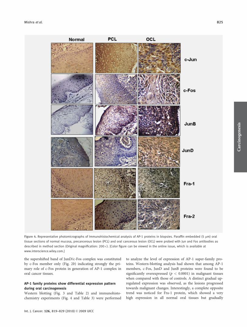
Figure 4. Representative photomicrographs of Immunohistochemical analysis of AP-1 proteins in biopsies. Paraffin embedded (5 lm) oral
tissue sections of normal mucosa, precancerous lesion (PCL) and oral cancerous lesion (OCL) were probed with Jun and Fos antibodies as
described in method section (Original magnification: 200�). [Color figure can be viewed in the online issue, which is available at
www.interscience.wiley.com.]
Carcinog
enesis
Mishra et al. 825
Int. J. Cancer: 126, 819–829 (2010) VC 2009 UICC

decreased as the lesions progressed and became nil in cancercases (p < 0.0001 in normal vs. cancer; Table 2). Although c-Jun and Fra-2 were also found to be upregulated in cancer-ous lesions (p ¼ 0.03, c-Jun; p ¼ 0.02, Fra-2), they were notelevated remarkably when compared with the high-expressionlevels of c-Fos, JunD and JunB in the same samples. To oursurprise, JunD, the only DNA-binding constituent of AP-1complex of all precancerous cases had not shown any re-markable overexpression when compared with the cancercases while corroborating its overexpression (Fig. 3) withtransactivation pattern (Fig. 2b). An inconsistent pattern ofFosB protein expression was also observed in all spectrumsof oral tissue biopsies from normal, premalignant to malig-nant lesions.
The expression profile of AP-1 protein members was fur-ther analyzed in situ by immunohistochemistry in paraffin-embedded sections and findings were very much in concord-ance with western blots. Intensity scoring of protein expres-sion in immunohistochemistry was performed on an arbi-trary 4-point scale; none (�); low (þ); moderate (þþ) andhigh (þþþþ) as described in Methods section. Results alongwith the statistical analyses are presented in Figure 4 andTable 3.
Expression of c-Fos protein was nil/low in 100% (10 of10) of the normal tissue sections examined, 80% precancer-ous biopsies (16 of 20) showed moderate and 95% tumors(19 of 20) showed a high expression of c-Fos (p < 0.0001 incancer vs. normal and precancer vs. normal). Conversely, forFra-1, most of the cancerous oral mucosa (19 of 20) showednil to low expression, 30% (6 of 20) of precancerous tissueshowed high positivity, but 70% of normal tissue sections (7of 10) have showed a high expression (p < 0.0001 in normalvs. cancer and precancer vs. cancer). About 75–90% of themalignant sections showed moderate to high immuno-stain-ing for JunB (18 of 20; p < 0.0001), JunD (16 of 20; p <
0.0004) and c-Jun (15 of 20; p ¼ 0.001) proteins, whereastheir expressions were either nil or low in normal as well asprecancerous mucosa. Low to high immunoreactivity againstFra-2 antibody was also observed in 85% of cancerous cases(17 of 20; p ¼ 0.04) each.
Transcriptional profiling of AP-1 members (c-fos, junD and
fra-1) and egfr mRNAs in oral tissue biopsies
The mRNA expression profiles from freshly collected normal,precancerous and cancerous biopsies for c-fos, junD and fra-1genes were further determined by northern-blot hybridization.
Densitometric analysis for mRNA transcripts in cancerouslesions revealed that c-fos, junD and egfr were significantlyoverexpressed with mean 6 SD values of 3.0 6 1.20, 2.11 60.80 and 1.91 6 0.73, respectively. On the contrary, fra-1expression was found to be gradually downregulated towardmalignant transformation of oral lesions with mean 6 SDvalue of 0.28 6 0.14 (Fig. 5). Interestingly, these members(c-fos, junD and fra-1) have also shown similar trend ofexpression at their protein level as observed by western blot-
ting (Fig. 3) and immunohistochemistry (Fig. 4), suggestingtheir regulation at transcriptional level during human oralcarcinogenesis.
c-fos upregulation, activation of NF-jB and induction of
EGFR expression are correlated in oral cancer tissues
Interestingly, we have observed that oral cancer biopsies haveparallel increase in c-fos mRNA (Fig. 5) along with overex-pressed egfr at both mRNA and protein levels (Figs. 5 andFig. 6a).
Because transcription of egfr gene is positively regulatedby NF-jB/p50 homodimers with recruitment of Bcl-3 as acoactivator,15 we further checked the expression and dimeri-zation pattern of NF-jB/p50 member in oral cancer cases. Asper our anticipation, upregulation of p50 (Fig. 6a) and Bcl-3(Fig. 6a) proteins with p50/p50 homodimers (Fig. 6b) wasfound in malignant specimens. Moreover, addition of anti-Bcl-3 antibody resulted in diminished p50/p50 NF-jB DNA-binding activity in supershift assay in malignant biopsies, asvisualized by densitometry analysis (Fig. 6b, last lane). Thisabsence of any supershifted band with a diminished DNAbinding indicates an interaction of Bcl-3 antibody withDNA-binding motif in p50/p50/Bcl-3 ternary complex pres-ent in oral cancer biopsies. To explore further the molecularmechanism involved in the induction of c-Fos in OSCC, we
Figure 5. Northern blotting of c-fos, fra-1, junD, egfr and b-actin
mRNA in oral biopsies. Increased expression of c-Fos, junD and
egfr and decreased expression of fra-1 mRNAs during development
of cancer lesions. Reprobing filters with b-actin probes confirmed
equal loading of mRNA.
Carcinog
enesis
826 AP-1 protein in human oral carcinogenesis
Int. J. Cancer: 126, 819–829 (2010) VC 2009 UICC

checked the expression of a critical upstream regulatoryMAPKinase-pERK1/2, which is affected by EGFR signals.Western-blot analysis (Fig. 6a) has shown that pERK expres-sion was highest in malignant oral cases. Taken together,these data suggest that NF-jB/p50 homodimers activatesEGFR expression, which is correlated with c-Fos inductionvia ERK activation in oral cancer tissues.
DiscussionHigh DNA-binding activity of AP-1 is reported in many epi-thelial tumors such as cervix, skin, breast and head andneck.15–17 AP-1 activation is linked with alcohol consump-tion9 and HPV infections in oral cancers.18 In agreementwith our data, constitutive activation of AP-1 is also reportedin several different head and neck as well as oral cancer celllines.7–9
Assuming that altered composition of AP-1 DNA bindingcomplex regulates its transactivation pattern and conse-quently expression of downstream targets during carcinogen-esis,19 we have analyzed the composition of AP-1 complex innuclear extracts from tissue biopsies of different grades.
We found a strong DNA-binding activity in oral cancertissue contributed by c-Fos/JunD heterodimers in majority ofcancerous tissues, whereas involvement of JunB was alsonoticed only in some of malignant cases. Interestingly, almostall precancerous biopsies have shown only preferential homo-dimerization of JunD/JunD. A very high expression of c-Fosprotein was confirmed in all malignant oral tissues by west-ern blotting and immunohistochemistry experiments, which
corroborates the results of supershift assays. Presence of over-expressed c-Fos was expected in oral cancer lesions as it con-trols several downstream target genes such as VEGF, MMPs,collagenase I involved in invasiveness during development ofmany cancers including head and neck cancers.10,11,20,21 Wehave also seen the presence of JunD with c-Fos in AP-1 com-plex in majority of oral cancer cases, which again corrobo-rates our IHC and western-blotting data, suggesting graduallyupregulated JunD protein during carcinogenesis. The rela-tively lower DNA binding of JunD containing AP-1 complexin PCL samples but highest in OCL can be explained by thefact that JunD homodimer binds its cognate DNA sequencewith lower affinity and also posses lower transactivationpotential.22 Similar to our present report, it has beenobserved that c-Fos/JunD heterodimer together showedhigher transcriptional activity than JunD homodimers.23
Therefore, we speculate that JunD/JunD homodimer forma-tion might prevent the precancerous cells entering into can-cerous condition, but as soon as participation of c-Fos mem-ber takes place in AP-1 complex formation, the precancerouscells are pushed further in the aggressive cancerous condi-tion. Moreover, it is also reported that JunD changes func-tionally from growth suppressor to growth promoter possiblyby the formation of mutant form of JunD.24 Similar to ourobservation of prevalent c-Fos/JunD complex, the same com-plex also positively regulates the expression of tyrosinase inmelanomas.25 In our supershift studies of cancerous tissues,involvement of c-Jun and JunB proteins in transcriptionallyactive AP-1 complex formation is found to be insignificantalbeit their consistent overexpression were shown by westernblotting and immunohistochemical methods. These resultsindicate that overexpressed JunD is the main dimerizing part-ner of c-Fos in the transactivated AP-1 complex. c-Jun/JunBcomplex in malignant cases demonstrated here are eithertranscriptionally inactive or have lower DNA bindingcapacity as reported by others too.23 Absence of c-Jun intranscriptionally active AP-1 DNA-binding complex might bealso possible due to the fact that JunB can substitute c-Jun invivo.5 In a perfect reverse correlation with upregulated c-fosand junD expression, the fra-1 mRNA transcripts hasrevealed a higher expression in control specimens with agradual downregulated trend with increasing severity oflesion as seen in many cancers.15,26 Our observation is insharp contrast to majority of other reports, which showed ageneralized overexpressed Fra-1 in different cancers20; butoverexpressed c-Fos in oral cancer cases found in our studyis in total agreement with other investigators.8,10,11,27
In contrast to other reports based on cell lines7 and tissuesbiopsies28 of head and neck cancers, we observed a very dis-tinct downregulated expression of Fra-1 protein as well asmRNA in freshly collected malignant oral tissue biopsies.Although, we understand that a more detailed studies areneeded to elucidate the exact molecular mechanism of fra-1gene downregulation in oral cancers, some plausible path-ways involved are interesting here to mention. The inhibited
Figure 6. Upregulated c-Fos expression is correlated with NF-jB/
p50:p50 homodimer mediated EGFR signals in oral cancer cells. (a)
Immunoblotting from the same biopsies shows the elevated
expression level of p50, pERK, EGFR, Bcl-3 and c-Fos proteins. (b)
OCL category of biopsies showed the preferential homodimeration
of p50/p50 in supershift assay. Nuclear extracts from OCL were
incubated with specific antibodies (2 lg each) against each
member for NF-jB proteins. Band intensities were quantified as
described in Material and methods section of text and indicated. Carcinog
enesis
Mishra et al. 827
Int. J. Cancer: 126, 819–829 (2010) VC 2009 UICC

positive autoregulation of fra-1 transcription might be operat-ing in oral cancer cells due to hampered nuclear shuttling ofFra-1 protein.29,30 Moreover, repressed fra-1 in oral cancertissues infected with human Papillomaviruses cannot be ruledout as seen in 1 of the other major human epithelial cancerof uterine cervix in females.13,15
Lack of activation domain in Fra-231 can explain the ab-sence of DNA binding in our gel-shift assays even it is upreg-ulated in many cancers.32,33 The low-binding activity of JunBeven after its elevated expression can be attributed to its in-herent lower DNA-binding activity seen in many oral cancercell lines.7 We expect that highly prevalent mutated p16 pro-teins in oral cancer cases might have altered the property ofJunB from antioncogenic to prooncogenic along with itsoverexpression in OSCC.
In this study, we have observed overexpressed EGFR in allmalignant cases as shown by others.34–37 Moreover, manyearlier reports38,39 also support our findings of p50/p50 homo-dimerization with participation of Bcl-3 (Fig. 6b) in NF-jBcomplex of oral cancer biopsies. Interestingly, Thornburget al.14 have shown overexpressed egfr due to the presence ofco-activator Bcl-3 in p50/NF-jB complex in nasopharyngealcancers. These data together indicate that p50 homodimeriza-tion in NF-jB complex positively regulates egfr expression inoral cancers.
Additionally, upregulated c-Fos due to activated ERK is awell-known signaling pathway.40 Similar to our findings ofupregulated pERK1/2 including elevated c-Fos and EGFRproteins in cancer biopsies (Fig. 6a), ERK1/2, an upstreamkinase of c-Fos, is reported to be activated in oral cancersthrough EGFR signals.41,42 In fact, c-Fos is also known as anestablished marker of anti-EGFR therapy in many cancer celllines including HNSCC.8 Moreover, studies have also docu-mented the cross-regulation of AP-1 and NF-jB43,44 path-ways through overexpressed EGFR-mediated activation byMAPKinase pathways in HNSCC cell lines.8,14 Therefore, inlight of findings of (i) p50/NF-jB activation along with egfroverexpression and (ii) elevated pERK and c-Fos proteins incancer biopsies, we are tempted to speculate that in humanoral cancers NF-jB and AP-1 signals might be coupled as:p50/p50 (NF-jB) ! EGFR : ! ERK ! c-Fos/AP-1 :.Additional functional studies and clinical correlation mayprovide further insights in signaling connections betweenAP-1 and NF-jB that may contributes toward better under-standing of oral carcinogenesis.
In conclusion, this study demonstrates an important roleof AP-1 as revealed by differential expression and transacti-vation of AP-1 super-family proteins that occur as a func-tion of severity of lesions during progression of oralcarcinogenesis.
References
1. Parkin DM, Bray F, Ferlay J, Pisani P.Estimating the world cancer burden:Globocan 2000. Int J Cancer 2001;94:153–6.
2. Herrero R, Castellsague X, Pawlita M,Lissowska J, Kee F, Balaram P, RajkumarT, Sridhar H, Rose B, Pintos J, FernandezL, Idris A, Sanchez MJ, Nieto A, TalaminiR, Tavani A, Bosch FX, Reidel U, SnijdersPJ, Meijer CJ, Viscidi R, Munoz N,Franceschi S; IARC Multicenter OralCancer Study Group. Humanpapillomavirus and oral cancer: theInternational Agency for Research onCancer Multicenter Study. J Natl CancerInst 2003;95:1772–83.
3. zur Hausen H. Papillomaviruses andcancer: from basic studies to clinicalapplication. Nat Rev Cancer 2002;2:342–50.
4. Angel P, Karin M. The role of Jun, Fosand the AP-1 complex in cell-proliferationand transformation. Biochim Biophys Acta1991;1072: 129–57.
5. Eferl R, Wagner EF. AP-1: a double-edgedsword in tumorigenesis. Nat Rev Cancer2003;3:859–68.
6. Young MR, Yang HS, Colburn NH.Promising molecular targets for cancerprevention: AP-1, NF-jB and Pdcd4.Trends Mol Med 2003;9:36–41.
7. Ondrey FG, Dong G, Sunwoo J, Chen Z,Wolf JS, Crowl-Bancroft CV, Mukaida N,Van Waes C. Constitutive activation oftranscription factors NF-jB, AP-1, andNF-IL6 in human head and necksquamous cell carcinoma cell lines thatexpress pro-inflammatory and pro-angiogenic cytokines. Mol Carcinog 1999;26:119–29.
8. Bancroft CC, Chen Z, Yeh J, Sunwoo JB,Yeh NT, Jackson S, Jackson C, Van WaesC. Effects of pharmacologic antagonists ofepidermal growth factor receptor, PI3Kand. MEK signal kinases on NF-kappaBand AP-1 activation and IL-8 and VEGFexpression in human head and necksquamous cell carcinoma lines. Int JCancer 2002;99:538–48.
9. Timmons SR, Nwankwo JO, Domann FE.Acetaldehyde activates Jun/AP-1 expressionand DNA binding activity in human oralkeratinocytes. Oral Oncol 2002;38:281–90.
10. de Sousa SO, Mesquita RA, Pinto DS,Gutkind S. Immunolocalization of c-Fosand c-Jun in human oral mucosa and inoral squamous cell carcinoma. J OralPathol Med 2002;31:78–81.
11. Turatti E, da Costa, Neves A, de MagalhaesMH, de Sousa SO. Assessment of c-Jun,c-Fos and cyclin D1 in premalignant and
malignant oral lesions. J Oral Sci 2005;47:71–6.
12. Dignam JD. Preparation of extracts fromhigher eukaryotes. Methods Enzymol 1990;182:194–203.
13. Prusty BK, Das BC. Constitutive activationof transcription factor AP-1 in cervicalcancer and suppression of humanpapillomavirus (HPV) transcription andAP-1 activity in HeLa cells by curcumin.Int J Cancer 2005;113:951–60.
14. Rosl F, Das BC, Lengert M, Geletneky K,zur Hausen H. Antioxidant-inducedchanges of the AP-1 transcription complexare paralleled by a selective suppression ofhuman papillomavirus transcription.J Virol 1997;71:362–70.
15. Thornburg NJ, Pathmanathan R, Raab-Traub N. Activation of nuclear factor-jBp50 homodimer/Bcl-3 complexes innasopharyngeal carcinoma. Cancer Res2003;63:8293–301.
16. Yuspa SH. The pathogenesis of squamouscell cancer: lessons learned from studies ofskin carcinogenesis. J Dermatol Sci 1998;17:1–7.
17. Chen TK, Smith LM, Gebhardt DK,Birrer MJ, Brown PH. Activation andinhibition of the AP-1 complex in humanbreast cancer cells. Mol Carcinog 1996;15:215–26.
Carcinog
enesis
828 AP-1 protein in human oral carcinogenesis
Int. J. Cancer: 126, 819–829 (2010) VC 2009 UICC

18. Ke LD, Adler-Storthz K, Mitchell MF,Clayman GL, Chen Z. Expression ofhuman papillomavirus E7 mRNA inhuman oral and cervical neoplasia and celllines. Oral Oncol 1999;35:415–20.
19. Chinenov Y, Kerppola TK. Closeencounters of many kinds: Fos-Juninteractions that mediate transcriptionregulatory specificity. Oncogene 2001;20:2438–52.
20. Milde-Langosch K. The Fos family oftranscription factors and their role intumourigenesis. Eur J Cancer 2005;41:2449–61.
21. Vairaktaris E, Spyridonidou S, PapakostaV, Vylliotis A, Lazaris A, Perrea D,Yapijakis C, Patsouris E. The hamstermodel of sequential oral oncogenesis. OralOncol 2008;44:315–24.
22. Pfarr CM, Mechta F, Spyrou G, LallemandD, Carillo S, Yaniv M. Mouse JunDnegatively regulates fibroblast growth andantagonizes transformation by ras. Cell1994;76:747–60.
23. Bakiri L, Matsuo K, Wisniewska M,Wagner EF, Yaniv M. Promoter specificityand biological activity of tethered AP-1dimers. Mol Cell Biol 2002;22:4952–64.
24. Agarwal SK, Novotny EA, Crabtree JS.Transcription factor JunD, deprived ofmenin, switches from growth suppressor togrowth promoter. Proc Natl Acad Sci USA2003;100:10770–5.
25. Englaro W, Rezzonico R, Durand-ClementM, Lallemand D, Ortonne JP, Ballotti R.Mitogen-activated protein kinase pathwayand AP-1 are activated during cAMP-induced melanogenesis in B-16melanoma cells. J Biol Chem 1995;270:24315–20.
26. de Wilde J, De-Castro Arce J, Snijders PJ,Meijer CJ, Rosl F, Steenbergen RD.Alterations in AP-1 and AP-1 regulatorygenes during HPV-induced carcinogenesis.Cell Oncol 2008;30:77–87.
27. Jimeno A, Kulesza P, Kincaid E, BouaroudN, Chan A, Forastiere A, Brahmer J, ClarkDP, Hidalgo M. c-Fos assessment as a
marker of anti-epidermal growth factorreceptor effect. Cancer Res 2006;66:2385–90.
28. Mangone FR, Brentani MM, Nonogaki S,Begnami MD, Campos AH, Walder F,Arvalho MB, Soares FA, Torloni H,Kowalski LP, Federico MH. Overexpressionof Fos-related antigen-1 in head and necksquamous cell carcinoma. Int J Exp Pathol2005;86:205–12.
29. Bergers G, Graninger P, Braselmann S,Wrighton C, Busslinger M. Transcriptionalactivation of the fra-1 gene by AP-1 ismediated by regulatory sequences in thefirst intron. Mol Cell Biol 1995;15:3748–58.
30. Burch PM, Yuan Z, Loonen A, Heintz NH.An extracellular signal-regulated kinase 1-and 2-dependent program of chromatintrafficking of c-Fos and Fra-1 is requiredfor cyclin D1 expression during cell cyclereentry. Mol Cell Biol 2003;24:4696–709.
31. Foletta VC. Transcription factor AP-1, andthe role of Fra-2. Immunol Cell Biol 1996;74:121–33.
32. Milde-Langosch K, Roder H, Andritzky B.The role of the transcription factors c-Fos,FosB, Fra-1 and Fra-2 in the invasionprocess of mammary carcinomas. BreastCancer Res Treat 2004;86:139–52.
33. Zoumpourlis V, Papassava P,Linardopoulos S, Gillespie D, Balmain A,Pintzas A. High levels of phosphorylatedc-Jun, Fra-1, Fra-2 and ATF-2 proteinscorrelate with malignant phenotypes in themultistage mouse skin carcinogenesismodel. Oncogene 2000;19:4011–21.
34. Pomerantz RG, Grandis JR. The role ofepidermal growth factor receptor in headand neck squamous cell carcinoma. CurrOncol Rep 2003;5:140–6.
35. Saranath D, Panchal RG, Nair R, MehtaAR, Sanghavi VD, Deo MG. Amplificationand overexpression of epidermal growthfactor receptor gene in humanoropharyngeal cancer. Eur J Cancer B OralOncol 1992;28:139–43.
36. Vairaktaris E, Loukeri S, Vassiliou S,Nkenke E, Spyridonidou S, Vylliotis A,
Papakosta V, Lazaris A, Agrogiannis G,Yapijakis C, Perrea D, Patsouris E. EGFRand c-Jun exhibit the same pattern ofexpression and increase gradually duringthe progress of oral oncogenesis. In Vivo2007;21:791–6.
37. Todd R, Wong DT. Epidermal growthfactor receptor (EGFR) biology and humanoral cancer. Histol Histopathol 1999;14:491–500.
38. Fujita T, Nolan GP, Liou HC, Scott ML,Baltimore D. The candidate proto-oncogene bcl-3 encodes a transcriptionalcoactivator that activates through NF-j Bp50 homodimers. Genes Dev 1993;7:1354–63.
39. Watanabe N, Iwamura T, Shinoda T,Fujita T. Regulation of NFKB1 proteinsby the candidate oncoprotein BCL-3:generation of NF-jB homodimersfrom the cytoplasmic pool of p50-p105\and nuclear translocation. EMBO J 1997;16:3609–20.
40. Monje P, Hernandez-Losa J, Lyons RJ,Castellone MD, Gutkind JS. Regulationof the transcriptional activity of c-Fosby ERK. A novel role for the prolylisomerase. PIN1. J Biol Chem 2005;280:35081–4.
41. Mishima K, Inoue K, Hayashi Y.Overexpression of extracellular-signalregulated kinases on oral squamous cellcarcinoma. Oral Oncol 2002;38:468–74.
42. Turjanski AG, Vaque JP, Gutkind JS. MAPkinases and the control of nuclear events.Oncogene 2007;26:3240–53.
43. Fujioka S, Niu J, Schmidt C, Sclabas GM,Peng B, Uwagawa T, Li Z, Evans DB,Abbruzzese JL, Chiao PJ. NF-jB and AP-1connection: mechanism of NF-jB-dependent regulation of AP-1 activity. MolCell Biol 2004;24:7806–19.
44. Stein B, Baldwin AS, Ballard DW, GreeneWC, Angel P, Herrlich P. Cross-couplingof the NF-j B p65 and Fos/Juntranscription factors produces potentiatedbiological function. EMBO J 1993;12:3879–91.
Carcinog
enesis
Mishra et al. 829
Int. J. Cancer: 126, 819–829 (2010) VC 2009 UICC