
A Decade of Children's Environmental Health Research: Highlights ...
<<NOTE TO USER: Please add details of the date, time, place and sponsorship of the meeting for which you are using this presentation in the space indicated.>>
<<NOTE TO USER: This is a large set of slides from which the presenter should select the most relevant ones to use in a specific presentation. These slides cover many facets of the problem. Present only those slides that apply most directly to the local situation in the region.>>
<<NOTE TO USER: This module presents several examples of risk factors that affect reproductive health. You can find more detailed information in other modules of the training package that deal with specific risk factors, such as lead, mercury, pesticides, persistent organic pollutants, endocrine disruptors, occupational exposures; or disease outcomes, such as developmental origins of disease, reproductive effects, neurodevelopmental effects, immune effects, respiratory effects, and others.>>
<<NOTE TO USER: For more information on reproductive health, please visit the website of the Department of Reproductive Health and Research at WHO: www.who.int/reproductivehealth/en/>>
1
November 2011
TRAINING FOR THE HEALTH SECTOR [Date ... Place ... Event ... Sponsor ... Organizer}
FEMALE REPRODUCTIVE HEALTH
AND THE ENVIRONMENT
Training Module 2
Children's Environmental Health Public Health and the Environment
World Health Organization
www.who.int/ceh
WHO/HSE/PHE/EPE/11.01.11
<<READ SLIDE.>>
According to the formal definition by the World Health Organization (WHO), health is more than absence of illness. It is a state of complete physical, mental and social well-being. Similarly, reproductive health also represents a state of complete physical, mental and social well-being, and not merely the absence of reproductive disease or infirmity.
This presentation will introduce you to the basics of female reproductive health disorders and the potential role that the environment may play in the development of these disorders.
Refs:•WHO. Department of Reproductive Health and Research, Partner Brief. Geneva, Switzerland, World Health Organization, 2009. WHO/RHR/09.02. Available at whqlibdoc.who.int/hq/2009/WHO_RHR_09.02_eng.pdf – accessed 15 June 2011•WHO. Preamble to the Constitution of the World Health Organization as adopted by the International Health Conference. New York, United States of America, World Health Organization, 1946.
2
Female Reproductive Health and the Environment
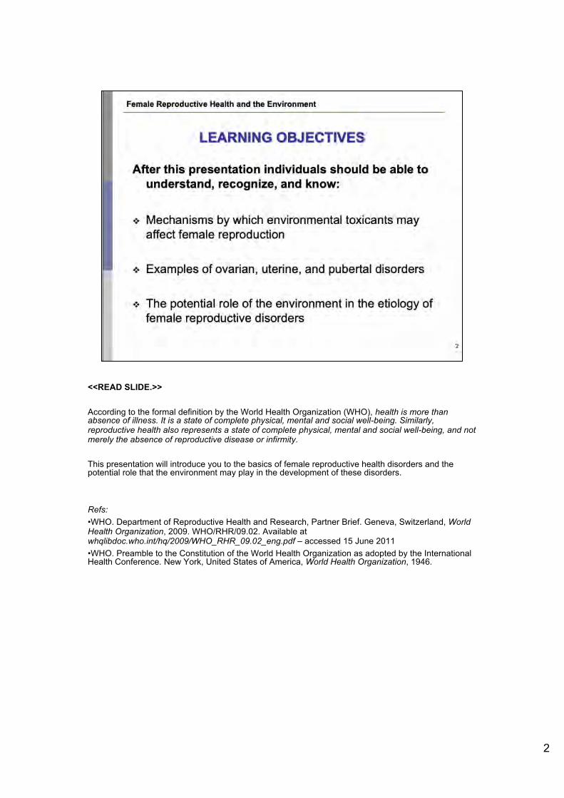
LEARNING OBJECTIVES
After this presentation individuals should be able to understand, recognize, and know:
❖ Mechanisms by which environmental toxicants may affect female reproduction
❖ Examples of ovarian, uterine, and pubertal disorders
❖ The potential role of the environment in the etiology of female reproductive disorders
2

<<READ SLIDE.>>
<<NOTE TO USER: You may decide to delete certain parts of the presentation depending on time. Please correct the outline accordingly.>>
3
Female Reproductive Health and the Environment
OUTLINE
❖ Considerations in female infertility and fecundity ❖ Potentia l connections to environmental exposures
❖ Potential mechanisms of action of environmental contaminants on reproductive health
❖ Overview of female hormonal disorders ❖ Ovarian disorders ❖ Uterine disorders ♦:♦ Pubertal development alterations

The World Health Organization defines the term primary infertility as the inability to bear any children, whether this is the result of the inability to conceive a child, or the inability to carry a child to full term after 12 months of unprotected sexual intercourse. Primary infertility is sometimes known as primary sterility. However, in many medical studies, the term primary infertility is only used to describe a situation where a couple is not able to conceive. Secondary infertility is defined as the inability to have a second child after a first birth. Secondary infertility has shown to have a high geographical correlation with primary infertility. Fecundity describes the ability to conceive after several years of exposure to risk of pregnancy. Fecundity is often evaluated as the time necessary for a couple to achieve pregnancy. The World Health Organization recommends defining fecundity as the ability for a couple to conceive after two years of attempting to become pregnant. The terms infertility and infecundity are often confused. Fertility describes the actual production of live offspring, while fecundity describes the ability to produce live offspring. Fecundity cannot be directly measured, though it may be assessed clinically. Typically, fecundity may be assessed by the time span between a couple’s decision to attempt to conceive and a successful pregnancy.
Ref:•Rutsein S and Iqbal S. Infecundity, infertility, and childlessness in the developing world. Geneva, Switzerland, World Health Organization and ORC Macro, 2004. DHS Comparative Report, No. 9.
Image: WHO
4
Female Reproductive Health and the Environment
INFERTILITY AND FECUNDITY
❖ Primary infertility- fa ilure to bear any children after 12
months of unprotected sexua l intercourse
❖ Secondary infertility- fa ilure to have a second child after
a first birth
❖ Fecundity- the ability of a
couple to conce ive afte r a
certain time of attempting to
become pregnant
WHO 4

Fertility is a concept directly related to a number of both biological and behavioural factors. These factors mediate the influence of socio-economic status, living conditions, cultural beliefs, and other determinants on individual reproductive behaviour. These biological and behavioural factors are known as proximate determinants of fertility. These determinants define how social and economic environments can influence individual reproduction. Essentially, these factors explain why women do not have the maximum number of children they could potentially have in their lifetime. Biological constraints on fertility include not only the time lost during pregnancy, but also the time required for a woman to recover from pregnancy and childbirth. This time frame is referred to as postpartum infecundity and includes necessary maternal functions such as breastfeeding. The averaged estimated time of postpartum infecundity is approximately 1.5 months but may vary widely between females. Other biological constraints may include such factors as sterility induced by age or pathology. The term “total fecundity” is used to describe the natural limit in physiological capability of childbearing for an average female due to biological constraints. Several behavioural considerations also exist that influence the fertility of a woman. However, these include factors that pertain mostly to the possibility of conception. For example, the time a women spends in a sexual relationship or married directly affects her engagement in sexual intercourse and thus potential pregnancy. The most important behavioral consideration relates to the woman’s decision to utilize contraception. This may include traditional methods or modern methods of family planning.
Ref:• Frank O. The demography of fertility and fecundity. Geneva, Switzerland, World Health Organization, 2007. Available at www.gfmer.ch/Books/Reproductive_health/The_demography_of_fertility_and_infertility.html –accessed 10 June 2010
5
Female Reproductive Health and the Environment
PROXIMATE DETERMINANTS OF FERTILITY
❖ Biological and behavioral factors that influence individual reproductive behavior ❖ Explain why women do not have as many children as
possible in lifetime
❖ Biological constraints ❖ Breastfeeding
,:. Pathologies
❖ Behavioral constraints
A variety of internal and external factors
may influence female fertility!
❖ Single most important factor : use of contraception
5

6
Some female reproductive disorders linked to fertility and fecundity may occur during fetal development. Female reproductive organs begin to develop between the fourth and fifth week of pregnancy, and continue until the 20th
week of pregnancy. Due to the complexity of the development of the reproductive system, many factors may alter the healthy growth of these essential tissues, organs, and hormonal messaging pathways. Alterations may be the result of genetic abnormalities from external factors that may change the normal development of specific tissues. The mechanisms of action of various female disorders will be discussed in upcoming slides. You may also refer back to module 1 for more information about reproductive health abnormalities and their etiologies. However, it is important to note that female reproductive disorders may also develop during various life phases of the female. Alterations in proper reproductive functioning may be the result of various occurrences and experiences throughout childhood, adolescence, or adulthood. While much is known about the female reproductive system, its development, and many causes of specific disorders, the research pertaining to the mechanisms of action for certain pathologies is still largely unknown. However, exposure to environmental contaminants has been proposed in recent years to potentially contribute to female reproductive disorders. Research has been focused on exposures that occur during critical periods of development, however this is an emerging field of research that demands greater scientific investigation.
Refs:•Caserta D et al. Impact of endocrine disruptor chemicals in gynaecology. Human Reproductive Update, 2008, 14:59–72. •Foster WG et al. Environmental contaminants and human infertility: hypothesis or cause for concern? Journal of Toxicol Environmental Health: Critical Review, 2008, 11:162–76.
Image: WHO. Biennial Report of HRP (2008-2009). Special programme of research, development and research training in human reproduction (HRP). Geneva, Switzerland, World Health Organization, 2009. Available at whqlibdoc.who.int/hq/2010/WHO_RHR_HRP_10.09_eng.pdfm - accessed 7 July 2010.
Female Reproductive Health and the Environment
FEMALE REPRODUCTIVE DISORDERS
❖ Disorders related to female reproductive health may develop during fetal development, childhood, adolescence, or adulthood
❖ Multiple causes for changes in
female reproductive functioning
❖ Recent focus on potential
environmental causes
UNDP/U NFPAIWHO/World Bank , 2009
6

7
Though evidence from demographic surveys in the industrialized world showed a clear decrease in fertility, surveys conducted in association with the WHO in the developing world demonstrate different trends throughout various nations. This figure portrays either a positive or negative change in the percentage of of non-contracepting, sexually experienced women (age 15-49) who have been married for the past five years but had no live births or pregnancies. The bars compare the positive or negative difference between the first survey that occurred in 1975 and the second survey that occurred between the years of 1995 and 2000. You can see that some nations, such as Colombia, Peru, and Jordan, experienced a very large increase in the percentage of women who experienced no live births or pregnancies despite being sexually active during five years marriage and not using contraception. This trend may indicate a decrease in overall fertility and potentially fecundity. However, notice that some nations, such as Burkina Faso, Senegal, and Kazakhstan experienced a decrease in the percentage of women who experienced no live births or pregnancies despite being sexually active during five years marriage and not using contraception.
Ref:• ORC Macro and the World Health Organization. Infecundity, Infertility, and Childlessness in Developing Countries. Calverton, Maryland, USA, ORC Macro and the World Health Organization, 2004. DHS Comparative Reports No. 9.
Image: ORC Macro and the World Health Organization. Infecundity, Infertility, and Childlessness in Developing Countries. Calverton, Maryland, USA, ORC Macro and the World Health Organization, 2004. DHS Comparative Reports No. 9.
Do
Female Reproductive Health and the Environment
VARIABILITY IN DEVELOPING NATIONS l v rk 1n,1 f.MO
0.Tl"roo G no Kc:- )'JI
M.KJ.ag~.a,r .,_,,. "' N ,~~t
N~ger :a
S.«1eg ol
1,.an7.;an,.a
To go
Ug..-id• l•mb.a, .. iMbwo
CgyD t
Jord•n
-0((0
Sa-,g~d~
lndof'lo('S:.a,
IC.iu.•kh"-'!4 Bo ,v:• 8,..~-·
COIOMb•
in.ca n Rep ub ic
Gu•te~• .... · '0
- -• I
I ■ -• ■ ---I -■
• -- ■ --I ·• 0
Percent d~r-enc~ (re<
WHO JORC macro
s
Figure depicts percentage of of non-contracepting , sexually experienced women (age 15-49) who have been married for the past five years but had no live births or pregnancies , 1st
survey (1970) compared to 2nd
survey (1995-2000)
lO
Significantly variable trends between
developing nations!
7

A critical window of susceptibility is a period in which there are numerous changing capabilities in the developing fetus. Exposures to environmental contaminants during this window may result in permanent damage to a fetus and may have lifelong impacts on health. Given that development continues after birth, critical and sensitive windows occur before, during, and shortly after the fertilization of the egg. Critical windows of development are also present during pregnancy, infancy, childhood, and puberty. The diagram provided demonstrates the particular windows of susceptibility for the developing fetus. The maternal environment at these specific temporal windows has important implications for the healthy development of the reproductive organs of a developing fetus. However, disorders related to female reproductive health may develop during sensitive windows throughout fetal development, childhood, adolescence, or adulthood.
<< NOTE TO USER: For further information, please refer to the module on "Developmental and Environmental Origins of Disease”>>
Ref:•Calabrese EJ. Sex differences in susceptibility to toxic industrial chemicals. British Journal of Industrial Medicine, 1986, 43:577–579.
Figure: Reprinted from The Developing Human, Moore, Elsevier Inc., 1973. Used with copyright permission (2004) from Elsevier.
8
Female Reproductive Health and the Environment
CRITICAL WINDOWS OF SUSCEPTIBILITY
Sensit ive time interval dur ing development when environmental exposures can interfere with physiology of cell, tissue, or organ
Exposure at specific windows may result in adverse and irreversible effects
8

Reproductive health and the environment focuses on exposures to environmental contaminants during critical periods of human development. These periods are directly related to reproductive health throughout the life course, including the period before conception, at conception, fertility, pregnancy, child and adolescent development, and adult health. Exposures to different environmental contaminants may influence reproductive health status of the individual and its offspring, through the process of epigenetics. Environmental toxins may potentially induce effects in human reproductive processes. However, the extent of this hypothesis must be supported through greater levels of research. Currently, women’s health care providers and gynecologists are growing increasingly aware of the potential for environmental factors to influence female health and reproductive status.
Refs:•WHO. Global assessment of the state of the science of endocrine disruptors. Geneva, Switzerland, WHO/PCS/EDC, 2002. Available at www.who.int/ipcs/publications/new_issues/endocrine_disruptors/en/ - accessed 23 June 2010. •Woodruff T. Proceedings of the Summit on Environmental Challenges to Reproductive Health and Fertility: executive summary. Fertility and Sterility, 2003, 89 (2),1-20.
<< NOTE TO USER: For further information, please refer to the module on "Developmental and Environmental Origins of Disease”>>
9
Female Reproductive Health and the Environment
REPRODUCTIVE HEALTH AND THE ENVIRONMENT
❖ Focuses on exposure to contaminants found in the environment, specifically during critical periods of development.
❖ All the physical , chemical, biological and social factors that may affect the origin, growth, development and surv ival of a person in a given setting.
Some examples include:
- Specific synthetic chemicals
- Some metals
- Air contaminants
Still an emerging issuel
9

Developmental toxicants are agents that adversely affect the developing embryo or fetus. Some mothers may be exposed to thesein the occupational setting. In addition to highly sensitive windows for morphological abnormalities (birth defects), there are also time windows important for the development of physiological defects and morphological changes at the tissue, cellular and subcellular levels. Most existing data are related to preconceptional and prenatal exposures. Data on prenatal exposures are based mainly on studies of maternal exposure to pharmaceuticals (e.g., diethylstilbestrol, thalidomide) and parental alcohol use, smoking, and occupational exposures. Information on critical windows for exposure during the postnatal period is scarce. Postnatal exposures have been examined in detail for only a few environmental agents, including lead, mercury, some pesticides, and radiation. Developmental exposures may result in health effects observed:•prenatally and at birth, such as spontaneous abortion, stillbirth, low birth weight, small size for gestational age, infant mortality, and malformation;•in childhood, such as asthma, cancer, neurological and behavioural effects;•at puberty, such as alterations in normal development and impaired reproductive capacity;•in adults, such as cancer, heart disease, and degenerative neurological and behavioural disorders.<<NOTE TO USER: For more information see module on how prenatal exposures affect the fetus, see modules on developmental environmental origins of disease; occupational exposures and child health.>>Refs: •Birnbaum L. The effects of environmental chemicals on human health. Fertility and Sterility, 2008, 89(1):e31•Blanck HM et al. Age at menarche and tanner stage in girls exposed in utero and postnatally to polybrominated biphenyl. Epidemiology, 2000, 11(6):641–7.•Canadian Partnership for Children's Health & Environment. Child health and the environment, a primer. Canadian Partnership for Children's Health & Environment, 2005.•Daniels JL, Olshan AF, Savitz DA. Pesticides and childhood cancers. Environmental health perspectives, 1997, 105(10):1068–77.•Gonzalez-Cossio T et al H. Decrease in birth weight in relation to maternal bonelead burden. Pediatrics, 1997, 100(5):856–62.•Hu FB et al. Prevalence of asthma and wheezing in public schoolchildren: association with maternal smoking during pregnancy. Annals of allergy, asthma and immunology, 1997, 79(1):80–4.•Krstevska-Konstantinova M et al. Sexual precocity after immigration from developing countries to Belgium: evidence of previous exposure to organochlorine pesticides. Human reproduction, 2001, 16(5):1020–6.•Mendola P. et al. Science linking environmental contaminant exposures with fertility and reproductive health impacts in the adult female. Fertility and Sterility, 2008, 89 (1):e81-e94.•Miller RW. Special susceptibility of the child to certain radiation-induced cancers. Environmental health perspectives, 103(Suppl. 6):41–4. •Moore KL, Persaud TVN. The developing human: clinically oriented embryology. W.B. Saunders, Philadelphia, 1973, 98.•Needleman HL et al. The long-term effects of exposure to low doses of lead in childhood. An 11-year follow-up report. New England journal of medicine, 1990, 322:83–8.•Osmond C et al. Early growth and death from cardiovascular disease in women. British medical journal, 1993. •Selevan SG, Kimmel CA, Mendola P. Windows of susceptibility to environmental exposures in children. In: Children's health and the environment: a global perspective. Pronczuk J, ed. WHO, Geneva, 2005, 2:17-25•Selevan SG, Lemasters GK. The dose-response fallacy in human reproductive studies of toxic exposures. Journal of occupational medicine, 1987, 29(5):451–4.•Wadsworth ME, Kuh DJ. Childhood influences on adult health: a review of recent work from the British 1946 national birth cohort study, the MRC National Survey of Health and Development. Paediatric and perinatal epidemiology, 1997, 11:2–20.
10
Female Reproductive Health and the Environment
PRENATAL ENVIRONMENTAL HEALTH
❖ Methylmercury ❖ Lead ❖ Ionizing radiations ❖ Polychlorinated biphenyls ❖ Polycyclic aromatic
compounds ❖ Other air contaminants ❖ Organic solvents ❖ Some pesticides ❖ Alcohol ❖ Others ...
Developmental toxicants' effects:
Spontaneous abortion
Stillbirth
Low
Birth weight
Decreased head circumference
Preterm delivery
Birth defects
Visual and hearing deficits
Chromosomal abnormalities
Intellectual deficits
Others 10

•Weinhold B. Environmental factors in birth defects: What we need to know. Environmental health perspectives, 2009, 117 (10) :A440-A447.•WHO. Principles for evaluating health risks in children associated with chemical exposure. Environmental Health Criteria 237. WHO, Geneva, Switzerland, 2006. Available at www.who.int/ipcs/publications/ehc/ehc237.pdf – accessed March 2011 •WHO. Children's health and the environment: a global perspective. Pronczuk J, ed. WHO, Geneva, 2005
10

Several chemicals, compounds (both synthetic and organic), metals, and other environmental toxicants have been associated with adverse human health effects at high concentrations but we do not know with certainty if there is a safe threshold. Significant scientific concerns over the potential impact of these environmental hazards on reproductive health have increased research and public debate on this issue. For instance, evidence is arising on relationships between spontaneous abortion as well as reduction of anogenital distance and exposure to DDT during pregnancy. In life, we are all exposed to a combination of environmental risk factors and mixtures of chemicals. We must learn more about low-level exposures, effects of combined exposures and mixtures and importance of the timing of exposures. Refs:•Collaborative on Health & the Environment (CHE). Chemical contaminants in the environment. Collaborative on Health & the Environment (CHE). www.healthandenvironment.org - accessed 20 March 2010. •Swan SH et al. Decrease in anogenital distance among male infants with prenatal phthalate exposure. Environ. Health Perspect.2005, 113 (8):1056–61. Prenatal phthalate exposure impairs testicular function and shortens anogenital distance (AGD) in male rodents. We present data from the first study to examine AGD and other genital measurements in relation to prenatal phthalate exposure in humans. A standardized measure of AGD was obtained in 134 boys 2–36 months of age. AGD was significantly correlated with penile volume (R = 0.27, p = 0.001) and the proportion of boys with incomplete testicular descent (R = 0.20, p = 0.02). We defined the anogenital index (AGI) as AGD divided by weight at examination [AGI = AGD/weight (mm/kg)] and calculated the age-adjusted AGI by regression analysis. We examined nine phthalate monoester metabolites, measured in prenatal urine samples, as predictors of age-adjusted AGI in regression and categorical analyses that included all participants with prenatal urine samples (n = 85). Urinary concentrations of four phthalate metabolites [monoethyl phthalate (MEP), mono-n-butyl phthalate (MBP), monobenzyl phthalate (MBzP), and monoisobutyl phthalate (MiBP)] were inversely related to AGI. After adjusting for age at examination, p-values for regression coefficients ranged from 0.007 to 0.097. Comparing boys with prenatal MBP concentration in the highest quartile with those in the lowest quartile, the odds ratio for a shorter than expected AGI was 10.2 (95% confidence interval, 2.5 to 42.2). The corresponding odds ratios for MEP, MBzP, and MiBP were 4.7, 3.8, and 9.1, respectively (all p-values < 0.05). We defined a summary phthalate score to quantify joint exposure to these four phthalate metabolites. The age-adjusted AGI decreased significantly with increasing phthalate score (p-value for slope = 0.009). The associations between male genital development andphthalate exposure seen here are consistent with the phthalate-related syndrome of incomplete virilization that has been reported in prenatally exposed rodents. The median concentrations of phthalate metabolites that are associated with short AGI and incompletetesticular descent are below those found in one-quarter of the female population of the United States, based on a nationwide sample. These data support the hypothesis that prenatal phthalate exposure at environmental levels can adversely affect male reproductive development in humans.•Torres-Sanchez L et al. Dichlorodiphenyldichloroethylene exposure during the first trimester of pregnancy alters the anal position in male infants. Annals of the New York Academy of Sciences, 2008, 1140:155–162. •WHO. Chemicals assessment. Geneva, Switzerland, WHO/IPCS, 2007. www.who.int/ipcs/assessment/en/index.html - accessed 20 June 2010. •WHO. Fact sheet: Dioxins and their effects on human health. Geneva, Switzerland, World Health Organization, 2008. www.who.int/mediacentre/factsheets/fs225/en/index.html - accessed 20 June 2010. •Woodruff T. Proceedings of the Summit on Environmental Challenges to Reproductive Health and Fertility: executive summary.Fertility and Sterility, 2003, 89 (2),1-20.
11
Female Reproductive Health and the Environment
CHEMICALS POT NTIALLY ASSOCIATED WITH REPRODUCTIVE HEAL TH EFFECTS
Commonly used pesticides
Flame retardants
Dioxin-like substances
Phthalates
Addit ives to consumer products (plasticizers)
DDT ( dichlorodiphenyltrichloroethane) Organophosphates
PBDEs (polybrominated dipheny1ethers)
PCBs (polychlorinated biphenyls)
PVC (polyviny l chloride) Di ethyl hexyl phthalate
SPA (bisphenol A)
Multiple case studies from wildlife exposures ; some human evidence
Animal exposure models/data
-Animal exposure models/data -Wildlife exposure studies -Weak human exposure data
-Animal exposure models/data - Emerging human studies (surveys , biomarker associat ion)
- Evidence from animal exposure models/data
11

The endocrine system is a complex network of hormones that regulates various bodily functions such as growth and development. The endocrine glands include the pituitary, thyroid, adrenal, thymus, pancreas, ovaries, and testes. These glands or organs release carefully-measured levels of hormones into the bloodstream that act as natural chemical messengers to control important processes of the body. Specific environmental toxicants directly effect the endocrine system. Endocrine disruptors are exogenous agents that interfere with the synthesis, secretion, transport, binding, action, or elimination of natural hormones in the body that are responsible for the maintenance of homeostasis, reproduction, development, and/or behavior. Endocrine disruptors can change normal hormone levels, stimulate or halt the production of certain hormones, or change the way hormones move through the body. However, greater research is still needed to ascertain this hypothesis. DDT: dichlorodiphenyltrichloroethaneDDE: dichlorodiphenyldichloroethylene <<NOTE TO USER: For more information see module on Introduction to reproductive health and environment and/or module on endocrine disruption.>>
Refs: •Calafat AM, Needham LL. Human exposures and body burdens of endocrine-disrupting chemicals. In: Gore AC, ed. Endocrine-disrupting chemicals: from basic research to clinical practice. Totowa, NJ: Humana Press, 2007, 253–268. •Gray LE et al. Adverse effects of environmental antiandrogens and androgens on reproductive development in mammals. Int Journal of Androgens, 2007, 29:96–104.•WHO. Global assessment of the state of the science of endocrine disruptors. Geneva, Switzerland, WHO/PCS/EDC, 2002. Available at www.who.int/ipcs/publications/new_issues/endocrine_disruptors/en/ - accessed 23 June 2010.
Image: WHO
12
Female Reproductive Health and the Environment
ENDOCRINE DISRUPTORS
Endocrine disruptors interfere with the production, metabolism, and action of natural hormones in the body - Disrupt hormones needed for homeostasis and developmental
processes
- Alter estrogen , androgen, thyroid, neuroendocrine and metabolic signaling
Endocrine disruptors include: - Some pesticides (dichlorod iphenyltrichloroethane (DDT),
dichlorodiphenyldichloroethylene (DOE))
- Some herbicides (atrazine)
- Some Persistent Organic Pollutants (POPs) (e.g. dioxin)
- Potential: e.g. phthalates

As increasingly more women enter the workforce, they may be exposed to a variety of occupational chemicals and hazards that may lead to adverse health and reproductive effects. In addition, smoking, alcohol consumption, and other lifestyle factors play an increasingly important role in determining the health status of women. There is now abundant evidence that environmental factors may contribute to many of the disease processes discussed above. Some examples of likely environmental impact on women's health include the following:
<<READ SLIDE.>>
Among the most widespread and persistent environmental toxicants are chlorinated hydrocarbons (such as DDT and polychlorinated biphenyls), which are known to possess estrogenic potential, i.e., the ability to mimic the biological effects of estrogens. Imbalanced or unopposed estrogen exposure is a leading risk factor for many gynecologic malignancies, as well as benign proliferative disorders such as endometriosis and leiomyoma. The potential impact of these compounds on hormone-dependent physiological processes such as conception and fetal development, as well as on disease processes such as osteoporosis and cardiovascular disease, demands further exploration.
DDT: dichlorodiphenyltrichloroethane
Ref:•Ontario's Maternal Newborn and Early Child Development Resource Centre. Workplace reproductive health: research and strategis. Best Start. Ontario's Maternal Newborn and Early Child Development Resource Centre. 2001. Available at www.beststart.org/resources/wrkplc_health/pdf/WorkplaceDocum.pdf - accessed 31 October 2011.
Female Reproductive Health and the Environment
ENDOCRINE DISRUPTORS
❖ The most widespread and persistent are chlorinated hydrocarbons: may mimic the biological effects of estrogens
❖ Excessive estrogen exposure is a key risk factor for gynaecologic malignancies and benign proliferative disorders, e.g. endometriosis and leiomyoma
Also on hormone-dependent physiological processes
e.g. conception; fetal development
Possibly on osteoporosis and cardiovascular disease
13

There are several mechanisms of action that environmental contaminants may have within the human body. However, it is only recently that research has discovered these pathways. Thus, perhaps more mechanisms exist of which we are currently unaware. An environmental contaminant acting directly on gene expression would alter hormone function and influence changes in reproductive processes and systems. This could either increase or decrease levels of endogenous hormones within the body. Neuroendocrine effects could occur by nervous system monitoring of the environment and neuronal signaling to the endocrine system. The endocrine system would then alter hormonal function in response. The epigenetic route includes the alteration in gene expression by environmental factors without a direct change in DNA sequence. It is important to note that epigenetic changes may sometimes confer developmental advantages, enabling the growing organism to modify development of organs and systems in response to downstream requirements. The genetic mechanism of action, however, directly changes the DNA sequence. This may include mutations of the DNA in the female egg cell, the male sperm cell, or in the developing fetus. Any of these direct genetic changes may impact reproductive processes. Finally, systemic toxicity indicates that an environmental exposure may result in widespread effects on many systems.
<<NOTE TO USER: The next slides will explain each mechanism in greater detail.>>
Refs:•Anway MD, Skinner MK. Epigenetic transgenerational actions of endocrine disruptors. Endocrinology, 2006, 147:S43-9.•Crain DA et al. Female reproductive disorders: the roles of endocrine-disrupting compounds and developmental timing. Fertility and Sterility, 2008, 90:911-940.
14
Female Reproductive Health and the Environment
MECHANISMS OF ACTION
1. Direct gene expression Environment directly acts on hormone function
2. Epigenetic route Environment augments gene expression , but does not act directly on DNA sequence
3. Genetic route Environmental exposure causes DNA mutations in the egg, sperm, or the fetus
4. Endocrine mimicking
5. Neuroendocrine route Effect nervous system that then acts on hormones
6. Systemic toxicity 14

The first way that an environmental agent can affect normal female reproductive function is through direct gene expression. Direct gene expression means that an environmental contaminant, once it enters the human body, will directly change the normal function of naturally occurring human hormones. This environmental contaminant will change normal hormonal functioning by acting directly on the gene responsible for this process. For example, a specific environmental toxin may enter the body, mimic a naturally occurring hormone, like estrogen, and bind to a cell’s receptor for estrogen. This binding process may directly change the normal hormonal functioning of a specific system and lead to augmentation in gene expression. In addition, an environmental contaminant may directly alter gene expression that regulates hormone production or secretion. This action may result in an increase or decrease in the levels of naturally occurring hormones in the body, leading to an imbalance of the endocrine system. Such an imbalance may have significant effects on the proper functioning of the reproductive system.
Refs:•Crews D, McLachlan JA. Epigenetics, evolution, endocrine disruption, health, and disease. Endocrinology 2006, 147:S4–10.•Jirtle RL, Skinner MK. Environmental epigenomics and disease susceptibility. National Review of Gene, 2007, 8:253–62.
Image: Phelps J. Headliners: Neurodevelopment: genome-wide screen reveals candidate genes for neural tube defects. Environmental Health Perspectives, 2006, 114:A351-A351. Reproduced with permission from Environmental Health Perspectives.
15
Female Reproductive Health and the Environment
1. DIRECT GENE EXPRESSION
❖ An environmental agent acts directly on gene expression to: ❖Change action of natural hormones
❖Change metabolism of natural hormones
❖ This mechanism indicates a
direct action of an
environmental agent on the
natural process of internal
hormones
ehsehp/p03.niohs..nih.govlar1icJe/fetchAlficJe.acUon?a,t;c/eUR/=ln/o%3Adoi"2FIO. I 289"2Feh p. 114-a35 I

The epigenetic mechanism of action suggests that environmental factors alter how a gene is expressed, but without directly changing the DNA sequence.Epigenetics is the study of inherited changes in phenotype (factors that account for appearance) that are not directly related to, nor explained by changes in our DNA pattern. For this reason, this field of study is known as "epi," the greek root for "above," indicating that a change has occurred that is not directly related to the genetic code, but “above it” somehow. In epigenetics, non-genetic causes are considered responsible for different expressions of phenotypes. Or, termed in a different way, epigenetics describes changes in the expression of our genes that are not caused by alterations in the DNA sequence. Essentially, a different factor accounts for the change in gene expression. Exogenous, or environmental components may affect gene regulation and thus, potentially, subsequent expression in the phenotype. Changes to gene expression induced by environmental contaminants can be permanent or transient. Research has shown that epigenetic changes may in fact be reversed.
<<NOTE TO USER: For more information about epigenetics, please refer to Module 1: Introduction to Environmental Reproductive Health.>>
Refs:•Anway MD, Skinner MK. Epigenetic transgenerational actions of endocrine disruptors. Endocrinology, 2006, 147:S43-9.•Diamanti-Kandarakis E et al. Endocrine-disrupting chemicals: an Endocrine Society scientific statement. The Endocrine Society. 2009.•Hartl D, Jones E. Genetics: analysis of genes and genomes. Sudbary, MA, USA: Jones and Bartlett Publishers, 2005. •Klug M. Concepts of genetics, 9th Edition. New York: Benjamin Cummings Publishing, 2008.
Image: WHO
16
Female Reproductive Health and the Environment
2. EPIGENETICS
❖ Changes in expression of genes
❖ May be caused by elements
in the environment
❖ Can alter gene express ion
❖ Can suppress or activate
spec ific genes
❖ Epigenetic changes may be
revers ible
WHO
16

17
• A genetic mechanism of action is one that directly impacts the genes of the female egg cell, male sperm cell, or fetus. This means that an environmental contaminant may directly induce changes in the DNA sequence. This may include mutations of the DNA in the female egg cell, the male sperm cell, or in the developing fetus. Any of these direct genetic changes may impact reproductive processes.• Aneuploidy is the most commonly identified chromosome abnormality in humans, occurring in at least 5% of pregnancies. Aneuploidy is also the most common known cause of mental retardation. Despite the devastating clinical consequences of aneuploidy, relatively little is known of how it originates in humans. • An example of how a genetic route of action may influence female reproductive health status comes from an animal study. Bisphenol A (BPA) is a synthetic compound that is added to many plastic consumer goods. When BPA enters the body, it has the ability to mimic naturally occurring estrogen, and is thus characterized as “estrogenic.” Research has found that BPA exposure may lead to disruptions in the proper formation of chromosome alignment. Laboratory mice kept in cages containing BPA were exposed to this chemical when it leached into their water sources. The female mice were found to have significant defects in the number and quality of their eggs, or oocytes. Researchers believe that this endpoint results from BPA’s known estrogenic activity, which would potentially lead to the death of an oocyte in specific situations. However, the evidence from this study is inadequate to determine a true causal pathway. More research on the true mechanism of action of BPA is needed to make any conclusive observations. It is important to note there is a lack of human studies that validate this genetic route of action. Refs:•Can A, Semiz O. Diethylstilbestrol (DES)-induced cell cycle delay and meiotic spindle disruption in mouse oocytes during in-vitro maturation. Mol Hum Reprod. 2000, 6:154–62. •Crain DA et al. Female reproductive disorders: the roles of endocrine-disrupting compounds and developmental timing. Fertility and Sterility, 2008,90:911-940.•Diamanti-Kandarakis E. et al. Endocrine-disrupting chemicals: an Endocrine Society scientific statement. 2009. Endocrine reviews. 30(4): 293-342.
•Hassold T, Hunt P. To err (meiotically) is human: the genesis of human aneuploidy. National Review of Genet, 2001;2:280-91.Aneuploidy (trisomy or monosomy) is the most commonly identified chromosome abnormality in humans, occurring in at least 5% of all clinically recognized pregnancies. Most aneuploid conceptuses perish in utero, which makes this the leading genetic cause of pregnancy loss. However, some aneuploid fetuses survive to term and, as a class, aneuploidy is the most common known cause of mental retardation. Despite the devastating clinical consequences of aneuploidy, relatively little is known of how trisomy and monosomy originate in humans. However, recent molecular and cytogenetic approaches are now beginning to shed light on the non-disjunctional processes that lead to aneuploidy.•Hunt PA et al. Bisphenol A exposure causes meiotic aneuploidy in the female mouse. Current Biology, 2003;13:546-53. There is increasing concern that exposure to man-made substances that mimic endogenous hormones may adversely affect mammalian reproduction. Although a variety of reproductive complications have been ascribed to compounds with androgenic or estrogenic properties, little attention has been directed at the potential consequences of such exposures to the genetic quality of the gamete. RESULTS: A sudden, spontaneous increase in meiotic disturbances, including aneuploidy, in studies of oocytes from control female mice in our laboratory coincided with the accidental exposure of our animals to an environmental source of bisphenol A (BPA). BPA is an estrogenic compound widely used in the production of polycarbonate plastics and epoxy resins. We identified damaged caging material as the source of the exposure, as we were able to recapitulate the meiotic abnormalities by intentionally damaging cages and water bottles. In subsequent studies of female mice, we administered daily oral doses of BPA to directly test the hypothesis that low levels of BPA disrupt female meiosis. Our results demonstrated that the meiotic effects were dose dependent and could be induced by environmentally relevant doses of BPA. CONCLUSIONS: Both the initial inadvertent exposure and subsequent experimental studies suggest that BPA is a potent meiotic aneugen. Specifically, in the female mouse, short-term, low-dose exposure during the final stages of oocyte growth is sufficient to elicit detectable meiotic effects. These results provide the first unequivocal link between mammalian meiotic aneuploidy and an accidental environmental exposure and suggest that the oocyte and its meiotic spindle will provide a sensitive assay system for the study of reproductive toxins.•Susiarjo M et al. Bisphenol A exposure in utero disrupts early oogenesis in the mouse. PLoS Genetics, 2007;3:e5.
Female Reproductive Health and the Environment
3. GENETIC ROUTE
❖ Directly changes DNA sequence
❖ Aneuploidy: abnormal number of chromosomes ,:. Commonly leads to miscarriage , congenital defects or
mental retardat ion
♦:♦ Mice kept in cages made with certain chemicals experienced reproductive defects - Bisphenol A (BPA): plasticizer with estrogen ic effects on
chromosomes - BPA effects include cell cycle arrest or death of oocyte
But, there are a lack of human studies supporting this endpoint I 17

Endocrine disrupting compounds act by mimicking or antagonizing naturally occurring hormones in the body. It is believed that endocrine disruptors act by interfering with synthesis, secretion, transport, metabolism, binding action, or elimination of natural hormones that are present in the body and are responsible for homeostasis, reproduction, and developmental process.
Refs: •Calafat AM, Needham LL. Human exposures and body burdens of endocrine-disrupting chemicals. In: Gore AC, ed. Endocrine-disrupting chemicals: from basic research to clinical practice. Totowa, NJ: Humana Press, 2007, 253–268. •Dickerson SM, Gore AC. Estrogenic environmental endocrine-disrupting chemical effects on reproductive neuroendocrine function and dysfunction across the life cycle. Rev Endocr Metab Disord, 2007, 8:143–159 15. •WHO. Global assessment of the state of the science of endocrine disruptors. Geneva, Switzerland, WHO/PCS/EDC, 2002. Available at www.who.int/ipcs/publications/new_issues/endocrine_disruptors/en/ - accessed 23 June 2010.
Image: Endocrine disruptor. National Institutes of Environmental Health Sciences. Available at www.niehs.nih.gov/news/newsletter/2009/july/images/endocrine-disruptor-graphic.jpg - accessed 20 March 2010.
18
Female Reproductive Health and the Environment
\ Endo<rine ~ Disruptor
4. ENDOCRINE MIMICKING
Hormone
❖ Endocrine disruptors may potentially disrupt the physiological function of naturally occurring hormones
"'-
❖ Endocrine disruptors (green) disrupt the normal binding process of hormones (orange) to their receptors (purple)
Response
NIEHS 18

The mechanism of action for some environmental contaminants is their capacity to interact with hormones necessary for reproductive processes. They can alter hormone synthesis, disrupt neural and immune signaling pathways, and alter the regulation of gene expression. Xenohormones interact with steroid hormones receptors, in particular those for estrogens and androgens. Xenohormones act through several mechanisms that can affect the reproductive system. They act through nuclear hormone receptors, the estrogen receptor (ER) and the androgen receptors (AR). Xenohormones could mimic estrogen action, antagonize testosterone action, or alter the secretion of follicle- stimulating hormone (FSH) and luteinizing hormone (LH). These actions could have an effect on reproductive health.Many documented incidents of decreased reproductive capacity in wildlife population are strongly associated with exposure to chemicals in the environment. Reproductive disorders in wildlife have included egg shell thinning of birds, widespread population declines, morphologic abnormalities, sex reversal, impaired viability of offspring, altered hormone concentration and changes in socio-sexual behavior.
<< NOTE TO USER: For more information on specific case studies of wildlife exposures, please refer to Module 1: Reproductive Health and the Environment.>>
Ref:• McLachlan JA, Environmental signaling: what embryos and evolution teach us about endocrine disrupting chemicals. Endocrine Review, 2000, 22: 319–341.
19
Female Reproductive Health and the Environment
INTERFERENCE WITH REPRODUCTIVE PROCESS S
❖ Some environmental contaminants may alter estrogen , androgen, and thyro id signal ing, essential for normal reproduct ive activity and embryonic development
❖ Xenohormones - Compounds that mimic the activity of the steroid hormones
❖ Multiple examp les from wildlife reproduct ive capacity
Exposure examples exist but mechanism of action largely unknown!
19

• More than 850 chemicals directly impact the nervous system and may cause adverse health effects. This includes some metals, organic solvent agrochemicals, poly-halogenated aromatic hydrocarbons, and pharmaceuticals. The neuro-endocrine system describes the collaborative functioning between the nervous and the endocrine system. These two systems are closely related because the secretion of certain important hormones in the body is regulated directly through the hypothalamus in the brain. The brain is a central feature of the nervous system.
• In the human body, the reproductive system and the hormonal pathways that dictate its proper functioning, are largely regulated by the neuro-endocrine system. When environmental contaminants affect the neuro-endocrine system, serious impacts can result on reproductive function. The neuro-endocrine mechanism of action describes how the nervous system senses changes in the environment and alerts the endocrine system of necessary changes that must be made in the body to maintain adequate health status. For example, if the nervous system notices the presence of a certain environmental contaminant, it may send signals to the endocrine system to augment production and secretion of a specific hormone. The induction of specific changes related to environmental exposures may result in adverse affects in reproductive function through the over-secretion or under-secretion of specific reproductive hormones.
• There are two different believed mechanisms of environmental contaminant on the neuro-endocrine system. Certain environmental contaminants may activate specific properties in adults and produce transient changes in the nervous system, or, exposure to environmental contaminants during neural development may induce changes in neurobehavioral function, specifically sex-related behaviours. Neurons monitor the environment and send signals to the endocrine system.
Ref:• Khan IA, Thomas P. Disruption of neuroendocrine control of luteinizing hormone secretion by
Aroclor 1254 involves inhibition of hypothalamic tryptophan hydroxylase activity. Biology of Reproduction, 2001, 64:955-964.
• Mensah-Nyagan AG et al. Neurosteroids: Expression of steroidogenic enzymes and regulation of steroid biosynthesis in the central nervous system. Pharmacology Review, 1999, 51:63-81.
Image: Terese Winslow, 2001. Scientific and Medical Illustrations. Used with copyright permission.
20
Female Reproductive Health and the Environment
5. NEUROENDOCRINE ROUTE
❖ Hundreds of environmental toxicants
can affect the nervous system
❖ Nervous system monitors environment
and sends signals to endocrine
system, which controls reproductive
processes
❖ Two potential processes: ❖ transient changes in adult nervous system
❖ permanent changes induced in neural development
r
Terese Winslow. 2001
20

21
Systemic toxicity indicates that an environmental exposure results in general health effects. This may result in adverse effects in reproductive health. There are several known pathologies that may result in adverse effects in reproductive function. For example, the sexually transmitted infections (e.g. gonorrhea) have been shown to lead to infertility in women when the infection was left untreated. Infections are very important and preventable causes of infertility in the developed as well as developing world. Chlamydia has been shown to cause fallopian tube infection which often does not present any symptoms, but can lead to significant reproductive dysfunction and subsequent infertility. Environmental risk factors, such as alcohol and mercury can also affect reproductive status. For instance, alcohol has been linked to irregular menstrual cycles and premature birth and alcohol consumption during pregnancy causes fetal alcohol spectrum disorders. Mercury is also neurotoxic to the unborn child.
Ref:•ORC Macro and the World Health Organization Infecundity, infertility, and childlessness in developing countries. Calverton, Maryland, USA, ORC Macro and the World Health Organization. DHS Comparative Reports No. 9.
Image: WHO. Reproductive Health Strategy to accelerate progress towards the attainment of international development goals and targets. Global strategy adopted by the 57th World Health Assembly. Geneva, Switzerland, World Health Organization, 2004. Available at www.who.int/reproductivehealth/publications/general/RHR_04_8/en/index.html - accessed 7 July 2010.
Female Reproductive Health and the Environment
6. SYSTEMIC TOXICITY
Maternal diseases may have adverse effects on reproduction ❖ Sexually transmitted diseases (HIV/AIDS, chlamydia)
❖ Infections (rubella)
Environmental exposures may result in systemic toxicity that later affects reproductive status
21 WHO

Hormonal balance of sexual hormones in particular is an important factor in maintaining fertility and regulating reproductive processes. Exogenous substances, such as environmental endocrine disruptors, may disturb the hormonal balance and thereby cause reproductive disorders. Three specific classes of female reproductive disorders are of particular concern. Ovarian disorders relate to the ovary, which is responsible for the production, storage, and release of the female reproductive cell, the egg, or the oocyte. Ovarian disorders also include pathologies that relate to the natural cyclicity of the female reproductive cycle. Uterine disorders relate to the internal female reproductive structure that will act as the future womb of the developing fetus. Pubertal disorders relate to the maturation phase of the female as she enters the fertile phase of her adolescent and adult life. Environmental factors may or may not be related to the development of these classes of disorders. The potential role of the environment will be overviewed in the upcoming slides.
<<NOTE TO USER: For more information about the basic physiology or anatomy of the female reproductive system, please refer to Module 1: Reproductive Health and the Environment.>>
Ref:•Crain DA et al. Female reproductive disorders: the roles of endocrine-disrupting compounds and developmental timing. Fertility and Sterility, 2008,90:911-940.
Image ref:•Providing the foundation for sexual and reproductive health: A record of achievement. Geneva, Switzerland, UNDP/UNFPA/WHO/World Bank Special Programme on Research, Development, and Research Training in Human Reproduction, 2008. Avilable at www.who.int/reproductivehealth/publications/general/hrp_brochure.pdf - accessed 23 June 2010.
22
Female Reproductive Health and the Environment
FEMALE REPRODUCTIVE DISORDERS OF CONCERN
1. Ovarian disorders
2. Uterine disorders
3. Pubertal disorders
22
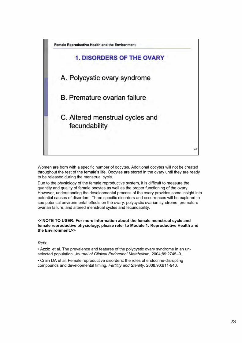
Women are born with a specific number of oocytes. Additional oocytes will not be created throughout the rest of the female’s life. Oocytes are stored in the ovary until they are ready to be released during the menstrual cycle. Due to the physiology of the female reproductive system, it is difficult to measure the quantity and quality of female oocytes as well as the proper functioning of the ovary. However, understanding the developmental process of the ovary provides some insight into potential causes of disorders. Three specific disorders and occurrences will be explored to see potential environmental effects on the ovary: polycystic ovarian syndrome, premature ovarian failure, and altered menstrual cycles and fecundability.
<<NOTE TO USER: For more information about the female menstrual cycle and female reproductive physiology, please refer to Module 1: Reproductive Health and the Environment.>>
Refs:• Azziz et al. The prevalence and features of the polycystic ovary syndrome in an un-selected population. Journal of Clinical Endocrinol Metabolism, 2004;89:2745–9. • Crain DA et al. Female reproductive disorders: the roles of endocrine-disrupting compounds and developmental timing. Fertility and Sterility, 2008,90:911-940.
23
Female Reproductive Health and the Environment
1. DISORDERS OF THE OVARY
A. Polycystic ovary syndrome
B. Premature ovarian failure
C. Altered menstrual cycles and fecundability
23

The female ovary first begins to develop between the second and third trimester of fetal development in the mother’s womb. Following this initial formation, the ovarian cells, called follicles, will remain dormant for 15 to 50 years. This extended period of dormancy indicates that these follicles may be exposed to a variety of environmental factors. Furthermore, this indicates that the ovarian tissue may be affected by the environment either during the development of the organ in utero, or during the dormancy period. This demonstrates an increased sensitivity for this specific female reproductive organ. However, it is still not known which environmental agents may influence the health of the ovarian follicle.
Refs:• Crain DA et al. Female reproductive disorders: the roles of endocrine-disrupting compounds and developmental timing. Fertility and Sterility, 2008,90:911-940.• Kipp JL et al.. Neonatal exposure to estrogens suppresses activin expression and signaling in the mouse ovary. Endocrinology, 2007;148:1968–76.• Pepe GJ, Billiar RB, Albrecht ED. Regulation of baboon fetal ovarian folliculogenesis by estrogen. Molecular Cellular Endocrinology, 2006;247:41–6.
Image: WHO
24
Female Reproductive Health and the Environment
DEVELOPMENT OF THE OVARY AND THE ENVIRONMENT
❖ Ovarian tissue develops during second and third trimester of fetal development
❖ Ovarian follicles remain dormant between 15-50 years - Longest lived, non-regenerating
cells in the body
❖ Cumulative exposures to environmental agents and contaminants
WHO
24

25
Some female reproductive disorders may have their origin in early life development, perhaps as early as in the first trimester. Research has shown that proper ovarian development is dependent on the proper balance of estrogen in the fetal environment. Furthermore, estrogenic activity at the time of ovarian follicle development is also crucial to achieve adequate oocyte quality. This process has important implications for future ovarian health during the fertile periods of the woman’s life. Thus, the maintenance of a proper balance of estrogen is crucial for healthy ovarian tissue and follicular development. Changes in the hormonal environment of the developing ovary may disrupt this fragile and sensitive process. Some environmental contaminants that mimic natural estrogen may disrupt or affect the ovarian development process. Two specific case studies illustrate this point. First, an animal model using laboratory mice has demonstrated that exposure to estrogenic compounds resulted in a failure of normal ovarian follicle formation. Likewise, a wildlife study of the American alligator showed that female alligators that had high levels of estrogenic environmental contaminants in their bodies, specifically, the pesticide difocol, also failed to produce healthy ovarian follicles. However, the exact mechanism of action for these animal examples remains unknown.
Refs:•Crain DA, Janssen SJ, Edwards TM, Heindel J, Ho S, Hunt P, et al., Female reproductive disorders: the roles of endocrine-disrupting compounds and developmental timing. Fertility and Sterility, 2008,90:911-940.•Kipp JL, Kilen SM, Bristol-Gould S, Woodruff TK, Mayo KE. Neonatal exposure to estrogens suppresses activin expression and signaling in the mouse ovary. Endocrinology. 2007;148:1968–76. •Pepe GJ, Billiar RB, Albrecht ED. Regulation of baboon fetal ovarian folliculogenesis by
Female Reproductive Health and the Environment
EVIDENCE OF ENVIRONMENTAL EFFECTS ON THE OVARY
❖ Proper development of ovarian tissue depends on estrogenic pathways ❖ Therefore, exposure to and withdrawal from estrogen &
exposure to estrogen-like substances may alter proper function
❖ Maintaining proper estrogen balance during follicle development is necessary for normal tissue development and oocyte quality
❖ Animal models show failure of normal follicle development when exposed to estrogenic endocrine disruptors
EHP 25

estrogen. Mol Cell Endocrinol. 2006;247:41–6.
Image: Environmental Health Perspectives (EHP). Available at ehp.niehs.nih.gov/docs/2005/113-8/fetus.jpg - accessed 29 June 2010. Reproduced with permission from EHP.
25

26
<<READ SLIDE.>>While it is known that proper functioning of the ovarian system is dependent on appropriate hormonal signaling, the pathogenesis of polycystic ovary syndrome is largely known. However, the most recent research has hypothesized that the development of polycystic ovary syndrome may be a combined effect from an environmental exposure and a genetic effect while a fetus is developing in utero. It is believed that excess exposure to the reproductive hormone testosterone in utero may potentially promote development of this syndrome. However, exposure to this excess testosterone may occur due to a genetic predisposition of hyper-secretion of testosterone and environmental toxin exposure may lead to elevation of prenatal testosterone. Thus, polycystic ovary syndrome may be a good example of a female reproductive disorder that relies on both environmental and genetic factors for development. Refs:• Azziz R et al. The prevalence and features of the polycystic ovary syndrome in an un-selected population. Journal of Clinical Endocrinol Metabolism, 2004;89:2745–9. Notwithstanding the potential public health impact of the polycystic ovary syndrome (PCOS), estimates regarding its prevalence are limited and unclear. Between July 1998 and October 1999, 400 unselected consecutive premenopausal women (18-45 yr of age) seeking a preemployment physical at the University of Alabama at Birmingham were studied (223 Black, 166 White, and 11 of other races). Evaluation included a history and physical examination, a modified Ferriman-Gallwey hirsutism score, and serum screening for hyperandrogenemia, hyperprolactinemia, and 21-hydroxylase-deficient nonclassical adrenal hyperplasia. PCOS was diagnosed by the presence of the following: 1) oligoovulation, 2) hyperandrogenemia and/or hirsutism (modified Ferriman-Gallwey score > or = 6), and 3) the exclusion of related disorders. Confirmed PCOS was established in those individuals whose evaluation was complete and indicative of PCOS, and possible PCOS was established when the hormonal evaluation was not complete or was unavailable, but the clinical phenotype was otherwise suggestive of the disorder. The individual probability of PCOS in women with possible PCOS was assigned a weight based on the findings in similar subjects whose evaluation was complete, and the total number of PCOS cases arising from these individuals was calculated (i.e. individual probability of PCOS x total number of subjects in the group). The cumulative prevalence of PCOS in our population was 6.6% (26.5 of 400), including 15 subjects among the 347 women completing their evaluation and a calculated prevalence of 11.5 subjects among the remainder. The prevalence rates of PCOS for Black and White women were 8.0 and 4.8%, respectively, not significantly different. These data from a large representative unselected population support the concept that PCOS is the most common endocrine abnormality of reproductive-aged women in the United States.•Knochenhauer ES et al. Prevalence of the polycystic ovary syndrome in unselected black and white women of the southeastern United States: a prospective study. Journal of Clinical Endcrinol Metabolism, 1998;83:3078-82. Estimates of the prevalence of the polycystic ovary syndrome (PCOS) in the general population have ranged from 2-20%. The vast majority of these reports have studied White populations in Europe, used limited definitions of the disorder, and/or used bias populations, such as those seeking medical care. To estimate the prevalence of this disorder in the United States and address these limitations, we prospectively determined the prevalence of PCOS in a reproductive-aged population of 369 consecutive women (174 White and 195 Black; aged 18-45 yr), examined at the time of their preemployment physical. Body measures were obtained, and body hair was quantified by a modified Ferriman-Gallwey (F-G) method. All exams were initially performed by 2 trained nurses, and any subject with an F-G score above 3 was reexamined by a physician, the same for all patients. Of the 369 women, 277 (75.1%) also agreed to complete a questionnaire and have additional blood drawn. Subjects were studied regardless of current estrogen/progestin hormonal use (28.5%). PCOS was defined as 1) oligoovulation, 2) clinical hyperandrogenism (i.e. hirsutism) and/or hyperandrogenemia, and 3) exclusion of other related disorders, such as hyperprolactinemia, thyroid abnormalities, and non-classic adrenal hyperplasia. Hirsutism was defined by a F-G score of 6 or more, and hyperandrogenemia was defined as a total or free testosterone, androstenedione, and/or dehydroepiandrosterone sulfate level above the 95th percentile of control values [i.e. all eumenorrheic women in the study, who had no hirsutism (F-G < or = 5) or acne and were receiving no hormonal therapy; n = 98]. Considering all 369 women studied, White and Black women had similar mean ages (29.4 +/- 7.1 and 31.1 +/- 7.8 yr, respectively), although White women had a lesser body mass than Black women (24.9 +/- 6.1 vs. 29.2 +/- 8.1 kg/m2, respectively; P < 0.001). Of these 7.6%, 4.6%, and 1.9% demonstrated a F-G score of 6 or more, 8 or 10, respectively, and there was no significant racial difference, with hirsutism prevalences of 8.0%, 2.8%, and 1.6% in Whites, and 7.1%, 6.1%, and 2.1% in Blacks, respectively. Of the 277 women consenting to a history and hormonal evaluation, 4.0% had PCOS as defined, 4.7% (6 of 129) of Whites and 3.4% (5 of 148) of Blacks. In conclusion, in our consecutive population of unselected women the prevalence of hirsutism varied from 2-8% depending on the chosen cut-off F-G score, with no significant difference between White and Black women. Using an F-G score of 6 or more as indicative of hirsutism, 3.4% of Blacks and 4.7% of Whites had PCOS as defined. These data suggest that PCOS may be one of most common reproductive endocrinological disorders of women.•Takeuchi T et al. Positive relationship between androgen and the endocrine disruptor, bisphenol A, in normal women and women with ovarian dysfunction. Endocrinology Journal, 2004;51:165-9. This study was performed to investigate the serum levels of bisphenol A (BPA), an endocrine disruptor, in women with ovarian dysfunction and obesity. Fasting serum samples were obtained from 19 non-obese and 7 obese women with normal menstrual cycles: 7 patients with hyperprolactinemia, 21 patients with hypothalamic amenorrhea, and 13 non-obese and 6 obese patients with polycystic ovary syndrome (PCOS). BPA was measured by an enzyme-linked immunosorbent assay. BPA was detected in all human sera. Serum BPA concentrations were significantly higher in both non-obese and obese women with polycystic ovary syndrome (1.05 +/- 0.10 ng/ml, 1.17 +/- 0.16 ng/ml; p<0.05, respectively) and obese normal women (1.04 +/- 0.09 ng/ml, p<0.05) compared with those in non-obese normal women (0.71 +/- 0.09 ng/ml). There was no difference among women with hyperprolactinemia, women with hypothalamic amenorrhea, and non-obese normal women. There were significant positive correlations between serum BPA and total testosterone (r = 0.391, p<0.001), free testosterone (r = 0.504, p<0.001), androstenedione (r = 0.684, p<0.001), and DHEAS (r = 0.514, p<0.001) concentrations in all subjects. These findings show that there is a strong
Female Reproductive Health and the Environment
1.A. POL YCYSTIC OVARY SYNDROME
❖ Very common endocrine related disorder in reproductive-aged women - 4-8% affected
❖ Health effects associated with polycystic ovary syndrome: insulin resistance , diabetes, endometrial cancer, and infertility
❖ Pathogenesis of polycystic ovary syndrome unknown!
❖ Potential pathway: excessive testosterone exposure in utero - Combined effects between genetics and the
environment 2s

relationship between serum BPA and androgen concentrations, speculatively due to the effect of androgen on the metabolism of BPA.
26

<<READ SLIDE.>>
Approximately 1% of the female population has premature ovarian failure. In girls and young women with premature ovarian failure, loss of eggs, a dysfunction of the eggs or the removal of the ovaries at a young age causes the end of menstruation. Unlike menopause, this is not a natural occurrence. Premature ovarian failure usually occurs in women under the age of 40 and can happen as early as the teen years. Premature ovarian failure is sometimes associated with autoimmune disorders (such as thyroid problems, diabetes or adrenal problems) that may require further medical treatment. Several chemicals, including mancozeb (pesticide), dibromoacetic acid (water disinfection by-product), polycyclic aromatic hydrocarbons, cyclophosphamide (a chemotherapy drug) and others have been shown in animals to interfere with the process of follicle maturation.The etiology of premature ovarian failure is unclear. However, researchers believe that it may result from a lack of ovarian follicle reserve at birth or that follicles activate too early and then die as a result during the fetal period. It is possible that some sort of environmental exposure may alter the development process of the ovarian follicle through changes in hormonal signaling, as described in previous slides.
Refs:•McGee EA, Hsueh AJ. Initial and cyclic recruitment of ovarian follicles. Endocrinology Review, 2000;21:200–14. •Panay N and Fenton P. Premature ovarian failure: a growing concern. Climacteric, 2008, 11, 1–3.•Program on Reproductive Health and the Environment. Shaping our legacy: reproductive health and the environment. University of California, San Francisco. 2008.•Stubbs SA. Abnormal preantral folliculogenesis in polycystic ovaries is associated with increased granulosa cell division. Journal of Clinical Endocrinol Metabolism, 2007; 92:4418–26. 69.
Image: WHO
27
Female Reproductive Health and the Environment
1.B. PREMATURE OVARIAN FAILURE
❖ End of menstruation prior to age 40 - 1 % of women affected
❖ May result from inadequate ovarian reserve and early activation and death of follicles in utero
❖ Pathogenesis not known- could environmental factors affect follicular development?
❖ Fertility treatment unlikely to aid in becoming pregnant
WHO
27

28
Interference with the regulation of the menstrual cycle hormones results in irregular cycles and may reduce the ability of a female to conceive. Thus, appropriate regulation of the female cycle is dependent on precise levels of endogenous hormones. Several environmental contaminants, that may act as endocrine disrupting compounds (EDs), may alter the hormonal balance of a female and thus affect her cyclicity and subsequent ability to conceive. Alterations in hormonal balance may be induced by changes in hormonal creation, release, signaling, or appropriate binding. Smoking has also been associated with altered menstrual cycles. Animal models show some evidence of altered menstrual cycles and fecundability after exposure to environmental contaminants that act as potential EDs. For example, the plasticizer bisphenol A (BPA), has shown to increase the natural length of the menstrual cycle of the female mouse. Similar effects have been seen in mice exposed to estrogenic compounds such as genistein and reseveratrol. These estrogenic compounds are naturally occurring and may be found in soy products, red wine, and certain berries.
Refs:•Chao et al. Placental transfer of polychlorinated dibenzo-p-dioxins, dibenzofurans, and biphenyls in Taiwanese mothers in relation to menstrual cycle characteristics. Food Chemistry and Toxicology, 2007; 45:259–65. •Jefferson WN, Padilla-Banks E, Folliculogenesis in polycystic ovaries is associated with increased granulosa cell division. Journal of Clinical Endocrinol Metabolism, 2007;92:4418–26. 49. Outbred female CD-1 mice were treated with genistein (Gen), the primary phytoestrogen in soy, by s.c. injections on Neonatal Days 1-5 at doses of 0.5, 5, or 50 mg/kg per day (Gen-0.5, Gen-5, and Gen-50). The day of vaginal opening was observed in mice treated with Gen and compared with controls, and although there were some differences, they were not statistically significant. Gen-treated mice had prolonged estrous cycles with a dose- and age-related increase in severity of abnormal cycles. Females treated with Gen-0.5 or Gen-5 bred to control males at 2, 4, and 6 mo showed statistically significant decreases in the number of live pups over time with increasing dose; at 6 mo, 60% of the females in the Gen-0.5 group and 40% in the Gen-5 group delivered live pups compared with 100% of controls. Mice treated with Gen-50 did not deliver live pups. At 2 mo, >60% of the mice treated with Gen-50 were fertile as determined by uterine implantation sites, but pregnancy was not maintained; pregnancy loss was characterized by fewer, smaller implantation sites and increased reabsorptions. Mice treated with lower doses of Gen had increased numbers of corpora lutea compared with controls, while mice treated with the highest dose had decreased numbers; however, superovulation with eCG/hCG yielded similar numbers of oocytes as controls. Serum levels of progesterone, estradiol, and testosterone were similar between Gen-treated and control mice when measured before puberty and during pregnancy. In summary, neonatal treatment with Gen caused abnormal estrous cycles, altered ovarian function, early reproductive senescence, and subfertility/infertility
Female Reproductive Health and the Environment
1.C. ALTERED MENSTRUAL CYCLES
♦:♦ Altered cycle length or irregular cycles may decrease fecundability
♦:♦ Proposed mechanism: - Change in hormone creation, release, signaling, receptor
binding
♦:♦ Animal evidence indicates environmental exposures potentially play a role - Exposure to a type of estrogen in utero causes altered
menstrual cycling
28

at environmentally relevant doses.•Nikaido Y et al. Effects of maternal xenoestrogen exposure on development of the reproductive tract and mammary gland in female CD-1 mouse offspring. Reproductive Toxicology, 2004;18:803–11.
28

29
Evidence of exposure to environmental contaminants and altered menstrual cycles and fecundability is limited. However, two specific studies exist that may demonstrate a potential link between exposures to contaminants and hormonal changes within the female reproductive system. As mentioned previously, interference with the regulation of the menstrual cycle hormones results in irregular cycles and may reduce the ability for a female to conceive. Many studies have focused on human occupational exposures to pesticides. Such studies have indicated a potential link between organochlorine pesticide exposure in females and shortened menstrual cycles. However, similar studies show that women exposed to hormonally active pesticides that are nonorganochlorines have a 60 to 100% odds of longer cycles and missed periods. These differing results demonstrate that cyclicity evidence from occupationally exposed females to pesticides remains inconclusive. Greater research is needed in this field to determine the mechanism of action of various classes of pesticides on female hormonal pathways and functioning.
Refs:• Axmon A, Rylander L, Stromberg U, Hagmar L. Altered menstrual cycles in women with a high dietary intake of persistent organochlorine compounds. Chemosphere, 2004;56:813–9.• Farr SL et al. Pesticide use and menstrual cycle characteristics among premenopausal women in the Agricultural Health Study. American Journal of Epidemiology, 2004;160:1194-204.Menstrual cycle characteristics may have implications for women's fecundability and risk of hormonally related diseases. Certain pesticides disrupt the estrous cycle in animals. The authors investigated the cross-sectional association between pesticide use and menstrual function among 3,103 women living on farms in Iowa and North Carolina. Women were aged 21-40 years, premenopausal, not pregnant or breastfeeding, and not taking oral contraceptives. At study enrollment (1993-1997), women completed two self-administered questionnaires on pesticide use and reproductive health. Exposures of interest were lifetime use of any pesticide and hormonally active pesticides. Menstrual cycle characteristics of interest included cycle length, missed periods, and intermenstrual bleeding. The authors used generalized estimating equations to assess the association between pesticide use and menstrual cycle characteristics, controlling for age, body mass index, and current smoking status. Women who used pesticides experienced longer menstrual cycles and increased odds of missed periods (odds ratio = 1.5, 95% confidence interval: 1.2, 1.9) compared with women who never used pesticides. Women who used probable hormonally active pesticides had a 60-100% increased odds of experiencing long cycles, missed periods, and intermenstrual bleeding compared with women who had never used pesticides. Associations remained after control for occupational physical activity.•Jefferson WN, Padilla-Banks E. Folliculogenesis in polycystic ovaries is associated with increased granulosa cell
Female Reproductive Health and the Environment
CASE STUDY: MENSTRUAL CYCLES AND PESTICIDE EXPOSURE
❖ Several studies measur ing cycles of women occupationally exposed to different types of pesticides
❖ Differing results from different pesticides
❖ Organochlo rines shown to decrease menstrual cycle
❖ Non-organochlorine pesticides shown to increase menstrual cycles WHO
29

division. Journal of Clinical Endocrinol Metabolism, 2007;92:4418–26. 49.
Image: WHO
29

30
The second part of the overview of female reproductive disorders describes disorders associated with the uterus. The two specific disorders that will be described are endometriosis and uterine fibroids. The figure depicts the female reproductive anatomy.
<<NOTE TO USER: For more information regarding the anatomical structures of the female reproductive system, please refer to Module 1: Reproductive Health and the Environment. For information on cancers, please go to the cancer module.>>
Ref:•Yin Y, Ma L. Development of the mammalian female reproductive tract. Journal of Biochemistry, 2005, 137:677–83.
Image: Uterus and Uterine Tubes. National Cancer Institute. Available at training.seer.cancer.gov/ss_module09_cervix_corpus/cervix_unit01_sec01_intro.html -accessed 20 July 2010. This image is public domain.
Female Reproductive Health and the Environment
2. DISORDERS OF THE UTERUS
Uterus a■d Uterine tules
nf.nJ bulum Uterne tube
j Uerus
A. Endometriosis
/ Fimbriae
Pe·~e1rum B. Uterine fibroids Cervix
National CancEN" Institute
30

31
At approximately 9.5 to 11.5 weeks of development in the maternal womb, the female fetus will undergo significant differentiation of the reproductive organs. During this process, the fallopian tubes are formed along with the cervix, uterus, and upper vagina. However, it is important to know that while development of the uterine tissue begins in utero, full development of the uterine tissue is completed during pubertal years. Therefore, similar of the development of the ovaries, a broad time frame exists for the development of this organ. This extended time frame may indicate a greater risk to exposure to a variety of environmental agents. Early developmental environmental exposures may act on the developing fetus and cause structural abnormalities in the growth and maturation of uterine tissue. Additionally, such defects may not be detected until post-pubertal development, although their initiation began much earlier. One strong example comes from the synthetic estrogen diethylstilbestrol (DES). This was a medicine given to pregnant women in the United States between the 1940s and late 1970s for a variety of symptoms. In a study of women who were born to mothers exposed to DES during pregnancy, it was found that some women experienced morphological abnormalities of the uterine tissue. In addition, studies of stillborn infants who were exposed to DES in utero (during the first half of gestation) detected an 80% increased prevalence of abnormal development of vaginal and uterine tissue. Exposure to this synthetic estrogen in utero emphasizes the critical window of susceptibility for the developing reproductive system of the female fetus. However, there is no conclusive evidence of a causal pathway between adverse uterine development and DES exposure in utero. Similarly, a mechanism of action for this described process has not yet been determined. Occupational exposures are also important. For instance, some results have related noise exposure and shift work to menstrual disturbance and infertility.
Refs: •Diamanti-Kandarakis E. et al. Endocrine-disrupting chemicals: an Endocrine Society scientific statement. 2009. Endocrine reviews. 30(4): 293-342.•Hoover RN et al. Adverse Health Outcomes in Women Exposed In Utero to Diethylstilbestrol. N Engl J Med. 2011; 365:1304-1314.•Jefferies JA et al. Structural anomalies of the cervix and vagina in women enrolled in the Diethylstilbestrol Adenosis (DESAD) Project. American Journal Obstetrics Gynecology, 1984, 148:59–66. 112. Among women exposed in utero to diethylstilbestrol (DES) and enrolled in the Diethylstilbestrol Adenosis (DESAD) Project, structural anomalies of the cervix or vagina were found in 25% of the 1,655 subjects identified by review of prenatal records, 43% of the 800 who themselves requested entry into the project, and 49% of the 1,089 referred by physicians but in only 2% of the 963 control subjects. Among the 367 cases found by record review to have complete information on the DES exposure, multivariate analysis indicated close association of the anomalies with the gestational week of first exposure and the total dose. Also, the prevalence rate of the anomalies was lower among subjects who had been pregnant and higher among those with later age at menarche.•Johnson LD et al. Vaginal adenosis in stillborn and neonates exposed to diethylstilbestrol and steroidal estrogens and progestins. Obstetrical Gynecology, 1979, 53:671-9.A histologic study was conducted of sagittal sections of the genital tracts of 281 autopsied female stillborns and neonates. The prevalence of vaginal adenosis among 43 offspring exposed in utero to diethylstilbestrol (DES) was 70%, a frequency 18 times greater than the 4% prevalence among 159 unexposed offspring. The relationship of the prevalence of vaginal adenosis to the gestational age at initial exposure was highly significant: 81% of those first exposed during the period of vaginogenesis had adenosis, whereas none exposed after 21 weeks' gestation had adenosis (P1 = 1 X 10(-4)). The relationship of the prevalence of vaginal adenosis to the total dose of DES prior to 22 weeks' gestation also was significant (P1 = 0.02), and this relationship was independent of gestational age at first exposure (P1 =0.01). In contrast, the prevalence of adenosis among 23 offspring exposed to steroidal estrogens and progestins was about the same as that among the unexposed offspring. Vaginal adenosis was unrelated to the complications of pregnancy for which the hormones were given, the calendar year of birth, and the gestational age at delivery).•Nurminen T. Female noise exposure, shift work, and reproduction. J Occup Environ Med.1995, 37(8):945-50.
Female Reproductive Health and the Environment
UTERIN EFFECTS OF ENVIRONMENTAL EXPOSURES
❖ Uterus formation occurs at 9.5-11.5 weeks gestat ion
❖ Environmenta l exposures in utero have led to poor differentiation of uterine tissues in humans
❖ Pre-pubertal alteration in hormones can lead to changes in uterine morphology later in life ❖ Diethylst ilbestrol (DES) exposure
❖ Occupational exposures during reproductive years also matter!
31

32
Endometriosis is a prevalent and painful condition among premenopausal women. Essentially, it is the presence of endometrial glands outside their proper location. These glands should rest inside of the uterine cavity, however, in this disease, the glands move outside of this area. Though estimates of the frequency of endometriosis vary widely, surveys have shown that up to 15% of women may be affected. The causes of endometriosis remain unclear, but it is widely accepted that endometriosis is an estrogen-dependent disease. This means that the movement of the endometrial glands is dependent upon the levels of estrogen circulating within the female reproductive system. Research has suggested that women who experience endometriosis have altered hormonal levels. Thus, most of the endometrial research has focused on the environmental exposures to agents that may affect the hormonal pathways of the female reproductive system.
Refs:•Cummings AM, Hedge JM, Birnbaum LS. Effect of prenatal exposure to TCDD on the promotion of endometriotic lesion growth by TCDD in adult female rats and mice. Toxicological Sciences, 1999, 52:45-9.•Missmer SA et al. In utero exposures and the incidence of endometriosis. Fertility Sterility, 2004, 82:1501-8. OBJECTIVE: To investigate the relation between the fetal environment and endometriosis. DESIGN: Prospective cohort study. SETTING: Nurses' Health Study II with 10 years of follow-up. PARTICIPANT(S): Eighty-four thousand, four hundred forty-six women aged 25-42 who had never been diagnosed with endometriosis, infertility, or cancer at baseline in 1989. MAIN OUTCOME MEASURE(S): Incidence of laparoscopically confirmed endometriosis according to birthweight, prematurity, multiple gestation, diethylstilbestrol (DES) exposure, and having been breastfed. RESULT(S): During 566,250 woman-years of follow-up, 1,226 cases of laparoscopically-confirmed endometriosis were reported among women with no past infertility. After adjusting for age, calendar time, parity, race, and body mass index at age 18, we observed a linear increase in the incidence rate with decreasing birthweight (rate ratio [RR] = 1.3 for birthweight <5.5 pounds versus 7.0-8.4 pounds, 95% confidence interval [CI] = 1.0-1.8, P value, test for trend = .01). In addition, women who were born as one of a multiple gestation (i.e., twins or greater number) were at increased risk even after controlling for birthweight (RR = 1.7, CI = 1.2-2.5). The rate of endometriosis was also 80% greater among women exposed to diethylstilbestrol in utero (RR = 1.8, CI = 1.2-2.8). Neither premature delivery nor having been breastfed were associated with the incidence of endometriosis. None of these effect estimates were modified by infertility status at the time of endometriosis diagnosis. CONCLUSION(S): The fetal environment is associated with subsequent laparoscopically confirmed endometriosis in this cohort of US women. •Vigano P et al. Endometriosis: epidemiology and aetiological factors. Best Practice Res Chil Obstet Gynaecology, 2004;18:177-200. Estimates of the frequency of endometriosis vary widely. Based on the few reliable data, the prevalence of the condition can reasonably be assumed to be around 10%. Although no consistent information is available on the incidence of the disease, temporal trends suggest an increase among women of reproductive age. This could be explained-at least in part-by changing reproductive habits. Numerous epidemiological studies have indicated that nulliparous women and women reporting short and heavy menstrual cycles are at increased risk of developing endometriosis; data on other risk factors are less consistent. These epidemiological findings strongly support the menstrual reflux hypothesis. Additional evidence in favour of this theory includes the
Female Reproductive Health and the Environment
2.A. ENDOMETRIOSIS
❖ Characterized by presence of endometrial glands outside uterine cavity
❖ 10-15% prevalence (estimates for Italy)
❖ Disease linked to estrogenic activity
❖ Potential link to environmental exposures
32

demonstration of viable endometrial cells in the menstrual effluent and peritoneal fluid, experimental implantation and growth of endometrium within the peritoneal cavity, observation of some degree of retrograde menstruation in most women undergoing laparoscopy during menses, and an association between obstructed menstrual outflow and endometriosis.
32

33
The most notable studies relating environmental exposures to endometrial abnormalities have been seen in laboratory animals exposed to the dioxin tetrachlorodibenzo-p-dioxin (TCDD). TCDD is produced as a by-product during a variety of industrial processes and during waste incineration. TCDD is also found in certain herbicides and wood preservers. Several studies have shown that TCDD may play a role in the development of endometriosis. For example, scientists have determined that exposure to this chemical may enhance the development and progression of endometriosis because of the chemical’s strong anti-estrogenic effects. This is a plausible assumption given the information in the previous slide: that endometriosis is known to be an estrogen-dependent disease. It must be noted that in all of the studies conducted, the dose at which results were seen were extremely high. These high doses do not appropriately reflect the doses at which humans may be exposed to in real-life scenarios.
Refs:•Cummings AM, Hedge JM, Birnbaum LS. Effect of prenatal exposure to TCDD on the promotion of endometriotic lesion growth by TCDD in adult female rats and mice. Toxicological Sciences, 1999, 52:45-9.•Missmer SA et al. In utero exposures and the incidence of endometriosis. Fertility Sterility, 2004, 82:1501-8. OBJECTIVE: To investigate the relation between the fetal environment and endometriosis. DESIGN: Prospective cohort study. SETTING: Nurses' Health Study II with 10 years of follow-up. PARTICIPANT(S): Eighty-four thousand, four hundred forty-six women aged 25-42 who had never been diagnosed with endometriosis, infertility, or cancer at baseline in 1989. MAIN OUTCOME MEASURE(S): Incidence of laparoscopically confirmed endometriosis according to birthweight, prematurity, multiple gestation, diethylstilbestrol (DES) exposure, and having been breastfed. RESULT(S): During 566,250 woman-years of follow-up, 1,226 cases of laparoscopically-confirmed endometriosis were reported among women with no past infertility. After adjusting for age, calendar time, parity, race, and body mass index at age 18, we observed a linear increase in the incidence rate with decreasing birthweight (rate ratio [RR] = 1.3 for birthweight <5.5 pounds versus 7.0-8.4 pounds, 95% confidence interval [CI] = 1.0-1.8, P value, test for trend = .01). In addition, women who were born as one of a multiple gestation (i.e., twins or greater number) were at increased risk even after controlling for birthweight (RR = 1.7, CI = 1.2-2.5). The rate of endometriosis was also 80% greater among women exposed to diethylstilbestrol in utero (RR = 1.8, CI = 1.2-2.8). Neither premature delivery nor having been breastfed were associated with the incidence of endometriosis. None of these effect estimates were modified by infertility status at the time of endometriosis diagnosis. CONCLUSION(S): The fetal environment is associated with subsequent laparoscopically confirmed endometriosis in this cohort of US women.
•Vigano P et al. Endometriosis: epidemiology and aetiological factors. Best Practice Res Chil Obstet Gynaecology, 2004;18:177-200. Estimates of the frequency of endometriosis vary widely. Based on the few reliable data, the prevalence of the condition can reasonably be assumed to be around 10%. Although no consistent information is available on the incidence of the disease, temporal trends suggest an increase among women of reproductive age. This could be explained-at least in part-by changing reproductive habits. Numerous epidemiological studies have indicated that nulliparous women and women reporting short and heavy menstrual cycles are at increased risk of developing endometriosis; data on other risk factors are less consistent. These epidemiological findings strongly support the menstrual reflux hypothesis. Additional evidence in favour of this theory includes the demonstration of viable endometrial cells in the menstrual effluent and peritoneal fluid, experimental implantation and growth of endometrium within the peritoneal cavity, observation of some degree of retrograde menstruation in most women undergoing laparoscopy during menses, and an association between obstructed menstrual outflow and endometriosis.
Female Reproductive Health and the Environment
CAS STUDY: DIOXINS AND ENDOMETRIOSIS
❖ TCDD (tetrachlorodibenzo-p-dioxin) ❖ A dioxin and industrial byproduct ❖ May be produced during waste incineration
❖ TCDD may enhance the development of endometriosis due to anti-estrogenic effects
❖ Animal models show correlation with endometriosis but only at high doses
33

34
Uterine fibroids are the most common tumour for the female reproductive tract and indicate localized pathology in the uterus. These uterine fibroids occur in high prevalence and may affect up to 50% of all women. The risk of uterine fibroids increases with agethough tends to decrease with the onset of menopause. Estrogenic exposures have been determined to be a risk factor for uterine fibroid development. This includes exposures in utero to excess estrogens, as well as throughout childhood, adolescence, and adulthood. However, the mechanism of action remains unclear. Several animal studies have suggested that environmental contaminants that act through estrogenic pathways may promote the development of uterine fibroids in women. This may be especially relevant during critical periods of development.
Refs:•Baird DD, Newbold R. Prenatal diethylstilbestrol (DES) exposure is associated with uterine leiomyoma development. Reproductive Toxicology, 2005;20:81–4.•Chiaffarino F et al. Diet and uterine myomas. Obstetrical Gynecology, 1999;94:395–8.•Diamanti-Kandarakis E. et al. Endocrine-disrupting chemicals: an Endocrine Society scientific statement. 2009. Endocrine reviews. 30(4): 293-342.•Hoover RN et al. Adverse Health Outcomes in Women Exposed In Utero to Diethylstilbestrol. N Engl J Med. 2011; 365:1304-1314.•Marshall LM et al. Variation in the incidence of uterine leiomyoma among premenopausal women by age and race. Obstetrical Gynecology, 1997, 90:967–73155. •Walker CL, Stewart EA. Uterine fibroids: the elephant in the room. Science, 2005, 308:1589–92.
Female Reproductive Health and the Environment
2.B. UTERINE FIBROIDS
♦:♦ Most common tumour of female reproductive tract ❖ 25% to 50% of women affected
♦:♦ Local disease of uterus, can cause benign tumour, abnormal heavy bleeding and pain, discomfort, and difficulties in reproduction
♦:♦ Potential etiology linked to hormonal irregularities, specifically estrogenic exposures
34

35
<<READ SLIDE.>>During the past decade, possible advancement in timing of puberty has been reported throughout several world regions. Though these surveys have focused on industrialized nations, some evidence exists for developing nations as well. In addition, earlypubertal development has been noticed in children, primarily girls, migrating for foreign adoption in several Western European countries. These observations are raising the issues of current differences and secular trends in timing of puberty in relation to ethnic, geographical, and socioeconomic background. Recently, the possible role of endocrine-disrupting chemicals from the environment has been considered. Refs:•Herman-Giddens ME et al. Secondary sexual characteristics and menses in young girls seen in office practice: a study from the Pediatric Research in Office Settings network. Pediatrics, 1997, 99:505-12. OBJECTIVE: To determine the current prevalence and mean ages of onset of pubertal characteristics in young girls seen in pediatric practices in the United States. METHODS: A cross-sectional study was conducted by 225 clinicians in pediatric practices belonging to Pediatric Research in Office Settings, a practice-based research network. After standardized training in the assessment of pubertal maturation, practitioners rated the level of sexual maturation on girls 3 through 12 years who were undergoing complete physical examinations. RESULTS: Data were analyzed for 17,077 girls, of whom 9.6% were African-American and 90.4% white. At age 3, 3% of African-American girls and 1% of white girls showed breast and/or pubic hair development, with proportions increasing to 27.2% and 6.7%, respectively, at 7 years of age. At age 8, 48.3% of African-American girls and 14.7% of white girls had begun development. At every age for each characteristic, African-American girls were more advanced than white girls. The mean ages of onset of breast development for African-American and white girls were 8.87 years (SD, 1.93) and 9.96 years (SD, 1.82), respectively; and for pubic hair development, 8.78 years (SD, 2.00) and 10.51 years (SD, 1.67), respectively. Menses occurred at 12.16 years (SD, 1.21) in African-American girls and 12.88 years (SD, 1.20) of age in white girls. CONCLUSIONS: These data suggest that girls seen in a sample of pediatric practices from across the United States are developing pubertal characteristics at younger ages than currently used norms. Practitioners may need to revise their criteria for referral of girls with precocious puberty, with attention to racial differences.•Parent AS et al. The timing of normal puberty and the age limits of sexual precocity: variations around the world, secular trends, and changes after migration. Endocrinology Review, 2003;24:668-93. During the past decade, possible advancement in timing of puberty has been reported in the United States. In addition, early pubertal development and an increased incidence of sexual precocity have been noticed in children, primarily girls, migrating for foreign adoption in several Western European countries. These observations are raising the issues of current differences and secular trends in timing of puberty in relation to ethnic, geographical, and socioeconomic background. None of these factors provide an unequivocal explanation for the earlier onset of puberty seen in the United States. In the formerly deprived migrating children, refeeding and catch-up growth may prime maturation. However, precocious puberty is seen also in some nondeprived migrating children. Attention has been paid to the changing milieu after migration, and recently, the possible role of endocrine- disrupting chemicals from the environment has been considered. These observations urge further study of the onset of puberty as a possible sensitive and early marker of the interactions between environmental conditions and genetic susceptibility that can influencephysiological and pathological processes.•Tanner JM. Growth at adolescence, 2nd ed. Oxford: Blackwell, 1962.
Female Reproductive Health and the Environment
3. PUBERTAL DISORDERS
❖ Puberty comprises multiple events resulting in reproductive function - Development of the hypothalamus, ovary, breast, and uterus
❖ Puberty is triggered by gonadotropin releasing hormone (GnRH), follicle stimulating hormone (FSH) and luteinizing hormone (LH)
❖ Female pubertal markers include : breast development (thelarche ), appearance of pubic hair (pubarche) , menstrual cycle (menarche)
❖ Are environmental factors contributing to decreasing age at puberty in girls?
WHO
35

Several environmental factors may influence the onset of early puberty in females. In is important to note that these factors vary widely between females and certain factors may play a larger role than others. As research in this field increases, it is possible that more environmental factors will be attributed to early onset of puberty.
<<NOTE TO USER: EDC in this figure is Endocrine Disrupting Compound. For more information on EDCs, please see the module "Endocrine Disorders.">>
Ref:•Crain DA et al. Female reproductive disorders: the roles of endocrine-disrupting compounds and developmental timing. Fertility and Sterility, 2008,90:911-940.
Image: Crain DA et al. Female reproductive disorders: the roles of endocrine-disrupting compounds and developmental timing. Fertility and Sterility, 2008,90:911-940. This figure was reproduced with copyright permission from Fertility and Sterility.
36
Female Reproductive Health and the Environment
ENVIRONMENTAL FACTORS INFLUENCING EARLY PUB RTY
t Nutrition t Photoperiod
EDC Exposure • • • • • • • • • • • • + Stressors
t Obesity + Lead
Fortility and Sterility, 2008,90:911-940 36

37
Puberty is divided into five stages, called Tanner Stages (numbered 1-5). Each stage represents a stage of breast and pubic hair growth. Early breast development is the first event in the Tanner stages of pubertal development. It is also known as thelarche. Early breast development is a more sensitive indicator of exposure to environmental endocrine disrupting chemicals (EDs) than age at menarche. Menarche occurs several years after the initiation of endogenous estrogen production/circulation. Menarche is an event that lends itself better to measurement in a population. Refs:• Colon I et al. Identification of phthalate esters in the serum of young Puerto Rican girls with premature breast development. Environmental Health Perspectives. 2000;108:895-900. Premature breast development (thelarche) is the growth of mammary tissue in girls younger than 8 years of age without other manifestations of puberty. Puerto Rico has the highest known incidence of premature thelarche ever reported. In the last two decades since this serious public health anomaly has been observed, no explanation for this phenomenon has been found. Some organic pollutants, including pesticides and some plasticizers, can disrupt normal sexual development in wildlife, and many of these have been widely used in Puerto Rico. This investigation was designed to identify pollutants in the serum of Puerto Rican girls with premature thelarche. A method for blood serum analysis was optimized and validated using pesticides and phthalate esters as model compounds of endocrine-disrupting chemicals. Recovery was > 80% for all compounds. We performed final detection by gas chromatography/mass spectrometry. We analyzed 41 serum samples from thelarche patients and 35 control samples. No pesticides or their metabolite residues were detected in the serum of the study or control subjects. Significantly high levels of phthalates [dimethyl, diethyl, dibutyl, and di-(2-ethylhexyl)] and its major metabolite mono-(2-ethylhexyl) phthalate were identified in 28 (68%) samples from thelarche patients. Of the control samples analyzed, only one showed significant levels of di-isooctyl phthalate. The phthalates that we identified have been classified as endocrine disruptors. This study suggests a possible association between plasticizers with known estrogenic and antiandrogenic activity and the cause of premature breast development in a human female population.• Den Hond E et al. Sexual maturation in relation to polychlorinated aromatic hydrocarbons: Sharpe and Skakkebaek’s hypothesis revisited. Environmental Health Perspectives, 2002;110:771-6.Polychlorinated aromatic hydrocarbons (PCAHs) have been described as endocrine disruptors in animals and in accidentally or occupationally exposed humans. In the present study we examined the effect of moderate exposure to PCAHs on sexual maturation. Two hundred adolescents (mean age, 17.4 years) who resided in two polluted suburbs and a rural control area in Flanders (Belgium) participated. We measured the serum concentration of polychlorinated biphenyl (PCB) congeners 138, 153, and 180 and dioxin-like compounds [chemically activated luciferase expression (CALUX) assay] as biomarkers of exposure. School physicians assessed the pubertal development of boys and girls and measured testicular volume. In one suburb near two waste incinerators, compared with the other suburb and the control area, fewer boys (p < 0.001) had reached the adult stages of genital development (62% vs. 92% and 100%, respectively) and pubic hair growth (48% vs. 77% and 100%). Also, in the same suburb, fewer girls (p = 0.04) had reached the adult stage of breast development (67% vs. 90% and 79%). In individual boys, a doubling of the serum concentration of PCB congener 138 increased the odds of not having matured into the adult stage of genital development by 3.5 (p = 0.04); similarly for PCB congener 153 in relation to male pubic hair growth, the odds ratio was 3.5 (p = 0.04). In girls, a doubling of the serum dioxin concentration increased the odds of not having reached the adult stage of breast development by 2.3 (p = 0.02). Left plus right testicular volume was lower in both polluted areas than in the control area (42.4 mL vs. 47.3 mL, p = 0.005) but was not related to the current exposure of the adolescents to PCAHs. Through endocrine disruption, environmental exposure to PCAHs may interfere with sexual maturation and in the long-run adversely affect human reproduction.• Freni-Titulaer LW et al. Premature thelarche in Puerto Rico. A search for environmental factors. American Journal of Diseases of Children, 1986;140:1263-7.Pediatric endocrinologists in Puerto Rico reported a threefold increase in the number of patients with premature thelarche seen between 1978 and 1981. A matched-pairs case-control study was conducted to evaluate associations with potential environmental exposures to substances with estrogenic activity, as well as with familial factors. Analysis was performed on 120 pairs, the case subjects of which were selected from those diagnosed between 1978 and 1982. In subjects 2 years of age or older at the onset of thelarche, no significant associations were found. In subjects with onset before 2 years of age, significant positive associations were found with a maternal history of ovarian cysts, consumption of soy-based formula, and consumption of various meat products. A statistically significant negative association was found with consumption of corn products. These statistical associations are probably not sufficient to explain the reported increase because in over 50% of the case subjects there was no exposure to any of the risk factors for which statistical associations were found. Exposure to other substances with possible estrogenic effect, such as waste products from pharmaceutical factories and pesticides, was also excluded as a possible cause. These findings suggest that better diagnosis and reporting, or conceivably the presence of entirely new, unsuspected factors, could account for the reported increase.•Qiao L, Zheng L, Cai D. Study on the di-n-butyl phthalate and di-2-ethylhexyl phthalate level of girl serum related with precocious puberty in Shanghai. Wei Sheng Yan Jiu, 2007;36:93-5.•Slyper AH. The pubertal timing controversy in the USA, and a review of possible causative factors for the advance in timing of onset of puberty. Clinical Endocrinology, 2006;65:1-8. Previously used standards for the diagnosis of precocious puberty in girls no longer appear to be appropriate in the USA, in that a significant number of girls are being seen in paediatricians' offices with breast budding before 8 years of age. The timing of menarche, however, has changed little over the past few decades. Early maturing girls are more likely to become obese in adolescence and adulthood than normal or late maturing girls. Early maturing white girls are heavier at the onset of puberty, but this is not the case for African-American girls or boys of either race. Boys and girls with premature pubarche may be more hyperinsulinaemic than normal children, and girls with premature pubarche more likely to develop functional ovarian and adrenal hyperandrogenism. Early menarche is preceded by prepubertal hyperinsulinaemia. It is proposed that pubertal onset, although not necessarily the tempo of puberty, is influenced by hyperinsulinaemia and insulin resistance. If this hypothesis is correct, insulin resistance may be more prevalent in US children than previously recognized. An
Female Reproductive Health and the Environment
EARLY BREAST DEVELOPMENT
,:. First developmental stage of puberty = thelarche (breast development)
,:. Premature thelarche= isolated breast development before 8 years of age
,:. Risk factors include : - Obesity, hyperinsulinemia, insulin resistance , improved nutrition
,:. Some environmental exposures may affect early breast development timing ❖ Pesticides ❖ Flame retardants
37

advance in timing of onset of puberty has not been noted in other countries, although it is likely that this phenomenon may become more prevalent as other countries adopt a more American lifestyle and diet.•Vom Saal FS, Richter CA, Mao J, Welshons WV. Commercial animal feed: variability in estrogenic activity and effects on body weight in mice. Birth Defects Res A Clin Mol Teratol, 2005;73:474-5. Image: WHO
37

38
A specific case study emphasizes the potential relationship between exposure to environmental contaminants that have endocrine disrupting activity and thelarche. The island of Puerto Rico has high levels of thelarche in their female population. This trend has been witnessed across ethnicities. A study determined that 68% of girls who experienced thelarche has significantly higher phthalate levels in their bodies than girls who did not experience thelarche. Phthalates are chemical compounds that are added to plastics to make them more durable. However, research has determined that phthalates are estrogenic and anti-androgenic in the human body. This indicates that they may directly interfere with normal hormonal functioning in the female reproductive system.
Refs:•Colon I et al. Identification of phthalate esters in the serum of young Puerto Rican girls with premature breast development.Environmental Health Perspectives, 2000;108:895-900.Premature breast development (thelarche) is the growth of mammary tissue in girls younger than 8 years of age without other manifestations of puberty. Puerto Rico has the highest known incidence of premature thelarche ever reported. In the last two decades since this serious public health anomaly has been observed, no explanation for this phenomenon has been found. Some organic pollutants, including pesticides and some plasticizers, can disrupt normal sexual development in wildlife, and many of these have been widely used in Puerto Rico. This investigation was designed to identify pollutants in the serum of Puerto Rican girls with premature thelarche. A method for blood serum analysis was optimized and validated using pesticides and phthalate esters as model compounds of endocrine-disrupting chemicals. Recovery was > 80% for all compounds. We performed final detection by gas chromatography/mass spectrometry. We analyzed 41 serum samples from thelarche patients and 35 control samples. No pesticides or their metabolite residues were detected in the serum of the study or control subjects. Significantly high levels of phthalates [dimethyl, diethyl, dibutyl, and di-(2-ethylhexyl)] and its major metabolite mono-(2-ethylhexyl) phthalate were identified in 28 (68%) samples from thelarche patients. Of the control samples analyzed, only one showed significant levels of di-isooctyl phthalate. The phthalates that we identified have been classified as endocrine disruptors. This study suggests a possible association between plasticizers with known estrogenic and antiandrogenic activity and the cause of premature breast developmentin a human female population.•Freni-Titulaer LW et al. Premature thelarche in Puerto Rico. A search for environmental factors. American Journal of Diseases of Children, 1986;140:1263-7.Pediatric endocrinologists in Puerto Rico reported a threefold increase in the number of patients with premature thelarche seen between 1978 and 1981. A matched-pairs case-control study was conducted to evaluate associations with potential environmental exposures to substances with estrogenic activity, as well as with familial factors. Analysis was performed on 120 pairs, the case subjects of which were selected from those diagnosed between 1978 and 1982. In subjects 2 years of age or older at the onset of thelarche, no significant associations were found. In subjects with onset before 2 years of age, significant positive associations were found with a maternal history of ovarian cysts, consumption of soy-based formula, and consumption of various meat products.A statistically significant negative association was found with consumption of corn products. These statistical associations areprobably not sufficient to explain the reported increase because in over 50% of the case subjects there was no exposure to any of the risk factors for which statistical associations were found. Exposure to other substances with possible estrogenic effect, such as waste products from pharmaceutical factories and pesticides, was also excluded as a possible cause. These findings suggest that better diagnosis and reporting, or conceivably the presence of entirely new, unsuspected factors, could account for the reportedincrease.
Female Reproductive Health and the Environment
CASESTUDY:PREMATURETHELARCHE
❖ Puerto Rico has highest national levels for premature thelarche incidence ,:. 3 fold increase between 1978 to 1981 ,:. Non-genetic cause: increase observed across ethnic groups
❖ Linked to consumption of soy-based formula and meat products
❖ Significantly high levels of phthalates found in 68% of females with premature thelarche. ❖ Matched-pairs case-control study (120 pairs) ,:. Only one of the controls showed high phthalate levels
❖ Phthalates are plasticizers with known estrogenic and antiandrogenic activity
38

A girl's first menstrual cycle commonly occurs several years after the occurrence of pubic hair growth and breast development. Age at menarche is an easy measurement to utilize in studies compared with other pubertal indices. However, age at menarche is less likely to be sensitive to endocrine disruptors than age at onset of breast development. It has been observed that girls exposed to estrogenic organochlorines have a younger age at menarche.Humans are exposed to numerous xenobiotics, each of which could have a different effect. Thus, determining the exact role of a particular endocrine disruptor in menarche alterations is difficult. Some studies have shown that organochlorine pesticides that are estrogenic in nature may reduce the age of menarche. However, these studies have determined that it is truly the cumulative effect of many environmental contaminants that is important in the determination of menarche. For this reason, greater research is necessary to understand which types of exposures may be responsible for changes in menarche occurrence.
Xenobiotic: chemical found in an organism but not normally produced nor expected to be present in it.
Refs:•Crain DA et al. Female reproductive disorders: the roles of endocrine-disrupting compounds and developmental timing. Fertility and Sterility, 2008, 90:911-940.•Ouyang MJ et al., Serum DDT, age at menarche, and abnormal menstrual cycle length. Occupational Environmental Medicine, 2005, 62, 878–884.•Vasiliu O, Muttineni J, and Karmaus, W. In utero exposure to organochlorines and age at menarche. Human Reproduction, 2004, 19, 1506–1512.
39
Female Reproductive Health and the Environment
MENARCHE
♦:♦ A girl's first menstrual cycle ❖ Early (precocious) if occurs at < 10 years
❖ Late (primary amenorrhea) if occurs at >16 years
♦:♦ Many variables can influence the time of occurrence
♦:♦ Estrogenic organochlorines may reduce the age of menarche - But, cumulative effects of environmental contaminants
complicate understanding of associat ion with change in age at menarche
39

Polybrominated biphenyls (PBBs) are flame retardants used in many different applications, including consumer goods. Animal studies have shown that PBBs in the mother’s blood can cross the placenta and expose the developing fetus to this chemical. Furthermore, newborns can also be exposed to the compound via breastfeeding because PBBs is also known to contaminate breast milk. Studies show that exposure to PBBs during gestation and early infancy may alter the hormonal balance necessary for proper growth and maturation. This hormonal imbalance may reduce the age at which girls experience pubertal development. There is also an example of PBBs' influence on pubertal development for a large scale chemical spill that occurred in the United States in 1973. It was found that the daughters of the women exposed to this very high level of PBB who were breastfed and exposed to high levels of PBBs in the womb had an onset of menstruation and pubic hair development one-half to one year earlier than breastfed girls who were exposed to lower levels of PBBs.
Refs:•Blanck HM et al. Age at menarche and Tanner stage in girls exposed in utero and postnatally to polybrominated biphenyl. Epidemiology, 2000;11:641–7.253.Accidental contamination of the Michigan food chain with polybrominated biphenyls (PBBs) led to the exposure of more than 4,000 individuals in 1973. Because PBB exposure is suspected to disrupt endocrine function, we assessed pubertal development in females 5-24 years of age (N = 327) who were exposed to PBB in utero and, in many cases, through breastfeeding. We estimated in utero PBB exposure using maternal serum PBB measurements taken after exposure (1976-1979) and extrapolated to time of pregnancy using a model of PBB decay. We found that breastfed girls exposed to high levels of PBB in utero (> or =7 parts per billion) had an earlier age at menarche (mean age = 11.6 years) than breastfed girls exposed to lower levels of PBB in utero (mean age = 12.2-12.6 years) or girls who were not breastfed (mean age = 12.7 years). This association persisted after adjustment for potential confounders (menarche ratio = 3.4, 95% confidence interval = 1.3-9.0). Perinatal PBB exposure was associated with earlier pubic hair stage in breastfed girls, but little association was found with breast development. The associations observed here lend support to the hypothesis that pubertal events may be affected by pre- and postnatal exposure to organohalogens.•Impacts of endocrine disrupting compounds. Washington DC, USA, National Institute of Environmental Health Sciences, 2008. Available at www.niehs.nih.gov/health/impacts/endocrine.cfm - accessed 19 March 2010.
40
Female Reproductive Health and the Environment
CASE STUDY: PBB EXPOSURE AND PUBERTAL DEVELOPMENT
♦:♦ PBBs (polybrominated biphenyls): common flame retardants
♦:♦ Fetus exposed to PBBs in utero or while breastfeeding
♦:♦ Exposure during gestation and infancy alters the hormonal balance necessary for maturation
♦:♦ Large scale chemical spill in the US Female children of women that were breastfed and had high PBB levels experienced puberty 6-12 months earlier than comparison group
40

41
Changes and alterations in reproductive function have been largely based on genetic as well as environmental factors. Due to the multiple effects that both factors may have on reproductive health, it has been difficult to discern the differences between genetic effects and environmental effects on male and female reproductive development. For this reason, reproductive health studies have been conducted on immigrants because with this cohort, it is possible to separate genetic and environmental effects on puberty. Interestingly, studies have indicated that internationally adopted children and immigrant children have an increased risk of developing precocious puberty. In a study of 145 patients seen in Belgium during a 9-year period for treatment of precocious puberty, 28% appeared to be foreign children (39 girls, one boy) who immigrated 4 to 5 years earlier from 22 developing countries, without any link to a particular ethnic or country background. This led researchers to believe that the mechanism of early onset of puberty might involve previous exposure to estrogenic endocrine disrupters. Similarly, a study in Denmark found that international boys and girls were 10 to 20 times more likely to develop precocious puberty compared with a Danish reference group. Also, older age at adoption significantly increased the risk of precocious puberty. Specifically, the study determined that foreign-adopted children originating from regions other than the Republic of Korea had a 15- to 20-fold increased risk of precocious puberty compared with Danish-born children. This means that the effect of country of origin might be explained by genetic factors or by different environmental exposures and living conditions in the different countries. Older age at adoption increased the risk for premature onset of puberty, which may suggest that environmental factors influence the risk of precocious pubertal development in adopted children.Refs:•Krstevska-Konstantinova M et al. Sexual precocity after immigration from developing countries to Belgium: evidence of previous exposure to organochlorine pesticides. Human Reproduction, 2001;16:1020-6. In a retrospective auxological study of 145 patients seen in Belgium during a 9-year period for treatment of precocious puberty, 28% appeared to be foreign children (39 girls, one boy) who immigrated 4 to 5 years earlier from 22 developing countries, without any link to a particular ethnic or country background. The patients were either adopted (n = 28) or non-adopted (n = 12), the latter having normal weight and height at immigration and starting early puberty without evidence of earlier deprivation. This led to the hypothesis that the mechanism of precocious puberty might involve previous exposure to oestrogenic endocrine disrupters. A toxicological plasma screening for eight pesticides detected p,p'-DDE, which is derived from the organochlorine pesticide DDT. Median p,p'-DDE concentrations were respectively 1.20 and 1.04 ng/ml in foreign adopted (n = 15) and non-adopted (n = 11) girls with precocious puberty, while 13 out of 15 Belgian native girls with idiopathic or organic precocious puberty showed undetectable concentrations (<0.1 ng/ml). A possible relationship between transient exposure to endocrine disrupters and sexual precocity is suggested, and deserves further studies in immigrant children with non-advanced puberty.•Teillmann G et al. Increased risk of precocious puberty in internationally adopted children in Denmark. Pediatrics, 2006;118:e391-9. Studies have indicated that internationally adopted children have an increased risk of developing precocious puberty, but no epidemiologic risk estimates have previously been calculated. We aimed to assess the risk of developing precocious puberty in intercountry adoptees, children immigrating with their family, and descendants of immigrants living in Denmark. METHODS: Patients who were registered with the diagnosis of precocious puberty during the period 1993-2001 were identified through the national patient registry. The background population of children born from 1983 to 2001 were identified through the unique Danish Civil Registration System and subsequently categorized as being Danish (N = 1,062,333), adopted (N = 10,997), immigrating with their family (N = 72,181), or being descendants of immigrants (N = 128,152). The incidence rate ratio of precocious puberty was estimated by log-linear Poisson regression. All rate ratios were adjusted for age and its interaction with gender and calendar year. P values were based on likelihood ratio tests, and 95% confidence intervals were calculated by Wald's test. RESULTS: In the study period, 655 children developed precocious puberty during 5,627,763 person-years at risk. Adopted children were followed during 39,978 person-years at risk, during which 45 girls and 6 boys developed precocious puberty. The risk of developing precocious puberty was significantly increased 10 to 20 times in adopted girls compared with girls with Danish background. The risk of developing precocious puberty depended on the country of origin. In children immigrating with their family, the risk of developing precocious puberty was only marginally increased. Older age at adoption significantly increased the risk of precocious puberty in adoptees independent of region of origin. The incidence rate ratio was significantly higher in children adopted after the age of 2. In children immigrating with their family, we found no effect of age at migration. DISCUSSION: In this large, nationwide, register-based study including 655 cases of precocious puberty, we found that intercountry boys and girls were 10 to 20 times more likely to develop precocious puberty compared with the Danish reference group. Older age at adoption significantly increased the risk of precocious puberty. Uncertainty of the exact age is a well-known problem in adopted children, and systematic underestimation of age might bias the result. However, using the worst-case scenario that all children who according to the Danish Civil Registration System were adopted after 2 years of age were in fact 1 year older, we still observed a highly increased risk of precocious puberty associated with adoption and especially with adoption after 2 years of age. Surprisingly, the risk of precocious puberty was not increased in the large group of children adopted from Korea. One case of precocious puberty was identified among Korean children, whereas > 20 cases of precocious puberty would have been expected if the risk for a Korean child was at the same level as observed among adopted children from India and South America. In the study population, 99% of Korean children were adopted before 2 years of age, which may contribute to explaining our finding. In Korea, children appointed for adoption are often living in foster care settings from birth to adoption, whereas most other countries are reported to take care of the children in orphanages before adoption. It can only be speculated whether a relation between preadoption living conditions and later risk of precocious puberty exists. Genetic factors play a key role in the timing of puberty, and large variations in age at menarche are observed worldwide. Age at menarche is reported to be in the same age range in South Korea as in well-off populations in other parts of the world, indicating that the different risk of precocious puberty observed between Korean and other adoptees probably cannot be explained by genetic factors alone. The finding that the risk of precocious puberty was significantly increased among adoptees in contrast to what was seen in children immigrating with their families contradicts a direct effect of migration. An increasing number of studies have shown long-term effects of certain prenatal and postnatal growth patterns, including advancement in pubertal maturation after poor intrauterine growth and catch-up growth during childhood. Different growth patterns and dietary habits between adoptees and children immigrating with their families
Female Reproductive Health and the Environment
IMMIGRANT STUDIES AND EARLY PUBERTY
❖ Studies of immigrants are useful to separate the genetic and environmental effects on puberty
❖ Belgium: 28% of girls with early puberty immigrated from regions of high dichlorodiphenyldichloroethylene (DOE) use
❖ Denmark: adoptees' increased incidence of early onset of puberty
WHO
41

might contribute to explain our findings. It has been hypothesized that stressful psychosocial factors in infancy and childhood may lead to earlier pubertal maturation. In general, adoptees have experienced several traumatic life events, and it may be speculated that these events alter the susceptibility for developing precocious puberty. CONCLUSIONS: Foreign-adopted children originating from regions other than Korea had a 15- to 20-fold increased risk of precocious puberty compared with Danish-born children, whereas adoptees originating from Korea had no increased risk of precocious puberty. In addition, children immigrating with their families had no increased risk of precocious puberty. The effect of country of origin might be explained by genetic factors or by different environmental exposures and living conditions in the different countries. Older age at adoption increased the risk for premature onset of puberty, which may suggest that environmental factors influence the risk of precocious pubertal development in adopted children.DDT: dichlorodiphenyltrichloroethaneDDE: dichlorodiphenyldichloroethylene Image: WHO
41

The developmental and environmental origins of disease hypothesis states that exposures to environmental toxicants and various environmental elements in utero or during the early life years may increase risk for various adult diseases. It is is relevant to both to the developing and industrialized world. However, more research is needed to better understand the roles played by environment, nutrition and genes. A key factor that may also play a role is paternal exposures to environmental contaminants.
Refs:•Gluckman P, Hanson M. Developmental origins of disease paradigm: a mechanistic and evolutionary perspective. Pediatric Research, 2004, 56(3): 311•Gluckman, P. et al. The fetal, neonatal, and infant environments - the long-term consequences for disease risk. Early Human Development, 2005, 81: 51-59•Gluckman, P et al. Effect of in utero and early-life conditions on adult health and disease. New England Journal of Medicine, 2008, 359:61-73
Image: Environmental Health Perspectives, 2003, 111:9. Reproduced with permission from Environmental Health Perspectives.
<<NOTE TO USER: You can find more detailed information in the modules on endocrine disruptors, occupational exposures and developmental origins of disease>>
42
Female Reproductive Health and the Environment
DEVELOPMENTAL / ENVIRONMENTAL ORIGINS OF DISEASE AND HEALTH
Exposures to environmental toxins in utero may increase risk for adult diseases, including: ❖ Cardiovascular disease
❖ Obesity
❖ Diabetes
,:. Infertility
,:. Cancers ❖ Parkinson's disease
❖ Alzheimer 's disease
Environ Health Perspectives 42

43
•Much of our understanding of environmental contaminants and their possible role in female reproductive health is based on animal exposure models. While the physiological structures of laboratory animal models and the human may be similar, it is incredibly difficult to understand how results from animal studies pertain to human health endpoints. For this reason, the future of female reproductive health and how it relates to environmental exposures should focus on human epidemiological research. These types of studies may provide a more accurate depiction of causal pathways, mechanisms of action, as well as dose-response relationships. • Furthermore, due to the plethora of chemicals that exist in the work place and the specific vulnerability a female may face when exposed to these compounds, future female reproductive health research should focus on potential occupational exposure scenarios. This is not only relevant for the reproductive health of the women, but potentially for her developing fetus if she is employed during pregnancy. Such research may lead to the development of appropriately protective policies in the occupational setting. • Finally, due to the specific susceptibility of the developing fetus, future reproductive health research should utilize biomarker technology. Increasing the use of biomarker technology may help prevent in utero exposures to harmful contaminants and thus prevent subsequent reproductive health disorders in later life. This initiative would not only potentially prevent reproductive health disorders, but may act as an effective forum for conducting research on environmental contaminants and resulting health indicators.
Image: WHO
Female Reproductive Health and the Environment
FUTURE INVESTIGATIONS OF FEMALE REPRODUCTIV H AL TH
❖ Greater emphasis on human epidemiological
studies
❖ Greater attention to
occupational exposure
scenarios
❖ Increased use of
biomarkers WHO
43

Researchers have identified four critical areas of need in the field of female environmental reproductive health. First, the major factor limiting the certainty of several environmental toxicants on human female reproduction is the limited data that links human fetal exposures to adult-onset reproductive disorders. Second, because multiple outcomes and endpoints could share etiologies, it would be very beneficial to analyze data from multiple settings and environments. Thus coordinating efforts between nations could contribute much needed samples and data to uncovering the extent of the association between environmental toxicants and female reproductive health. A coordinated approach is necessary to improve research and potentially reduce the detrimental impact of environmental toxicants on reproductive health. Numerous organizations currently work to clarify and reduce the detrimental effects of environmental toxicants, however a coordinated consortium does not currently exist to organize such efforts. Finally, increasing the power of detection methods for certain environmental exposures may potentially prevent specific female reproductive health disorders. Research to develop more effective biomarkers for environmental exposures may also expand our knowledge of dose-response relationships and mechanisms of action between chemical exposures and health endpoints.
Ref:•Crain DA et al. Female reproductive disorders: the roles of endocrine-disrupting compounds and developmental timing. Fertility and Sterility, 2008,90:911-940.
44
Female Reproductive Health and the Environment
MORE RESEARCH IS NECESSARY
Identified areas of need:
1. Studies of early life exposures with sufficient follow-up
2. National and international coordination of data
3. Establishment of an interdisciplinary consortium to improve research
4. Improved detection methods such as more accurate biomarkers
44

<<NOTE TO USER: Add points for discussion according to the needs of your audience.>>
45
Female Reproductive Health and the Env ironment
POINTS FOR DISCUSSION
45

46
Female Reproductive Health and the Env ironment
ACKNOWLEDGEMENTS
WHO and its partners are grateful to the US EPA Office of Children 's Health Protection and the UK Department of Health for the financial support that made this
project possible .
First draft prepared by a working group at Johns Hopkins Bloomberg School of Public Health (Halshka Graczyk, MPH, with the lead of Dr. Lynn Goldman) and WHO (Marie-Noel Brune, MSc)
Expert reviewers: Dr. Heli Bathija (WHO); Dr. Bremen De Mucio (CLAP/SMR) ; Dr. Pablo Duran (CLAP/SMR); Dr. Ricardo Fescina (CLAP/SMR); Prof. Dr. Jean Golding (UK); Prof. Dr. Eun-Hee Ha (Rep. of Kore~; Dr. Woong Ju (Rep. of Koreal; Prof. Dr. Young Ju Kim (Rep. of Korea ; Prof. Dr. Merci Kusel (Austra la); Dr. Hanns Moshammer (ISDE); r. Joanne Perron (US); 1:>r. Suzanne Serruya (CLAP/SMR); Prof. Dr. Oriol Vall Combelles (Spain) ; Dr. Sheryl Vanderpoel (WHO).
Additional Collaborators: Dr. Brenda Eskenazi (US), Dr. Jenny Pronczuk (WHO), Anne Sweeney (US), Anna Pollack (05)
WHO Training Project Coordination :
Last update : November 2011
Dr. Ruth Etzel (WHO) Marie-Noel Brune , MSc (WHO)
46

47
Female Reproductive Health and th e Env ironm ent
Disclaimer
WHO/t!SE/PHE/EP E/11 .01.11 • 0 World Hoalth Organization 2011 . All rights reservod. This e•leam ing tra ining was developed by the World Health Organization (WHO~. It is intended to be used as a self•learning course on Children' s Health and the EnvironmenLAII reasonable precautions have been taken by WHO to verify the information contained in t his elearning traini ng.. However, the e-learning training is being dist ribute d wit hout warranty of any kind, eithe r expressed or implied. The respons ibility for the interpretation and use of the e-learning tra ining lies with the read er. In no event shall WHO be liable for damages a rising from its use . The me ntion of specific compan ies or of certain manufacturers' products does not imply that they are endoBed or reco mmended by the World Health Organization in prefe rence to others of a similar natu re that are not mentioned . Errors and omissions excepted , the names of prop rietary products are distinguished by initial capita l lette.rs.
47


