Tracheary Element Differentiation - Plant Cell · The Plant Cell, Vol. 9, 1147-1 156, July 1997 O...
11
The Plant Cell, Vol. 9, 1147-1 156, July 1997 O 1997 American Society of Plant Physiologists Tracheary Element Differentiation Hiroo Fukuda’ Botanical Gardens, Faculty of Science, University of Tokyo, 3-7-1 Hakusan, Tokyo 112, Japan INTRODUCTION Vascular plants, which are adapted for life on land, first ap- peared in the late Silurian period, some 400 million years ago. Since then they have evolved to fill a diverse range of habitats all over the earth. The vascular systems of land plants are composed of specialized conducting tissues, the xylem and the phloem, which provide both a pathway for water and nutrient transport and mechanical support for slender plants. The vascular system is also an important conduit for signal-transducing molecules. Tracheary elements (TEs), which are the distinctive cells of the xylem, are characterized by the formation of a secondary cell wall with annular, spiral, reticulate, or pitted wall thicken- ings. In the primary xylem, TEs differentiate from procambial cells, whereas in the secondary xylem, they arise from cells produced by the vascular cambium. As they mature, TEs lose their nuclei and cell contents, leaving hollow dead cells that form vessels or tracheids. The final stage of TE differentia- tion represents a typical example of programmed cell death in higher plants (see Pennell and Lamb, 1997, in this issue). TEs can also be induced to form in vitro from various types of cells, including cells of the phloem parenchyma and the cortex in roots, the pith parenchyma in shoots, the tuber pa- renchyma, and the mesophyll and epidermis in leaves (Roberts et al., 1988; Fukuda, 1992). In Zinnia elegans cell cultures, sin- gle mesophyll cells transdifferentiate directly into TEs with- out cell division in response to phytohormones (Fukuda and Komamine, 1980). The Zinnia system has proven to be partic- ularly useful for studies of the sequence of events during TE differentiation.This is largely becausedifferentiation occurs at a high frequency in Zinnia cultures and because the process can be followed in single cells (Chasan, 1994; Fukuda, 1994, 1996). Recently, I presented a general overview of xylogene- sis (Fukuda, 1996). In this article, I focus on efforts to elucidate the molecular mechanisms underlying the in vitro differentia- tion of parenchyma cells into TEs. INlTlATlON OF TE DIFFERENTIATION Phytohormonal lnduction The continuity of xylem tissues along the plant axis has been suggested to result from the steady polar flow of auxin from E-mail sfukudat2hongo.ecc.u-tokyo.ac.jp; fax 81-3-3814-0139. leaves to roots (Aloni, 1987; Sachs, 1991). Indeed, overpro- duction of the product of an Agrobacferium tumefaciens auxin biosynthetic gene in transgenic petunia plants causes an in- crease in the number of TEs and a decrease in their size (Klee et al., 1987). By contrast, the inactivationof endogenous auxin in tobacco plants transformed with the Pseudomonas savas- fanoi indoleaceticacid (IAA)-lysine synthetase gene decreases the number of TEs and increases their diameter (Romano et al., 1991). Therefore, the endogenous leve1 of auxin seems to play a key role in controlling the initiation of TE differentiation and the size of TEs (Aloni and Zimmermann, 1983). Also of relevance are recent data demonstrating that in Pinus trees there is a steep gradient of IAA across the vas- cular cambium and its derivatives, with a peak at the cam- bium (Uggla et al., 1996). Uggla et al. (1996) suggest that positional information triggering the differentiation of differ- ent types of vascular cells may be derived from the gradient of IAA rather than from its concentration. Although this idea is very stimulating, it cannot explain the requirement for auxin in the induction and progression of TE differentiation from isolated single mesophyll cells in vitro (Fukuda, 1992). Cytokinins also participate in the control of TE differentia- tion both in vivo, in which case cytokinins may be produced in roots, and in vitro (Fukuda, 1992; Aloni, 1995). However, the relatively high levels of endogenous cytokinin in plant tissues often mask the requirement for cytokinin in TE differ- entiation (Fukuda, 1992). Cytokinins promote TE formation in the acropetal direction in the presence of IAA, suggesting that cytokinins increase the sensitivity of cambial initials and their derivatives to auxin, which in turn stimulates the initials to differentiate into TEs (Baum et al., 1991). As is the case with auxin, cytokinins are necessary for the progression of TE differentiation as well as for its induction (Church and Galston, 1988). lnhibitors of ethylene biosynthesis often suppress TE dif- ferentiation in vitro. This suggests that ethylene is also involved in the induction and/or progression of TE differenti- ation, although there is no direct evidence for such a role (Fukuda, 1996). Wound lnduction Mechanical wounding often induces transdifferentiation of parenchyma cells into TEs, as typically demonstrated by the
Transcript of Tracheary Element Differentiation - Plant Cell · The Plant Cell, Vol. 9, 1147-1 156, July 1997 O...

The Plant Cell, Vol. 9, 1147-1 156, July 1997 O 1997 American Society of Plant Physiologists
Tracheary Element Differentiation
Hiroo Fukuda’ Botanical Gardens, Faculty of Science, University of Tokyo, 3-7-1 Hakusan, Tokyo 112, Japan
INTRODUCTION
Vascular plants, which are adapted for life on land, first ap- peared in the late Silurian period, some 400 million years ago. Since then they have evolved to fill a diverse range of habitats all over the earth. The vascular systems of land plants are composed of specialized conducting tissues, the xylem and the phloem, which provide both a pathway for water and nutrient transport and mechanical support for slender plants. The vascular system is also an important conduit for signal-transducing molecules.
Tracheary elements (TEs), which are the distinctive cells of the xylem, are characterized by the formation of a secondary cell wall with annular, spiral, reticulate, or pitted wall thicken- ings. In the primary xylem, TEs differentiate from procambial cells, whereas in the secondary xylem, they arise from cells produced by the vascular cambium. As they mature, TEs lose their nuclei and cell contents, leaving hollow dead cells that form vessels or tracheids. The final stage of TE differentia- tion represents a typical example of programmed cell death in higher plants (see Pennell and Lamb, 1997, in this issue).
TEs can also be induced to form in vitro from various types of cells, including cells of the phloem parenchyma and the cortex in roots, the pith parenchyma in shoots, the tuber pa- renchyma, and the mesophyll and epidermis in leaves (Roberts et al., 1988; Fukuda, 1992). In Zinnia elegans cell cultures, sin- gle mesophyll cells transdifferentiate directly into TEs with- out cell division in response to phytohormones (Fukuda and Komamine, 1980). The Zinnia system has proven to be partic- ularly useful for studies of the sequence of events during TE differentiation. This is largely because differentiation occurs at a high frequency in Zinnia cultures and because the process can be followed in single cells (Chasan, 1994; Fukuda, 1994, 1996). Recently, I presented a general overview of xylogene- sis (Fukuda, 1996). In this article, I focus on efforts to elucidate the molecular mechanisms underlying the in vitro differentia- tion of parenchyma cells into TEs.
INlTlATlON OF TE DIFFERENTIATION
Phytohormonal lnduction
The continuity of xylem tissues along the plant axis has been suggested to result from the steady polar flow of auxin from
E-mail sfukudat2hongo.ecc.u-tokyo.ac.jp; fax 81 -3-381 4-01 39.
leaves to roots (Aloni, 1987; Sachs, 1991). Indeed, overpro- duction of the product of an Agrobacferium tumefaciens auxin biosynthetic gene in transgenic petunia plants causes an in- crease in the number of TEs and a decrease in their size (Klee et al., 1987). By contrast, the inactivation of endogenous auxin in tobacco plants transformed with the Pseudomonas savas- fanoi indoleacetic acid (IAA)-lysine synthetase gene decreases the number of TEs and increases their diameter (Romano et al., 1991). Therefore, the endogenous leve1 of auxin seems to play a key role in controlling the initiation of TE differentiation and the size of TEs (Aloni and Zimmermann, 1983).
Also of relevance are recent data demonstrating that in Pinus trees there is a steep gradient of IAA across the vas- cular cambium and its derivatives, with a peak at the cam- bium (Uggla et al., 1996). Uggla et al. (1996) suggest that positional information triggering the differentiation of differ- ent types of vascular cells may be derived from the gradient of IAA rather than from its concentration. Although this idea is very stimulating, it cannot explain the requirement for auxin in the induction and progression of TE differentiation from isolated single mesophyll cells in vitro (Fukuda, 1992).
Cytokinins also participate in the control of TE differentia- tion both in vivo, in which case cytokinins may be produced in roots, and in vitro (Fukuda, 1992; Aloni, 1995). However, the relatively high levels of endogenous cytokinin in plant tissues often mask the requirement for cytokinin in TE differ- entiation (Fukuda, 1992). Cytokinins promote TE formation in the acropetal direction in the presence of IAA, suggesting that cytokinins increase the sensitivity of cambial initials and their derivatives to auxin, which in turn stimulates the initials to differentiate into TEs (Baum et al., 1991). As is the case with auxin, cytokinins are necessary for the progression of TE differentiation as well as for its induction (Church and Galston, 1988).
lnhibitors of ethylene biosynthesis often suppress TE dif- ferentiation in vitro. This suggests that ethylene is also involved in the induction and/or progression of TE differenti- ation, although there is no direct evidence for such a role (Fukuda, 1996).
Wound lnduction
Mechanical wounding often induces transdifferentiation of parenchyma cells into TEs, as typically demonstrated by the

1148 The Plant Cell
formation of wound vessel members around wound sites(Jacobs, 1952). Wounding interrupts vascular bundles andtherefore disturbs hormonal transport. This disturbance maytrigger the formation of new vascular tissues around thewound (Aloni, 1995; see Nelson and Dengler, 1997, in thisissue). However, Church and Galston (1989) have shownthat TE formation from mesophyll cells is substantially pro-moted in Zinnia leaf disks whose upper or lower epidermis ispeeled off. Because this effect is not associated withchanges in hormonal transport, wounding itself may be in-volved in the induction of TE differentiation.
STAGE I
A number of cytological, biochemical, and molecular mark-ers for TE differentiation have been identified in the Zinnia
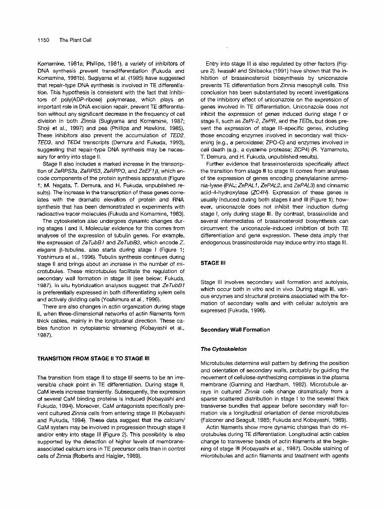
cell culture system. These markers have facilitated the divi-sion of the process of transdifferentiation into three stages:stage I, stage II, and stage III (Figure 1; Fukuda, 1994, 1996).
Stage I, which immediately follows the induction of differ-entiation, corresponds to the functional dedifferentiationprocess during which isolated mesophyll cells lose theirability to perform photosynthesis. This dedifferentiation pro-cess also involves the expression of wound-induced genesand the acquisition of the cells' ability to grow and differenti-ate in a new environment. However, this dedifferentiationprocess is not accompanied by cell division.
A typical example of functional dedifferentiation duringstage I is the change in the organization of the reticulate ar-rays of actin filaments that are thought to anchor chloro-plasts to the plasma membrane (Kobayashi et al., 1987).These arrays are reorganized into a three-dimensional net-work over the entire length of the cell, causing chloroplaststo leave the vicinity of the plasma membrane and the meso-
24 48 72 96 hr
Stage I Stage II Stage III
Dedifferentiation Restriction ofdevelopmental potential TE specific development
BZeRT, ZePR, ZePI-1.ZePI-2
ZeRPS3a, ZeRPS3. ZeRPPO. ZeEFW_____ZeTubBL ZeTubBS
TED2, TEDS, TED4
ZCAD1
ZePAU, ZePAL2, ZePAL3, ZC4H ZePALI, ZePAL2, ZePAL3, ZC4H
ZPO-C
ZEN1, ZRNase I, ZCP4, p48h-17
Figure 1. Accumulation of Various Transcripts during the Transdifferentiation of Single Zinnia Mesophyll Cells into TEs.
(A) The three stages of transdifferentiation. Stage I corresponds to the functional dedifferentiation process. In stage II, the developmental poten-tial of the dedifferentiated cells becomes restricted from the pluripotent ability to differentiate into immature xylem and/or phloem cells to thesingle ability to differentiate into TEs. Stage III involves TE-specific events.(B) Gene expression during transdifferentiation. The accumulation patterns of transcripts for various genes isolated from Z. elegans are depictedas bars. Green, yellow, and red bars show transcripts whose accumulation starts during stages I, II, and III, respectively.

Tracheary Element Differentiation 1149
B
AuxinCytoklnlnWounding
Accumulationof hydrolytlcenzymesSW synthesis
tCa/CaMcystelne proteasesbrasslnosterolds
Figure 2. Cellular Events That Occur during Transdifferentiation of Zinnia Mesophyll Cells into TEs.(A) Isolated mesophyll cell.(B) Dedifferentiated cell.(C) TE precursor cell.(D) Immature TE.(E) Maturing TE.(F) Mature TE.Transdifferentiation is induced by wounding and a combination of auxin and cytokinin. Green, yellow, and red arrows show the progress ofstages I, II, and III, respectively. The transition from stage II to stage III appears to be regulated by calcium (Ca)/CaM, cysteine proteases, andendogenous brassinosteroids. At the start of stage III, genes that are involved in both secondary wall synthesis and autolysis are expressed. Hy-drolytic enzymes, such as DNases, RNases, and proteases, may accumulate in the vacuole. The disruption of the tonoplast causes these en-zymes to invade the cytoplasm and attack various organelles, resulting in the formation of a mature TE that has lost its cell contents. CP,chloroplast; CT, cytoplasm; NC, nucleus; PW, primary wall; SW, secondary wall; VC, vacuole.
phyll cells to lose their ability to perform photosynthesis.Calmodulin (CaM) antagonists inhibit TE differentiation whenadded early in stage I (Roberts and Haigler, 1990), suggest-ing that progression through dedifferentiation may involvecalcium/CaM regulation.
We have isolated cDNA clones corresponding to 12 genesthat are induced during stage I (Figure 1; M. Nagata, T. Demura,and H. Fukuda, unpublished results). These genes havebeen categorized into three groups: group 1 compriseswound-related genes, such as genes encoding protease in-hibitors (e.g., Z. elegans protease inhibitor 1 and 2 [ZePI-1and ZePI-2]), and group 2 is made up of genes that encodecomponents of the protein synthesis apparatus, such as ri-bosomal proteins (e.g., Z. elegans ribosomal protein geneS3a, S3, and PO [ZeRPSSa, ZeRPS3, and ZeRPPO}) and anelongation factor (Z. elegans elongation factor 1 b [ZeEFIb]).The remainder of the cDNAs make up group 3. The tran-scription of all of these genes is induced by wounding butnot by phytohormones. However, phytohormones do affectthe changes in the level of their transcripts that occur duringstage II and stage III of TE differentiation. For example, ei-ther naphthaleneacetic acid or benzyladenine must be presentfor substantial levels of group 2 transcripts to be maintainedthroughout development. Moreover, the accumulation oftranscripts of group 1 genes is reduced only in cells thathave been cultured in differentiation-inducing media thatcontain specific ratios of naphthaleneacetic acid and benzyl-adenine. These results suggest that the dedifferentiation
process involves wound-induced events and the activationof protein synthesis, both of which are regulated by phyto-hormones at a later stage in the transdifferentiation process.
STAGE I
The accumulation of transcripts of the TED2, TED3, andTED4 (tracheary element differentiation-related 2, 3, and 4)genes, which begins 12 to 24 hr before secondary wallthickening, defines the second stage of transdifferentiationin cultured Zinnia cells (Figure 1; Demura and Fukuda, 1993,1994). In situ hybridization experiments have also demon-strated that the expression of these genes is restricted tocells that are involved in vascular differentiation in intactplants. For example, TEDS transcripts are expressed specif-ically in TE precursor cells. Similarly, TED4 transcripts arerestricted to vascular cells or future vascular cells and inparticular to immature xylem cells that do not show anymorphological changes. TED2 transcripts are restricted toprocambial regions, immature phloem, and immature xylemcells. These data suggest that stage II of transdifferentiationin vitro corresponds to the process of differentiation fromprocambial initials to precursors of TEs in vivo (Figure 2).
Although transdifferentiation of Zinnia and Jerusalemartichoke cells does not require cell division or progres-sion through the S phase of the cell cycle (Fukuda and
1150 The Plant Cell
Komamine, 1981a; Phillips, 1981), a variety of inhibitors of DNA synthesis prevent transdifferentiation (Fukuda and Komamine, 1981 b). Sugiyama et al. (1 995) have suggested that repair-type DNA synthesis is involved in TE differentia- tion. This hypothesis is consistent with the fact that inhibi- tors of poly(ADP-ribose) polymerase, which plays an important role in DNA excision repair, prevent TE differentia- tion without any significant decrease in the frequency of cell division in both Zinnia (Sugiyama and Komamine, 1987; Shoji et al., 1997) and pea (Phillips and Hawkins, 1985). These inhibitors also prevent the accumulation of TED2, TED3, and TED4 transcripts (Demura and Fukuda, 1993), suggesting that repair-type DNA synthesis may be neces- sary for entry into stage II.
Stage II also includes a marked increase in the transcrip- tion of ZeRPS3a, ZeRPS3, ZeRPPO, and ZeEFlp, which en- code components of the protein synthesis apparatus (Figure 1 ; M. Nagata, T. Demura, and H. Fukuda, unpublished re- sults). The increase in the transcription of these genes corre- lates with the dramatic elevation of protein and RNA synthesis that has been demonstrated in experiments with radioactive tracer molecules (Fukuda and Komamine, 1983).
The cytoskeleton also undergoes dynamic changes dur- ing stages I and II. Molecular evidence for this comes from analyses of the expression of tubulin genes. For example, the expression of ZeTubBl and ZeTubB3, which encode Z. elegans p-tubulins, also starts during stage I (Figure 1; Yoshimura et al., 1996). Tubulin synthesis continues during stage I I and brings about an increase in the number of mi- crotubules. These microtubules facilitate the regulation of secondary wall formation in stage III (see below; Fukuda, 1987). In situ hybridization analyses suggest that ZeTubBl is preferentially expressed in both differentiating xylem cells and actively dividing cells (Yoshimura et al., 1996).
There are also changes in actin organization during stage 11, when three-dimensional networks of actin filaments form thick cables, mainly in the longitudinal direction. These ca- bles function in cytoplasmic streaming (Kobayashi et al., 1987).
TRANSlTlON FROM STAGE II TO STAGE 111
The transition from stage II to stage III seems to be an irre- versible check point in TE differentiation. During stage 11, CaM levels increase transiently. Subsequently, the expression of several CaM binding proteins is induced (Kobayashi and Fukuda, 1994). Moreover, CaM antagonists specifically pre- vent cultured Zinnia cells from entering stage 111 (Kobayashi and Fukuda, 1994). These data suggest that the calcium/ CaM system may be involved in progression through stage II and/or efitry into stage 111 (Figure 2). This possibility is also supported by the detection of higher levels of membrane- associated calcium ions in TE precursor cells than in control cells of Zinnia (Roberts and Haigler, 1989).
Entry into stage 111 is also regulated by other factors (Fig- ure 2). lwasaki and Shibaoka (1991) have shown that the in- hibition of brassinosteroid biosynthesis by uniconazole prevents TE differentiation from Zinnia mesophyll cells. This conclusion has been substantiated by recent investigations of the inhibitory effect of uniconazole on the expression of genes involved in TE differentiation. Uniconazole does not inhibit the expression of genes induced during stage I or stage 11, such asZePl-2, ZePR, and the TEDs, but does pre- vent the expression of stage Ill-specific genes, including those encoding enzymes involved in secondary wall thick- ening (e.g., a peroxidase; ZPO-C) and enzymes involved in cell death (e.g., a cysteine protease; ZCP4) (R. Yamamoto, T. Demura, and H. Fukuda, unpublished results).
Further evidence that brassinosteroids specifically affect the transition from stage II to stage 111 comes from analyses of the expression of genes encoding phenylalanine ammo- nia-lyase (PAL; ZePALl,ZePALZ, and ZePAL3) and cinnamic acid-4-hydroxylase (ZC4H). Expression of these genes is usually induced during both stages I and 111 (Figure 1); how- ever, uniconazole does not inhibit their induction during stage I, only during stage 111. By contrast, brassinolide and several intermediates of brassinosteroid biosynthesis can circumvent the uniconazole-induced inhibition of both TE differentiation and gene expression. These data imply that endogenous brassinosteroids may induce entry into stage 111.
STAGE 111
Stage 111 involves secondary wall formation and autolysis, which occur both in vitro and in vivo. During stage 111, vari- ous enzymes and structural proteins associated with the for- mation of secondary walls and with cellular autolysis are expressed (Fukuda, 1996).
Secondary Wall Formation
The Cytoskeleton
Microtubules determine wall pattern by defining the position and orientation of secondary walls, probably by guiding the movement of cellulose-synthesizing complexes in the plasma membrane (Gunning and Hardham, 1982). Microtubule ar- rays in cultured Zinnia cells change dramatically from a sparse scattered distribution in stage I to the several thick transverse bundles that appear before secondary wall for- mation via a longitudinal orientation of dense microtubules (Falconer and Seagull, 1985; Fukuda and Kobayashi, 1989).
Actin filaments show more dynamic changes than do mi- crotubules during TE differentiation. Longitudinal actin cables change to transverse bands of actin filaments at the begin- ning of stage 111 (Kobayashi et al., 1987). Double staining of microtubules and actin filaments and treatment with agents

Tracheary Element Differentiation 1151
that depolymerize cytoskeletal components indicate that there is a coordinated mechanism in which actin filaments are involved in the reorganization of microtubules, which in turn regulate the spatial disposition of secondary walls (Fukuda and Kobayashi, 1989).
Secondary Wall-Specific Proteins
Severa1 proteins are specifically associated with secondary walls of TEs. These proteins include an extensin-like protein, which was isolated from the xylem of loblolly pine (Bao et al., 1992). This protein is present in secondary cell walls of xylem cells during lignification and remains as a structural component of cell walls in wood. Arabinogalactan proteins are also present in secondary walls of TEs, and severa1 anti- bodies directed against arabinogalactan proteins bind to im- mature xylem cells in some species of plant (Schindler et al., 1995). For instance, the JIM13 monoclonal antibody binds to immature and mature TEs in cultured Zinnia cells (Stacey et al., 1995).
Wheat germ agglutinin, which has a strong affinity for a sequence of three p-(1-4)-linked N-acetyl-D-glucosamine residues, also binds specifically to secondary walls of TEs (Hogetsu, 1990; Wojtaszek and Bolwell, 1995). Wojtaszek and Bolwell (1 995) have isolated three novel glycoproteins that bind to wheat germ agglutinin. By using an antibody against one of these glycoproteins, SWGPSO, they show that the protein is localized in secondary walls of TEs, xylem fibers, and phloem fibers. Another novel type of wall protein, a Tyr- and Lys-rich protein from tomato, has also been lo- calized to secondary walls (Domingo et al., 1994). Finally, GRP 7.8 was isolated as a cDNA clone encoding a protein that is localized in the cell walls of the primary xylem ele- ments and the primary phloem of many plant species (Keller et al., 1988; Ye et al., 1991). Detailed immunoelectron microscopy studies indicate that GRPl.8 is produced by xylem parenchyma cells and exported to the walls of proto- xylem vessels (Ryser and Keller, 1992).
The bean GRP 7.8 promoter has been analyzed in detail. The promoter possesses both a negative element that re- presses promoter activity in nonvascular cells and a positive element that directs vascular tissue-specific expression (Keller and Baumgartner, 1991 ; Keller and Heierli, 1994). Re- cently, Keller’s group indicated that a novel basic leucine zipper transcription factor binds to a 28-bp element, termed vs-7, that partially overlaps with the negative element in the GRP 7.8 promoter (Schumann et al., 1996).
Expression of Lignin Biosynthetic Enzymes
Lignin is one of the most characteristic components of sec- ondary walls of TEs; its biosynthesis involves the shikimate, general phenylpropanoid, and specific lignin pathways (Boudet et al., 1995). In cultured Zinnia cells, activities of
PAL and caffeoyl-COA-3-O-methyltransferase are induced preferentially during stages I and 111, mirroring the increased accumulation of the corresponding transcripts (Lin and Northcote, 1990; Ye et al., 1994). Detailed analysis ofZePAL7, ZePAL2, ZePAL3, and ZC4H transcript accumulation sug- gests that the expression of these phenylpropanoid path- way-related genes is coordinately induced during stages I and III (Figure 1; R. Yamamoto, T. Demura, and H. Fukuda, unpublished results). During stage I, the expression of these genes is induced by wounding, whereas during stage 111, it is associated with lignin synthesis (Fukuda, 1996).
The promoters of genes encoding PAL and 4-coumaric acid:CoA ligase (4CL) share conserved sequences that may mediate their coordinate regulation. For example, the AC- rich sequence ACII, which has been identified in PAL- and 4CL-encoding genes of a number of plants, functions as a negative element that suppresses gene expression in the phloem (Hauffe et al., 1993; Hatton et al., 1995). ACll is thought to be a Myb binding site, which implies that Myb proteins may be involved in the coordinated expression of genes encoding phenylpropanoid pathway-related enzymes.
By contrast, the activity of cinnamyl alcohol dehydroge- nase, which is involved in the specific lignin pathway, is highest at stage 111, but its transcript (ZCAD7) begins to ac- cumulate during stage II (Figure 1 ; Sato et al., 1997). Inter- estingly, uniconazole does not inhibit the expression of ZCAD7, which is consistent with the above-mentioned ob- servation that uniconazole does not inhibit the expression of genes induced during stages I and 11.
A second enzyme in the specific lignin pathway, P5, which is a cationic peroxidase isozyme that is ionically bound to the cell walls, is known to be involved in the final step of lig- nin synthesis in differentiating Zinnia cells-the polymeriza- tion of cinnamyl alcohols into lignin (Sato et al., 1993, 1995). Transcripts of the corresponding gene, ZPO-C, accumulate specifically and transiently during stage 111 (Figure 1; Y. Sato and H. Fukuda, unpublished results). The accumulation of ZPO-C transcripts precedes that of the ZePALs and ZC4H, and ZPO-C transcript levels are elevated for a shorter period of time. Therefore, genes encoding lignin biosynthetic en- zymes appear to be regulated in a complex manner during TE differentiation in cultured Zinnia cells.
Programmed Cell Death
Differentiation into TEs is a typical example of developmen- tally programmed cell death in higher plants (see Pennell and Lamb, 1997, in this issue). TEs reach maturity after the loss of cell contents, including the nucleus, and the partia1 digestion of primary walls. Pores open at the ends of individ- ual vessel elements, which are longitudinally arranged to form a long vessel tube. The cell death process that takes place during stage III of TE differentiation is coupled tightly to the formation of secondary walls. Indeed, it has not been

11 52 The Plant Cell
possible to separate experimentally cell death from second- ary wall formation during TE differentiation.
lnduction of Cell Death
In animals, a variety of factors, such as Fas ligands, tumor necrosis factors, and hormones, are involved in the induc- tion of apoptosis. Death signals induced by these factors activate the interleukin-1 p-converting enzyme (ICE)-like protease cascade, which triggers cell death (White, 1996). Genetic analysis of the nematode Caenorhabdifis elegans has provided the most direct evidence that the initiation of cell death during development is controlled by specific genes, such as ced-3, cedd, and ced-9 (Yuan and Horvitz, 1990; Hengartner et al., 1992). The nematode ced-3 and ced-9 genes have mammalian counterparts that are respon- sible for initiating apoptosis in mammals (White, 1996).
In plants, toxins, high concentrations of salts, and some chemicals can induce the apoptosis-like death of particular cells (Katsuhara and Kawasaki, 1996; Wang et al., 1996). However, inducers of developmental cell death have not been identified in higher plants. During TE differentiation, inhibitors of CaM action (Kobayashi and Fukuda, 1994), cys- teine protease activity (Y. Watanabe and H. Fukuda, unpub- lished results), and brassinosteroid synthesis (R. Yamamoto, T. Demura, and H. Fukuda, unpublished results) prevent TE precursor cells from entering stage 111 (Figure 2).
The transcripts for both cell death-related enzymes and enzymes involved in secondary wall formation begin to ac- cumulate at the same time (Figure 1). These observations imply that a common signal(s) may induce both cell death and secondary wall formation. Furthermore, the inhibition of the entry into stage III by cysteine protease inhibitors may suggest the presence of an unknown protease cascade sim- ilar to the ICE cascade, although inhibitors of ICE do not prevent cell death during TE differentiation (H. Kuriyama and H. Fukuda, unpublished results; see also Pennell and Lamb, 1997, in this issue).
Morphological Features of the Cell Death Process
Apoptosis in animals involves nuclear shrinkage, cellular shrinkage, membrane bubbling, the formation of apoptotic bodies, and digestion by macrophages (Kerr and Harmon, 1991; see also Pennell and Lamb, 1997, in this issue). The process of cell death during TE differentiation, which has been studied in detail by electron microscopy, does not ex- hibit any substantial similarity to these typical morphological features of animal apoptosis (reviewed in Fukuda, 1996). For example, in differentiating Zinnia cells, the visible degenera- tion of all organelles, including the nucleus, starts only after the tonoplast ruptures, which occurs severa1 hours after the secondary wall thickenings become visible. After the disrup- tion of the tonoplast, organelles with a single membrane,
such as Golgi bodies and the endoplasmic reticulum, be- come swollen and then rupture. Subsequently, organelles with double membranes are degraded. The nucleus is also degraded, but this degradation is not preceded by nuclear shrinkage and fragmentation. TEs lose most of their or- ganelles within a few hours after disruption of the tonoplast, and the entire contents of the cell disappear within -6 hr af- ter the first visible evidence of secondary wall thickening. These data imply that disruption of the tonoplast may be a critical event in TE cell death.
Concomitant with autolysis, cell walls of TEs are modified, although lignification of secondary walls begins before tono- plast disruption. Around the time of tonoplast rupture, lignin is deposited on the outer layer of primary walls of TEs, after which the regions of primary walls that have not been ligni- fied are digested. Perforation of the ends of elongated TEs, which presumably are not thickened, also follows tonoplast disruption.
These cell wall modifications must be controlled strictly. Indeed, we recently identified a cell wall degrading activity that is specific to differentiating TEs in cultured Zinnia cells and that is initiated before tonoplast disruption (Y. Ohdaira, M. Sugiyama, and H. Fukuda, unpublished results). More- over, Burgess and Linstead (1984) have reported that the middle lamella between a cell that is differentiating into TEs and one that is not is resistant to degradation, whereas the middle lamella between two neighboring cells that are both differentiating is digested completely. These observations imply that both cell wall modification and expression of hy- drolytic enzymes may be necessary for the characteristic degradation of TE walls.
Molecular Features of the Cell Death Process
The rapid degeneration of organelles during autolysis must be the result of a variety of highly active hydrolytic enzymes. Minami and Fukuda (1995) have found that the activity of a 30-kD cysteine protease increases transiently just before the start of autolysis and is specific to differentiating Zinnia TEs; this protease is most active at pH 5.5. Moreover, the spe- cific inhibition of intracellular cysteine protease activity in differentiating TEs suppresses nuclear degeneration, sug- gesting that a cysteine protease(s) plays a key role in this process (Y. Watanabe and H. Fukuda, unpublished results).
Two cDNAs, p48h-17 (Ye and Varner, 1996) and ZCf4 (A. Minami, T. Demura, and H. Fukuda, unpublished results), that may encode cysteine proteases have been isolated from Zinnia. The deduced amino acid sequences of these cysteine proteases suggest that their precursors, which re- semble papine, are probably transported into the vacuole, where they are processed into activated forms. The p48h-17 and ZCP4 transcripts also accumulate transiently just before autolysis and are found specifically in differentiating TEs in vitro and in vivo (Ye and Varner, 1996; A. Minami, T. Demura, and H. Fukuda, unpublished results).

Tracheaty Element Differentiation 1153
In addition to the cysteine proteases, a 145-kD serine pro- tease has been detected specifically in differentiating TEs of cultured Zinnia cells on substrate-impregnated gels (Beers and Freeman, 1997). Moreover, an -60-kD serine protease has been reported to appear preferentially in differentiating TEs (Ye and Varner, 1996; Beers and Freeman, 1997). These results indicate that a complex set of proteases is involved in the autolytic process. Nonetheless, the substrates, cellu- lar location, and mode of induction of each of these pro- teases are unknown.
During apoptosis, DNA laddering is caused by the diges- tion of DNA into nucleosome-sized units by endonucleases. Among the candidates for these nucleases in animal cells are NUC 18, DNase I, DNase 11, and DNase (Peitsch et al., 1993; Tanuma and Shiokawa, 1994). DNase is present in the nucleus, activated by Ca2+ and Mg2+, and inhibited by Zn2+ (Tanuma and Shiokawa, 1994). Similarly, Mittler and Lam (1995a) have detected a 36-kD DNase, NUC 111, that is ex- pressed in association with cell death during the hypersensi- tive reaction (see Pennell and Lamb, 1997, in this issue). This nuclease has characteristics similar to apoptosis- related DNases in that it is present in the nucleus and shows Ca2+-mediated activation and Zn2+-mediated inhibition. However, this DNase is not expressed during cell death in senescence.
Thelen and Northcote (1989) have found that severa1 DNase and RNase activities are closely associated with TE differentiation in Zinnia cells. These 43-, 22-, and 25-kD nu- cleases appeared 12 hr before the visible formation of TEs, and their levels increased conspicuously during the matura- tion phase of differentiation. The 43-kD nuclease is the only one that has DNase activity in differentiating TEs. This nu- clease requires Zn2+ for activation and can hydrolyze RNA, single-stranded DNA, and double-stranded DNA. Recently, Mittler and Lam (199513) detected fragmented nuclear DNA in differentiating TEs. However, their results could be ex- plained by invoking the activity of a general nuclease such as this rather than that of apoptosis-specific endonucleases.
The characteristics of the 43-kD nuclease resemble those of a nuclease that is expressed in barley aleurone cells dur- ing germination (Brown and Ho, 1986). Moreover, a Zn2+- activated DNase has also been found in senescing leaves (Blank and McKeon, 1989). We have isolated two cDNA clones, BfN7 @arley gndonuclease 1) and ZfN7 @mia gn- dopclease l ) , that correspond to the Znz+-activated nu- cleases from the barley aleurone and from differentiating Zinnia TEs, respectively. The nucleotide and deduced amino acid sequences show that BENI and ZENl are similar to each other and to the S1 nuclease of Aspergillus (S. Aoyagi, M. Sugiyama, and H. Fukuda, unpublished results). These data suggest that Zn2+-activated nucleases similar to S1 are commonly involved in programmed cell death processes that take place throughout plant development.
Recently, ZRNasel, a cDNA encoding the 22-kD (or 25-kD) RNase detected by Thelen and Northcote (1 989), has been isolated from Zinnia (Ye and Droste, 1996). The mRNA for
ZRNasel also accumulates specifically in differentiating TEs in vitro and in vivo. Like those for ZEN7, ZCP4, and p48h-77, ZRNasel transcripts also accumulate transiently just before autolysis starts (Figure 1). Therefore, these genes may be regulated via a common mechanism that relies on the same cis- and trans-activation factors. Interestingly, ZPO-C, a gene encoding lignin-related peroxidase, is also expressed in a pattern very similar to that of the autolysis-related genes, suggesting that gene expression, in association with both secondary wall formation and autolysis, may be regu- lated at least in part by a common mechanism.
Their deduced amino acid sequences show that ZEN1, ZRNasel, ZCP4, and p48h-17 have signal peptides at their N termini. This implies that these proteins may be transported to the vacuole (Figure 2). Disruption of the tonoplast during stage 111 of TE differentiation causes hydrolytic enzymes to invade the cytoplasm and attack various organelles. It is in- teresting that the optimal pH of both the DNase and cysteine protease is 5.5, which corresponds to the pH in the vacuole and probably in the cytoplasm after the tonoplast ruptures. Even so, one of the most pressing issues regarding the cell death process during TE differentiation-the mechanism controlling tonoplast disruption-is still open for investigation.
CONCLUSIONS AND FUTURE PERSPECTIVES
Transdifferentiation into TEs in vitro is induced by a combi- nation of auxin and cytokinin and, in some cases, by wound- ing. The transdifferentiation process can be divided into three stages: stage I, stage 1 1 , and stage III (Figures 1 and 2). Stage I corresponds to the functional dedifferentiation pro- cess. In stage II, the developmental potential of the de- differentiated cells becomes restricted from the pluripotent ability to differentiate into immature xylem and/or phloem cells to the single ability to differentiate into TEs. This pro- cess seems to correspond to the in vivo process during which meristematic cells change into procambial cells and then into TE precursor cells. Stage III involves TE-specific events, such as secondary wall formation and autolysis, and is shared both in vitro and in vivo. A two-step process un- derlying the differentiation of parenchyma cells into stele cells and then of stele cells into TEs has also been sug- gested for transdifferentiation in wounded pea roots (Rana and Gahan, 1983).
One of the most promising approaches to the elucidation of the mechanism of TE differentiation is the analysis of mu- tants that have a defect in the process of TE differentiation. However, because plants that have a severe defect in TE differentiation are likely to be inviable, they may be hard to identify. Moreover, transgenic plants in which a TE-essential gene is overexpressed or suppressed may also face prob- lems with lethality during regeneration.
The use of transgenic organs may overcome this problem. Indeed, we have recently established a transgenic Zinnia

11 54 The Plant Cell
root system into which we plan to introduce TE-specific genes so that we may analyze their function in TE differenti- ation during the development of vascular bundles in roots (Y. Tateishi, H. Choi, and H. Fukuda, unpublished results). We are using this system to investigate the roles of the TEDs and of severa1 genes involved in lignin biosynthesis, and ini- tia1 indications are that this system may be useful for the study of the role of various gene products in TE differentia- tion. Such new approaches should provide marked in- creases in our understanding of TE differentiation in plants in the near future.
ACKNOWLEDGMENTS
I thank Dr. Roni Aloni for critical reading of the manuscript. Financia1 support from the Ministry of Education, Science, and Culture of Japan (Grant Nos. 05276103,06278102, and 07454215), the Japan Society for the Promotion of Science (Grant No. JSPS-RFTF96L00605), and the Toray Science Foundation is gratefully acknowledged.
REFERENCES
Aloni, R. (1987). Differentiation of vascular tissues. Annu. Rev. Plant Physiol. 38, 179-204.
Aloni, R. (1995). The induction of vascular tissues by auxin and cytokinin. In Plant Hormones, Physiology, Biochemistry, and Molecular Biology, P.J. Davies, ed (Dordrecht, The Netherlands: Kluwer Academic Publishers), pp. 531-546.
Aloni, R., and Zimmermann, M.H. (1983). The control of vessel size and density along the plant axis-A new hypothesis. Differentia- tion 24, 203-208.
Bao, W., O’Malley, D.M., and Sederoff, R.R. (1992). Wood con- tains a cell-wall structural protein. Proc. Natl. Acad. Sci. USA 89, 66044608.
Baum, S.F, Aloni, R., and Peterson, C.A. (1991). The role of cytoki- nin in vessel regeneration in wounded Coleus internodes. Ann. Bot. 67, 543-548.
Beers, E.P., and Freeman, T.B. (1997). Protease activity during tra- cheary element differentiation in Zinnia mesophyll cultures. Plant Physiol. 113, 873-880.
Blank, A., and McKeon, T.A. (1 989). Single-strand-preferring nuclease activity in wheat leaves is increased in senescence and is negatively photoregulated. Proc. Natl. Acad. Sci. USA 86,
Boudet, A.M., Lapierre, C., and Grima-Pettenati, J. (1995). Bio- chemistry and molecular biology of lignification. New Phytol. 129,
31 69-31 73.
203-236.
Brown, P.H., and Ho, T.-H.D. (1986). Barley (Hordeum vulgare culti- var Himalaya) aleurone layers secrete a nuclease in response to gibberellic acid: Purification and partia1 characterization of the associated ribonuclease, deoxyribonuclease, and 3‘-nucleotidase activities. Plant Physiol. 82, 801-806.
Burgess, J., and Linstead, P. (1984). In vitro tracheary element for- mation: Structural studies and the effect of triiodobenzoic acid. Planta 160, 481-489.
Chasan, R. (1 994). Tracing tracheary element development. Plant Cell6,917-919.
Church, D.L., and Galston, A.W. (1988). Kinetics of determination in the differentiation of isolated mesophyll cells of Zinnia elegans to tracheary elements. Plant Physiol. 88, 92-96.
Church, D.L., and Galston, A.W. (1989). Hormonal induction of vascular differentiation in cultured Zinnia leaf disks. Plant Cell Physiol. 30, 73-78.
Demura, T., and Fukuda, H. (1993). Molecular cloning and charac- terization of cDNAs associated with tracheary element differentia- tion in cultured Zinnia cells. Plant Physiol. 103, 815-821.
Demura, T., and Fukuda, H. (1994). Nove1 vascular cell-specific genes whose expression is regulated temporally and spatially dur- ing vascular system development. Plant Cell 6, 967-981.
Domingo, C., Gómez, M.D., Caiias, L., Hernández-Yago, J., Conejero, V., and Vera, P. (1994). A nove1 extracellular matrix protein from tomato associated with lignified secondary cell walls. Plant Cell 6, 1035-1 047.
Falconer, M.M., and Seagull, R.W. (1 985). lmmunofluorescent and Calcofluor white staining of developing tracheary elements in Zin- nia elegans L. suspension cultures. Protoplasma 125, 190-1 98.
Fukuda, H. (1987). A change in tubulin synthesis in the process of tracheary element differentiation and cell division of isolated Zin- nia mesophyll cells. Plant Cell Physiol. 28, 51 7-528.
Fukuda, H. (1992). Tracheary element formation as a model system of cell differentiation. Int. Rev. Cytol. 136, 289-332.
Fukuda, H. (1994). Redifferentiation of single mesophyll cells into tracheary elements. Int. J. Plant Sci. 155, 262-271.
Fukuda, H. (1 996). Xylogenesis: Initiation, progression and cell death. Annu. Rev. Plant Physiol. Plant MOI. Biol. 47, 299-325.
Fukuda, H., and Kobayashi, H. (1989). Dynamic organization of the cytoskeleton during tracheary-element differentiation. Dev. Growth Differ. 31, 9-1 6.
Fukuda, H., and Komamine, A. (1980). Establishment of an experi- mental system for the tracheary element differentiation from sin- gle cells isolated from the mesophyll of Zinnia elegans. Plant Physiol. 65, 57-60.
Fukuda, H., and Komamine, A. (1981a). Relationship between tra- cheary element differentiation and the cell cycle in single cells iso- lated from the mesophyll of Zinnia elegans. Physiol. Plant. 52,
Fukuda, H., and Komamine, A. (1 981 b). Relationship between tra- cheary element differentiation and DNA synthesis in single cells isolated from the mesophyll of Zinnia elegans-Analysis by inhibi- tors of DNA synthesis. Plant Cell Physiol. 22, 41-49.
Fukuda, H., and Komamine, A. (1983). Changes in the synthesis of RNA and protein during tracheary element differentiation in single cells isolated from the mesophyll of Zinnia elegans. Plant Cell Physiol. 24, 603-614.
Gunning, B.E.S., and Hardham, A.R. (1 982). Microtubules. Annu. Rev. Plant Physiol. 33,651-698.
Hatton, D., Sablowski, R., Yung, M.-H., Smith, C., Schuch, W., and Bevan, M. (1995). Two classes of cis sequences contribute
423-430.

Tracheary Element Differentiation 1155
to tissue-specific expression of a PAL2 promoter in transgenic tobacco. Plant J. 7, 859-876.
Hauffe, K.D., Lee, S.P., Subramaniam, R., and Douglas, C.J. (1993). Combinatorial interactions between positive and negative cis-acting elements control spatial patterns of 4CL-7 expression in transgenic tobacco. Plant J. 4, 235-253.
Hengartner, M.O., Ellis, R.E., and Horovitz, H.R. (1992). Cae- norhabditis elegans gene ced-9 protects cells from programmed cell death. Nature 356, 494-499.
Hogetsu, T. (1990). Detection of hemicelluloses specific to the cell wall of tracheary elements and phloem cells by fluorescein-conju- gated lectins. Protoplasma 156, 67-73.
Iwasaki, T., and Shibaoka, H. (1991). Brassinosteroids act as regu- lators of tracheary-element differentiation in isolated Zinnia meso- phyll cells. Plant Cell Physiol. 32, 1007-1014.
Jacobs, W.P. (1952). The role of auxin in differentiation of xylem around a wound. Am. J. Bot. 39,301-309.
Katsuhara, K., and Kawasaki, T. (1 996). Salt stress-induced nuclear and DNA degradation in meristematic cells of barley roots. Plant Cell Physiol. 37, 169-173.
Keller, B., and Baumgartner, C. (1 991). Vascular-specific expres- sion of the bean GRP 1.8 gene is negatively regulated. Plant Cell
Keller, B., and Heierli, D. (1994). Vascular expression of the grp7.8 promoter is controlled by three specific regulatory elements and one unspecific activating sequence. Plant MOI. Biol. 26, 747-756.
Keller, B., Sauer, N., and Lamb, C.J. (1988). Glycine-rich cell wall proteins in bean: Gene structure and association of the protein with the vascular system. EMBO J. 7,3625-3633.
Kerr, J.F.R., and Harmon, B.V. (1991). Definition and incidence of apoptosis: An historical perspective. In Apoptosis: The Molecular Basis of Cell Death, L.D. Tomei and F.O. Cope, eds (Cold Spring Harbor, NY: Cold Spring Harbor Laboratory Press), pp. 5-29.
Klee, H.J., Horsch, R.B., Hinchee, M.A., Hein,' M.B., and Hoffmann, N.L. (1987). The effects of overproduction of two Agrobacterium tumefaciens T-DNA auxin biosynthetic gene prod- ucts in transgenic petunia plants. Genes Dev. I, 86-96.
Kobayashi, H., and Fukuda, H. (1994). lnvolvement of calmodulin and calmodulin-binding proteins in the differentiation of tracheary elements in Zinnia cells. Planta 194, 388-394.
Kobayashi, H., Fukuda, H., and Shibaoka, H. (1987). Reorganiza- tion of actin filaments associated with the differentiation of trache- ary elements in Zinnia mesophyll cells. Protoplasma 138, 69-71.
Lin, Q., and Northcote, D.H. (1990). Expression of phenylalanine ammonia-lyase gene during tracheary-element differentiation from cultured mesophyll cells of Zinnia elegans L. Planta 182,
Minami, A., and Fukuda, H. (1995). Transient and specific expres- sion of a cysteine endopeptidase during autolysis in differentiating tracheary elements from Zinnia mesophyll cells. Plant Cell Physiol.
Mittler, R., and Lam, E. (1 995a). Identification, characterization, and purification of a tobacco endonuclease activity induced upon hypersensitive response cell death. Plant Cell 7, 1951-1962.
Mittler, R., and Lam, E. (1995b). In situ detection of nDNAfragmen- tation during the differentiation of tracheary elements in higher plants. Plant Physiol. 108, 489-493.
3,1051-1061.
591 -598.
36,1599-1 606.
Nelson, T., and Dengler, N. (1997). Leaf vascular pattern formation. Plant Cell 9, 1121-1 135.
Peitsh, M.C., Polzar, B., Stepham, H., Crompton, T., MacDonald, H.R., Mannherz, H.G., and Tshopp, J. (1 993). Characterization of the endogenous deoxyribonuclease involved in nuclear DNA deg- radation during apoptosis (programmed cell death). EMBO J. 12,
Pennell, R.I., and Lamb, C. (1997). Programmed cell death in plants. Plant Cell 9, 11 57-1 168.
Phillips, R. (1981). Direct differentiation of tracheary elements in cul- tured explants of gamma-irradiated tubers of Helianthus tubero- sus. Planta 153,262-266.
Phillips, R., and Hawkins, S.W. (1985). Characteristics of the inhibi- tion of induced tracheary element differentiation by 3-aminoben- zamide and related compounds. J. Exp. Bot. 36, 119-128.
Rana, M.A., and Gahan, P.B. (1983). A quantitative cytochemical study of determination for xylem-element formation in response to wounding in roots of Pisum sativum L. Planta 157, 307-316.
Roberts, A.W., and Haigler, C.H. (1 989). Rise in chlorotetracycline accompanies tracheary element differentiation in suspension cul- tures of Zinnia. Protoplasma 152,3745.
Roberts, A.W., and Haigler, C.H. (1 990). Tracheary-element differ- entiation in suspension-cultured cells of Zinnia requires uptake of extracellular Ca2+. Planta 180, 502-509.
Roberts, L.W., Gahan, P.B., and Aloni, R. (1988). Vascular Differ- entiation and Plant Growth Regulators. (Berlin: Springer-Verlag).
Romano, C.P., Hein, M.B., and Klee, H.J. (1991). lnactivation of auxin in tobacco transformed with the indoleacetic acid-lysine synthetase gene of Pseudomonas savastanoi. Genes Dev. 5,
Ryser, U., and Keller, B. (1992). Ultrastructural localization of a bean glycine-rich protein in unlignified primary walls of protoxy- lem cells. Plant Cell 4, 773-783.
Sachs, T. (1991). Cell polarity and tissue patterning in plants. Devel- opment 1 (suppl.), 83-93.
Sato, Y., Sugiyama, M., Gorecki, R.J., Fukuda, H., and Komamine, A. (1 993). lnterrelationship between lignin deposition and the activities of peroxidase isozymes in differentiating trache- ary elements of Zinnia: Analysis with L-ol-aminooxy-p-phenylpro- pionic acid and 2-aminoindan-2-phosphonic acid. Planta 189,
Sato, Y., Sugiyama, M., Komamine, A., and Fukuda, H. (1995). Separation and characterization of the isozymes of wall-bound peroxidase from cultured Zinnia cells during tracheary-element differentiation. Planta 196, 141-147.
Sato, Y., Watanabe, T., Komamine, A., Hibino, T., Shibata, D., Sugiyama, M., and Fukuda, H. (1997). Changes in the activity and mRNA of cinnamyl alcohol dehydrogenase during tracheary element differentiation in Zinnia. Plant Physiol. I 13, 425-430.
Schindler, T., Bergfeld, R., and Schopfer, P. (1 995). Arabinogalac- tan proteins in maize coleoptiles: Developmental relationship to cell death during xylem differentiation but not to extension growth. Plant J. 7, 25-36.
Schumann, S.T., Ringli, C., Heierli, D., Amrhein, N., and Keller, B. (1996). In vitro binding of the tomato bZlP transcriptional activator VSF-1 to a regulatory element that controls xylem-specific gene expression. Plant J. 9, 283-296.
371-377.
438-446.
584-589.

1156 The Plant Cell
Shoji, Y., Sugiyama, M., and Komamine, A. (1997). lnvolvement of poly(ADP-ribose) synthesis in transdifferentiation of isolated mesophyll cells of Zinnia elegans into tracheary elements. Plant Cell Physiol. 38, 36-43.
Stacey, N.J., Roberts, K., Carpita, N.C., Wells, B., and McCann, M.C. (1995). Dynamic changes in cell surface molecules are very early events in the differentiation of mesophyll cells from Zinnia elegans into tracheary elements. Plant J. 8, 891-906.
Sugiyama, M., and Komamine, A. (1987). Effect of inhibitors of ADP-ribosyltransferase on the differentiation of tracheary ele- ments from isolated mesophyll cells of Zinnia elegans. Plant Cell Physiol. 28, 541-544.
Sugiyama, M., Yeung, E.C., Shoji, Y., and Komamine, A. (1995). Possible involvement of DNA-repair events in the transdifferentia- tion of mesophyll cells of Zinnia elegans into tracheary elements. J. Plant Res. 108, 351-361.
Tanuma, S.I., and Shiokawa, D. (1994). Multiple forms of nuclear deoxyribonuclease in rat thymocytes. Biochem. Biophys. Res. Commun. 203,789-797.
Thelen, M.P., and Northcote, D.H. (1 989). ldentification and purifi- cation of a nuclease from Zinnia elegans L.: A potential molecular marker for xylogenesis. Planta 179, 181-195.
Uggla, C., Moritz, T., Sandberg, G., and Sundberg, B. (1996). Auxin as a positional signal in pattern formation in plants. Proc. Natl. Acad. Sci. USA 93,9282-9286.
Wang, H., Li, J., Bostock, R.M., and Gilchrist, D.G. (1996). Apop- tosis: A functional paradigm for programmed plant cell death
induced by a host-selective phytotoxin and invoked during devel- opment. Plant Cell8,375-391.
White, E. (1996). Life, death, and the pursuit of apoptosis. Genes Dev. 10, 1-15.
Wojtaszek, P., and Bolwell, G.P. (1 995). Secondary cell-wall-spe- cific glycoprotein(s) from French bean (fhaseolus vulgaris L.) hypocotyls. Plant Physiol. 108, 1001-1012.
Ye, L H . , and Droste, D.L. (1996). lsolation and characterization of cDNAs encoding xylogenesis-associated and wound-induced ribonucleases in Zinnia elegans. Plant MOI. Biol. 30, 697-709.
Ye, Z.-H., and Varner, J.E. (1996). lnduction of cysteine and serine proteases during xylogenesis in Zinnia elegans. Plant MOI. Biol.
Ye, Z.-H., Song, Y.-R., Marcus, A., and Varner, J.E. (1991). Com- parative localization of three classes of cell wall proteins. Plant J. I, 175-1 83.
Ye, Z.-H., Kneusel, R.E., Matern, U., and Varner, J.E. (1994). An alternative methylation pathway in lignin biosynthesis in Zinnia. Plant Cell 6, 1427-1439.
Yoshimura, T., Demura, T., Igarashi, M., and Fukuda, H. (1996). Differential expression of three genes for different p-tubulin iso- forms during the initial culture of Zinnia mesophyll cells that divide and differentiate into tracheary elements. Plant Cell Physiol. 37,
Yuan, J., and Horvitz, H.R. (1990). The Caenorhabditis elegans genes ced-3 and ced-4 act autonomously to cause programmed cell death. Dev. Biol. 138, 33-41.
30,1233-1246.
1 167-1 176.

DOI 10.1105/tpc.9.7.1147 1997;9;1147-1156Plant Cell
H. FukudaTracheary Element Differentiation.
This information is current as of January 2, 2020
Permissions X
https://www.copyright.com/ccc/openurl.do?sid=pd_hw1532298X&issn=1532298X&WT.mc_id=pd_hw1532298
eTOCs http://www.plantcell.org/cgi/alerts/ctmain
Sign up for eTOCs at:
CiteTrack Alerts http://www.plantcell.org/cgi/alerts/ctmain
Sign up for CiteTrack Alerts at:
Subscription Information http://www.aspb.org/publications/subscriptions.cfm
is available at:Plant Physiology and The Plant CellSubscription Information for
ADVANCING THE SCIENCE OF PLANT BIOLOGY © American Society of Plant Biologists