Toxicity of TiO2, SiO2, ZnO, CuO, Au and Ag engineered ... · Prodac International, Cittadella PD,...
16
Toxicity of TiO 2 , SiO 2 , ZnO, CuO, Au and Ag engineered nanoparticles on hatching and early nauplii of Artemia sp. Rohit Rekulapally*, Lakshmi Narsimha Murthy Chavali*, Mohammed M. Idris and Shashi Singh Centre for Cellular and Molecular Biology, Hyderabad, Telangana, India * These authors contributed equally to this work. ABSTRACT The potential of environmental release enhances with increased commercial applications of the nanomaterials. In this work, a simple and efficient test to estimate the acute toxicity of nanoparticles is carried out on Artemia species and their hatching rates. We have tested six different engineered nanoparticles (silver, gold, copper oxide, zinc oxide, TiO 2 and SiO 2 nanoparticles) and three soluble salts (CuSO 4 , ZnSO 4 and AgNO 3 ) on Artemia sp. The physicochemical properties of the nanoparticles involved in this study were analyzed in normal water and marine water. Hydrated and bleached Artemia cysts were allowed to hatch in continuously aerated, filtered sterile salt water containing nanoparticles; hatching of viable nauplii and total hatchlings have been recorded. In parallel, standard Artemia toxicity test was conducted on the nauplii monitoring the viability. In hatching experiments, a reduction in hatching rate was observed along with mortality of newly hatched nauplii. The results of the hatching experiment and of the standard Artemia test showed a good correlation. The toxicity of the nanoparticles was compared and the order of toxicity was estimated as Ag>CuO>ZnO>Au>TiO 2 >SiO 2 . The study thus suggests that the hatching test itself is a reliable assay for determining the toxicity of nanomaterials. Subjects Toxicology, Ecotoxicology Keywords Nanomaterial, Toxicity, Hatching, Method, Nauplii, Salt water INTRODUCTION Nanomaterials with their ever-expanding diversity, unique properties and endless applications pose risk to environment and human health. There is a dearth of information on the impact or the risk of nanoscale objects to the environment. Increasing utility would mean enhanced exposure of the ecosystems to these unknown risks. Their release into the environment may start with production, during their applications, by weathering and finally through wastes. Nanomaterials can be designed from almost any material—metal/oxides, carbon, organic, biomaterial etc., and in any form, by strictly adhering to the size range of nanometer at least in one dimension. Nanomaterials behave differently from the bulk due to their unique physicochemical properties. Information on predictability of their How to cite this article Rekulapally R, Murthy Chavali LN, Idris MM, Singh S. 2019. Toxicity of TiO 2 , SiO 2 , ZnO, CuO, Au and Ag engineered nanoparticles on hatching and early nauplii of Artemia sp. PeerJ 6:e6138 DOI 10.7717/peerj.6138 Submitted 18 July 2018 Accepted 20 November 2018 Published 3 January 2019 Corresponding authors Mohammed M. Idris, [email protected] Shashi Singh, [email protected] Academic editor Monika Mortimer Additional Information and Declarations can be found on page 13 DOI 10.7717/peerj.6138 Copyright 2019 Rekulapally et al. Distributed under Creative Commons CC-BY 4.0
Transcript of Toxicity of TiO2, SiO2, ZnO, CuO, Au and Ag engineered ... · Prodac International, Cittadella PD,...
Toxicity of TiO2, SiO2, ZnO, CuO, Au andAg engineered nanoparticles on hatchingand early nauplii of Artemia sp.Rohit Rekulapally*, Lakshmi Narsimha Murthy Chavali*,Mohammed M. Idris and Shashi Singh
Centre for Cellular and Molecular Biology, Hyderabad, Telangana, India* These authors contributed equally to this work.
ABSTRACTThe potential of environmental release enhances with increased commercialapplications of the nanomaterials. In this work, a simple and efficient test to estimatethe acute toxicity of nanoparticles is carried out on Artemia species and theirhatching rates. We have tested six different engineered nanoparticles (silver, gold,copper oxide, zinc oxide, TiO2 and SiO2 nanoparticles) and three soluble salts(CuSO4, ZnSO4 and AgNO3) on Artemia sp. The physicochemical properties ofthe nanoparticles involved in this study were analyzed in normal water and marinewater. Hydrated and bleached Artemia cysts were allowed to hatch in continuouslyaerated, filtered sterile salt water containing nanoparticles; hatching of viablenauplii and total hatchlings have been recorded. In parallel, standard Artemiatoxicity test was conducted on the nauplii monitoring the viability. In hatchingexperiments, a reduction in hatching rate was observed along with mortality of newlyhatched nauplii. The results of the hatching experiment and of the standard Artemiatest showed a good correlation. The toxicity of the nanoparticles was comparedand the order of toxicity was estimated as Ag>CuO>ZnO>Au>TiO2>SiO2. The studythus suggests that the hatching test itself is a reliable assay for determining thetoxicity of nanomaterials.
Subjects Toxicology, EcotoxicologyKeywords Nanomaterial, Toxicity, Hatching, Method, Nauplii, Salt water
INTRODUCTIONNanomaterials with their ever-expanding diversity, unique properties and endlessapplications pose risk to environment and human health. There is a dearth of informationon the impact or the risk of nanoscale objects to the environment. Increasing utilitywould mean enhanced exposure of the ecosystems to these unknown risks. Their releaseinto the environment may start with production, during their applications, by weatheringand finally through wastes.
Nanomaterials can be designed from almost any material—metal/oxides, carbon,organic, biomaterial etc., and in any form, by strictly adhering to the size range ofnanometer at least in one dimension. Nanomaterials behave differently from the bulkdue to their unique physicochemical properties. Information on predictability of their
How to cite this article Rekulapally R, Murthy Chavali LN, Idris MM, Singh S. 2019. Toxicity of TiO2, SiO2, ZnO, CuO, Au and Agengineered nanoparticles on hatching and early nauplii of Artemia sp. PeerJ 6:e6138 DOI 10.7717/peerj.6138
Submitted 18 July 2018Accepted 20 November 2018Published 3 January 2019
Corresponding authorsMohammed M. Idris,[email protected] Singh, [email protected]
Academic editorMonika Mortimer
Additional Information andDeclarations can be found onpage 13
DOI 10.7717/peerj.6138
Copyright2019 Rekulapally et al.
Distributed underCreative Commons CC-BY 4.0

behavior or state in due course is almost negligible. Not only is there limited informationon the bioavailability and biopersistence of these particles, the behavior of the particlesin various conditions like pH, salinity and other biotic factors is not clearly known(Musee, 2011; Moore, 2006). Efforts are currently on to identify the risks associated withthe nanomaterial applications.
Hazard identification is an important step for risk assessment of nanomaterials. Most ofthe methodologies used in hazard identification are the ones used for chemicals in general.Being different from bulk material, the nanoforms of the same materials may posesome restraints or advantages in the use of methodologies.
Aquatic systems are the inevitable receptacles for the materials released in theenvironment. The organisms therein get exposed to the nanomaterials accumulatedfrom seepage and flow through. Many assays for ecotoxicology have been tested fornanomaterials—using bacteria (Suppi et al., 2015), fishes and fish embryos (Rizzo et al.,2013; Zhu et al., 2008); copepods (Templeton et al., 2006); Daphnia (Adam et al., 2014;Völker et al., 2013). In this study, Artemia sp. are used to study the nanomaterialtoxicity. Artemia is a nonselective filter feeder organism which is available worldwide inhighly saline waters. Due to their ubiquitous distribution, robust nature and cost-effectiveculturing conditions they make a good model organism for toxicological assays.Though Artemia are considered to be one of the insensitive models for a lot of chemicals,the early developmental stages of Artemia are highly vulnerable to many of the testmaterials. Few studies are reported for nanomaterial toxicity using Artemia sp. also(Arulvasu et al., 2014; Ates et al., 2012, 2013, 2015; Rajabi et al., 2015). Selectionof end point is a factor worth considering while evaluating sensitivity of the species.Most of the studies are carried out in newly hatched nauplii that are exposed to varyingconcentration of nanomaterial; hatching of hydrated cysts has not been used as an end pointfor toxicity except in (Carballo et al., 2002; Migliore et al., 1997; Rotini et al., 2015;Sarabia et al., 2008) though some earlier studies had shown sensitivity of hatching rateof Artemia to metals (Go, Pandey & MacRae, 1990; Sarabia et al., 1998; Brix et al., 2006).
In this study, the nanomaterial we have evaluated are metals and metal oxidenanoparticles (NP); these have a high production globally due to their demand for diverseapplicability. Generally, these nanomaterials find widespread applications as theircorresponding bulk materials are nontoxic.
Acute toxicity of four metal oxide nanoparticles (TiO2, SiO2, ZnO and CuO) andtwo metal nanoparticles (Ag and Au) to nauplii of Artemia and on the hatching of theirhydrated cysts has been studied. For the standard Artemia test and hatching test, mortalityand hatching rates were considered as end points. Both the tests exhibit a good inversecorrelation in sensitivity to nanoparticles, thereby implying that a bioassay based onhatching can be developed for hazard assessment of nanoparticles.
MATERIALS AND METHODSPreparation of nanoparticles and their characterizationAll the nanoparticles used in the study were obtained from the consortium of theNANOVALID (Nordmiljö AB, Sundsvall, Sweden) project. SiO2 nanoparticles (powder
Rekulapally et al. (2019), PeerJ, DOI 10.7717/peerj.6138 2/16

form) were supplied by Nanologica AB (Södertälje, Sweden), silver nanoparticles wereobtained as a suspension of 4% from Colorobbia, Firenze, Italy; gold nanoparticles as asuspension of 0.006% from INMETRO (Rio de Janeiro, Brazil); copper oxide nanoparticles(powder form) from Intrinsiq Materials (Hampshire, UK); zinc oxide nanoparticles(powder form) from Nanogate (Quierschied-Göttelborn, Germany) and TiO2 particleswere synthesized in house using the existing protocol (Reddy et al., 2004). Thenanoparticles obtained in powder form were made into a suspension. A suspension of NPswas made by suspending SiO2, TiO2, CuO and ZnO NPs at a concentration of two mg/mL.The suspension was sonicated at ∼30 W with pulse of 50% for 10 min. Silver nanoparticleswere obtained as suspension at a concentration of 4% with polyvinylpyrrolidone (PVP) asa stabilizing agent, gold nanoparticles in water at a concentration of 0.006%. Workingdilutions of the nanoparticles were prepared in salt water (SW) (Oceanfish marine salt;Prodac International, Cittadella PD, Italy) for the experiments.
The nanoparticles used in this study were characterized in the NANOVALID consortiumin round robin manner. Transmission electron microscopy (TEM) and dynamic lightscattering (DLS) were used in this study for basic characterization of nanoparticles beforeevery experiment. In TEM studies the nanoparticle suspension of 50 mg/mL was appliedto formvar coated copper grids and air dried. The grids were examined in JEOL 2010TEM at 100 kV using 20 m aperture, about 10 images were taken using GATAN camera andlater analyzed using GATAN software. The images of about 100 particles were used foranalysis. The nanoparticle suspensions at concentration of 25 mg/mL in milliQ water (mQW)and SW were measured in DLS system (Horiba Nanopartica SZ100, Kyoto, Japan).Each suspension was analyzed thrice up to 48 h and the data was represented as mean ± SEM.Solvents used to prepare the nanoparticles were used as controls for size determination.
Hatching of ArtemiaFor every one L culture, one g of Artemia cysts were used (San Francisco Bay Strain,UT, USA). These cysts were hydrated in distilled water under aeration for 45 min and thenbleached to decapsulate the cysts wall using 20% sodium hypochlorite solution for 15 min.The bleached Artemia eggs were then washed for six times with distilled water and allowedto hatch in 2.5% SW (pH 8.0) with continuous light and aeration for 24 h (Sorgeloos, 1973).The temperature was maintained at 28 �C, the optimum temperature for hatching ofArtemia cysts. Nanoparticles were added to the culture in concentrations of 100 and10 mg/L. pH of the SW containing nanoparticles was always determined using pH meterafter setting the pH meters with two reference standards at pH 7 and 9.
Toxicity studies using hatching rate as end pointSix types of nanoparticles (TiO2, SiO2, ZnO, CuO, Au and Ag) are tested for their toxicityon brine shrimp hatching. The Artemia cysts were allowed to hatch under standardculture conditions and in presence of two different concentrations of each nanomaterial(100 and 10 mg/L). The hatching rate, mortality and viability were checked after 24 h foreach Artemia cultures exposed to nanoparticle. Three aliquots of 100 mL was takenfrom each flask after thorough mixing and counted under the stereo-microscope.
Rekulapally et al. (2019), PeerJ, DOI 10.7717/peerj.6138 3/16

Both, hatched Artemia (immotile vs. motile) (h), and unhatched (u) cysts were counted.Hatching rate was expressed as percent of total number of fully hatched Artemia incomparison to total Artemia (H) in the aliquot. The experiment was repeated six times.The average and SEM was calculated for each sample.
Hatching rate ¼ h=H� 100
Standard Artemia toxicity testIn a second set of experiments, following the protocol of Solis et al. (1993), Artemiahatched under normal conditions in SW were exposed to various nanoparticles. A total of10 nauplii were transferred to six-well plates and the nanoparticles at 100, 10, 1, 0.1and 0.01 mg/L concentrations were added. The number of live larvae was countedat intervals of 6, 12, 18, 24, 36 and 48 h. Mortality of the larvae was used as end point.Mortality was recorded as cessation of swimming or any movement by nauplii. LC50 wascalculated for each particles using Graphpad (www.graphpad.com/quickcalcs/).
Oxidative stress testStresses due to reactive oxidation species can be observed by treating the nauplii exposedto different concentrations with 10 mM dichloroflourescien diacetate (DCFDA). DCFDA orwith 20 mM dihydroethidium (DHE), both of which are chemiluminiscent probesand react with ROS generated due to oxidative stress. The nanoparticle treated naupliiexposed to DCFDA or DHE are imaged in Zeiss Axiovert imager using appropriate filters.
Validity of resultsThe hatching studies have been conducted six times and the validity criterion used is,low mortality in the control. The data was analyzed for statistic validation using Graphpad(www.graphpad.com/quickcalcs/). In the Artemia toxicity tests the validity criterion wasmortality below 10% in 24 h period. The correlation test was performed for both sets of datausing on line software for correlation coefficient and comparing data using Microsoft excel.
RESULTSAnalysis of nanoparticlesThe size of nanoparticles both in distilled water and SW was measured. The nanoparticlesshowed reduced size in SW by TEM. The TEM studies showed no apparent change inaggregation or dispersion of particles (Fig. 1). Measurement of particle size by DLS whichallows examination of particles sizes as well as aggregates when in suspension revealedthat nanoparticles exhibited different aggregation profiles in mQW and SW. TiO2
(7,082 nm in mQW: 8,009 nm in SW), CuO (329 nm in mQW: 468 nm in SW) and ZnO(117 nm in mQW: 185 nm in SW) nanoparticles showed higher values for aggregate sizesin SW as shown in Fig. 2. Silver (169 nm in mQW: 101 nm in SW), gold (5,542 nmin DW: 3,712 nm in SW) and SiO2 (7,704 nm in DW: 6,096 nm in SW) had smalleraggregates in SW as compared to particles in mQW (Fig. 2). Visible flocculationwas observed in case of copper and zinc oxide nanoparticles. DLS measurements forthese nanoparticles gave very high values corroborating the flocculence seen (Fig. 2).
Rekulapally et al. (2019), PeerJ, DOI 10.7717/peerj.6138 4/16

Though size changes in the mean size of particles in mQW and SW was noticed both byTEM and DLS, it was not found to be significant in statistical analysis (Table 1).
The nanoparticles suspended both in mQW and 2.5% NaCl solution (SW) weremeasured up to 48 h to observe change, if any in their dispersion pattern (Fig. S1). SilverNPs, TiO2 and SiO2 did not show much difference in dispersion in mQW over 48 h but theparticles showed higher aggregate sizes in SW in 48 h. In CuO and ZnO NPs visibleflocculation and sedimentation was seen in case of both MilliQ and salt solution by 48 h.
Hatching studiesBrine shrimp Artemia were allowed to hatch in filtered sterile SW kept at continuousaeration that also keeps the particles in suspension throughout the experiment.
Figure 1 TEM of nanoparticles in distilled and salt water. Transmission electron micrograph ofnanoparticles suspended in distilled water and high salt solution. Panel on the left represents particles inmilliQ water and on right particles in salt water. The particles are TiO2 (A, B); SiO2 (C, D); ZnO (E, F);CuO (G, H); Gold (I, J) and Silver (K, L) nanoparticles. The average size of nanoparticles is mentionedin nm. Full-size DOI: 10.7717/peerj.6138/fig-1
Rekulapally et al. (2019), PeerJ, DOI 10.7717/peerj.6138 5/16
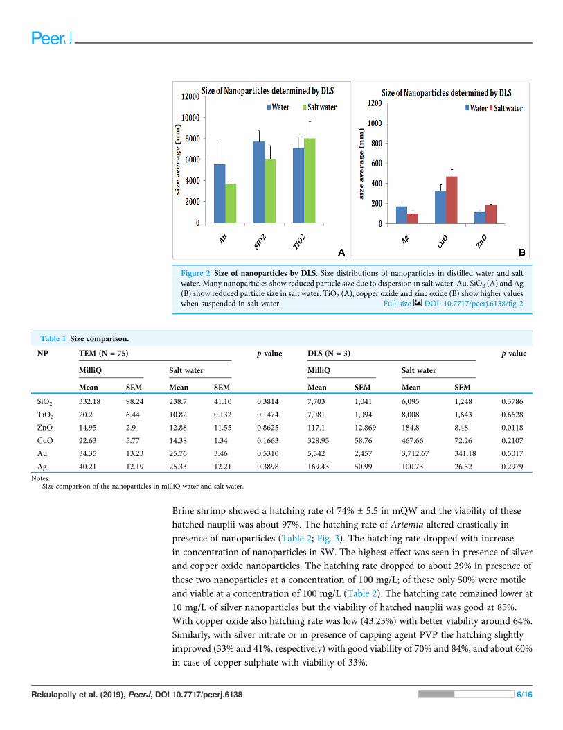
Brine shrimp showed a hatching rate of 74% ± 5.5 in mQW and the viability of thesehatched nauplii was about 97%. The hatching rate of Artemia altered drastically inpresence of nanoparticles (Table 2; Fig. 3). The hatching rate dropped with increasein concentration of nanoparticles in SW. The highest effect was seen in presence of silverand copper oxide nanoparticles. The hatching rate dropped to about 29% in presence ofthese two nanoparticles at a concentration of 100 mg/L; of these only 50% were motileand viable at a concentration of 100 mg/L (Table 2). The hatching rate remained lower at10 mg/L of silver nanoparticles but the viability of hatched nauplii was good at 85%.With copper oxide also hatching rate was low (43.23%) with better viability around 64%.Similarly, with silver nitrate or in presence of capping agent PVP the hatching slightlyimproved (33% and 41%, respectively) with good viability of 70% and 84%, and about 60%in case of copper sulphate with viability of 33%.
Figure 2 Size of nanoparticles by DLS. Size distributions of nanoparticles in distilled water and saltwater. Many nanoparticles show reduced particle size due to dispersion in salt water. Au, SiO2 (A) and Ag(B) show reduced particle size in salt water. TiO2 (A), copper oxide and zinc oxide (B) show higher valueswhen suspended in salt water. Full-size DOI: 10.7717/peerj.6138/fig-2
Table 1 Size comparison.
NP TEM (N = 75) p-value DLS (N = 3) p-value
MilliQ Salt water MilliQ Salt water
Mean SEM Mean SEM Mean SEM Mean SEM
SiO2 332.18 98.24 238.7 41.10 0.3814 7,703 1,041 6,095 1,248 0.3786
TiO2 20.2 6.44 10.82 0.132 0.1474 7,081 1,094 8,008 1,643 0.6628
ZnO 14.95 2.9 12.88 11.55 0.8625 117.1 12.869 184.8 8.48 0.0118
CuO 22.63 5.77 14.38 1.34 0.1663 328.95 58.76 467.66 72.26 0.2107
Au 34.35 13.23 25.76 3.46 0.5310 5,542 2,457 3,712.67 341.18 0.5017
Ag 40.21 12.19 25.33 12.21 0.3898 169.43 50.99 100.73 26.52 0.2979
Notes:Size comparison of the nanoparticles in milliQ water and salt water.
Rekulapally et al. (2019), PeerJ, DOI 10.7717/peerj.6138 6/16
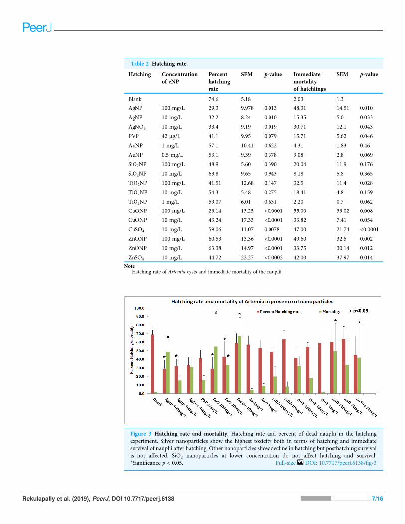
Table 2 Hatching rate.
Hatching Concentrationof eNP
Percenthatchingrate
SEM p-value Immediatemortalityof hatchlings
SEM p-value
Blank 74.6 5.18 2.03 1.3
AgNP 100 mg/L 29.3 9.978 0.013 48.31 14.51 0.010
AgNP 10 mg/L 32.2 8.24 0.010 15.35 5.0 0.033
AgNO3 10 mg/L 33.4 9.19 0.019 30.71 12.1 0.043
PVP 42 mg/L 41.1 9.95 0.079 15.71 5.62 0.046
AuNP 1 mg/L 57.1 10.41 0.622 4.31 1.83 0.46
AuNP 0.5 mg/L 53.1 9.39 0.378 9.08 2.8 0.069
SiO2NP 100 mg/L 48.9 5.60 0.390 20.04 11.9 0.176
SiO2NP 10 mg/L 63.8 9.65 0.943 8.18 5.8 0.365
TiO2NP 100 mg/L 41.51 12.68 0.147 32.5 11.4 0.028
TiO2NP 10 mg/L 54.3 5.48 0.275 18.41 4.8 0.159
TiO2NP 1 mg/L 59.07 6.01 0.631 2.20 0.7 0.062
CuONP 100 mg/L 29.14 13.25 <0.0001 55.00 39.02 0.008
CuONP 10 mg/L 43.24 17.33 <0.0001 33.82 7.41 0.054
CuSO4 10 mg/L 59.06 11.07 0.0078 47.00 21.74 <0.0001
ZnONP 100 mg/L 60.53 13.36 <0.0001 49.60 32.5 0.002
ZnONP 10 mg/L 63.38 14.97 <0.0001 33.75 30.14 0.012
ZnSO4 10 mg/L 44.72 22.27 <0.0002 42.00 37.97 0.014
Note:Hatching rate of Artemia cysts and immediate mortality of the nauplii.
Figure 3 Hatching rate and mortality. Hatching rate and percent of dead nauplii in the hatchingexperiment. Silver nanoparticles show the highest toxicity both in terms of hatching and immediatesurvival of nauplii after hatching. Other nanoparticles show decline in hatching but posthatching survivalis not affected. SiO2 nanoparticles at lower concentration do not affect hatching and survival.�Significance p < 0.05. Full-size DOI: 10.7717/peerj.6138/fig-3
Rekulapally et al. (2019), PeerJ, DOI 10.7717/peerj.6138 7/16
Hatching rate in presence of other nanoparticles (gold, SiO2, TiO2, copper oxide andzinc oxide) was low at about 50–60% as compared to control (Table 2; Fig. 3) andposthatching mortality was not very high. In presence of zinc oxide nanoparticles, 60% ofthe nauplii hatched but the cysts had a tendency to clump together. In TiO2 at 100 mg/L,hatching rate was 41.50% with 67.8% viability; the hatching rate was at 54% with∼90% viability in 10 mg/L TiO2 dioxide NPs. Gold and SiO2 nanoparticles displayed verygood viability after hatching (Fig. 3). SiO2 nanoparticles at a concentration of 10 mg/Ldisplayed hatching rate and viability equivalent to the controls.
The pH of SW did not change upon addition of nanoparticles and remained around8.3–8.5 up to 48 h. Live hatched nauplii appeared normal and swimming vigorously.There were no visible changes in morphology of nauplii hatched in presence ofnanoparticles. Some of the nauplii had emerged but were immotile/dead; some weretrapped in the membrane and still-dead (Figs. S2.1–S2.6). Silver and copper oxidenanoparticles which had maximum toxic effect on hatching rate also did notshow any gross morphological abnormalities. Staining of hatched Artemia with DCFDArevealed oxidative stress in a few still trapped nauplii and the gut of dead nauplii(Fig. S3).
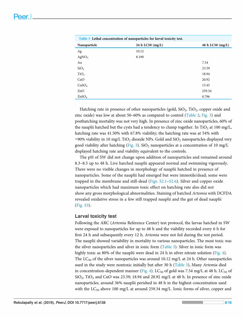
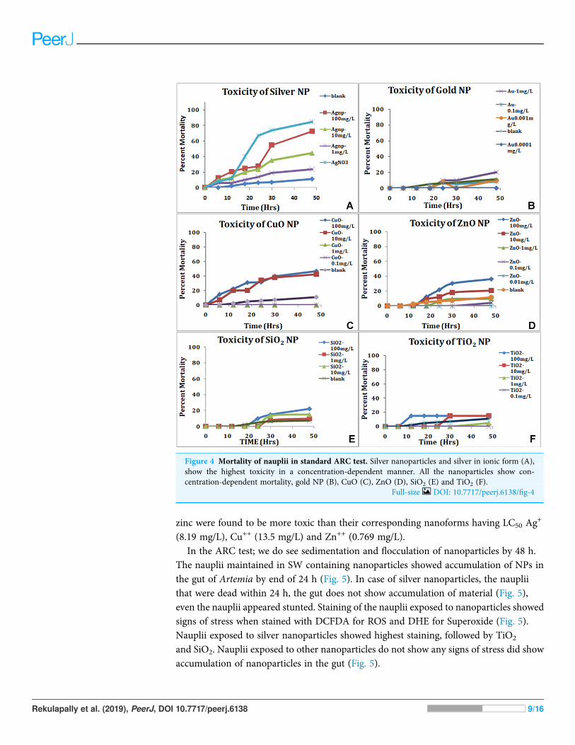
Larval toxicity testFollowing the ARC (Artemia Reference Center) test protocol, the larvae hatched in SWwere exposed to nanoparticles for up to 48 h and the viability recorded every 6 h forfirst 24 h and subsequently every 12 h. Artemia were not fed during the test period.The nauplii showed variability in mortality to various nanoparticles. The most toxic wasthe silver nanoparticles and silver in ionic form (Table 3). Silver in ionic form washighly toxic as 80% of the nauplii were dead in 24 h in silver nitrate solution (Fig. 4).The LC50 of the silver nanoparticles was around 10.12 mg/L at 24 h. Other nanoparticlesused in the study were nontoxic initially but after 30 h (Table 3), Many Artemia diedin concentration-dependent manner (Fig. 4). LC50 of gold was 7.54 mg/L at 48 h. LC50 ofSiO2, TiO2 and CuO was 23.59; 18.94 and 20.92 mg/L at 48 h. In presence of zinc oxidenanoparticles, around 36% nauplii perished in 48 h in the highest concentration usedwith the LC50 above 100 mg/L at around 259.34 mg/L. Ionic forms of silver, copper and
Table 3 Lethal concentration of nanoparticles for larval toxicity test.
Nanoparticle 24 h LC50 (mg/L) 48 h LC50 (mg/L)
Ag 10.12
AgNO3 8.190
Au 7.54
SiO2 23.59
TiO2 18.94
CuO 20.92
CuSO4 13.45
ZnO 259.34
ZnSO4 0.796
Rekulapally et al. (2019), PeerJ, DOI 10.7717/peerj.6138 8/16
zinc were found to be more toxic than their corresponding nanoforms having LC50 Ag+
(8.19 mg/L), Cu++ (13.5 mg/L) and Zn++ (0.769 mg/L).In the ARC test; we do see sedimentation and flocculation of nanoparticles by 48 h.
The nauplii maintained in SW containing nanoparticles showed accumulation of NPs inthe gut of Artemia by end of 24 h (Fig. 5). In case of silver nanoparticles, the naupliithat were dead within 24 h, the gut does not show accumulation of material (Fig. 5),even the nauplii appeared stunted. Staining of the nauplii exposed to nanoparticles showedsigns of stress when stained with DCFDA for ROS and DHE for Superoxide (Fig. 5).Nauplii exposed to silver nanoparticles showed highest staining, followed by TiO2
and SiO2. Nauplii exposed to other nanoparticles do not show any signs of stress did showaccumulation of nanoparticles in the gut (Fig. 5).
Figure 4 Mortality of nauplii in standard ARC test. Silver nanoparticles and silver in ionic form (A),show the highest toxicity in a concentration-dependent manner. All the nanoparticles show con-centration-dependent mortality, gold NP (B), CuO (C), ZnO (D), SiO2 (E) and TiO2 (F).
Full-size DOI: 10.7717/peerj.6138/fig-4
Rekulapally et al. (2019), PeerJ, DOI 10.7717/peerj.6138 9/16

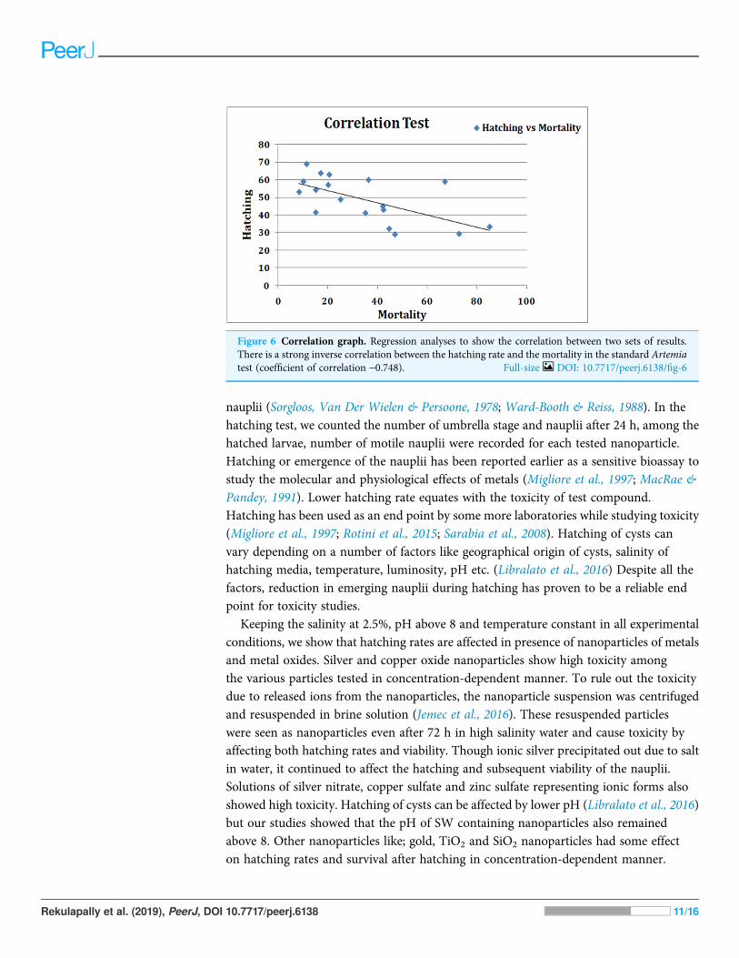
DISCUSSIONIn this study, we have compared hatching rate of Artemia cyst in presence ofnanoparticles and the toxicity of nauplii using standard Artemia test at variousconcentrations of nanoparticle suspension in high salinity conditions. We found a strongcorrelation in hatching rates and larval mortality in various nanoparticle suspensions(Fig. 6).
Artemia are able to tolerate high salinity and adapt to extreme conditions. Theyhave wide distribution globally and the collection of cysts is easy. Artemia cysts aredormant gastrula states during development encased in hard outer shell; formed underunfavorable conditions. Under simple laboratory conditions, the cysts can be hatched inhigh salinity water when by about 20 h the umbrella stage emerges and yields free swimming
Figure 5 ROS staining of dead nauplii. Nauplii of Artemia in standard ARC test stained with DCFDA(green) for ROS and DHE (Red) for superoxide accumulation due to stress. Nauplii exposed to silver NPs,TiO2 and SiO2 NP show higher staining for both ROS and superoxide at 24 h. (A–D) Control; (E–H)silver NP; (I–L) gold NP; (M–P) CuO NP; (Q–T) ZnO—NP; (U–X) titania and (Y–BB) SiO2NP.
Full-size DOI: 10.7717/peerj.6138/fig-5
Rekulapally et al. (2019), PeerJ, DOI 10.7717/peerj.6138 10/16
nauplii (Sorgloos, Van Der Wielen & Persoone, 1978; Ward-Booth & Reiss, 1988). In thehatching test, we counted the number of umbrella stage and nauplii after 24 h, among thehatched larvae, number of motile nauplii were recorded for each tested nanoparticle.Hatching or emergence of the nauplii has been reported earlier as a sensitive bioassay tostudy the molecular and physiological effects of metals (Migliore et al., 1997; MacRae &Pandey, 1991). Lower hatching rate equates with the toxicity of test compound.Hatching has been used as an end point by some more laboratories while studying toxicity(Migliore et al., 1997; Rotini et al., 2015; Sarabia et al., 2008). Hatching of cysts canvary depending on a number of factors like geographical origin of cysts, salinity ofhatching media, temperature, luminosity, pH etc. (Libralato et al., 2016) Despite all thefactors, reduction in emerging nauplii during hatching has proven to be a reliable endpoint for toxicity studies.
Keeping the salinity at 2.5%, pH above 8 and temperature constant in all experimentalconditions, we show that hatching rates are affected in presence of nanoparticles of metalsand metal oxides. Silver and copper oxide nanoparticles show high toxicity amongthe various particles tested in concentration-dependent manner. To rule out the toxicitydue to released ions from the nanoparticles, the nanoparticle suspension was centrifugedand resuspended in brine solution (Jemec et al., 2016). These resuspended particleswere seen as nanoparticles even after 72 h in high salinity water and cause toxicity byaffecting both hatching rates and viability. Though ionic silver precipitated out due to saltin water, it continued to affect the hatching and subsequent viability of the nauplii.Solutions of silver nitrate, copper sulfate and zinc sulfate representing ionic forms alsoshowed high toxicity. Hatching of cysts can be affected by lower pH (Libralato et al., 2016)but our studies showed that the pH of SW containing nanoparticles also remainedabove 8. Other nanoparticles like; gold, TiO2 and SiO2 nanoparticles had some effecton hatching rates and survival after hatching in concentration-dependent manner.
Figure 6 Correlation graph. Regression analyses to show the correlation between two sets of results.There is a strong inverse correlation between the hatching rate and the mortality in the standard Artemiatest (coefficient of correlation -0.748). Full-size DOI: 10.7717/peerj.6138/fig-6
Rekulapally et al. (2019), PeerJ, DOI 10.7717/peerj.6138 11/16
SiO2 nanoparticles at low concentration were very conducive to and in fact showed betterhatching as compared to control (Table S1). Hatching rates in presence of nanoparticleswere not much different at 20 and 24 h but the mortality of hatched nauplii washigher in some nanoparticles at 24 h (data not shown). Locomotion of the hatchednauplii was also affected in presence of nanoparticles.
In the standard Artemia test, nauplii hatched in SW are exposed to nanoparticlesuspensions. The mortality and toxic effects expressed as LC50; the concentration of anagent at which 50% of the tested animals are dead after 24 h; were chosen as criteriaof the toxicity (Nunes et al., 2006). The standard Artemia toxicity test results werecomparable and silver nanoparticles ranked very high in toxicity, followed by copper andzinc oxide nanoparticles. TiO2, gold and SiO2 nanoparticles in that order, showed abetter survival of nauplii up to 48 h. On comparing the two methods, a good inversecorrelation was observed between the results obtained, that is, effects of NPs on hatchingrates and effect on mortality of the hatched larvae (r = -0.79). The particles thatreduced the hatching rate showed an increase in mortality in the ARC test using thenormally hatched nauplii. Our results correlate well with some previous reports, wheretoxicity assays were performed on Artemia using nanoparticles (Lee, Chen & Chou, 1999;Rajasree et al., 2011; Ates et al., 2013). Comparing the viability of nauplii (inverse ofmortality) in ARC test with the hatching rate showed no statistically significant differencein the mean values for each concentration of different particles (p = 0.683) in thepaired t-test.
The advantage of the hatching assay is that it is easy to perform, short term anduses mortality as the end point for assessing toxicity of the test material. The question ofaltered solubility may not arise though sedimentation of these suspensions sometimesmay vary for different particles. So far, we have seen that nanoparticles tested inthis work remain stable in seawater up to 1 week, though the ionic or dissolvedcomponent may settle down due to precipitation. The sedimentation factor is taken careof by the continuous aeration in the hatching test that keeps the particles insuspended form.
Organisms in aquatic environment like daphnia incorporate NPs via gut. Artemia is alsoa filter feeder and the NPs may enter the guts of Artemia through ingestion. The mortalityobserved may be due to the uptake of the NPs clogging the gut as most of the naupliiin presence of NPs show presence of particles in the digestive tract. In the hatching test,the dead Artemia and partially hatched cysts show increased oxidative stress inpresence of nanoparticles as indicated by DCDFA staining. Even the nauplii exposed tosome nanoparticles like silver NPs showed accumulation of ROS and superoxide butwith remaining nanoparticles it is likely that the gut accumulation of the NPs is the causeof mortality.
Artemia with capacity to homeostasis in variable external salt concentrations certainlyappears to be a model organism to assess the nanoparticle behavior and ecotoxicity.Suitability of Artemia for toxicity tests is established and is often used in pharmaceuticalindustry and for metal and radiation toxicity (Pelka et al., 2000; Dvorak & Benova, 2002;Mayorga et al., 2010). We propose that Artemia hatchability test along with standard
Rekulapally et al. (2019), PeerJ, DOI 10.7717/peerj.6138 12/16
lethality/toxicity test definitely could also be used as prescreening of nanoparticle toxicityprior to their validation through in vivo toxicity studies. These acute toxicity testseliminate the need of laboratory maintenance of test species which otherwise take up spaceand time. Use of cysts which are available in enormous quantities as batch also cut downon the variability in test species.
CONCLUSIONSHatchability of Artemia can be used as a quick screening test for toxicity of materialswithout much need for laboratory space and maintenance. With strictly laid out protocolsand test conditions, this assay can be adopted for the hazard identification ofnanomaterials and high throughput methods.
ADDITIONAL INFORMATION AND DECLARATIONS
FundingThis work was supported by European Union FP7 project no. 263147 NANOVALID.There was no additional external funding received for this study. The funders had no rolein study design, data collection and analysis, decision to publish, or preparation of themanuscript.
Grant DisclosureThe following grant information was disclosed by the authors:European Union FP7: 263147 NANOVALID.
Competing InterestsThe authors declare that they have no competing interests.
Author Contributions� Rohit Rekulapally performed the experiments.� Lakshmi Narsimha Murthy Chavali performed the experiments.� Mohammed M. Idris analyzed the data, contributed reagents/materials/analysis tools,prepared figures and/or tables, approved the final draft.
� Shashi Singh conceived and designed the experiments, analyzed the data, contributedreagents/materials/analysis tools, prepared figures and/or tables, authored or revieweddrafts of the paper, approved the final draft.
Data AvailabilityThe following information was supplied regarding data availability:
The raw data is available in the Supplemental Files.
Supplemental InformationSupplemental information for this article can be found online at http://dx.doi.org/10.7717/peerj.6138#supplemental-information.
Rekulapally et al. (2019), PeerJ, DOI 10.7717/peerj.6138 13/16
REFERENCESAdam N, Schmitt C, Galceran J, Companys E, Vakurov A, Wallace R, Knapen D, Blust R. 2014.
The chronic toxicity of ZnO nanoparticles and ZnCl2 to Daphnia magna and the use of differentmethods to assess nanoparticle aggregation and dissolution. Nanotoxicology 8(7):709–717DOI 10.3109/17435390.2013.822594.
Arulvasu C, Jennifer SM, Prabhu D, Chandhirasekar D. 2014. Toxicity effect of silvernanoparticles in brine shrimp Artemia. Scientific World Journal 2014:256919DOI 10.1155/2014/256919.
Ates M, Daniels J, Arslan Z, Farah IO. 2012. Effects of aqueous suspensions titanium dioxidenanoparticles on Artemia salina: assessment of nanoparticle aggregation, accumulation andtoxicity. Environmental Monitoring and Assessment 185(4):3339–3348DOI 10.1007/s10661-012-2794-7.
Ates M, Daniels J, Arslan Z, Farah IO, Rivera HF. 2013. Comparative evaluation of impact ofZn and ZnO nanoparticles on brine shrimp (Artemia salina) larvae: effects of particle size andsolubility on toxicity. Environmental Science Process & Impacts 15(1):225–233DOI 10.1039/c2em30540b.
Ates M, Demir V, Arslan Z, Daniels J, Farah IO, Bogatu C. 2015. Evaluation of alpha and gammaaluminum oxide nanoparticle accumulation, toxicity, and depuration in Artemia salina larvae.Environmental Toxicology 30(1):109–118 DOI 10.1002/tox.21917.
Brix KV, Gerdes RM, Adams WJ, Grosell M. 2006. Effects of copper, cadmium, and zinc on thehatching success of brine shrimp (Artemia franciscana). Archives of EnvironmentalContamination and Toxicology 51(4):580–583 DOI 10.1007/s00244-005-0244-z.
Carballo JL, Hernández-Inda ZL, Pérez P, García-Grávalos MD. 2002. A comparisonbetween two brine shrimp assays to detect in vitro cytotoxicity in marine natural products.BMC Biotechnology 2:17 DOI 10.1186/1472-6750-2-17.
Dvorak P, Benova K. 2002. The investigation of interactions of low doses of ionizing radiationand risk factors by means of Artemia salina biotest. Folia Veterinaria 46:195–197.
Go EC, Pandey AS, MacRae TH. 1990. Effect of inorganic mercury on the emergence andhatching of the brine shrimp Artemia franciscana. Marine Biology 107(1):93–102DOI 10.1007/bf01313246.
Jemec A, Kahru A, Potthoff A, Drobne D, Heinlaan M, Bohme S, Geppert M, Novak S,Schirmer K, Rekulapally R, Singh S, Aruoja V, Sihtmae M, Jganson K, Kakinen A, Kuhnel D.2016. An interlaboratory comparison of nanosilver characterisation and hazard identification:harmonizing techniques for high quality data. Environmental International 87:20–32DOI 10.1016/j.envint.2015.10.014.
Lee TH, Chen YM, Chou HN. 1999. Toxicity assay of cyanobacterial strains using Artemia salinain comparison with the mouse bioassay. Acta Zoologica Taiwanica 10:1–8.
Libralato G, Prato E, Migliore L, Cicero AM, Manfra L. 2016. A review of toxicity testingprotocols and endpoints with Artemia spp. Ecological Indicators 69:35–49DOI 10.1016/j.ecolind.2016.04.017.
MacRae TH, Pandey AS. 1991. Effects of metals on early life stages of the brine shrimp,Artemia: a developmental toxicity assay. Archives of Environmental Contamination andToxicology 20(2):247–252 DOI 10.1007/bf01055911.
Mayorga P, Pérez KR, Cruz SM, Cáceres S. 2010. Comparison of bioassays using the anostracancrustaceans Artemia salina and Thamnocephalus platyurus for plant extract toxicity screening.RevistaBrasileira de Farmacognosia. Brazilian Journal of Pharmacognosy 20(6):897–903DOI 10.1590/s0102-695x2010005000029.
Rekulapally et al. (2019), PeerJ, DOI 10.7717/peerj.6138 14/16
Migliore L, Civitareale C, Brambilla G, Di Delupis GD. 1997. Toxicity of severalimportant agricultural antibiotics to Artemia. Water Research 31(7):1801–1806DOI 10.1016/s0043-1354(96)00412-5.
Moore MN. 2006. Do nanoparticles present exotoxicological risks for the health of the aquaticenvironment? Environment International 32(8):967–976 DOI 10.1016/j.envint.2006.06.014.
Musee N. 2011. Nanowaste and the environment: potential new waste management paradigm.Environment International 37(1):112–128 DOI 10.1016/j.envint.2010.08.005.
Nunes BS, Carvalho FD, Guilhermino LM, Van Stappen G. 2006. Use of the genusArtemia in ecotoxicity testing. Environmental Pollution 144(2):453–462DOI 10.1016/j.envpol.2005.12.037.
Pelka M, Danzl C, Distler W, Petschelt A. 2000. A new screening test toxicity testing of dentalmaterials. Journal of Dental Research 28(5):341–345 DOI 10.1016/s0300-5712(00)00007-5.
Rajabi S, Ramazani A, Hamidi M, Naji T. 2015. Artemia salina as a model organism in toxicityassessment of nanoparticles. DARU Journal of Pharmaceutical Sciences 23(1):20DOI 10.1186/s40199-015-0105-x.
Rajasree SRR, Kumar KG, Abraham LS, Manoharan N. 2011. Assessment on the toxicity ofengineered nanoparticles on the lifestages of marine aquatic invertebrate Artemia salina.International Journal of NanoScience 10(04n05):1153–1159 DOI 10.1142/s0219581x11009428.
Reddy KM, Guin D, Manorama SV, Ramachandra Reddy A. 2004. Selective synthesis of nanosizeTiO2 by hydrothermal route: characterization, structure property relation and photochemicalapplications. Journal of Material Research 19(9):2567–2595 DOI 10.1557/jmr.2004.0335.
Rizzo LY, Golombek SK, Mertens ME, Pan Yu, Laaf D, Broda J, Jayapaul J, Möckel D, Subr V,Hennink WE, Storm G, Simon U, Jahnen-Dechent W, Kiesslinga F, Lammers T. 2013.In vivo nanotoxicity testing using the zebrafish embryo assay. Journal of Materials Chemistry B1(32):3918–3925 DOI 10.1039/C3TB20528B.
Rotini A, Manfra L, Canepa S, Tornambe A, Migliore L. 2015. Can Artemia hatching assay canbe a (sensitive) alternative tool to acute toxicity test? Bulletin of Environmental Contaminationand Toxicology 95(6):745–751 DOI 10.1007/s00128-015-1626-1.
Sarabia R, Del Ramo J, Varó I, Díaz-Mayans J, Torreblanca A. 2008. Sublethal zinc exposurehas a detrimental effect on reproductive performance but not on the cyst hatching success ofArtemia partenogenetica. Science of The Total Environment 398(1–3):48–52DOI 10.1016/j.scitotenv.2008.03.002.
Sarabia R, Torreblanca A, Delramo JJ, Diaz-Mayans J. 1998. Effects of lowmercury concentrationexposure on hatching, growth and survival in Artemia strain La Mata parthenogenesis diploid.Comparative Biochemistry and Physiology Part A: Molecular & Integrative Physiology120(1):93–97 DOI 10.1016/s1095-6433(98)10015-6.
Solis PN, Wright CW, Anderson MM, Gupta MP, Phillipson JD. 1993. A microwellcytotoxicity assay using Artemia salina (Brine Shrimp). Planta Medica 59(3):250–252DOI 10.1055/s-2006-959661.
Sorgeloos P. 1973. First report on the triggering effect of light on the hatching mechanism ofArtemia salina dry cysts. Marine Biology 22(1):75–76 DOI 10.1007/bf00388912.
Sorgloos P, Van Der Wielen CR, Persoone G. 1978. The use of Artemia nauplii for toxicitytests—a critical analysis. Ecotoxicology and Environmental Safety 2(3–4):249–255DOI 10.1016/s0147-6513(78)80003-7.
Suppi S, Kasemets K, Ivask A, Künnis-Beres K, Sihtmäe M, Kurvet I, Aruoja V, Kahru A. 2015.A novel method for comparison of biocidal properties of nanomaterials to bacteria, yeasts andalgae. Journal of Hazardous Materials 286:75–84 DOI 10.1016/j.jhazmat.2014.12.027.
Rekulapally et al. (2019), PeerJ, DOI 10.7717/peerj.6138 15/16
Templeton RC, Ferguson PL, Washburn KM, Scrivens WA. 2006. Chandler GT life-cycle effectsof single-walled carbon nanotubes (SWNTs) on an estuarine meiobenthic copepod.Environmental Science & Technology 40(23):7387–7393 DOI 10.1021/es060407p.
Völker C, Boedicker C, Daubenthaler J, Oetken M, Oehlmann J. 2013. Comparative toxicityassessment of nanosilver on three daphnia species in acute, chronic and multi-generationexperiments. PLOS ONE 8(10):e75026 DOI 10.1371/journal.pone.0075026.
Ward-Booth K, Reiss M. 1988. Artemia salina: an easily cultured invertebrate ideallysuited for ecological studies. Journal of Biology Education 22(4):247–251DOI 10.1080/00219266.1988.9654995.
Zhu X, Zhu L, Duan Z, Qi R, Li Y, Lang Y. 2008. Comparative toxicity of several metal oxidenanoparticle aqueous suspensions to Zebrafish (Danio rerio) early developmental stage.Journal of Environmental Science and Health, Part A 43(3):278–284DOI 10.1080/10934520701792779.
Rekulapally et al. (2019), PeerJ, DOI 10.7717/peerj.6138 16/16


![АКВАРИУМНЫЕ РАСТЕНИЯ...[ 2 ] Prodac International АКВАРИУМНЫЕ РАСТЕНИЯ ПОЗВОЛЬТЕ СВОЕМУ АКВАРИУМУ ДЫШАТЬ Растения](https://static.fdocuments.in/doc/165x107/5f46fbe300ae3178a0728d95/oe-2-prodac-international-oe.jpg)