Toxic dilatation of the colon
3
GI EMERGENCIES MEDICINE 35:3 169 © 2006 Elsevier Ltd. All rights reserved. Toxic dilatation of the colon Kate Evans Simon Travis Abstract Toxic megacolon is a potentially fatal condition that represents the end of a spectrum of severe colitis. Typically, a complication of ulcerative colitis, it is increasingly a consequence of infective colitis. Diagnosis of toxic megacolon requires radiographic evidence of a dilated colon and tachycardia or fever in a patient with severe colitis of any cause. Unprepared flexible sigmoidoscopy should be performed on admission to confirm colitis and exclude complications such as cytomegalovirus infection. CT scanning is more sensitive than plain films for detecting perforation. Joint management between surgeons and physicians is fundamental. Medical therapy includes steroids, antibiotics, fluid and electrolyte management. Up to half respond to medical treatment. Colec- tomy is indicated if dilatation persists beyond the first 24 hours of ad- mission. Delayed decision-making increases the likelihood of perforation with a concomitant rise in morbidity and mortality. Keywords colectomy; pseudomembranous colitis; toxic dilatation; ulcerative colitis Incidence and aetiology Toxic megacolon (TM) is defined as total or segmental non- obstructive dilatation of the colon ≥6 cm, associated with sys- temic toxicity. 1 It is typically a complication of ulcerative colitis, but may be associated with other colitides. Crohn’s disease, pseu- domembranous colitis, infection (Salmonella enteritidis, Campy- lobacter sp., amoebic colitis, Shigella sp., cytomegalovirus) and ischaemic colitis are all recognized causes. Toxic megacolon simply represents the end of a spectrum of severe colitis that has been unrecognized or undertreated. The incidence has never been studied systematically. For ulcerative colitis, around 15% of all patients are admitted to hospital with a severe attack and about 5% of these will have toxic dilata- tion. 2,3 The risk may be highest early on in the disease, since 16/55 (30%) of an early case series developed TM within three months of diagnosis. 4 Earlier diagnosis, more intensive medical management and earlier surgery has reduced the incidence of Kate Evans MRCP is a Specialist Registrar at the Gastroenterology Unit, John Radcliffe Hospital, Oxford, UK. Competing interests: none declared. Simon Travis DPhil FRCP is a Consultant Gastroenterologist at the Gastroenterology Unit, John Radcliffe Hospital, Oxford, UK. Competing interests: none declared. TM complicating ulcerative colitis. 3 In contrast, TM complicating infective colitis is rising, reflecting the increasing prevalence and severity of pseudomembranous colitis. 5 Pathology and pathogenesis The pathogenesis of TM is unclear, but chemical mediators such as nitric oxide may play a pivotal role. Acute mucosal inflam- mation becomes transmural and is associated with neurogenic loss of motor tone in the smooth muscle layer which leads to dilatation of the colon. Nitric oxide inhibits smooth muscle tone. The amount and activity of inducible nitric oxide synthase is sig- nificantly increased in those with TM. 6 The potential for TM is enhanced by any factor that adversely affects colonic motility; this includes hypokalaemia, hypomagnesaemia, anticholinergic or anti-diarrhoeal drugs. This is as relevant in pseudomembra- nous colitis as it is in ulcerative colitis. Histology demonstrates acute inflammation in all layers of the colon, with muscle necro- sis and replacement by granulation tissue infiltrated by leuko- cytes and plasma cells. Perforation occurs in about 20%. Clinical features and investigations Typical features of severe colitis (diarrhoea, usually bloody, with anorexia, tachycardia and fever), predate the onset of acute dilatation by a week or more. Symptoms are masked in patients receiving steroids. Distension is not prominent, although the alert clinician may detect the abdominal asymmetry of a dilated transverse colon. The abdomen is usually tender in the line of the colon. If peritonism is present, perforation (which may initially be localized) is likely. Diagnosis of TM is simple: radiographic evidence of dilated colon (≥6 cm; Figure 1) and a tachycardia or temperature in a patient with severe colitis of any cause. This requires that a plain abdominal radiograph is performed on any patient presenting with severe colitis. 2,7 While some have suggested multiple crite- ria for diagnosis, 4 these serve little purpose since the condition must be recognized at an early rather than terminal stage. Laboratory findings in TM are those of a systemic inflamma- tory response, including leukocytosis, raised erythrocyte sedi- mentation rate or C-reactive protein, and hypoalbuminaemia. Hypokalaemia and hypomagnesaemia are common and exacer- bate the condition. The plain abdominal radiograph is pivotal for diagnosis. The external diameter is measured at the widest point of the colon, which is usually the transverse colon because it is the least dependent part of the bowel. Mucosal islands (oedematous rem- nants of mucosa projecting into the lumen, or seen en face), or an associated ileus with 3 or more small bowel loops have been associated with the need for colectomy in severe colitis indepen- dent of dilatation. 8,9 CT scanning is unnecessary for diagnosis, but is more sensitive than plain films for detecting localized perforation. 10 In a series of 18 patients, 12 with pseudomembranous colitis, 2 colonic perforations and 2 septic portal thromboses were identified. 10 Gentle flexible sigmoidoscopy without preparation and with minimal air insufflation is necessary on admission to confirm the diagnosis of colitis and take biopsies to exclude complications such as cytomegalovirus infection. Urgent histology is appropriate
-
Upload
kate-evans -
Category
Documents
-
view
223 -
download
1
Transcript of Toxic dilatation of the colon
Gi emerGencies
Toxic dilatation of the colonKate evans
simon Travis
AbstractToxic megacolon is a potentially fatal condition that represents the end
of a spectrum of severe colitis. Typically, a complication of ulcerative
colitis, it is increasingly a consequence of infective colitis. Diagnosis
of toxic megacolon requires radiographic evidence of a dilated colon
and tachycardia or fever in a patient with severe colitis of any cause.
Unprepared flexible sigmoidoscopy should be performed on admission
to confirm colitis and exclude complications such as cytomegalovirus
infection. cT scanning is more sensitive than plain films for detecting
perforation. Joint management between surgeons and physicians is
fundamental. medical therapy includes steroids, antibiotics, fluid and
electrolyte management. Up to half respond to medical treatment. colec-
tomy is indicated if dilatation persists beyond the first 24 hours of ad-
mission. Delayed decision-making increases the likelihood of perforation
with a concomitant rise in morbidity and mortality.
Keywords colectomy; pseudomembranous colitis; toxic dilatation;
ulcerative colitis
Incidence and aetiology
Toxic megacolon (TM) is defined as total or segmental non-obstructive dilatation of the colon ≥6 cm, associated with sys-temic toxicity.1 It is typically a complication of ulcerative colitis, but may be associated with other colitides. Crohn’s disease, pseu-domembranous colitis, infection (Salmonella enteritidis, Campy-lobacter sp., amoebic colitis, Shigella sp., cytomegalovirus) and ischaemic colitis are all recognized causes.
Toxic megacolon simply represents the end of a spectrum of severe colitis that has been unrecognized or undertreated. The incidence has never been studied systematically. For ulcerative colitis, around 15% of all patients are admitted to hospital with a severe attack and about 5% of these will have toxic dilata-tion.2,3 The risk may be highest early on in the disease, since 16/55 (30%) of an early case series developed TM within three months of diagnosis.4 Earlier diagnosis, more intensive medical management and earlier surgery has reduced the incidence of
Kate Evans MRCP is a Specialist Registrar at the Gastroenterology Unit,
John Radcliffe Hospital, Oxford, UK. Competing interests: none declared.
Simon Travis DPhil FRCP is a Consultant Gastroenterologist at the
Gastroenterology Unit, John Radcliffe Hospital, Oxford, UK. Competing
interests: none declared.
meDicine 35:3 1
TM complicating ulcerative colitis.3 In contrast, TM complicating infective colitis is rising, reflecting the increasing prevalence and severity of pseudomembranous colitis.5
Pathology and pathogenesis
The pathogenesis of TM is unclear, but chemical mediators such as nitric oxide may play a pivotal role. Acute mucosal inflam-mation becomes transmural and is associated with neurogenic loss of motor tone in the smooth muscle layer which leads to dilatation of the colon. Nitric oxide inhibits smooth muscle tone. The amount and activity of inducible nitric oxide synthase is sig-nificantly increased in those with TM.6 The potential for TM is enhanced by any factor that adversely affects colonic motility; this includes hypokalaemia, hypomagnesaemia, anticholinergic or anti-diarrhoeal drugs. This is as relevant in pseudomembra-nous colitis as it is in ulcerative colitis. Histology demonstrates acute inflammation in all layers of the colon, with muscle necro-sis and replacement by granulation tissue infiltrated by leuko-cytes and plasma cells. Perforation occurs in about 20%.
Clinical features and investigations
Typical features of severe colitis (diarrhoea, usually bloody, with anorexia, tachycardia and fever), predate the onset of acute dilatation by a week or more. Symptoms are masked in patients receiving steroids. Distension is not prominent, although the alert clinician may detect the abdominal asymmetry of a dilated transverse colon. The abdomen is usually tender in the line of the colon. If peritonism is present, perforation (which may initially be localized) is likely.
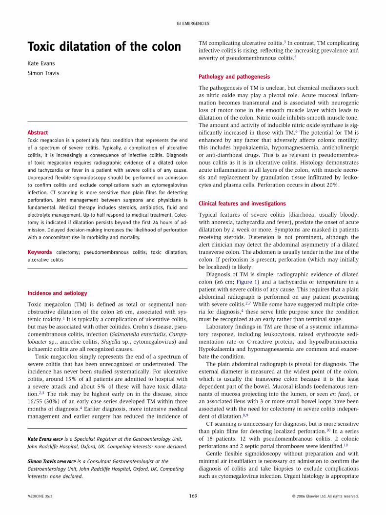
Diagnosis of TM is simple: radiographic evidence of dilated colon (≥6 cm; Figure 1) and a tachycardia or temperature in a patient with severe colitis of any cause. This requires that a plain abdominal radiograph is performed on any patient presenting with severe colitis.2,7 While some have suggested multiple crite-ria for diagnosis,4 these serve little purpose since the condition must be recognized at an early rather than terminal stage.
Laboratory findings in TM are those of a systemic inflamma-tory response, including leukocytosis, raised erythrocyte sedi-mentation rate or C-reactive protein, and hypoalbuminaemia. Hypokalaemia and hypomagnesaemia are common and exacer-bate the condition.
The plain abdominal radiograph is pivotal for diagnosis. The external diameter is measured at the widest point of the colon, which is usually the transverse colon because it is the least dependent part of the bowel. Mucosal islands (oedematous rem-nants of mucosa projecting into the lumen, or seen en face), or an associated ileus with 3 or more small bowel loops have been associated with the need for colectomy in severe colitis indepen-dent of dilatation.8,9
CT scanning is unnecessary for diagnosis, but is more sensitive than plain films for detecting localized perforation.10 In a series of 18 patients, 12 with pseudomembranous colitis, 2 colonic perforations and 2 septic portal thromboses were identified.10
Gentle flexible sigmoidoscopy without preparation and with minimal air insufflation is necessary on admission to confirm the diagnosis of colitis and take biopsies to exclude complications such as cytomegalovirus infection. Urgent histology is appropriate
69 © 2006 elsevier Ltd. All rights reserved.
Gi emerGencies
if cytomegalovirus colitis is suspected, because a decision about colectomy is needed within 24 hours of admission.
Management
The key aspects of management are aggressive medical therapy and early surgical decision-making. Fluid and electrolyte resus-citation, with intravenous hydrocortisone (100 mg four times daily) should be commenced immediately, together with anti-biotics (metronidazole 500 mg three times daily and ciprofloxa-cin 500 mg twice daily) if an infective aetiology is suspected. The combination of steroids and antibiotics is safe even for infective colitis; steroids reduce inflammation in pseudomem-branous colitis.11 Large amounts of potassium may be needed to correct hypokalaemia. All anti-motility agents, opioids and anticholinergics should be stopped. Thromboprophylaxis with subcutaneous heparin should be started. Nasogastric suction cannot be expected to decompress the colon and is unnecessary. A senior surgical opinion is best sought on the day of admission. It should be made clear to all that there is a 24-hour window of opportunity for medical treatment to work and that if there is no improvement, then early colectomy will be necessary.
On the day after admission, the plain abdominal radiograph should be repeated. If the diameter has decreased then medi-cal treatment can be continued and the radiograph repeated the following day. If the diameter has increased, or if it remains
figure 1 An abdominal radiograph showing marked colonic dilatation
and loss of haustral pattern from the splenic flexure distally. courtesy
of Dr Andrew slater, Oxford, UK.
meDicine 35:3 1
unchanged with an accompanying tachycardia or tempera-ture, then urgent colectomy is indicated. If there is doubt, or if there is focal tenderness, then a CT scan to look for localized perforation is appropriate. Management decisions should be made jointly between an experienced gastroenterologist and colorectal surgeon.
Nutritional support is vital, especially with TM complicat-ing pseudomembranous colitis. If there has been an extended period of poor nutrition, intravenous vitamins, phosphate (if <0.4 mmol/litre) and magnesium (if <0.5 mmol/litre) replace-ment should be given along with potassium in the first 24 hours. This will reduce the risk of re-feeding syndrome. On the day after admission, if the decision is to continue medical therapy then fine-bore nasogastric feeding is best started under the supervision of a dietitian. If the decision is to proceed to colectomy, paren-teral nutrition is appropriate. Enteral feeding promotes intesti-nal epithelial restitution and repair and can be given safely to patients with an ileus.12
Prognosis
There are no randomized trials and it should be understood that the condition is potentially fatal. The mortality increases if perforation occurs, while inappropriate persistence with medical treatment ‘because the patient is too sick for surgery’ is a self-fulfilling prophecy. That said, around 50% of patients with toxic megacolon due to ulcerative colitis respond to medical therapy,3 even if a high proportion (88%) with refractory colitis ultimately require colectomy.13 In a series of 70 patients over 20 years with surgically managed TM (half of whom had ulcerative colitis and half some other aetiology), the total mortality rate was 16%.14 The mortality of TM associated with ulcerative colitis is now less than 5% if early colectomy is performed. TM associated with Cl. difficile has a higher mortality (up to 67%),3,5 reflecting serious comorbidity with more difficult (and thereby delayed) decision- making. ◆
RefeRenCes
1 sheth s G, Lamont J T. Toxic megacolon. Lancet 1998; 351: 509–13.
2 Jakobovits s, Travis s P L. The management of severe ulcerative
colitis. Br Med Bull 2006; 76: 131–44.
3 Gan s i, Beck P L. A new look at toxic megacolon: an update and
review of incidence, etiology, pathogenesis, and management.
Am J Gastroenterol 2003; 98: 2363–71.
4 Jalan K n, sircus W, card W i et al. An experience of ulcerative
colitis: toxic dilatation in 55 cases. Gastroenterology 1969; 57:
68–82.
5 Oldfield 3rd e c. Clostridium difficile-associated diarrhea: resurgence
with a vengeance. Rev Gastroenterol Disord 2006; 6: 79–96.
6 mourelle m, casellas F, Guarner F et al. induction of nitric oxide
synthase in colonic smooth muscle from patients with toxic
megacolon. Gastroenterology 1995; 109: 1497–502.
7 Truelove s c, Witts L J. cortisone in ulcerative colitis: final report on
a therapeutic trial. BMJ 1955; ii: 1041–8.
70 © 2006 elsevier Ltd. All rights reserved.
Gi emerGencies
8 Bartram c. imaging the gut in inflammatory bowel disease:
radiology. in: satsangi J, sutherland L r, eds. Inflammatory bowel
diseases. London: churchill Livingstone, 2003: 219–36.
9 Travis s P L. Predicting outcome in severe ulcerative colitis.
Dig Liver Dis 2004; 36: 448–50.
10 imbriaco m, Balthazar e J. Toxic megacolon: role of cT in evaluation
and detection of complications. Clin Imaging 2001; 25: 349–54.
11 Goodman m J, Truelove s c. intensive intravenous regimen for
membranous colitis. BMJ 1976; 2: 354.
meDicine 35:3 1
12 Kaur n, Gupta m K, minocha V r. early enteral feeding by
nasoenteric tubes in patients with perforation peritonitis.
World J Surg 2005; 29: 1023–8.
13 moskovitz D n, Van Assche G, maenhout B et al. incidence of
colectomy during long-term follow-up after cyclosporine-induced
remission of severe ulcerative colitis. Clin Gastroenterol Hepatol
2006; 4: 760–5.
14 Ausch c, madoff r, Gnant m et al. Aetiology and surgical
management of toxic megacolon. Colorectal Dis 2006; 8: 195–201.
71 © 2006 elsevier Ltd. All rights reserved.