Towards the Creation of Cell Arrays for Rapid Bioanalysis Barry Cheung University of...
28
Towards the Creation of Cell Arrays for Rapid Bioanalysis Barry Cheung University of Nebraska-Lincoln Neil Lawrence Gonghua Wang Students: Collaborat or: Fereydoon Namavar University of Nebraska Medical Center Jan 16 2009, COBRE Retreat
-
date post
21-Dec-2015 -
Category
Documents
-
view
213 -
download
0
Transcript of Towards the Creation of Cell Arrays for Rapid Bioanalysis Barry Cheung University of...

Towards the Creation of Cell Arrays for Rapid Bioanalysis
Barry Cheung
University of Nebraska-Lincoln
Neil Lawrence Gonghua WangStudents: Collaborator:
Fereydoon Namavar
University of NebraskaMedical Center
Jan 16 2009, COBRE Retreat

Cell-based Array AssaysCommon application:High-throughput screening for drug development and genetic profiling.
Current methodology / technology :Dispensing cells onto microtiter plates (with up to hundreds of wells)
Microtiter plate assay requirements:• A few thousand cells per well• Growth of cell culture is typically necessary for this assay
Limitation:when the cell culture sample is limited or cannot be easily cultured
Potential solution: Cell micro-arrays and their advantages
• Potential to track the growth & heterogeneity of individual cell’s phenotypic responses to therapeutic drugs• Requirement: good cell adhesion is crucial to the generation of successful cell micro-arrays.

Desirable surface for cell patterning:
Too strong an interaction between the cells and the substrates may lead to activation of undesirable cell responses.
Cell adhesion schemes:
1. Molecule-directed cell patterning
• Promote location-selective cell adhesion by patterning cell adhesion biomolecules, antibodies, and organic functionalized surfaces
Potential limiting factor: Proteolysis of these adhesion molecules by secreted cellular enzymes
2. Chemically-robust silicon surfaces with engineered nanostructures
• Promote and control cell adhesion by increasing the adhesion surface. Potential limiting factor: Silicon is not optically transparent. This prohibits the use of conventional optical detection methods, such as optical fluorescence microscopy for cell arrays investigation.
Possible opportunity:
Substrates: optical transparency, biocompatible with engineered structures.
Cell-based Array Assays: Criteria

Hypothesis
Central hypothesis:
By optimizing the nanostructured surface morphology, optically transparent
bioceramic films will enhance the attachment of cells and serve as ideal substrates
for cell-based arrays chips.
Rationale:
Nanograin size of nanostructured bioceramic films brings about a significant increase
in grain boundaries, which changes the films' overall physico-chemical
characteristics.
Short-term goal:
To fabricate, verify, and select transparent zirconium oxide films with optimal
nanostructures for enhanced attachment of wild type E. coli and mouse lymphocytic
leukemia cells.

Specific Aims
Specific Aim 1: Determine surface characteristics (e.g., topography and hydrophilicity) of nanograinular zirconium oxide films that optimize the adhesion of E. coli and mouse lymphocytic leukemia cells.
Working hypothesis:Engineered nanograin structures and hydrophilicity of zirconium oxide films can be optimized to greatly increase cell adhesion with minimal disturbance of normal cell behavior on substrates for cell assays.
Specific Aim 2: Develop cell-based array chips with patterned nanostructured zirconium oxide films.
Postulation: Nanostructured zirconium oxide films patterned in micro-array format will enhance and modulate cell adhesion for the application of high density cell-based micro-array assays.

Experimental Design
Long-term goal
To develop chemically-robust nanostructured platforms as a means for controlling optimal adherent cell attachment to develop high throughput cancer cell-based array assays.
Objective of this application:
To create proof-of-principle cell array chips based on patterned bioceramic nanostructured films for enhancing cell attachment and therapeutic screening.
Nanostructured bioceramic films to be examined:Hydrophilic zirconium oxide films made by ion beam assisted deposition (IBAD)
Model cell systems:
Wild type E. coli and mouse lymphocytic leukemia cells

Ion Beam Assisted Deposition (IBAD)
* Physical vapor deposition technique + concurrent ion beam bombardment
* Advantages: Use of IBAD for meta-stable films production 1. Produce films with strong adherence to the substrates. 2. Produce films with high density 3. Modulate films chemical composition and structures with suitable ion beams.
* Advantages: Use of IBAD for meta-stable films production 1. Produce films with strong adherence to the substrates. 2. Produce films with high density 3. Modulate films chemical composition and structures with suitable ion beams.
Materials are “stitched” to the substrate by ion bombardment like “billions of ionic hammers”.
Materials are “stitched” to the substrate by ion bombardment like “billions of ionic hammers”.

IBAD ZrO2 film
Optical Image
* IBAD ZrO2 films on quartz: Transparent films with a green tint. * IBAD ZrO2 films on quartz: Transparent films with a green tint.
slide

IBAD ZrO2 films: AFM study
AFM study of IBAD ZrO2 suggests that the as-deposited films have low roughness even though the surface is bombarded with high energy ion beams.
AFM study of IBAD ZrO2 suggests that the as-deposited films have low roughness even though the surface is bombarded with high energy ion beams.
AFM height imageAFM height image
Roughness, Ra = 2.8 nmGrain size = 12.3 ± 2.0 nm

IBAD ZrO2 film: TEM study
Electron diffraction patternElectron diffraction pattern
Cubic Phase
Monoclinc Phase
Tetragonal Phase
X-section bright field imageX-section bright field image
X-section TEM study suggests that these IBAD ZrO2 films are made of mainly cubic phase nanocrystals of 5 -15 nm diameters.
10 nm

IBAD ZrO2 film: XRD study
1. XRD study suggests that the formation of single cubic phase in these films. 2. It demonstrates an excellent fit for cubic phase with average grain sizes of 12 nm (2 = 1.43) with a lattice constant a = 5.12 Å. 3. The fitted lattice constant is 0.6% larger than the literature reported value a = 5.088Å (J. Appl. Cryst. (2002). 35, 517-525).

IBAD ZrO2 film: XRD study
The XRD peak positions match cubic/tetragonal phase. → The existence of a (400) peak at ca. 74.3 instead of split (004) and (400) lines as in tetragonal phase, support the formation of pure cubic phase ZrO2.
Medical grade tetragonal ZrO2
IBAD ZrO2
Cubic Tetragonal
c/a = 1.02

Effect of Substrate Temperature on IBAD ZrO2 films
IBAD zirconia thin films deposited with N2 ion beam at 137 ºC and 473 ºC substrate temperature.
IBAD zirconia thin films deposited with N2 ion beam at 137 ºC and 473 ºC substrate temperature.
AFM study suggests that IBAD with N2 ion beam preferentially produce zirconium oxide films of higher roughness @ 473 ºC, than at lower substrate temperature 137 ºC.
137 ºC 473 ºCRa = 2.79 nm Ra = 19 nm

IBAD ZrO2 film: Contact angle study
Video Contact Angle measurement instrument Video Contact Angle measurement instrument
5º
Contact Angle measurement with waterContact Angle measurement with water
time = 1.75 s time = 2.08 s
Spreading of a 0.25 l water droplet on IBAD ZrO2
Typically measured angles: IBAD ZrO2 films: 0 to 10 °Bulk yttria stabilized cubic ZrO2: ~ 45 °Bulk monoclinic ZrO2: ~ 45 °
IBAD ZrO2 films exhibit hydrophilicity behavior compared to yttria stabilized cubic zirconium oxides and monoclinic zirconium oxides.

Fabrication of micro-array patterned nanostructured ZrO2 films
UV exposure
7. Deposit cells solution droplets using array robots

Fabrication of micro-array patterned nanostructured ZrO2 films
7. Deposit cells solution droplets using an array robot under humid environment
Quartz / Glass
Fluorinated silane
ZrO2
Dispensing cell solutionand drug
8. Staining and imaging cell arrays with microscopy Record the shape and other important cell morphology information to determine their response to drug.
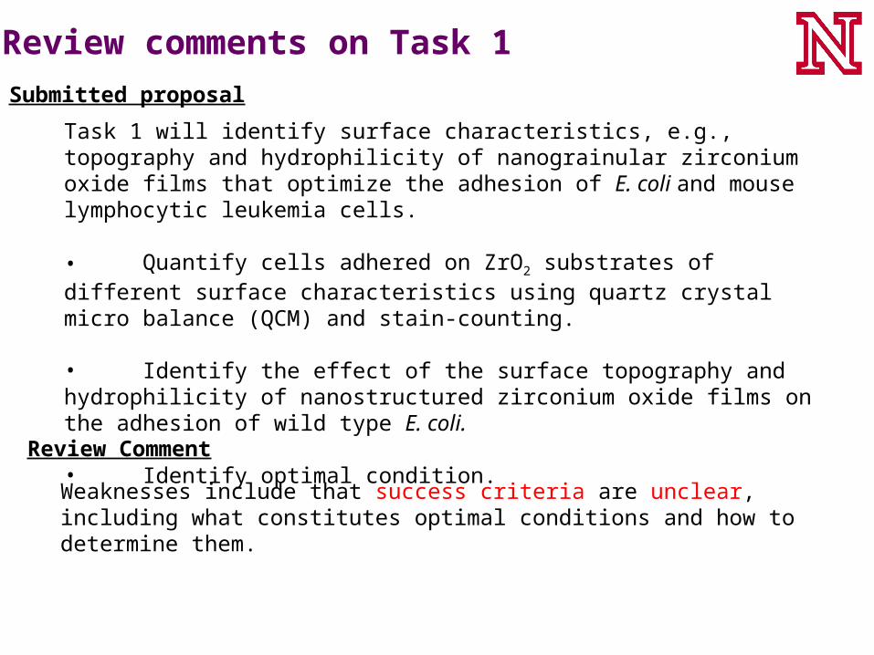
Review comments on Task 1
Task 1 will identify surface characteristics, e.g., topography and hydrophilicity of nanograinular zirconium oxide films that optimize the adhesion of E. coli and mouse lymphocytic leukemia cells.
• Quantify cells adhered on ZrO2 substrates of different surface characteristics using quartz crystal micro balance (QCM) and stain-counting.
• Identify the effect of the surface topography and hydrophilicity of nanostructured zirconium oxide films on the adhesion of wild type E. coli.
• Identify optimal condition.
Weaknesses include that success criteria are unclear, including what constitutes optimal conditions and how to determine them.
Submitted proposal
Review Comment

Plans to address review comments on Task 1
Weaknesses include that success criteria are unclear, including what constitutes optimal conditions and how to determine them.
Review Comment
Define success criteria for Task 1.a. Live cell coverage area of bioceramic surface by adhesion needs to be
> 80% and with improvement over glass or plastic.• This would increase the success of patterning cells in all patterned areas
for the fabrication of cell array.• Potential experiments: Stain counting and QCM measurement of controls
b. Cells grown on bioceramic surface should show normal behavior.• Normal cell growth behavior would further provide the basics to support
the feasibility of the cell array concept.• Potential experiments: Determine cell metabolic activities or by-products
c. Verification of better cell adhesion on the proposed bioceramic surface than glass• Potential experiments: Determine and compare the adhesion and force
profiles between the cell and glass/bioceramic coated beads by atomic force microscopy.
Response

Task 2 will develop cell-based array chips with patterned nanostructured zirconium oxide films.
Deliverables: 1. Verification: E. coli-based array chips made with nanostructured ZrO2
patterned films will yield similar LD50 value as those perform in solution. 2. Expectation: E. coli-based array chips are expected to provide an alternative
platform for therapeutic screening.
Review comments on Task 2
Submitted proposal
The project investigator states that this will provide the basis to extend the present platform for testing anticancer drugs with mammalian tumor cells. This seems to be an optimistic statement and a big leap.
Review Comment

Plans to address review comments on Task 1Review Comment
1. The claim for the E. coli–based cell array chips will be toned down.• Instead, we will state that the technical knowledge obtained in success of
E. coli-based cell array chips will be served as the basis to develop the patterning mouse lymphocytic leukemia cells arrays.
• If time and resource permitted, preliminary results for the growth of mouse tumor cell on bioceramic surface will also be demonstrated for the feasibility of the study.
2. Besides proving that E. coli–based cell array chips would have similar drug response as the solution assay, we will show that less than 100 addressable units in the cell array are needed to obtain the statistically significant data for the chip assays.
Response
The project investigator states that this will provide the basis to extend the present platform for testing anticancer drugs with mammalian tumor cells. This seems to be an optimistic statement and a big leap.

Preliminary Results: Study of E. coli XL-Blue adhesion on glass
Goals:
• Determine the coverage of E. coli growing on glass slides as a control for bioceramic films – find out the % of live/damage & dead bacteria • Develop protocols for the evaluation of the growth and adhesion of E. coli on bioceramic films
AFM Height Image
Ra = 0.3 nmGlass surface:
• Sample: Optical microscopy slides
• Morphology: Smooth compared to cell surface
• Roughness: 0.3 nm

Preliminary Results: Study of E. coli XL-Blue adhesion on glass
Goals:
• Determine the coverage of E. coli growing on glass slides as a control for bioceramic films – find out the % of live/damage & dead bacteria • Develop protocols for the evaluation of the growth and adhesion of E. coli on bioceramic films.
Method:
• Inoculate 1ml of E. coli XL-blue (106/ml) in wells of tilter plates
with 1 cm2 glass with or without bioceramic coating
• Incubate plates statically at 30°C for 4 to 26 hours
• Remove sample from well and rinse both sides in a gentle stream of 0.85% saline
• Stain with 0.75ml LIVE/DEAD BacLight for 15 min.
• Preserve samples in 3.7% formaldehyde and store in 0.85% saline
• Examine sample with confocal fluorescence microscopy
• Analyze data with Image J using the particle counting function.
sample

Preliminary Results: E. coli XL-Blue adhesion on glass
Confocal fluorescence image of stained E. coli on glass using the LIVE/DEAD BacLight kit
Live: green Dead: redLive damage: orange
Observations:
• E. coli shown growing on the glass
• E. coli adhered well enough to withstand the washing procedure
• The majority of the bacteria stained alive
• Unwashed samples showed multiple layer colonies
• Staining procedure developed worked well
• Further testing and controls with the K12 strain and proper rinse will be performed
After 26 h. incubation in LB media & a brief rinse with water

Coverage of E. coli XL-Blue on a microscope glass:• Area covered with bacteria = 0.22 ± 0.04%• Percentage of live bacteria = 87 ± 5%• Percentage of dead bacteria = 12 ± 6%• Percentage of live but damaged bacteria = 1 ± 2%
Note: 1. Average projected area of one E. coli: 1.6µm2
2. Fluorescence images was analyzed with ImageJ.
3. Experiments will be repeated with wild type E. coli strain K12-JM109 and rinsed with 0.85% NaCl or 10 mM pH7 MOP buffer instead.
Preliminary Results:E. coli XL-Blue adhesion on glass
After 26 h. incubation in LB media & a brief rinse with water

Continuing work to obtain preliminary results
1. Quantify and compare E. coli adhesion on one type of ZrO2 films and glass substrates by the stain-and-count method.
2. Quantify and compare mouse lymphocytic leukemia cell adhesion on one type of ZrO2 films and glass substrates by the stain-and-count method.

Alternative Bioceramic Film for Cell Array:Tantalum oxide
Optical Image AFM Height Image
Ra = 2.0 nm
Properties• Bioceramics used coating on templates for bone growth• Demonstrated to be made as transparent films by IBAD• Surface roughness potentially could be modulated by IBAD

Conclusions
• Hydrophilic, transparent nanostructured ZrO2 films have been hypothesized as possible substrates to enhance cell attachment and patterning.
• By taking advantage of the potential benefits of such films, a bioassay platform to create cell arrays has bee proposed to assist drug testing.
• Present focus of this project is on testing the adhesion and coverage of E. coli on bioceramic films and glass control substrates
• Short term focus of this project will be on studying the adhesion and coverage of mouse lymphocytic leukemia cells on bioceramic films and glass substrates.

Acknowledgement
Funding Support:
Development of bioceramic filmsNebraska Research Initiative
Development of cell-based array assay University of Nebraska-LincolnOffice of Sponsored Research
Dr. Fereydoon Namavar Gonghua Wang Neil Lawrence
University of Nebraska-Lincoln University of Nebraska Medical Center