Toward advanced neural interfaces for the peripheral ... · Neural interface for the peripheral...
8
Toward advanced neural interfaces for the peripheral nervous system (PNS) and their future applications Sanghoon Lee 1,2,3 and Chengkuo Lee 1,2,3 Abstract Modulation of nerve signals from the peripheral nervous system (PNS) is a growing field of neurotechnology for ther- apeutic effects of the human body and for interfacing with neural prostheses. For this, neural interfaces, that provide the basis for direct communication with neuron tissues and mapping neural signals, are preferentially developed. Even though various types of peripheral nerve interfaces have been developed for many years, advanced neural interfaces need to be developed in conjunction with cutting-edge tech- nology for the modulation of the human body and advanced neural prostheses. This paper reviews the evolution of pe- ripheral neural interfaces (PNI) and their applications. The emerging requirements of the next-generation PNI are also explored. Addresses 1 Electrical & Computer Engineering, National University of Singapore, Singapore, 117583, Singapore 2 Singapore Institute for Neurotechnology (SINAPSE), Singapore, 117456, Singapore 3 Center for Intelligent Sensors and MEMS, National University of Singapore, Singapore, 117608, Singapore Corresponding author: Lee, Chengkuo ([email protected]) Current Opinion in Biomedical Engineering 2018, 6:130 – 137 Edited by Wutian Wu and Seeram Ramakrishna This review comes from a themed issue on Biomaterials: Neural In- terfaces & Neuro-prosthetics Received 26 January 2018, revised 2 April 2018, accepted 16 May 2018 https://doi.org/10.1016/j.cobme.2018.05.004 2468-4511/© 2018 Published by Elsevier Inc. Keywords Bioelectronic medicine, Electroceutical, Peripheral nerve, Neural inter- face, Wireless neural interface, Self-sustainable platform. Introduction Neurotechnology has been developed for decades with the development of cutting-edge technology and neuroscience. From the discovery of electrical activity in the human brain in 1924 [1], variety of studies and tools have been demonstrated to explore the human brain. After demonstration of action potential recording from inside a nerve fiber by Hodgkin and Huxley in 1939 [2], neural electrodes have been widely used as a key tool to record action potentials from neurons or to stimulate neurons. Interfacing with the human nervous system opened new era of neurotechnology not only in neuro- science field to explore the human, but also in medical field to treat human neurological and psychiatric con- ditions. To investigate various neural characteristics of cortical and sensory areas in the brain, neural electrodes need to be implanted inside the brain. Multi-electrode arrays (MEA) such as Utah array (Figure 1a) [3] and Michigan probe (Figure 1b) [4] were designed and demonstrated using microelectromechanical systems (MEMS) technologies. However, higher density and resolution of neural electrodes are required constantly for a profound understanding of neuroscience, where limitations of those approaches are also reported. Accordingly, new approaches have recently been devel- oped using optic (Figure 1c) [5,6], magnetic (Figure 1d) [7], and ultrasonic manners [8] for the next generation of neurotechnology. Those novel approaches meet with cutting-edge technology such as flexible and stretchable electronics (Figure 1e and f) [6,9,10], showing prom- ising possibility of novel neurotechnology. Another research trend has been developed to connect the human nervous systems to devices or robotic systems, called neural prostheses, replacing or enhancing sensory, motor, and cognitive modalities of the human [11]. Starting with cochlear implant development in 1957 [12], visual [13] and motor prostheses [14] have been demon- strated. Naturally, the target of neural interfaces extended from the brain to the spinal cord, and then to peripheral nerves, changing the type and requirements of neural interfaces. Brain neurons are located under stiff skull and connected complicatedly each other and, making precise and functional modulation difficult (Figure 2a). The spinal cord is a long, thin, tubular bundle of nervous tissue, acting as the main pathway for information connecting the brain and the peripheral nervous system (PNS) [15]. Furthermore, with newly reported results by M. Capogrosso et al., there is increasing interests in the brain- spine interface research to support locomotion after spinal cord injury (Figure 2b) [16]. Peripheral nerves extending from the spinal cord are functionally separated than the spinal cord and can be experimentally accessed with less risk than approaching the brain (Figure 2c). This paradigm-shift approach from the brain and spinal cord to peripheral nerves provides higher and promising possi- bility of modulating bodily function via peripheral and visceral nerves with cutting-edge technologies. This field is now requiring advanced neural interfaces, opening another era of neurotechnology. Available online at www.sciencedirect.com ScienceDirect Current Opinion in Biomedical Engineering Current Opinion in Biomedical Engineering 2018, 6:130 – 137 www.sciencedirect.com
Transcript of Toward advanced neural interfaces for the peripheral ... · Neural interface for the peripheral...
Available online at www.sciencedirect.com
ScienceDirectCurrent Opinion in
Biomedical Engineering
Toward advanced neural interfaces for the peripheralnervous system (PNS) and their future applicationsSanghoon Lee1,2,3 and Chengkuo Lee1,2,3
Abstract
Modulation of nerve signals from the peripheral nervoussystem (PNS) is a growing field of neurotechnology for ther-apeutic effects of the human body and for interfacing withneural prostheses. For this, neural interfaces, that provide thebasis for direct communication with neuron tissues andmapping neural signals, are preferentially developed. Eventhough various types of peripheral nerve interfaces havebeen developed for many years, advanced neural interfacesneed to be developed in conjunction with cutting-edge tech-nology for the modulation of the human body and advancedneural prostheses. This paper reviews the evolution of pe-ripheral neural interfaces (PNI) and their applications. Theemerging requirements of the next-generation PNI are alsoexplored.
Addresses1 Electrical & Computer Engineering, National University of Singapore,Singapore, 117583, Singapore2 Singapore Institute for Neurotechnology (SINAPSE), Singapore,117456, Singapore3 Center for Intelligent Sensors and MEMS, National University ofSingapore, Singapore, 117608, Singapore
Corresponding author: Lee, Chengkuo ([email protected])
Current Opinion in Biomedical Engineering 2018, 6:130–137
Edited by Wutian Wu and Seeram Ramakrishna
This review comes from a themed issue on Biomaterials: Neural In-terfaces & Neuro-prosthetics
Received 26 January 2018, revised 2 April 2018, accepted 16 May2018
https://doi.org/10.1016/j.cobme.2018.05.004
2468-4511/© 2018 Published by Elsevier Inc.
KeywordsBioelectronic medicine, Electroceutical, Peripheral nerve, Neural inter-face, Wireless neural interface, Self-sustainable platform.
IntroductionNeurotechnology has been developed for decades withthe development of cutting-edge technology and
neuroscience. From the discovery of electrical activity inthe human brain in 1924 [1], variety of studies and toolshave been demonstrated to explore the human brain.After demonstration of action potential recording frominside a nerve fiber by Hodgkin and Huxley in 1939 [2],neural electrodes have been widely used as a key tool torecord action potentials from neurons or to stimulate
Current Opinion in Biomedical Engineering 2018, 6:130–137
neurons. Interfacing with the human nervous systemopened new era of neurotechnology not only in neuro-science field to explore the human, but also in medical
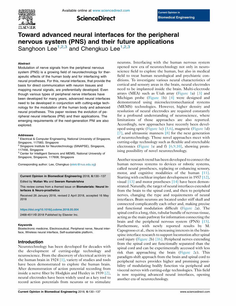
field to treat human neurological and psychiatric con-ditions. To investigate various neural characteristics ofcortical and sensory areas in the brain, neural electrodesneed to be implanted inside the brain. Multi-electrodearrays (MEA) such as Utah array (Figure 1a) [3] andMichigan probe (Figure 1b) [4] were designed anddemonstrated using microelectromechanical systems(MEMS) technologies. However, higher density andresolution of neural electrodes are required constantlyfor a profound understanding of neuroscience, wherelimitations of those approaches are also reported.
Accordingly, new approaches have recently been devel-oped using optic (Figure 1c) [5,6], magnetic (Figure 1d)[7], and ultrasonic manners [8] for the next generationof neurotechnology. Those novel approaches meet withcutting-edge technology such as flexible and stretchableelectronics (Figure 1e and f) [6,9,10], showing prom-ising possibility of novel neurotechnology.
Another research trendhas beendeveloped to connect thehuman nervous systems to devices or robotic systems,called neural prostheses, replacing or enhancing sensory,
motor, and cognitive modalities of the human [11].Starting with cochlear implant development in 1957 [12],visual [13] and motor prostheses [14] have been demon-strated.Naturally, the target of neural interfaces extendedfrom the brain to the spinal cord, and then to peripheralnerves, changing the type and requirements of neuralinterfaces. Brain neurons are located under stiff skull andconnected complicatedly each other and, making preciseand functional modulation difficult (Figure 2a). Thespinal cord is a long, thin, tubular bundle of nervous tissue,acting as themain pathway for information connecting the
brain and the peripheral nervous system (PNS) [15].Furthermore, with newly reported results by M.Capogrosso et al., there is increasing interests in the brain-spine interface research to support locomotion after spinalcord injury (Figure 2b) [16]. Peripheral nerves extendingfrom the spinal cord are functionally separated than thespinal cord and can be experimentally accessed with lessrisk than approaching the brain (Figure 2c). Thisparadigm-shift approach from the brain and spinal cord toperipheral nerves provides higher and promising possi-bility of modulating bodily function via peripheral andvisceral nerves with cutting-edge technologies. This field
is now requiring advanced neural interfaces, openinganother era of neurotechnology.
www.sciencedirect.com
Figure 1
Photos of (a) Utah array with 400 mm shank spacing and 100 channels demonstrated in human studies [3], and (b) several designs of silicon-basedMichigan probes [4]. (c) A picture of microfluidic channel integrated with micro-ILEDs delivering light and drug simultaneously. The inlet image shows acomparison of the flexible device (top) and conventional metal cannula (bottom) [6]. (d) Schematic of the microfabricated coil consisting of a coppertrace (red) on a silicon substrate (yellow) [7]. (e) Schematic illustration of the overall construction, highlighting a freely adjustable needle with a m-ILEDat the tip end, connected to a receiver coil with matching capacitors, a rectifier, and a separate m-ILED indicator [9]. (f) A bright-field microscope imageshowing partially ejected mesh electronics with significant expansion in solution [10].
Toward Advanced Neural interfaces for the Peripheral Nervous System (PNS) Lee and Lee 131
Neural interface for the peripheral nervoussystem (PNS)Peripheral nerves have different anatomical and physi-ological characteristics compared with the brain andspinal cord. Axons are surrounded by insulating myelin,then bundled into fascicles which are further surroun-ded by dense protective outer layers, known as the
perineurium and epineurium (Figure 2d) [17]. It makesus difficult to elucidate which specific pathways, orfascicles, signals are originating from. Furthermore, anyneural signals that one may achieve to record areinherently weaker than the brain or cortex, and arealways with additional interfering sources such as elec-trical signals, caused by muscle contraction, and move-ment artifacts that might corrupt measurements fromperipheral nerves. In addition, nerves are typicallyaffected by physiological motion (respiratory and car-diovascular movement) and kinematic movement
(muscles and organs), so that considerable complianceand flexibility of peripheral neural interface are highlyrequired. The ideal peripheral neural interfaces shouldhave the highest selectivity for recording and stimula-tion with the minimal invasiveness. The neural interfaceshould also be implanted for longer period withoutdegradation of own functionalities. Due to theanatomical characteristics of peripheral nerves, selec-tivity and invasiveness are trade-off. Accordingly, varioustypes of peripheral neural interfaces have been
www.sciencedirect.com
investigated for many years, such as extraneural (CUFF,FINE) [18,19], intrafascicular (LIFE and TIME)[20,21], penetrating (USEA) [22] and regenerative in-
terfaces [23] (Figure 3a).
The regenerative interface may ultimately allow tointerface a high number of nerve fibers for bidirectionalcommunication between a nerve and a prosthetic deviceor system. However, it is difficult for regenerating fibersto reconnect to its original target since the fibersrandomly grow. As a result, the success of functionalrecovery remains challenging. In addition, slow regen-eration (1e2 mm per day) and progressive reduction inthe capacity of Schwann cells to support regrowth result
in neuronal apoptosis and muscle atrophy [24]. Toovercome these issues, several approaches were inves-tigated such as use in biomaterials [25,26] as well asapplying external energies such as electrical stimulation[27] and pulsed electromagnetic fields [28].
Multi-electrode arrays (MEAs) also used for peripheralnerves as a penetrating interface. To minimize thenumber of redundant electrodes and to enhance theselectivity of fascicles within the nerve, Utah SlantedElectrode Array (USEA) was demonstrated for neuro-
modulation in the PNS [29]. Some critical issues,however, should be seriously considered for penetratingelectrodes. Firstly, this interface typically has stiff
Current Opinion in Biomedical Engineering 2018, 6:130–137

Figure 2
Schematic representation of neurotechnology in (a) a brain, (b) a spinal cord [16], and (c) peripheral nerves [40]. (d) Schematic diagram of peripheralnerve organization [17].
132 Biomaterials: Neural Interfaces & Neuro-prosthetics
electrode arrays which are enough to be penetrated innerves, leading to the damage of the nerve or fascicles aswell as electrode during the implantation and move-
ments of the object due to mechanical mismatch be-tween the nervous tissues and the electrodes [30].Secondly, selectivity is limited in the area where elec-trodes inserted. Thirdly, the large number of electrodesis not efficient for selective stimulation or recording aswell as requires many wires and connectors, which de-mands additional and complicated mapping procedures.
Intrafascicular electrodes show a higher level of selec-tivity for stimulation and recording by implanting insidea nerve as well as has considerable compliance and
flexibility for peripheral nerves using flexible materials[31]. However, the successful use of these interfacesstrongly depends on implantation techniques such as animplantation angle between the electrode and a nerve,implantation depth and secure fixation of the interfaceon the surface of a nerve. Furthermore, it is doubtful toimplant intra-neural approaches on autonomous nervesfor electroceutical applications. This is because theseapproaches themselves potentially cause nerve damage[32], which leads to serious side effect.
On the other hand, extraneural interfaces are relativelynon-invasive approach and allow easy implantation,showing reasonable effective selectivity for stimulation[33]. However, the low signal-to-noise ratio (SNR) re-mains a major challenge of the cuff-type electrodes forrecording since tight cuff for achieving close contact to a
Current Opinion in Biomedical Engineering 2018, 6:130–137
nerve could causes eventual nerve damage during long-term implantation [34]. In addition, a long length (atleast 20 mm) of cuff is usually required for high ampli-
tude signal recording when tripolar configuration ofrecording is used [15]. It is also limited due to higherpossibility of foreign body response, biomechanicalissues and difficulty of implanting on small nerves forlong term implantations [35].
Recently, flexible epineural electrodes have been sug-gested and investigated (Figure 3b), where contactelectrodes are closely on epineurium of nerves(Figure 3c) or partially implanted in epineurium(Figure 3d), not only to reduce pressure applied on the
nerve, but also to enhance selectivity of recording andstimulation [36e39]. Selective stimulation and recordingof rat sciatic nerves were demonstrated with less pressureon the nerve by the interface than that applied by acommercial cuff electrode (Figure 3e) [40]. The highquality of neural recordings was demonstrated inbranches of sciatic nerves in a rat to map a sciatic nerveand leg muscles (Figure 3f) [41] as well as sensory in-formation was decoded to restore sensory feedback [42].
Peripheral neural interface forneuroprosthesis and future directionNeural prostheses are assistive devices or systems thatreplace or restore sensory, motor and cognitive functionsof the human resulting from neural damage [11].Cutting-edge bionic limb as a motor prosthesis has been
developed with robotics, engineering, and 3D printing
www.sciencedirect.com

Figure 3
Schematic diagrams of various types of peripheral nerve interfaces; (a) invasiveness versus selectivity and (b) availability to small nerves versusselective functionality; (c– i) Schematic diagram of flexible strip electrodes and (c-ii) the result of neural recordings [38]; (d– i) Schematic diagram of splitring electrodes and (d-ii) the result of selective stimulation [36]; (e– i) Schematic diagram and picture of flexible sling electrodes and (e-ii) the result ofselective recording [40]; (f– i) Schematic diagram and picture of flexible neural ribbon electrodes and (f-ii) the result of mapping a sciatic nerve in a rat[41].
Toward Advanced Neural interfaces for the Peripheral Nervous System (PNS) Lee and Lee 133
technologies to replace arms or legs of amputees. Sen-sory feedback plays an important role in improving theperformance of the neural prostheses to achieve finer
and dexterous movement control [43]. Natural touchperception was elicited by electrical stimulation on theperipheral nerves in amputees [44], and tactile sensa-tions in patients who cannot benefit from peripheralnerve sensory stimulation was also evoked by intra-cortical microstimulation of the primary somatosensorycortex [45]. These studies have started to use fairlydense electrode arrays, which make difficult to stimu-late neurons that represent different parts of the body aswell as to represent different sensory modalities [46].Currently, artificial sensors are employed on the skin
surface to collect sensory information, however, it isdifficult for these artificial sensors to substitute thefunctions of the natural tactile and proprioceptive re-ceptors due to their density and complexity. These pe-ripheral receptors, together with the primary sensoryneurons that relay their signals to the CNS, are typicallyintact in persons with tetraplegia due to spinal cordinjury. Recording of sensory signals from these primary
www.sciencedirect.com
sensory neurons that are part of the PNS, and thetransformation of the information into a feedback signal,provides an alternative but yet relatively unexplored way
to restore high-fidelity sensory feedback to persons withtetraplegia. With the recent developments of implant-able self-powered energy harvesters [47], it may now bepossible to realize long-term nerve recording for sensoryfeedback. The PNS of the upper and lower limb conveysboth afferent sensory information to the brain andefferent motor commands to the muscles. Although theperipheral nerves are small in size, they are made up ofseveral nerve fascicles holding hundreds of nerve fibers.As an example, the human median nerve trunk, whichhas around 20 nerve fascicles (with an average area of
0.16 mm2), holds 20,000 axons [17]. Thus, an effectiveneural interface needs to record from a large number ofnerve fibers in a highly selective manner. To achievethis, various intrafascicular multichannel neural in-terfaces have been developed, including LIFE, TIME,and UEA. To extend the UEA along the nerve fiber di-rection, it is desirable to integrate the penetratingmicroneedle electrodes with a flexible substrate, which
Current Opinion in Biomedical Engineering 2018, 6:130–137
134 Biomaterials: Neural Interfaces & Neuro-prosthetics
will deform with the nerve. A manually integratedstretchable microneedle electrode array has been shownto maintain stable contact with the muscle tissue duringelectromyographic recording and stimulation [48].However, the manual integration process lacks preci-sion, repeatability, and scalability compared to standardmicroelectromechanical systems (MEMS) fabricationprocess. Recently, a highly selective 3D spiked neural
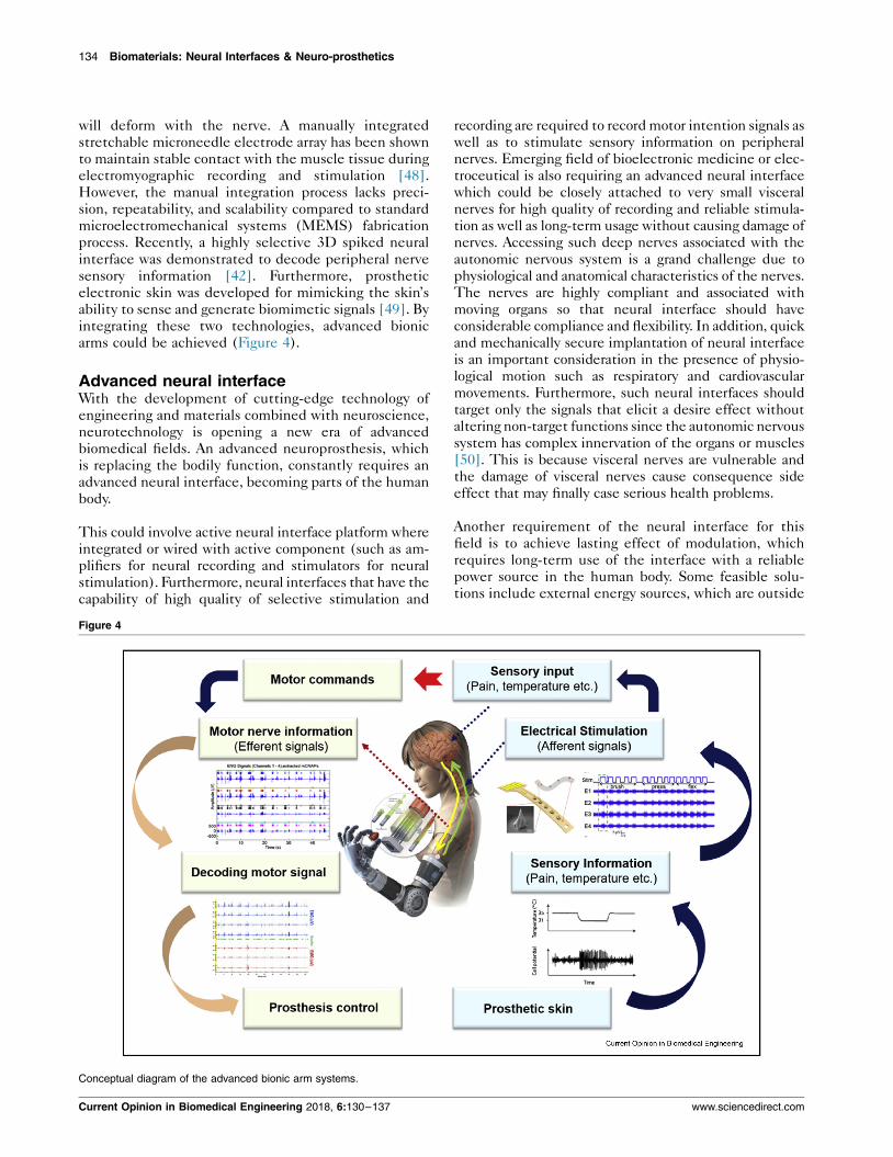
interface was demonstrated to decode peripheral nervesensory information [42]. Furthermore, prostheticelectronic skin was developed for mimicking the skin’sability to sense and generate biomimetic signals [49]. Byintegrating these two technologies, advanced bionicarms could be achieved (Figure 4).
Advanced neural interfaceWith the development of cutting-edge technology ofengineering and materials combined with neuroscience,neurotechnology is opening a new era of advancedbiomedical fields. An advanced neuroprosthesis, whichis replacing the bodily function, constantly requires anadvanced neural interface, becoming parts of the humanbody.
This could involve active neural interface platform whereintegrated or wired with active component (such as am-plifiers for neural recording and stimulators for neuralstimulation). Furthermore, neural interfaces that have thecapability of high quality of selective stimulation and
Figure 4
Conceptual diagram of the advanced bionic arm systems.
Current Opinion in Biomedical Engineering 2018, 6:130–137
recording are required to recordmotor intention signals aswell as to stimulate sensory information on peripheralnerves. Emerging field of bioelectronic medicine or elec-troceutical is also requiring an advanced neural interfacewhich could be closely attached to very small visceralnerves for high quality of recording and reliable stimula-tion as well as long-term usage without causing damage ofnerves. Accessing such deep nerves associated with the
autonomic nervous system is a grand challenge due tophysiological and anatomical characteristics of the nerves.The nerves are highly compliant and associated withmoving organs so that neural interface should haveconsiderable compliance and flexibility. In addition, quickand mechanically secure implantation of neural interfaceis an important consideration in the presence of physio-logical motion such as respiratory and cardiovascularmovements. Furthermore, such neural interfaces shouldtarget only the signals that elicit a desire effect withoutaltering non-target functions since the autonomic nervous
system has complex innervation of the organs or muscles[50]. This is because visceral nerves are vulnerable andthe damage of visceral nerves cause consequence sideeffect that may finally case serious health problems.
Another requirement of the neural interface for thisfield is to achieve lasting effect of modulation, whichrequires long-term use of the interface with a reliablepower source in the human body. Some feasible solu-tions include external energy sources, which are outside
www.sciencedirect.com

Toward Advanced Neural interfaces for the Peripheral Nervous System (PNS) Lee and Lee 135
of body and provide energy to the devices via wired andwireless communication. The integration of neuralinterface with wireless powering by either ultrasound[51] or electromagnetic powers [52] is a promising di-rection for future bioelectronic medicine. Therefore,next generation of neural interface for bioelectronicmedicine should have been adapted and miniaturizedenough to interrogate visceral nerves non-invasively,
wirelessly and securely.
Recently, nanoclip interface was suggested anddemonstrated in a very small vagus nerve for stimulationand recording [53], but it still needs to be connected tothe outside of sources or components via wires. Highperformance wireless powering at low gigahertz fre-quencies combined with neural cuff was demonstratedto regulate heart rate and blood pressure in a porchineanimal model [54]. Flexible neural clip interface wassuggested and demonstrated in branches of sciatic
nerves, vagus nerves and bladder pelvic nerves for leg
Figure 5
Schematic diagram of wireless and self-sustainable platforms for advanced n
www.sciencedirect.com
muscle activation, heart rate control and modulation ofbladder function, respectively, then finally demon-strated wireless bladder modulation combined with themid-field powering (Figure 5) [55].
Furthermore, the self-powered concept using variousmechanisms could be a promising solution for achievinglasting effect of modulation, which is recently attracting
significant attention for biomedical devices as a novelself-sustainable platform for long-term use [56]. Forinstance, triboelectric nanogenerators (TENGs), thatare operated by any mechanical sources, generate risingexponential waveforms as an output signal which can beused for direct neuro-stimulation as a power source.This has an advantage in that a rising exponentialwaveform stimulated neuron more effectively than asquare-wave pulse [57e59]. Previous study showedclear muscle activations with TENGs by stimulating asciatic nerve and branches [40], and TENGs show a
promising candidate for a battery-free stimulator
eural interfaces and their applications.
Current Opinion in Biomedical Engineering 2018, 6:130–137
136 Biomaterials: Neural Interfaces & Neuro-prosthetics
(Figure 5b). Furthermore, comparative demonstration ofthe efficiency of nerve stimulation induced by expo-nential and square pulsed waveforms was verified thatTENGs can effectively stimulate nerves and are goodcandidates for neurostimulators [60].
Also, with the development of cutting-edge self-powered device, any kinds of mechanical movements of
muscles or organs from the human body could be usedfor sustainable power sources of TENGs. This could actas not only a battery-free stimulator by combining neuralinterfaces, but also a self-sustainable power source forneuroprostheses.
Summary and outlooksNeural interfaces for peripheral nerve applications haveshown promising potential in emerging research field,such asmodulation of the human bodily function and fineconnection with bionic prostheses. For neural prostheses,bidirectional communication between prostheses andperipheral nerves will be required with neural interfacesthat record the high quality of motor signal to control theprosthesis as well as stimulate a peripheral nerve withsensory information to provide sensory feedback. For
bioelectronic medicine, neural interfaces should non-invasively and securely stick on visceral nerves. Integra-tion of energy sources with neural interfaces in small formfactormay lead to novel self-sustainable neuroprostheses.
AcknowledgementsThe authors would like to acknowledge financial support from followingresearch grants: NRF-CRP10-2012-01 “Peripheral Nerve Prostheses: AParadigm Shift in Restoring Dexterous Limb Function” and NRFCRP8-2011-01 “Self-powered body sensor for disease management andprevention-orientated healthcare” from the National Research Foundation(NRF), Singapore, and Faculty Research Committee (FRC) grant “Ther-moelectric Power Generator (TEG) Based Self-Powered ECG Plas-terdSystem Integration (Part 3)” at the National University of Singapore.
Conflict of interestThe authors declare no conflict of interest.
ReferencesPapers of particular interest, published within the period of review,have been highlighted as:
� of special interest�� of outstanding interest
1. Hass L: Hans Berger (1873-1941), Richard Caton (1842-1926),and electroencephalography. J Neurol Neurosurg Psychiatry2003, 74:9.
2. Hodgkin AL, Huxley AF: Action potentials recorded from insidea nerve fibre. Nature 1939, 114:710–711.
3. Rousche PJ, Norman RA: Chronic intracortical micro-stimulation (ICMS) of cat sensory cortex using the Utahintracortical electrode array. IEEE Trans Rehabil Eng 1999, 7:56–68.
4. The Michigan Probe: Changing the course of brain research, ece.umich.edu.
5. Montgomery KL, Yeh AJ, Ho JS, Tsao V, Mohan Iyer S,Grosenick L, Ferenczi EA, Tanabe Y, Deisseroth K, Delp SL,
Current Opinion in Biomedical Engineering 2018, 6:130–137
Poon AS: Wirelessly powered, fully internal optogenetics forbrain, spinal and peripheral circuits in mice. Nat Methods2015, 12:969–974.
6�. Jeong JW, McCall JG, Shin G, Zhang Y, Al-Hasani R, Kim M, Li S,
Sim JY, Jang KI, Shi Y, Hong DY, Liu Y, Schmitz GP, Xia L, He Z,Gamble P, Ray WZ, Huang Y, Bruchas MR, Rogers JA: Wirelessoptofluidic systems for programmable in vivo pharmacologyand optogenetics. Cell 2015, 162:662–674.
7��. Lee SW, Fallegger F, Casse BDF, Fried SI: Implantable micro-
coils for intracortical magnetic stimulation. Sci Adv 2016, 2,e1600889.
8. Seo D, Carmena JM, Rabaey JM, Alon E, Maharbiz MM: Neuraldust: an ultrasonic, low power solution for chronic brain-machineinterfaces, arXiv preprint. 2013. arXiv:1307.2196.
9. Shin G, Gomez AM, Al-Hasani R, Jeong YR, Kim J, Xie Z,Banks A, Lee SM, Han SY, Yoo CJ, Lee JL, Lee SH, Kurniawan J,Tureb J, Guo Z, Yoon J, Park SI, Bang SY, Nam Y, Walicki MC,Samineni VK, Mickle AD, Lee K, Heo SY, McCall JG, Pan T,Wang L, Feng X, Kim TI, Kim JK, Li Y, Huang Y, Gereau RWt,Ha JS, Bruchas MR, Rogers JA: Flexible near-field wirelessoptoelectronics as subdermal implants for broad applica-tions in optogenetics. Neuron 2017, 93:509–521.e3.
10. Hong G, Yang X, Zhou T, Lieber CM: Mesh electronics: a newparadigm for tissue-like brain probes. Curr Opin Neurobiol2017, 50:33–41.
11. Prochazka A, Mushahwar VK, McCreery DB: Neural prostheses.J Physiol 2001, 533:99–109.
12. Eshraghi AA, Nazarian R, Telischi FF, Rajguru SM, Truy E,Gupta C: The cochlear implant: historical aspects and futureprospects. Anat Rec (Hoboken) 2012, 295:1967–1980.
13. Schiller PH, Tehovnik EJ: Visual prosthesis. Perception 2008,37:1529–1559.
14. Scherberger H: Neural control of motor prostheses. Curr OpinNeurobiol 2009, 19:629–633.
15. Horch KW, Dhillon GS: Neuroprosthetics: theory and practice.World Scientific Publishing Co. Pte. Ltd; 2004.
16��
. Capogrosso M, Milekovic T, Borton D, Wagner F, Moraud EM,Mignardot JB, Buse N, Gandar J, Barraud Q, Xing D, Rey E,Duis S, Jianzhong Y, Ko WK, Li Q, Detemple P, Denison T,Micera S, Bezard E, Bloch J, Courtine G: A brain-spine interfacealleviating gait deficits after spinal cord injury in primates.Nature 2016, 539:284–288.
17. Patton KT: Anatomy & physiology. Elsevier; 2010.
18. Loeb GE, Peck RA: Cuff electrodes for chronic stimulationand recording of peripheral nerve activity. J Neurosci Methods1996, 64:95–103.
19. Tyler DJ, Durand DM: Functionally selective peripheral nervestimulation with a flat interface nerve electrode. IEEE TransNeural Syst Rehabil Eng 2002, 10:294–303.
20. Lago N, Yoshida K, Koch KP, Navarro X: Assessment ofbiocompatibility of chronically implanted polyimide andplatinum intrafascicular electrodes. IEEE Trans Biomed Eng2007, 54:281–290.
21. Boretius T, Badia J, Pascual-Font A, Schuettler M, Navarro X,Yoshida K, Stieglitz T: A transverse intrafascicular multi-channel electrode (TIME) to interface with the peripheralnerve. Biosens Bioelectron 2010, 26:62–69.
22. Christensen MB, Pearce SM, Ledbetter NM, Warren DJ,Clark GA, Tresco PA: The foreign body response to the UtahSlant Electrode Array in the cat sciatic nerve. Acta Biomater2014, 10:4650–4660.
23. Ortiz-Catalan M, Marin-Millan J, Delbeke J, Hakansson B,Branemark R: Effect of signal-to-noise ratio of splitting thecontinous contacts of cuff electrodes into smaller recordingareas. J Neuroeng Rehabil 2013, 10:1–15.
24. Grinsell D, Keating CP: Peripheral nerve reconstruction afterinjury: a review of clinical and experimental therapies.Biomed Res Int 2014, 2014, 698256.
www.sciencedirect.com
Toward Advanced Neural interfaces for the Peripheral Nervous System (PNS) Lee and Lee 137
25. Musick KM, Rigosa J, Narasimhan S, Wurth S, Capogrosso M,Chew DJ, Fawcett JW, Micera S, Lacour SP: Chronic multi-channel neural recordings from soft regenerative micro-channel electrodes during gait. Sci Rep 2015, 5, 14363.
26. Rutkowski GE, Miller CA, Jeftinija S, Mallapragada SK: Syner-gistic effects of micropatterned biodegradable conduits andSchwann cells on sciatic nerve regeneration. J Neural Eng2004, 1:151–157.
27. Xu C, Kou Y, Zhang P, Han N, Yin X, Deng J, Chen B, Jiang B:Electrical stimulation promotes regeneration of defectiveperipheral nerves after delayed repair intervals lasting underone month. PLoS One 2014, 9, e105045.
28. Mohammadi R, Faraji D, Alemi H, Mokarizadeh A: Pulsed elec-tromagnetic fields accelerate functional recovery of trans-ected sciatic nerve bridged by chitosan conduit: an animalmodel study. Int J Surg 2014, 12:1278–1285.
29. Wark HA, Mathews KS, Normann RA, Fernandez E: Behavioraland cellular consequences of high-electrode count UtahArrays chronically implanted in rat sciatic nerve. J Neural Eng2014, 11, 046027.
30. Lacour SP, Courtine G, Guck J: Materials and technologies forsoft implantable neuroprostheses. Nat Rev Mater 2016, 1,16063.
31. Raspopovic S, Capogrosso M, Petrini FM, Bonizzato M, Rigosa J,Di Pino G, Carpaneto J, Controzzi M, Boretius T, Fernandez E,Granata G, Oddo CM, Citi L, Ciancio AL, Cipriani C, Carrozza MC,Jensen W, Guglielmelli E, Stieglitz T, Rossini PM, Micera S:Restoring natural sensory feedback in real-time bidirectionalhand prostheses. Sci Transl Med 2014, 6. 222ra19.
32. Di Pino G, Denaro L, Vadala G, Marinozzi A, Tombini M, Ferreri F,Papalia R, Accoto D, Guglielmelli E, Di Lazzaro V, Denaro V:Invasive neural interfaces: the perspective of the surgeon.J Surg Res 2014, 188:77–87.
33�
. Tan DW, Schiefer MA, Keith MW, Anderson JR, Tyler J, Tyler DJ:A neural interface provides long-term stable natural touchperception. Sci Transl Med 2014, 6. 257ra138.
34. Hoffer JA, Kallesoe K: How to use nerve cuffs to stimulate, recordor modulate neural activity. 2001.
35. Restaino SM, Abliz E, Wachrathit K, Krauthamer V, Shah SB:Biomechanical and functional variation in rat sciatic nervefollowing cuff electrode implantation. J Neuroeng Rehabil2014, 11:73.
36. Lee S, Sheshadri S, Xiang Z, Delgado-Martinez I, Xue N, Sun T,Thakor NV, Yen S-C, Lee C: Selective stimulation and neuralrecording on peripheral nerves using flexible split ring elec-trodes. Sensor Actuators B Chem 2017, 242:1165–1170.
37. Lee S, Yen SC, Liao LD, Gammad GGL, Thakor NV, Lee C:Flexible sling electrode for bidirectional neural signalrecording and selective stimulation. In IEEE 29th InternationalConference on Micro Electro Mechanical Systems (MEMS); 2016:375–378.
38. Lee S, Yen SC, Sheshadri S, Delgado-Martinez I, Xue N, Xiang Z,Thakor NV, Lee C: Flexible epineural strip electrode forrecording in fine nerves. IEEE Trans Biomed Eng 2016, 63:581–587.
39. Xiang Z, Yen SC, Sheshadri S, Wang J, Lee S, Liu YH, Liao LD,Thakor NV, Lee C: Progress of flexible electronics in neuralinterfacing - a self-adaptive non-invasive neural ribbon elec-trode for small nerves recording. Adv Mater 2016, 28:4472–4479.
40. Lee S, Wang H, Shi Q, Dhakar L, Wang J, Thakor NV, Yen S-C,Lee C: Development of battery-free neural interface andmodulated control of tibialis anterior muscle via commonperoneal nerve based on triboelectric nanogenerators(TENGs). Nano Energy 2017, 33:1–11.
41. Xiang Z, Sheshadri S, Lee SH, Wang J, Xue N, Thakor NV,Yen SC, Lee C: Mapping of small nerve trunks and branchesusing adaptive flexible electrodes. Adv Sci 2016, 3, 1500386.
42. Wang J, Thow XY, Wang H, Lee S, Voges K, Thakor NV, Yen SC,Lee C: A highly selective 3D spiked ultraflexible neural (SUN)
www.sciencedirect.com
interface for decoding peripheral nerve sensory information.Adv Healthc Mater 2017, 1700987.
43. Saunders I, Vijayakumar S: The role of feed-forward andfeedback processes for closed-loop prosthesis control.J NeuroEng Rehabil 2011, 8:60.
44. Davis T, Wark H, Hutchinson D, Warren D, O’Neill K,Scheinblum T, Clark GA, Normann R, Greger B: Restoringmotor control and sensory feedback in people with upperextremity amputations using arrays of 96 microelectrodesimplanted in the median and ulnar nerves. J Neural Eng 2016,13, 036001.
45. Flesher SN, Collinger JL, Foldes ST, Weiss JM, Downey JE,Tyler-Kabara EC, Bensmaia SJ, Schwartz AB, Boninger ML,Gaunt RA: Intracortical microstimulation of human somato-sensory cortex. Sci Transl Med 2016, 8. 361ra141–361ra141.
46. Tomlinson T, Miller LE: Toward a proprioceptive neural interfacethat mimics natural cortical activity, progress in motor control.Springer; 2016:367–388.
47. Zheng Q, Zou Y, Zhang Y, Liu Z, Shi B, Wang X, Jin Y, Ouyang H,Li Z, Wang ZL: Biodegradable triboelectric nanogenerator as alife-time designed implantable power source. Sci Adv 2016, 2,e1501478.
48. Guvanasen GS, Guo L, Aguilar RJ, Cheek AL, Shafor CS,Rajaraman S, Nichols TR, DeWeerth SP: A stretchable micro-needle electrode array for stimulating and measuring intra-muscular electromyographic activity. IEEE Trans Neural SystRehabil Eng 2017, 25:1440–1452.
49. Chortos A, Liu J, Bao Z: Pursuing prosthetic electronic skin.Nat Mater 2016, 15:937–950.
50. Birmingham K, Gradinaru V, Anikeeva P, Grill WM, Pikov V,McLaughlin B, Pasricha P, Weber D, Ludwig K, Famm K: Bio-electronic medicines: a research roadmap. Nat Rev DrugDiscov 2014, 13:399–400.
51. Charthad J, Weber MJ, Chang TC, Arbabian A: A mm-sizedimplantable medical device (IMD) with ultrasonic powertransfer and a hybrid bi-directional data link. IEEE J SolidState Circ 2015, 50:1741–1753.
52. Jegadeesan R, Nag S, Agarwal K, Thakor NV, Guo YX: Enablingwireless powering and telemetry for peripheral nerve im-plants. IEEE J Biomed Health Inform 2015, 19:958–970.
53�
. Lissandrello CA, Gillis WF, Shen J, Pearre BW, Vitale F,Pasquali M, Holinski BJ, Chew DJ, White AE, Gardner TJ:A micro-scale printable nanoclip for electrical stimulationand recording in small nerves. J Neural Eng 2017, 14, 036006.
54�
. Tanabe Y, Ho JS, Liu J, Liao SY, Zhen Z, Hsu S, Shuto C,Zhu ZY, Ma A, Vassos C, Chen P, Tse HF, Poon ASY: High-performance wireless powering for peripheral nerve neuro-modulation systems. PLoS One 2017, 12, e0186698.
55. Lee S, Peh WYX, Wang J, Yang F, Ho JS, Thakor NV, Yen SC,Lee C: Toward bioelectronic medicine-neuromodulation ofsmall peripheral nerves using flexible neural clip. Adv Sci(Weinh) 2017, 4, 1700149.
56. Zheng Q, Zhang H, Shi B, Xue X, Liu Z, Jin Y, Ma Y, Zou Y,Wang X, An Z, Tang W, Zhang W, Yang F, Liu Y, Lang X, Xu Z,Li Z, Wang ZL: In vivo self-powered wireless cardiac moni-toring via implantable triboelectric nanogenerator. ACS Nano2016, 10:6510–6518.
57. Wongsarnpigoon A, Grill WM: Genetic algorithm revealsenergy-efficient waveforms for neural stimulation. IEEEEMBS 2009:634–637.
58. Yip M, Bowers P, Noel V, Chandrakasan A, Stankovic KM:Energy-efficient waveform for electrical stimulation of thecochlear nerve. Sci Rep 2017, 7, 13582.
59. Brocker DT, Grill WM: Principles of electrical stimulation ofneural tissue. Handb Clin Neurol 2013, 116:3–18.
60. Lee S, Wang H, Wang J, Shi Q, Yen SC, Thakor NV, Lee C:Battery-free neuromodulator for peripheral nerve directstimulator. Nano Energy 2018, 50:148–158.
Current Opinion in Biomedical Engineering 2018, 6:130–137


![yp ological - UMasspeople.umass.edu/bhatt/papers/others/deo-sharma.pdf · yp ological V ariation in the Ergativ e Morphology of Indo-Ary an Languages Ash wini Deo [adeo@stanford.edu]](https://static.fdocuments.in/doc/165x107/5e65e585e06131514964bb2d/yp-ological-yp-ological-v-ariation-in-the-ergativ-e-morphology-of-indo-ary-an.jpg)