
Totipotency: What It Is and What It Is Not
17
COMPREHENSIVE REVIEW Totipotency: What It Is and What It Is Not Maureen L. Condic There is surprising confusion surrounding the concept of biological totipotency, both within the scientific community and in society at large. Increasingly, ethical objections to scientific research have both practical and political implications. Ethical controversy surrounding an area of research can have a chilling effect on in- vestors and industry, which in turn slows the development of novel medical therapies. In this context, clarifying precisely what is meant by ‘‘totipotency’’ and how it is experimentally determined will both avoid unnecessary controversy and potentially reduce inappropriate barriers to research. Here, the concept of totipotency is discussed, and the confusions surrounding this term in the scientific and nonscientific literature are considered. A new term, ‘‘plenipotent,’’ is proposed to resolve this confusion. The requirement for specific, oocyte-derived cytoplasm as a component of totipotency is outlined. Finally, the implications of twinning for our understanding of totipotency are discussed. Highlights Inaccurate use of the term ‘‘totipotent’’ by scientists creates unnecessary ethical controversy. Public concern over producing embryos by repro- gramming reflects confusion over totipotency. Twinning by blastocyst splitting does not provide sci- entific evidence for totipotency. What Is Totipotency? T he medical dictionary administered by the National Institutes of Health gives two contrasting definitions for the term totipotent: ‘‘capable of developing into a complete organism’’ or ‘‘differentiating into any of its cells or tissues’’ (www.merriam-webster.com/medlineplus/totipotent; ac- cessed 6/23/2013). Much of the confusion surrounding the term totipotency centers on the important differences be- tween these two definitions. A one-cell embryo (zygote) is ‘‘totipotent’’ in both senses; yet, some authors character- ize tumors [1,2] and stem cells [3,4] as ‘‘totipotent,’’ based only on the second definition (ie, the ability of these cells to produce a wide range of cell types). The difference between these two definitions is not trivial. Producing a mature organism requires the ability to both generate all the cells of the body and to organize them in a specific temporal and spatial sequence, that is, to undergo a coordinated process of development. Totipotency in this strict sense is demonstrated by the ability of an isolated cell to produce a fertile, adult individual. Consequently, a cell that is totipotent is also a one-cell embryo; that is, a cell that is capable of generating a globally coordinated develop- mental sequence. While stem cells, tumors, and embryos have many mo- lecular features in common, embryos are clearly organisms [5–8]. Embryos develop in a predictable manner toward a species-specific adult form (human embryos do not mature into mice, monkeys, or tumors). Embryos repair injury. They adapt to changing environmental conditions. Most importantly, they show coordinated interactions between parts (molecules, cells, tissues, structures, and organs) that promote the survival, health, and continued development of the organism as a whole; that is, interactions that are char- acteristic of ‘‘an individual constituted to carry on the ac- tivities of life by means of organs separate in function but mutually dependent: a living being,’’ (www.merriam- webster.com/medlineplus/organism; accessed 6/23/2013). In contrast, stem cells and tumors do not produce cells or structures in the functionally integrated progression that is characteristic of an organism. They are not capable of de- velopment. The ability to produce an orchestrated developmental sequence should not be misconstrued as some kind of mystical element that is merely attributed to an embryo. The fact that the embryo undergoes a self-directed process of maturation is entirely a matter of empirical observation; embryos construct themselves. Scientists tend to view this developmental capability as a series of cellular/molecular events. Others may view human development in more Department of Neurobiology, School of Medicine, University of Utah, Salt Lake City, Utah. STEM CELLS AND DEVELOPMENT Volume 23, Number 8, 2014 Ó Mary Ann Liebert, Inc. DOI: 10.1089/scd.2013.0364 796
Transcript of Totipotency: What It Is and What It Is Not
COMPREHENSIVE REVIEW
Totipotency:What It Is and What It Is Not
Maureen L. Condic
There is surprising confusion surrounding the concept of biological totipotency, both within the scientificcommunity and in society at large. Increasingly, ethical objections to scientific research have both practical andpolitical implications. Ethical controversy surrounding an area of research can have a chilling effect on in-vestors and industry, which in turn slows the development of novel medical therapies. In this context, clarifyingprecisely what is meant by ‘‘totipotency’’ and how it is experimentally determined will both avoid unnecessarycontroversy and potentially reduce inappropriate barriers to research. Here, the concept of totipotency isdiscussed, and the confusions surrounding this term in the scientific and nonscientific literature are considered.A new term, ‘‘plenipotent,’’ is proposed to resolve this confusion. The requirement for specific, oocyte-derivedcytoplasm as a component of totipotency is outlined. Finally, the implications of twinning for our understandingof totipotency are discussed.
Highlights
� Inaccurate use of the term ‘‘totipotent’’ by scientistscreates unnecessary ethical controversy.
� Public concern over producing embryos by repro-gramming reflects confusion over totipotency.
� Twinning by blastocyst splitting does not provide sci-entific evidence for totipotency.
What Is Totipotency?
The medical dictionary administered by the NationalInstitutes of Health gives two contrasting definitions for
the term totipotent: ‘‘capable of developing into a completeorganism’’ or ‘‘differentiating into any of its cells or tissues’’(www.merriam-webster.com/medlineplus/totipotent; ac-cessed 6/23/2013). Much of the confusion surrounding theterm totipotency centers on the important differences be-tween these two definitions. A one-cell embryo (zygote) is‘‘totipotent’’ in both senses; yet, some authors character-ize tumors [1,2] and stem cells [3,4] as ‘‘totipotent,’’ basedonly on the second definition (ie, the ability of these cellsto produce a wide range of cell types).
The difference between these two definitions is not trivial.Producing a mature organism requires the ability to bothgenerate all the cells of the body and to organize them in aspecific temporal and spatial sequence, that is, to undergo acoordinated process of development. Totipotency in thisstrict sense is demonstrated by the ability of an isolated cellto produce a fertile, adult individual. Consequently, a cell
that is totipotent is also a one-cell embryo; that is, a cell thatis capable of generating a globally coordinated develop-mental sequence.
While stem cells, tumors, and embryos have many mo-lecular features in common, embryos are clearly organisms[5–8]. Embryos develop in a predictable manner toward aspecies-specific adult form (human embryos do not matureinto mice, monkeys, or tumors). Embryos repair injury.They adapt to changing environmental conditions. Mostimportantly, they show coordinated interactions betweenparts (molecules, cells, tissues, structures, and organs) thatpromote the survival, health, and continued development ofthe organism as a whole; that is, interactions that are char-acteristic of ‘‘an individual constituted to carry on the ac-tivities of life by means of organs separate in function butmutually dependent: a living being,’’ (www.merriam-webster.com/medlineplus/organism; accessed 6/23/2013). Incontrast, stem cells and tumors do not produce cells orstructures in the functionally integrated progression that ischaracteristic of an organism. They are not capable of de-velopment.
The ability to produce an orchestrated developmentalsequence should not be misconstrued as some kind ofmystical element that is merely attributed to an embryo. Thefact that the embryo undergoes a self-directed process ofmaturation is entirely a matter of empirical observation;embryos construct themselves. Scientists tend to view thisdevelopmental capability as a series of cellular/molecularevents. Others may view human development in more
Department of Neurobiology, School of Medicine, University of Utah, Salt Lake City, Utah.
STEM CELLS AND DEVELOPMENT
Volume 23, Number 8, 2014
� Mary Ann Liebert, Inc.
DOI: 10.1089/scd.2013.0364
796
spiritual or poetic terms. But neither of these views alters thescientific facts; embryos manifest a unique molecularcomposition and pattern of behavior that is characteristic ofan organism (ie, ‘‘a living being’’) that has not been ob-served in tumors or other human cells.
Regardless how individuals or societies ultimately weighthe value of the embryo relative to the value of scientificresearch, it is important to appreciate that in all cases, theethical consideration given to human embryos does not reflectthe status they will achieve at some point in the future (ie,what they will mature into). If this was the case, then therecould be no possible objection to embryo-destructive researchsince, by definition, adult status is never attained in suchsituations. Rather, ethical consideration is given to humanembryos based on the status they already possess; that is, theirunique and fully operative ability to function as a humanorganism. Therefore, ethical controversy regarding totipotenthuman cells only concerns cells that are totipotent in thestrict, organismal sense; that is, a cell that is a human embryo.
The term totipotent describes the properties of an indi-vidual cell (not a group of cells) with the two meanings ofthis term roughly corresponding to the progressive restrictionin potential cells exhibit during normal development. At theexpanded blastocyst stage (Fig. 1A), cells in specific regionsof the embryo are restricted to produce only a subset of the
cells observed at more-mature stages (Table 1). Restrictionsin potency occur gradually as development proceeds, with theability to independently initiate a developmental sequence (ie,totipotency in the first sense) being lost relatively early duringdevelopment, and individual cells of the late morula and earlyblastocyst producing a wider range of derivatives than cells atthe expanded blastocyst stage (Fig. 1B).
The confusion between the two senses of totipotency hasled some to propose new terminology, with one authorsuggesting the term ‘‘totipotent’’ be reserved for organisms,while stem cells and tumors that produce all cell types butdo not organize them into a coherent body plan would bereferred to as ‘‘omnipotent’’ [9]. Yet the strong connotationof this word outside the field of science compromises itsutility. ‘‘Plenipotent’’ (from the Latin plenus, or ‘‘full’’)would be preferable for individual cells that are totipotent inthe cellular or weak sense, reserving ‘‘pluripotent’’ (fromthe Latin pluris, or ‘‘more’’) for cells that make only asubset of the normal derivatives of the embryo (Fig. 1B andTable 2). These terms would better reflect the progressiverestrictions in potency that occur during normal develop-ment (Fig. 1B), and avoid the unfortunate application of theterm ‘‘totipotent’’ to cells that are not embryos.
In many cases, there are significant limitations in the dataavailable to classify the developmental capabilities of cell
FIG. 1. Cell types and potency at different developmental stages. (A) Anatomy and cell types of the expanded blastocystembryo (human development, *5–6 days; shown as a mid-sagittal section). The position of the ICM (green and pink)defines the embryonic-abembryonic axis, which has a consistent relationship to the animal-vegetal axis of the oocyte, andmay be determined by both the shape of the zygote [153,189] and the early cleavage patterns of the embryo [156,164]. Theentire embryo is surrounded by an acellular protein layer known as the zona pellucida (gray). TE associated with theembryonic pole (light blue) induces the formation of PE (pink), that is initially interspersed with the presumptive epiblast(green), and segregates by cell sorting [77]. Together, epiblast and PE constitute the ICM. The blastocyst cavity is a fluid-filled space. Polar (light blue) and mural (dark blue) TE have distinct molecular properties [61,190,191] and distinctdevelopmental fates [162]. (B) During human development, only the zygote and early cleavage-stage blastomeres (possiblyup to the four-cell stage) remain totipotent, that is, capable of independently initiating a developmental sequence (Table 2).Cells of the late morula/early blastocyst are plenipotent; that is, they are able to produce all or most of the cells of the body,but not organize them into a coherent body plan (Table 2). Cells of the epiblast at the expanded blastocyst stage (the cellsfrom which many embryonic stem cell lines are derived) are pluripotent; that is, they are able to produce cell types found inthe mature body, but are not derivatives of the TE and PE (Table 2). PE, primitive endoderm; TE, trophectoderm.
TOTIPOTENCY: WHAT IT IS AND WHAT IT ISN’T 797
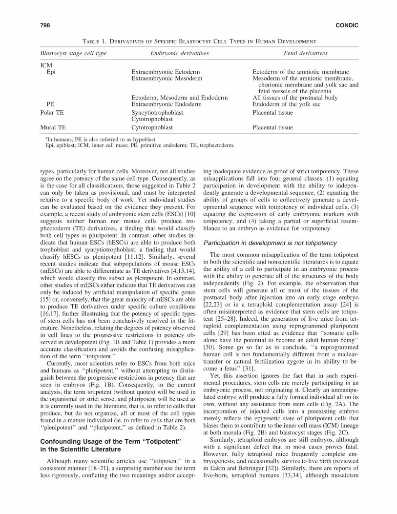
types, particularly for human cells. Moreover, not all studiesagree on the potency of the same cell type. Consequently, asis the case for all classifications, those suggested in Table 2can only be taken as provisional, and must be interpretedrelative to a specific body of work. Yet individual studiescan be evaluated based on the evidence they present. Forexample, a recent study of embryonic stem cells (ESCs) [10]suggests neither human nor mouse cells produce tro-phectoderm (TE) derivatives, a finding that would classifyboth cell types as pluripotent. In contrast, other studies in-dicate that human ESCs (hESCs) are able to produce bothtrophoblast and syncytiotrophoblast, a finding that wouldclassify hESCs as plenipotent [11,12]. Similarly, severalrecent studies indicate that subpopulations of mouse ESCs(mESCs) are able to differentiate as TE derivatives [4,13,14],which would classify this subset as plenipotent. In contrast,other studies of mESCs either indicate that TE derivatives canonly be induced by artificial manipulation of specific genes[15] or, conversely, that the great majority of mESCs are ableto produce TE derivatives under specific culture conditions[16,17], further illustrating that the potency of specific typesof stem cells has not been conclusively resolved in the lit-erature. Nonetheless, relating the degrees of potency observedin cell lines to the progressive restrictions in potency ob-served in development (Fig. 1B and Table 1) provides a moreaccurate classification and avoids the confusing misapplica-tion of the term ‘‘totipotent.’’
Currently, most scientists refer to ESCs from both miceand humans as ‘‘pluripotent,’’ without attempting to distin-guish between the progressive restrictions in potency that areseen in embryos (Fig. 1B). Consequently, in the currentanalysis, the term totipotent (without quotes) will be used inthe organismal or strict sense, and pluripotent will be used asit is currently used in the literature, that is, to refer to cells thatproduce, but do not organize, all or most of the cell typesfound in a mature individual (ie, to refer to cells that are both‘‘plenipotent’’ and ‘‘pluripotent,’’ as defined in Table 2).
Confounding Usage of the Term ‘‘Totipotent’’in the Scientific Literature
Although many scientific articles use ‘‘totipotent’’ in aconsistent manner [18–21], a surprising number use the termless rigorously, conflating the two meanings and/or accept-
ing inadequate evidence as proof of strict totipotency. Thesemisapplications fall into four general classes: (1) equatingparticipation in development with the ability to indepen-dently generate a developmental sequence, (2) equating theability of groups of cells to collectively generate a devel-opmental sequence with totipotency of individual cells, (3)equating the expression of early embryonic markers withtotipotency, and (4) taking a partial or superficial resem-blance to an embryo as evidence for totipotency.
Participation in development is not totipotency
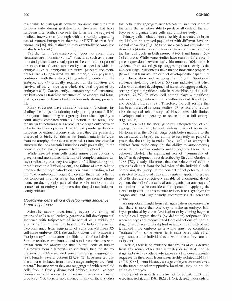
The most common misapplication of the term totipotentin both the scientific and nonscientific literatures is to equatethe ability of a cell to participate in an embryonic processwith the ability to generate all of the structures of the bodyindependently (Fig. 2). For example, the observation thatstem cells will generate all or most of the tissues of thepostnatal body after injection into an early stage embryo[22,23] or in a tetraploid complementation assay [24] isoften misinterpreted as evidence that stem cells are totipo-tent [25–28]. Indeed, the generation of live mice from tet-raploid complementation using reprogrammed pluripotentcells [29] has been cited as evidence that ‘‘somatic cellsalone have the potential to become an adult human being’’[30]. Some go so far as to conclude, ‘‘a reprogrammedhuman cell is not fundamentally different from a nuclear-transfer or natural fertilization zygote in its ability to be-come a fetus’’ [31].
Yet, this assertion ignores the fact that in such experi-mental procedures, stem cells are merely participating in anembryonic process, not originating it. Clearly an unmanipu-lated embryo will produce a fully formed individual all on itsown, without any assistance from stem cells (Fig. 2A). Theincorporation of injected cells into a preexisting embryomerely reflects the epigenetic state of pluripotent cells thatbiases them to contribute to the inner cell mass (ICM) lineageat both morula (Fig. 2B) and blastocyst stages (Fig. 2C).
Similarly, tetraploid embryos are still embryos, althoughwith a significant defect that in most cases proves fatal.However, fully tetraploid mice frequently complete em-bryogenesis, and occasionally survive to live birth (reviewedin Eakin and Behringer [32]). Similarly, there are reports oflive-born, tetraploid humans [33,34], although mosaicism
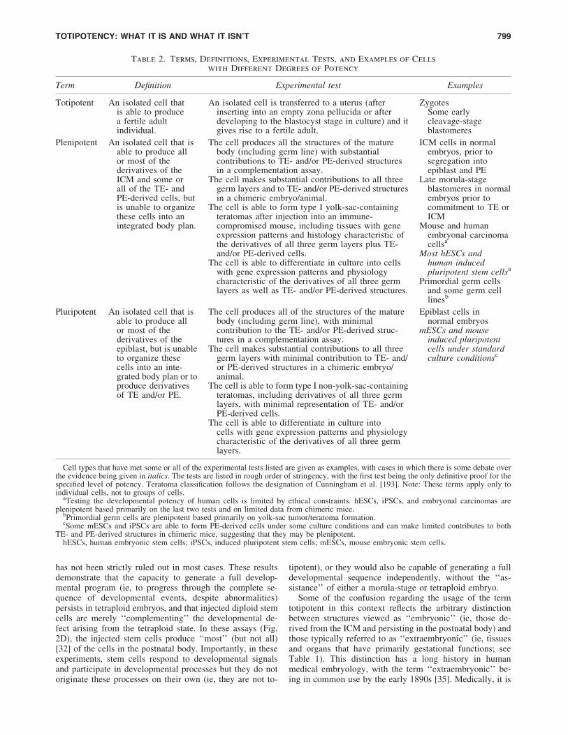
Table 1. Derivatives of Specific Blastocyst Cell Types in Human Development
Blastocyst stage cell type Embryonic derivatives Fetal derivatives
ICMEpi Extraembryonic Ectoderm Ectoderm of the amniotic membrane
Extraembryonic Mesoderm Mesoderm of the amniotic membrane,chorionic membrane and yolk sac andfetal vessels of the placenta
Ectoderm, Mesoderm and Endoderm All tissues of the postnatal bodyPE Extraembryonic Endoderm Endoderm of the yolk sac
Polar TE Syncytiotrophoblast Placental tissueCytotrophoblast
Mural TE Cytotrophoblast Placental tissue
aIn humans, PE is also referred to as hypoblast.Epi, epiblast; ICM, inner cell mass; PE, primitive endoderm; TE, trophectoderm.
798 CONDIC
has not been strictly ruled out in most cases. These resultsdemonstrate that the capacity to generate a full develop-mental program (ie, to progress through the complete se-quence of developmental events, despite abnormalities)persists in tetraploid embryos, and that injected diploid stemcells are merely ‘‘complementing’’ the developmental de-fect arising from the tetraploid state. In these assays (Fig.2D), the injected stem cells produce ‘‘most’’ (but not all)[32] of the cells in the postnatal body. Importantly, in theseexperiments, stem cells respond to developmental signalsand participate in developmental processes but they do notoriginate these processes on their own (ie, they are not to-
tipotent), or they would also be capable of generating a fulldevelopmental sequence independently, without the ‘‘as-sistance’’ of either a morula-stage or tetraploid embryo.
Some of the confusion regarding the usage of the termtotipotent in this context reflects the arbitrary distinctionbetween structures viewed as ‘‘embryonic’’ (ie, those de-rived from the ICM and persisting in the postnatal body) andthose typically referred to as ‘‘extraembryonic’’ (ie, tissuesand organs that have primarily gestational functions; seeTable 1). This distinction has a long history in humanmedical embryology, with the term ‘‘extraembryonic’’ be-ing in common use by the early 1890s [35]. Medically, it is
Table 2. Terms, Definitions, Experimental Tests, and Examples of Cells
with Different Degrees of Potency
Term Definition Experimental test Examples
Totipotent An isolated cell thatis able to producea fertile adultindividual.
An isolated cell is transferred to a uterus (afterinserting into an empty zona pellucida or afterdeveloping to the blastocyst stage in culture) and itgives rise to a fertile adult.
ZygotesSome earlycleavage-stageblastomeres
Plenipotent An isolated cell that isable to produce allor most of thederivatives of theICM and some orall of the TE- andPE-derived cells, butis unable to organizethese cells into anintegrated body plan.
The cell produces all the structures of the maturebody (including germ line) with substantialcontributions to TE- and/or PE-derived structuresin a complementation assay.
The cell makes substantial contributions to all threegerm layers and to TE- and/or PE-derived structuresin a chimeric embryo/animal.
The cell is able to form type I yolk-sac-containingteratomas after injection into an immune-compromised mouse, including tissues with geneexpression patterns and histology characteristic ofthe derivatives of all three germ layers plus TE-and/or PE-derived cells.
The cell is able to differentiate in culture into cellswith gene expression patterns and physiologycharacteristic of the derivatives of all three germlayers as well as TE- and/or PE-derived structures.
ICM cells in normalembryos, prior tosegregation intoepiblast and PE
Late morula-stageblastomeres in normalembryos prior tocommitment to TE orICM
Mouse and humanembryonal carcinomacellsa
Most hESCs andhuman inducedpluripotent stem cellsa
Primordial germ cellsand some germ celllinesb
Pluripotent An isolated cell that isable to produce allor most of thederivatives of theepiblast, but is unableto organize thesecells into an inte-grated body plan or toproduce derivativesof TE and/or PE.
The cell produces all of the structures of the maturebody (including germ line), with minimalcontribution to the TE- and/or PE-derived struc-tures in a complementation assay.
The cell makes substantial contributions to all threegerm layers with minimal contribution to TE- and/or PE-derived structures in a chimeric embryo/animal.
The cell is able to form type I non-yolk-sac-containingteratomas, including derivatives of all three germlayers, with minimal representation of TE- and/orPE-derived cells.
The cell is able to differentiate in culture intocells with gene expression patterns and physiologycharacteristic of the derivatives of all three germlayers.
Epiblast cells innormal embryos
mESCs and mouseinduced pluripotentcells under standardculture conditionsc
Cell types that have met some or all of the experimental tests listed are given as examples, with cases in which there is some debate overthe evidence being given in italics. The tests are listed in rough order of stringency, with the first test being the only definitive proof for thespecified level of potency. Teratoma classification follows the designation of Cunningham et al. [193]. Note: These terms apply only toindividual cells, not to groups of cells.
aTesting the developmental potency of human cells is limited by ethical constraints. hESCs, iPSCs, and embryonal carcinomas areplenipotent based primarily on the last two tests and on limited data from chimeric mice.
bPrimordial germ cells are plenipotent based primarily on yolk-sac tumor/teratoma formation.cSome mESCs and iPSCs are able to form PE-derived cells under some culture conditions and can make limited contributes to both
TE- and PE-derived structures in chimeric mice, suggesting that they may be plenipotent.hESCs, human embryonic stem cells; iPSCs, induced pluripotent stem cells; mESCs, mouse embryonic stem cells.
TOTIPOTENCY: WHAT IT IS AND WHAT IT ISN’T 799
reasonable to distinguish between transient structures thatfunction only during gestation and structures that havefunctions after birth, since only the latter are the subject ofmedical intervention (although with the rapidly expandinguse of exutero intrapartum therapy, or EXIT, to treat fetalanomalies [36], this distinction may eventually become lessmedially relevant.).
Yet the term ‘‘extraembryonic’’ does not mean thesestructures are ‘‘nonembryonic.’’ Structures such as the am-nion and placenta are clearly part of the embryo, not part ofthe mother or of some other entity that coexists with theembryo. Like all embryonic structures, placenta and mem-branes are (1) generated by the embryo, (2) physicallycontinuous with the embryo, (3) genetically identical to theembryo, and (4) critically required for the function andsurvival of the embryo as a whole (ie, vital organs of theembryo itself). Consequently, ‘‘extraembryonic’’ structuresare best seen as transient structures of the embryo and fetus;that is, organs or tissues that function only during prenatallife.
Many structures have similarly transient functions, in-cluding the lungs (functioning only during postnatal life),the thymus (functioning in a greatly diminished capacity atadult stages, compared with its function in the fetus), andthe uterus (functioning as a reproductive organ only betweenpuberty and menopause). Due to the purely gestationalfunctions of extraembryonic structures, they are physicallydiscarded at birth, but this is not fundamentally differentfrom the atrophy of the ductus arteriosis (a transient vascularstructure that has essential functions only prenatally) in theneonate, or the loss of primary teeth in childhood.
While injected stem cells make minor contributions toplacenta and membranes in tetraploid complementation as-says (indicating that they are capable of differentiating intothese tissues to a limited extent), the failure of stem cells toproduce the embryo entirely on their own (including all ofthe ‘‘extraembryonic’’ organs) indicates that stem cells arenot totipotent in either sense, but rather are merely plurip-otent, producing only part of the whole embryo in thecontext of an embryonic process that they do not indepen-dently initiate.
Collectively generating a developmental sequenceis not totipotency
Scientific authors occasionally equate the ability ofgroups of cells to collectively generate a full developmentalsequence with totipotency of individual cells within thegroup (Fig. 3). For example, based on the failure to generatelive-born mice from aggregates of cells derived from 32-cell-stage embryos [37], the authors assert that blastomere‘‘totipotency’’ is lost after the fifth round of cell division.Similar results were obtained and similar conclusions weredrawn from the observation that ‘‘outer’’ cells of humanblastocysts form blastocyst-like structures that initiate ex-pression of ICM-associated genes following reaggregation[38]. Finally, several authors [27,39–42] have asserted thatblastomeres isolated from morula-stage embryos are ‘‘toti-potent,’’ because when they are reaggregated with tetraploidcells from a freshly dissociated embryo, either live-bornanimals or what appear to be normal blastocysts can beproduced. Yet, there is no evidence in any of these studies
that cells in the aggregate are ‘‘totipotent’’ in either sense ofthe term; that is, either able to produce all cells of the em-bryo or to organize these cells into a mature body.
Primary cells isolated from a freshly dissociated embryosare likely to be a mixed population with different develop-mental capacities (Fig. 3A) and are clearly not equivalent tostem cells [43–47]. Zygotic transcription commences duringthe first cell cycle in both mouse [48–51] and human [52–59] embryos. While some studies have seen no difference ingene expression between early blastomeres [60], there isevidence from several groups suggesting that as early as the4–8-cell stage, blastomeres have unique molecular properties[61–71] that translate into distinct developmental capabilitiesafter dissociation and reaggregation [72,73]. Substantialevidence stretching back over 60 years indicates that whencells with distinct developmental states are aggregated, cellsorting plays a significant role in re-establishing the initialpattern [74,75]. In mice, cell sorting plays a significantrole in the segregation of cells within normal 16-cell [76]and 32-cell embryos [77]. Therefore, the cell sorting thathas been observed in some studies [37] is likely to reorga-nize the spatial relationships of blastomeres with limiteddevelopmental competency to reconstitute a full embryo(Fig. 3B, E).
Yet even with the most generous interpretation of cellaggregation studies (that cell sorting does not occur andblastomeres at the 16-cell stage contribute randomly to thereconstituted embryo), the ability to respecify as part of agroup (ie, the ability to make ‘‘any’’ cell of an embryo) isdistinct from totipotency (ie, the ability to autonomouslymake all cells of an embryo and to organize them into acoherent whole). The significant role of ‘‘community ef-fects’’ in development, first described by Sir John Gurdon in1988 [78], clearly illustrates that the behavior of cells ingroups is distinct from the behavior of the individual cellscomprising the group. If the concept of totipotency is notrestricted to individual cells and is instead applied to groupsof cells that are collectively capable of undergoing devel-opment, then all of the cells of any organism at any stage ofmaturation must be considered ‘‘totipotent.’’ Applying theterm ‘‘totipotent’’ in this manner reduces it to a synonym for‘‘organism’’ and significantly compromises its scientificutility.
An important insight from cell aggregation experiments isthat there is more than one way to make an embryo. Em-bryos produced by either fertilization or by cloning begin asa single-cell zygote that is (by definition) totipotent. Yet,when embryos are reconstituted from collections of morula-stage blastomeres (either diploid or a mixture of diploid andtetraploid), the embryo as a whole must be considered‘‘totipotent’’ in some sense (ie, it must be considered anorganism), but the individual cells within the embryo are nottotipotent.
To date, there is no evidence that groups of cells derivedfrom any source other than a freshly dissociated morula-stage embryo can collectively generate a full developmentalsequence on their own. Even when freshly isolated ICM [79]or TE [80,81] from blastocyst-stage embryos are transferredto the uterus or other supportive locations, they do not de-velop as embryos.
Groups of stem cells are also not totipotent. mES lineswere first isolated in 1981 [82,83]. Yet, despite thousands of
800 CONDIC
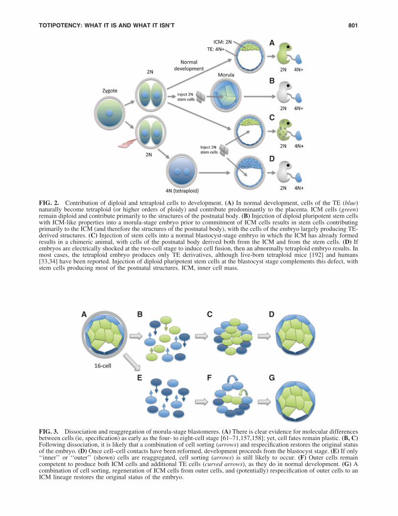
FIG. 3. Dissociation and reaggregation of morula-stage blastomeres. (A) There is clear evidence for molecular differencesbetween cells (ie, specification) as early as the four- to eight-cell stage [61–71,157,158]; yet, cell fates remain plastic. (B, C)Following dissociation, it is likely that a combination of cell sorting (arrows) and respecification restores the original statusof the embryo. (D) Once cell–cell contacts have been reformed, development proceeds from the blastocyst stage. (E) If only‘‘inner’’ or ‘‘outer’’ (shown) cells are reaggregated, cell sorting (arrows) is still likely to occur. (F) Outer cells remaincompetent to produce both ICM cells and additional TE cells (curved arrows), as they do in normal development. (G) Acombination of cell sorting, regeneration of ICM cells from outer cells, and (potentially) respecification of outer cells to anICM lineage restores the original status of the embryo.
FIG. 2. Contribution of diploid and tetraploid cells to development. (A) In normal development, cells of the TE (blue)naturally become tetraploid (or higher orders of ploidy) and contribute predominantly to the placenta. ICM cells (green)remain diploid and contribute primarily to the structures of the postnatal body. (B) Injection of diploid pluripotent stem cellswith ICM-like properties into a morula-stage embryo prior to commitment of ICM cells results in stem cells contributingprimarily to the ICM (and therefore the structures of the postnatal body), with the cells of the embryo largely producing TE-derived structures. (C) Injection of stem cells into a normal blastocyst-stage embryo in which the ICM has already formedresults in a chimeric animal, with cells of the postnatal body derived both from the ICM and from the stem cells. (D) Ifembryos are electrically shocked at the two-cell stage to induce cell fusion, then an abnormally tetraploid embryo results. Inmost cases, the tetraploid embryo produces only TE derivatives, although live-born tetraploid mice [192] and humans[33,34] have been reported. Injection of diploid pluripotent stem cells at the blastocyst stage complements this defect, withstem cells producing most of the postnatal structures. ICM, inner cell mass.
TOTIPOTENCY: WHAT IT IS AND WHAT IT ISN’T 801
articles that describe the production of stem cell aggregatesover the last three decades, there have been no reports ofspontaneous generation of embryos from ESCs. Similarly,there have been no reports of generating embryos from acombination of mES and trophoblast stem cells [84]. Thefailure of aggregated stem cell lines derived thus far tospontaneously form embryos is likely to reflect the fact thatstem cell lines have a range of properties [85–87] that arenot identical either to zygotes [88,89] or to cells that havebeen freshly isolated from embryos [43–47].
While these negative findings do not strictly rule out thepossibility that later-stage blastomeres or pluripotent stemcells ‘‘could’’ combine in some way to generate an embryo (itis logically impossible to prove something from a negativefinding), they strongly suggest that collective reconstitution ofan embryo from cells of a reaggregated morula-stage embryo(Fig. 3) reflects a unique balance of developmental propertieswithin those freshly dissociated cells—a balance that is notpresent in cultured stem cell lines or in cells derived fromlater-stage embryos.
Expression of molecular markers found in earlyembryos is not totipotency
Scientific authors occasionally conflate expression ofmolecular markers characteristic of specific embryonicstages with the corresponding developmental capacities ofcells at those stages. For example, several studies have de-termined that most or all mESCs cycle in and out of a un-ique transcriptional state that shares some elements incommon with blastomeres at the two-cell (2C) stage [14,90–92]. Cells in this 2C-like state lack pluripotency markersnormally associated with the ICM, as do human zygotes andembryos at the two-cell stage [88]. The ‘‘2C-like’’ cells alsoshow expanded developmental potential when injected intoembryos. The authors present no evidence that isolated 2C-like cells are capable of generating embryos on their ownand are careful not to pronounce their 2C-like cells totipo-tent in the organismal sense, but the repeated observationthat the 2C stage correlates with a period in which ‘‘blas-tomeres are totipotent’’ [14] is strongly suggestive of thisunwarranted conclusion and illustrates the confusing usageof the term ‘‘totipotent’’ in the scientific literature. Simi-larly, the ability of stem cells [3,4] or carcinomas [1,2] toexpress a molecular marker of ‘‘extraembryonic’’ cell lin-eages in addition to markers associated with ICM has beentaken as evidence of totipotency, despite there being noindication that these cells can initiate a developmental se-quence on their own.
Behaving like part of an embryo or merely lookinglike an embryo is not totipotency
Finally, some scientific authors suggest that the ability ofstem cells to replicate limited aspects of normal embryonicdevelopment is evidence for totipotency, or something veryclose to it. For example, a number of studies have shownthat ESC aggregates treated with specific signaling mole-cules exhibit some of the molecular cascades and cell be-haviors observed during normal gastrulation [93–97]. Whilethese studies do not equate stem cells with embryos, severalauthors conclude that stem cell aggregates are surprisingly
‘‘embryo like.’’ For example, one article states that aggre-gates ‘‘resemble normal embryonic development muchcloser than previously thought,’’ exhibiting ‘‘an unexpecteddegree of self-organization’’ [97]. Yet stem cell aggregates donot resemble organisms so much as they resemble tumors,which can also show a surprising degree of self-organization,producing well-formed teeth [98] and, even in one case, aremarkably normal eye [99].
Similarly, ESC aggregates [100,101] and tumors [102]occasionally generate cystic structures that have some visualsimilarity to a blastocyst-stage embryo, which has led someauthors to ask whether ESCs and tumors might also beembryos [9,103]. Yet many cell types with clearly restrictedpotency will form such cystic structures, including liver,heart, and cartilage [104]; neurons [105]; fibroblasts [106];kidney [107]; and umbilical cells [108], indicating that themere formation of such structures is not sufficient evidencefor totipotency.
Confusions Regarding Totipotency in the Non-scientific Literature: Reprogramming ‘‘Too Far’’
Since the advent of cellular reprogramming [109–111],the concern that embryos might be inadvertently generatedduring reprogramming [112] has frequently been raised,with authors asking, for example, ‘‘how to ensure that de-differentiation goes only so far and no further?’’ [113]. Thepossibility of producing embryos by reprogramming hasbeen raised by some scientific authors as well [31,103].
The belief that reprogramming could ‘‘accidentally’’bring a cell into a totipotent state seems to stem from thestubbornly compelling notion that unipotent, pluripotent,and totipotent represent points along a continuum [114] andthat reprogramming progressively rewinds developmentbackward toward the beginning. In this view, if repro-gramming goes one step beyond pluripotency, then a toti-potent zygote will accidentally be produced. Some authorshave speculated that this could be done intentionally, gen-erating an ‘‘induced totipotent stem’’ cell, [31] despite theobvious illogic of this assertion; totipotent zygotes aremanifestly not capable of self-renewal, and are therefore not‘‘stem cells.’’
Reprogramming does not merely ‘‘rewind’’development
As plausible as this concern may seem, it is not possiblefor reprogramming to accidently produce a totipotent cellfor at least two important reasons. First, reprogrammingdoes not simply ‘‘reverse’’ development, like rewinding anold audiocassette tape reverses the recording; that is, re-programming does not move a cell backward along thedevelopmental pathway that initially produced its maturestate. Instead, reprogramming is more of a single leap fromone cellular state to another [115,116]. For example, re-programming a skin cell into a heart cell [117,118] un-doubtedly involves intermediate steps between these twostates, but it does not rewind development in any rationalsense, since cardiac cells are not part of the developmentalhistory of skin cells.
Pluripotency is as much of a specific cellular state as anyother. To reprogram an adult cell to pluripotency, factors are
802 CONDIC
introduced that result in a highly specific cascade of geneexpression that is not seen in a totipotent zygote [88]. Forexample, in human embryos, the transcription factors Nanogand Pou5f1 (also called Oct3/4) are expressed in the ICMand are not found in the zygotes [119–121]. Consequently, acell that expresses such factors cannot be identical to azygote [122].
The state of the cytoplasm
The second important reason a totipotent zygote cannotbe accidently produced during reprogramming involves thenature of totipotency—specifically the reasons pluripotentand totipotent are radically different cellular states, eventhough they occur close together in developmental time.Totipotency is not merely a state of the cell’s nucleus; it alsorequires a very specific type of cellular cytoplasm that is acritical component of totipotency.
At this time, the only known totipotent cytoplasm isproduced by an oocyte and contributed to the embryo atfertilization. The fact that oocytes produce the cytoplasmicfactors that are required for an embryo to be totipotent is thereason oocytes are used for cloning. During cloning, oocytecomponents can, over the first several days of development,induce an adult nucleus to assume a state similar to that of anormal embryo (ie, a state that is capable of driving acomplete developmental sequence), mimicking the pro-cesses that naturally occur after sperm-egg fusion [123]. If acompetent nuclear state is achieved, then the oocyte-specificcomponents in the cytoplasm work with the reprogrammednucleus to produce an embryonic pattern of development,despite the significant differences observed between clonedembryos and embryos derived from fertilization [124–127].
Oocytes are highly structured cells that are uniquelyproduced by the complex process of oogenesis, which in-volves a characteristic sequence of gene activation [128]that is distinct from the pattern observed in the maternalpronucleus after fertilization or following zygotic gene ac-tivation [58,59]. Oogenesis also requires information fromother cells in the ovary [129–131]. Moreover, recent workclearly documents multiple oocyte-derived components thatare essential for mammalian embryonic development (re-viewed in Li et al. [132] and Matzuk and Burns [133]). Forexample, oocyte-expressed Ooep [134], PADi6 [135], Nlrp5[136], Ecat1 [137], and Tle6 [138] are required for thenormal function of a critical subcortical cytoplasmic com-plex, with loss of any of these genes resulting in embryolethality at the two-cell stage. Maternal expression ofKdm1B [139], Dmap1 [140], Dppa3 [141], and severalothers is required for correct DNA methylation and main-tenance of genomic imprinting, with maternal gene dele-tions resulting in death at early embryonic stages. Finally,maternally supplied Brg1 [142] and Brwd1 [143] are re-quired for zygotic gene activation, with loss of these genesresulting in arrest at the two-cell stage.
Simple reprogramming of a somatic nucleus does not es-tablish the cytoplasmic components required for totipotencyand these factors are not produced during normal embryonicdevelopment; that is, the transcriptomes of oocytes and earlyembryos are clearly distinct [58,59]. Without the requiredcytoplasmic components contributed by oocyte, there can beno zygote—regardless of the state of the nucleus [19].
In other words, even if the nucleus of a somatic cell isfully reprogrammed to be identical in every respect to thatof a zygote (a state that is distinct from a pluripotent stemcell and one that would require different reprogrammingfactors), it would still not be totipotent, because it lacks thenongenetic factors (proteins, RNA, miRNA, and specificmacromolecular complexes) that are critical components oftotipotency. Conversely, if a nucleus from a bona fide zy-gote produced by fertilization was transferred to a differ-entiated cell (eg, a muscle cell), it would not continue to be azygotic nucleus in this new location. It would be (or wouldrapidly become) a muscle cell nucleus that functions in themanner specified by the muscle cell cytoplasm [144].
The requirement for totipotent cytoplasm in order for a cellto actually be a zygote is precisely the reason that totipotencypersists for such a short time in development. In mostmammals, only the first two cells of the embryo remain to-tipotent, that is, able to generate a complete embryo on theirown when separated [145–147], perhaps in part because cellsno longer have sufficient mass to continue development[148,149]. In rare cases, totipotency is preserved until thefour-cell stage [150], with there being one reported case of achild born after transfer of a four-cell embryo in which onlyone of the blastomeres was judged to be alive [151]. Therehas also been one report in pigs of totipotency persisting untilthe eight-cell stage [152]. But there is no evidence for toti-potent cells persisting beyond this stage. As soon as a cell hasonly a subset of the specialized cytoplasmic componentscontributed by the oocyte, it is no longer totipotent.
The cell fate restrictions observed in mice and otherspecies from the two-cell stage onward are likely to reflectinheritance of required oocyte-derived components as wellas cell–cell interactions and stochastic events [72,153–156].There is clear evidence from multiple laboratories that bythe four- to eight-cell stage these two processes have re-sulted in blastomeres that have distinct molecular, func-tional, and developmental properties [61–71,157,158],indicating that from the four-cell stage onward, mammaliandevelopment is mosaic to some extent.
The requirement for totipotent cytoplasm in no way de-nies the clearly regulative aspects of mammalian develop-ment that depend on cell–cell signaling, cell interaction, andrandom events. Neither does it imply that inheritance ofmaternal factors is the only mechanism underlying celldifferentiation in mammals, or that such factors must bedifferentially inherited by specific blastomeres, althoughdifferential inheritance is possible mechanism underlyingearly lineage restrictions. Rather it suggests that the dis-tinction between regulative and mosaic development is afalse dichotomy [159]. Cell fate decisions are produced by acontinuum of overlapping and redundant mechanisms[76], with oocyte-derived factors clearly playing a criticalrole in the developmental competency of early blastomeres[160–164].
Is it possible to make a cloned embryo from an adultcell by reprogramming?
Reprogramming can generate pluripotent stem cells be-cause it initiates a chain of events that brings an adult cellnucleus into a pluripotent state and this ‘‘converted’’ nucleussubsequently produces the non-nuclear factors that are
TOTIPOTENCY: WHAT IT IS AND WHAT IT ISN’T 803

required to actually be a pluripotent stem cell. Yet totipotentzygotes are unusual, because unlike stem cells or any othercells in the body, zygotes do not produce the highly specializedcytoplasm they require on their own. Rather they inherit thisspecialized cytoplasm from a very different kind of cell with avery different nuclear state, that is, from the oocyte. Once a cellnucleus has the configuration of a totipotent zygote, it does notproduce the same factors produced by the oocyte.
This does not mean that it would be impossible to makean embryo by reprogramming, but it does mean that itcannot happen ‘‘accidently.’’ And converting an adult cellinto an embryo using reprogramming (making an ‘‘inducedtotipotent cell’’) would be difficult to accomplish, even in-tentionally (Fig. 4).
To convert an adult cell into a zygote, it would first haveto be reprogrammed to become a cell that is capable ofproviding the factors that are normally generated during theprocess of oogenesis. The simplest way of accomplishingthis would be to reprogram the adult cell into an immatureoocyte (a distinct state from a pluripotent stem cell thatwould require different reprogramming factors). The im-mature oocyte would then have to be provided with all of thecell interactions and ovarian factors required for it to become amature oocyte. During this process, the normal epigenetic re-programming and meiotic divisions that occur as part of oo-genesis [123,165] would have to be suppressed in order topreserve the nucleus in a state that is capable of driving humandevelopment (this may not be technically or even logicallypossible.). Once this unnaturally suppressed oocyte had beenmade, the nucleus would again have to be reprogrammed to azygotic state, a significant remodeling that normally reflectsfactors derived from both sperm and egg [21,123,166]. If all ofthis could be achieved, then the ‘‘secondarily reprogrammed’’totipotent cell would have to be activated to begin the processof development. Then, and only then, would a cloned embryobe produced from an adult cell by reprogramming. And thiscould hardly happen ‘‘by accident.’’
Twinning
It is sometimes asserted that because twinning can occurby splitting of a blastocyst-stage embryo (Fig. 5), this‘‘proves’’ that the blastocyst contains a mixture of totipotentand pluripotent cells [167–169]. This view raises the con-cern that because pluripotent stem cells are similar to cellsof the ICM, stem cell cultures could also contain a mixtureof pluripotent and totipotent cells.
Twinning can potentially occur by a number of differentmechanisms (a topic that has recently been re-examined inan excellent critical review; [170]). However, there is noscientific evidence to date that twinning at the blastocyststage involves totipotent cells, and multiple studies haveconcluded exactly the opposite; that is, that blastocyst cellsare quite unlike totipotent zygotes on multiple parameters[43–47,85–87] and that blastocyst cells are completely in-capable of producing a whole embryo when isolated fromeach other [145–147], instead producing only stem celllines. These observations argue strongly that twinning doesnot involve totipotent cells, but rather relies on some otherdevelopmental mechanism.
The simplest way twinning could occur at the blastocyststage is by the process of ‘‘regulation,’’ first described by
Driesch over 100 years ago [171]. Regulation does not meanthat when an embryo is split, cells revert to totipotency andstart the process of development all over again. It simplymeans that a split embryo remains an embryo. Just as anindividual who loses a limb continues to be a human being,although a damaged one, a blastocyst-stage embryo that haslost half of its cells is still an organism. And one of thecharacteristics of organisms is that they repair injuries. Yetunlike adult human organisms, who have limited ability toregenerate lost tissue, human organisms at the embryonicstage are very good at regenerating missing parts (Fig. 5).
When an embryo is split, each half can (in some cases)become a ‘‘demi-embryo’’ that proceeds with development[172–175]. Direct observation indicates that in such demi-embryos, the ratio of cells in the ICM and TE is eithermaintained or restored by cell proliferation [176–178].Lineage analysis indicates that cells of the TE or ICM lar-gely contribute cells to their own lineage (Fig. 5B), just asthey do in normal development [179–181]. There is no ev-idence for totipotent cells producing most or all of thestructures of the twin after embryo splitting.
Similar results are seen for even more catastrophic injuries.When the cells of the ICM are isolated from blastocyst-stageembryos, the outer cell layer [either TE or primitive endo-derm (PE)] can be partially regenerated in some cases. De-tailed analysis of these imperfectly restored embryos showsthat the regenerated tissues are not produced by ‘‘totipotent’’cells within the ICM, but rather by committed TE or PE cellsthat were isolated along with the ICM [182,183].
After blastocyst splitting, embryonic development is notreinitiated (Fig. 5C), but rather proceeds in synchrony withnormal (un-split), sibling embryos [184]. There is no reca-pitulation of the events of early development. The embryorepairs itself by the same kind of process an adult humanuses to repair injury; cells in each of the embryo’s spe-cialized tissues replace the specialized cells that have beenlost, and once the damage has been repaired, developmentproceeds from the blastocyst stage forward without return-ing to a zygote-like state. Twinning appears to be anotherexample of a community effect, whereby a group of cellshas properties that exceed the potency of any of the indi-vidual cells comprising the group. Importantly, twinning atthe blastocyst-stage provides absolutely no evidence for thepresence of totipotent cells within the blastocyst. Therefore,the ability of embryos to generate twins raises no credibleconcerns about the potency of stem cells derived either fromreprogramming or from blastocyst-stage embryos.
Conclusions
Totipotent zygotes are distinct from pluripotent stem cellsor tumors because they can originate development. Theability to both produce all cell types and to organize theminto a coherent body plan is the defining feature of an or-ganism [5,6] and also the strict definition of totipotency.Misapplication of this term in the scientific literature createsartificial controversy over areas of research that are ethicallyunproblematic. In the interest of both scientific accuracy andpublic education, scientists should confine the use of theterm totipotent to embryos, and apply a rigorous scientificstandard to defining totipotency; the ability of an isolatedcell to mature into a fully formed individual when placed in
804 CONDIC

a supportive environment (for mammals, a uterus). To de-scribe cells that produce all the derivatives of the zygote, yetare not capable of undergoing development (eg, hESCs andembryonal carcinomas), either new terminology, such as‘‘plenipotent,’’ or a qualified use of ‘‘pluripotent’’ would bepreferable to a misleading application of the term ‘‘totipo-tent’’ to cells that are not organisms.
Clearly, there is more than one way to make an embryo.Naturally conceived human embryos are produced by fu-sion of sperm and egg in a woman’s reproductive tract, aprocess that can be replicated, although imperfectly [185–187], by in vitro fertilization. Recent work has shownthat human embryos capable of surviving to the blasto-cyst stage can be produced by cloning [188]. All three of
FIG. 5. Twinning at the blastocyststage does not require totipotent cellsto be present. (A) Cells of the blasto-cyst have distinct molecular propertiesand restricted developmental cap-abilities. Subsequent panels show theresult of splitting at the dotted line. (B)After splitting, a closed sphere rapidlyreforms (curved gray arrows), andwithin the sphere, cells of each of theembryonic lineages replace cellswithin their own tissues (colored ar-rows). Cells within specific lineages(TE, epiblast, or PE) are likely to as-sume a new positional identity thatreflects their new location. There is noevidence for respecification acrosslineages contributing to regenerationof the blastocyst. (C) The smaller,‘‘demi-embryos’’ resulting from split-ting have approximately half thenumber of cells as the original blas-tocyst, and proceed from the blasto-cyst stage in synchrony with unsplitsibling controls [184].
FIG. 4. The multiple steps to re-program a mature cell to a zygotecannot happen unintentionally. (A) Asomatic cell must first be repro-grammed to an immature oocyte. (B)The oocyte must be provided with thenecessary cell–cell interactions tofully mature. (C) Meiosis and genomicreprogramming must be intentionallysuppressed during the maturationprocess. (D) Once mature, the oocytenucleus must be reprogrammed to azygotic state. (E) The newly formedzygote stimulated to divide. Cells arenot drawn to scale; oocytes haveroughly 1000 · greater volume thanskin cells (illustration is *200 ·greater).
TOTIPOTENCY: WHAT IT IS AND WHAT IT ISN’T 805
these approaches require oocyte cytoplasm to generate atotipotent one-cell embryo.
Importantly, all other ways of ‘‘reconstituting’’ an em-bryo start with an embryo produced by one of these threemethods (natural fertilization, in vitro fertilization, orcloning). Injecting pluripotent cells into an existing morula,blastocyst, or tetraploid embryo generates a chimeric ani-mal, with injected cells contributing in varying degrees tothe postnatal body (Fig. 2). Under some conditions, freshlydissociated early embryos (up to the 16-cell stage) can bereaggregated and produce live-born animals (Fig. 3). Split-ting an existing embryo can result in the regeneration of twodemi-embryos (Fig. 5). Yet none of these methods producean embryo—they merely reconfigure an existing embryo orcontribute additional cells to an ongoing embryonic process.
Ultimately, we may discover additional ways of consti-tuting a full organism from cells that are not themselvestotipotent or identify new ways of producing a totipotentcell. Yet in all cases (both actual and theoretical), the uniquecapacity of an organism to undergo development clearlydistinguishes an embryo from a cell that is not totipotent.And it is the capacity of an organism to generate a completedevelopmental program that warrants serious ethical con-cern. We do not owe any particular ethical consideration tohuman gametes, skin cells, or stem cells. Yet, if human cellsare manipulated so that they participate in the developmentof an existing embryo (eg, by tetraploid complementation)or are used to generate a human embryo de novo (eg, byfertilization or cloning), then the embryo merits substantialethical consideration due to the fact that it is a human or-ganism, that is, a human being.
Acknowledgments
The author thanks Drs. H.J. Yost and D. Prentice forsuggestions on the article and figures.
Author Disclosure Statement
No competing financial interests exist.
References
1. Feigin ME and CC Malbon. (2008). OSTM1 regulatesbeta-catenin/Lef1 interaction and is required for Wnt/beta-catenin signaling. Cell Signal 20:949–957.
2. Honecker F, H Stoop, F Mayer, C Bokemeyer, DH Cas-trillon, YF Lau, LH Looijenga and JW Oosterhuis. (2006).Germ cell lineage differentiation in non-seminomatousgerm cell tumours. J Pathol 208:395–400.
3. Abad M, L Mosteiro, C Pantoja, M Canamero, T Rayon, IOrs, O Grana, D Megias, O Dominguez, et al. (2013).Reprogramming in vivo produces teratomas and iPS cellswith totipotency features. Nature 502: 340–345.
4. Morgani SM, MA Canham, J Nichols, AA Sharov, RPMigueles, MS Ko and JM Brickman. (2013). Totipotentembryonic stem cells arise in ground-state culture condi-tions. Cell Rep 3:1945–1957.
5. Condic M. (2011). Pre-implantation stages of human de-velopment: the biological and moral status of early em-bryos. In: Is This Cell a Human Being?: Exploring theStatus of Embryos, Stem Cells and Human-Animal Hy-brids. Suarez A, J Huarte, eds. Springer, New York, p 209.
6. Condic ML. (2008). When does human life begin? Ascientific perspective. In: Westchester Institute WhitePaper Series. The Westchester Institute for Ethics and theHuman Person, Thornwood, NY, pp 1–18.
7. Ingebrigtsen R. (1912). Studies upon the characteristics ofdifferent culture media and their influence upon the growthof tissue outside of the organism. J Exp Med 16:421–431.
8. Murray HA. (1926). Physiological ontogeny: A. Chickenembryos. Viii. Accelerations of integration and differentia-tion during the embryonic period. J Gen Physiol 9:603–619.
9. Denker HW. (2004). Early human development: new dataraise important embryological and ethical questions rele-vant for stem cell research. Naturwissenschaften 91:1–21.
10. Bernardo AS, T Faial, L Gardner, KK Niakan, D Ort-mann, CE Senner, EM Callery, MW Trotter, M Hem-berger, et al. (2011). BRACHYURY and CDX2 mediateBMP-induced differentiation of human and mouse plu-ripotent stem cells into embryonic and extraembryoniclineages. Cell Stem Cell 9:144–155.
11. Amita M, K Adachi, AP Alexenko, S Sinha, DJ Schust,LC Schulz, RM Roberts and T Ezashi. (2013). Completeand unidirectional conversion of human embryonic stemcells to trophoblast by BMP4. Proc Natl Acad Sci U S A110:E1212–E1221.
12. Das P, T Ezashi, LC Schulz, SD Westfall, KA Livingstonand RM Roberts. (2007). Effects of fgf2 and oxygen in thebmp4-driven differentiation of trophoblast from humanembryonic stem cells. Stem Cell Res 1:61–74.
13. Canham MA, AA Sharov, MS Ko and JM Brickman.(2010). Functional heterogeneity of embryonic stem cellsrevealed through translational amplification of an earlyendodermal transcript. PLoS Biol 8:e1000379.
14. Macfarlan TS, WD Gifford, S Driscoll, K Lettieri, HMRowe, D Bonanomi, A Firth, O Singer, D Trono and SLPfaff. (2012). Embryonic stem cell potency fluctuates withendogenous retrovirus activity. Nature 487:57–63.
15. Lu R, A Yang and Y Jin. (2011). Dual functions of T-box3 (Tbx3) in the control of self-renewal and extraembry-onic endoderm differentiation in mouse embryonic stemcells. J Biol Chem 286:8425–8436.
16. Hayashi Y, MK Furue, S Tanaka, M Hirose, N Wakisaka,H Danno, K Ohnuma, S Oeda, Y Aihara, et al. (2010).BMP4 induction of trophoblast from mouse embryonicstem cells in defined culture conditions on laminin. InVitro Cell Dev Biol Anim 46:416–430.
17. Mfopou JK, M Geeraerts, R Dejene, S Van Langenhoven,A Aberkane, LA Van Grunsven and L Bouwens. (2013).Efficient definitive endoderm induction from mouse em-bryonic stem cell adherent cultures: a rapid screening modelfor differentiation studies. Stem Cell Res 12:166–177.
18. Do JT, DW Han and HR Scholer. (2006). Reprogrammingsomatic gene activity by fusion with pluripotent cells.Stem Cell Rev 2:257–264.
19. Chan MM, ZD Smith, D Egli, A Regev and A Meissner.(2012). Mouse ooplasm confers context-specific repro-gramming capacity. Nat Genet 44:978–980.
20. Messerschmidt DM. (2012). Should I stay or should I go:protection and maintenance of DNA methylation at im-printed genes. Epigenetics 7:969–975.
21. Wasson JA, CC Ruppersburg and DJ Katz. (2013). Re-storing totipotency through epigenetic reprogramming.Brief Funct Genomics 12:118–128.
22. Huang J, K Deng, H Wu, Z Liu, Z Chen, S Cao, L Zhou, XYe, DL Keefe and L Liu. (2008). Efficient production of
806 CONDIC
mice from embryonic stem cells injected into four- oreight-cell embryos by piezo micromanipulation. StemCells 26:1883–1890.
23. Poueymirou WT, W Auerbach, D Frendewey, JF Hickey,JM Escaravage, L Esau, AT Dore, S Stevens, NC Adams,et al. (2007). F0 generation mice fully derived from gene-targeted embryonic stem cells allowing immediate phe-notypic analyses. Nat Biotechnol 25:91–99.
24. Nagy A, J Rossant, R Nagy, W Abramow-Newerly and JCRoder. (1993). Derivation of completely cell culture-derived mice from early-passage embryonic stem cells.Proc Natl Acad Sci U S A 90:8424–8428.
25. Hiriart MI, RJ Bevacqua, NG Canel, R Fernandez-Martinand DF Salamone. (2013). Production of chimeric em-bryos by aggregation of bovine egfp eight-cell stageblastomeres with two-cell fused and asynchronic embryos.Theriogenology 80:357–364.
26. Sagan A and P Singer. (2007). The moral status of stemcells. Metaphilosophy 38:264–284.
27. Tarkowski AK, W Ozdzenski and R Czolowska. (2001).Mouse singletons and twins developed from isolateddiploid blastomeres supported with tetraploid blastomeres.Int J Dev Biol 45:591–596.
28. Wang ZQ, F Kiefer, P Urbanek and EF Wagner. (1997).Generation of completely embryonic stem cell-derivedmutant mice using tetraploid blastocyst injection. MechDev 62:137–145.
29. Zhao XY, W Li, Z Lv, L Liu, M Tong, T Hai, J Hao, CLGuo, QW Ma, et al. (2009). iPS cells produce viable micethrough tetraploid complementation. Nature 461:86–90.
30. Sagan A and P Singer. (2009). Embryos, stem cells andmoral status: a response to George and Lee. EMBO Rep10:1283.
31. Magill G and WB Neaves. (2009). Ontological and ethicalimplications of direct nuclear reprogramming. KennedyInst Ethics J 19:23–32.
32. Eakin GS and RR Behringer. (2003). Tetraploid devel-opment in the mouse. Dev Dyn 228:751–766.
33. Guc-Scekic M, J Milasin, M Stevanovic, LJ Stojanov andM Djordjevic. (2002). Tetraploidy in a 26-month-oldgirl (cytogenetic and molecular studies). Clin Genet 61:62–65.
34. Nakamura Y, M Takaira, E Sato, K Kawano, O Miyoshiand N Niikawa. (2003). A tetraploid liveborn neonate:cytogenetic and autopsy findings. Arch Pathol Lab Med127:1612–1614.
35. Marshall A. (1893). Vertebrate Embryology: A Text-Bookfor Students and Practitioners. G.P. Putnam’s sons,New York.
36. Moldenhauer JS. (2013). Ex utero intrapartum therapy.Semin Pediatr Surg 22:44–49.
37. Suwinska A, R Czolowska, W Ozdzenski and AK Tar-kowski. (2008). Blastomeres of the mouse embryo losetotipotency after the fifth cleavage division: expression ofCdx2 and Oct4 and developmental potential of inner andouter blastomeres of 16- and 32-cell embryos. Dev Biol322:133–144.
38. De Paepe C, G Cauffman, A Verloes, J Sterckx, P Dev-roey, H Tournaye, I Liebaers and H Van de Velde. (2013).Human trophectoderm cells are not yet committed. HumReprod 28:740–749.
39. Schramm RD and AM Paprocki. (2004). In vitro devel-opment and cell allocation following aggregation of splitembryos with tetraploid or developmentally asynchronous
blastomeres in rhesus monkeys. Cloning Stem Cells6:302–314.
40. Shinozawa T, A Sugawara, A Matsumoto, YJ Han, ITomioka, K Inai, H Sasada, E Kobayashi, H Matsumotoand E Sato. (2006). Development of rat tetraploid andchimeric embryos aggregated with diploid cells. Zygote14:287–297.
41. Tarkowski AK, W Ozdzenski and R Czolowska. (2005).Identical triplets and twins developed from isolated blas-tomeres of 8- and 16-cell mouse embryos supported withtetraploid blastomeres. Int J Dev Biol 49:825–832.
42. Tarkowski AK, A Suwinska, R Czolowska and W Ozd-zenski. (2010). Individual blastomeres of 16- and 32-cellmouse embryos are able to develop into foetuses andmice. Dev Biol 348:190–198.
43. Brink TC, S Sudheer, D Janke, J Jagodzinska, M Jung andJ Adjaye. (2008). The origins of human embryonic stemcells: a biological conundrum. Cells Tissues Organs 188:9–22.
44. O’Leary T, B Heindryckx, S Lierman, D van Bruggen, JJGoeman, M Vandewoestyne, D Deforce, SM de SousaLopes and P De Sutter. (2012). Tracking the progressionof the human inner cell mass during embryonic stem cellderivation. Nat Biotechnol 30:278–282.
45. Reijo Pera RA, C DeJonge, N Bossert, M Yao, JY HwaYang, NB Asadi, W Wong, C Wong and MT Firpo.(2009). Gene expression profiles of human inner cellmass cells and embryonic stem cells. Differentiation 78:18–23.
46. Tang F, C Barbacioru, S Bao, C Lee, E Nordman, XWang, K Lao and MA Surani. (2010). Tracing the deri-vation of embryonic stem cells from the inner cell massby single-cell RNA-Seq analysis. Cell Stem Cell 6:468–478.
47. Varela E, RP Schneider, S Ortega and MA Blasco. (2011).Different telomere-length dynamics at the inner cell massversus established embryonic stem (ES) cells. Proc NatlAcad Sci U S A 108:15207–15212.
48. Martin-McCaffrey L, FS Willard, AJ Oliveira-dos-Santos,DR Natale, BE Snow, RJ Kimple, A Pajak, AJ Watson, LDagnino, et al. (2004). RGS14 is a mitotic spindle proteinessential from the first division of the mammalian zygote.Dev Cell 7:763–769.
49. Bouniol C, E Nguyen and P Debey. (1995). Endogenoustranscription occurs at the 1-cell stage in the mouse em-bryo. Exp Cell Res 218:57–62.
50. Aoki F, DM Worrad and RM Schultz. (1997). Regulationof transcriptional activity during the first and second cellcycles in the preimplantation mouse embryo. Dev Biol181:296–307.
51. Zeng F and RM Schultz. (2005). RNA transcript profilingduring zygotic gene activation in the preimplantationmouse embryo. Dev Biol 283:40–57.
52. Ao A, RP Erickson, RM Winston and AH Handyside.(1994). Transcription of paternal Y-linked genes in thehuman zygote as early as the pronucleate stage. Zygote2:281–287.
53. Daniels R, M Zuccotti, T Kinis, P Serhal and M Monk.(1997). XIST expression in human oocytes and preim-plantation embryos. Am J Hum Genet 61:33–39.
54. Dobson AT, R Raja, MJ Abeyta, T Taylor, S Shen, CHaqq and RA Pera. (2004). The unique transcriptomethrough day 3 of human preimplantation development.Hum Mol Genet 13:1461–1470.
TOTIPOTENCY: WHAT IT IS AND WHAT IT ISN’T 807
55. Daniels R, T Kinis, P Serhal and M Monk. (1995). Ex-pression of the myotonin protein kinase gene in preim-plantation human embryos. Hum Mol Genet 4:389–393.
56. Fiddler M, B Abdel-Rahman, DA Rappolee and E Per-gament. (1995). Expression of SRY transcripts in preim-plantation human embryos. Am J Med Genet 55:80–84.
57. Daniels R, S Lowell, V Bolton and M Monk. (1997).Transcription of tissue-specific genes in human preim-plantation embryos. Hum Reprod 12:2251–2256.
58. Xue Z, K Huang, C Cai, L Cai, CY Jiang, Y Feng, Z Liu,Q Zeng, L Cheng, et al. (2013). Genetic programs inhuman and mouse early embryos revealed by single-cellRNA sequencing. Nature 500:593–597.
59. Yan L, M Yang, H Guo, L Yang, J Wu, R Li, P Liu, YLian, X Zheng, et al. (2013). Single-cell RNA-Seq pro-filing of human preimplantation embryos and embryonicstem cells. Nat Struct Mol Biol 20:1131–1139.
60. VerMilyea MD, M Maneck, N Yoshida, I Blochberger, ESuzuki, T Suzuki, R Spang, CA Klein and AC Perry.(2011). Transcriptome asymmetry within mouse zygotesbut not between early embryonic sister blastomeres.EMBO J 30:1841–1851.
61. Antczak M and J Van Blerkom. (1997). Oocyte influenceson early development: the regulatory proteins leptin andSTAT3 are polarized in mouse and human oocytes anddifferentially distributed within the cells of the preim-plantation stage embryo. Mol Hum Reprod 3:1067–1086.
62. Hansis C, JA Grifo and LC Krey. (2004). Candidate lin-eage marker genes in human preimplantation embryos.Reprod Biomed Online 8:577–583.
63. Jedrusik A, DE Parfitt, G Guo, M Skamagki, JB Grabarek,MH Johnson, P Robson and M Zernicka-Goetz. (2008).Role of Cdx2 and cell polarity in cell allocation andspecification of trophectoderm and inner cell mass in themouse embryo. Genes Dev 22:2692–2706.
64. Torres-Padilla ME, DE Parfitt, T Kouzarides and M Zer-nicka-Goetz. (2007). Histone arginine methylation regu-lates pluripotency in the early mouse embryo. Nature445:214–218.
65. Sun JH, Y Zhang, BY Yin, JX Li, GS Liu, W Xu and STang. (2012). Differential expression of Axin1, Cdc25cand Cdkn2d mRNA in 2-cell stage mouse blastomeres.Zygote 20:305–310.
66. Plachta N, T Bollenbach, S Pease, SE Fraser and P Pan-tazis. (2011). Oct4 kinetics predict cell lineage patterning inthe early mammalian embryo. Nat Cell Biol 13:117–123.
67. Galan A, D Montaner, ME Poo, D Valbuena, V Ruiz, CAguilar, J Dopazo and C Simon. (2010). Functional ge-nomics of 5- to 8-cell stage human embryos by blastomeresingle-cell cDNA analysis. PLoS One 5:e13615.
68. Hartshorn C, JJ Eckert, O Hartung and LJ Wangh. (2007).Single-cell duplex RT-LATE-PCR reveals Oct4 and XistRNA gradients in 8-cell embryos. BMC Biotechnol 7:87.
69. May A, R Kirchner, H Muller, P Hartmann, N El Hajj, ATresch, U Zechner, W Mann and T Haaf. (2009). Multi-plex rt-PCR expression analysis of developmentally im-portant genes in individual mouse preimplantationembryos and blastomeres. Biol Reprod 80:194–202.
70. Wang CW, DS Yao, SG Horng, HC Chiu, CK Chen, CLLee, HY Huang, HS Wang, YK Soong and CC Pao.(2004). Feasibility of human telomerase reverse tran-scriptase mRNA expression in individual blastomeres asan indicator of early embryo development. J Assist Re-prod Genet 21:163–168.
71. Roberts RM, M Katayama, SR Magnuson, MT Faldutoand KE Torres. (2011). Transcript profiling of individualtwin blastomeres derived by splitting two-cell stage mu-rine embryos. Biol Reprod 84:487–494.
72. Piotrowska-Nitsche K, A Perea-Gomez, S Haraguchi andM Zernicka-Goetz. (2005). Four-cell stage mouse blasto-meres have different developmental properties. Develop-ment 132:479–490.
73. Lorthongpanich C, SH Yang, K Piotrowska-Nitsche, RParnpai and AW Chan. (2008). Development of singlemouse blastomeres into blastocysts, outgrowths and theestablishment of embryonic stem cells. Reproduction135:805–813.
74. Townes P and J Holtfreter. (1955). Directed movementsand selective adhesion of embryonic amphibian cells. J ExpZool 128:53–120.
75. Amack JD and ML Manning. (2012). Knowing theboundaries: extending the differential adhesion hypothesisin embryonic cell sorting. Science 338:212–215.
76. McDole K, Y Xiong, PA Iglesias and Y Zheng. (2011).Lineage mapping the pre-implantation mouse embryo bytwo-photon microscopy, new insights into the segregationof cell fates. Dev Biol 355:239–249.
77. Meilhac SM, RJ Adams, SA Morris, A Danckaert, JF LeGarrec and M Zernicka-Goetz. (2009). Active cell move-ments coupled to positional induction are involved in lineagesegregation in the mouse blastocyst. Dev Biol 331:210–221.
78. Gurdon JB. (1988). A community effect in animal de-velopment. Nature 336:772–774.
79. Rossant J. (1975). Investigation of the determinative stateof the mouse inner cell mass. II. The fate of isolated innercell masses transferred to the oviduct. J Embryol ExpMorphol 33:991–1001.
80. Carr DH. (1977). An experimental study of trophoblastgrowth in the lung. Obstet Gynecol 50:473–478.
81. Tutton DA and DH Carr. (1984). The fate of trophoblastretained within the oviduct in the mouse. Gynecol ObstetInvest 17:18–24.
82. Evans MJ and MH Kaufman. (1981). Establishment inculture of pluripotential cells from mouse embryos. Nat-ure 292:154–156.
83. Martin GR. (1981). Isolation of a pluripotent cell linefrom early mouse embryos cultured in medium condi-tioned by teratocarcinoma stem cells. Proc Natl Acad SciU S A 78:7634–7638.
84. Tanaka S, T Kunath, AK Hadjantonakis, A Nagy and JRossant. (1998). Promotion of trophoblast stem cell pro-liferation by FGF4. Science 282:2072–2075.
85. Kim C, J Park, T Amano, RH Xu, G Lin, MG Carter andXC Tian. (2012). Established preblastocyst- and blasto-cyst-derived ES cell lines have highly similar gene ex-pression profiles, despite their differing requirements forderivation culture conditions. Cell Reprogram 14:1–7.
86. Tesar PJ. (2005). Derivation of germ-line-competent em-bryonic stem cell lines from preblastocyst mouse em-bryos. Proc Natl Acad Sci U S A 102:8239–8244.
87. Tesar PJ, JG Chenoweth, FA Brook, TJ Davies, EP Evans,DL Mack, RL Gardner and RD McKay. (2007). New celllines from mouse epiblast share defining features withhuman embryonic stem cells. Nature 448:196–199.
88. Cauffman G, M De Rycke, K Sermon, I Liebaers and HVan de Velde. (2009). Markers that define stemness inESC are unable to identify the totipotent cells in humanpreimplantation embryos. Hum Reprod 24:63–70.
808 CONDIC
89. Galan A, P Diaz-Gimeno, ME Poo, D Valbuena, E San-chez, V Ruiz, J Dopazo, D Montaner, A Conesa and CSimon. (2013). Defining the genomic signature of toti-potency and pluripotency during early human develop-ment. PLoS One 8:e62135.
90. Falco G, SL Lee, I Stanghellini, UC Bassey, T Hamataniand MS Ko. (2007). Zscan4: a novel gene expressed ex-clusively in late 2-cell embryos and embryonic stem cells.Dev Biol 307:539–550.
91. Hung SS, RC Wong, AA Sharov, Y Nakatake, H Yu andMS Ko. (2013). Repression of global protein synthesis byEif1a-like genes that are expressed specifically in the two-cell embryos and the transient Zscan4-positive state ofembryonic stem cells. DNA Res 20:391–402.
92. Zalzman M, G Falco, LV Sharova, A Nishiyama, MThomas, SL Lee, CA Stagg, HG Hoang, HT Yang, et al.(2010). Zscan4 regulates telomere elongation and geno-mic stability in ES cells. Nature 464:858–863.
93. Arima N, Y Uchida, R Yu, K Nakayama and H Nishina.(2013). Acetylcholine receptors regulate gene expression thatis essential for primitive streak formation in murine embry-oid bodies. Biochem Biophys Res Commun 435:447–453.
94. Ding H, KC Keller, IK Martinez, RM Geransar, KO zurNieden, SG Nishikawa, DE Rancourt and NI zur Nieden.(2012). NO-beta-catenin crosstalk modulates primitivestreak formation prior to embryonic stem cell osteogenicdifferentiation. J Cell Sci 125:5564–5577.
95. Gadue P, TL Huber, PJ Paddison and GM Keller. (2006).Wnt and TGF-beta signaling are required for the inductionof an in vitro model of primitive streak formation usingembryonic stem cells. Proc Natl Acad Sci U S A 103:16806–16811.
96. Nakanishi M, A Kurisaki, Y Hayashi, M Warashina, SIshiura, M Kusuda-Furue and M Asashima. (2009). Di-rected induction of anterior and posterior primitive streakby Wnt from embryonic stem cells cultured in a chemi-cally defined serum-free medium. FASEB J 23:114–122.
97. ten Berge D, W Koole, C Fuerer, M Fish, E Eroglu and RNusse. (2008). Wnt signaling mediates self-organizationand axis formation in embryoid bodies. Cell Stem Cell3:508–518.
98. Devoize L, D Collangettes, G Le Bouedec, F Mishellany,T Orliaguet, R Dallel and M Baudet-Pommel. (2008).Giant mature ovarian cystic teratoma including more than300 teeth. Oral Surg Oral Med Oral Pathol Oral RadiolEndod 105:e76–e79.
99. Sergi C, V Ehemann, B Beedgen, O Linderkamp and HFOtto. (1999). Huge fetal sacrococcygeal teratoma with acompletely formed eye and intratumoral DNA ploidyheterogeneity. Pediatr Dev Pathol 2:50–57.
100. Joza N, SA Susin, E Daugas, WL Stanford, SK Cho, CYLi, T Sasaki, AJ Elia, HY Cheng, et al. (2001). Essentialrole of the mitochondrial apoptosis-inducing factor inprogrammed cell death. Nature 410:549–554.
101. Thomson JA, J Kalishman, TG Golos, M Durning, CPHarris and JP Hearn. (1996). Pluripotent cell lines derivedfrom common marmoset (Callithrix jacchus) blastocysts.Biol Reprod 55:254–259.
102. Andrews PW. (2002). From teratocarcinomas to embry-onic stem cells. Philos Trans R Soc Lond B Biol Sci357:405–417.
103. Denker HW. (2008). Human embryonic stem cells: thereal challenge for research as well as for bioethics is stillahead of us. Cells Tissues Organs 187:250–256.
104. Kelm JM and M Fussenegger. (2004). Microscale tissueengineering using gravity-enforced cell assembly. TrendsBiotechnol 22:195–202.
105. Suslov ON, VG Kukekov, TN Ignatova and DA Steindler.(2002). Neural stem cell heterogeneity demonstrated bymolecular phenotyping of clonal neurospheres. Proc NatlAcad Sci U S A 99:14506–14511.
106. Armstrong MT and PB Armstrong. (2003). Growth factormodulation of the extracellular matrix. Exp Cell Res288:235–245.
107. Guo Q, B Xia, S Moshiach, C Xu, Y Jiang, Y Chen, YSun, JM Lahti and XA Zhang. (2008). The microenvi-ronmental determinants for kidney epithelial cyst mor-phogenesis. Eur J Cell Biol 87:251–266.
108. Zaibak F, P Bello, J Kozlovski, D Crombie, H Ang, MDottori and R Williamson. (2009). Unrestricted somaticstem cells from human umbilical cord blood grow in se-rum-free medium as spheres. BMC Biotechnol 9:101.
109. Takahashi K and S Yamanaka. (2006). Induction of plu-ripotent stem cells from mouse embryonic and adult fi-broblast cultures by defined factors. Cell 126:663–676.
110. Takahashi K, K Tanabe, M Ohnuki, M Narita, T Ichisaka,K Tomoda and S Yamanaka. (2007). Induction of plu-ripotent stem cells from adult human fibroblasts by de-fined factors. Cell 131:861–872.
111. Yu J, MA Vodyanik, K Smuga-Otto, J Antosiewicz-Bourget, JL Frane, S Tian, J Nie, GA Jonsdottir, V Ruotti,et al. (2007). Induced pluripotent stem cell lines derivedfrom human somatic cells. Science 318:1917–1920.
112. Power C and JE Rasko. (2011). Will cell reprogrammingresolve the embryonic stem cell controversy? A narrativereview. Ann Intern Med 155:114–121.
113. Neri D. (2011). The race toward ‘ethically universallyacceptable’ human pluripotent (embryonic-like) stemcells: only a problem of sources? Bioethics 25:260–266.
114. Baertschi B and A Mauron. (2010). Moral status revisited:the challenge of reversed potency. Bioethics 24:96–103.
115. Takahashi K. (2012). Cellular reprogramming—loweringgravity on Waddington’s epigenetic landscape. J Cell Sci125:2553–2560.
116. Worsdorfer P, M Thier, A Kadari and F Edenhofer.(2013). Roadmap to cellular reprogramming—manipu-lating transcriptional networks with DNA, RNA, proteinsand small molecules. Curr Mol Med 13:868–878.
117. Ieda M, JD Fu, P Delgado-Olguin, V Vedantham, YHayashi, BG Bruneau and D Srivastava. (2010). Directreprogramming of fibroblasts into functional cardiomyo-cytes by defined factors. Cell 142:375–386.
118. Song K, YJ Nam, X Luo, X Qi, W Tan, GN Huang, AAcharya, CL Smith, MD Tallquist, et al. (2012). Heartrepair by reprogramming non-myocytes with cardiactranscription factors. Nature 485:599–604.
119. Guo G, M Huss, GQ Tong, C Wang, L Li Sun, ND Clarkeand P Robson. (2010). Resolution of cell fate decisionsrevealed by single-cell gene expression analysis fromzygote to blastocyst. Dev Cell 18:675–685.
120. Cauffman G, H Van de Velde, I Liebaers and A VanSteirteghem. (2005). Oct-4 mRNA and protein expressionduring human preimplantation development. Mol HumReprod 11:173–181.
121. Niakan KK and K Eggan. (2013). Analysis of humanembryos from zygote to blastocyst reveals distinct geneexpression patterns relative to the mouse. Dev Biol375:54–64.
TOTIPOTENCY: WHAT IT IS AND WHAT IT ISN’T 809
122. Mitalipov S and D Wolf. (2009). Totipotency, plur-ipotency and nuclear reprogramming. Adv Biochem EngBiotechnol 114:185–199.
123. Morgan HD, F Santos, K Green, W Dean and W Reik.(2005). Epigenetic reprogramming in mammals. Hum MolGenet 14 Spec No 1:R47–R58.
124. Fukuda A, F Cao, S Morita, K Yamada, Y Jincho, S Tane,Y Sotomaru and T Kono. (2010). Identification of inap-propriately reprogrammed genes by large-scale tran-scriptome analysis of individual cloned mouse blastocysts.PLoS One 5:e11274.
125. Jincho Y, Y Sotomaru, M Kawahara, Y Ono, H Ogawa, YObata and T Kono. (2008). Identification of genes aber-rantly expressed in mouse embryonic stem cell-clonedblastocysts. Biol Reprod 78:568–576.
126. Somers J, C Smith, M Donnison, DN Wells, H Henderson,L McLeay and PL Pfeffer. (2006). Gene expression pro-filing of individual bovine nuclear transfer blastocysts.Reproduction 131:1073–1084.
127. Tong GQ, BC Heng, LG Tan and SC Ng. (2006). Aberrantprofile of gene expression in cloned mouse embryos derivedfrom donor cumulus nuclei. Cell Tissue Res 325:231–243.
128. Grondahl ML, R Borup, J Vikesa, E Ernst, CY Andersenand K Lykke-Hartmann. (2013). The dormant and thefully competent oocyte: comparing the transcriptome ofhuman oocytes from primordial follicles and in metaphaseII. Mol Hum Reprod 19:600–617.
129. Li R and DF Albertini. (2013). The road to maturation:somatic cell interaction and self-organization of themammalian oocyte. Nat Rev Mol Cell Biol 14:141–152.
130. Sanchez F and J Smitz. (2012). Molecular control of oo-genesis. Biochim Biophys Acta 1822:1896–1912.
131. Mtango NR, S Potireddy and KE Latham. (2008). Oocytequality and maternal control of development. Int Rev CellMol Biol 268:223–290.
132. Li L, P Zheng and J Dean. (2010). Maternal control ofearly mouse development. Development 137:859–870.
133. Matzuk MM and KH Burns. (2012). Genetics of mam-malian reproduction: modeling the end of the germline.Annu Rev Physiol 74:503–528.
134. Tashiro F, M Kanai-Azuma, S Miyazaki, M Kato, T Ta-naka, S Toyoda, E Yamato, H Kawakami, T Miyazaki andJ Miyazaki. (2010). Maternal-effect gene Ces5/Ooep/Moep19/Floped is essential for oocyte cytoplasmic latticeformation and embryonic development at the maternal-zygotic stage transition. Genes Cells 15:813–828.
135. Yurttas P, AM Vitale, RJ Fitzhenry, L Cohen-Gould, WWu, JA Gossen and SA Coonrod. (2008). Role for PADI6and the cytoplasmic lattices in ribosomal storage in oo-cytes and translational control in the early mouse embryo.Development 135:2627–2636.
136. Fernandes R, C Tsuda, AL Perumalsamy, T Naranian, JChong, BM Acton, ZB Tong, LM Nelson and A Jurisi-cova. (2012). NLRP5 mediates mitochondrial function inmouse oocytes and embryos. Biol Reprod 86:138, 1–10.
137. Zheng P and J Dean. (2009). Role of Filia, a maternaleffect gene, in maintaining euploidy during cleavage-stagemouse embryogenesis. Proc Natl Acad Sci U S A 106:7473–7478.
138. Li L, B Baibakov and J Dean. (2008). A subcortical ma-ternal complex essential for preimplantation mouse em-bryogenesis. Dev Cell 15:416–425.
139. Ciccone DN, H Su, S Hevi, F Gay, H Lei, J Bajko, G Xu,E Li and T Chen. (2009). KDM1B is a histone H3K4
demethylase required to establish maternal genomic im-prints. Nature 461:415–418.
140. Mohan KN, F Ding and JR Chaillet. (2011). Distinct rolesof DMAP1 in mouse development. Mol Cell Biol 31:1861–1869.
141. Payer B, M Saitou, SC Barton, R Thresher, JP Dixon, DZahn, WH Colledge, MB Carlton, T Nakano and MASurani. (2003). Stella is a maternal effect gene requiredfor normal early development in mice. Curr Biol 13:2110–2117.
142. Bultman SJ, TC Gebuhr, H Pan, P Svoboda, RM Schultzand T Magnuson. (2006). Maternal BRG1 regulates zy-gotic genome activation in the mouse. Genes Dev 20:1744–1754.
143. Philipps DL, K Wigglesworth, SA Hartford, F Sun, SPattabiraman, K Schimenti, M Handel, JJ Eppig and JCSchimenti. (2008). The dual bromodomain and WD re-peat-containing mouse protein BRWD1 is required fornormal spermiogenesis and the oocyte-embryo transition.Dev Biol 317:72–82.
144. Blau HM, GK Pavlath, EC Hardeman, CP Chiu, L Sil-berstein, SG Webster, SC Miller and C Webster. (1985).Plasticity of the differentiated state. Science 230:758–766.
145. Katayama M, MR Ellersieck and RM Roberts. (2010).Development of monozygotic twin mouse embryos fromthe time of blastomere separation at the two-cell stage toblastocyst. Biol Reprod 82:1237–1247.
146. Rossant J. (1976). Postimplantation development of blas-tomeres isolated from 4- and 8-cell mouse eggs. J EmbryolExp Morphol 36:283–290.
147. Tarkowski AK and J Wroblewska. (1967). Developmentof blastomeres of mouse eggs isolated at the 4- and 8-cellstage. J Embryol Exp Morphol 18:155–180.
148. Tarkowski AK, W Ozdzenski and R Czolowska. (2001).How many blastomeres of the 4-cell embryo contrib-ute cells to the mouse body? Int J Dev Biol 45:811–816.
149. Van de Velde H, G Cauffman, H Tournaye, P Devroeyand I Liebaers. (2008). The four blastomeres of a 4-cellstage human embryo are able to develop individually intoblastocysts with inner cell mass and trophectoderm. HumReprod 23:1742–1747.
150. Johnson WH, NM Loskutoff, Y Plante and KJ Betteridge.(1995). Production of four identical calves by the sepa-ration of blastomeres from an in vitro derived four-cellembryo. Vet Rec 137:15–16.
151. Veiga A, G Calderon, PN Barri and B Coroleu. (1987).Pregnancy after the replacement of a frozen-thawed em-bryo with less than 50% intact blastomeres. Hum Reprod2:321–323.
152. Saito S and H Niemann. (1991). Effects of extracellularmatrices and growth factors on the development of iso-lated porcine blastomeres. Biol Reprod 44:927–936.
153. Gardner RL and TJ Davies. (2006). An investigationof the origin and significance of bilateral symmetry ofthe pronuclear zygote in the mouse. Hum Reprod 21:492–502.
154. Held E, D Salilew-Wondim, M Linke, U Zechner, F Rings,D Tesfaye, K Schellander and M Hoelker. (2012). Tran-scriptome fingerprint of bovine 2-cell stage blastomeres isdirectly correlated with the individual developmentalcompetence of the corresponding sister blastomere. BiolReprod 87:154.
810 CONDIC
155. Plusa B, AK Hadjantonakis, D Gray, K Piotrowska-Nitsche, A Jedrusik, VE Papaioannou, DM Glover and MZernicka-Goetz. (2005). The first cleavage of themouse zygote predicts the blastocyst axis. Nature 434:391–395.
156. Bischoff M, DE Parfitt and M Zernicka-Goetz. (2008).Formation of the embryonic-abembryonic axis of themouse blastocyst: relationships between orientation of earlycleavage divisions and pattern of symmetric/asymmetricdivisions. Development 135:953–962.
157. Skamagki M, KB Wicher, A Jedrusik, S Ganguly and MZernicka-Goetz. (2013). Asymmetric localization of Cdx2mRNA during the first cell-fate decision in early mousedevelopment. Cell Rep 3:442–457.
158. Niwa H, Y Toyooka, D Shimosato, D Strumpf, K Taka-hashi, R Yagi and J Rossant. (2005). Interaction betweenOct3/4 and Cdx2 determines trophectoderm differentia-tion. Cell 123:917–929.
159. Zernicka-Goetz M and S Huang. (2010). Stochasticityversus determinism in development: a false dichotomy?Nat Rev Genet 11:743–744.
160. Lawrence PA and M Levine. (2006). Mosaic and regula-tive development: two faces of one coin. Curr Biol16:R236–R239.
161. Tabansky I, A Lenarcic, RW Draft, K Loulier, DB Keskin,J Rosains, J Rivera-Feliciano, JW Lichtman, J Livet, et al.(2013). Developmental bias in cleavage-stage mouseblastomeres. Curr Biol 23:21–31.
162. Takaoka K and H Hamada. (2012). Cell fate decisions andaxis determination in the early mouse embryo. Develop-ment 139:3–14.
163. Johnson MH. (2009). From mouse egg to mouse embryo:polarities, axes, and tissues. Annu Rev Cell Dev Biol25:483–512.
164. Zernicka-Goetz M. (2005). Cleavage pattern and emerg-ing asymmetry of the mouse embryo. Nat Rev Mol CellBiol 6:919–928.
165. Stringer JM, S Barrand and P Western. (2013). Fine-tuningevolution: germ-line epigenetics and inheritance. Repro-duction 146:R37–R48.
166. Potok ME, DA Nix, TJ Parnell and BR Cairns. (2013).Reprogramming the maternal zebrafish genome after fer-tilization to match the paternal methylation pattern. Cell153:759–772.
167. Hsu YC and MA Gonda. (1980). Monozygotic twinformation in mouse embryos in vitro. Science 209:605–606.
168. Koch-Hershenov R. (2006). Totipotency, twinning, andensoulment at fertilization. J Med Philos 31:139–164.
169. Velez JR. (2005). Immediate animation: thomistic prin-ciples applied to Norman Ford’s objections. Ethics Med21:11–28.
170. Herranz G. (2013). The timing of monozygotic twinning:a criticism of the common model. Zygote [Epub ahead ofprint]; DOI:10.1017/S0967199413000257.
171. Driesch H. (1892). The potency of the first two cleavagecells in echinoderm development: experimental produc-tion of partial and double formations. In: Foundations ofExperimental Embryology, 1974. Willier B, J Oppenhei-mer, eds. Hafner, New York.
172. Gatica R, MP Boland, TF Crosby and I Gordon. (1984).Micromanipulation of sheep morulae to produce mono-zygotic twins. Theriogenology 21:555–560.
173. Mitalipov SM, RR Yeoman, HC Kuo and DP Wolf.(2002). Monozygotic twinning in rhesus monkeys bymanipulation of in vitro-derived embryos. Biol Reprod66:1449–1455.
174. Reichelt B and H Niemann. (1994). Generation ofidentical twin piglets following bisection of embryos atthe morula and blastocyst stage. J Reprod Fertil 100:163–172.
175. Wang ZJ, A Trounson and M Dziadek. (1990). Develop-mental capacity of mechanically bisected mouse morulaeand blastocysts. Reprod Fertil Dev 2:683–691.
176. Hardy K and AH Handyside. (1993). Cell allocation intwin half mouse embryos bisected at the 8-cell stage:implications for preimplantation diagnosis. Mol ReprodDev 36:16–22.
177. Rho GJ, WH Johnson and KJ Betteridge. (1998). Cellularcomposition and viability of demi- and quarter-embryosmade from bisected bovine morulae and blastocysts pro-duced in vitro. Theriogenology 50:885–895.
178. Tao T, B Reichelt and H Niemann. (1995). Ratio of innercell mass and trophoblastic cells in demi- and intact pigembryos. J Reprod Fertil 104:251–258.
179. Papaioannou VE. (1982). Lineage analysis of inner cellmass and trophectoderm using microsurgically recon-stituted mouse blastocysts. J Embryol Exp Morphol 68:199–209.
180. Rossant J, BA Croy, DA Clark and VM Chapman. (1983).Interspecific hybrids and chimeras in mice. J Exp Zool228:223–233.
181. Zheng YL, MX Jiang, YC OuYang, QY Sun and DYChen. (2005). Production of mouse by inter-strain innercell mass replacement. Zygote 13:73–77.
182. Gardner RL. (1985). Regeneration of endoderm fromprimitive ectoderm in the mouse embryo: fact or artifact?J Embryol Exp Morphol 88:303–326.
183. Szczepanska K, L Stanczuk and M Maleszewski. (2011).Isolated mouse inner cell mass is unable to reconstructtrophectoderm. Differentiation 82:1–8.
184. Morris SA, Y Guo and M Zernicka-Goetz. (2012).Developmental plasticity is bound by pluripotency andthe Fgf and Wnt signaling pathways. Cell Rep 2:756–765.
185. Farhi A, B Reichman, V Boyko, S Mashiach, A Hourvitz,EJ Margalioth, D Levran, I Calderon, R Orvieto, et al.(2013). Congenital malformations in infants conceivedfollowing assisted reproductive technology in comparisonwith spontaneously conceived infants. J Matern FetalNeonatal Med 26:1171–1179.
186. Hansen M, JJ Kurinczuk, E Milne, N de Klerk and CBower. (2013). Assisted reproductive technology andbirth defects: a systematic review and meta-analysis. HumReprod Update 19:330–353.
187. Lu YH, N Wang and F Jin. (2013). Long-term follow-upof children conceived through assisted reproductivetechnology. J Zhejiang Univ Sci B 14:359–371.
188. Tachibana M, P Amato, M Sparman, NM Gutierrez, RTippner-Hedges, H Ma, E Kang, A Fulati, HS Lee, et al.(2013). Human embryonic stem cells derived by somaticcell nuclear transfer. Cell 153:1228–1238.
189. Gray D, B Plusa, K Piotrowska, J Na, B Tom, DM Gloverand M Zernicka-Goetz. (2004). First cleavage of themouse embryo responds to change in egg shape at fertil-ization. Curr Biol 14:397–405.
TOTIPOTENCY: WHAT IT IS AND WHAT IT ISN’T 811
190. Hierholzer A and R Kemler. (2009). Cdx1::Cre allele forgene analysis in the extraembryonic ectoderm and the threegerm layers of mice at mid-gastrulation. Genesis 47:204–209.
191. Matsumoto H, T Daikoku, H Wang, E Sato and SK Dey.(2004). Differential expression of ezrin/radixin/moesin(ERM) and ERM-associated adhesion molecules in theblastocyst and uterus suggests their functions during im-plantation. Biol Reprod 70:729–736.
192. Snow MH. (1975). Embryonic development of tetraploidmice during the second half of gestation. J Embryol ExpMorphol 34:707–721.
193. Cunningham JJ, TM Ulbright, MF Pera and LH Looi-jenga. (2012). Lessons from human teratomas to guidedevelopment of safe stem cell therapies. Nat Biotechnol30:849–857.
Address correspondence to:Maureen L. Condic
Department of NeurobiologySchool of MedicineUniversity of Utah
401 MREB, 20 North 1900 EastSalt Lake City, UT 84132-3401
E-mail: [email protected]
Received for publication August 3, 2013Accepted after revision December 23, 2013
Prepublished on Liebert Instant Online December 24, 2013
812 CONDIC