Total Exosome RNA and Protein Isolation Kit - Life Technologies
40
USER GUIDE For Research Use Only. Not for use in diagnostic procedures. Total Exosome RNA and Protein Isolation Kit For isolation of RNA and protein from exosomes Catalog Number 4478545 Revision A Publication Number MAN0006962
Transcript of Total Exosome RNA and Protein Isolation Kit - Life Technologies

USER GUIDE
For Research Use Only. Not for use in diagnostic procedures.
Total Exosome RNA and Protein Isolation KitFor isolation of RNA and protein from exosomesCatalog Number 4478545 Revision A
Publication Number MAN0006962

For Research Use Only. Not for use in diagnostic procedures.
Information in this document is subject to change without notice. DISCLAIMER LIFE TECHNOLOGIES CORPORATION AND/OR ITS AFFILIATE(S) DISCLAIM ALL WARRANTIES WITH RESPECT TO THIS DOCUMENT, EXPRESSED OR IMPLIED, INCLUDING BUT NOT LIMITED TO THOSE OF MERCHANTABILITY, FITNESS FOR A PARTICULAR PURPOSE, OR NON-INFRINGEMENT. TO THE EXTENT ALLOWED BY LAW, IN NO EVENT SHALL LIFE TECHNOLOGIES AND/OR ITS AFFILIATE(S) BE LIABLE, WHETHER IN CONTRACT, TORT, WARRANTY, OR UNDER ANY STATUTE OR ON ANY OTHER BASIS FOR SPECIAL, INCIDENTAL, INDIRECT, PUNITIVE, MULTIPLE OR CONSEQUENTIAL DAMAGES IN CONNECTION WITH OR ARISING FROM THIS DOCUMENT, INCLUDING BUT NOT LIMITED TO THE USE THEREOF. IMPORTANT LICENSING INFORMATION This product may be covered by one or more Limited Use Label Licenses. By use of this product, you accept the terms and conditions of all applicable Limited Use Label Licenses. TRADEMARKS © 2014 Thermo Fisher Scientific Inc. All rights reserved. All trademarks are the property of Thermo Fisher Scientific and its subsidiaries unless otherwise specified.
2

Contents Product Information ....................................................................................................... 4
Product Description ...................................................................................................................................... 4 Product Contents ........................................................................................................................................... 6
Methods .......................................................................................................................... 7
Procedural Overview .................................................................................................................................... 7 Troubleshooting ........................................................................................................................................... 13
Appendix A: Enriching for Small RNAs ...................................................................... 15
Enrichment Procedure ................................................................................................................................ 15
Appendix B: Preparing RNA for Sequencing ............................................................. 17
Procedural Overview .................................................................................................................................. 17 (Optional) RNase III Fragmentation Procedure ....................................................................................... 18 Small RNA Library Construction Procedure ........................................................................................... 20 Perform Sequencing on the Ion PGM™ System ....................................................................................... 28
Appendix C: Western Blotting..................................................................................... 29
Western Blotting Procedure ....................................................................................................................... 29
Appendix D: Isolation of RNA and Protein from Cells or Serum Samples .............. 30
Introduction ................................................................................................................................................. 30 Isolation of RNA and Protein from Cells ................................................................................................. 31 Isolation of RNA and Protein from Serum .............................................................................................. 34
Appendix E ................................................................................................................... 35
Accessory Products ..................................................................................................................................... 35 Technical Support ........................................................................................................................................ 36 References ..................................................................................................................................................... 37 Purchaser Notification ............................................................................... Error! Bookmark not defined.
3
Product Information
Product Description
Introduction The Total Exosome RNA and Protein Isolation Kit is designed for isolation of RNA and protein from a single enriched exosome preparation.
Resuspended exosomes are divided, with one portion being used directly for common protein analysis applications such as western blotting, and the second proceeding to organic extraction and purification of total RNA.
Maximal yields of ultra-pure RNA can be prepared in about 30 minutes, and are suitable for studies of RNA expression (specifically miRNA), processing, or function. The RNA can be used in downstream application, including RT-PCR, sequencing (e.g., Ion PGM™, Ion Proton™, or SOLiD® Systems), RNA amplification, microarray analyses, solution hybridization assays, and blot hybridization.
Exosomes Exosomes are small vesicles (30–120 nm) secreted by all types of cultured cells,
and found in abundance in body fluids including blood, saliva, urine, and breast milk. They contain nucleic acid and protein and are thought to transport this cargo between diverse locations in the body (Valadi et. al., 2007).
Originating from endosomes that fuse to form multivesicular bodies (MVBs) in the cytoplasm, exosomes are released to extracellular fluids in exocytic bursts by fusion of the MVBs with the cell surface, but the control of exosome formation, the makeup of the cargo, and biological pathways in which they are involved are not completely understood. As a result, interest in the isolation, identification, detection, and use of small RNA molecules and protein contents from exosomes has rapidly expanded (Vlassov et. al., 2012).
About the kit The Total Exosome RNA and Protein Isolation Kit has been developed to meet
the need for a reliable method to examine the RNA and protein cargo of enriched exosomes.
The Total Exosome RNA and Protein Isolation Kit uses Acid-Phenol:Chloroform extraction to provide a robust front-end RNA purification step (Chomczynski and Sacchi, 1987), followed by a final RNA purification over a glass-fiber filter.
Ethanol is added to samples that are passed through a Filter Cartridge containing a glass-fiber filter which immobilizes the RNA. The filter is washed, and the RNA is eluted with a low ionic-strength solution.
The majority of RNA contained in purified exosomes ranges in size from ~15–500 nt. It is possible to enrich the sample for RNA <200 nt, but we recommend total RNA isolation in order to recover all fragments of RNA.
4

Product Description, Continued
The number of reverse transcription (RT) reactions possible from a single
exosomal RNA purification eluted in 100 uL of buffer is shown in the following table:
Number of Reverse Transcription Reactons
Volume of exosomal RNA/RT reaction 5 uL
Total RT reaction volume 15 uL
RT reactions/100 uL exosomal RNA 20 each
The number of real-time PCR reactions possible from a single exosomal RNA purification eluted in 100 uL of buffer is shown in the following table:
Number of qPCR Reactons
Volume of RT product/qPCR reaction 2 uL
qPCR reactions/15 uL RT reaction 7 each
qPCR reactions/100 uL exosomal RNA 140 each*
* This number assumes that RT reactions are not pooled.
5

Product Contents
Kit contents and storage
This kit contains reagents for up to 40 isolations of protein and total RNA, or 20 isolations of protein with optional enrichment of small RNA.
Components Amount Storage
miRNA Wash Solution 1* Add 21 mL of 100% ethanol before use
9 mL Room temp†
Wash Solution 2/3 Add 40 mL of 100% ethanol before use
10 mL Room temp†
Collection Tubes 2 × 40 each Room temp
Filter Cartridges 2 × 20 each Room temp
Exosome Resuspension Buffer 25 mL 4°C
2X Denaturing Solution* Add 375 µL of 2-mercaptoethanol before use
25 mL 4°C
Acid-Phenol:Chloroform‡ 2 × 25 mL 4°C
Elution Solution 5 mL 4°C * These reagents contain guanidinium thiocyanate. This is a potentially
hazardous substance and should be used with appropriate caution. Contact with acids or bleach liberates toxic gases. DO NOT ADD acids or bleach to any liquid wastes containing this product. † Store at room temp for up to 1 month. For longer term storage, store at 2°C to 8°C, but warm to room temperature before use. ‡ This reagent contains phenol, which is a poison and an irritant. Use gloves and other personal protection equipment when working with this reagent.
Note: The kit is shipped at room temperature which will not affect its stability.
6
Methods
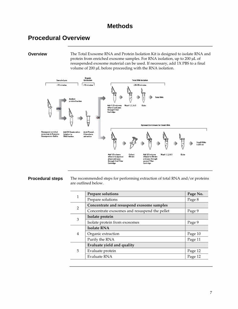
Procedural Overview
Overview The Total Exosome RNA and Protein Isolation Kit is designed to isolate RNA and
protein from enriched exosome samples. For RNA isolation, up to 200 µL of resuspended exosome material can be used. If necessary, add 1X PBS to a final volume of 200 µL before proceeding with the RNA isolation.
Procedural steps The recommended steps for performing extraction of total RNA and/or proteins
are outlined below.
1
Prepare solutions Page No. Prepare solutions Page 8
2 Concentrate and resuspend exosome samples Concentrate exosomes and resuspend the pellet Page 9
3 Isolate protein Isolate protein from exosomes Page 9
4 Isolate RNA Organic extraction Page 10 Purify the RNA Page 11
5 Evaluate yield and quality Evaluate protein Page 12 Evaluate RNA Page 12
7

Procedure
Additional required materials
• 2-mercaptoethanol (14.3 M)
• 100% ethanol, ACS grade or better
• Phosphate buffered saline (PBS)
• Microcentrifuge capable of at least 10,000 × g
• Heat block set to 95°C to 100°C
• (optional) vacuum manifold: to pull solutions through the Filter Cartridges
• RNase-free 1.5 mL or 2.0 mL polypropylene microfuge tubes, adjustable pipettors and RNase-free tips
• Gloves
Before starting Before working with RNA, clean the lab bench, and pipettors with an RNase decontamination solution (e.g., Ambion® RNaseZap®).
Wear laboratory gloves at all times and change them frequently. They will protect you from the reagents, and they will protect the RNA from nucleases that are present on your skin. Use RNase-free pipette tips to handle the kit reagents.
Prepare solutions 2X Denaturing Solution
Add 375 μL of 2-mercaptoethanol to the 2X Denaturing Solution. Mix well. Place a check mark in the box on the label to indicate that the 2-mercaptoethanol has been added.
Note: The 2X Denaturing Solution may solidify at 2°C to 8°C. Before use, warm the solution at 37°C with occasional agitation for 5–10 minutes, or until it is completely in solution. To avoid solidification, you may store the 2X Denaturing Solution at room temp for up to 1 month if desired.
miRNA Wash Solution 1
Add 21 mL of ACS grade 100% ethanol to the miRNA Wash Solution 1. Mix well. Place a check mark in the box on the label to indicate that the ethanol has been added.
Wash Solution 2/3
Add 40 mL of ACS grade 100% ethanol to the Wash Solution 2/3. Mix well. Place a check mark in the box on the label to indicate that the ethanol has been added.
Note: A precipitate may form in the Wash Solution 2/3 bottle as excess EDTA precipitates out of solution. Simply leave these crystals in the bottle when removing Wash Solution 2/3 for use.
Continued on next page
8

Procedure, Continued
Concentrate exosomes and resuspend pellet
After pelleting exosomes by precipitation (using Total Exosome Isolation reagents), ultracentrifugation, or other procedure, resuspend the exosome pellet according to the following directions:
1. Resuspend the exosome pellet with ice cold Exosome Resuspension Buffer or 1X PBS.
Note: Always use the Exosome Resuspension Buffer if protein analysis is required. Resuspension volume depends on the exosome source, and desired volume and sample concentration, and can range from 25 μL to 1 mL or higher.
2. Incubate the sample for 5–10 minutes at room temperature to allow the pellet to dissolve.
3. Gently pipet up and down to thoroughly resuspend the sample.
4. Proceed to protein and/or RNA isolation.
• If both RNA and protein isolation is desired, split the sample into two fractions and proceed immediately to Isolate RNA – Organic extraction (page 10), followed by Isolate protein (page 9).
• If only protein isolation is desired, proceed immediately to Isolate protein (page 9).
• If only RNA isolation is desired, proceed immediately to Isolate RNA – Organic extraction (page 10).
Important: If you are not immediately proceeding to isolate protein after resuspending the sample in the Exosome Resuspension Buffer, store it for up to 5–10 minutes on ice, or at –20°C for longer term.
If you are not immediately proceeding to isolate RNA after resuspending the sample in the Exosome Resuspension Buffer, store it at –20°C until needed.
Isolate protein After the exosome sample is resuspended, it can be used directly for protein
analysis applications.
Use the resuspended exosome sample for protein analysis immediately, or place the sample on ice for 5–10 minutes, or store it at –20°C until ready for use.
See Appendix C (page 29) for details on performing SDS-PAGE and western blotting.
Continued on next page
9

Procedure, Continued
Isolate RNA –Organic extraction
1. Add 1X PBS to the exosome sample in an RNase-free tube so that the total volume is 200 µL.
Note: For some exosome samples isolated from cell culture media the volume may be greater than 200 μL. In such cases, take 200 μL for processing, or process the entire volume. If processing >200 μL, keep the 2X Denaturing Solution and the Acid-Phenol:Chloroform proportionate to the starting sample volume.
2. Add one volume of 2X Denaturing Solution and mix thoroughly.
Important: Pre-warm the 2X Denaturing Solution at 37°C to dissolve precipitate if necessary.
3. Incubate the mixture on ice for 5 minutes.
4. Add one volume of Acid-Phenol:Chloroform to each sample.
The volume of Acid-Phenol:Chloroform should be equal to the total volume of the sample plus the 2X Denaturing Solution (e.g., if the initial sample lysate volume was 200 μL and it was mixed with 200 μL of 2X Denaturing Solution in step 1, add 400 μL Acid-Phenol:Chloroform).
Important: Be sure to withdraw the bottom phase containing Acid-Phenol:Chloroform, not the aqueous buffer that lies on top of the mixture.
5. Mix samples by vortexing for 30–60 seconds.
6. Centrifuge for 5 minutes at maximum speed (≥10,000 x g) at room temperature to separate the mixture into aqueous and organic phases. Repeat the centrifugation if the interphase is not compact.
7. Carefully remove the aqueous (upper) phase without disturbing the lower phase or the interphase, and transfer it to a fresh tube. Note the volume recovered.
8. Proceed to Isolate RNA – Purify the RNA (page 11)
Continued on next page
10

Procedure, Continued
Isolate RNA –Purify the RNA
Final RNA isolation is performed on the aqueous phase from acid-phenol: chloroform extraction. Before starting, be sure to:
• Preheat Elution Solution or nuclease-free water to 95°C for use in eluting the RNA from the filter at the end of the procedure.
Note: Nuclease-free water can be used in place of the elution buffer, especially if concentrating the RNA with a centrifugal vacuum concentrator.
• 100% ethanol must be at room temperature. If the 100% ethanol is stored cold, warm it to room temperature before starting the RNA isolation.
Note: To isolate RNA that is enriched for small RNA species (<200 bases) perform the procedure described in Appendix A: Enriching for Small RNAs (page 15).
Bind the RNA 1. Add 1.25 volumes 100% ethanol to the aqueous phase, and mix thoroughly
(e.g., if 300 μL was recovered, add 375 μL ethanol).
2. For each sample, place a Filter Cartridge into one of the Collection Tubes (supplied in kit).
3. Pipet 700 μL of the lysate/ethanol mixture onto the Filter Cartridge. For sample volumes >700 μL, apply the mixture in successive applications to the same filter.
4. Centrifuge at 10,000 × g for ~15 seconds, or until the mixture has passed through the filter. Alternatively, use a vacuum to pull the samples through the filter.
5. Discard the flow-through, and repeat until all of the lysate/ethanol mixture has been passed through the filter. Save the Collection Tube for the washing steps.
Wash the RNA 6. Add 700 μL miRNA Wash Solution 1 (working solution mixed with ethanol) to the Filter Cartridge.
7. Centrifuge at 10,000 × g for ~15 seconds or use a vacuum to pull the solution through the filter. Discard the flow-through from the Collection Tube, and replace the Filter Cartridge into the same Collection Tube.
8. Apply 500 μL Wash Solution 2/3 (working solution mixed with ethanol) and draw it through the filter as in the previous step.
9. Repeat with a second 500 μL of Wash Solution 2/3.
10. After discarding the flow-through from the last wash, replace the Filter Cartridge in the same Collection Tube and centrifuge the assembly at 10,000 × g for 1 minute to remove residual fluid from the filter.
Continued on next page
11

Procedure, Continued
Elute the RNA 11. Transfer the Filter Cartridge into a fresh Collection Tube (supplied in kit).
12. Apply 50 μL of preheated (95°C) Elution Solution or nuclease-free water to the center of the filter.
13. Centrifuge for ~30 seconds to recover the RNA.
14. Repeat the elution once more with an addition alquot of 50 μL of Elution Solution or nuclease-free water.
15. Collect the eluate (which contains the RNA) and place on ice for immediate use, or store it at ≤ –20°C.
Evaluate yield and quality of protein
Storage of protein samples
Protein samples can be stored at –20°C for up to 1 week. For long term storage, or if the protein is to be used in functional assays, we recommend freezing the samples in liquid nitrogen and storing at ≤ –70°C. The concentration of protein samples can be assessed using any standard protein quantitation procedure, including the BCA method, the Bradford assay, the Lowry assay or other commercially available protein quantitation assays.
Protein quality and downstream applications
• Total protein samples can be used directly for applications such as Western blotting. For use in applications such as enzymatic assays, gel shift assays, and two-dimensional gel electrophoresis, it may be necessary to further purify protein samples.
• If salts and/or detergents present in the Exosome Resuspension Buffer interfere with your experiments, they can be removed by dialysis or by protein purification on gel-filtration columns.
Evaluate yield and quality of RNA
• Concentration of RNA from exosome samples is extremely low and is often not detectable when measured using standard spectrophotometers or fluorometers. Instead, the concentration of an RNA solution can be determined by using the Agilent® 2100 Bioanalyzer® with the RNA 6000 Pico Kit, or the Small RNA Kit.
• Total RNA yield from exosomes can vary widely according to the type and amount of starting sample.
12

Troubleshooting
Review the information below to troubleshoot your experiments when performing sample isolation with this kit.
Observation Cause Solution
Poor RNA quality Long RNA (mRNA) contained in exosomes is often highly degraded compared to RNA isolated from tissue or liquid samples. Sizes can typically range from 15–500 nt
Refer to the Ambion® Technical Reference Library at www.lifetechnologies.com/support for comprehensive information on maximizing yield and quality when performing RNA isolation.
RNA degradation from incorrect storage in Exosome Resuspension Buffer
Follow correct storage conditions. Store at –20°C until needed, or isolate RNA immediately after resuspending the sample in the Exosome Resuspension Buffer.
RNA contaminated with RNase Follow the guidelines on page 8 to prevent RNase contamination.
Poor quality starting samples Always use fresh samples or, if appropriate, samples frozen at –80°C and thawed using best practice.
Poor RNA yield RNA degradation from incorrect storage in Exosome Resuspension Buffer
Follow correct storage conditions. Store at –20°C until needed, or isolate RNA immediately after resuspending the sample in the Exosome Resuspension Buffer.
Poor quality starting sample Always use fresh samples or samples frozen at –80°C. For lysis, process the sample quickly to avoid degradation.
Starting sample contains less exosomes than expected
Expected yields of exosome can vary widely between sample types. For example, higher yields of exosomes are often recovered from cells cultured with depleted fetal bovine serum (FBS) than cells grown without FBS.
RNA contains excess DNA
During the organic extraction, the aqueous phase was contaminated by aspirating some of the interphase or the lower phase
Contaminating DNA can be removed by DNase digestion, with subsequent removal of DNase and buffer from the sample. We recommend TURBO DNA-free™
Kit for this type of application.
13

Troubleshooting, Continued
Observation Cause Solution
High Ct values in real-time RT-PCR
RNA degradation from incorrect storage in Exosome Resuspension Buffer
Follow correct storage conditions. Store at –20°C until needed, or isolate RNA immediately after resuspending the sample in the Exosome Resuspension Buffer.
Serum or cells contain less RNA than expected
Expected yields of RNA may vary widely between sample types. Specific miRNAs also have a characteristic tissue distribution, and some tissues are devoid of detectable levels of miRNAs.
No protein activity or poor protein activity
Improper handling of sample It is important to keep samples cold during the procedure. Keep samples on ice between each step and centrifuge at 2°C to 8°C when required. Also, complete the procedure as quickly as possible to get the highest quality protein (and RNA)
Follow correct storage conditions. Store at –20°C until needed, or isolate RNA immediately after resuspending the sample in the Exosome Resuspension Buffer.
Protein degradation or modification
If required, protease inhibitors, phosphatase inhibitors, and anti-oxidants can be added to the Exosome Resuspension Buffer just before use. These reagents will not adversely affect subsequent RNA isolation from samples.
14

Appendix A: Enriching for Small RNAs
Enrichment Procedure
Overview This variation of traditional glass-fiber filter RNA purification yields RNA that is significantly enriched for small RNAs. This enrichment is accomplished by first immobilizing large RNAs on the filter with a relatively low ethanol concentration and collecting the flow-through containing mostly small RNA species. More ethanol is then added to this flow-through, and the mixture is passed through a second glass filter where the small RNAs are immobilized. The filter is then washed a few times, and the small-RNA enriched fraction is eluted.
Bind the large RNA 1. Complete the Organic extraction procedure (page 10), and transfer the aqueous phase to a new tube.
2. Add 1∕3 volume of 100% ethanol to the aqueous phase, and mix thoroughly (e.g., if 300 μL is recovered, add 100 μL 100% ethanol).
3. For each sample, place a Filter Cartridge into a Collection Tube (supplied in kit).
4. Add up to 700 μL of the lysate/ethanol mixture onto the Filter Cartridge at a time. For volumes >700 μL, add any remaining sample to the same Filter Cartridge in successive steps.
5. Centrifuge for ~30 seconds or until the mixture has passed through the filter. Alternatively, use a vacuum to pull samples through the filter.
6. Collect the flow-through (containing the small RNA). If the sample volume was >700 μL, transfer the flow-through to a fresh tube, and repeat steps 4–6 until all of the sample has been passed through the Filter Cartridge. Pool the collected flow-through for samples >700 μL, and measure the total volume.
Note: At this point, the filter contains an RNA fraction depleted of small RNA, while the flow-through contains the small RNA.
Continued on next page
15

Enrichment Procedure, Continued
Bind the small RNA 7. Add 2∕3 volume 100% ethanol to the flow-through containing the small RNA, and mix thoroughly (e.g., if 400 μL of flow-through is recovered, add 266 μL 100% ethanol).
8. Place a new Filter Cartridge into a Collection Tube (supplied in kit).
9. Add up to 700 μL of the flow-through/ethanol mixture containing the small RNA onto the second Filter Cartridge at a time. For volumes >700 μL, add any remaining sample to the same Filter Cartridge in successive steps.
10. Centrifuge for ~30 seconds or until the mixture has passed through the filter, and discard the flow-through. Alternatively, use a vacuum to pull samples through the filter.
11. If the sample volume was >700 μL, repeat steps 9–10 until all of the sample has been passed through the Filter Cartridge. Reuse the Collection Tube for the washing steps.
Wash the small RNA
12. Add 700 μL of miRNA Wash Solution 1 (containing ethanol) to the Filter Cartridge.
13. Centrifuge for ~15 seconds, or use a vacuum to pull the solution through the filter. Discard the flow-through from the Collection Tube, and replace the Filter Cartridge into the same Collection Tube.
14. Apply 500 μL Wash Solution 2/3 (containing ethanol).
15. Centrifuge for ~15 seconds, or use a vacuum to pull the solution through the filter. Discard the flow-through from the Collection Tube, and replace the Filter Cartridge into the same Collection Tube.
16. Repeat steps 14–15 once.
17. After discarding the flow-through from the final wash, replace the Filter Cartridge in the same Collection Tube and spin the assembly for 1 minute to remove residual fluid from the filter.
Elute the small RNA
18. Transfer the Filter Cartridge into a fresh Collection Tube (supplied in kit).
19. Add 50 μL of preheated (95°C) Elution Solution, or nuclease-free water to the center of the filter.
20. Centrifuge for ~30 seconds to recover the RNA.
21. Repeat the step 19–20 once.
22. Collect the eluate (which contains the RNA) and store at ≤ –20°C.
Continued on next page
16

Appendix B: Preparing RNA for Sequencing
Procedural Overview
Step (Optional) Fragment RNA Page No.
1 Fragment RNA Page 18
2 Purify fragmented RNA Page 19
Prepare small RNA library
3 Hybridize and ligate RNA Page 21
4 Perform reverse transcription Page 22
5 Purify cDNA Page 23
6 Amplify cDNA Page 25
7 Purify amplified cDNA Page 26
Perform sequencing on the Ion PGM™ System
8 Assess yield and size distribution of amplified cDNA Page 28
9 Set up the Ion PGM™ System for RNA libraries Page 28
General guidelines For sequencing the RNA content of exosomes derived from various sources, we recommend to extract exosomes from:
• At least 200 μL of serum isolated using the Total Exosome Isolation (from serum) reagent.
or
• At least 5 mL of cell media isolated using the Total Exosome Isolation (from cell culture media) reagent.
For exosomes isolated by ultracentrifugation protocols (with or without sucrose gradient), we recommend increasing the starting volume by 10-fold, due to very low recovery when using this method.
After the exosomal RNA is isolated using Total Exosome RNA and Protein Isolation Kit, or other appropriate kit, the small RNA libraries can be conveniently prepared with the Ion Total RNA-Seq Kit v2 and sequencing performed on Ion Torrent™ PGM™.
The protocol below is based on the Ion Total RNA-Seq Kit v2 with a number of modifications introduced due to specifics of the exosome samples:
1. Relatively low amount of RNA 2. Majority of the RNA cargo is short < 200 nt
Note: This protocol is just a guide, and further modifications may be required, depending on the objectives of your study, the origin of the exosomes, the concentration and purity of RNA, the availability of certain kits/reagents, and other factors. For additional details on small RNA library construction, refer to the Ion Total RNA-Seq Kit v2 User Guide at www.lifetechnologies.com.
Continued on next page
17
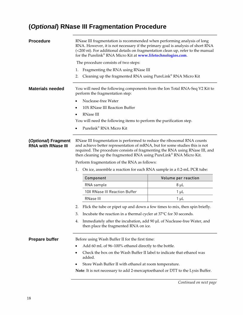
(Optional) RNase III Fragmentation Procedure
Procedure RNase III fragmentation is recommended when performing analysis of long RNA. However, it is not necessary if the primary goal is analysis of short RNA (<200 nt). For additional details on fragmentation clean up, refer to the manual for the Purelink® RNA Micro Kit at www.lifetechnologies.com.
The procedure consists of two steps:
1. Fragmenting the RNA using RNase III
2. Cleaning up the fragmented RNA using PureLink® RNA Micro Kit
Materials needed You will need the following components from the Ion Total RNA-Seq V2 Kit to perform the fragmentation step:
• Nuclease-free Water
• 10X RNase III Reaction Buffer
• RNase III
You will need the following items to perform the purification step.
• Purelink® RNA Micro Kit
(Optional) Fragment RNA with RNase III
RNase III fragmentation is performed to reduce the ribosomal RNA counts and achieve better representation of mRNA, but for some studies this is not required. The procedure consists of fragmenting the RNA using RNase III, and then cleaning up the fragmented RNA using PureLink® RNA Micro Kit.
Perform fragmentation of the RNA as follows:
1. On ice, assemble a reaction for each RNA sample in a 0.2-mL PCR tube:
Component Volume per reaction
RNA sample 8 μL
10X RNase III Reaction Buffer 1 μL
RNase III 1 μL
2. Flick the tube or pipet up and down a few times to mix, then spin briefly.
3. Incubate the reaction in a thermal cycler at 37°C for 30 seconds.
4. Immediately after the incubation, add 90 μL of Nuclease-free Water, and then place the fragmented RNA on ice.
Prepare buffer Before using Wash Buffer II for the first time:
• Add 60 mL of 96–100% ethanol directly to the bottle.
• Check the box on the Wash Buffer II label to indicate that ethanol was added.
• Store Wash Buffer II with ethanol at room temperature.
Note: It is not necessary to add 2-mercaptoethanol or DTT to the Lysis Buffer.
Continued on next page
18

RNase III Fragmentation Procedure, Continued
Clean up the fragmented RNA
1. Transfer the 100 μL fragmented RNA digestion mix to a 1.5 mL tube. Add 100 μL of Lysis Buffer and 400 μL of 100% ethanol, then mix well.
2. Obtain a PureLink® RNA Micro Kit Spin Column with a collection tube, then verify that the column has a red o-ring. Note: The red o-ring differentiates the PureLink® RNA Micro Kit Spin Column from the unmarked PureLink® PCR Micro Kit Spin Column.
Bind the RNA 3. Load 650 μL of the RNA sample containing Lysis Buffer and ethanol onto
the Spin Column.
4. Spin the column at 12,000 × g for 15 seconds.
5. Discard the flow through.
6. Return the Spin Column to the Collection Tube.
Wash the RNA 7. Add 500 μL of Wash Buffer II (W5) with ethanol to the Spin Column.
8. Spin the column at 12,000 × g for 15 seconds.
9. Discard the flow through.
10. Return the Spin Column to the Collection Tube.
11. Add another 500 μL of Wash Buffer II (W5) with ethanol to the Spin Column.
12. Spin the column at 12,000 × g for 15 seconds.
13. Discard the flow through.
14. Return the Spin Column to the Collection tube.
15. Using a pipette, carefully remove any buffer adhering to the top of the o-ring inside of the column.
16. Spin the column at 12,000 × g for 1 minute.
Elute the RNA 17. Place the Spin Column in a clean Recovery Tube.
18. Add 12 μL of Nuclease-free Water to the center of the Spin Column.
19. Wait 1 minute.
20. Spin the column at 12,000 × g for 1 minute.
Note: You should recover approximately 10 μL of fragmented low input RNA
21. In a speed-vac dry sample to 3 μL.
19

Small RNA Library Construction Procedure
Procedure Constructing the small RNA library includes the following:
1. Hybridize and ligate the small RNA
2. Perform reverse transcription
3. Purify cDNA using MagMAX™ beads
4. Amplify the cDNA
5. Purify the amplified DNA using MagMAX™ beads
6. Assess the yield and size distribution of the amplified DNA
Materials needed You will need the following components from the Ion Total RNA-Seq V2 Kit to perform the hybridization and ligation step:
• Hybridization Solution
• Nuclease-free Water
• 2X Ligation Buffer
• Ligation Enzyme Mix
• Ion Adaptor Mix v2
You will need the following components from the Ion Total RNA-Seq V2 Kit to perform the reverse transcription step.
• Nuclease-free Water
• dNTP Mix
• Ion RT Primer v2
• 10X SuperScript® III Enzyme Mix
• 10X RT buffer
Continued on next page
20

Small RNA Library Construction Procedure, Continued
Hybridize and ligate the small RNA
1. On ice, prepare the hybridization master mix:
Component Volume per reaction
Hybridization Solution 3 μL
Ion Adaptor Mix v2 2 μL
Total volume 5 μL
2. Transfer 5 μL hybridization master mix to 3 μL RNA sample.
3. Slowly pipet up and down a few times to mix well, then spin briefly.
4. Run the hybridization reaction in a thermal cycler:
Temperature Time
65ºC 10 minutes
16ºC 5 minutes
5. On ice, prepare the ligation master mix
Component Volume per reaction
2X Ligation Buffer 10 μL
Ligation Enzyme Mix 2 μL
Important! You may observe white precipitate in the 2X Ligation Buffer. If so, warm the tube at 37 °C for 2–5 minutes or until the precipitate is dissolved. 2X Ligation Buffer is very viscous; pipette slowly to dispense it accurately.
6. Slowly pipet up and down a few times to mix well, then spin briefly.
7. Transfer 12 μL ligation master mix to each of the 8 μL hybridization reactions.
8. Incubate the 20 μL ligation reaction in a thermal cycler at 16°C for 16 hours (overnight).
Note: If possible, set the temperature of the thermal cycler lid to match the block temperature. Otherwise, incubate the reaction with the heated lid turned off, or do not cover the reaction tubes with the heated lid.
Continued on next page
21

Small RNA Library Construction Procedure, Continued
Perform reverse transcription
1. Prepare RT master mix:
Component Volume per reaction
Nuclease-free water 2 μL
10X RT Buffer 4 μL
dNTP Mix 2 μL
Ion RT Primer 8 μL
Total volume 16 μL
2. Add 16 μL of RT master mix to each 20 μL ligation reaction.
3. Pipet up and down a few times to mix, then spin briefly.
4. Incubate in a thermal cycler with a heated lid at 70°C for 10 minutes, then snap-cool on ice.
5. Add 4 μL 10X SuperScript® Enzyme Mix to each ligated RNA sample.
6. Gently vortex to mix thoroughly, then spin briefly.
7. Incubate in a thermal cycler with a heated lid at 42°C for 30 minutes.
Note: The cDNA can be stored at –20°C for a few weeks, stored at –80°C for long-term storage, or used immediately.
Continued on next page
22

Small RNA Library Construction Procedure, Continued
Purify cDNA using MagMAX™ beads
Use the Magnetic Bead Clean-Up Module to clean up the cDNA products. After washing the beads, the desired cDNA products are eluted with pre-warmed (37°C) Nuclease-Free Water.
Required materials from the Magnetic Bead Clean-Up Module
• Wash Solution Concentrate
• Binding Solution Concentrate
• Nucleic Acid Binding Beads
• Processing Plate
• Nuclease-Free Water
Other reagents/Equipment needed:
• 100% ethanol, ACS-grade or higher quality
• Magnetic stand for 96-well plates
• 37°C heat block or water bath
• (Optional) MicroAmp® Clear Adhesive Film
Before starting
• Add 44 mL of 100% ethanol to bottle of the Wash Solution Concentrate and mix well. Mark the label on the bottle to indicate that you added ethanol. Store the solution at ambient temperature (15°C to 30°C).
• Incubate the Nuclease-Free Water at 37°C for ≥5 minutes.
NOTE: To reduce the chance of cross-contamination, we strongly recommend sealing unused wells on the Processing Plate with MicroAmp® Clear Adhesive Film. Alternatively, you can skip a row between sample rows.
IMPORTANT! Accurate Pipetting of bead cleanup reagents is critical for the success of size selection. Please follow the instruction exactly to minimize pipetting variations.
Continued on next page
23

Small RNA Library Construction Procedure, Continued
Prepare MagMAX™ beads
1. Prepare beads for each sample
2. Gently vortex the Nucleic Acid Binding Beads tube to resuspend the magnetic beads.
3. Add 5 µL beads to wells on a 96-well plate.
4. Add 250 µL Binding Solution Concentrate to each well, and mix by pipeting up and down 10 times.
Bind cDNA to MagMAX™ beads
5. Add 60 µL of nuclease-free water to each of the 40 µL RT reaction.
6. Transfer 100 µL RT reaction to the beads on 96-well plate.
7. Add 275 µL 100% ethanol to each well.
8. Mix thoroughly by pipette mixing 10 times. Note: The color of the mixture should appear homogenous after mixing.
9. Incubate samples for 5 minutes at room temperature off the magnetic stand.
10. Once complete, let samples collect on magnetic stand for 2–5 minutes or until the supernatant is clear.
11. Carefully aspirate and discard the supernatant without disturbing the beads. Leave the plate on the magnetic stand.
IMPORTANT! Do not disturb the beads. If the beads are drawn out during aspiration, leave a few microliters of supernatant behind.
Wash MagMAX™ beads
12. Add 150 µL Wash Buffer Concentrate to each well while on the magnetic stand.
13. Incubate samples for 30 seconds.
14. Aspirate and discard the supernatant.
15. Air dry the beads at room temperature for 2–3 minutes to remove all traces of ethanol. (Note: Do not over dry the beads. Over dried beads appear cracked.)
16. Remove the plate from the magnetic stand.
Elute cDNA from MagMAX™ beads
17. Add 12 µL pre-warmed nuclease-free water (at 37°C) to each well. Resuspend the beads thoroughly by pipette mixing 10 times.
18. Incubate for 1 minute at room temperature.
19. Place 1.2 mL 96-well plate back on magnetic stand for 1 minute to separate the beads from solution.
20. Collect the 12 µL eluant.
Continued on next page
24

Small RNA Library Construction Procedure, Continued
Amplify the cDNA Use components from the Ion Total RNA-Seq Kit v2:
• Ion 3’ PCR Primer v2
• Ion 5’ PCR primer v2
• Platinum® PCR SuperMix High Fidelity
1. For each cDNA sample, prepare 47 μL PCR master mix:
Component Volume per reaction
Platinum® PCR SuperMix High Fidelity
45 µL
Ion 3’ PCR Primer v2 1 µL
Ion 5' PCR Primer v2 1 µL
Total volume 47 µL
2. Transfer 6 μL of cDNA to a new PCR tube.
3. Transfer 47 μL PCR master mix into each 6 µL cDNA sample.
4. Run the PCR reactions in a thermal cycler:
Stage Temp Time
Hold 94ºC 2 min
Cycle (2 cycles)
94ºC 30 sec
50ºC 30 sec
68ºC 30 sec
Cycle (16 cycles)
94ºC 30 sec
62ºC 30 sec
68ºC 30 sec
Hold 68ºC 5 min
Continued on next page
25

Small RNA Library Construction Procedure, Continued
Purify the amplified DNA
Required materials from the Magnetic Bead Clean-Up Module
• Wash Solution Concentrate
• Binding Solution Concentrate
• Nucleic Acid Binding Beads
• Processing Plate
• Nuclease-Free Water
Other reagents/Equipment needed:
• 100% ethanol, ACS-grade or higher quality
• Magnetic stand for 96-well plates
• 37°C heat block or water bath
• (Optional) MicroAmp® Clear Adhesive Film
Before you begin
• Add 44 mL of 100% ethanol to bottle of the Wash Solution Concentrate and mix well. Mark the label on the bottle to indicate that you added ethanol. Store the solution at ambient temperature (15°C to 30°C).
• Incubate the Nuclease-Free Water at 37°C for ≥5 minutes.
NOTE: To reduce the chance of cross-contamination, we strongly recommend sealing unused wells on the Processing Plate with MicroAmp® Clear Adhesive Film. Alternatively, you can skip a row between sample rows.
IMPORTANT! Accurate Pipetting of bead cleanup reagents is critical for the success of size selection. Please follow the instruction exactly to minimize pipetting variations.
26
Small RNA Library Construction Procedure, Continued
Prepare beads 1. Gently vortex the Nucleic Acid Binding Beads tube to resuspend the
magnetic beads.
2. Add 5 µL beads to wells on a 96-well plate that you plan to use.
3. Add 280 µL Binding Solution Concentrate to the beads and mix by pipetting up and down 10 times.
Bind amplified cDNA to beads
4. Add 27 µL of nuclease-free water to each of the 53 µL PCR reaction.
5. Transfer 80 µL PCR reaction to the beads on 96-well plate.
6. Add 230 µL 100% ethanol to each well.
7. Mix thoroughly by pipette mixing 10 times . Note: The color of the mixture should appear homogenous after mixing.
8. Incubate samples for 5 minutes at room temperature off the magnet.
9. Once complete, let samples collect on magnet for 2–5 minutes or until the supernatant is clear.
10. Carefully aspirate and discard the supernatant without disturbing the beads, and leave the plate on the magnetic stand.
Important! Do not disturb the beads. If the beads are drawn out during aspiration, leave a few microliters of supernatant behind.
Wash beads 11. Add 150 µL Wash Buffer Concentrate to each well while on magnetic stand.
12. Incubate samples for 30 seconds.
13. Aspirate and discard the supernatant.
14. Air dry the beads at room temperature for 2–3 minutes to remove all traces of ethanol. (Note: Do not over dry the beads. Over dried beads appear cracked.)
15. Remove the plate from the Magnetic Stand.
Elute cDNA 16. Add 10 µL pre-warmed nuclease-free water (at 37°C) to each well.
17. Incubate for 1 minute at room temperature.
18. Place 1.2 mL 96-well plate back on Magnetic Stand-96 for 1 minute to separate the beads from solution.
19. Collect the 10 µL eluant.
Continued on next page
27

Perform Sequencing on the Ion PGM™ System
Assess cDNA Assess the yield and size distribution of the amplified DNA
1. Run 1 µL of the sample on Agilent® DNA High Sensitivity chip to assess the yield and size distribution.
2. Determine the molar concentration of the library with the Agilent® 2100 Bioanalyzer® Instrument Expert software. Perform a smear analysis by following the manufacturer’s instructions. In the Bioanalyzer® Instrument Expert software select: View > Setpoints > Smear Analysis.
For additional details on analyzing the cDNA and sequencing, refer to the Preparation and Sequencing of RNA Libraries with the Ion Personal Genome Machine® (PGM™) System user bulletin available from the Ion Community at www.lifetechnologies.com/support.
Set up Ion PGM™ System
1. Enter these settings on the Run Info screen fo the Ion PGM™ sequencer.
If the touchscreen prompt says… Select…
Runtype GENS
Library Appropriate library reference
Barcode • Non-barcoded libraries: RNA_Barcode_None
• Barcoded libraries prepared with the Ion Express™ RNA-Seq Barcode 01-16 Kit: IonXpressRNA
Number of flows Small RNA libraries: 160 flows (40 cycles)
Note: To change cycling conditions for different read lengths, enter a different number of flows.
Important! For all libraries prepared with the Ion Total RNA-seq Kit v2, you must select an option from the barcode pull-down menu to ensure proper trimming of the sequences prior to alignment with a reference. If the RNA-seq library does not have barcodes, select RNA_Barcode_None in the barcode dialog box. If the library was prepared with the Ion Express™ RNA-Seq Barcode 01-16 Kit, select IonXpressRNA.
2. Enter the remaining information as needed.
3. Click Next, then follow the remaining prompts to start the run.
28

Appendix C: Western Blotting
Western Blotting Procedure
Perform electrophoresis
1. Mix 5 µL of the exosome sample (isolated from cell culture media or serum) with an equal volume of Novex® Tris-Glycine SDS Sample Buffer.
2. Heat the sample at 75°C for 5 minutes.
3. Load the sample on a Novex® 4-20% Tris-Glycine Gel.
4. Load 5 µL of Benchmark™ Prestained Protein Ladder to the gel.
5. Run the gel at 150V, for 1.5 hours.
Perform detection 1. Transfer the proteins from the gel to a nitrocellulose or PVDF membrane using your method of choice (wet blotting, semi-wet blotting, etc.). For the most rapid results, we recommend using theiBlot® Gel Transfer Device.
2. Wash and block the membranes.
3. Incubate the membranes with an antibody against an exosomal marker (e.g., CD63) at the appropriate concentration suggested by the manufacturer.
4. Use Westernbreeze® Chemiluminescence kit to detect the antibody.
5. Expose the membrane to X-ray film for 1–10 minutes.
29

Appendix D: Isolation of RNA and Protein from Cells or Serum Samples
Introduction
Overview For certain types of experiments, comparison of the exosomal RNA and protein content to the “parental” cells and blood serum samples is required. The Total Exosome RNA and Protein Isolation Kit is suitable for isolation of RNA from both cells and serum samples, and protein from the cells.
Serum sample comparison
Cell culture media sample comparison
Continued on next page
30

Isolation of RNA and Protein from Cells
Introduction This procedure is set up to prepare an amount of lysate that can be processed
on a single Filter Cartridge. Using more than the recommended amount of material on a single Filter Cartridge can result in cellular debris clogging the filter.
Prepare cell sample
Cultured cells should be processed fresh (i.e. not frozen). If you need to store the cells before RNA isolation, it is recommended that they be stored in RNAlater® Solution, or pelleted and snap-frozen in liquid nitrogen, and stored at below –70°C.
Suspension cells:
1. Centrifuge a volume of cell culture media containing 102–107 cells at low speed (appropriate for your cells).
2. Discard the culture medium, and resuspend the cells in ~1 mL PBS.
3. Centrifuge the cells at low speed, and discard the supernatant.
4. Place the cell pellet on ice.
Adherent cells:
1. Aspirate and discard the culture medium.
2. Rinse the culture plate with PBS.
3. Place the culture plate on ice.
OR
1. Trypsinize adherent cells, and perform a cell count.
2. Inactivate the trypsin, and centrifuge a volume containing 102–107 cells.
3. Discard the culture medium, and resuspend the cells in ~1 mL PBS.
4. Centrifuge the cells at low speed, and discard the supernatant.
5. Place the cell pellet on ice.
31

Isolation of RNA and Protein from Cells, Continued
Prepare cell lysate
Disrupt the cells with Exosome Resuspension Buffer. If your sample is near the suggested maximum (107 cells), then use near the maximum recommended volume of Exosome Resuspension Buffer, and conversely, for smaller amounts of cells, use a lower amount of buffer.
The volume of Exosome Resuspension Buffer can also be adjusted according to the amount/ concentration of lysate wanted for protein analysis.
Protease, phosphatase and/or RNase inhibitors can be added to an aliquot of Exosome Resuspension Buffer immediately before use if required for specific applications.
Large frozen cell pellets (i.e. containing more than about 107 cells may need to be ground to a powder prior to lysis.
If your sample was stored in RNAlater® Solution, make sure you pellet the cells and remove the RNAlater® Solution prior to disrupting the cells.
Sample should be kept cold on ice while performing lysis.
1. Add 100–625 μL of ice-cold Exosome Resuspension Buffer to the cell pellet or culture plate. Use at least 300 μL for ≥106 cells.
2. Scrape adherent cells lysed directly on a culture plate with a rubber spatula, and collect the lysate in a microcentrifuge tube.
3. Vortex or pipet the cells vigorously to achieve complete cell lysis, and to obtain a homogenous lysate.
5. If both RNA and protein isolation is desired, split the sample into two equal aliquots.
For protein isolation protocol, proceed immediately to Protein isolation from cells (page 33).
For RNA isolation protocol, proceed immediately to RNA isolation from cells (page 33).
32
Isolation of RNA and Protein from Cells, Continued
Protein isolation from cells
1. Incubate the cell lysate on ice for 5–10 minutes to ensure complete cell lysis.
2. (Optional): Clarified the sample by centrifugation at top speed in a microcentrifuge for 1–2 minutes at 2°C to 8°C.
3. (Optional): Reduce sample viscosity by sonicating the lysate, or by passing it through a syringe needle several times.
4. Use the protein lysate directly for protein analysis applications.
RNA isolation from cells
Perform RNA isolation at room temperature.
1. If the lysate volume is less than 100 μL, add Exosome Resuspension Buffer to bring the sample to ≥100 μL
2. Immediately add one volume of 2X Denaturing Solution to the cell lysate, and mix thoroughly.
Important: Pre-warm the 2X Denaturing Solution at 37°C to dissolve precipitate if required.
3. Add a volume of Acid-Phenol:Chloroform equal to the total volume of the sample plus the 2X Denaturing Solution.
4. Proceed to Isolate RNA – Organic extraction starting at step 5, page 10.
33
Isolation of RNA and Protein from Serum
Introduction Serum samples are presumed not to contain cells, so any protein is either free
plasma proteins not captured during the clotting process, or whatever protein is freed from microvesicles with the Denaturing Solution. Additionally, any RNA isolated from serum can be considered to be circulating extracellular RNA, which includes exosomal RNA.
Isolate protein If the samples are only going to be analyzed by SDS-PAGE followed by western
blot, add the serum directly to the SDS loading buffer, and load the samples directly onto the gel.
Isolate RNA Perform RNA isolation at room temperature.
1. If the lysate volume is less than 200 μL, add 1X PBS to bring the sample to 200 μL.
2. Immediately add 200 μL of 2X Denaturing Solution to the sample, and mix thoroughly.
Important: Pre-warm the 2X Denaturing Solution at 37°C to dissolve precipitate if necessary.
3. Add a volume of Acid-Phenol:Chloroform equal to the total volume of the sample plus the 2X Denaturing Solution.
4. Proceed to Isolate RNA – Organic extraction starting at step 5, page 10.
34

Appendix E
Accessory Products
Additional Products
Ordering information on a variety of reagents and apparatus available from Life Technologies is provided below. For more information, visit our website at www.lifetechnologies.com or call Technical Support (see page 36).
Product Quantity Catalog no.
Total Exosome Isolation (from serum) 6 mL 4478360
Total Exosome Isolation (from cell culture media) 50 mL 4478359
RNaseZap® Solution 250 mL
6 bottles 4 L
AM9780 AM9782 AM9784
TURBO DNA-free™ Kit 50 reactions AM1907
RNAlater® Solution 100 mL AM7020
PureLink® RNA Micro Kit 50 preps 12183-016
MicroAmp® Clear Adhesive Film 100 films 4306311
Magnetic Stand-96 1 each AM10027
Magnetic-Ring Stand (96 Well) 1 each AM10050
Ion Total RNA-Seq V2 Kit 12 reactions 4475936
iBlot® Gel Transfer Device 1 each IB1001
WesternBreeze® Chemiluminescent Kit–Anti-Goat 1 kit WB7108
WesternBreeze® Chemiluminescent Kit–Anti-Mouse 1 kit WB7104
WesternBreeze® Chemiluminescent Kit–Anti-Rabbit 1 kit WB7106
Novex® Tris-Glycine SDS Sample Buffer (2X) 20 mL LC2676
Novex® 4-20% Tris-Glycine Gel, 1.5 mm, 15 Well 1 box EC60285BOX
Benchmark™ Prestained Protein Ladder 2 × 250 µL 10748-010
Exosome Immunoprecipitation (Protein A) 10610D
Exosome Immunoprecipitation (Protein G ) 10612D
35

Technical Support
Obtaining support For the latest services and support information for all locations, go to www.lifetechnologies.com
At the website, you can:
• Access worldwide telephone and fax numbers to contact Technical Support and Sales facilities
• Search through frequently asked questions (FAQs)
• Submit a question directly to Technical Support ([email protected])
• Search for user documents, SDSs, vector maps and sequences, application notes, formulations, handbooks, certificates of analysis, citations, and other product support documents
• Obtain information about customer training
• Download software updates and patches
Safety Data Sheets (SDS)
Safety Data Sheets (SDSs) are available at www.lifetechnologies.com/support.
Certificate of Analysis
The Certificate of Analysis provides detailed quality control and product qualification information for each product. Certificates of Analysis are available on our website. Go to www.lifetechnologies.com/support and search for the Certificate of Analysis by product lot number, which is printed on the box.
Limited warranty Life Technologies and/or its affiliate(s) warrant their products as set forth in the Life Technologies General Terms and Conditions of Sale found on the Life Technologies web site at http://www.lifetechnologies.com/termsandconditions. If you have any questions, please contact Life Technologies.
36

References
Chomczynski P., and Sacchi N. (1987). Single-step Method of RNA Isolation by Acid Guanidinium Thiocyanate- Phenol-Chloroform Extraction. Analytical Biochem. 162, 156–159.
Valadi H., Ekström K., Bossios A., Sjöstrand M, Lee J.J., Lötvall J.O. (2007). Exosome-mediated transfer of mRNAs and microRNAs is a novel mechanism of genetic exchange between cells. Nat Cell Biol. 9(6):654–9.
Vlassov A. V., Magdaleno S., Setterquist R., Conrad R. (2012). Exosomes: Current knowledge of their composition, biological functions, and diagnostic and therapeutic potentials. Biochim Biophys Acta. 1820 (7): 940–948.
37


For support visit www.lifetechnologies.com/support or email [email protected]
www.lifetechnologies.com
18 August 2014