Visions Going Forward Donald Beagle Belmont Abbey College February 23, 2005.
Upload
jhon-arriaga-cordovaCategory
view
922download
2description
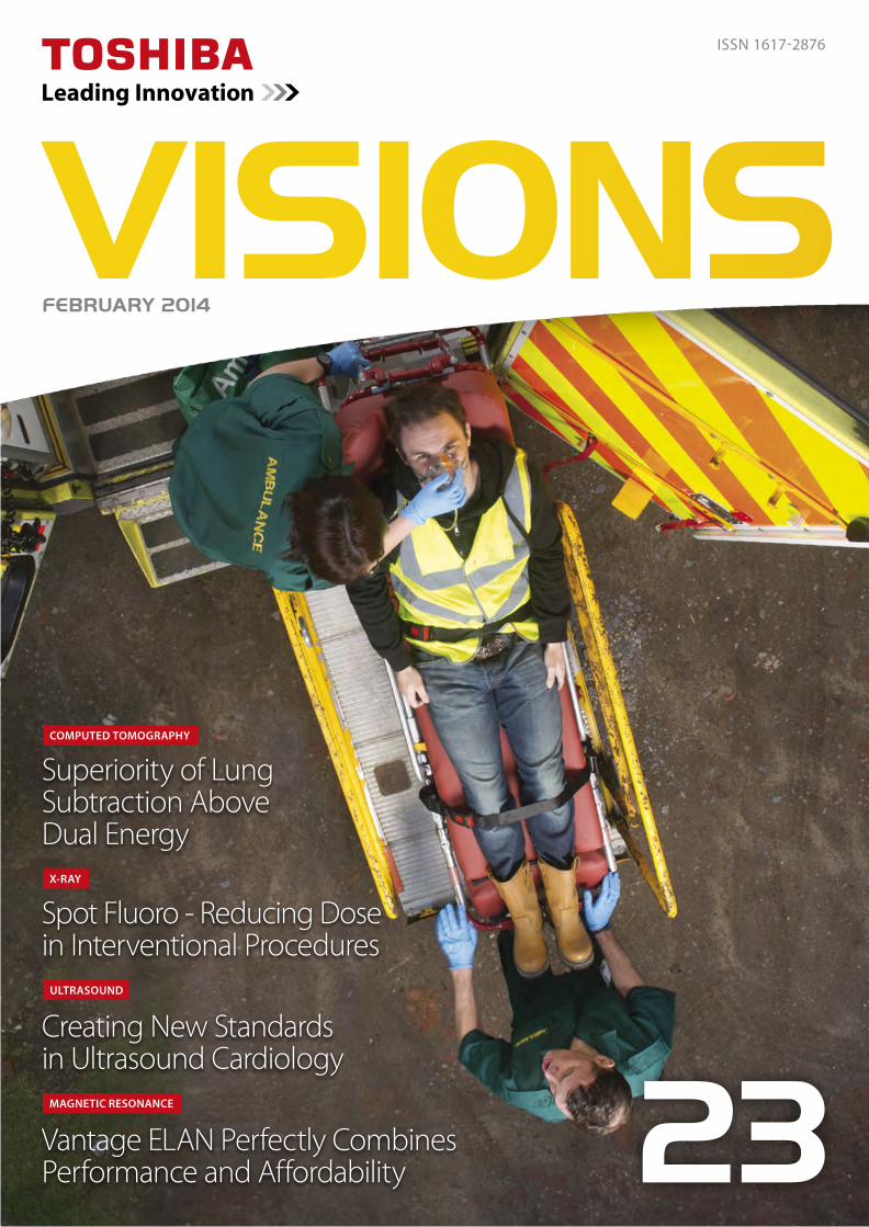
Spot Fluoro - Reducing Dose in Interventional Procedures
Superiority of Lung Subtraction Above Dual Energy
Creating New Standards in Ultrasound Cardiology
Vantage ELAN Perfectly Combines Performance and Affordability 23
X-RAY
COMPUTED TOMOGRAPHY
ULTRASOUND
MAGNETIC RESONANCE
ISSN 1617-2876
VISIONSFEBRUARY 2014
Caspar Berger Caspar Berger - Bone at Beelden aan Zee
Vita Subsidiaria Calva/Self-portrait 31 (excerpt)
Caspar Berger, 2014
Material: Print, argmented reality dimensions: 102 x 72
This artwork is about the question
of whether we can rethink life,
faced with the possibility of science
cracking the code of life, of being
able to make and fix life. If we were
offered the choice, would we dare
to be immortal?
Download the ‘Caspar Berger
Skeleton’ app, point your mobile
device at the artwork and give the
skull a check-up!
About the Bone Exhibition
For his project ‘Skeleton’, award-winning Dutch artist, Caspar Berger, made an exact copy of
his own skeleton using a Toshiba Aquilion CT scanner and 3D-print technology, which he then
incorporated into a wide variety of creative interpretations. The result is a lifelike ‘interior’
portrait of the artist, although none of the individual works features his fully intact skeleton.
For ‘Bone’, Berger explored, for example, the concept of humans in fossil form. The hard
parts of his body were effectively ‘preserved’ in stone objects. The piece entitled “Do Not be
Afraid of Becoming a Bench/Self-Portrait 28” contains casts of all the bones of his skeleton.
While seated on the stone benches, which can be interpreted as symbolizing the possible
role of Man in the history of the World , the viewer can watch the video projection, entitled
‘Attraction/Self-Portrait 30’ which shows a reversed process of creation. In the video, silver-
grey balloons with cast parts of Berger’s skeleton attached descend from a cloudy sky.
Then the sun emerges, encouraging viewers to question if Man’s ‘place’ is actually between
heaven and Earth.
About Caspar Berger
Sculptor Caspar Berger (1965) studied at the AKI, Enschede and at the Jan van Eyck Academy
in Maastricht, the Netherlands. In his work Berger investigates the relationship between
the interior and exterior, between reality and image. Berger gives shape to his ideas using
silicone casts of models and of his own skin or even what’s underneath, leading to its end
result in bronze, silver and sometimes gold; more recently, video installations, concrete and
print. In 2013 he was awarded Singer prijs 2013 and recently he received the Sacha Tanja
Penning 2014.
www.casparberger.nl
AdmissionFree
Museum Beelden aan Zee
Harteveltstraat 1
2586 EL Den Haag
T: +31 70 358 58 57
www.beeldenaanzee.nl
This voucher entitles free admission for one person to Museum Beelden aan Zee.
Valid until June 1st 2014
Hidden in the dunes of Scheveningen, like
a pearl in the sand, you will find museum
Beelden aan Zee, a truly unique museum in the
Netherlands that focuses on modern and con-
temporary international sculpture. Man - the
human image - is the leitmotiv of the collection
of Beelden aan Zee. The collection holds work
from sculptors as: Karel Appel, Atelier van
Lieshout, Fernando Botero, Cesar, Tony Cragg,
Igor Mitoraj, Jan Meefout and Ossip Zadkine.
As from January 31st 2014 the following expo-
sition can be seen at museum Beelden aan Zee:
• Caspar Berger - Bone.
• Yubi Kirindongo - Rebel in Art & Soul.
• George Minne - Voorbode van de Moderne
Kunst & Nick Ervinck - GNI-RI.
www.beeldenaanzee.nl
This artwork is made possible in part by: Toshiba Medical Systems Europe and Beyond Reality
Imprint
Publisher:TOSHIBA Medical Systems Europe B.V.,Zilverstraat 1NL-2718 RP ZoetermeerTel.: +31 79 368 92 22Fax: +31 79 368 94 44Web: www.toshiba-medical.euEmail: [email protected]
Editor-in-chief: Jack Hoogendoorn ([email protected])
Modality coordinators:CT: Roy IrwanUL: Joerg Schlegel XR: Jaco Terlouw
“Creating New Standards in Ultrasound Cardiology” & “Customer Focus: Rigshospitalet, Copenhagen” by The Creative Practice (www.thecreativepractice.com)
Design & Layout:Boerma Reclame (www.boermareclame.com)
Printing: Schefferdrukkerij (www.schefferdrukkerij.nl)
Subscription Service:www.toshiba-medical.eu/visions
© 2014 by TOSHIBA Medical Systems EuropeAll rights reserved
Museum Beelden aan Zee
Harteveltstraat 1
2586 EL Den Haag
T: +31 70 358 58 57
www.beeldenaanzee.nl
Photography: Erik en Petra Hesmerg
VISIONS23 | 3
I grew up following the science fiction TV series, ‘Star Trek’. The original series was broadcast in the Netherlands between 1966 and 1969 and at that time, I was already intrigued by the foresight of the series’ Writer and Producer, Gene Roddenberry, and his team, particularly on technology and medical science.
Devices in the series, such as The Communicator, Hypospray and Medical Tricorder, were presented as everyday utilitarian objects. Central characters, such as Captain James T. Kirk, used communicators to talk across galaxies with the starship, ‘The USS Enterprise’. And the ship’s Chief Medical Officer, Dr. McCoy, collected physiological information about patients and diagnosed diseases in just a couple of seconds. Further extensive clinical diag-noses were possible on board the ship using even more futuristic and astonishing systems and technologies. ‘Completely logical’, I imagine Science Officer, Spock, would say.
Now, almost 50 years later, we consider mobile phones as an essential part of our lives, ourselves, and some-times, even our personality. While they are true commodities, promising new concepts, technologies and products are emerging on the healthcare horizon. For example, Google Glass1 that enables live transmission of surgeries to medical colleagues and students. Or the Google Smart Contact Lens2 that can help people with diabetes control blood sugar level by measuring glucose levels in their tears second-by-second. Last but not least, the Scanadu Scout3. A scanner packed with sensors that enable anyone to conduct sophisticated physical examinations in an instance. A first step in the realization of Dr. McCoy’s Medical Tricorder? These are all fascinating developments that might have immediate impact on medicine, or might become important in the very near future.
We, at Toshiba, follow all kinds of medical developments with great interest, but nevertheless, prefer to focus on continuously creating leading innovations in our field of expertise: medical imaging and treatment. Every year, we file thousands of patents, making innovation a key part of Toshiba’s fabric. And with each new product and technology that we develop, we keep in mind that the best patient treatment starts with a fast, safe and accurate examination to enable reliable diagnosis.
Our aim and dedication is to provide you with amazing, unprecedented, state-of-the-art imaging and treat-ment products, systems and technologies that you never thought to be true. Or, as Dr. McCoy would say: “It is medical imaging, Jim, but not as we know it”.
Kind regards,
Dear reader,
EDITORIAL
Jack HoogendoornSr. Manager Marketing Communications
Toshiba Medical Systems Europe BV
1 http://tinyurl.com/nfdfuwc2 http://tinyurl.com/mfd8aua3 http://www.scanadu.com
©2014 TOSHIBA MEDICAL SYSTEMS4 | VISIONS23
10 ECR 2014 abstracts
15 Time to ditch the chest X-ray? A comparison with CT
17 Usefulness of 320-row CT in the oil industry
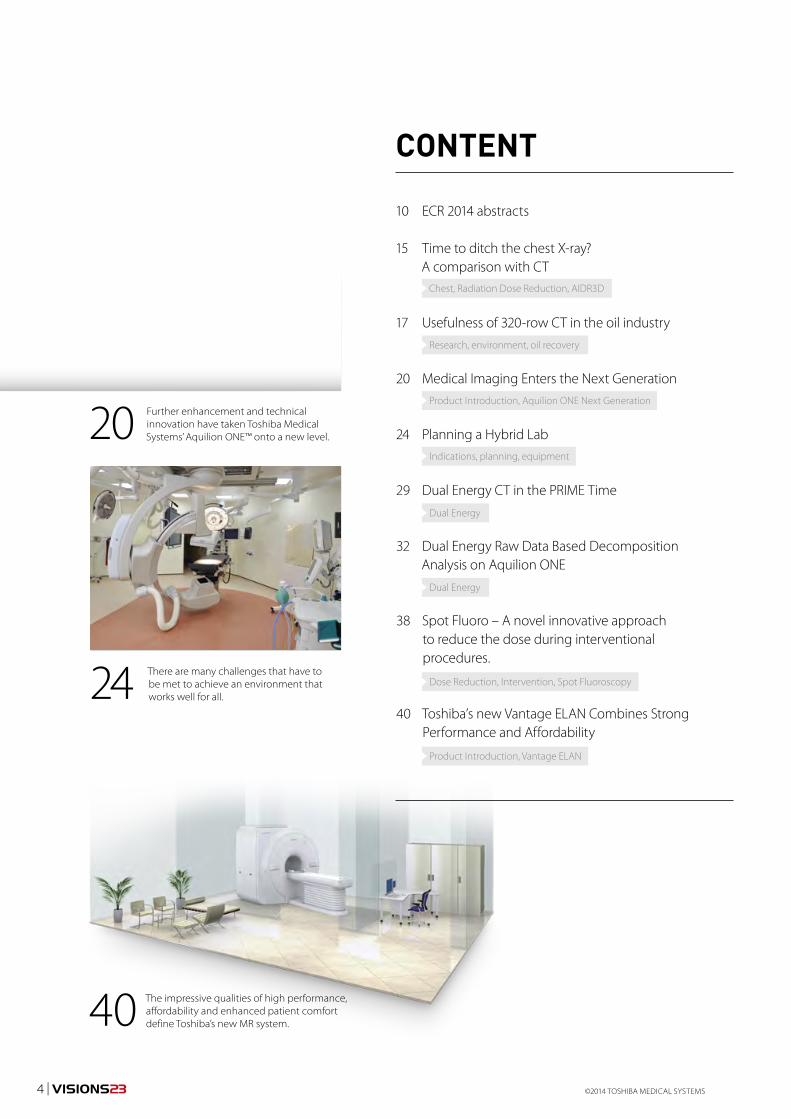
20 Medical Imaging Enters the Next Generation
24 Planning a Hybrid Lab
29 Dual Energy CT in the PRIME Time
32 Dual Energy Raw Data Based Decomposition Analysis on Aquilion ONE
38 Spot Fluoro – A novel innovative approach to reduce the dose during interventional procedures.
40 Toshiba’s new Vantage ELAN Combines Strong Performance and Affordability
20
24
40
Further enhancement and technical innovation have taken Toshiba Medical Systems’ Aquilion ONE™ onto a new level.
There are many challenges that have to be met to achieve an environment that works well for all.
The impressive qualities of high performance, affordability and enhanced patient comfort define Toshiba’s new MR system.
CONTENT
Chest, Radiation Dose Reduction, AIDR3D
Research, environment, oil recovery
Product Introduction, Aquilion ONE Next Generation
Indications, planning, equipment
Dual Energy
Dual Energy
Dose Reduction, Intervention, Spot Fluoroscopy
Product Introduction, Vantage ELAN
VISIONS23 | 5
43 Single energy metal artifact reduction algorithm for CT evaluation of periprosthetic soft tissues: Clinical applications
46 Creating New Standards in Ultrasound Cardiology
49 Superiority of Lung Subtracton Above Dual Energy
52 Violation of longitudinal strain of the left ventricular myocardium as a predictor of positive stress echocardiography results
55 CUSTOMER FOCUS: Rigshospitalet
67 Subtraction CTA of the brain detects cerebral aneurysms
68 Diagnosis and treatment of primary sarcoma of the liver
74 320-row CT as a single and effective platform for anatomical and functional evaluation of coronary artery disease: the CORE320 trial
76 Secondlife Mobile CT
46
55
68
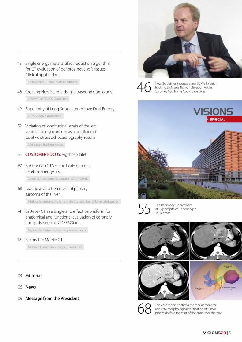
New Guidelines Incorporating 2D Wall Motion Tracking to Assess Non-ST Elevation Acute Coronary Syndrome Could Save Lives
The Radiology Departmentat Rigshospitalet Copenhagen in Denmark
This case report confirms the requirement for accurate morphological verification of tumor process before the start of the antitumor therapy.
03 Editorial
06 News
09 Message from the President
VISIONSSPECIAL
Orthopedics, SEMAR, metallic artifacts
2D WMT, NSTE-ACS, Guidelines
CTPA, Lungs, substraction
2D Speckle Tracking, Artida
Cerebral Aneurysms, Subtraction CTA, AIDR 3D
Histiocytic sarcoma, malignant histiocytosis, liver, differential diagnosis
Myocardial Perfusion, Coronary Angiography
Mobile CT, temporary imaging, Secondlife
©2014 TOSHIBA MEDICAL SYSTEMS6 | VISIONS23
The page on the right is part of the VISIONS Photo Page Series reflecting an eye for the beauty of
our planet, the environment and the direct surroundings where Toshiba’s systems are installed
by Toshiba and its customers. Not the actual imaging products but photos of sceneries, cities,
countries or other cultural aspects are highlighted on this photo page. The Photo Page is based
upon an idea of Prof. Edwin van Beek.
Every reader of VISIONS can participate and get their picture published. The submitted content should
include: high resolution (300dpi) image, photo of the hospital and a brief text, name of photographer
and Toshiba system(s) installed. The complete result is shown on the opposite page.
Send your pictures and texts to: [email protected], Subject: Photo Page
►
NEWS
Edinburgh: Transforming Global Healthcare
Edinburgh is home to world-leading medical researchers
who work in partnership with the NHS and the private
sector. This close cooperation helps to accelerate
medical discoveries and transform them into treatments
for patients.
Toshiba is featured in a promotional clip from the
inspiring captial of Scotland. Have a look and pay spe-
cial attention to the frames from 1:23m-1:44m where
Dr. Ken Sutherland explains about Toshiba’s activities
in Edinburgh. http://tinyurl.com/punkwhp
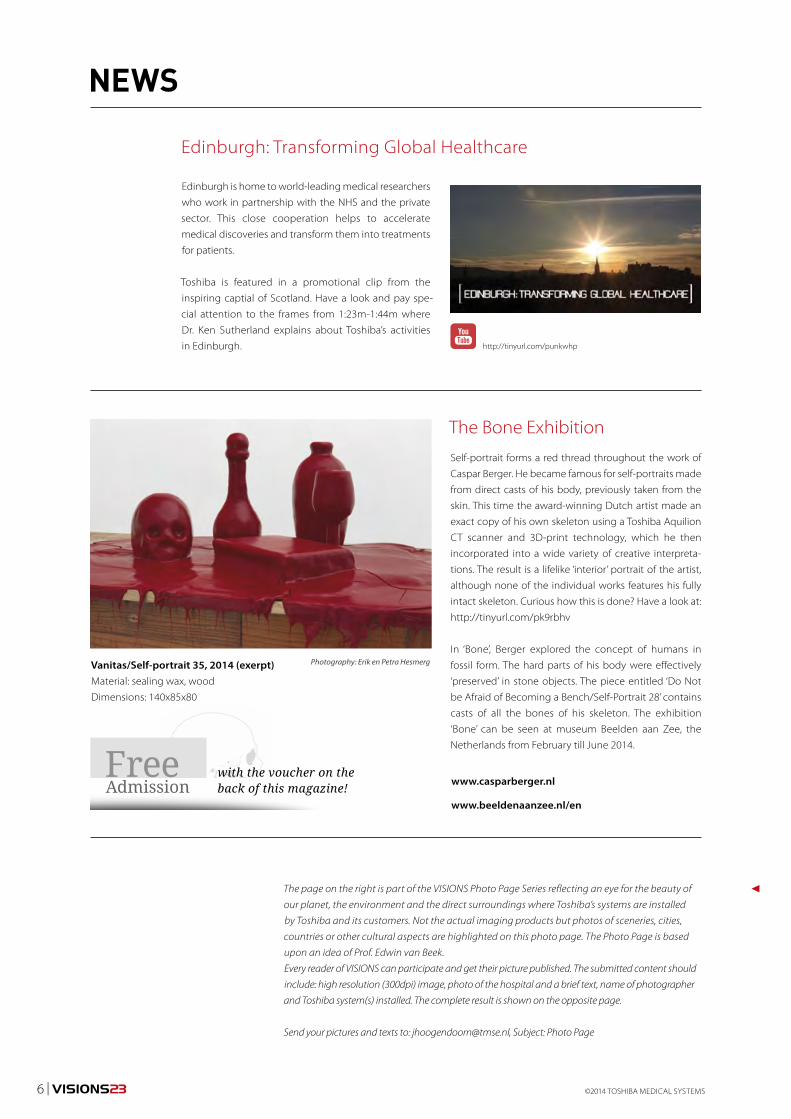
Self-portrait forms a red thread throughout the work of
Caspar Berger. He became famous for self-portraits made
from direct casts of his body, previously taken from the
skin. This time the award-winning Dutch artist made an
exact copy of his own skeleton using a Toshiba Aquilion
CT scanner and 3D-print technology, which he then
incorporated into a wide variety of creative interpreta-
tions. The result is a lifelike ‘interior’ portrait of the artist,
although none of the individual works features his fully
intact skeleton. Curious how this is done? Have a look at:
http://tinyurl.com/pk9rbhv
In ‘Bone’, Berger explored the concept of humans in
fossil form. The hard parts of his body were effectively
‘preserved’ in stone objects. The piece entitled ‘Do Not
be Afraid of Becoming a Bench/Self-Portrait 28’ contains
casts of all the bones of his skeleton. The exhibition
‘Bone’ can be seen at museum Beelden aan Zee, the
Netherlands from February till June 2014.
The Bone Exhibition
Vanitas/Self-portrait 35, 2014 (exerpt) Material: sealing wax, wood
Dimensions: 140x85x80
Photography: Erik en Petra Hesmerg
www.casparberger.nl
www.beeldenaanzee.nl/en
Follow us
AdmissionFree with the voucher on the
back of this magazine!
VISIONS23 | 7
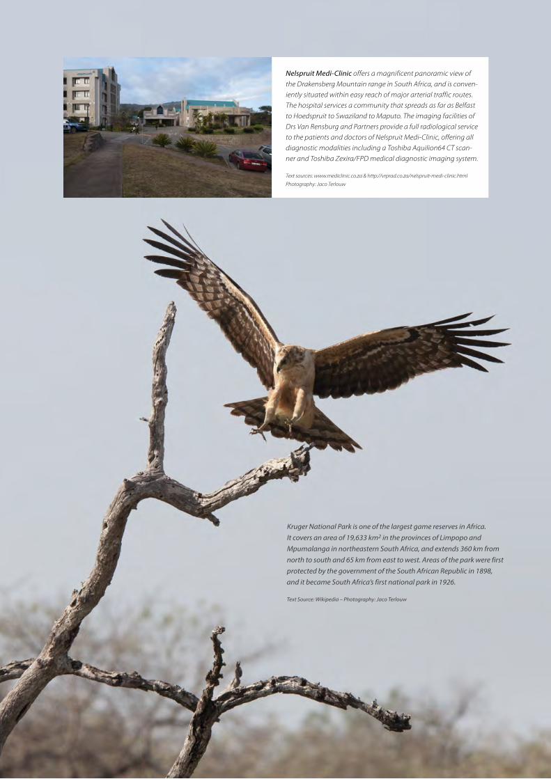
Kruger National Park is one of the largest game reserves in Africa. It covers an area of 19,633 km2 in the provinces of Limpopo and Mpumalanga in northeastern South Africa, and extends 360 km from north to south and 65 km from east to west. Areas of the park were first protected by the government of the South African Republic in 1898, and it became South Africa’s first national park in 1926.
Text Source: Wikipedia – Photography: Jaco Terlouw
Nelspruit Medi-Clinic offers a magnificent panoramic view of the Drakensberg Mountain range in South Africa, and is conven-iently situated within easy reach of major arterial traffic routes. The hospital services a community that spreads as far as Belfast to Hoedspruit to Swaziland to Maputo. The imaging facilities of Drs Van Rensburg and Partners provide a full radiological service to the patients and doctors of Nelspruit Medi-Clinic, offering all diagnostic modalities including a Toshiba Aquilion64 CT scan-ner and Toshiba Zexira/FPD medical diagnostic imaging system.
Text sources: www.mediclinic.co.za & http://vrprad.co.za/nelspruit-medi-clinic.html
Photography: Jaco Terlouw
www.toshiba-medical.eu ULTRASOUND C T MRI X-RAY SERVICES
Our lives and social environment are subject to constant change and create
ever-increasing needs and high demand for better medical solutions. We at
Toshiba aim to maximize the quality, safety, and efficiency of medical care,
supporting clinical practice with reliable quality products and innovative,
cutting-edge technologies.
The high image resolution and superior operability of our medical systems
creates new clinical value. While our advanced applications, supported by
highly reliable technologies, open the door to the next stage of medical care.
We will continue to provide a wide variety of leading-edge solutions for the
benefit of all people around the world, and seek to further development in the
field of healthcare following our basic commitments: “Improving the quality of
life”, “Lifelong commitment to innovation”, and “Achieving lifetime partnerships”.
Toshiba: Made for Life!
MADE FOR LIFE.
VISIONS23 | 9
PRESIDENT’SMESSAGE
Satoshi TsunakawaPresident and Chief Executive Officer
Toshiba Medical Systems Corporation
Toshiba’s primary focus for FY2013 was to better
understand our customer and the needs of their
business in medical imaging – we always want to keep
the customers first.
In our CT business, we reached a very significant
milestone with the manufacture of our 30,000th CT
system in November 2013. We have continuously offered
leading, innovative products based on the clinical needs
of our customers since our first CT introduction. We
believe that lowering CT dose and improving workflow
are not choices that clinicians should have to make, so
Toshiba CT is putting customers first by providing the
industry’s best solutions to solve these challenges. We
introduced Toshiba’s most advanced dose reduction
technology, AIDR 3D, which enables our customers
to achieve the best clinical results at the lowest dose.
More recently we have introduced the suite of Adaptive
Diagnostic CT technology, which makes complex
examinations easier while simultaneously improving
diagnostic accuracy and reproducibility to solve clinical
challenges faced daily in routine practice.
In our X-Ray business, we had the worldwide
introduction of the DTS (Dose Tracking System) at RSNA
2013. This technology provides a real-time display of the
cumulative skin dose distribution during interventional
procedures. The push for developing this technology
came from the strong desire from the doctors to be
able to monitor and minimize patient radiation which
had risen sharply in recent years during interventions.
Our team listened to this VOC (Voice of Customers)
from around the world and successfully developed this
system – understanding our customers’ business needs
to protect their patients.
In the MRI business unit, we launched Vantage
Elan™, which is providing excellent image quality, while
achieving ease-of-use, in a quiet environment, and with
a very compact footprint. The operation of Vantage
Elan is simple for any level of user which means better
images for every customer while increasing operational
efficiency. Vantage Elan requires only 23m2, and
5 working days for the complete installation after system
delivery which means real savings for the customer.
Vantage Elan is the realization of the MRI team’s
design goal of “No Compromises in Image Quality”,
in spite of its compactness. I am confident that this
system will become the new standard of the next
generation of MRI systems.
Our Ultrasound team launched a new technology
called SMI (Superb Micro vascular Imaging). This new
algorithm isolates and removes clutter while preserving
the underlying hemodynamic flow information. This
technology allows you to see the flow information that
is behind the clutter and visualize extremely low velocity
flows that are typically obscured with conventional
Color Doppler. See the unseen – A giant step towards
achieving our goal of “Picture Perfect Ultrasound” starts
from here.
Finally, last but not least, as of October 1st, healthcare
has become one of the three business pillars of the
Toshiba Corporation, and TMSC will be the core member
of the new Healthcare Systems & Services Group.
We will be working closely with the new established
Healthcare Business Development Division in Toshiba
Corporation. The mission of the Healthcare Group is to
extend the scope of business from medical diagnostics
to new healthcare related business, including disease
prevention and patient care. With this initiative, and
carefully expanding our businesses, we are continuing
in our corporate philosophy to keep the customers and
more importantly their patients first.
“ We always keep our customers first.”
By focusing on low dose, high-quality imaging technologiesfor accurate diagnosis and treatment, Toshiba continues toimprove the quality of life for all people.
©2014 TOSHIBA MEDICAL SYSTEMS10 | VISIONS23
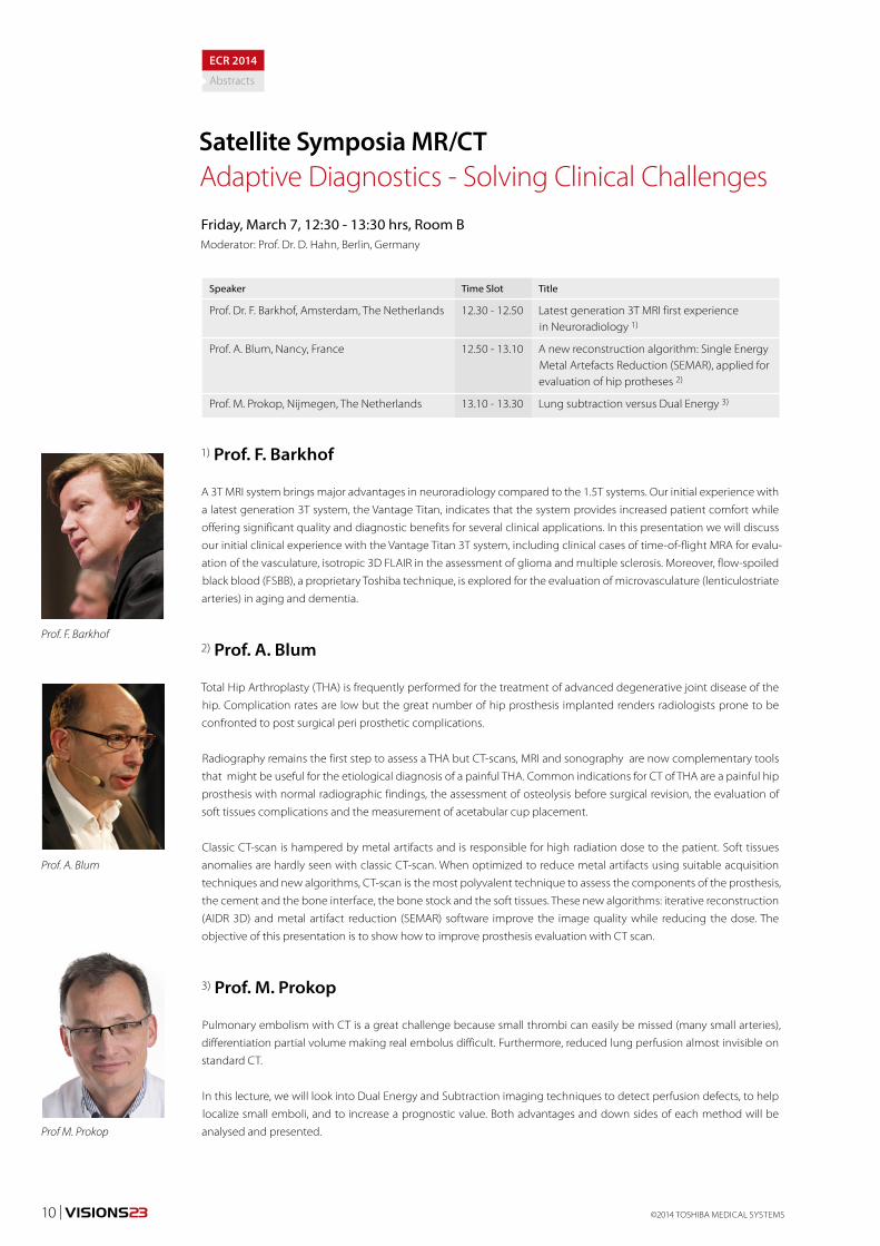
1) Prof. F. Barkhof
A 3T MRI system brings major advantages in neuroradiology compared to the 1.5T systems. Our initial experience with
a latest generation 3T system, the Vantage Titan, indicates that the system provides increased patient comfort while
offering significant quality and diagnostic benefits for several clinical applications. In this presentation we will discuss
our initial clinical experience with the Vantage Titan 3T system, including clinical cases of time-of-flight MRA for evalu-
ation of the vasculature, isotropic 3D FLAIR in the assessment of glioma and multiple sclerosis. Moreover, flow-spoiled
black blood (FSBB), a proprietary Toshiba technique, is explored for the evaluation of microvasculature (lenticulostriate
arteries) in aging and dementia.
2) Prof. A. Blum
Total Hip Arthroplasty (THA) is frequently performed for the treatment of advanced degenerative joint disease of the
hip. Complication rates are low but the great number of hip prosthesis implanted renders radiologists prone to be
confronted to post surgical peri prosthetic complications.
Radiography remains the first step to assess a THA but CT-scans, MRI and sonography are now complementary tools
that might be useful for the etiological diagnosis of a painful THA. Common indications for CT of THA are a painful hip
prosthesis with normal radiographic findings, the assessment of osteolysis before surgical revision, the evaluation of
soft tissues complications and the measurement of acetabular cup placement.
Classic CT-scan is hampered by metal artifacts and is responsible for high radiation dose to the patient. Soft tissues
anomalies are hardly seen with classic CT-scan. When optimized to reduce metal artifacts using suitable acquisition
techniques and new algorithms, CT-scan is the most polyvalent technique to assess the components of the prosthesis,
the cement and the bone interface, the bone stock and the soft tissues. These new algorithms: iterative reconstruction
(AIDR 3D) and metal artifact reduction (SEMAR) software improve the image quality while reducing the dose. The
objective of this presentation is to show how to improve prosthesis evaluation with CT scan.
3) Prof. M. Prokop
Pulmonary embolism with CT is a great challenge because small thrombi can easily be missed (many small arteries),
differentiation partial volume making real embolus difficult. Furthermore, reduced lung perfusion almost invisible on
standard CT.
In this lecture, we will look into Dual Energy and Subtraction imaging techniques to detect perfusion defects, to help
localize small emboli, and to increase a prognostic value. Both advantages and down sides of each method will be
analysed and presented.
Satellite Symposia MR/CT
Friday, March 7, 12:30 - 13:30 hrs, Room BModerator: Prof. Dr. D. Hahn, Berlin, Germany
ECR 2014
Abstracts
Adaptive Diagnostics - Solving Clinical Challenges
Speaker Time Slot Title
Prof. Dr. F. Barkhof, Amsterdam, The Netherlands 12.30 - 12.50 Latest generation 3T MRI first experience in Neuroradiology 1)
Prof. A. Blum, Nancy, France 12.50 - 13.10 A new reconstruction algorithm: Single Energy Metal Artefacts Reduction (SEMAR), applied for evaluation of hip protheses 2)
Prof. M. Prokop, Nijmegen, The Netherlands 13.10 - 13.30 Lung subtraction versus Dual Energy 3)
Prof. A. Blum
Prof. F. Barkhof
Prof M. Prokop
VISIONS23 | 11
Satellite Symposia ULTRASOUND
Saturday, March 8, 2014, 12.30 - 13.30 hrs, Room E1Moderator: Prof. V. Mitkov, Russia
1) Dr. T. Fischer
Prostate: Men with an elevated serum level of prostate-specific antigen (PSA) or suspicious findings on digital rectal
examination (DRE) are examined by transrectal ultrasound (TRUS). For histological confirmation and therapeutic plan-
ning, ultrasound guided systematic biopsy of the prostate is performed. However, in a subgroup of patients with
elevated PSA levels, no malignancy is detected by biopsy or up to four biopsies are performed before prostate cancer is
detected. A negative biopsy therefore does not exclude prostate cancer. In consequence, unnecessary biopsies with an
increase of complications are performed in healthy men. Real-time MR/US image fusion may enhance cancer detection
rates of TRUS-guided biopsies and contributes to lesion characterization by state-of-the-art US techniques. The study
presented here for the first time compares state-of-the art US techniques after contrast medium administration with
MRI and histology as the standard of reference.
Scrotum: Ultrasound is the imaging modality of choice for the examination of the scrotum. The propblem of tumour
/ haematoma misinterpretation can result in unnecessary orchiectomy. CEUS for the testis can be currently recom-
mended for the differentiation between hypovascular and avascular lesions (benign).
Learning objects:
- To investigate whether multiparametric magnetic resonance imaging (MRI) allows lesion localization in prostate can-
cer in patients scheduled for MR/US fusion biopsy and whether the findings correlate with new ultrasound techniques
- Prostate lesions were classified on the basis of MRI and US (B-mode scan, power Doppler, elastography/TDI, CEUS)
- Targeted biopsies were performed in the MR/US fusionmode
- Indications for the use of CEUS in focal testicular lesions, secmental infartion, after trauma and abscess formation in
severe epididymoorchitis.
2) Prof. A. Lim
This talk will encompass the latest technological developments of the Aplio 500 with particular focus on a few advanced
applications for abdominal and small parts ultrasound. These newest developments include the improved visualisation
of the microvasculature without the need for contrast and its potential clinical applications will be discussed. There
have also been significant improvement in specialist applications such as elastography and also improved sensitivity in
high frequency contrast technology. The latter now also allows reliable detection of the sentinel lymph node and their
clinical impact will be outlined and illustrated.
Expanding Clinical Boundries in Ultrasound
Speaker Time Slot Title
Dr. T. Fischer, Berlin, Germany 12.30 - 12.50 Genitourinary Ultrasound: Advanced Diagnostics in Prostate Cancer and Scrotal Lesions 1)
Prof. A. Lim, London, United Kingdom 12.50 - 13.10 Abdominal and high-frequency Ultrasound imaging: Latest technologies for improved lesion detection and characterisation 2)
Prof. J. Hata, Kawasaki, Japan 13.10 - 13.30 Seeing the Unseen - New Clinical Findings by Novel Imaging Techniques 3)
Dr. T. Fischer
Prof. A. Lim
BRINGING INNOVATION TO LIFE
©2014 TOSHIBA MEDICAL SYSTEMS12 | VISIONS23
3) Prof. J. Hata
In this session, the clinical experience of Superb microflow imaging (SMI), which is the novel application of Aplio 500
from Toshiba, is discussed. SMI is a unique ultrasound Doppler imaging employing multi-dimensional filter that makes
it possible to depict minute vessels with slow velocity, without using contrast agents. In other words, the advantages
of SMI are; 1.high frame rate, 2. high sensitivity, 3. high resolution, and 4. less motion artifact.
Therefore, SMI is useful in various situations encountered in daily practice. The evaluation of the density and the shape
of tumor vessels with SMI help us diagnose and differentiate tumorous lesions and also could be the non-invasive
modality for the assessment of therapeutic effect of chemotherapy. Since SMI clearly visualizes minute vessels, it can
also be used to evaluate the disease activity in inflammatory disorders, even in bowel inflammation such as ulcerative
colitis, Crohn’s disease, and so forth. Furthermore, the diagnosis of organ ischemia can be done accurately with SMI,
which is expressed as the focal defect of minute vessels in a real-time fashion, with high spatial resolution. In addition,
SMI provides more sensitive view of minute vessels after the injection of contrast agent, which helps us to obtain the
sustained dynamic image of microflow even after the first pass of the contrast agent.
In conclusion, SMI, which provides the dynamic image of microvessels, is an essential tool for the diagnosis and the
evaluation of various clinical conditions.
Toshiba Workshop
Sessions will be offered Friday, March 7 thru Sunday, March 9. Times as listed below in room Lounge 6 - Level 01
(max 20 seats).
Workshops are open to any ECR 2014 attendees. On-line registration in the “myuserArea” of the ECR web page.
Toshiba is offering a series of educational sessions for radiologists throughout ECR 2014. Each mentored workshop is
preceded by a short lecture providing an overview on data acquisition, clinical use and image interpretation. Clinical
cases will be analyzed and interpreted by participants on the workstations provided. The maximum number of
participants per session is 20 (2 participants per workstation). CME credits will be provided, and the workshop language
is English.
Date Time Slot Title
March 7, 2013 1 10:00 - 11:00 Ultrasound - Elastography Quantification, guest speaker: Dr. V. Cantisani, Rome, Italy
March 7, 2013 2 11:15 - 12:15 Ultrasound - Fly Thru Acquisition and Reconstructions, guest speaker: Dr. B. Smith, London, UK
March 7, 2013 3 12:30 - 13:30 Ultrasound - DCEUS in a multicenter study, guest speaker: Dr. N. Lassau, Paris, France
March 7, 2013 4 13:45 - 14:45 Ultrasound - Elastography Quantification, guest speaker: Dr. V. Cantisani, Rome, Italy
March 7, 2013 5 15:00 - 16:00 Ultrasound - Fly Thru Acquisition and Reconstructions, guest speaker: Dr. B. Smith, London, UK
March 7, 2013 6 16:15 - 17:15 Ultrasound - DCEUS in a multicenter study, guest speaker: Dr. N. Lassau, Paris, France
March 8, 2013 7 10:00 - 11:00 CT - Ultra low dose brain perfusion, guest speaker: Dr. E. Smit, Nijmegen, The Netherlands
March 8, 2013 8 11:15 - 12:15 ViTAL - Lung Density Analysis, guest speaker: Dr. S. Niehues, Berlin, Germany
March 8, 2013 9 13:45 - 14:45 CT - Myocardial perfusion, guest speaker: Dr. K. Kofoed, Copenhagen, Denmark
March 8, 2013 10 15:00 - 16:00 CT - Liver perfusion, guest speaker: Dr. H. Schöllnast, Graz, Austria
March 8, 2013 11 16:15 - 17:15 CT - Ultra Low Cardiac CTA, guest speaker: Dr. R. Bull, Bournemouth, UK
March 9, 2013 12 10:00 - 11:00 ViTAL - TAVR planning, guest speaker: Dr. S. Niehues, Berlin, Germany
March 9, 2013 13 11:15 - 12:15 CT - Ultra Low Cardiac CTA, guest speaker: Dr. R. Bull, Bournemouth, UK
March 9, 2013 14 12:30 - 13:30 ViTAL - Lung Density Analysis, guest speakers: Dr. S. Niehues, Berlin, Germany
March 9, 2013 15 13:45 - 14:45 CT - Liver perfusion, guest speaker: Dr. H. Schöllnast, Graz, Austria
March 9, 2013 16 15:00 - 16:00 CT - Myocardial perfusion, guest speaker: Dr. K. Kofoed, Copenhagen, Denmark
March 9, 2013 17 16:15 - 17:15 CT - Ultra low dose brain perfusion, guest speaker: Dr. E. Smit, Nijmegen, The Netherlands
Prof. J. Hata
VISIONS23 | 13
BRINGING INNOVATION TO LIFE
DCEUS Quantification Workshop Bracco/ToshibaSaturday, March 8, 2014, 12.30 - 13.20 hrs, Room Lounge 1 - Level 01 (max 30 seats)
Moderator: Prof. Jean-Michel Correas, Paris, France
Quantification of Renal Perfusion
Bracco and Toshiba offer a 50 minutes educational session for radiologists on quantification of Dynamic Contrast
Enhanced Ultrasound (DCEUS) based on the use of the Bracco quantification software VueBox™ and contrast-enhanced
ultrasound sequences acquired with the Toshiba Aplio 500. Brief introductionary lectures will provide an overview of
concepts in DCEUS quantification and in renal perfusion. In the hands on session, the participants will perform their
own analysis of Toshiba Aplio 500 clips under the supervision of Prof. Jean-Michel Correas using laptops equipped with
the VueBox software.
Floor plan is an indication.
Please follow the signage
TOSHIBA IWS.
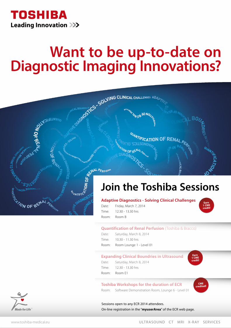
Want to be up-to-date onDiagnostic Imaging Innovations?
www.toshiba-medical.eu ULTRASOUND C T MRI X-RAY SERVICES
Adaptive Diagnostics - Solving Clinical ChallengesDate: Friday, March 7, 2014
Time: 12.30 - 13.30 hrs
Room: Room B
Quantification of Renal Perfusion (Toshiba & Bracco)Date: Saturday, March 8, 2014
Time: 10.30 - 11.30 hrs
Room: Room Lounge 1 - Level 01
Expanding Clinical Boundries in UltrasoundDate: Saturday, March 8, 2014
Time: 12.30 - 13.30 hrs
Room: Room E1
Toshiba Workshops for the duration of ECRRoom: Software Demonstration Room, Lounge 6 - Level 01
Sessions open to any ECR 2014 attendees.
On-line registration in the “myuserArea” of the ECR web page.
Join the Toshiba Sessions
CME credited!
Earn 1 CME credit!
Earn 1 CME credit!
VISIONS23 | 15
MATERIALS AND METHODSWe examined 13 patients with suspected pulmonary
lesions, following chest radiography with ultralow-dose
CT acquired on an Aquilion ONE scanner. Images were
acquired at 135 kV and 10 mA, with 0.5-mm slice thick-
ness and rotation time of 350 msec.
Three experienced chest radiologists rated both sets
of images for pathological findings on a three-point
scale, and image quality was rated according to nine
criteria of the European guidelines for chest CT and chest
radiographs.
RESULTSBoth image quality and the ability to detect and exclude
pathology was strikingly better for ultralow-dose CT than
for radiography. The radiation dose for CT and conven-
tional radiographs was comparable and the laboratory
time only marginally longer for CT.
In summary, we found the following:
• 33 relevant findings including 19 masses, nodules, and
micronodules.
• Detection sensitivity for all observers and all findings
was 18% ± 3% for radiography and 89% ± 2% for
ultralow-dose CT.
• The positive predictive value for radiography was 37%,
including 31 false-positive findings, while the positive
predictive value for CT was 98% with two false-positive
findings.
• Image quality for all readers was significantly higher for
the CT images (p < 0.05).
• The average effective dose was 0.05 mSv for chest radi-
ography and 0.105 mSv for CT.
• The average laboratory time was five minutes for chest
radiography versus seven minutes for CT.
Time to ditch the chest X-ray? A comparison with CT
It is widely accepted that Computer Tomography (CT) is both more time consuming and expensive than chest radiography. Furthermore, CT incurs a considerably higher radiation dose than the conventional chest X-ray. Ideally we wish to combine the low-dose of the chest X-ray with the high image quality of CT, but this ambitious aim has, until now, not been possible. In this study we compared conventional chest radiography to 320-detector-row CT Aquilion ONE™ using advanced iterative reconstruction (AIDR 3D) in terms of image quality, radiation dose and laboratory time.
Dr. T.M. Aaløkken
J.F. Kristiansen MSc
Dr. A. Günther
A.C.T. Martinsen PhD
1-4) Oslo University
Hospital, Norway
CTEU130074
CLINICAL CASE COMPUTED TOMOGRAPHY
Chest, Radiation Dose Reduction, AIDR3D
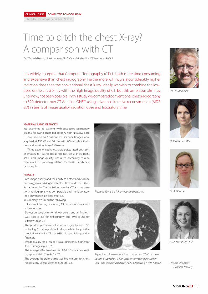
Figure 1: Above is a false-negative chest X-ray.
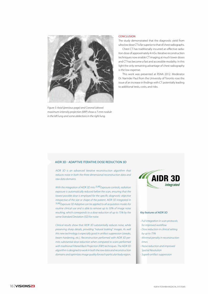
Figure 2: an ultralow-dose 3-mm axial chest CT of the same
patient acquired on a 320-detector-row scanner (Aquilion
ONE) and reconstructed with AIDR 3D shows a 7-mm nodule.
Dr. T.M.Aaløkken 1), J.F. Kristiansen MSc 2), Dr. A. Günther 3), A.C.T. Martinsen PhD 4)
©2014 TOSHIBA MEDICAL SYSTEMS16 | VISIONS23
CONCLUSIONThe study demonstrated that the diagnostic yield from
ultra low dose CT is far superior to that of chest radiographs.
Chest CT has traditionally incurred an effective radia-
tion dose of approximately 4 mSv. Iterative reconstruction
techniques now enable CT imaging at much lower doses
and CT has become a fast and accessible modality. In this
light the only remaining advantage of chest radiography
is the low expense.
This work was presented at RSNA 2012. Moderator
Dr. Narinder Paul from the University of Toronto rose the
issue of an increase in findings with CT potentially leading
to additional tests, costs, and risks.
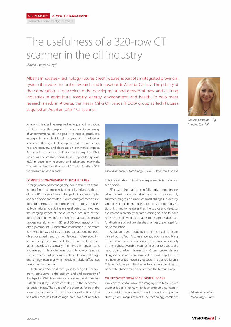
Figure 3: Axial (previous page) and Coronal (above)
maximum-intensity projection (MIP) show a 7-mm nodule
in the left lung and some atelectasis in the right lung.
AIDR 3D is an advanced iterative reconstruction algorithm that
reduces noise in both the three dimensional reconstruction data and
raw data domains.
With the integration of AIDR 3D into SUREExposure controls, radiation
exposure is automatically reduced before the scan, ensuring that the
lowest possible dose is employed for the specific diagnostic objective
irrespective of the size or shape of the patient. AIDR 3D integrated in SUREExposure 3D Adaptive can be applied to all acquisition modes for
routine clinical use and is able to remove up to 50% of image noise
resulting, which corresponds to a dose reduction of up to 75% by the
same Standard Deviation (SD) for noise.
Clinical results show that AIDR 3D substantially reduces noise, while
preserving sharp details, providing “natural looking” images. As well
this new technology is especially good in artifact suppression (streaks,
beam-hardening, etc.). Reconstruction performed with AIDR 3D per-
mits substantial dose reduction when compared to scans performed
with traditional Filtered Back Projection (FBP) techniques. The AIDR 3D
algorithm is designed to work in both the raw data and reconstruction
domains and optimizes image quality for each particular body region.
AIDR 3D - ADAPTIVE ITERATIVE DOSE REDUCTION 3D
Key features of AIDR 3D
- Full integration in scan protocols
for improved workflow
- Dose reduction in clinical setting
by up to 75%
- Minimal penalty in reconstruction
times
- Noise reduction and improved
Spatial Resolution
- Superb artifact suppression
VISIONS23 | 17
As a world leader in energy technology and innovation,
HOOS works with companies to enhance the recovery
of unconventional oil. The goal is to help oil producers
engage in sustainable development of Alberta’s
resources through technologies that reduce costs,
improve recovery, and decrease environmental impact.
Research in this area is facilitated by the Aquilion ONE,
which was purchased primarily as support for applied
R&D in petroleum recovery and advanced materials.
This article describes the use of CT with Aquilion ONE
for research at Tech Futures.
COMPUTED TOMOGRAPHY AT TECH FUTURESThrough computed tomography, non-destructive exami-
nation of internal structure is accomplished and high-res-
olution 3D images of items like geological core samples
and sand packs are created. A wide variety of reconstruc-
tion algorithms and post-processing options are used
at Tech Futures to suit the material being scanned and
the imaging needs of the customer. Accurate extrac-
tion of quantitative information from advanced image
processing, along with 2D and 3D reconstructions, is
often paramount. Quantitative information is delivered
to clients by way of customized calibrations for each
object or experiment scanned. Targeted noise-reduction
techniques provide methods to acquire the best reso-
lution possible. Specifically, this involves repeat scans
and averaging data whenever possible to reduce noise.
Further discrimination of materials can be done through
dual energy scanning, which exploits subtle differences
in attenuation spectra.
Tech Futures’ current strategy is to design CT experi-
ments conducive to the energy level and geometry of
the Aquilion ONE. Low-attenuation vessels and materials
suitable for X-ray use are considered in the experimen-
tal design stage. The speed of the scanner, for both the
acquisition and reconstruction of data, makes it possible
to track processes that change on a scale of minutes.
The usefulness of a 320-row CT scanner in the oil industry
Alberta Innovates - Technology Futures (Tech Futures) is part of an integrated provincial system that works to further research and innovation in Alberta, Canada. The priority of the corporation is to accelerate the development and growth of new and existing industries in agriculture, forestry, energy, environment, and health. To help meet research needs in Alberta, the Heavy Oil & Oil Sands (HOOS) group at Tech Futures acquired an Aquilion ONE™ CT scanner.
CTEU130078
Shauna Cameron, P.Ag,
Imaging Specialist
1) Alberta Innovates –
Technology Futures.
Alberta Innovates - Technology Futures, Edmonton, Canada
Shauna Cameron, P.Ag 1)
This is invaluable for fluid flow experiments in cores and
sand packs.
Efforts are also made to carefully register experiments
when repeat scans are taken in order to successfully
subtract images and uncover small changes in density.
Orbital sync has been a useful tool in securing registra-
tion. This function ensures that the source and detector
are located in precisely the same starting position for each
repeat scan allowing the images to be either subtracted
for discrimination of tiny density changes or averaged for
noise reduction.
Radiation dose reduction is not critical to scans
carried out at Tech Futures since subjects are not living.
In fact, objects or experiments are scanned repeatedly
at the highest available settings in order to extract the
best quantitative information. Often, protocols are
designed so objects are scanned in short lengths, with
multiple volumes necessary to cover the desired length.
This technique permits the highest allowable dose to
penetrate objects much denser than the human body.
OIL RECOVERY FROM ROCK: DIGITAL ROCKSOne application for advanced imaging with Tech Futures’
scanner is digital rocks, which is an emerging concept in
characterizing reservoirs by obtaining physical properties
directly from images of rocks. The technology combines
OIL INDUSTRY COMPUTED TOMOGRAPHY
Research, environment, oil recovery
data can be very useful in studying pore architecture and
physics of the processes happening during enhanced oil
recovery (EOR). It is especially important for unconven-
tional resources like shale, carbonate, tight gas, and coal
seam gas. One of the most interesting aspects at this time
is characterizing bitumen-bearing carbonates, especially
fracture characterization.
Digital rocks enables Tech Futures to engage in a
unique up-scaling effort of merging micro and macro
imaging to observe a wide range of fluids, conditions,
and detailed behaviour in EOR processes. The advantage
of the technique is the considerable cost savings over
securing individual laboratory analysis for each desired
property. The procedure involves first obtaining full core
mm-scale resolution scans with the Aquilion ONE scan-
ner to identify sites representative of a range of image
properties. This is followed by micro CT scans (approxi-
mately 10 micron resolution), available from various out-
sourced labs, which are suitable for sand packs or other
very small samples. Imaging with sufficient resolution to
resolve individual pores in a sand pack or a core is inevita-
bly restricted to a very small volume. Simulation software
is used to upscale the results of pore scale multi-phase
flow simulations to obtain flow properties at each site
and look for correlations between flow properties and
CT at different scales (mm to micron resolution) with
X-ray and scanning-electron microscopy (SEM) to create
images of the internal structure of the pore spaces as
well as minerals and organic matter within them. Such
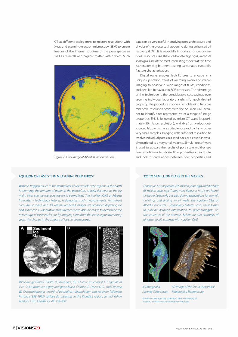
Figure 2: Axial image of Alberta Carbonate Core
Dinosaurs first appeared 225 million years ago and died out
65 million years ago. Today most dinosaur fossils are found
by doing fieldwork, but also during excavations for tunnels,
buildings and drilling for oil wells. The Aquilion ONE at
Alberta Innovates - Technology Futures scans these fossils
to provide detailed information to paleontologists on
the structures of the animals. Below are two examples of
dinosaur fossils scanned with Aquilion ONE.
3D Image of a
Juvenile Ceratopsian
3D image of the Snout (Antorbital
Region) of a Tyrannosaur
225 TO 65 MILLION YEARS IN THE MAKING
Specimens are from the collections of the University of Alberta, Laboratory of Vertebrate Paleontology
AQUILION ONE ASSISTS IN MEASURING PERMAFROST
Water is trapped as ice in the permafrost of the world’s artic regions. If the Earth
is warming, the amount of water in the permafrost should decrease as the ice
melts. How can we measure the ice in permafrost? The Aquilion ONE at Alberta
Innovates - Technology Futures, is doing just such measurements. Permafrost
cores are scanned and 3D volume rendered images are produced depicting ice
and sediment. Quantitative measurements can also be made to determine the
percentage of ice in each core. By imaging cores from the same region over many
years, the change in the amount of ice can be measured.
Three images from CT data. (A) Axial slice; (B) 3D reconstruction; (C) Longitudinal
slice. Soil is white, ice is gray and gas is black. Calmels, F., Froese D.G., and Clavano,
W. Cryostratigraphic record of permafrost degradation and recovery following
historic (1898-1992) surface disturbances in the Klondike region, central Yukon
Territory. Can. J. Earth Sci. 49: 938–952
18 | VISIONS23 ©2014 TOSHIBA MEDICAL SYSTEMS
VISIONS23 | 19
image properties. Properties are then mapped back onto
the macro CT scan using the correlations and core scale
flow simulations are performed with the results to make
predictions for recovery from the oil reservoir.
OIL RECOVERY FROM SAND: IMAGING SAND PACKSOther HOOS applications of the Tech Futures scanner
include calculating porosities and saturations in sand
packs, tracking solute in sand packs, testing solute uptake
in oil, studying turbulent flow in water as well as laminar
flow in oil, monitoring sand failure or production, and
investigating the waterflood process used in oil recovery
at the scale of sand grain pores.
CONCLUSIONThe use of CT to image and quantify materials greatly
assists scientists in determining potential sites for com-
mercial oil extraction in unconventional materials such
as carbonates and oil sands. This is a very interesting and
different application of CT from its usual medical uses.
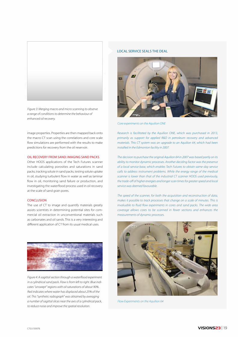
Figure 3: Merging macro and micro scanning to observe
a range of conditions to determine the behaviour of
enhanced oil recovery.
Figure 4: A sagittal section through a waterflood experiment
in a cylindrical sand pack. Flow is from left to right. Blue indi-
cates “unswept” regions with oil saturations of about 90%.
Red indicates where water has displaced about 25% of the
oil. This “synthetic radiograph” was obtained by averaging
a number of sagittal slices near the axis of a cylindrical pack,
to reduce noise and improve the spatial resolution.
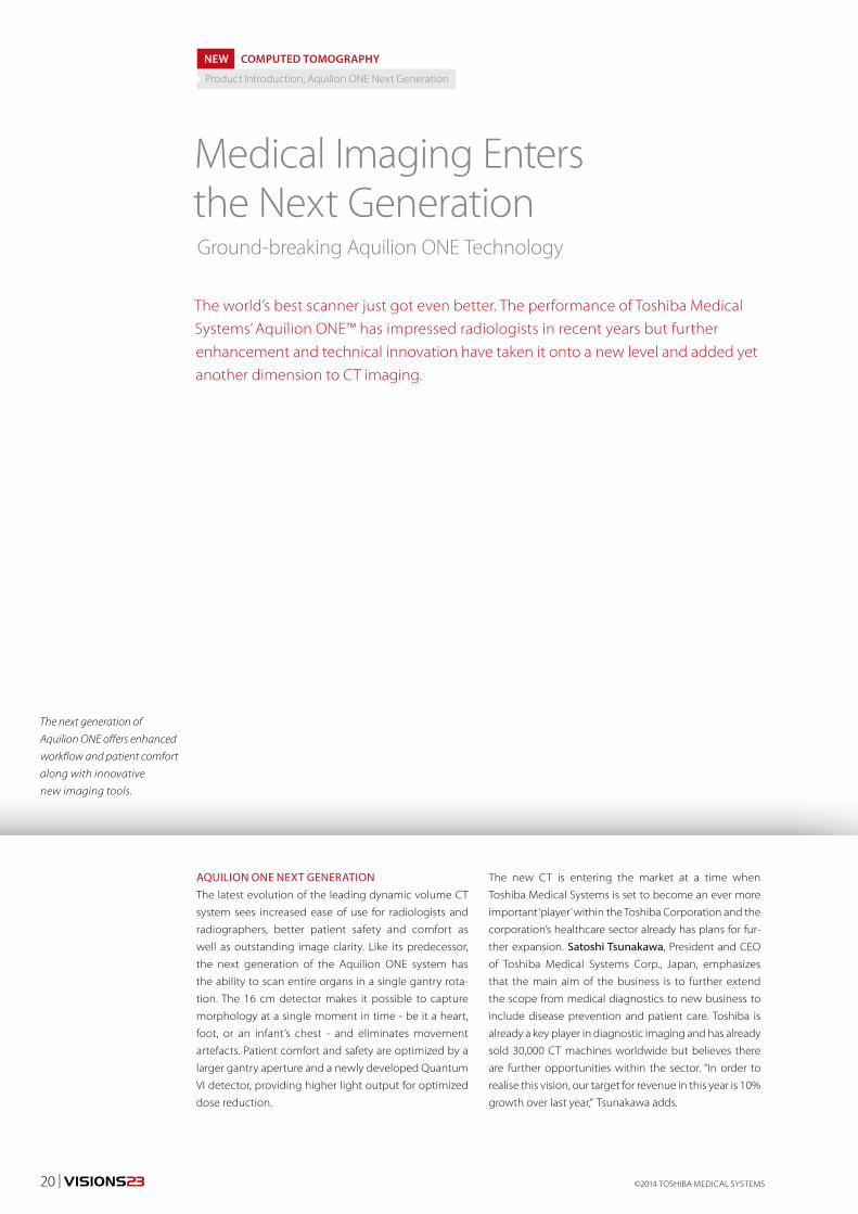
Research is facilitated by the Aquilion ONE, which was purchased in 2013,
primarily as support for applied R&D in petroleum recovery and advanced
materials. This CT system was an upgrade to an Aquilion 64, which had been
installed in the Edmonton facility in 2007.
The decision to purchase the original Aquilion 64 in 2007 was based partly on its
ability to monitor dynamic processes. Another deciding factor was the presence
of a local service base, which enables Tech Futures to obtain same-day service
calls to address instrument problems. While the energy range of the medical
scanner is lower than that of the industrial CT scanner HOOS used previously,
the trade-off of higher energies and longer scan times for greater speed and local
service was deemed favourable.
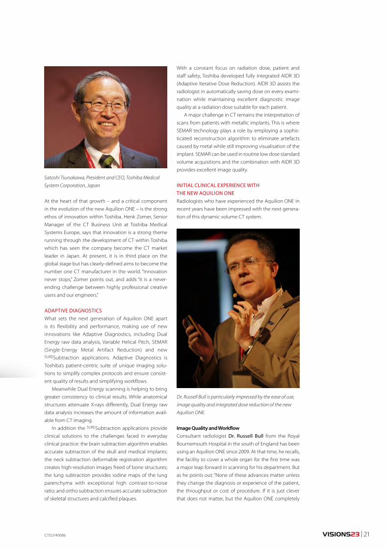
The speed of the scanner, for both the acquisition and reconstruction of data,
makes it possible to track processes that change on a scale of minutes. This is
invaluable to fluid flow experiments in cores and sand packs. The wide area
coverage allows cores to be scanned in fewer sections and enhances the
measurements of dynamic processes.
LOCAL SERVICE SEALS THE DEAL
Core experiments on the Aquilion ONE
Flow Experiments on the Aquilion 64
CTEU130078
©2014 TOSHIBA MEDICAL SYSTEMS20 | VISIONS23
AQUILION ONE NEXT GENERATIONThe latest evolution of the leading dynamic volume CT
system sees increased ease of use for radiologists and
radiographers, better patient safety and comfort as
well as outstanding image clarity. Like its predecessor,
the next generation of the Aquilion ONE system has
the ability to scan entire organs in a single gantry rota-
tion. The 16 cm detector makes it possible to capture
morphology at a single moment in time - be it a heart,
foot, or an infant’s chest - and eliminates movement
artefacts. Patient comfort and safety are optimized by a
larger gantry aperture and a newly developed Quantum
VI detector, providing higher light output for optimized
dose reduction.
The new CT is entering the market at a time when
Toshiba Medical Systems is set to become an ever more
important ‘player’ within the Toshiba Corporation and the
corporation’s healthcare sector already has plans for fur-
ther expansion. Satoshi Tsunakawa, President and CEO
of Toshiba Medical Systems Corp., Japan, emphasizes
that the main aim of the business is to further extend
the scope from medical diagnostics to new business to
include disease prevention and patient care. Toshiba is
already a key player in diagnostic imaging and has already
sold 30,000 CT machines worldwide but believes there
are further opportunities within the sector. “In order to
realise this vision, our target for revenue in this year is 10%
growth over last year,” Tsunakawa adds.
Medical Imaging Enters the Next Generation
The world’s best scanner just got even better. The performance of Toshiba Medical Systems’ Aquilion ONE™ has impressed radiologists in recent years but further enhancement and technical innovation have taken it onto a new level and added yet another dimension to CT imaging.
NEW COMPUTED TOMOGRAPHY
Product Introduction, Aquilion ONE Next Generation
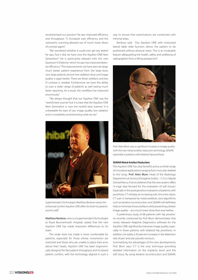
The next generation of
Aquilion ONE offers enhanced
workflow and patient comfort
along with innovative
new imaging tools.
Ground-breaking Aquilion ONE Technology
VISIONS23 | 21CTEU140086
At the heart of that growth – and a critical component
in the evolution of the new Aquilion ONE – is the strong
ethos of innovation within Toshiba. Henk Zomer, Senior
Manager of the CT Business Unit at Toshiba Medical
Systems Europe, says that innovation is a strong theme
running through the development of CT within Toshiba
which has seen the company become the CT market
leader in Japan. At present, it is in third place on the
global stage but has clearly-defined aims to become the
number one CT manufacturer in the world. “Innovation
never stops,” Zomer points out, and adds “it is a never-
ending challenge between highly professional creative
users and our engineers.”
ADAPTIVE DIAGNOSTICSWhat sets the next generation of Aquilion ONE apart
is its flexibility and performance, making use of new
innovations like Adaptive Diagnostics, including Dual
Energy raw data analysis, Variable Helical Pitch, SEMAR
(Single-Energy Metal Artifact Reduction) and new SURESubtraction applications. Adaptive Diagnostics is
Toshiba’s patient-centric suite of unique imaging solu-
tions to simplify complex protocols and ensure consist-
ent quality of results and simplifying workflows.
Meanwhile Dual Energy scanning is helping to bring
greater consistency to clinical results. While anatomical
structures attenuate X-rays differently, Dual Energy raw
data analysis increases the amount of information avail-
able from CT imaging.
In addition the SURESubtraction applications provide
clinical solutions to the challenges faced in everyday
clinical practice: the brain subtraction algorithm enables
accurate subtraction of the skull and medical implants;
the neck subtraction deformable registration algorithm
creates high-resolution images freed of bone structures;
the lung subtraction provides iodine maps of the lung
parenchyma with exceptional high contrast-to-noise
ratio; and ortho subtraction ensures accurate subtraction
of skeletal structures and calcified plaques.
With a constant focus on radiation dose, patient and
staff safety, Toshiba developed fully integrated AIDR 3D
(Adaptive Iterative Dose Reduction). AIDR 3D assists the
radiologist in automatically saving dose on every exami-
nation while maintaining excellent diagnostic image
quality at a radiation dose suitable for each patient.
A major challenge in CT remains the interpretation of
scans from patients with metallic implants. This is where
SEMAR technology plays a role by employing a sophis-
ticated reconstruction algorithm to eliminate artefacts
caused by metal while still improving visualisation of the
implant. SEMAR can be used in routine low dose standard
volume acquisitions and the combination with AIDR 3D
provides excellent image quality.
INITIAL CLINICAL EXPERIENCE WITH THE NEW AQUILION ONERadiologists who have experienced the Aquilion ONE in
recent years have been impressed with the next genera-
tion of this dynamic volume CT system.
Image Quality and WorkflowConsultant radiologist Dr. Russell Bull from the Royal
Bournemouth Hospital in the south of England has been
using an Aquilion ONE since 2009. At that time, he recalls,
the facility to cover a whole organ for the first time was
a major leap forward in scanning for his department. But
as he points out: “None of these advances matter unless
they change the diagnosis or experience of the patient,
the throughput or cost of procedure. If it is just clever
that does not matter, but the Aquilion ONE completely
Satoshi Tsunakawa, President and CEO, Toshiba Medical
System Corporation, Japan
Dr. Russell Bull is particularly impressed by the ease of use,
image quality and integrated dose reduction of the new
Aquilion ONE.
©2014 TOSHIBA MEDICAL SYSTEMS22 | VISIONS23
revolutionised our practice.” He saw improved efficiency
and throughput. “It increased scan efficiency and the
volumetric scanning allowed use of much lower doses
of contrast agent.”
“We wondered whether it could ever get any better,”
he says, “but it did, we have now the Aquilion ONE Next
Generation.” He is particularly pleased with the new
Quantum Vi Detector which he says has improved detec-
tor efficiency. “The improvements we have seen are large;
much better patient experience from the large bore,
very large patients receive low radiation dose and image
quality is again better. There are fewer artefacts and less
IV contrast is needed. Furthermore we have the ability
to scan a wider range of patients as well seeing much
faster reporting. As a result, the workflow has improved
enormously.”
“We always thought that our Aquilion ONE was the
‘world’s best scanner’ but it is clear that the Aquilion ONE
Next Generation is now the ‘world’s best scanner’. It is
unbeatable for ease of use, image quality, low radiation
and is completely revolutionising what we do.”
Matthew Benbow, who is a Superintendent Technologist
at Royal Bournemouth Hospital, stated that the new
Aquilion ONE has made important differences to his
team.
The wider bore has made it more comfortable for
patients, especially for those whose movements are
restricted and those who are unable to place their arms
above their heads. Aquilion ONE has been ergonomi-
cally designed for fast patient throughput and increased
patient comfort, with the technology aligned in such a
way to ensure that examinations are conducted with
minimal steps.
Benbow said: “Our Aquilion ONE with motorized
lateral table slide function allows the patient to be
positioned without physical strain. This is an invaluable
feature safeguarding the health, safety and wellbeing of
radiographers from a lifting perspective”.
SEMAR Metal Artefact ReductionThe Aquilion ONE has clear benefits across a whole range
of innovative applications ranging from muscular-skeletal
to the lungs. Prof. Alain Blum, Head of the Radiology
Department at Service d’Imagerie Guilloz – C.H.U. Hôpital
Central Nancy, France, believes that the new system offers
“a huge step forward for the evaluation of soft tissues.”
Especially in the postoperative evaluation of patients with
prosthesis, CT will play an increasing role. Since the classic
CT scan is hampered by metal artefacts, new algorithms
such as iterative reconstruction and SEMAR will definitely
help to minimize those artefacts while presenting a better
image quality – at a much lower dose than ever before.
A preliminary study of 68 patients with hip prosthe-
sis recently conducted by Prof. Blum demonstrates that
newly released Adaptive Diagnostics software for the
Aquilion ONE significantly improves image quality, espe-
cially in those patients with bilateral hip prosthesis. In
addition, he states a 30 percent increase in the detection
rate of peri-articular pseudo-tumours.
Summarizing the advantages of the new developments
Prof. Blum says: “CT is the only technique providing
detailed information on the implants, bone and the
soft tissue. By using iterative reconstruction and SEMAR,
Superintendent Technologist Matthew Benbow values the
enhanced comfort Aquilion ONE offers for both his patients
and his staff.
Prof. Alain Blum sees a significant increase in image quality
with the new metal artefact reduction technology SEMAR,
especially in patients with bilateral hip prosthesis.
VISIONS23 | 23
artefacts are significantly reduced and diagnostic image
quality is dramatically improved. This will surely lead to an
increase of CT-examinations for patients with hip pros-
thesis, so far difficult to examine.”
SURESUBTRACTION LUNG IODINE MAPPINGProf. Mathias Prokop, Chairman of the Department
of Radiology at Radboud University Medical Center
in Nijmegen in The Netherlands, has evaluated SURESubtraction Lung.
This newly developed Adaptive Diagnostics software
creates iodine maps by subtracting a low-dose pre-
contrast CT from a CT angiogram of the lung. “The iodine
maps created by SURESubtraction on the Aquilion ONE,”
Prof. Prokop points out, “have greatly improved signal
and higher spatial resolution compared to iodine maps
derived from dual energy acquisitions. SURESubtraction
Lung shows perfusion defects and thus provides direct
insight in the effect of embolism on lung perfusion.
By providing a perfusion map of the lung it can help
SURESubtraction Lung provides iodine maps of the lung
parenchyma with a high contrast/noise ratio. The color
overlay allows easy identification of hypo-perfused areas.
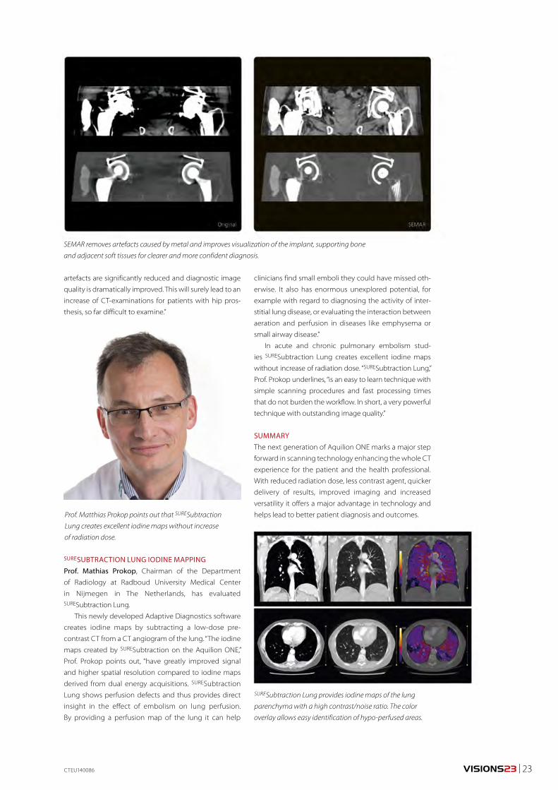
SEMAR removes artefacts caused by metal and improves visualization of the implant, supporting bone
and adjacent soft tissues for clearer and more confident diagnosis.
Prof. Matthias Prokop points out that SURESubtraction
Lung creates excellent iodine maps without increase
of radiation dose.
clinicians find small emboli they could have missed oth-
erwise. It also has enormous unexplored potential, for
example with regard to diagnosing the activity of inter-
stitial lung disease, or evaluating the interaction between
aeration and perfusion in diseases like emphysema or
small airway disease.”
In acute and chronic pulmonary embolism stud-
ies SURESubtraction Lung creates excellent iodine maps
without increase of radiation dose. “SURESubtraction Lung,”
Prof. Prokop underlines, “is an easy to learn technique with
simple scanning procedures and fast processing times
that do not burden the workflow. In short, a very powerful
technique with outstanding image quality.”
SUMMARYThe next generation of Aquilion ONE marks a major step
forward in scanning technology enhancing the whole CT
experience for the patient and the health professional.
With reduced radiation dose, less contrast agent, quicker
delivery of results, improved imaging and increased
versatility it offers a major advantage in technology and
helps lead to better patient diagnosis and outcomes.
CTEU140086
©2014 TOSHIBA MEDICAL SYSTEMS24 | VISIONS23
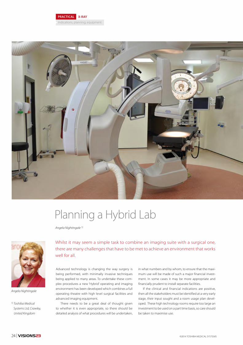
Whilst it may seem a simple task to combine an imaging suite with a surgical one, there are many challenges that have to be met to achieve an environment that works well for all.
Angela Nightingale 1)
Angela Nightingale
1) Toshiba Medical
Systems Ltd, Crawley,
United Kingdom
PRACTICAL X-RAY
Indications, planning, equipment
Advanced technology is changing the way surgery is
being performed, with minimally invasive techniques
being applied to many areas. To undertake these com-
plex procedures a new ‘Hybrid’ operating and imaging
environment has been developed which combines a full
operating theatre with high level surgical facilities and
advanced imaging equipment.
There needs to be a great deal of thought given
to whether it is even appropriate, so there should be
detailed analysis of what procedures will be undertaken,
in what numbers and by whom, to ensure that the maxi-
mum use will be made of such a major financial invest-
ment. In some cases it may be more appropriate and
financially prudent to install separate facilities.
If the clinical and financial indications are positive,
then all the stakeholders must be identified at a very early
stage, their input sought and a room usage plan devel-
oped. These high technology rooms require too large an
investment to be used on a part time basis, so care should
be taken to maximise use.
Planning a Hybrid Lab
VISIONS23 | 25
CLINICAL INDICATIONS FOR A HYBRID LAB
CardiacInitially developed for Paediatric Cardiology, use of hybrid
labs has expanded into adult surgery with coronary revas-
cularisation, trans catheter valve replacement (TAVI) and
repair, left ventricular assist devices (LVAD’s) and aortic stent
placement ideally performed in such an environment.
TaviTrans catheter replacement of aortic valves is still limited
to patients at high risk during conventional surgical tech-
niques. The European Societies of Cardiac Surgery and
Cardiology have recommended the hybrid environment
as the ideal for these new less invasive techniques.
Figure 1: Tavi procedure picture
Congenital heart diseaseIn certain groups of patients and conditions the combi-
nation of imaging and a percutaneous approach reduces
the challenge of navigating complex anatomy, bypass
time, overall risk and therefore improves outcomes.
Coronary artery diseasePrimary diagnosis will always be via CT or a conventional
Cath, but in cases of graft failures research suggests
that 13-20% could be diagnosed and then immediately
repaired, but currently the two procedures are generally
regarded as separate options. A hybrid approach can
decrease morbidity and mortality when compared with
conventional surgery.
Endovascular aortic repairEndovascular repair of the descending aorta (EVAR) is a
well-established technique with a higher survival rate than
open surgery, but only recently has the same technique
been applied to the rest of the aorta. This is often com-
bined with open surgery, a situation for which a hybrid lab
is ideally suited and minimises risk for the patient.
XREU130018
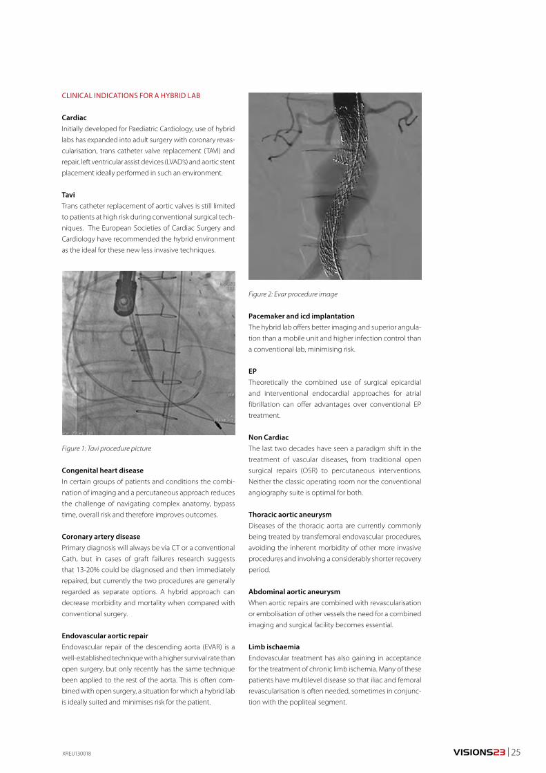
Figure 2: Evar procedure image
Pacemaker and icd implantationThe hybrid lab offers better imaging and superior angula-
tion than a mobile unit and higher infection control than
a conventional lab, minimising risk.
EPTheoretically the combined use of surgical epicardial
and interventional endocardial approaches for atrial
fibrillation can offer advantages over conventional EP
treatment.
Non CardiacThe last two decades have seen a paradigm shift in the
treatment of vascular diseases, from traditional open
surgical repairs (OSR) to percutaneous interventions.
Neither the classic operating room nor the conventional
angiography suite is optimal for both.
Thoracic aortic aneurysmDiseases of the thoracic aorta are currently commonly
being treated by transfemoral endovascular procedures,
avoiding the inherent morbidity of other more invasive
procedures and involving a considerably shorter recovery
period.
Abdominal aortic aneurysmWhen aortic repairs are combined with revascularisation
or embolisation of other vessels the need for a combined
imaging and surgical facility becomes essential.
Limb ischaemiaEndovascular treatment has also gaining in acceptance
for the treatment of chronic limb ischemia. Many of these
patients have multilevel disease so that iliac and femoral
revascularisation is often needed, sometimes in conjunc-
tion with the popliteal segment.
©2014 TOSHIBA MEDICAL SYSTEMS26 | VISIONS23
Neuro-interventionalFor Neuro-interventional procedures, a hybrid theatre
can provide clinical benefits in stroke, aneurysm, trauma,
paediatric and more complex cases. New procedures,
such as endovascular neurosurgery, are evolving as
the latest imaging equipment can provide advanced
image guidance to allow more accurate positioning of
Catheters, stents, coils and guide wires.
PLANNING A HYBRID THEATRE
Addressing all clinical needsFor the successful development of a hybrid lab the roles
of each of the stake holding groups need to be under-
stood and taken into account. The key internal people
who should be involved on hybrid suite planning com-
mittees include the surgeons and cardiologists who will
use the facility, the theatre and imaging lab staff, surgical
nurses, IT staff, facilities/estates manager, infection con-
trol personnel, hospital engineers and directorate man-
agement. External stakeholders are major equipment
manufacturers, the architect, M and E consultants, struc-
tural engineers, electricians and IT.
Location, location, locationIt is generally simpler to put a hybrid room close to or in
an Operating Theatre environment as the infrastructure
to support its use is close at hand, rather than try and
reproduce a full surgical environment in an open depart-
ment. In a theatre, positive pressure ventilation, sterilisa-
tion equipment, scrubs, etc., are all available so do not
need to be duplicated.
Size does matterA hybrid lab needs to be substantially larger than either a
conventional surgical theatre or an interventional imag-
ing room as, unlike an imaging only room, there needs to
be space to park the imaging equipment out of the way
of the surgical area.
Physical room considerationsInterventional imaging equipment may suspended from
the ceiling, located on the floor, or both, so the floor and
ceiling strength needs to be determined, before a choice
of equipment is made. Ceiling mounted systems require
a minimum and maximum height for installation which
may be difficult to achieve in an existing space.
Ancillary roomsThese should include patient preparation and recovery
areas, clean and dirty utility, staff areas and offices. If the
Hybrid room is located in the same area as other operat-
ing theatres, this will allow sharing of some of these facili-
ties, reducing cost and increasing usage rates.
Construction accessTypically the imaging equipment in a hybrid room is
delivered in one or two very large pieces which require
a delivery route with minimum door and ceiling heights
and widths, which should be taken into account when
looking at potential locations.
IN-ROOM PLANNING
Radiation protectionA radiation protection advisor should be consulted when
planning the layout of the area so that they can advise
on the level of protection required, which may include
lead shielding of wall/doors, radiation warning lights and
local rules.
When selecting the imaging equipment for the room,
input should be sought from radiology or cardiology staff
to look at clinical examination dose levels v image quality
and potential dose saving features of each system.
Figure 3: Aortic aneurysm repair
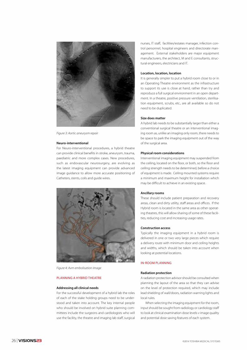
Figure 4: Avm embolisation image
VISIONS23 | 27XREU130018
LightingThere are many different types of lighting that need to be
planned in this type of suite – general/ambient, general
surgical and procedure specific. The various users of the
room should suggest the lighting they need and the pro-
ject manager and building works contractor will plan a
scheme in conjunction with specialists if needed.
Anaesthetic considerationsIn an ideal situation the positioning and type of these
facilities would suit every surgeon’s/anaesthetist’s
preference and procedure type, but this is rarely possi-
ble. Ceiling mounted units will provide the maximum
flexibility whilst keeping the floor clear of obstructions,
facilitating high levels of infection control and the best
access to the patient.
Air conditioningFull compliance with current codes of practice and
guidelines for a surgical theatre will be required. Imaging
equipment has an temperature range for optimum reli-
ability and image quality and its heat output and that of
other equipment in the room will need to be included in
all air conditioning calculations. Specific surgical proce-
dures may also require specialist environmental condi-
tions and these should be taken into account.
It considerationsHybrid surgical suites are often one of the most techno-
logically advanced rooms in a hospital, so the suite will
require multiple network points to access patient records,
previous studies and other data, which may also require
additional display monitors.
External AV conferencing is also frequently required
which will need to be planned for – microphone and
camera positions, network points, bandwidth etc. all have
to be part of the suite specification.
Infection controlIt is important that the site’s IC department is consulted at
an early stage to ensure that their recommendations are
incorporated into the design of the suite and possibly the
choice of equipment within the lab.
Storage/cathetersOpinion on storage for Catheters, devices, consumables
and other theatre items varies widely and will largely
depend on local opinion and the predominant use of
the room. For imaging there may be a need to store
Catheters within the room and appropriate cabinets
will need to be installed to comply with local infection
controls regulations. If space permits it may be better to
have a storage area adjacent to and accessible from the
theatre itself.
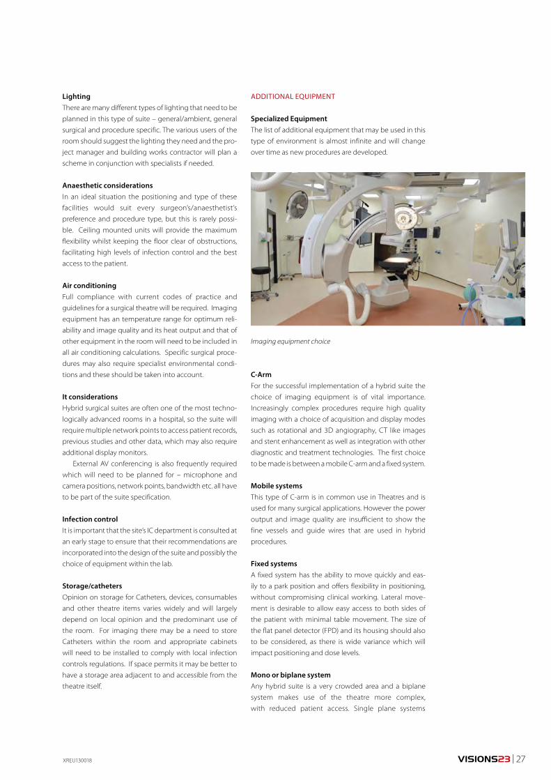
ADDITIONAL EQUIPMENT
Specialized EquipmentThe list of additional equipment that may be used in this
type of environment is almost infinite and will change
over time as new procedures are developed.
Imaging equipment choice
C-ArmFor the successful implementation of a hybrid suite the
choice of imaging equipment is of vital importance.
Increasingly complex procedures require high quality
imaging with a choice of acquisition and display modes
such as rotational and 3D angiography, CT like images
and stent enhancement as well as integration with other
diagnostic and treatment technologies. The first choice
to be made is between a mobile C-arm and a fixed system.
Mobile systemsThis type of C-arm is in common use in Theatres and is
used for many surgical applications. However the power
output and image quality are insufficient to show the
fine vessels and guide wires that are used in hybrid
procedures.
Fixed systemsA fixed system has the ability to move quickly and eas-
ily to a park position and offers flexibility in positioning,
without compromising clinical working. Lateral move-
ment is desirable to allow easy access to both sides of
the patient with minimal table movement. The size of
the flat panel detector (FPD) and its housing should also
to be considered, as there is wide variance which will
impact positioning and dose levels.
Mono or biplane systemAny hybrid suite is a very crowded area and a biplane
system makes use of the theatre more complex,
with reduced patient access. Single plane systems
©2014 TOSHIBA MEDICAL SYSTEMS28 | VISIONS23
are generally the system of choice for most vascular,
cardiac, GI and orthopaedic suites. A biplane system may
be considered for dedicated neuroradiology, paediatric
cardiology or electrophysiology hybrid theatres, but
the actual use should be clarified with all stakeholders
to ensure that the need for Biplane imaging outweighs
the drawbacks of the room complexity.
Floor or ceiling mountCeiling mounted systems generally have a greater range
of movement, take up no floor space and are easier to
park away from the operating table. Floor-mounted sys-
tems have no overhead cabling or ceiling tracks above
the operative field, reducing the infection control burden.
Reconciling these two aspects is an individual choice, but
the majority of hospitals decide on ceiling mount as they
provide best access to the patient without the need to
move the table and the most flexible use of the theatre.
TableThe challenge in selecting a table for a hybrid theatre is
the compromise between imaging and surgical needs.
Surgery needs a breakable table that can be customised
with different tops and accessories to cater for the varying
types of procedures that will be undertaken, but imaging
requires a radiolucent floating table top which is not
available with surgical tables. For 3D and CT like imaging
the table needs to be integrated into the imaging system
and so the choice of tables is normally limited to the one
that is standard on the system and one or two theatre
type tables.
SUMMARYImplementation of a hybrid surgical/imaging suite is a
complex process involving many different clinical and
non-clinical disciplines with different requirements and
priorities. Keys to a successful and economically viable
outcome are consultation, accurate understanding of the
purpose of the suite and detailed planning.
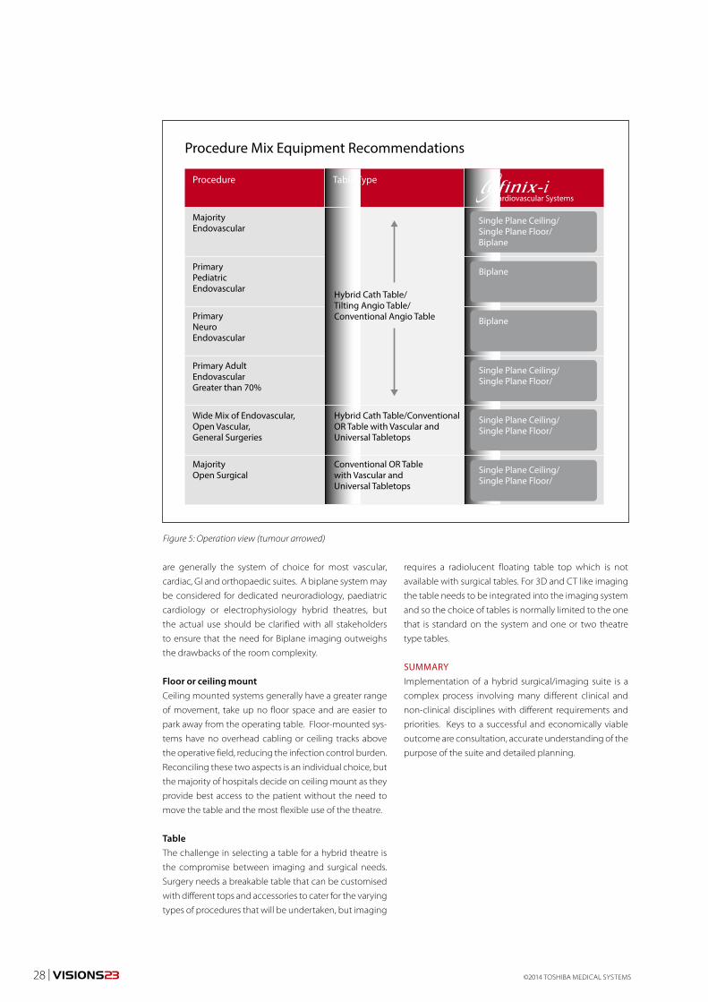
Procedure Mix Equipment Recommendations
Procedure
MajorityEndovascular
PrimaryPediatricEndovascular
PrimaryNeuroEndovascular
Primary AdultEndovascularGreater than 70%
Wide Mix of Endovascular,Open Vascular,General Surgeries
MajorityOpen Surgical
Hybrid Cath Table/Conventional OR Table with Vascular and Universal Tabletops
Hybrid Cath Table/Tilting Angio Table/Conventional Angio Table
Conventional OR Tablewith Vascular andUniversal Tabletops
Table Type
Cardiovascular Systems
Single Plane Ceiling/Single Plane Floor/Biplane
Biplane
Biplane
Single Plane Ceiling/Single Plane Floor/
Single Plane Ceiling/Single Plane Floor/
Single Plane Ceiling/Single Plane Floor/
Figure 5: Operation view (tumour arrowed)
VISIONS23 | 29
By virtue of the interaction of emitted xrays from the CT
tube with the k-edge of matter, CT has not had difficulty
differentiating anatomic structures with significantly dif-
ferent composition or atomic number. The attenuation
coefficient of photons varies significantly with differing
k-edge binding energies. Fat, bone or air for example will
not be mistaken on CT. The challenge arises for matter
with similar k-edge binding energies. Two materials will
behave differently at differing photon energies, depend-
ing on the elements they are composed. Matter with a
high atomic number such as calcium has a higher change
in attenuation at differing kVp energies than uric acid for
example, which has a lower atomic number.
By emitting two different photon energies, 135 and
80 kVp, matter with close k-edge binding energies can
be differentiated. Aquilion ONE and Aquilion PRIME
(Toshiba), are amongst the scanners that are leading the
way in this new era of CT technology.
PRACTICAL CONSIDERATIONS FOR DUAL ENERGY CT
1. Noise and image quality: How to prevent noise on the low dose scan?
Although two separate volumes with separate energies
are acquired, the Toshiba’s DE technology automatically
adjusts the mA settings for each kVp to provide image
volumes with similar signal to noise ratios.
2. Coverage:Complete coverage of a region is acquired at both
energies by using a very low pitch. Scan start times
are synchronized. With the z axis resolution of 16cm in
the Aquilion ONE, volumetric scanning of a solid organ
can be acquired with two temporally uniform data sets
provided. Any potential for motion artifact is eliminated
Dual Energy CT in the PRIME Time
The storms of the ‘slice war’, ‘gantry rotation war’ and ‘dose war’ have calmed, for now. Significant technologic advances are now available such as 320 detector row panels with 16cm volume coverage and remarkable gantry rotation times (275ms) on the Aquilion ONE™ (Toshiba). Toshiba AIDR 3D technology has shown the effective dose exposures can be reduced by up to 75%. What is next for CT, what battle to choose now? Huge advances in spatial and temporal resolution aren’t enough? CT now leaps further into the realm of tissue characterisation like its rival MRI?
Dr. Orla Buckley 1)
Dr. Orla Buckley
1) Tallaght Hospital
Ireland
CTEU140081
TECHNOLOGY COMPUTED TOMOGRAPHY
Dual Energy
with this capability. Scanners with less detector rows take
advantage of a very low pitch to ensure coverage of the
required volume.
3. Radiation exposure:Low pitch, dual energy exposures… Is this recipe for high
radiation exposure? Depending on the body mass of the
patient and the volume of the target organ, the mA can
be manually set to a higher or lower on the console prior
to scanning. Secondly, exposure can be manually turned
OFF in the upper part of gantry rotation that would
expose the ventral side of the patient and potentially
radiation sensitive areas such as breast tissue in females
(Fig. 1).
Figure 1: Tube exposure can be manually turned OFF in the
upper 180° of gantry rotation that would expose the ventral
side of the patient and potentially more radiation sensitive
areas such as breast tissue in females.
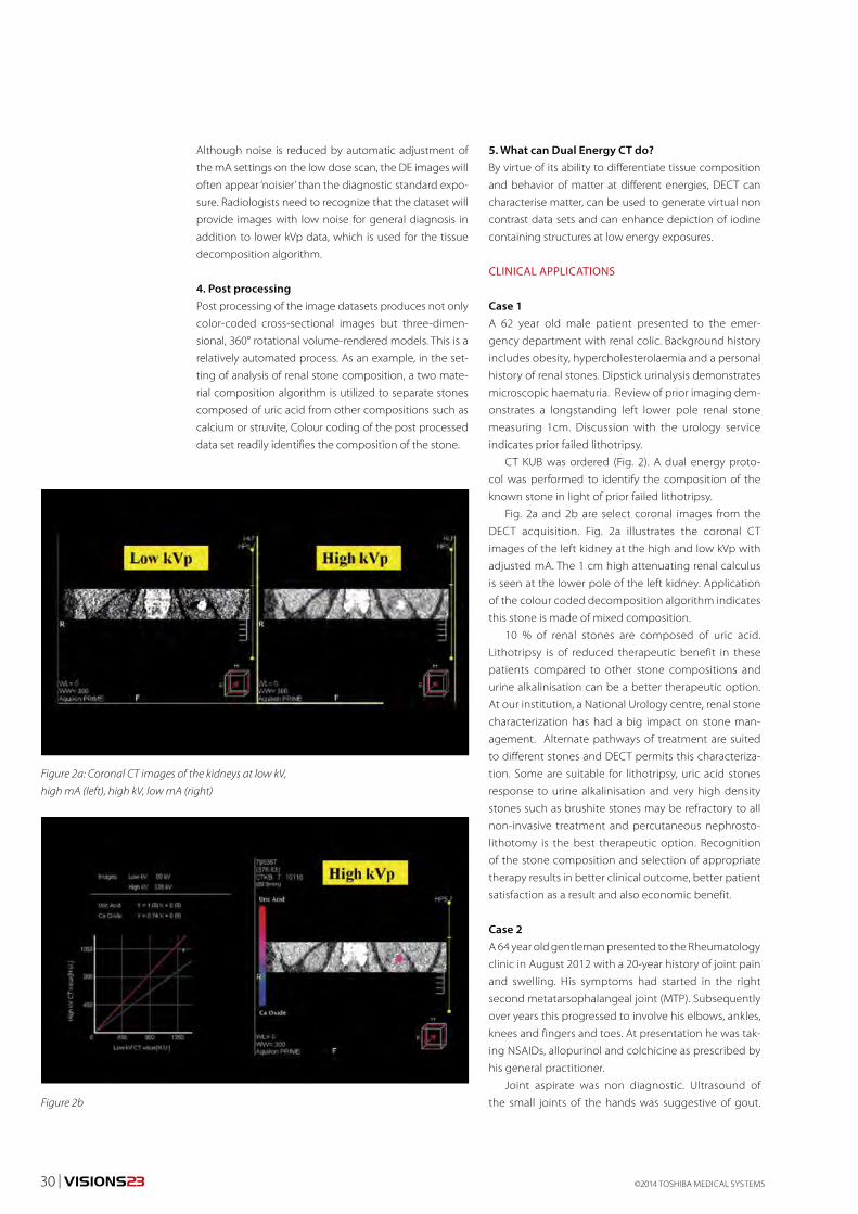
©2014 TOSHIBA MEDICAL SYSTEMS30 | VISIONS23
Figure 2a: Coronal CT images of the kidneys at low kV,
high mA (left), high kV, low mA (right)
Figure 2b
Although noise is reduced by automatic adjustment of
the mA settings on the low dose scan, the DE images will
often appear ‘noisier’ than the diagnostic standard expo-
sure. Radiologists need to recognize that the dataset will
provide images with low noise for general diagnosis in
addition to lower kVp data, which is used for the tissue
decomposition algorithm.
4. Post processingPost processing of the image datasets produces not only
color-coded cross-sectional images but three-dimen-
sional, 360° rotational volume-rendered models. This is a
relatively automated process. As an example, in the set-
ting of analysis of renal stone composition, a two mate-
rial composition algorithm is utilized to separate stones
composed of uric acid from other compositions such as
calcium or struvite, Colour coding of the post processed
data set readily identifies the composition of the stone.
5. What can Dual Energy CT do?By virtue of its ability to differentiate tissue composition
and behavior of matter at different energies, DECT can
characterise matter, can be used to generate virtual non
contrast data sets and can enhance depiction of iodine
containing structures at low energy exposures.
CLINICAL APPLICATIONS
Case 1A 62 year old male patient presented to the emer-
gency department with renal colic. Background history
includes obesity, hypercholesterolaemia and a personal
history of renal stones. Dipstick urinalysis demonstrates
microscopic haematuria. Review of prior imaging dem-
onstrates a longstanding left lower pole renal stone
measuring 1cm. Discussion with the urology service
indicates prior failed lithotripsy.
CT KUB was ordered (Fig. 2). A dual energy proto-
col was performed to identify the composition of the
known stone in light of prior failed lithotripsy.
Fig. 2a and 2b are select coronal images from the
DECT acquisition. Fig. 2a illustrates the coronal CT
images of the left kidney at the high and low kVp with
adjusted mA. The 1 cm high attenuating renal calculus
is seen at the lower pole of the left kidney. Application
of the colour coded decomposition algorithm indicates
this stone is made of mixed composition.
10 % of renal stones are composed of uric acid.
Lithotripsy is of reduced therapeutic benefit in these
patients compared to other stone compositions and
urine alkalinisation can be a better therapeutic option.
At our institution, a National Urology centre, renal stone
characterization has had a big impact on stone man-
agement. Alternate pathways of treatment are suited
to different stones and DECT permits this characteriza-
tion. Some are suitable for lithotripsy, uric acid stones
response to urine alkalinisation and very high density
stones such as brushite stones may be refractory to all
non-invasive treatment and percutaneous nephrosto-
lithotomy is the best therapeutic option. Recognition
of the stone composition and selection of appropriate
therapy results in better clinical outcome, better patient
satisfaction as a result and also economic benefit.
Case 2A 64 year old gentleman presented to the Rheumatology
clinic in August 2012 with a 20-year history of joint pain
and swelling. His symptoms had started in the right
second metatarsophalangeal joint (MTP). Subsequently
over years this progressed to involve his elbows, ankles,
knees and fingers and toes. At presentation he was tak-
ing NSAIDs, allopurinol and colchicine as prescribed by
his general practitioner.
Joint aspirate was non diagnostic. Ultrasound of
the small joints of the hands was suggestive of gout.
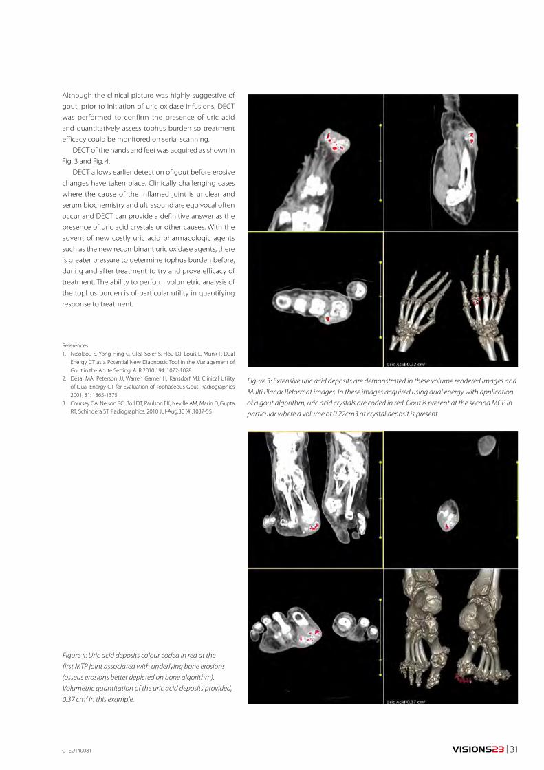
VISIONS23 | 31
Although the clinical picture was highly suggestive of
gout, prior to initiation of uric oxidase infusions, DECT
was performed to confirm the presence of uric acid
and quantitatively assess tophus burden so treatment
efficacy could be monitored on serial scanning.
DECT of the hands and feet was acquired as shown in
Fig. 3 and Fig. 4.
DECT allows earlier detection of gout before erosive
changes have taken place. Clinically challenging cases
where the cause of the inflamed joint is unclear and
serum biochemistry and ultrasound are equivocal often
occur and DECT can provide a definitive answer as the
presence of uric acid crystals or other causes. With the
advent of new costly uric acid pharmacologic agents
such as the new recombinant uric oxidase agents, there
is greater pressure to determine tophus burden before,
during and after treatment to try and prove efficacy of
treatment. The ability to perform volumetric analysis of
the tophus burden is of particular utility in quantifying
response to treatment.
References1. Nicolaou S, Yong-Hing C, Glea-Soler S, Hou DJ, Louis L, Munk P. Dual
Energy CT as a Potential New Diagnostic Tool in the Management of Gout in the Acute Setting. AJR 2010 194: 1072-1078.
2. Desai MA, Peterson JJ, Warren Garner H, Kansdorf MJ. Clinical Utility of Dual Energy CT for Evaluation of Tophaceous Gout. Radiographics 2001; 31: 1365-1375.
3. Coursey CA, Nelson RC, Boll DT, Paulson EK, Neville AM, Marin D, Gupta RT, Schindera ST. Radiographics. 2010 Jul-Aug;30 (4):1037-55
Figure 3: Extensive uric acid deposits are demonstrated in these volume rendered images and
Multi Planar Reformat images. In these images acquired using dual energy with application
of a gout algorithm, uric acid crystals are coded in red. Gout is present at the second MCP in
particular where a volume of 0.22cm3 of crystal deposit is present.
Figure 4: Uric acid deposits colour coded in red at the
first MTP joint associated with underlying bone erosions
(osseus erosions better depicted on bone algorithm).
Volumetric quantitation of the uric acid deposits provided,
0.37 cm³ in this example.
CTEU140081
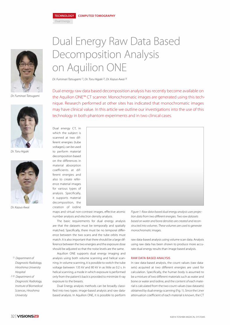
©2014 TOSHIBA MEDICAL SYSTEMS32 | VISIONS23
Dual energy CT, in
which the subject is
scanned at two dif-
ferent energies (tube
voltages), can be used
to perform material
decomposition based
on the differences in
material absorption
coefficients at dif-
ferent energies and
also to create refer-
ence material images
for various types of
analysis. Specifically,
it supports material
decomposition, the
creation of iodine
maps and virtual non-contrast images, effective atomic
number analysis and electron density analysis.
The basic requirements for dual energy analysis
are that the datasets must be temporally and spatially
matched. Specifically, there must be no temporal differ-
ence between the two scans and the tube orbits must
match. It is also important that there should be a large dif-
ference between the two energies and the exposure dose
should be adjusted so that the noise levels are the same.
Aquilion ONE supports dual energy imaging and
analysis using both volume scanning and helical scan-
ning. In volume scanning, it is possible to switch the tube
voltage between 135 kV and 80 kV in as little as 0.2 s. In
helical scanning, a mode in which exposure is performed
only from the patient’s back is provided to minimize X-ray
exposure to the breasts.
Dual Energy analysis methods can be broadly classi-
fied into two types: image-based analysis and raw data-
based analysis. In Aquilion ONE, it is possible to perform
Dual Energy Raw Data Based Decomposition Analysis on Aquilion ONE
Dual energy raw data based decomposition analysis has recently become available on the Aquilion ONE™ CT scanner. Monochromatic images are generated using this tech-nique. Research performed at other sites has indicated that monochromatic images may have clinical value. In this article we outline our investigations into the use of this technology in both phantom experiments and in two clinical cases.
Dr. Fuminari Tatsugami 1), Dr. Toru Higaki 2), Dr. Kazuo Awai 3)
Dr. Fuminari Tatsugami
Dr. Toru Higaki
Dr. Kazuo Awai
1) Department of
Diagnostic Radiology,
Hiroshima University
Hospital2-3) Department of
Diagnostic Radiology,
Institute of Biomedical
Sciences, Hiroshima
University
TECHNOLOGY COMPUTED TOMOGRAPHY
Dual Energy
raw data-based analysis using volume scan data. Analysis
using raw data has been shown to produce more accu-
rate dual energy results than image based analysis.
RAW DATA-BASED ANALYSISIn raw data-based analysis, the count values (raw data-
sets) acquired at two different energies are used for
calculation. Specifically, the human body is assumed to
be a mixture of two different materials such as water and
bone or water and iodine, and the content of each mate-
rial is calculated from the two count values (raw datasets)
obtained by dual energy scanning (Fig. 1). Since the Liner
attenuation coefficient of each material is known, the CT
Figure 1: Raw data-based dual energy analysis uses projec-
tion data from two different energies. Two raw datasets
based on water and bone densities are created and recon-
structed into volumes. These volumes are used to generate
monochromatic images.
VISIONS23 | 33CTEU130080
Figure 2: Beam hardening artifacts seen in a single energy 120kV image, image based dual energy image
and monochromatic raw data based dual energy image.
number at a given energy can be determined by combi-
nation with the calculated content, making it possible to
create monochromatic images (keV images).
Novel analysis software which utilizes raw data
based dual energy data available on the Aquilion
ONE includes reduction of beam hardening artifacts,
automatic generation of the best CNR images, virtual
non-contrast image, iodine map, effective atomic num-
ber and electron density imaging. Among these new
technologies, electron density images has gained atten-
tion of radiologists and radiation oncologists because
it is a promising method for generating more accurate
electron density images than conventional methods in
radiotherapy planning.
We performed phantom experiments to verify the
accuracy of this method.
PHANTOM EXPERIMENTS
Improvement of beam hardening artifactsWhen two syringes containing a high-absorption mate-
rial (iodine with a CT number of approximately 1000 HU)
are placed in a phantom filled with water and scanned
at a single energy, severe beam hardening artifacts are
observed between the two syringes. The beam hard-
ening artifacts are still seen in the Virtual 120kV image
generated using image based dual energy analysis. In a
monochromatic image created by raw data-based dual
energy analysis, the beam hardening artifacts are signifi-
cantly reduced (Fig. 2).
Best CNR imageThe use of monochromatic images makes it possible to
obtain an image with the highest possible contrast-to-
noise ratio in the region of interest (called the Best CNR
image). The results of comparison between image-based
analysis and raw data-based analysis showed that the
CNR was approximately 5% higher for raw data-based
analysis. It is considered that beam hardening artifacts in
high-absorption materials are reduced, resulting in more
accurate CT number measurement in the subject.
Virtual non-contrast imageInvestigations using a water phantom containing
syringes filled with fat and with iodinated contrast
medium at seven different concentrations showed that
both the iodine map and virtual non-contrast image
obtained using the raw data-based analysis tool were
superior. In the raw data-based analysis, the influence
of beam hardening artifacts was reduced and the CT
number accuracy in the virtual non-contrast image was
improved.
Effective atomic number and electron densityIt is possible to determine the effective atomic number
and the electron density of an object using the raw
Aquilion ONE
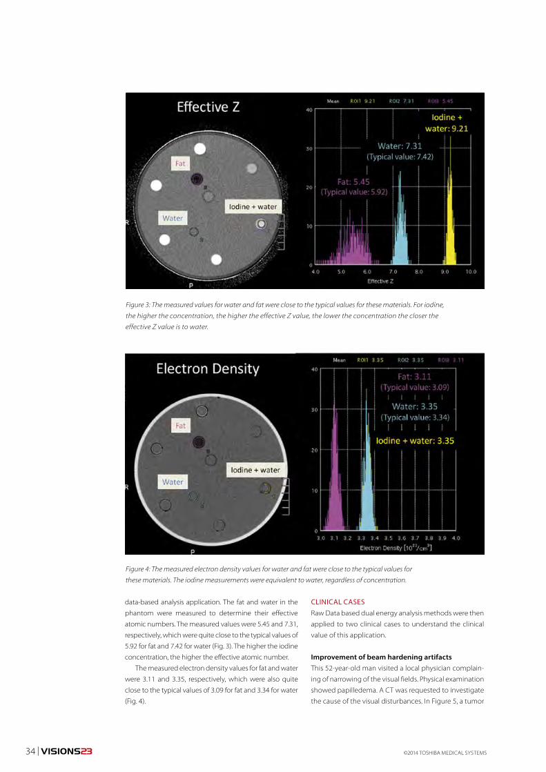
©2014 TOSHIBA MEDICAL SYSTEMS34 | VISIONS23
data-based analysis application. The fat and water in the
phantom were measured to determine their effective
atomic numbers. The measured values were 5.45 and 7.31,
respectively, which were quite close to the typical values of
5.92 for fat and 7.42 for water (Fig. 3). The higher the iodine
concentration, the higher the effective atomic number.
The measured electron density values for fat and water
were 3.11 and 3.35, respectively, which were also quite
close to the typical values of 3.09 for fat and 3.34 for water
(Fig. 4).
CLINICAL CASESRaw Data based dual energy analysis methods were then
applied to two clinical cases to understand the clinical
value of this application.
Improvement of beam hardening artifactsThis 52-year-old man visited a local physician complain-
ing of narrowing of the visual fields. Physical examination
showed papilledema. A CT was requested to investigate
the cause of the visual disturbances. In Figure 5, a tumor
Figure 3: The measured values for water and fat were close to the typical values for these materials. For iodine,
the higher the concentration, the higher the effective Z value, the lower the concentration the closer the
effective Z value is to water.
Figure 4: The measured electron density values for water and fat were close to the typical values for
these materials. The iodine measurements were equivalent to water, regardless of concentration.
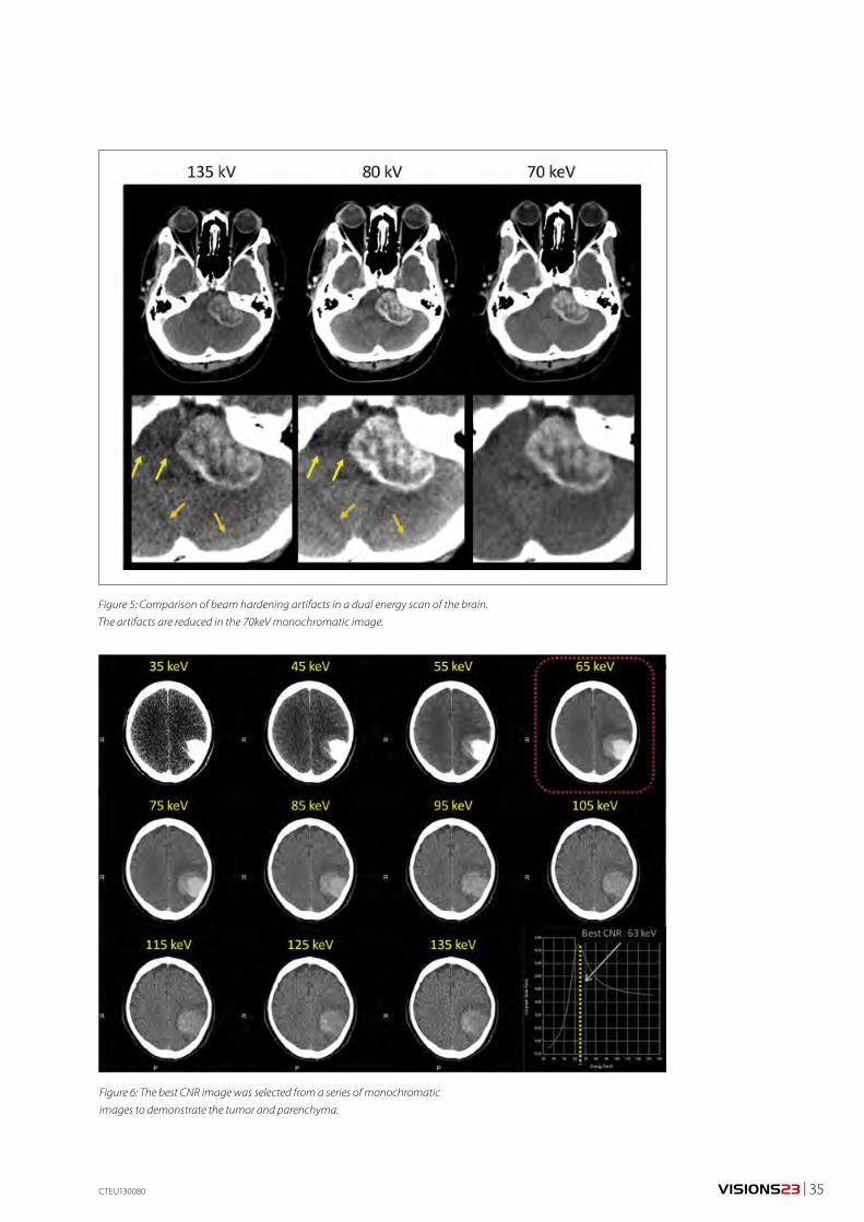
VISIONS23 | 35
Figure 5: Comparison of beam hardening artifacts in a dual energy scan of the brain.
The artifacts are reduced in the 70keV monochromatic image.
CTEU130080
Figure 6: The best CNR image was selected from a series of monochromatic
images to demonstrate the tumor and parenchyma.
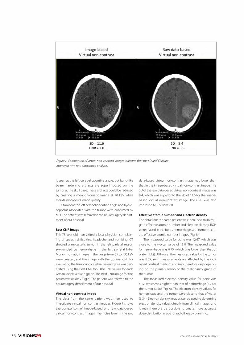
©2014 TOSHIBA MEDICAL SYSTEMS36 | VISIONS23
Figure 7: Comparison of virtual non-contrast images indicates that the SD and CNR are
improved with raw data based analysis.
is seen at the left cerebellopontine angle, but band-like
beam hardening artifacts are superimposed on the
tumor at the skull base. These artifacts could be reduced
by creating a monochromatic image at 70 keV while
maintaining good image quality.
A tumor at the left cerebellopontine angle and hydro-
cephalus associated with the tumor were confirmed by
MRI. The patient was referred to the neurosurgery depart-
ment of our hospital.
Best CNR imageThis 75-year-old man visited a local physician complain-
ing of speech difficulties, headache, and vomiting. CT
showed a metastatic tumor in the left parietal region
surrounded by hemorrhage in the left parietal lobe.
Monochromatic images in the range from 35 to 135 keV
were created, and the image with the optimal CNR for
evaluating the tumor and cerebral parenchyma was gen-
erated using the Best CNR tool. The CNR values for each
keV are displayed as a graph. The Best CNR image for this
patient was 63 keV (Fig 6). The patient was referred to the
neurosurgery department of our hospital.
Virtual non-contrast imageThe data from the same patient was then used to
investigate virtual non contrast images. Figure 7 shows
the comparison of image-based and raw data-based
virtual non-contrast images. The noise level in the raw
data-based virtual non-contrast image was lower than
that in the image-based virtual non-contrast image. The
SD of the raw data-based virtual non-contrast image was
8.4, which was superior to the SD of 11.6 for the image-
based virtual non-contrast image. The CNR was also
improved to 3.5 from 2.0.
Effective atomic number and electron densityThe data from the same patient was then used to investi-
gate effective atomic number and electron density. ROIs
were placed in the bone, hemorrhage, and tumor to cre-
ate effective atomic number images (Fig. 8).
The measured value for bone was 12.67, which was
close to the typical value of 13.8. The measured value
for hemorrhage was 6.75, which was lower than that of
water (7.42). Although the measured value for the tumor
was 8.69, such measurements are affected by the iodi-
nated contrast medium and may therefore vary depend-
ing on the primary lesion or the malignancy grade of
the tumor.
The measured electron density value for bone was
5.12, which was higher than that of hemorrhage (3.7) or
the tumor (3.58) (Fig. 9). The electron density values for
hemorrhage and the tumor were close to that of water
(3.34). Electron density images can be used to determine
electron density values directly from clinical images, and
it may therefore be possible to create more accurate
dose distribution maps for radiotherapy planning.
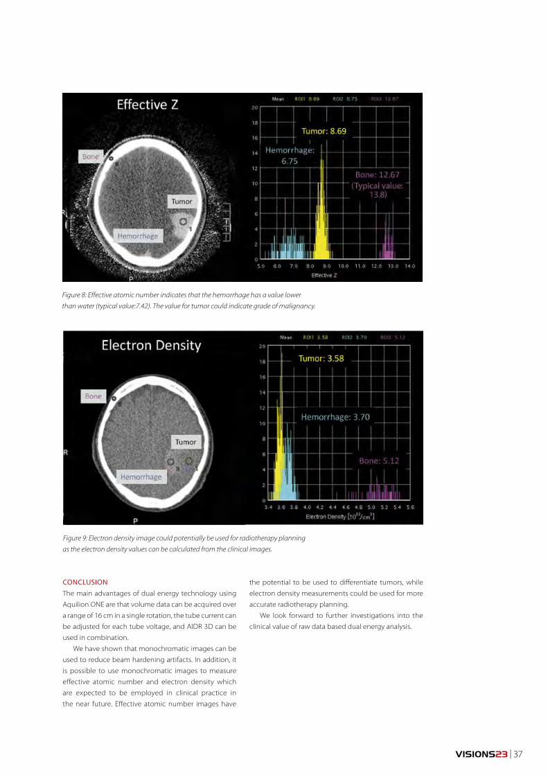
VISIONS23 | 37
Figure 8: Effective atomic number indicates that the hemorrhage has a value lower
than water (typical value:7.42). The value for tumor could indicate grade of malignancy.
Figure 9: Electron density image could potentially be used for radiotherapy planning
as the electron density values can be calculated from the clinical images.
CONCLUSIONThe main advantages of dual energy technology using
Aquilion ONE are that volume data can be acquired over
a range of 16 cm in a single rotation, the tube current can
be adjusted for each tube voltage, and AIDR 3D can be
used in combination.
We have shown that monochromatic images can be
used to reduce beam hardening artifacts. In addition, it
is possible to use monochromatic images to measure
effective atomic number and electron density which
are expected to be employed in clinical practice in
the near future. Effective atomic number images have
the potential to be used to differentiate tumors, while
electron density measurements could be used for more
accurate radiotherapy planning.
We look forward to further investigations into the
clinical value of raw data based dual energy analysis.
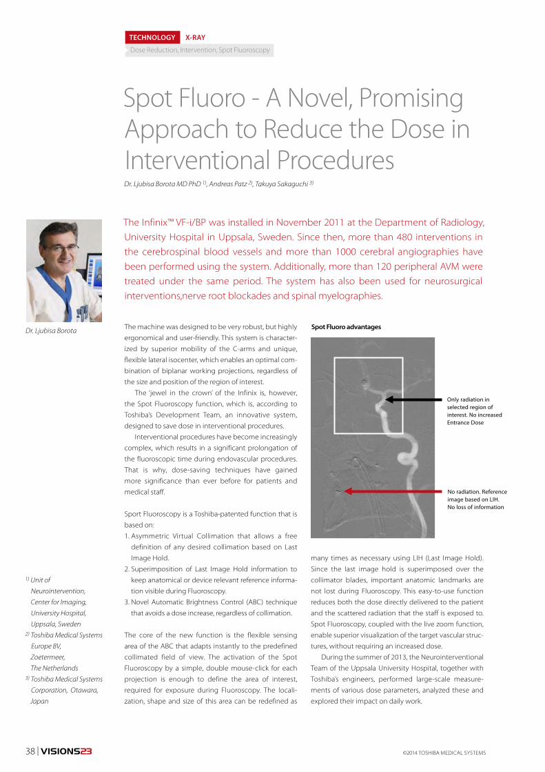
©2014 TOSHIBA MEDICAL SYSTEMS38 | VISIONS23
The machine was designed to be very robust, but highly
ergonomical and user-friendly. This system is character-
ized by superior mobility of the C-arms and unique,
flexible lateral isocenter, which enables an optimal com-
bination of biplanar working projections, regardless of
the size and position of the region of interest.
The ‘jewel in the crown’ of the Infinix is, however,
the Spot Fluoroscopy function, which is, according to
Toshiba’s Development Team, an innovative system,
designed to save dose in interventional procedures.
Interventional procedures have become increasingly
complex, which results in a significant prolongation of
the fluoroscopic time during endovascular procedures.
That is why, dose-saving techniques have gained
more significance than ever before for patients and
medical staff.
Sport Fluoroscopy is a Toshiba-patented function that is
based on:
1. Asymmetric Virtual Collimation that allows a free
definition of any desired collimation based on Last
Image Hold.
2. Superimposition of Last Image Hold information to
keep anatomical or device relevant reference informa-
tion visible during Fluoroscopy.
3. Novel Automatic Brightness Control (ABC) technique
that avoids a dose increase, regardless of collimation.
The core of the new function is the flexible sensing
area of the ABC that adapts instantly to the predefined
collimated field of view. The activation of the Spot
Fluoroscopy by a simple, double mouse-click for each
projection is enough to define the area of interest,
required for exposure during Fluoroscopy. The locali-
zation, shape and size of this area can be redefined as
The Infinix™ VF-i/BP was installed in November 2011 at the Department of Radiology, University Hospital in Uppsala, Sweden. Since then, more than 480 interventions in the cerebrospinal blood vessels and more than 1000 cerebral angiographies have been performed using the system. Additionally, more than 120 peripheral AVM were treated under the same period. The system has also been used for neurosurgical interventions,nerve root blockades and spinal myelographies.
1) Unit of
Neurointervention,
Center for Imaging,
University Hospital,
Uppsala, Sweden2) Toshiba Medical Systems
Europe BV,
Zoetermeer,
The Netherlands3) Toshiba Medical Systems
Corporation, Otawara,
Japan
TECHNOLOGY X-RAY
Dose Reduction, Intervention, Spot Fluoroscopy
Dr. Ljubisa Borota MD PhD 1), Andreas Patz 2), Takuya Sakaguchi 3)
many times as necessary using LIH (Last Image Hold).
Since the last image hold is superimposed over the
collimator blades, important anatomic landmarks are
not lost during Fluoroscopy. This easy-to-use function
reduces both the dose directly delivered to the patient
and the scattered radiation that the staff is exposed to.
Spot Fluoroscopy, coupled with the live zoom function,
enable superior visualization of the target vascular struc-
tures, without requiring an increased dose.
During the summer of 2013, the Neurointerventional
Team of the Uppsala University Hospital, together with
Toshiba’s engineers, performed large-scale measure-
ments of various dose parameters, analyzed these and
explored their impact on daily work.
Spot Fluoro - A Novel, Promising Approach to Reduce the Dose in Interventional Procedures
Dr. Ljubisa Borota
No radiation. Reference image based on LIH.No loss of information
Only radiation in selected region of interest. No increased Entrance Dose
Spot Fluoro advantages
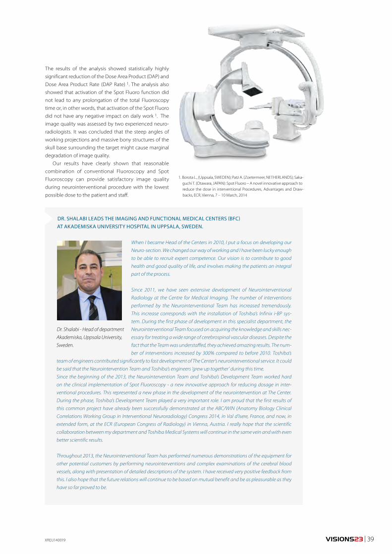
VISIONS23 | 39XREU140019
The results of the analysis showed statistically highly
significant reduction of the Dose Area Product (DAP) and
Dose Area Product Rate (DAP Rate) 1. The analysis also
showed that activation of the Spot Fluoro function did
not lead to any prolongation of the total Fluoroscopy
time or, in other words, that activation of the Spot Fluoro
did not have any negative impact on daily work 1. The
image quality was assessed by two experienced neuro-
radiologists. It was concluded that the steep angles of
working projections and massive bony structures of the
skull base surrounding the target might cause marginal
degradation of image quality.
Our results have clearly shown that reasonable
combination of conventional Fluoroscopy and Spot
Fluoroscopy can provide satisfactory image quality
during neurointerventional procedure with the lowest
possible dose to the patient and staff.
When I became Head of the Centers in 2010, I put a focus on developing our
Neuro-section. We changed our way of working and I have been lucky enough
to be able to recruit expert competence. Our vision is to contribute to good
health and good quality of life, and involves making the patients an integral
part of the process.
Since 2011, we have seen extensive development of Neurointerventional
Radiology at the Centre for Medical Imaging. The number of interventions
performed by the Neurointerventional Team has increased tremendously.
This increase corresponds with the installation of Toshiba’s Infinix i-BP sys-
tem. During the first phase of development in this specialist department, the
Neurointerventional Team focused on acquiring the knowledge and skills nec-
essary for treating a wide range of cerebrospinal vascular diseases. Despite the
fact that the Team was understaffed, they achieved amazing results. The num-
ber of interventions increased by 300% compared to before 2010. Toshiba’s
team of engineers contributed significantly to fast development of The Center’s neurointerventional service. It could
be said that the Neurointervention Team and Toshiba’s engineers ‘grew up together’ during this time.
Since the beginning of the 2013, the Neurointervention Team and Toshiba’s Development Team worked hard
on the clinical implementation of Spot Fluoroscopy - a new innovative approach for reducing dosage in inter-
ventional procedures. This represented a new phase in the development of the neurointervention at The Center.
During the phase, Toshiba’s Development Team played a very important role. I am proud that the first results of
this common project have already been successfully demonstrated at the ABC/WIN (Anatomy Biology Clinical
Correlations Working Group in Interventional Neuroradiology) Congress 2014, in Val d’Isere, France, and now, in
extended form, at the ECR (European Congress of Radiology) in Vienna, Austria. I really hope that the scientific
collaboration between my department and Toshiba Medical Systems will continue in the same vein and with even
better scientific results.
Throughout 2013, the Neurointerventional Team has performed numerous demonstrations of the equipment for
other potential customers by performing neurointerventions and complex examinations of the cerebral blood
vessels, along with presentation of detailed descriptions of the system. I have received very positive feedback from
this. I also hope that the future relations will continue to be based on mutual benefit and be as pleasurable as they
have so far proved to be.
Dr. Shalabi - Head of department
Akademiska, Uppsala University,
Sweden.
DR. SHALABI LEADS THE IMAGING AND FUNCTIONAL MEDICAL CENTERS (BFC) AT AKADEMISKA UNIVERSITY HOSPITAL IN UPPSALA, SWEDEN.
1. Borota L., (Uppsala, SWEDEN); Patz A. (Zoetermeer, NETHERLANDS); Saka-guchi T. (Otawara, JAPAN): Spot Fluoro – A novel innovative approach to reduce the dose in interventional Procedures, Advantages and Draw-backs, ECR, Vienna, 7 – 10 March, 2014
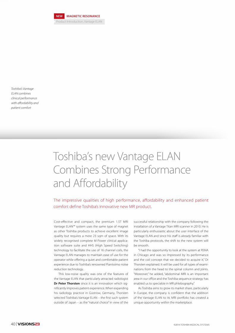
©2014 TOSHIBA MEDICAL SYSTEMS40 | VISIONS23
The impressive qualities of high performance, affordability and enhanced patient comfort define Toshiba’s innovative new MR product.
Cost-effective and compact, the premium 1.5T MRI
Vantage ELAN™ system uses the same type of magnet
as other Toshiba products to achieve excellent image
quality but requires a mere 23 sqm of space. With its
widely recognised complete M-Power clinical applica-
tion software suite and HHS (High Speed Switching)
technology to facilitate the use of 16 channel coils, the
Vantage ELAN manages to maintain ease of use for the
operator while offering a quiet and comfortable patient
experience due to Toshiba’s renowned Pianissimo noise
reduction technology.
This low-noise quality was one of the features of
the Vantage ELAN that particularly attracted radiologist
Dr Peter Thorsten since it is an innovation which sig-
nificantly improves patient experience. When expanding
his radiology practice in Güstrow, Germany, Thorsten
selected Toshiba’s Vantage ELAN – the first such system
outside of Japan – as the “natural choice” in view of the
successful relationship with the company following the
installation of a Vantage Titan MRI scanner in 2010. He is
particularly enthusiastic about the user interface of the
Vantage ELAN and since his staff is already familiar with
the Toshiba protocols, the shift to the new system will
be smooth.
“I had the opportunity to look at the system at RSNA
in Chicago and was so impressed by its performance
and the coil concept that we decided to acquire it,” Dr
Thorsten explained. It will be used for all types of exami-
nations from the head to the spinal column and joints.
“Moreover,” he added, “abdominal MRI is an important
area in our office and the Toshiba sequence strategy has
enabled us to specialize in MR phlebography.”
As Toshiba aims to grow its market share, particularly
in Europe, the company is confident that the addition
of the Vantage ELAN to its MRI portfolio has created a
unique opportunity within the marketplace.
Toshiba’s new Vantage ELAN Combines Strong Performance and Affordability
Toshiba’s Vantage
ELAN combines
clinical performance
with affordability and
patient comfort
NEW MAGNETIC RESONANCE
Product Introduction, Vantage ELAN
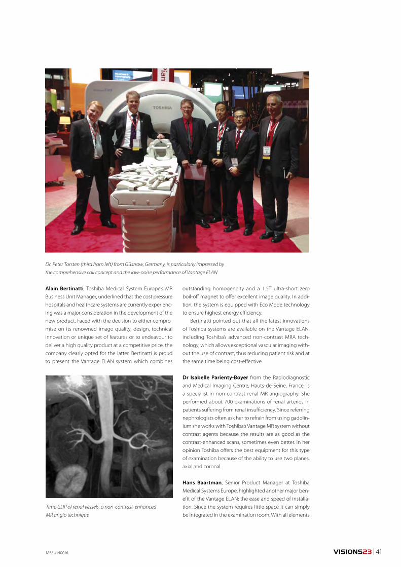
VISIONS23 | 41MREU140016
Alain Bertinatti, Toshiba Medical System Europe’s MR
Business Unit Manager, underlined that the cost pressure
hospitals and healthcare systems are currently experienc-
ing was a major consideration in the development of the
new product. Faced with the decision to either compro-
mise on its renowned image quality, design, technical
innovation or unique set of features or to endeavour to
deliver a high quality product at a competitive price, the
company clearly opted for the latter. Bertinatti is proud
to present the Vantage ELAN system which combines
outstanding homogeneity and a 1.5T ultra-short zero
boil-off magnet to offer excellent image quality. In addi-
tion, the system is equipped with Eco Mode technology
to ensure highest energy efficiency.
Bertinatti pointed out that all the latest innovations
of Toshiba systems are available on the Vantage ELAN,
including Toshiba’s advanced non-contrast MRA tech-
nology, which allows exceptional vascular imaging with-
out the use of contrast, thus reducing patient risk and at
the same time being cost-effective.
Dr Isabelle Parienty-Boyer from the Radiodiagnostic
and Medical Imaging Centre, Hauts-de-Seine, France, is
a specialist in non-contrast renal MR angiography. She
performed about 700 examinations of renal arteries in
patients suffering from renal insufficiency. Since referring
nephrologists often ask her to refrain from using gadolin-
ium she works with Toshiba’s Vantage MR system without
contrast agents because the results are as good as the
contrast-enhanced scans, sometimes even better. In her
opinion Toshiba offers the best equipment for this type
of examination because of the ability to use two planes,
axial and coronal.
Hans Baartman, Senior Product Manager at Toshiba
Medical Systems Europe, highlighted another major ben-
efit of the Vantage ELAN: the ease and speed of installa-
tion. Since the system requires little space it can simply
be integrated in the examination room. With all elements
Dr. Peter Torsten (third from left) from Güstrow, Germany, is particularly impressed by
the comprehensive coil concept and the low-noise performance of Vantage ELAN
Time-SLIP of renal vessels, a non-contrast-enhanced
MR angio technique
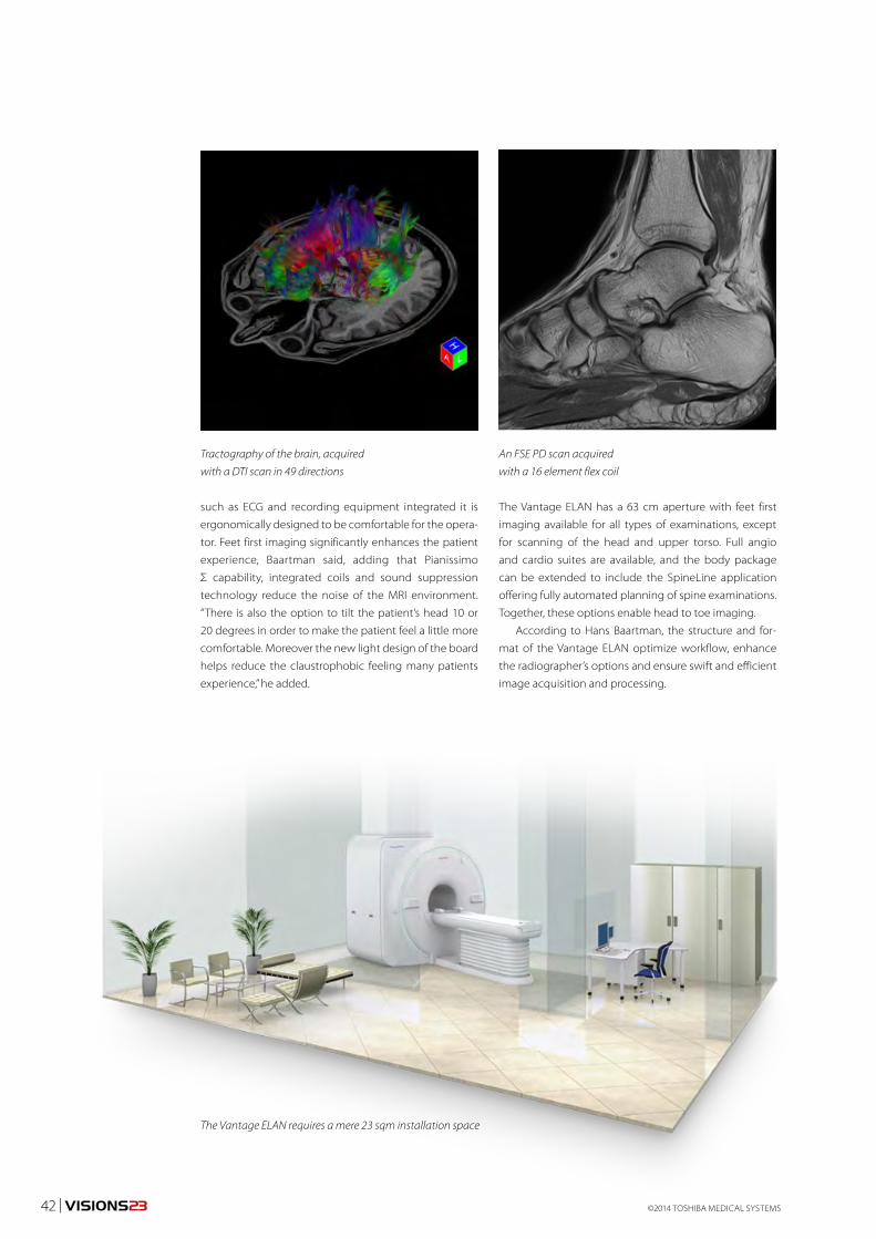
©2014 TOSHIBA MEDICAL SYSTEMS42 | VISIONS23
Tractography of the brain, acquired
with a DTI scan in 49 directions
An FSE PD scan acquired
with a 16 element flex coil
The Vantage ELAN requires a mere 23 sqm installation space
such as ECG and recording equipment integrated it is
ergonomically designed to be comfortable for the opera-
tor. Feet first imaging significantly enhances the patient
experience, Baartman said, adding that Pianissimo
Σ capability, integrated coils and sound suppression
technology reduce the noise of the MRI environment.
“There is also the option to tilt the patient’s head 10 or
20 degrees in order to make the patient feel a little more
comfortable. Moreover the new light design of the board
helps reduce the claustrophobic feeling many patients
experience,” he added.
The Vantage ELAN has a 63 cm aperture with feet first
imaging available for all types of examinations, except
for scanning of the head and upper torso. Full angio
and cardio suites are available, and the body package
can be extended to include the SpineLine application
offering fully automated planning of spine examinations.
Together, these options enable head to toe imaging.
According to Hans Baartman, the structure and for-
mat of the Vantage ELAN optimize workflow, enhance
the radiographer’s options and ensure swift and efficient
image acquisition and processing.
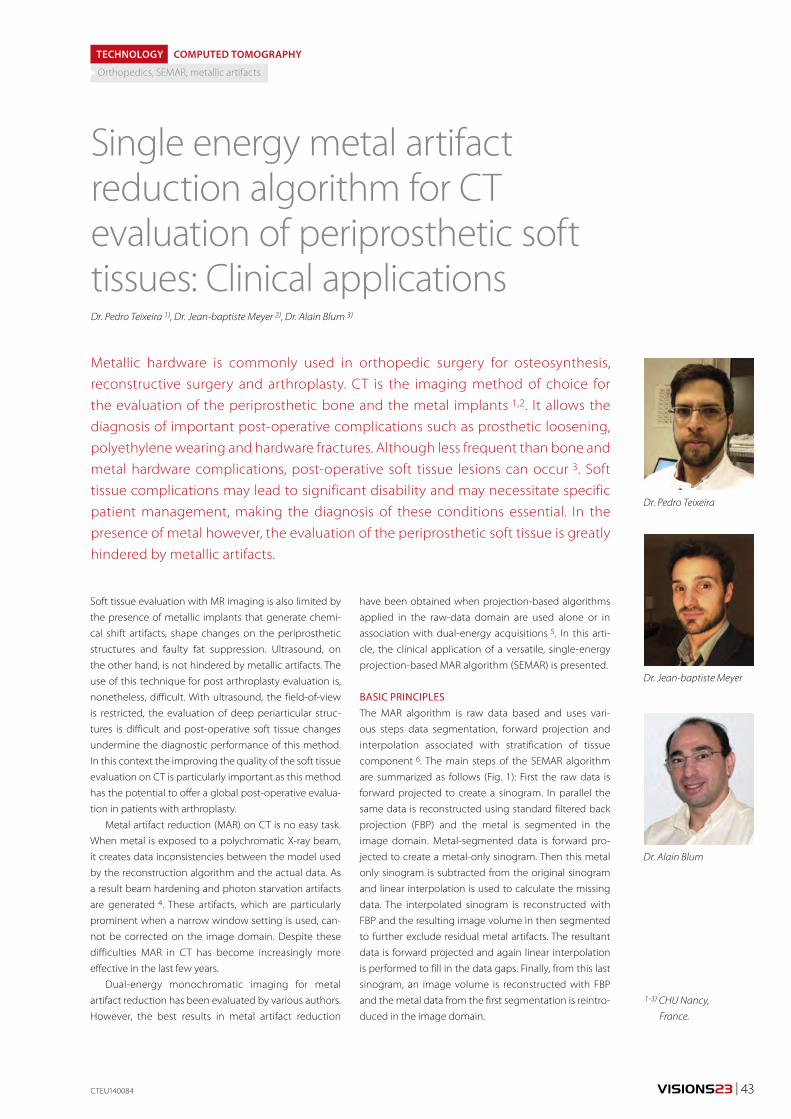
VISIONS23 | 43
Soft tissue evaluation with MR imaging is also limited by
the presence of metallic implants that generate chemi-
cal shift artifacts, shape changes on the periprosthetic
structures and faulty fat suppression. Ultrasound, on
the other hand, is not hindered by metallic artifacts. The
use of this technique for post arthroplasty evaluation is,
nonetheless, difficult. With ultrasound, the field-of-view
is restricted, the evaluation of deep periarticular struc-
tures is difficult and post-operative soft tissue changes
undermine the diagnostic performance of this method.
In this context the improving the quality of the soft tissue
evaluation on CT is particularly important as this method
has the potential to offer a global post-operative evalua-
tion in patients with arthroplasty.
Metal artifact reduction (MAR) on CT is no easy task.
When metal is exposed to a polychromatic X-ray beam,
it creates data inconsistencies between the model used
by the reconstruction algorithm and the actual data. As
a result beam hardening and photon starvation artifacts
are generated 4. These artifacts, which are particularly
prominent when a narrow window setting is used, can-
not be corrected on the image domain. Despite these
difficulties MAR in CT has become increasingly more
effective in the last few years.
Dual-energy monochromatic imaging for metal
artifact reduction has been evaluated by various authors.
However, the best results in metal artifact reduction
Single energy metal artifact reduction algorithm for CT evaluation of periprosthetic soft tissues: Clinical applications
Metallic hardware is commonly used in orthopedic surgery for osteosynthesis, reconstructive surgery and arthroplasty. CT is the imaging method of choice for the evaluation of the periprosthetic bone and the metal implants 1,2. It allows the diagnosis of important post-operative complications such as prosthetic loosening, polyethylene wearing and hardware fractures. Although less frequent than bone and metal hardware complications, post-operative soft tissue lesions can occur 3. Soft tissue complications may lead to significant disability and may necessitate specific patient management, making the diagnosis of these conditions essential. In the presence of metal however, the evaluation of the periprosthetic soft tissue is greatly hindered by metallic artifacts.
CTEU140084
Dr. Pedro Teixeira
Dr. Jean-baptiste Meyer
Dr. Alain Blum
1-3) CHU Nancy,
France.
Dr. Pedro Teixeira 1), Dr. Jean-baptiste Meyer 2), Dr. Alain Blum 3)
have been obtained when projection-based algorithms
applied in the raw-data domain are used alone or in
association with dual-energy acquisitions 5. In this arti-
cle, the clinical application of a versatile, single-energy
projection-based MAR algorithm (SEMAR) is presented.
BASIC PRINCIPLESThe MAR algorithm is raw data based and uses vari-
ous steps data segmentation, forward projection and
interpolation associated with stratification of tissue
component 6. The main steps of the SEMAR algorithm
are summarized as follows (Fig. 1): First the raw data is
forward projected to create a sinogram. In parallel the
same data is reconstructed using standard filtered back
projection (FBP) and the metal is segmented in the
image domain. Metal-segmented data is forward pro-
jected to create a metal-only sinogram. Then this metal
only sinogram is subtracted from the original sinogram
and linear interpolation is used to calculate the missing
data. The interpolated sinogram is reconstructed with
FBP and the resulting image volume in then segmented
to further exclude residual metal artifacts. The resultant
data is forward projected and again linear interpolation
is performed to fill in the data gaps. Finally, from this last
sinogram, an image volume is reconstructed with FBP
and the metal data from the first segmentation is reintro-
duced in the image domain.
TECHNOLOGY COMPUTED TOMOGRAPHY
Orthopedics, SEMAR, metallic artifacts
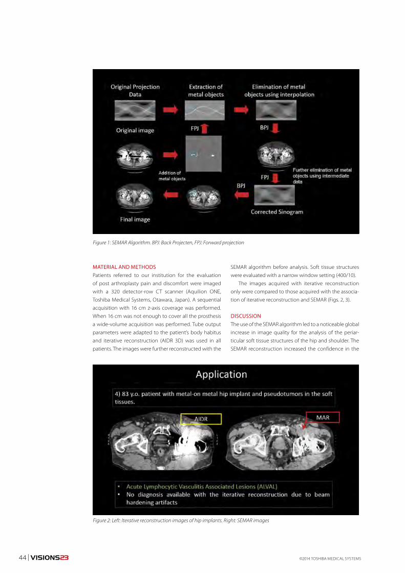
SEMAR algorithm before analysis. Soft tissue structures
were evaluated with a narrow window setting (400/10).
The images acquired with iterative reconstruction
only were compared to those acquired with the associa-
tion of iterative reconstruction and SEMAR (Figs. 2, 3).
DISCUSSIONThe use of the SEMAR algorithm led to a noticeable global
increase in image quality for the analysis of the periar-
ticular soft tissue structures of the hip and shoulder. The
SEMAR reconstruction increased the confidence in the
MATERIAL AND METHODSPatients referred to our institution for the evaluation
of post arthroplasty pain and discomfort were imaged
with a 320 detector-row CT scanner (Aquilion ONE,
Toshiba Medical Systems, Otawara, Japan). A sequential
acquisition with 16 cm z-axis coverage was performed.
When 16 cm was not enough to cover all the prosthesis
a wide-volume acquisition was performed. Tube output
parameters were adapted to the patient’s body habitus
and iterative reconstruction (AIDR 3D) was used in all
patients. The images were further reconstructed with the
Figure 1: SEMAR Algorithm. BPJ: Back Projecten, FPJ: Forward projection
Figure 2: Left: Iterative reconstruction images of hip implants. Right: SEMAR images
44 | VISIONS23 ©2014 TOSHIBA MEDICAL SYSTEMS
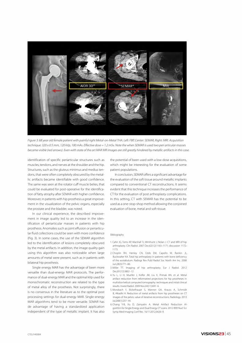
VISIONS23 | 45
Figure 3: 68 year old female patient with painful right Metal-on-Metal THA. Left: FBP, Center: SEMAR, Right: MRI. Acquisition
technique: 320 x 0.5 mm, 120 kVp, 100 mAs. Effective dose = 1.2 mSv. Note the when SEMAR is used two peri articular masses
became visible (red arrows). Even with state of the art MAR MR images are still greatly hindered by metallic artifacts in this case.
CTEU140084
Bibliography
1. Cahir JG, Toms AP, Marshall TJ, Wimhurst J, Nolan J. CT and MRI of hip arthroplasty. Clin Radiol. 2007 Dec;62(12):1163–1171; discussion 1172–1173.
2. Choplin RH, Henley CN, Edds EM, Capello W, Rankin JL, Buckwalter KA. Total hip arthroplasty in patients with bone deficiency of the acetabulum. Radiogr Rev Publ Radiol Soc North Am Inc. 2008 Jun;28(3):771–86.
3. Miller TT. Imaging of hip arthroplasty. Eur J Radiol. 2012 Dec;81(12):3802–12.
4. Yu L, Li H, Mueller J, Kofler JM, Liu X, Primak AN, et al. Metal artifact reduction from reformatted projections for hip prostheses in multislice helical computed tomography: techniques and initial clinical results. Invest Radiol. 2009 Nov;44(11):691–6.
5. Morsbach F, Bickelhaupt S, Wanner GA, Krauss A, Schmidt B, Alkadhi H. Reduction of metal artifacts from hip prostheses on CT images of the pelvis: value of iterative reconstructions. Radiology. 2013 Jul;268(1):237–44.
6. Chang Y-B, Xu D, Zamyatin A. Metal Artifact Reduction Al-gorithm for Single Energy and Dual Energy CT scans. 2012 IEEE Nucl Sci Symp Med Imaging Conf Rec. 16/112012;3426–9.
identification of specific periarticular structures such as
muscles, tendons, and nerves at the shoulder and the hip.
Structures, such as the gluteus minimus and medius ten-
dons, that were often completely obscured by the metal-
lic artifacts became identifiable with good confidence.
The same was seen at the rotator cuff muscle belies, that
could be evaluated for post-operative for the identifica-
tion of fatty atrophy after SEMAR with higher confidence.
Moreover, in patients with hip prosthesis a great improve-
ment in the visualization of the pelvic organs, especially
the prostate and the bladder, was noted.
In our clinical experience, the described improve-
ment in image quality led to an increase in the iden-
tification of periarticular masses in patients with hip
prosthesis. Anomalies such as joint effusion or periarticu-
lar fluid collections could be seen with more confidence
(Fig. 3). In some cases, the use of the SEMAR algorithm
led to the identification of lesions completely obscured
by the metal artifacts. In addition, the image quality gain
using this algorithm was also noticeable when large
amounts of metal were present, such as in patients with
bilateral hip prosthesis.
Single energy MAR has the advantage of been more
versatile than dual-energy MAR protocols. The perfor-
mance of dual-energy MAR and the optimal kVp used for
monochromatic reconstruction are related to the type
of metal alloy of the prosthesis. Not surprisingly, there
is no consensus in the literature as to the optimal post
processing settings for dual-energy MAR. Single-energy
MAR algorithms tend to be more versatile. SEMAR has
de advantage of having a standardized application
independent of the type of metallic implant. It has also
the potential of been used with a low-dose acquisitions,
which might be interesting for the evaluation of some
patient populations.
In conclusion, SEMAR offers a significant advantage for
the evaluation of the soft tissue around metallic implants
compared to conventional CT reconstructions. It seems
evident that this technique increases the performance of
CT for the evaluation of post arthroplasty complications.
In this setting, CT with SEMAR has the potential to be
used as a one-stop-shop method allowing the conjoined
evaluation of bone, metal and soft-tissue.
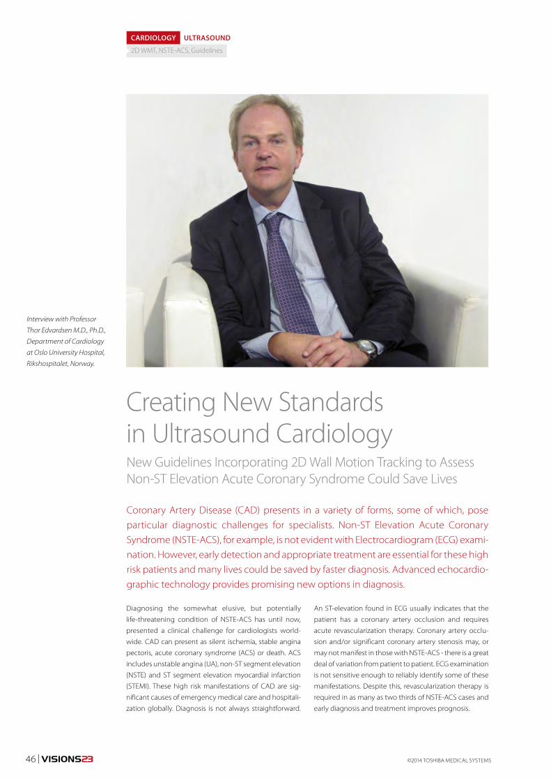
46 | VISIONS23 ©2014 TOSHIBA MEDICAL SYSTEMS
An ST-elevation found in ECG usually indicates that the
patient has a coronary artery occlusion and requires
acute revascularization therapy. Coronary artery occlu-
sion and/or significant coronary artery stenosis may, or
may not manifest in those with NSTE-ACS - there is a great
deal of variation from patient to patient. ECG examination
is not sensitive enough to reliably identify some of these
manifestations. Despite this, revascularization therapy is
required in as many as two thirds of NSTE-ACS cases and
early diagnosis and treatment improves prognosis.
Diagnosing the somewhat elusive, but potentially
life-threatening condition of NSTE-ACS has until now,
presented a clinical challenge for cardiologists world-
wide. CAD can present as silent ischemia, stable angina
pectoris, acute coronary syndrome (ACS) or death. ACS
includes unstable angina (UA), non-ST segment elevation
(NSTE) and ST segment elevation myocardial infarction
(STEMI). These high risk manifestations of CAD are sig-
nificant causes of emergency medical care and hospitali-
zation globally. Diagnosis is not always straightforward.
Creating New Standardsin Ultrasound Cardiology
Coronary Artery Disease (CAD) presents in a variety of forms, some of which, pose particular diagnostic challenges for specialists. Non-ST Elevation Acute Coronary Syndrome (NSTE-ACS), for example, is not evident with Electrocardiogram (ECG) exami-nation. However, early detection and appropriate treatment are essential for these high risk patients and many lives could be saved by faster diagnosis. Advanced echocardio-graphic technology provides promising new options in diagnosis.
CARDIOLOGY ULTRASOUND
2D WMT, NSTE-ACS, Guidelines
New Guidelines Incorporating 2D Wall Motion Tracking to Assess Non-ST Elevation Acute Coronary Syndrome Could Save Lives
Interview with Professor
Thor Edvardsen M.D., Ph.D.,
Department of Cardiology
at Oslo University Hospital,
Rikshospitalet, Norway.
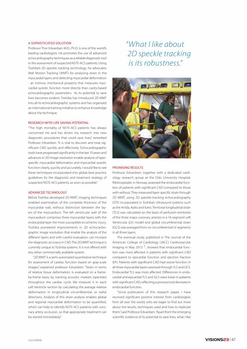
VISIONS23 | 47
PROMISING RESULTSProfessor Edvardsen, together with a dedicated cardi-
ology research group at the Oslo University Hospital,
Rikshospitalet, in Norway, assessed the endocardial func-
tion of patients with significant CAD compared to those
with without. They measured layer-specific strain through
2D WMT, using 2D speckle-tracking echocardiography
(STE) incorporated in Toshiba’s Ultrasound systems such
as the Artida, Aplio and Xario. Territorial longitudinal strain
(TLS) was calculated on the basis of perfusion territories
of the three major coronary arteries in a 16-segment Left
Ventricular (LV) model and global circumferential strain
(GCS) was averaged from six circumferential LV segments
in all three layers.
The eventual study, published in The Journal of the
American College of Cardiology (JACC) Cardiovascular
Imaging in May 2013 1, showed that endocardial func-
tion was more affected in patients with significant CAD
compared to epicardial function and ejection fraction
(EF). Patients with significant CAD had worse function in
all three myocardial layers assessed through TLS and GCS.
Endocardial TLS was most affected. Differences in endo-
cardial and epicardial TLS and GCS were lower in patients
with significant CAD, reflecting a pronounced decrease in
endocardial function.
“Since publication of this research paper, I have
received significant positive interest from cardiologists
from all over the world, who are eager to find out more
about the results, techniques used and how to replicate
them,” said Professor Edvardsen. “Apart from the emerging
scientific evidence of its potential to save lives, what I like
A SOPHISTICATED SOLUTION Professor Thor Edvardsen M.D., Ph.D. is one of the world’s
leading cardiologists. He promotes the use of advanced
echocardiography techniques as a reliable diagnostic tool
in the assessment of suspected NSTE-ACS patients. Using
Toshiba’s 2D speckle tracking technology, he advocates
Wall Motion Tracking (WMT) for analyzing strain in the
myocardial layers and detecting myocardial deformation
- an intrinsic mechanical property that measures myo-
cardial systolic function more directly than cavity-based
echocardiographic parameters. As its potential to save
lives becomes evident, Toshiba has introduced 2D WMT
into all its echocardiographic systems and has organized
an international training initiative to enhance knowledge
about the technique.
RESEARCH WITH LIFE SAVING POTENTIAL“The high mortality of NSTE-ACS patients has always
concerned me and has driven my research into new
diagnostic procedures that could save lives,” remarked
Professor Edvardsen. “It is vital to discover and treat sig-
nificant CAD quickly and effectively. Echocardiographic
tools have progressed significantly in the last 10 years and
advances in 2D image resolution enable analysis of layer-
specific myocardial deformation and myocardial systolic
function clearly, quickly and accurately. I would like to see
these techniques incorporated into global best practice
guidelines for the diagnostic and treatment strategy of
suspected NSTE-ACS patients, as soon as possible.”
ADVANCED TECHNOLOGYBefore Toshiba developed 2D WMT, imaging techniques
enabled examination of the complete thickness of the
myocardial wall, without distinction between the lay-
ers of the myocardium. The left ventricular wall of the
myocardium comprises three myocardial layers with the
endocardial layer the most susceptible to ischemic injury.
Toshiba pioneered improvements in 2D echocardio-
graphic image resolution that enable the analysis of the
different layers and with careful evaluation, can increase
the diagnostic accuracy in CAD. The 2D WMT technique is
currently unique to Toshiba systems. It is not offered with
any other commercially available system.
“2D WMT is a semi-automated quantitative technique
for assessment of cardiac function based on gray-scale
images,” explained professor Edvardsen. “Strain in terms
of relative tissue deformation, is evaluated on a frame-
by-frame basis, by tracking acoustic markers (speckles)
throughout the cardiac cycle. We measure it in each
Left Ventricle section by calculating the average relative
deformation in longitudinal circumferential, or radial
directions. Analysis of this strain analysis enables global
and regional myocardial deformation to be quantified,
which can help to identify NSTE-ACS patients with coro-
nary artery occlusion, so that appropriate treatment can
be started immediately.”
ULEU130038
“ What I like about 2D speckle tracking is its robustness.”
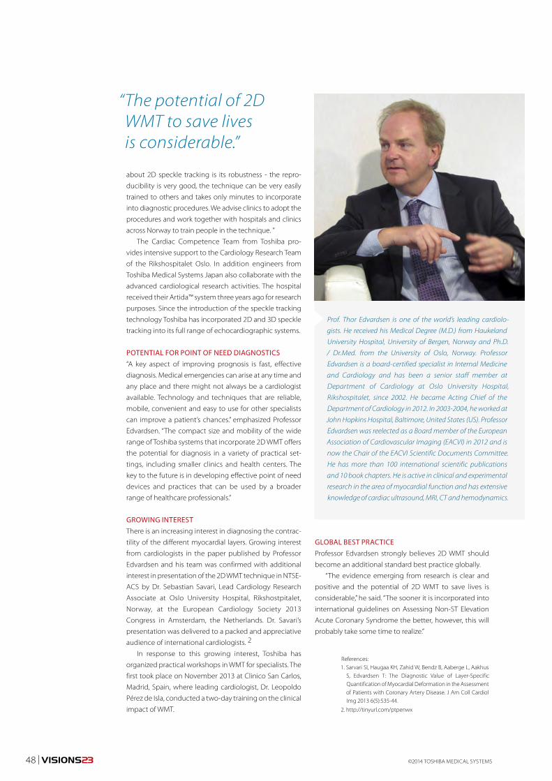
48 | VISIONS23 ©2014 TOSHIBA MEDICAL SYSTEMS
about 2D speckle tracking is its robustness - the repro-
ducibility is very good, the technique can be very easily
trained to others and takes only minutes to incorporate
into diagnostic procedures. We advise clinics to adopt the
procedures and work together with hospitals and clinics
across Norway to train people in the technique. ”
The Cardiac Competence Team from Toshiba pro-
vides intensive support to the Cardiology Research Team
of the Rikshospitalet Oslo. In addition engineers from
Toshiba Medical Systems Japan also collaborate with the
advanced cardiological research activities. The hospital
received their Artida™ system three years ago for research
purposes. Since the introduction of the speckle tracking
technology Toshiba has incorporated 2D and 3D speckle
tracking into its full range of echocardiographic systems.
POTENTIAL FOR POINT OF NEED DIAGNOSTICS “A key aspect of improving prognosis is fast, effective
diagnosis. Medical emergencies can arise at any time and
any place and there might not always be a cardiologist
available. Technology and techniques that are reliable,
mobile, convenient and easy to use for other specialists
can improve a patient’s chances.” emphasized Professor
Edvardsen. “The compact size and mobility of the wide
range of Toshiba systems that incorporate 2D WMT offers
the potential for diagnosis in a variety of practical set-
tings, including smaller clinics and health centers. The
key to the future is in developing effective point of need
devices and practices that can be used by a broader
range of healthcare professionals.”
GROWING INTERESTThere is an increasing interest in diagnosing the contrac-
tility of the different myocardial layers. Growing interest
from cardiologists in the paper published by Professor
Edvardsen and his team was confirmed with additional
interest in presentation of the 2D WMT technique in NTSE-
ACS by Dr. Sebastian Savari, Lead Cardiology Research
Associate at Oslo University Hospital, Rikshostpitalet,
Norway, at the European Cardiology Society 2013
Congress in Amsterdam, the Netherlands. Dr. Savari’s
presentation was delivered to a packed and appreciative
audience of international cardiologists. 2
In response to this growing interest, Toshiba has
organized practical workshops in WMT for specialists. The
first took place on November 2013 at Clinico San Carlos,
Madrid, Spain, where leading cardiologist, Dr. Leopoldo
Pérez de Isla, conducted a two-day training on the clinical
impact of WMT.
“ The potential of 2D WMT to save lives is considerable.”
Prof. Thor Edvardsen is one of the world’s leading cardiolo-
gists. He received his Medical Degree (M.D.) from Haukeland
University Hospital, University of Bergen, Norway and Ph.D.
/ Dr.Med. from the University of Oslo, Norway. Professor
Edvardsen is a board-certified specialist in Internal Medicine
and Cardiology and has been a senior staff member at
Department of Cardiology at Oslo University Hospital,
Rikshospitalet, since 2002. He became Acting Chief of the
Department of Cardiology in 2012. In 2003-2004, he worked at
John Hopkins Hospital, Baltimore, United States (US). Professor
Edvardsen was reelected as a Board member of the European
Association of Cardiovascular Imaging (EACVI) in 2012 and is
now the Chair of the EACVI Scientific Documents Committee.
He has more than 100 international scientific publications
and 10 book chapters. He is active in clinical and experimental
research in the area of myocardial function and has extensive
knowledge of cardiac ultrasound, MRI, CT and hemodynamics.
GLOBAL BEST PRACTICEProfessor Edvardsen strongly believes 2D WMT should
become an additional standard best practice globally.
“The evidence emerging from research is clear and
positive and the potential of 2D WMT to save lives is
considerable,” he said. “The sooner it is incorporated into
international guidelines on Assessing Non-ST Elevation
Acute Coronary Syndrome the better, however, this will
probably take some time to realize.”
References:1. Sarvari SI, Haugaa KH, Zahid W, Bendz B, Aaberge L, Aakhus
S, Edvardsen T: The Diagnostic Value of Layer-Specific Quantification of Myocardial Deformation in the Assessment of Patients with Coronary Artery Disease. J Am Coll Cardiol Img 2013 6(5):535-44.
2. http://tinyurl.com/ptpenwx
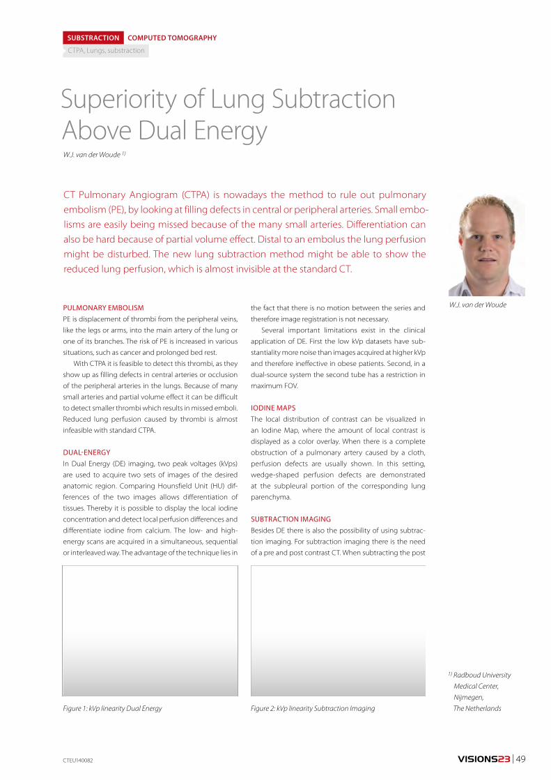
VISIONS23 | 49
PULMONARY EMBOLISMPE is displacement of thrombi from the peripheral veins,
like the legs or arms, into the main artery of the lung or
one of its branches. The risk of PE is increased in various
situations, such as cancer and prolonged bed rest.
With CTPA it is feasible to detect this thrombi, as they
show up as filling defects in central arteries or occlusion
of the peripheral arteries in the lungs. Because of many
small arteries and partial volume effect it can be difficult
to detect smaller thrombi which results in missed emboli.
Reduced lung perfusion caused by thrombi is almost
infeasible with standard CTPA.
DUAL-ENERGY In Dual Energy (DE) imaging, two peak voltages (kVps)
are used to acquire two sets of images of the desired
anatomic region. Comparing Hounsfield Unit (HU) dif-
ferences of the two images allows differentiation of
tissues. Thereby it is possible to display the local iodine
concentration and detect local perfusion differences and
differentiate iodine from calcium. The low- and high-
energy scans are acquired in a simultaneous, sequential
or interleaved way. The advantage of the technique lies in
Superiority of Lung Subtraction Above Dual Energy
CT Pulmonary Angiogram (CTPA) is nowadays the method to rule out pulmonary embolism (PE), by looking at filling defects in central or peripheral arteries. Small embo-lisms are easily being missed because of the many small arteries. Differentiation can also be hard because of partial volume effect. Distal to an embolus the lung perfusion might be disturbed. The new lung subtraction method might be able to show the reduced lung perfusion, which is almost invisible at the standard CT.
W.J. van der Woude 1)
W.J. van der Woude
1) Radboud University
Medical Center,
Nijmegen,
The Netherlands
CTEU140082
SUBSTRACTION COMPUTED TOMOGRAPHY
CTPA, Lungs, substraction
the fact that there is no motion between the series and
therefore image registration is not necessary.
Several important limitations exist in the clinical
application of DE. First the low kVp datasets have sub-
stantiality more noise than images acquired at higher kVp
and therefore ineffective in obese patients. Second, in a
dual-source system the second tube has a restriction in
maximum FOV.
IODINE MAPSThe local distribution of contrast can be visualized in
an Iodine Map, where the amount of local contrast is
displayed as a color overlay. When there is a complete
obstruction of a pulmonary artery caused by a cloth,
perfusion defects are usually shown. In this setting,
wedge-shaped perfusion defects are demonstrated
at the subpleural portion of the corresponding lung
parenchyma.
SUBTRACTION IMAGINGBesides DE there is also the possibility of using subtrac-
tion imaging. For subtraction imaging there is the need
of a pre and post contrast CT. When subtracting the post
Figure 1: kVp linearity Dual Energy Figure 2: kVp linearity Subtraction Imaging
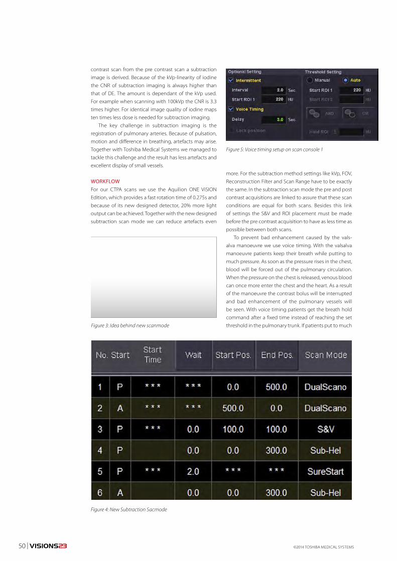
©2014 TOSHIBA MEDICAL SYSTEMS50 | VISIONS23
Figure 4: New Subtraction Sacmode
Figure 5: Voice timing setup on scan console 1
contrast scan from the pre contrast scan a subtraction
image is derived. Because of the kVp-linearity of iodine
the CNR of subtraction imaging is always higher than
that of DE. The amount is dependant of the kVp used.
For example when scanning with 100kVp the CNR is 3.3
times higher. For identical image quality of iodine maps
ten times less dose is needed for subtraction imaging.
The key challenge in subtraction imaging is the
registration of pulmonary arteries. Because of pulsation,
motion and difference in breathing, artefacts may arise.
Together with Toshiba Medical Systems we managed to
tackle this challenge and the result has less artefacts and
excellent display of small vessels.
WORKFLOWFor our CTPA scans we use the Aquilion ONE ViSION
Edition, which provides a fast rotation time of 0.275s and
because of its new designed detector, 20% more light
output can be achieved. Together with the new designed
subtraction scan mode we can reduce artefacts even
more. For the subtraction method settings like kVp, FOV,
Reconstruction Filter and Scan Range have to be exactly
the same. In the subtraction scan mode the pre and post
contrast acquisitions are linked to assure that these scan
conditions are equal for both scans. Besides this link
of settings the S&V and ROI placement must be made
before the pre contrast acquisition to have as less time as
possible between both scans.
To prevent bad enhancement caused by the vals-
alva manoeuvre we use voice timing. With the valsalva
manoeuvre patients keep their breath while putting to
much pressure. As soon as the pressure rises in the chest,
blood will be forced out of the pulmonary circulation.
When the pressure on the chest is released, venous blood
can once more enter the chest and the heart. As a result
of the manoeuvre the contrast bolus will be interrupted
and bad enhancement of the pulmonary vessels will
be seen. With voice timing patients get the breath hold
command after a fixed time instead of reaching the set
threshold in the pulmonary trunk. If patients put to much Figure 3: Idea behind new scanmode
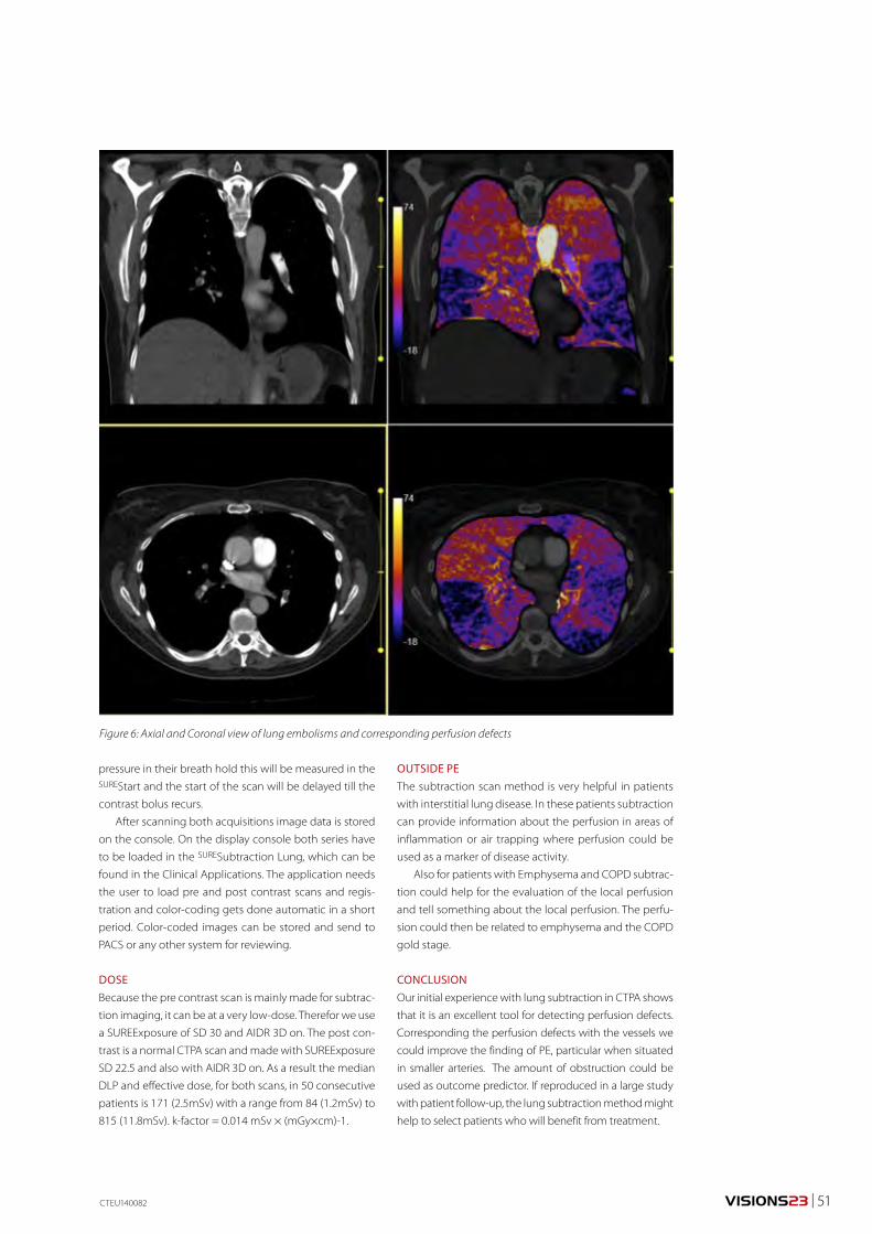
VISIONS23 | 51
Figure 6: Axial and Coronal view of lung embolisms and corresponding perfusion defects
CTEU140082
pressure in their breath hold this will be measured in the SUREStart and the start of the scan will be delayed till the
contrast bolus recurs.
After scanning both acquisitions image data is stored
on the console. On the display console both series have
to be loaded in the SURESubtraction Lung, which can be
found in the Clinical Applications. The application needs
the user to load pre and post contrast scans and regis-
tration and color-coding gets done automatic in a short
period. Color-coded images can be stored and send to
PACS or any other system for reviewing.
DOSEBecause the pre contrast scan is mainly made for subtrac-
tion imaging, it can be at a very low-dose. Therefor we use
a SUREExposure of SD 30 and AIDR 3D on. The post con-
trast is a normal CTPA scan and made with SUREExposure
SD 22.5 and also with AIDR 3D on. As a result the median
DLP and effective dose, for both scans, in 50 consecutive
patients is 171 (2.5mSv) with a range from 84 (1.2mSv) to
815 (11.8mSv). k-factor = 0.014 mSv × (mGy×cm)-1.
OUTSIDE PEThe subtraction scan method is very helpful in patients
with interstitial lung disease. In these patients subtraction
can provide information about the perfusion in areas of
inflammation or air trapping where perfusion could be
used as a marker of disease activity.
Also for patients with Emphysema and COPD subtrac-
tion could help for the evaluation of the local perfusion
and tell something about the local perfusion. The perfu-
sion could then be related to emphysema and the COPD
gold stage.
CONCLUSIONOur initial experience with lung subtraction in CTPA shows
that it is an excellent tool for detecting perfusion defects.
Corresponding the perfusion defects with the vessels we
could improve the finding of PE, particular when situated
in smaller arteries. The amount of obstruction could be
used as outcome predictor. If reproduced in a large study
with patient follow-up, the lung subtraction method might
help to select patients who will benefit from treatment.
52 | VISIONS23 ©2013 TOSHIBA MEDICAL SYSTEMS
RESULTSThe study at rest showed no visual violations of contractil-
ity in any patient, and the ejection fraction ranged from
58% to 71% (64.2 ± 4.7%). Transmural (both local and
global) deformation indices of the LV myocardium were
normal. However, the evaluation of longitudinal strain
before stress testing identified two subgroups: 9 patients
with the range of all indices of global and local longitudi-
nal strain from -15% to -18% (the first subgroup), and 11
patients (the second subgroup) with violations of local
longitudinal strain in some segments (-7% to -12%), with-
out any effect on the overall local myocardial contractility
at rest. Visual examination of the working report of stress
echocardiography at a maximum frequency of stimula-
tion showed some zones of hypokinesia in all patients
from the second group, accompanied by a significant
expansion of the regions of longitudinal strain violation,
and also by reduction of transmural strain in segments
AIMThe study was aimed at comparison of the longitudinal
strain at rest with the results of subsequent stress echo-
cardiography in patients without clinical manifestations
of coronary heart disease.
MATERIALS AND METHODSThe study involved 20 patients without cardiac com-
plaints (16 men and 4 women) aged 40–50 years (mean
age 43.7 ± 7.2) with a variety of nonspecific electrocar-
diographic (ECG) changes: extrasystole, negative T waves,
etc. listed in Table 1. All patients had undergone stress
ECG (bicycle ergometry or treadmill test), but for various
reasons this kind of exercise was not included in the
diagnostic criteria (subjective reasons, a significant blood
pressure increase during the procedure, etc.). Therefore,
in all 20 individuals stress echocardiography with
transesophageal atrial pacing was performed. In addition
to the standard study protocol with compara-
tive visual assessment of the kinetics of the left
ventricular myocardium at rest and at different
stages of stimulation (120, 140, and 160 pulses
per minute), the quantitative characteristics of
the longitudinal and transmural myocardial
strain before the exercise and at its peak were
studied using 2D Speckle Tracking technology
(“Artida” from Toshiba, Japan). Statistical analy-
sis was performed on a personal computer
using the Student’s test criterion with the
Microsoft Excel software package. Differences
with p<0.05 were considered significant.
Violation of longitudinal strain of the left ventricular myocardium as a predictor of positive stress echocardiography results.
Table 1: The distribution of electrocardiographic
changes at rest in the patients examined.
CARDIOLOGY ULTRASOUND
Violations of longitudinal strain of the left ventricular (LV) myocardium are known as one of the earliest manifestations of systolic dysfunction, appearing when the global contractility of the heart is preserved due to compensatory increase of other types of strain (circular, radial, and torsion). To maintain a normal level of the left ventricle global contractility and pumping function parameters, sufficient reserves are needed to enable functioning of these compensation mechanisms at rest. However, during exercise or other stress these reserves may be exhausted, and the compensation will fail.
Electrocardiographic signs
Atrial extrasystole 8
Ventricular extrasystole 7
Negative T waves 8
Uncertain ST-segment depression 4
Dr. V.V. Potapov
Dr. V.E. Smyalovsky
1-7) The budget-funded
healthcare facility
“Clinical Diagnostic
Centre”, Omsk region.
Number of patients
Dr. V.V. Potapov 1), Dr. A.M. Vishnjakov 2), Dr. E.A. Efremova 3), Dr. I.L. Kudryashov 4), Dr. V.E. Smyalovsky 5),
Dr. G.Y. Khlynova 6), Dr. S.E. Nikolsky 7)
2D Speckle Tracking, Artida
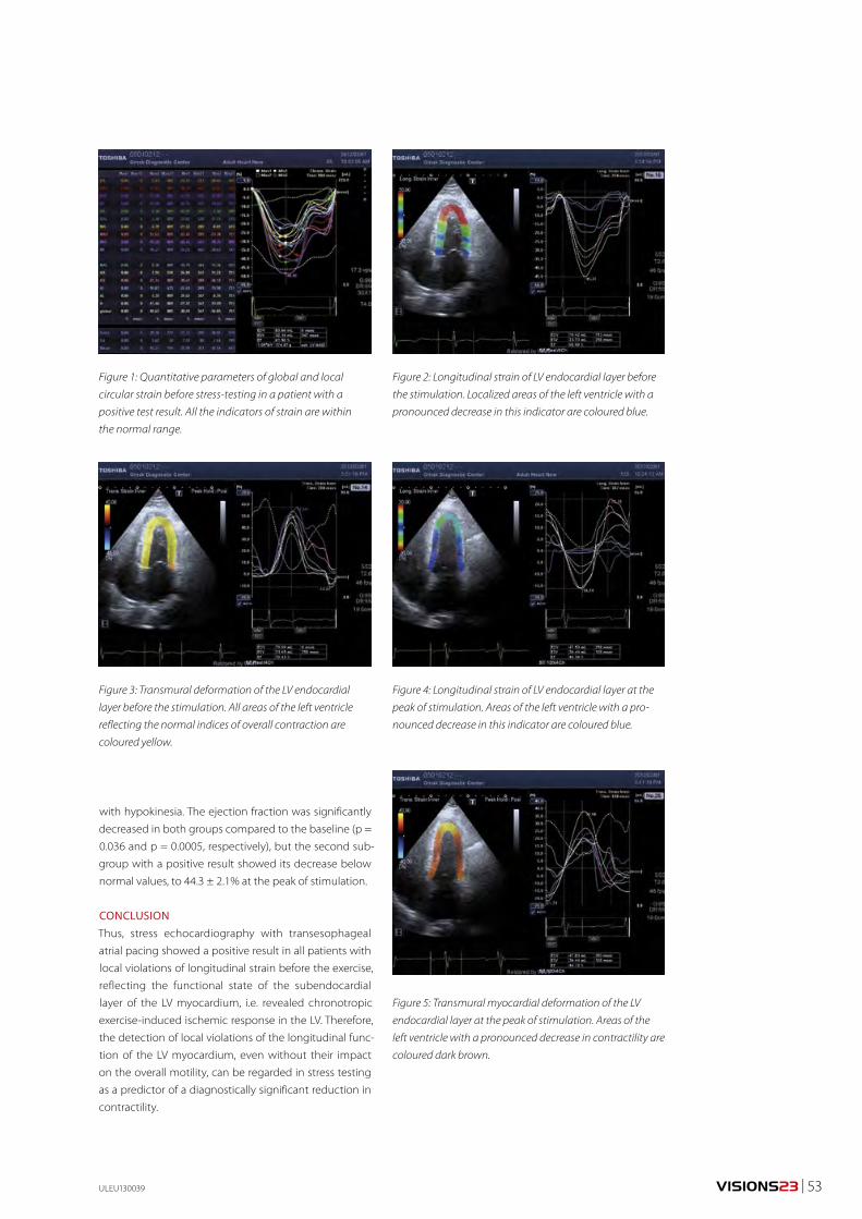
VISIONS23 | 53
with hypokinesia. The ejection fraction was significantly
decreased in both groups compared to the baseline (p =
0.036 and p = 0.0005, respectively), but the second sub-
group with a positive result showed its decrease below
normal values, to 44.3 ± 2.1% at the peak of stimulation.
CONCLUSIONThus, stress echocardiography with transesophageal
atrial pacing showed a positive result in all patients with
local violations of longitudinal strain before the exercise,
reflecting the functional state of the subendocardial
layer of the LV myocardium, i.e. revealed chronotropic
exercise-induced ischemic response in the LV. Therefore,
the detection of local violations of the longitudinal func-
tion of the LV myocardium, even without their impact
on the overall motility, can be regarded in stress testing
as a predictor of a diagnostically significant reduction in
contractility.
ULEU130039
Figure 1: Quantitative parameters of global and local
circular strain before stress-testing in a patient with a
positive test result. All the indicators of strain are within
the normal range.
Figure 3: Transmural deformation of the LV endocardial
layer before the stimulation. All areas of the left ventricle
reflecting the normal indices of overall contraction are
coloured yellow.
Figure 2: Longitudinal strain of LV endocardial layer before
the stimulation. Localized areas of the left ventricle with a
pronounced decrease in this indicator are coloured blue.
Figure 4: Longitudinal strain of LV endocardial layer at the
peak of stimulation. Areas of the left ventricle with a pro-
nounced decrease in this indicator are coloured blue.
Figure 5: Transmural myocardial deformation of the LV
endocardial layer at the peak of stimulation. Areas of the
left ventricle with a pronounced decrease in contractility are
coloured dark brown.
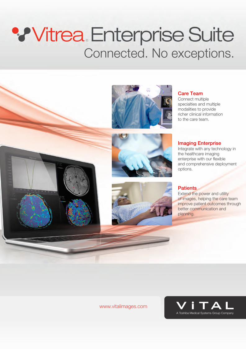
Connected. No exceptions.
www.vitalimages.com
Care TeamConnect multiplespecialties and multiple modalities to providericher clinical informationto the care team.
Imaging EnterpriseIntegrate with any technology in the healthcare imaging enterprise with our flexibleand comprehensive deployment options.
PatientsExtend the power and utilityof images, helping the care team improve patient outcomes through better communication and planning.
www.vitalimages.com
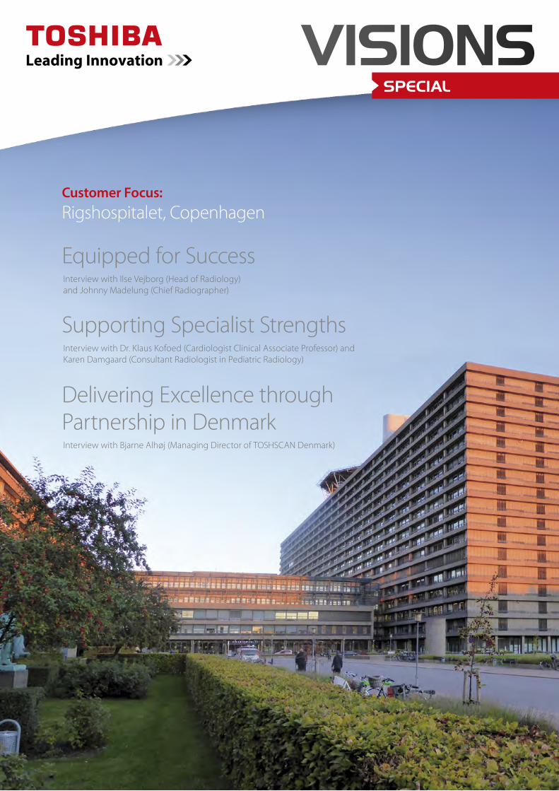
VISIONSSPECIAL
Delivering Excellence through Partnership in Denmark
Equipped for SuccessInterview with Ilse Vejborg (Head of Radiology) and Johnny Madelung (Chief Radiographer)
Interview with Dr. Klaus Kofoed (Cardiologist Clinical Associate Professor) and Karen Damgaard (Consultant Radiologist in Pediatric Radiology)
Interview with Bjarne Alhøj (Managing Director of TOSHSCAN Denmark)
Supporting Specialist Strengths
Customer Focus: Rigshospitalet, Copenhagen
©2014 TOSHIBA MEDICAL SYSTEMS56 | VISIONSSPECIAL
Inside the Rigshospitalet, the spacious reception and route
to the Radiology Department, where the VISIONS inter-
views are scheduled, have a calm, relaxed atmosphere,
which gives little indication that this is one of Denmark’s
largest, busiest and most prestigious specialized hospitals,
with 1200 beds, more than 100 departments and func-
tions, over 50 professional specialties and 8000 staff. Using
resourceful and clever spatial design, including use of
underground room for housing medical equipment, the
hospital has created a welcoming and friendly environ-
ment, in which, cutting-edge diagnostics, treatment,
patient care, research and medical education are carried
out efficiently and effectively.
THE HEART OF HEALTHCAREAt the heart of the Radiology Department, literally and
figuratively, is the staff. In the center of the Department’s
main isle, hangs a large board featuring the staff –
their photographs, names, specialisms and job title. It
includes each and every employee at the facility. There
are nearly 300 profiles on display, arranged within
five specialist sectors, including Body Imaging, Breast
Imaging, Neuro radiology, Ultrasound and Vascular
Intervention. Alongside their own specialisms, the
Department’s staff also played a major role in develop-
ing a brand new trauma center that has recently been
opened at the hospital. Its highly skilled and highly
motivated individuals, working together within a non-
hierarchical structure, enable this facility to achieve
formidable results in clinical practice and research. The
Department’s cutting-edge equipment, which includes
15 ultrasound-, 11 CT- and five MRIs scanners. Most of
the CT systems are made by Toshiba, and it has its own
dedicated TOSHSCAN Denmark service engineer onsite,
along with ongoing support from staff at TOSHSCAN
Denmark, Toshiba Europe and Toshiba Japan. This
ensures it deliver the high quality imaging necessary
Equipped for SuccessThe Radiology Department at Rigshospitalet Copenhagen in Denmark is an emerg-ing global center of excellence and comprises nearly 300 staff, carries out more than 160,000 examinations per year it has significant capabilities in both specialist clinical practice and pioneering research and is gaining increasing recognition as a global center of excellence. The Department has been equipped to the highest standards with a large proportion of its systems made by Toshiba. Ilse Vejborg, Head of Radiology, and Johnny Madelung, Chief Radiographer, lead the Department and have been largely responsible for driving its long term development into a world class diagnostics centre, together with highly specialised teams. VISIONS spoke to Ilse and Johnny to find out exactly how this has been achieved.
for accurate diagnosis, treatment and research into the
specialist cases that the hospital receives.
Ilse Vejborg, was recently appointed as the new Head
of the Radiology Department. She has worked at the
hospital for more than 24 years. She described the team
dynamic, which enables the Department to achieve the
high standards that it has become reknown for.
“This is the biggest radiology department in Denmark,
295 highly specialized staff – You might think it would be
a challenge to simply remember who everyone working
here is, and yet the sheer size of the department offers
more of a benefit than challenging to manage!” she
remarked. “This is because our staff are clearly organized
according to specialized sector, which gives everyone
unique opportunities to utilize and develop their exper-
tise. I rely completely on, and trust in, their specialist
skills. All our specialists are very experienced and 100%
engaged in delivering a very high level of service. We have
cultivated a strong sense of teamwork, despite the large
size of the department, and there are no barriers between
different staff. We all work together to solve problems and
get procedures done, and this is a nice feeling.”
Johnny Madelung, Chief Radiographer, originally
joined the department 26 years ago and has been work-
ing in his current post for the last 12 years.
“The Department is a really nice place to work,” he said.
“Specialists and other staff work together well in its open
and communicative environment. We have a multina-
tional team, with Danish, English, Swedish, or Norwegian
languages spoken. This positive environment has been
cultivated over many years. This doesn’t, of course, just
happen easily by itself. The Department is well-equipped
with state-of-the-art systems and spatially arranged to
optimize the environment for patient and staff alike. The
positive reputation of our great department attracts radi-
ographers from all over the world, who are ‘queuing up’
to work here.
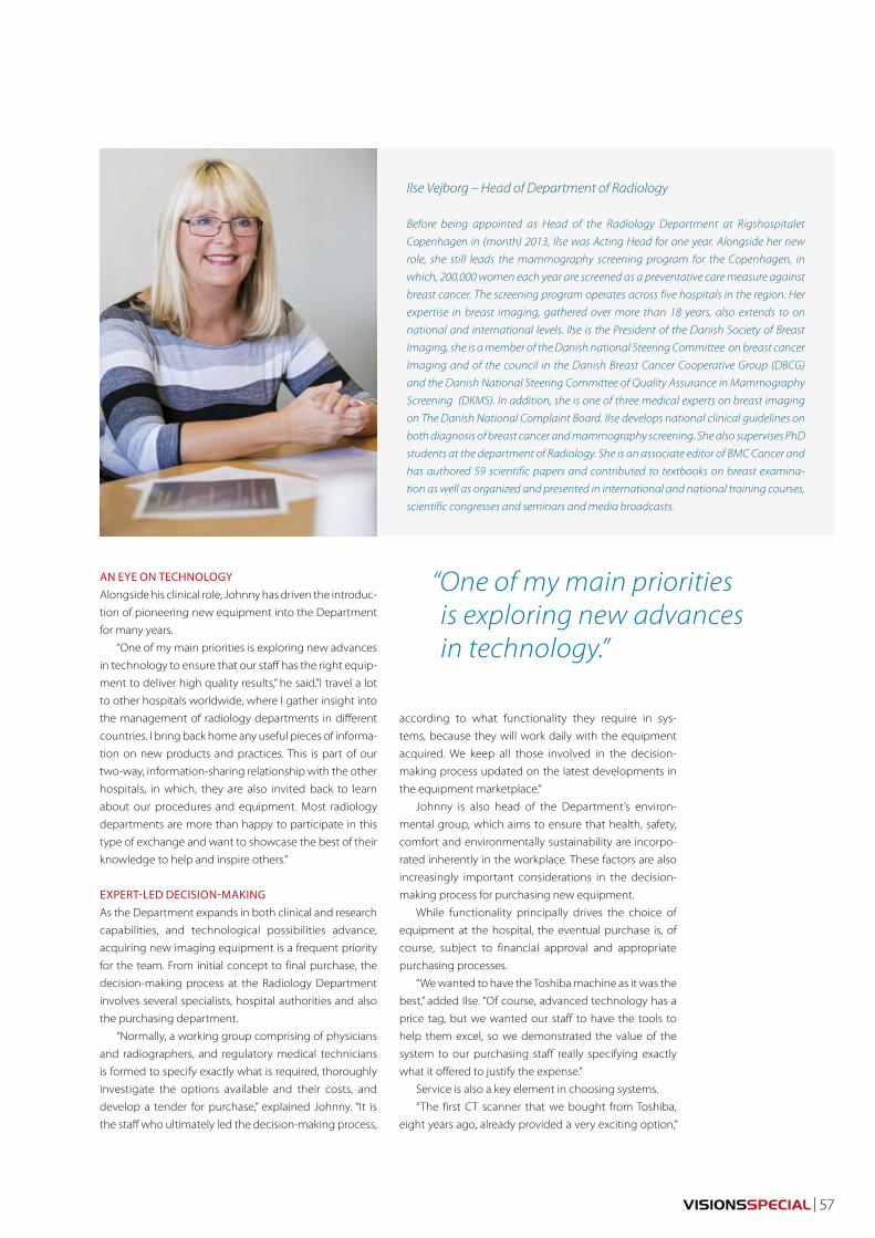
VISIONSSPECIAL | 57
AN EYE ON TECHNOLOGY Alongside his clinical role, Johnny has driven the introduc-
tion of pioneering new equipment into the Department
for many years.
“One of my main priorities is exploring new advances
in technology to ensure that our staff has the right equip-
ment to deliver high quality results,” he said.”I travel a lot
to other hospitals worldwide, where I gather insight into
the management of radiology departments in different
countries. I bring back home any useful pieces of informa-
tion on new products and practices. This is part of our
two-way, information-sharing relationship with the other
hospitals, in which, they are also invited back to learn
about our procedures and equipment. Most radiology
departments are more than happy to participate in this
type of exchange and want to showcase the best of their
knowledge to help and inspire others.”
EXPERT-LED DECISION-MAKINGAs the Department expands in both clinical and research
capabilities, and technological possibilities advance,
acquiring new imaging equipment is a frequent priority
for the team. From initial concept to final purchase, the
decision-making process at the Radiology Department
involves several specialists, hospital authorities and also
the purchasing department.
“Normally, a working group comprising of physicians
and radiographers, and regulatory medical technicians
is formed to specify exactly what is required, thoroughly
investigate the options available and their costs, and
develop a tender for purchase,” explained Johnny. “It is
the staff who ultimately led the decision-making process,
according to what functionality they require in sys-
tems, because they will work daily with the equipment
acquired. We keep all those involved in the decision-
making process updated on the latest developments in
the equipment marketplace.”
Johnny is also head of the Department’s environ-
mental group, which aims to ensure that health, safety,
comfort and environmentally sustainability are incorpo-
rated inherently in the workplace. These factors are also
increasingly important considerations in the decision-
making process for purchasing new equipment.
While functionality principally drives the choice of
equipment at the hospital, the eventual purchase is, of
course, subject to financial approval and appropriate
purchasing processes.
“We wanted to have the Toshiba machine as it was the
best,” added Ilse. “Of course, advanced technology has a
price tag, but we wanted our staff to have the tools to
help them excel, so we demonstrated the value of the
system to our purchasing staff really specifying exactly
what it offered to justify the expense.”
Service is also a key element in choosing systems.
“The first CT scanner that we bought from Toshiba,
eight years ago, already provided a very exciting option,”
Ilse Vejborg – Head of Department of Radiology
Before being appointed as Head of the Radiology Department at Rigshospitalet
Copenhagen in (month) 2013, Ilse was Acting Head for one year. Alongside her new
role, she still leads the mammography screening program for the Copenhagen, in
which, 200,000 women each year are screened as a preventative care measure against
breast cancer. The screening program operates across five hospitals in the region. Her
expertise in breast imaging, gathered over more than 18 years, also extends to on
national and international levels. Ilse is the President of the Danish Society of Breast
Imaging, she is a member of the Danish national Steering Committee on breast cancer
Imaging and of the council in the Danish Breast Cancer Cooperative Group (DBCG)
and the Danish National Steering Committee of Quality Assurance in Mammography
Screening (DKMS). In addition, she is one of three medical experts on breast imaging
on The Danish National Complaint Board. Ilse develops national clinical guidelines on
both diagnosis of breast cancer and mammography screening. She also supervises PhD
students at the department of Radiology. She is an associate editor of BMC Cancer and
has authored 59 scientific papers and contributed to textbooks on breast examina-
tion as well as organized and presented in international and national training courses,
scientific congresses and seminars and media broadcasts.
“ One of my main priorities is exploring new advances in technology.”
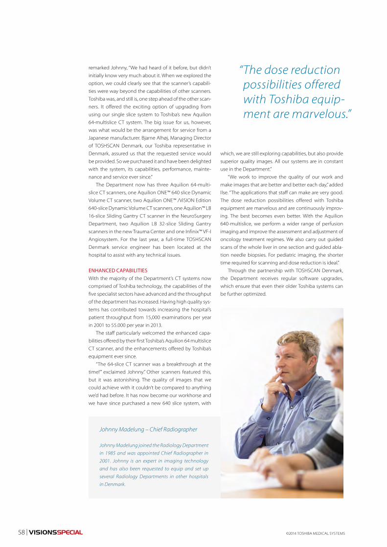
©2014 TOSHIBA MEDICAL SYSTEMS58 | VISIONSSPECIAL
remarked Johnny, “We had heard of it before, but didn’t
initially know very much about it. When we explored the
option, we could clearly see that the scanner’s capabili-
ties were way beyond the capabilities of other scanners.
Toshiba was, and still is, one step ahead of the other scan-
ners. It offered the exciting option of upgrading from
using our single slice system to Toshiba’s new Aquilion
64-multislice CT system. The big issue for us, however,
was what would be the arrangement for service from a
Japanese manufacturer. Bjarne Alhøj, Managing Director
of TOSHSCAN Denmark, our Toshiba representative in
Denmark, assured us that the requested service would
be provided. So we purchased it and have been delighted
with the system, its capabilities, performance, mainte-
nance and service ever since.”
The Department now has three Aquilion 64-multi-
slice CT scanners, one Aquilion ONE™ 640 slice Dynamic
Volume CT scanner, two Aquilion ONE™ /ViSION Edition
640-slice Dynamic Volume CT scanners, one Aquilion™ LB
16-slice Sliding Gantry CT scanner in the NeuroSurgery
Department, two Aquilion LB 32-slice Sliding Gantry
scanners in the new Trauma Center and one Infinix™ VF-I
Angiosystem. For the last year, a full-time TOSHSCAN
Denmark service engineer has been located at the
hospital to assist with any technical issues.
ENHANCED CAPABILITIESWith the majority of the Department’s CT systems now
comprised of Toshiba technology, the capabilities of the
five specialist sectors have advanced and the throughput
of the department has increased. Having high quality sys-
tems has contributed towards increasing the hospital’s
patient throughput from 15,000 examinations per year
in 2001 to 55.000 per year in 2013.
The staff particularly welcomed the enhanced capa-
bilities offered by their first Toshiba’s Aquilion 64 multislice
CT scanner, and the enhancements offered by Toshiba’s
equipment ever since.
“The 64-slice CT scanner was a breakthrough at the
time!”’ exclaimed Johnny.” Other scanners featured this,
but it was astonishing. The quality of images that we
could achieve with it couldn’t be compared to anything
we’d had before. It has now become our workhorse and
we have since purchased a new 640 slice system, with
which, we are still exploring capabilities, but also provide
superior quality images. All our systems are in constant
use in the Department.”
“We work to improve the quality of our work and
make images that are better and better each day,” added
Ilse. “The applications that staff can make are very good.
The dose reduction possibilities offered with Toshiba
equipment are marvelous and are continuously improv-
ing. The best becomes even better. With the Aquilion
640-multislice, we perform a wider range of perfusion
imaging and improve the assessment and adjustment of
oncology treatment regimes. We also carry out guided
scans of the whole liver in one section and guided abla-
tion needle biopsies. For pediatric imaging, the shorter
time required for scanning and dose reduction is ideal.”
Through the partnership with TOSHSCAN Denmark,
the Department receives regular software upgrades,
which ensure that even their older Toshiba systems can
be further optimized.
Johnny Madelung – Chief Radiographer
Johnny Madelung joined the Radiology Department
in 1985 and was appointed Chief Radiographer in
2001. Johnny is an expert in imaging technology
and has also been requested to equip and set up
several Radiology Departments in other hospitals
in Denmark.
“ The dose reduction possibilities offered with Toshiba equip-ment are marvelous.”
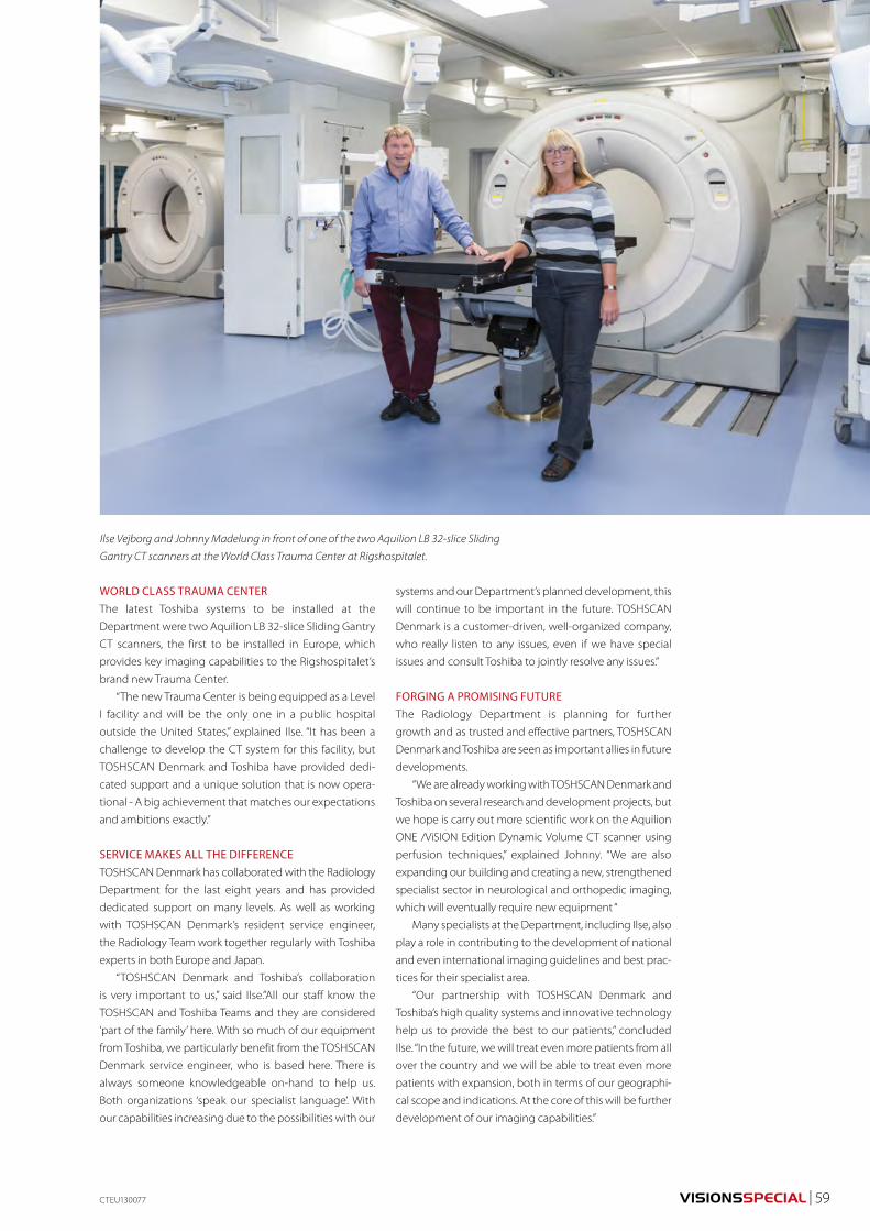
VISIONSSPECIAL | 59
WORLD CLASS TRAUMA CENTERThe latest Toshiba systems to be installed at the
Department were two Aquilion LB 32-slice Sliding Gantry
CT scanners, the first to be installed in Europe, which
provides key imaging capabilities to the Rigshospitalet’s
brand new Trauma Center.
“The new Trauma Center is being equipped as a Level
I facility and will be the only one in a public hospital
outside the United States,” explained Ilse. “It has been a
challenge to develop the CT system for this facility, but
TOSHSCAN Denmark and Toshiba have provided dedi-
cated support and a unique solution that is now opera-
tional - A big achievement that matches our expectations
and ambitions exactly.”
SERVICE MAKES ALL THE DIFFERENCETOSHSCAN Denmark has collaborated with the Radiology
Department for the last eight years and has provided
dedicated support on many levels. As well as working
with TOSHSCAN Denmark’s resident service engineer,
the Radiology Team work together regularly with Toshiba
experts in both Europe and Japan.
“TOSHSCAN Denmark and Toshiba’s collaboration
is very important to us,” said Ilse.”All our staff know the
TOSHSCAN and Toshiba Teams and they are considered
‘part of the family’ here. With so much of our equipment
from Toshiba, we particularly benefit from the TOSHSCAN
Denmark service engineer, who is based here. There is
always someone knowledgeable on-hand to help us.
Both organizations ‘speak our specialist language’. With
our capabilities increasing due to the possibilities with our
systems and our Department’s planned development, this
will continue to be important in the future. TOSHSCAN
Denmark is a customer-driven, well-organized company,
who really listen to any issues, even if we have special
issues and consult Toshiba to jointly resolve any issues.”
FORGING A PROMISING FUTUREThe Radiology Department is planning for further
growth and as trusted and effective partners, TOSHSCAN
Denmark and Toshiba are seen as important allies in future
developments.
“We are already working with TOSHSCAN Denmark and
Toshiba on several research and development projects, but
we hope is carry out more scientific work on the Aquilion
ONE /ViSION Edition Dynamic Volume CT scanner using
perfusion techniques,” explained Johnny. “We are also
expanding our building and creating a new, strengthened
specialist sector in neurological and orthopedic imaging,
which will eventually require new equipment “
Many specialists at the Department, including Ilse, also
play a role in contributing to the development of national
and even international imaging guidelines and best prac-
tices for their specialist area.
“Our partnership with TOSHSCAN Denmark and
Toshiba’s high quality systems and innovative technology
help us to provide the best to our patients,” concluded
Ilse. “In the future, we will treat even more patients from all
over the country and we will be able to treat even more
patients with expansion, both in terms of our geographi-
cal scope and indications. At the core of this will be further
development of our imaging capabilities.”
CTEU130077
Ilse Vejborg and Johnny Madelung in front of one of the two Aquilion LB 32-slice Sliding
Gantry CT scanners at the World Class Trauma Center at Rigshospitalet.
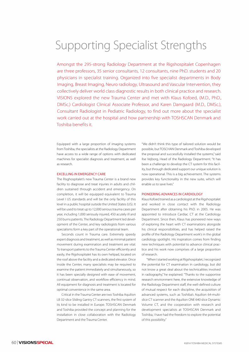
©2014 TOSHIBA MEDICAL SYSTEMS60 | VISIONSSPECIAL
Equipped with a large proportion of imaging systems
from Toshiba, the specialists at the Radiology Department
have access to a wide range of options with dedicated
machines for specialist diagnosis and treatment, as well
as research.
EXCELLING IN EMERGENCY CAREThe Risghospitalet’s new Trauma Center is a brand new
facility to diagnose and treat injuries in adults and chil-
dren sustained through accident and emergency. On
completion, it will be equipped equivalent to Trauma
Level I US standards and will be the only facility of this
level in a public hospital outside the United States (US). It
will be used to treat up to 12,000 serious trauma cases per
year, including 1,000 seriously injured, 450 acutely ill and
250 burns patients. The Radiology Department led devel-
opment of the Center, and key radiologists from various
specialisms form a key part of the operational team.
Seconds count in Trauma care. Extremely speedy
expert diagnosis and treatment, as well as minimal patient
movement during examination and treatment are vital.
To transport patients to the Trauma Center efficiently and
easily, the Rigshospitalet has its own helipad, located on
the roof above the facility and a dedicated elevator. Once
inside the Center, many specialists may be required to
examine the patient immediately and simultaneously, so
it has been specially designed with ease of movement,
continual observation, and workflow efficiency in mind.
All equipment for diagnosis and treatment is located for
optimal convenience in the same area.
Critical in the Trauma Center are two Toshiba Aquilion
LB 32-slice Sliding Gantry CT scanners, the first system of
its kind to be installed in Europe. TOSHSCAN Denmark
and Toshiba provided the concept and planning for the
installation in close collaboration with the Radiology
Department and the Trauma Center.
“We didn’t think this type of tailored solution would be
possible, but TOSCHAN Denmark and Toshiba developed
the proposal and successfully installed the systems,” said
Ilse Vejborg, Head of the Radiology Department. “It has
been a challenge to develop the CT system for this facil-
ity, but through dedicated support our unique solution is
now operational. This is a big achievement. The systems
provides key functionality in the new suite, which will
enable us to save lives.”
PIONEERING ADVANCES IN CARDIOLOGYKlaus Kofoed trained as a cardiologist at the Rigshospitalet
and worked in close contact with the Radiology
Department after obtaining his PhD. in 2005. He was
appointed to introduce Cardiac CT at the Cardiology
Department. Since then, Klaus has pioneered new ways
of exploring the heart with CT examinations alongside
his clinical responsibilities, and has helped raised the
profile of the Radiology Department work’s in the global
cardiology spotlight. His inspiration comes from finding
new techniques with potential to advance clinical prac-
tice and his work now comprises of a large proportion
of research.
“When I started working at Rigshospitalet, I recognized
the potential for CT examination in cardiology, but did
not know a great deal about the technicalities involved
in radiography,” he explained. “Thanks to the supportive
research environment here, the extensive knowledge of
the Radiology Department staff, the well-defined culture
of mutual respect for each discipline, the acquisition of
advanced systems, such as Toshiba’s Aquilion 64-multi-
slice CT scanner and the Aquilion ONE 640 slice Dynamic
Volume CT, and the cooperation with research and
development specialists at TOSHSCAN Denmark and
Toshiba, I have had the freedom to explore the potential
of this possibility.”
Supporting Specialist StrengthsAmongst the 295-strong Radiology Department at the Rigshospitalet Copenhagen are three professors, 35 senior consultants, 12 consultants, nine PhD. students and 20 physicians in specialist training. Organized into five specialist departments in Body Imaging, Breast Imaging, Neuro radiology, Ultrasound and Vascular Intervention, they collectively deliver world class diagnostic results in both clinical practice and research. VISIONS explored the new Trauma Center and met with Klaus Kofoed, (M.D., PhD., DMSc.) Cardiologist Clinical Associate Professor, and Karen Damgaard (M.D., DMSc.), Consultant Radiologist in Pediatric Radiology, to find out more about the specialist work carried out at the hospital and how partnership with TOSHSCAN Denmark and Toshiba benefits it.
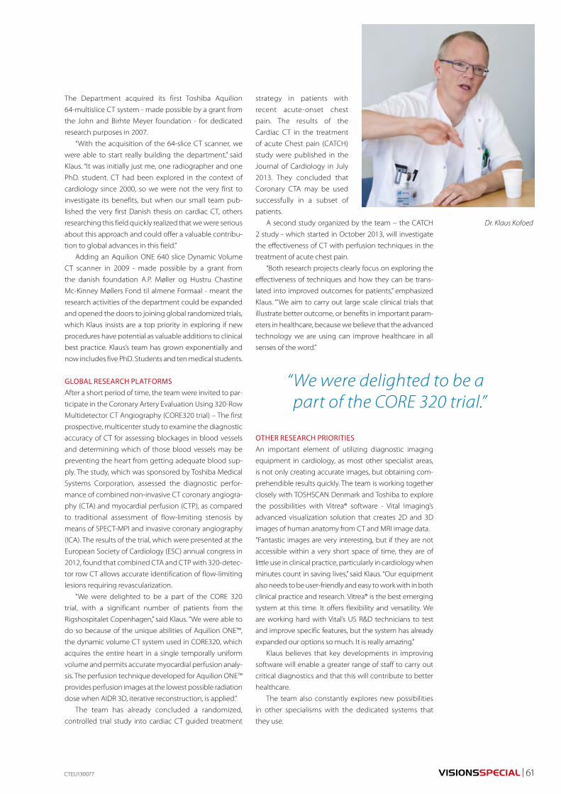
VISIONSSPECIAL | 61
The Department acquired its first Toshiba Aquilion
64-multislice CT system - made possible by a grant from
the John and Birhte Meyer foundation - for dedicated
research purposes in 2007.
“With the acquisition of the 64-slice CT scanner, we
were able to start really building the department,” said
Klaus. “It was initially just me, one radiographer and one
PhD. student. CT had been explored in the context of
cardiology since 2000, so we were not the very first to
investigate its benefits, but when our small team pub-
lished the very first Danish thesis on cardiac CT, others
researching this field quickly realized that we were serious
about this approach and could offer a valuable contribu-
tion to global advances in this field.”
Adding an Aquilion ONE 640 slice Dynamic Volume
CT scanner in 2009 - made possible by a grant from
the danish foundation A.P. Møller og Hustru Chastine
Mc-Kinney Møllers Fond til almene Formaal - meant the
research activities of the department could be expanded
and opened the doors to joining global randomized trials,
which Klaus insists are a top priority in exploring if new
procedures have potential as valuable additions to clinical
best practice. Klaus’s team has grown exponentially and
now includes five PhD. Students and ten medical students.
GLOBAL RESEARCH PLATFORMSAfter a short period of time, the team were invited to par-
ticipate in the Coronary Artery Evaluation Using 320-Row
Multidetector CT Angiography (CORE320 trial) – The first
prospective, multicenter study to examine the diagnostic
accuracy of CT for assessing blockages in blood vessels
and determining which of those blood vessels may be
preventing the heart from getting adequate blood sup-
ply. The study, which was sponsored by Toshiba Medical
Systems Corporation, assessed the diagnostic perfor-
mance of combined non-invasive CT coronary angiogra-
phy (CTA) and myocardial perfusion (CTP), as compared
to traditional assessment of flow-limiting stenosis by
means of SPECT-MPI and invasive coronary angiography
(ICA). The results of the trial, which were presented at the
European Society of Cardiology (ESC) annual congress in
2012, found that combined CTA and CTP with 320-detec-
tor row CT allows accurate identification of flow-limiting
lesions requiring revascularization.
“We were delighted to be a part of the CORE 320
trial, with a significant number of patients from the
Rigshospitalet Copenhagen,” said Klaus. “We were able to
do so because of the unique abilities of Aquilion ONE™,
the dynamic volume CT system used in CORE320, which
acquires the entire heart in a single temporally uniform
volume and permits accurate myocardial perfusion analy-
sis. The perfusion technique developed for Aquilion ONE™
provides perfusion images at the lowest possible radiation
dose when AIDR 3D, iterative reconstruction, is applied.”
The team has already concluded a randomized,
controlled trial study into cardiac CT guided treatment
strategy in patients with
recent acute-onset chest
pain. The results of the
Cardiac CT in the treatment
of acute Chest pain (CATCH)
study were published in the
Journal of Cardiology in July
2013. They concluded that
Coronary CTA may be used
successfully in a subset of
patients.
A second study organized by the team – the CATCH
2 study - which started in October 2013, will investigate
the effectiveness of CT with perfusion techniques in the
treatment of acute chest pain.
“Both research projects clearly focus on exploring the
effectiveness of techniques and how they can be trans-
lated into improved outcomes for patients,” emphasized
Klaus. “’We aim to carry out large scale clinical trials that
illustrate better outcome, or benefits in important param-
eters in healthcare, because we believe that the advanced
technology we are using can improve healthcare in all
senses of the word.”
CTEU130077
“ We were delighted to be a part of the CORE 320 trial.”
OTHER RESEARCH PRIORITIESAn important element of utilizing diagnostic imaging
equipment in cardiology, as most other specialist areas,
is not only creating accurate images, but obtaining com-
prehendible results quickly. The team is working together
closely with TOSHSCAN Denmark and Toshiba to explore
the possibilities with Vitrea® software - Vital Imaging’s
advanced visualization solution that creates 2D and 3D
images of human anatomy from CT and MRI image data.
“Fantastic images are very interesting, but if they are not
accessible within a very short space of time, they are of
little use in clinical practice, particularly in cardiology when
minutes count in saving lives,” said Klaus. “Our equipment
also needs to be user-friendly and easy to work with in both
clinical practice and research. Vitrea® is the best emerging
system at this time. It offers flexibility and versatility. We
are working hard with Vital’s US R&D technicians to test
and improve specific features, but the system has already
expanded our options so much. It is really amazing.”
Klaus believes that key developments in improving
software will enable a greater range of staff to carry out
critical diagnostics and that this will contribute to better
healthcare.
The team also constantly explores new possibilities
in other specialisms with the dedicated systems that
they use.
Dr. Klaus Kofoed
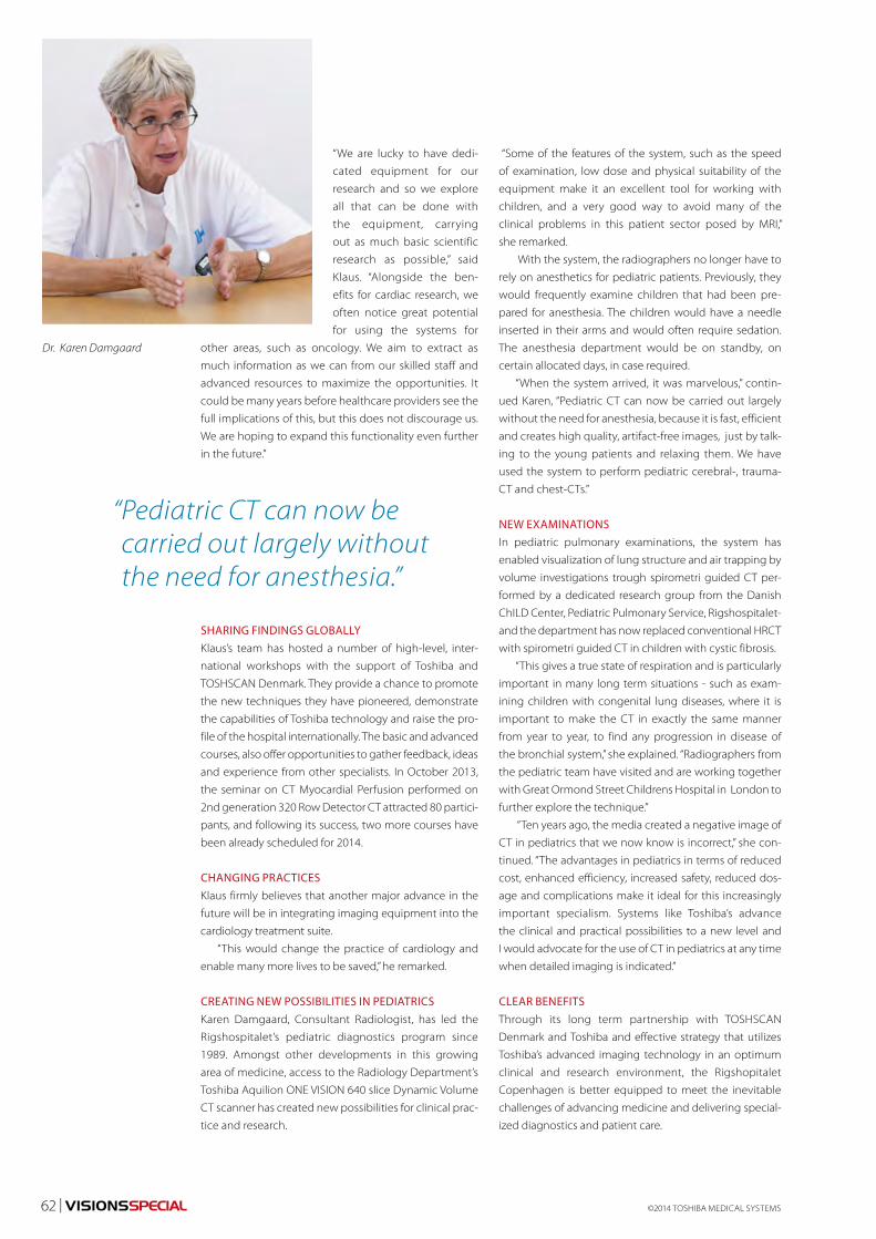
©2014 TOSHIBA MEDICAL SYSTEMS62 | VISIONSSPECIAL
“We are lucky to have dedi-
cated equipment for our
research and so we explore
all that can be done with
the equipment, carrying
out as much basic scientific
research as possible,” said
Klaus. “Alongside the ben-
efits for cardiac research, we
often notice great potential
for using the systems for
other areas, such as oncology. We aim to extract as
much information as we can from our skilled staff and
advanced resources to maximize the opportunities. It
could be many years before healthcare providers see the
full implications of this, but this does not discourage us.
We are hoping to expand this functionality even further
in the future.”
“Some of the features of the system, such as the speed
of examination, low dose and physical suitability of the
equipment make it an excellent tool for working with
children, and a very good way to avoid many of the
clinical problems in this patient sector posed by MRI,”
she remarked.
With the system, the radiographers no longer have to
rely on anesthetics for pediatric patients. Previously, they
would frequently examine children that had been pre-
pared for anesthesia. The children would have a needle
inserted in their arms and would often require sedation.
The anesthesia department would be on standby, on
certain allocated days, in case required.
“When the system arrived, it was marvelous,” contin-
ued Karen, “Pediatric CT can now be carried out largely
without the need for anesthesia, because it is fast, efficient
and creates high quality, artifact-free images, just by talk-
ing to the young patients and relaxing them. We have
used the system to perform pediatric cerebral-, trauma-
CT and chest-CTs.”
NEW EXAMINATIONSIn pediatric pulmonary examinations, the system has
enabled visualization of lung structure and air trapping by
volume investigations trough spirometri guided CT per-
formed by a dedicated research group from the Danish
ChILD Center, Pediatric Pulmonary Service, Rigshospitalet-
and the department has now replaced conventional HRCT
with spirometri guided CT in children with cystic fibrosis.
“This gives a true state of respiration and is particularly
important in many long term situations - such as exam-
ining children with congenital lung diseases, where it is
important to make the CT in exactly the same manner
from year to year, to find any progression in disease of
the bronchial system,” she explained. “Radiographers from
the pediatric team have visited and are working together
with Great Ormond Street Childrens Hospital in London to
further explore the technique.”
“Ten years ago, the media created a negative image of
CT in pediatrics that we now know is incorrect,” she con-
tinued. “The advantages in pediatrics in terms of reduced
cost, enhanced efficiency, increased safety, reduced dos-
age and complications make it ideal for this increasingly
important specialism. Systems like Toshiba’s advance
the clinical and practical possibilities to a new level and
I would advocate for the use of CT in pediatrics at any time
when detailed imaging is indicated.”
CLEAR BENEFITSThrough its long term partnership with TOSHSCAN
Denmark and Toshiba and effective strategy that utilizes
Toshiba’s advanced imaging technology in an optimum
clinical and research environment, the Rigshopitalet
Copenhagen is better equipped to meet the inevitable
challenges of advancing medicine and delivering special-
ized diagnostics and patient care.
Dr. Karen Damgaard
“ Pediatric CT can now be carried out largely without the need for anesthesia.”
SHARING FINDINGS GLOBALLYKlaus’s team has hosted a number of high-level, inter-
national workshops with the support of Toshiba and
TOSHSCAN Denmark. They provide a chance to promote
the new techniques they have pioneered, demonstrate
the capabilities of Toshiba technology and raise the pro-
file of the hospital internationally. The basic and advanced
courses, also offer opportunities to gather feedback, ideas
and experience from other specialists. In October 2013,
the seminar on CT Myocardial Perfusion performed on
2nd generation 320 Row Detector CT attracted 80 partici-
pants, and following its success, two more courses have
been already scheduled for 2014.
CHANGING PRACTICESKlaus firmly believes that another major advance in the
future will be in integrating imaging equipment into the
cardiology treatment suite.
“This would change the practice of cardiology and
enable many more lives to be saved,” he remarked.
CREATING NEW POSSIBILITIES IN PEDIATRICSKaren Damgaard, Consultant Radiologist, has led the
Rigshospitalet’s pediatric diagnostics program since
1989. Amongst other developments in this growing
area of medicine, access to the Radiology Department’s
Toshiba Aquilion ONE VISION 640 slice Dynamic Volume
CT scanner has created new possibilities for clinical prac-
tice and research.
VISIONSSPECIAL | 63
A STORY OF SUCCESS Bjarne has led TOSHSCAN Denmark since 2001. He joined
the organization with a burning ambition to make Toshiba’s
pioneering technology widely available to public hospitals
and private clinics in Denmark. He built the organization
from scratch, using a focused, modular approach – a strat-
egy to consolidate growth, modality by modality, eventu-
ally encompassing them all to ensure solid development.
TOSHSCAN Denmark’s initial focus was on Toshiba’s CT
and Ultrasound capabilities, because Bjarne felt its offer
in this was particularly strong and addressed the greatest
immediate needs within the Danish market. X-Ray was
later added as an additional focus.
“I studied medical engineering and worked as a
healthcare engineer before joining TOSHSCAN Denmark.
Throughout my career, I worked closely with Toshiba,
knew how high quality their products were and was
familiar with Toshiba’s background, the stability of the
company and its products,” he explained. “I also knew that
many Danish healthcare specialists had recognized the
same qualities and wanted to purchase Toshiba’s scan-
ners, however, at the time, they weren’t available here.
When I was offered the position of Managing Director
of the new TOSHSCAN Denmark organization, it was the
perfect opportunity to build a dedicated Toshiba organi-
zation in Denmark and bring Toshiba’s products to the
specialists that appreciated and wanted them.”
Providing a supportive and professional service
alongside Toshiba’s high quality products and technol-
ogy, TOSHSCAN Denmark has many satisfied customers
nationally and has also enabled many Danish specialists
contribute to the global medical imaging technology
platform, by engaging them in open, two-way partner-
ships, for example, in R&D. This is particularly true of the
Rigshospitalet Copenhagen in Denmark’s capital city - a
large, specialized hospital with significant capabilities.
Toshiba systems and long term support from TOSHSCAN
Delivering Excellence through Partnership in DenmarkTOSHSCAN Denmark, the well-known face of Toshiba in Denmark, provides a full range of imaging products and service to the Danish market. Established 13 years ago in response to demand for Toshiba’s systems in the country, TOSHSCAN Denmark has developed a robust business within the technophile Danish healthcare landscape. Epitomizing Toshiba’s core values of dedicated partnership, high quality and innovative solutions and service, the organization is highly regarded across the country. VISIONS talked to Bjarne Alhøj, Managing Director of TOSHSCAN Denmark, to find out more about why excellence and partnership are such key ingredients in success.
Denmark has helped equip the radiology department to
the highest standards and help it emerge as a center of
global excellence.
“One of my very first priorities was to support the
prestigious Rigshospitalet Copenhagen, which at the
time, were using just a few Toshiba ultrasound systems
at the hospital. My ultimate goal was to supply Toshiba
technology in all modalities, because I knew this could
help the Radiology Team realize their ambitions,” he con-
tinued. “When we first approached the Team, they were
impressed by Toshiba’s products, but had concerns that
service would be provided from Toshiba headquarters in
Japan, which, of course, would have been impractical. I
believe that both high quality products and equally high
quality support in optimizing their application are the
most vital aspects of our work. Therefore, we provided
the hospital with dedicated local service and have since
supplied the majority of the department’s CT solutions,
including, most recently, those for the hospital’s new
Trauma Center – the first in world to be equipped with
two CT systems set within a gantry that moves over the
patient on the surgery table”
Because of the sheer number of Toshiba systems
that the department now has, TOSHSCAN Denmark has
a dedicated service engineer located onsite for mainte-
nance of the systems.
PRESTIGIOUS PARTNERSHIPTOSHSCAN Denmark’s relationship with the
Rigshospitalet quickly evolved into a truly two-way part-
nership. Danish specialists are globally renowned for their
interest in innovation and what technology can offer, as
well as their professionalism. With its strong active focus
on research and medical education, alongside clinical
practice, the hospital provides a prime example of how
Toshiba’s strategic priority to incorporate ‘The Voice of the
Customer’ helps advance products and service.
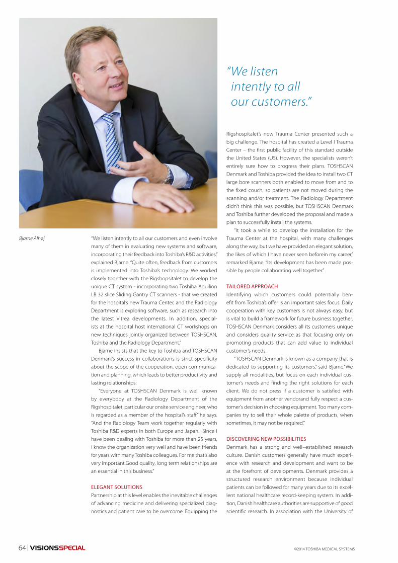
©2014 TOSHIBA MEDICAL SYSTEMS64 | VISIONSSPECIAL
“We listen intently to all our customers and even involve
many of them in evaluating new systems and software,
incorporating their feedback into Toshiba’s R&D activities,”
explained Bjarne. “Quite often, feedback from customers
is implemented into Toshiba’s technology. We worked
closely together with the Rigshopsitalet to develop the
unique CT system - incorporating two Toshiba Aquilion
LB 32 slice Sliding Gantry CT scanners - that we created
for the hospital’s new Trauma Center, and the Radiology
Department is exploring software, such as research into
the latest Vitrea developments. In addition, special-
ists at the hospital host international CT workshops on
new techniques jointly organized between TOSHSCAN,
Toshiba and the Radiology Department.”
Bjarne insists that the key to Toshiba and TOSHSCAN
Denmark’s success in collaborations is strict specificity
about the scope of the cooperation, open communica-
tion and planning, which leads to better productivity and
lasting relationships:
“Everyone at TOSHSCAN Denmark is well known
by everybody at the Radiology Department of the
Rigshospitalet, particular our onsite service engineer, who
is regarded as a member of the hospital’s staff” he says.
“And the Radiology Team work together regularly with
Toshiba R&D experts in both Europe and Japan. Since I
have been dealing with Toshiba for more than 25 years,
I know the organization very well and have been friends
for years with many Toshiba colleagues. For me that’s also
very important.Good quality, long term relationships are
an essential in this business.”
ELEGANT SOLUTIONSPartnership at this level enables the inevitable challenges
of advancing medicine and delivering specialized diag-
nostics and patient care to be overcome. Equipping the
Rigshospitalet’s new Trauma Center presented such a
big challenge. The hospital has created a Level I Trauma
Center – the first public facility of this standard outside
the United States (US). However, the specialists weren’t
entirely sure how to progress their plans. TOSHSCAN
Denmark and Toshiba provided the idea to install two CT
large bore scanners both enabled to move from and to
the fixed couch, so patients are not moved during the
scanning and/or treatment. The Radiology Department
didn’t think this was possible, but TOSHSCAN Denmark
and Toshiba further developed the proposal and made a
plan to successfully install the systems.
“It took a while to develop the installation for the
Trauma Center at the hospital, with many challenges
along the way, but we have provided an elegant solution,
the likes of which I have never seen beforein my career,”
remarked Bjarne. “Its development has been made pos-
sible by people collaborating well together.”
TAILORED APPROACHIdentifying which customers could potentially ben-
efit from Toshiba’s offer is an important sales focus. Daily
cooperation with key customers is not always easy, but
is vital to build a framework for future business together.
TOSHSCAN Denmark considers all its customers unique
and considers quality service as that focusing only on
promoting products that can add value to individual
customer’s needs.
“TOSHSCAN Denmark is known as a company that is
dedicated to supporting its customers,” said Bjarne.“We
supply all modalities, but focus on each individual cus-
tomer’s needs and finding the right solutions for each
client. We do not press if a customer is satisfied with
equipment from another vendorand fully respect a cus-
tomer’s decision in choosing equipment. Too many com-
panies try to sell their whole palette of products, when
sometimes, it may not be required.”
DISCOVERING NEW POSSIBILITIESDenmark has a strong and well–established research
culture. Danish customers generally have much experi-
ence with research and development and want to be
at the forefront of developments. Denmark provides a
structured research environment because individual
patients can be followed for many years due to its excel-
lent national healthcare record-keeping system. In addi-
tion, Danish healthcare authorities are supportive of good
scientific research. In association with the University of
Bjarne Alhøj
“ We listen intently to all our customers.”
VISIONSSPECIAL | 65
Copenhagen and the Danish University of Technology,
the Radiology Department at the Rigshospitalet
Copenhagen has a strong active research program
and has some dedicated research equipment within its
resources.
Bjarne has also found that Danish customers are par-
ticularly interested to get the most possible from new
developments and optimize the use of equipment. The
versatility and advanced capabilities of Toshiba’s systems
mean that customers themselves may find new opportu-
nities in clinical practice and research that had not been
previously explored. For example, the Rigshospitalet
Copenhagen, have discovered that Toshiba systems can
support outstanding CT Perfusion studies.
“Sometimes, it is the customer, who discovers new
applications for our systems, because Toshiba’s technol-
ogy has so many facets that even our own developers
may not be completely aware of the full capabilities,”
explained Bjarne. “When these offer benefits for patients
they create new research and development avenues for
the company.Toshiba provides support to selected spe-
cialists for larger scale research to explore promising new
techniques in some cases.”
DISSEMINATING KNOWLEDGERigshospitalet Copenhagen has hosted a number of
high-level, international CT workshops with the sup-
port of Toshiba and TOSHSCAN Denmark. Attended by
specialists from all over the world, they provide a chance
for physicians to promote new techniques, demon-
strate the capabilities of Toshiba technology and raise
the profile of the hospital internationally.The courses
also create opportunities to gather feedback, ideas and
experience from other specialists and users.Most recently,
the Rigshospitalet’s seminar on Advanced Myocardial
CT Perfusion performed on 2nd generation 320 Row
Detector CT, run by Dr. Klaus Kofoed, Cardiologist at
Rigshospitalet.
MEETING THE CHALLENGES OF CHANGEWhile the specialist consultants at Rigshospitalet
Copenhagen are convinced of the capabilities of Toshiba
systems and work closely with TOSHSCAN Denmark, they
are not the only ones involved in TOSHSCAN Denmark’s
partnerships.
“We also work closely with purchasing departments at
our customers because they are an integral part of the
decision-making team in Denmark,” said Bjarne. “Toshiba
technology offers many new exciting clinical and research
options, but of course, this has a price.We keep both cli-
nicians and purchasers informed of what technology is
coming up and what it costs.”
Denmark’s healthcare system includes an increasing
proportion of governmental national tenders, in which
certain equipment is purchased for hospitals across
the country. Over the next six years, it will focus on the
development of six new ‘super hospitals’ - located in
each of the five regions in Denmark and incorporating
a full range of medical specialization. The system is also
being revised to put patients firmly at the center of care,
which will result in better care, but also drives changes
for healthcare providers.
“TOSHSCAN Denmark and Toshiba are geared to
meet the challenges of change head on,”’ said Bjarne.
“We must meet the increasing demands on equip-
ment, but also continue to develop new things.While
we are advancing fast, there are still huge opportunities
to improve the speed and accuracy of diagnosis and
help patients become healthy again through exploring
new areas that continually emerge in our industry. One
of the biggest challenges is defining how to structure
healthcare in the best way and how we can improve our
support, so that the benefits to hospitals and patients
are optimized. We sell solutions, not just equipment -
TOSHIBA Made for Life.”
ENVIRONMENTAL PRIORITIESIn line with growing focus on sustainability and the
environment, TOSHSCAN Denmark implements Toshiba’s
environmental policy, as well as its own.
“In addition to the environmental benefits offered by
Toshiba’s systems, such as energy-saving features, reduced
packaging and other eco-standards, TOSHSCAN Denmark
strives to contribute to making our business more sus-
tainable in any way that we can,” said Bjarne. “We want to
ensure that our staff and our customers and their patients
have safe, healthy and environmentally-compatible sur-
roundings. This is also a personal goal for me.”
AWARD-WINNINGGuided by Bjarne’s careful strategic approach and deliv-
ering on TOSHSCAN Denmark’s substantial promise of
high quality support and service in providing Toshiba’s
innovative diagnostic solutions, the organization has
established an unparalleled reputation in Denmark with
customers and the medical imaging industry alike. Its
long term clients include prestigious world class medi-
cal establishments, like the Rigshospitalet Copenhagen.
The organization has recently been recognized with
Toshiba Medical Systems Europe awards for Outstanding
Performance 2012 DABO Region North and Outstanding
CT Performance 2012.
BRIGHT FUTURENow comprising a strong, modern organization,
TOSHSCAN Denmark continues to grow year by year. In
2012, it became Denmark’s market leader in CT systems.
“Further growth is important, but we must ensure that
this is stable and only grow in a way that creates a solid,
sustainable organization that will be around to support its
clients, their patients and contribute to the global Toshiba
organization well into the future,” concluded Bjarne.
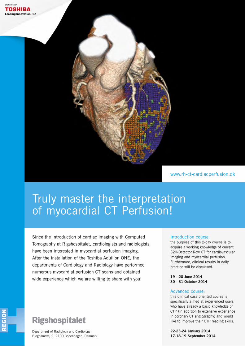
Truly master the interpretation of myocardial CT Perfusion!
SPONSORED BY
www.rigshospitalet.dk
SPONSORED BY
Since the introduction of cardiac imaging with Computed
Tomography at Rigshospitalet, cardiologists and radiologists
have been interested in myocardial perfusion imaging.
After the installation of the Toshiba Aquilion ONE, the
departments of Cardiology and Radiology have performed
numerous myocardial perfusion CT scans and obtained
wide experience which we are willing to share with you!
Introduction course: the purpose of this 2-day course is to acquire a working knowledge of current 320-Detector Row CT for cardiovascular imaging and myocardial perfusion. Furthermore, clinical results in daily practice will be discussed. 19 - 20 June 201430 - 31 October 2014
Advanced course: this clinical case oriented course is specifically aimed at experienced users who have already a basic knowledge of CTP (in addition to extensive experience in coronary CT angiography) and would like to improve their CTP reading skills.
22-23-24 January 201417-18-19 September 2014
Department of Radiology and CardiologyBlegdamsvej 9, 2100 Copenhagen, Denmark
www.rh-ct-cardiacperfusion.dk
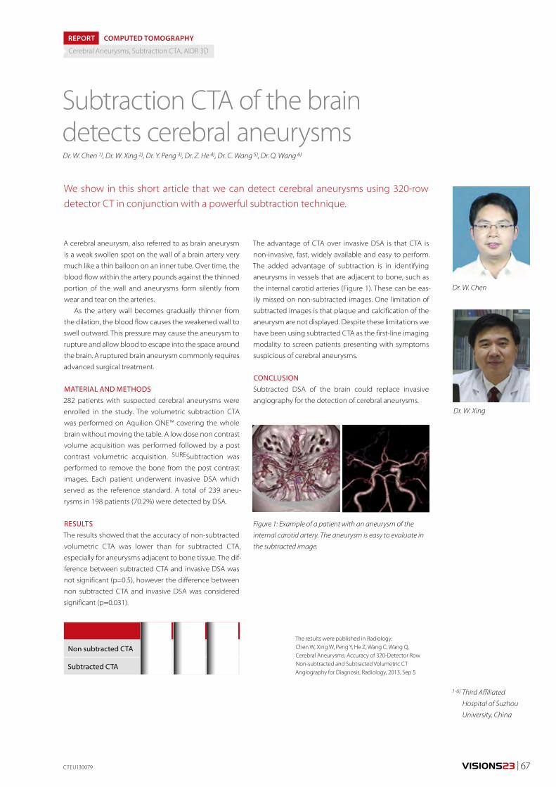
VISIONS23 | 67
A cerebral aneurysm, also referred to as brain aneurysm
is a weak swollen spot on the wall of a brain artery very
much like a thin balloon on an inner tube. Over time, the
blood flow within the artery pounds against the thinned
portion of the wall and aneurysms form silently from
wear and tear on the arteries.
As the artery wall becomes gradually thinner from
the dilation, the blood flow causes the weakened wall to
swell outward. This pressure may cause the aneurysm to
rupture and allow blood to escape into the space around
the brain. A ruptured brain aneurysm commonly requires
advanced surgical treatment.
MATERIAL AND METHODS282 patients with suspected cerebral aneurysms were
enrolled in the study. The volumetric subtraction CTA
was performed on Aquilion ONE™ covering the whole
brain without moving the table. A low dose non contrast
volume acquisition was performed followed by a post
contrast volumetric acquisition. SURESubtraction was
performed to remove the bone from the post contrast
images. Each patient underwent invasive DSA which
served as the reference standard. A total of 239 aneu-
rysms in 198 patients (70.2%) were detected by DSA.
RESULTSThe results showed that the accuracy of non-subtracted
volumetric CTA was lower than for subtracted CTA,
especially for aneurysms adjacent to bone tissue. The dif-
ference between subtracted CTA and invasive DSA was
not significant (p=0.5), however the difference between
non subtracted CTA and invasive DSA was considered
significant (p=0.031).
Subtraction CTA of the braindetects cerebral aneurysms
REPORT
We show in this short article that we can detect cerebral aneurysms using 320-row detector CT in conjunction with a powerful subtraction technique.
Dr. W. Chen
Dr. W. Xing
1-6) Third Affiliated
Hospital of Suzhou
University, China
CTEU130079
Dr. W. Chen 1), Dr. W. Xing 2), Dr. Y. Peng 3), Dr. Z. He 4), Dr. C. Wang 5), Dr. Q. Wang 6)
Figure 1: Example of a patient with an aneurysm of the
internal carotid artery. The aneurysm is easy to evaluate in
the subtracted image.
Cerebral Aneurysms, Subtraction CTA, AIDR 3D
The advantage of CTA over invasive DSA is that CTA is
non-invasive, fast, widely available and easy to perform.
The added advantage of subtraction is in identifying
aneurysms in vessels that are adjacent to bone, such as
the internal carotid arteries (Figure 1). These can be eas-
ily missed on non-subtracted images. One limitation of
subtracted images is that plaque and calcification of the
aneurysm are not displayed. Despite these limitations we
have been using subtracted CTA as the first-line imaging
modality to screen patients presenting with symptoms
suspicious of cerebral aneurysms.
CONCLUSIONSubtracted DSA of the brain could replace invasive
angiography for the detection of cerebral aneurysms.
Sens Spec Acc
Non subtracted CTA 96.7% 100% 97.5%
Subtracted CTA 99.2% 100% 99.4%
COMPUTED TOMOGRAPHY
The results were published in Radiology: Chen W, Xing W, Peng Y, He Z, Wang C, Wang Q, Cerebral Aneurysms: Accuracy of 320-Detector Row Non-subtracted and Subtracted Volumetric CT Angiography for Diagnosis, Radiology, 2013, Sep 5
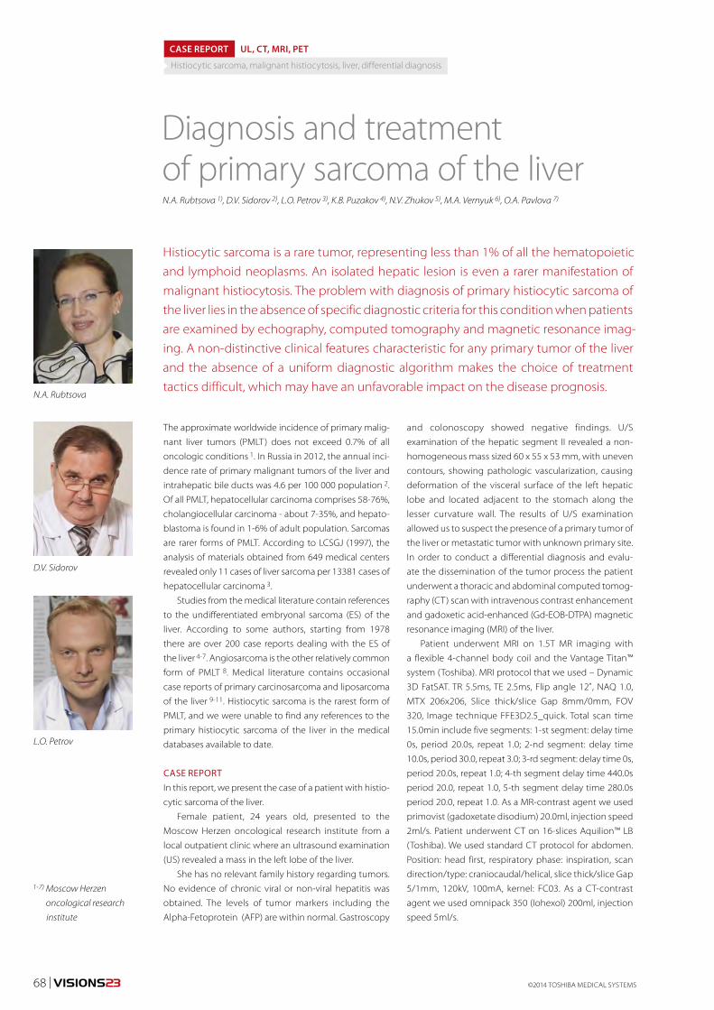
©2014 TOSHIBA MEDICAL SYSTEMS68 | VISIONS23
The approximate worldwide incidence of primary malig-
nant liver tumors (PMLT) does not exceed 0.7% of all
oncologic conditions 1. In Russia in 2012, the annual inci-
dence rate of primary malignant tumors of the liver and
intrahepatic bile ducts was 4.6 per 100 000 population 2.
Of all PMLT, hepatocellular carcinoma comprises 58-76%,
cholangiocellular carcinoma - about 7-35%, and hepato-
blastoma is found in 1-6% of adult population. Sarcomas
are rarer forms of PMLT. According to LCSGJ (1997), the
analysis of materials obtained from 649 medical centers
revealed only 11 cases of liver sarcoma per 13381 cases of
hepatocellular carcinoma 3.
Studies from the medical literature contain references
to the undifferentiated embryonal sarcoma (ES) of the
liver. According to some authors, starting from 1978
there are over 200 case reports dealing with the ES of
the liver 4-7. Angiosarcoma is the other relatively common
form of PMLT 8. Medical literature contains occasional
case reports of primary carcinosarcoma and liposarcoma
of the liver 9-11. Histiocytic sarcoma is the rarest form of
PMLT, and we were unable to find any references to the
primary histiocytic sarcoma of the liver in the medical
databases available to date.
CASE REPORTIn this report, we present the case of a patient with histio-
cytic sarcoma of the liver.
Female patient, 24 years old, presented to the
Moscow Herzen oncological research institute from a
local outpatient clinic where an ultrasound examination
(US) revealed a mass in the left lobe of the liver.
She has no relevant family history regarding tumors.
No evidence of chronic viral or non-viral hepatitis was
obtained. The levels of tumor markers including the
Alpha-Fetoprotein (AFP) are within normal. Gastroscopy
Diagnosis and treatment of primary sarcoma of the liver
Histiocytic sarcoma is a rare tumor, representing less than 1% of all the hematopoietic and lymphoid neoplasms. An isolated hepatic lesion is even a rarer manifestation of malignant histiocytosis. The problem with diagnosis of primary histiocytic sarcoma of the liver lies in the absence of specific diagnostic criteria for this condition when patients are examined by echography, computed tomography and magnetic resonance imag-ing. A non-distinctive clinical features characteristic for any primary tumor of the liver and the absence of a uniform diagnostic algorithm makes the choice of treatment tactics difficult, which may have an unfavorable impact on the disease prognosis.
N.A. Rubtsova 1), D.V. Sidorov 2), L.O. Petrov 3), K.B. Puzakov 4), N.V. Zhukov 5), M.A. Vernyuk 6), O.A. Pavlova 7)
N.A. Rubtsova
D.V. Sidorov
L.O. Petrov
1-7) Moscow Herzen
oncological research
institute
CASE REPORT UL, CT, MRI, PET
Histiocytic sarcoma, malignant histiocytosis, liver, differential diagnosis
and colonoscopy showed negative findings. U/S
examination of the hepatic segment II revealed a non-
homogeneous mass sized 60 x 55 x 53 mm, with uneven
contours, showing pathologic vascularization, causing
deformation of the visceral surface of the left hepatic
lobe and located adjacent to the stomach along the
lesser curvature wall. The results of U/S examination
allowed us to suspect the presence of a primary tumor of
the liver or metastatic tumor with unknown primary site.
In order to conduct a differential diagnosis and evalu-
ate the dissemination of the tumor process the patient
underwent a thoracic and abdominal computed tomog-
raphy (CT) scan with intravenous contrast enhancement
and gadoxetic acid-enhanced (Gd-EOB-DTPA) magnetic
resonance imaging (MRI) of the liver.
Patient underwent MRI on 1.5T MR imaging with
a flexible 4-channel body coil and the Vantage Titan™
system (Toshiba). MRI protocol that we used – Dynamic
3D FatSAT. TR 5.5ms, TE 2.5ms, Flip angle 12˚, NAQ 1.0,
MTX 206х206, Slice thick/slice Gap 8mm/0mm, FOV
320, Image technique FFE3D2.5_quick. Total scan time
15.0min include five segments: 1-st segment: delay time
0s, period 20.0s, repeat 1.0; 2-nd segment: delay time
10.0s, period 30.0, repeat 3.0; 3-rd segment: delay time 0s,
period 20.0s, repeat 1.0; 4-th segment delay time 440.0s
period 20.0, repeat 1.0, 5-th segment delay time 280.0s
period 20.0, repeat 1.0. As a MR-contrast agent we used
primovist (gadoxetate disodium) 20.0ml, injection speed
2ml/s. Patient underwent CT on 16-slices Aquilion™ LB
(Toshiba). We used standard CT protocol for abdomen.
Position: head first, respiratory phase: inspiration, scan
direction/type: craniocaudal/helical, slice thick/slice Gap
5/1mm, 120kV, 100mA, kernel: FC03. As a CT-contrast
agent we used omnipack 350 (Iohexol) 200ml, injection
speed 5ml/s.
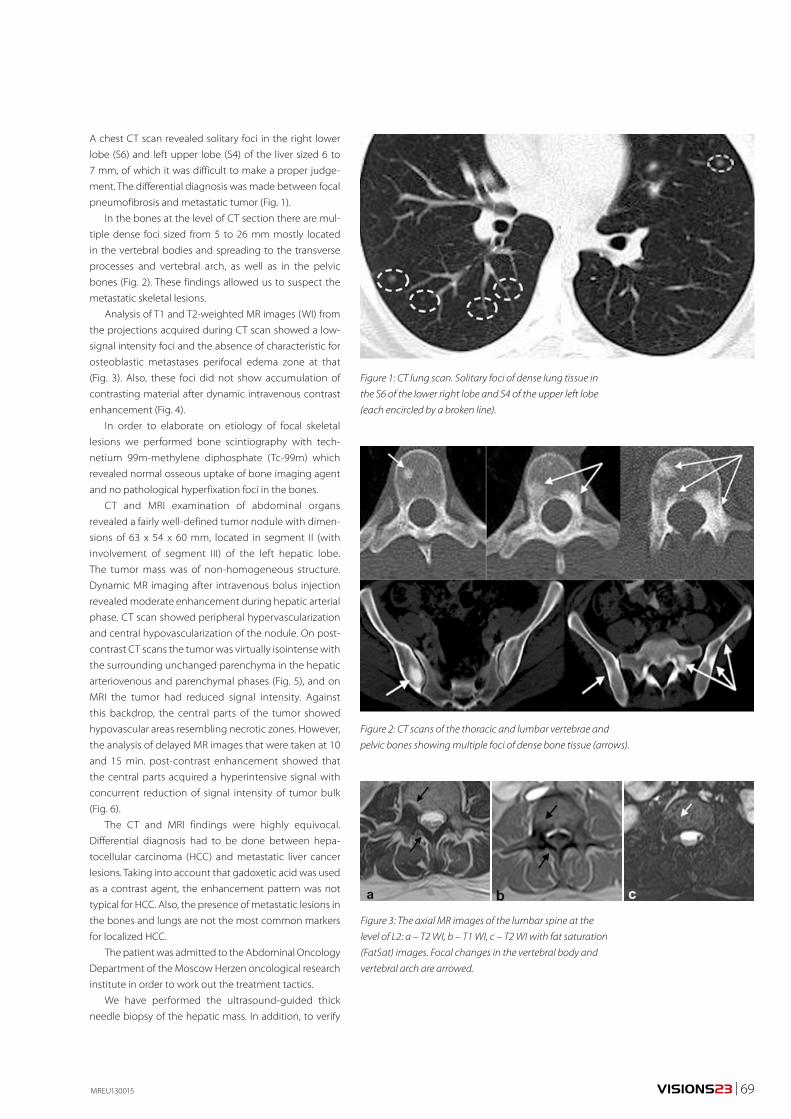
VISIONS23 | 69
A chest CT scan revealed solitary foci in the right lower
lobe (S6) and left upper lobe (S4) of the liver sized 6 to
7 mm, of which it was difficult to make a proper judge-
ment. The differential diagnosis was made between focal
pneumofibrosis and metastatic tumor (Fig. 1).
In the bones at the level of CT section there are mul-
tiple dense foci sized from 5 to 26 mm mostly located
in the vertebral bodies and spreading to the transverse
processes and vertebral arch, as well as in the pelvic
bones (Fig. 2). These findings allowed us to suspect the
metastatic skeletal lesions.
Analysis of T1 and T2-weighted MR images (WI) from
the projections acquired during CT scan showed a low-
signal intensity foci and the absence of characteristic for
osteoblastic metastases perifocal edema zone at that
(Fig. 3). Also, these foci did not show accumulation of
contrasting material after dynamic intravenous contrast
enhancement (Fig. 4).
In order to elaborate on etiology of focal skeletal
lesions we performed bone scintiography with tech-
netium 99m-methylene diphosphate (Tc-99m) which
revealed normal osseous uptake of bone imaging agent
and no pathological hyperfixation foci in the bones.
CT and MRI examination of abdominal organs
revealed a fairly well-defined tumor nodule with dimen-
sions of 63 х 54 х 60 mm, located in segment II (with
involvement of segment III) of the left hepatic lobe.
The tumor mass was of non-homogeneous structure.
Dynamic MR imaging after intravenous bolus injection
revealed moderate enhancement during hepatic arterial
phase. CT scan showed peripheral hypervascularization
and central hypovascularization of the nodule. On post-
contrast CT scans the tumor was virtually isointense with
the surrounding unchanged parenchyma in the hepatic
arteriovenous and parenchymal phases (Fig. 5), and on
MRI the tumor had reduced signal intensity. Against
this backdrop, the central parts of the tumor showed
hypovascular areas resembling necrotic zones. However,
the analysis of delayed MR images that were taken at 10
and 15 min. post-contrast enhancement showed that
the central parts acquired a hyperintensive signal with
concurrent reduction of signal intensity of tumor bulk
(Fig. 6).
The CT and MRI findings were highly equivocal.
Differential diagnosis had to be done between hepa-
tocellular carcinoma (HCC) and metastatic liver cancer
lesions. Taking into account that gadoxetic acid was used
as a contrast agent, the enhancement pattern was not
typical for HCC. Also, the presence of metastatic lesions in
the bones and lungs are not the most common markers
for localized HCC.
The patient was admitted to the Abdominal Oncology
Department of the Moscow Herzen oncological research
institute in order to work out the treatment tactics.
We have performed the ultrasound-guided thick
needle biopsy of the hepatic mass. In addition, to verify
Figure 1: CT lung scan. Solitary foci of dense lung tissue in
the S6 of the lower right lobe and S4 of the upper left lobe
(each encircled by a broken line).
Figure 2: CT scans of the thoracic and lumbar vertebrae and
pelvic bones showing multiple foci of dense bone tissue (arrows).
MREU130015
Figure 3: The axial MR images of the lumbar spine at the
level of L2: а – Т2 WI, b – Т1 WI, c – Т2 WI with fat saturation
(FatSat) images. Focal changes in the vertebral body and
vertebral arch are arrowed.
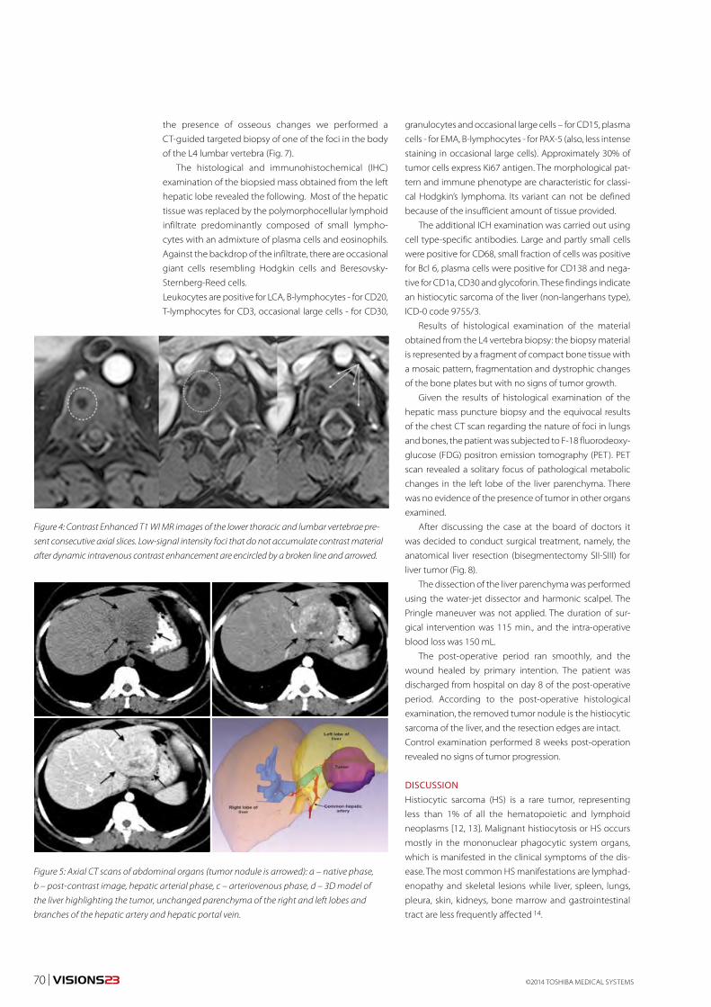
©2014 TOSHIBA MEDICAL SYSTEMS70 | VISIONS23
Figure 4: Contrast Enhanced T1 WI MR images of the lower thoracic and lumbar vertebrae pre-
sent consecutive axial slices. Low-signal intensity foci that do not accumulate contrast material
after dynamic intravenous contrast enhancement are encircled by a broken line and arrowed.
Figure 5: Axial CT scans of abdominal organs (tumor nodule is arrowed): а – native phase,
b – post-contrast image, hepatic arterial phase, c – arteriovenous phase, d – 3D model of
the liver highlighting the tumor, unchanged parenchyma of the right and left lobes and
branches of the hepatic artery and hepatic portal vein.
the presence of osseous changes we performed a
CT-guided targeted biopsy of one of the foci in the body
of the L4 lumbar vertebra (Fig. 7).
The histological and immunohistochemical (IHC)
examination of the biopsied mass obtained from the left
hepatic lobe revealed the following. Most of the hepatic
tissue was replaced by the polymorphocellular lymphoid
infiltrate predominantly composed of small lympho-
cytes with an admixture of plasma cells and eosinophils.
Against the backdrop of the infiltrate, there are occasional
giant cells resembling Hodgkin cells and Beresovsky-
Sternberg-Reed cells.
Leukocytes are positive for LCA, B-lymphocytes - for CD20,
T-lymphocytes for CD3, occasional large cells - for CD30,
granulocytes and occasional large cells – for CD15, plasma
cells - for ЕМА, B-lymphocytes - for РАХ-5 (also, less intense
staining in occasional large cells). Approximately 30% of
tumor cells express Ki67 antigen. The morphological pat-
tern and immune phenotype are characteristic for classi-
cal Hodgkin’s lymphoma. Its variant can not be defined
because of the insufficient amount of tissue provided.
The additional ICH examination was carried out using
cell type-specific antibodies. Large and partly small cells
were positive for CD68, small fraction of cells was positive
for Bсl 6, plasma cells were positive for CD138 and nega-
tive for CD1a, CD30 and glycoforin. These findings indicate
an histiocytic sarcoma of the liver (non-langerhans type),
ICD-0 code 9755/3.
Results of histological examination of the material
obtained from the L4 vertebra biopsy: the biopsy material
is represented by a fragment of compact bone tissue with
a mosaic pattern, fragmentation and dystrophic changes
of the bone plates but with no signs of tumor growth.
Given the results of histological examination of the
hepatic mass puncture biopsy and the equivocal results
of the chest CT scan regarding the nature of foci in lungs
and bones, the patient was subjected to F-18 fluorodeoxy-
glucose (FDG) positron emission tomography (PET). PET
scan revealed a solitary focus of pathological metabolic
changes in the left lobe of the liver parenchyma. There
was no evidence of the presence of tumor in other organs
examined.
After discussing the case at the board of doctors it
was decided to conduct surgical treatment, namely, the
anatomical liver resection (bisegmentectomy SII-SIII) for
liver tumor (Fig. 8).
The dissection of the liver parenchyma was performed
using the water-jet dissector and harmonic scalpel. The
Pringle maneuver was not applied. The duration of sur-
gical intervention was 115 min., and the intra-operative
blood loss was 150 mL.
The post-operative period ran smoothly, and the
wound healed by primary intention. The patient was
discharged from hospital on day 8 of the post-operative
period. According to the post-operative histological
examination, the removed tumor nodule is the histiocytic
sarcoma of the liver, and the resection edges are intact.
Control examination performed 8 weeks post-operation
revealed no signs of tumor progression.
DISCUSSION Histiocytic sarcoma (HS) is a rare tumor, representing
less than 1% of all the hematopoietic and lymphoid
neoplasms [12, 13]. Malignant histiocytosis or HS occurs
mostly in the mononuclear phagocytic system organs,
which is manifested in the clinical symptoms of the dis-
ease. The most common HS manifestations are lymphad-
enopathy and skeletal lesions while liver, spleen, lungs,
pleura, skin, kidneys, bone marrow and gastrointestinal
tract are less frequently affected 14.
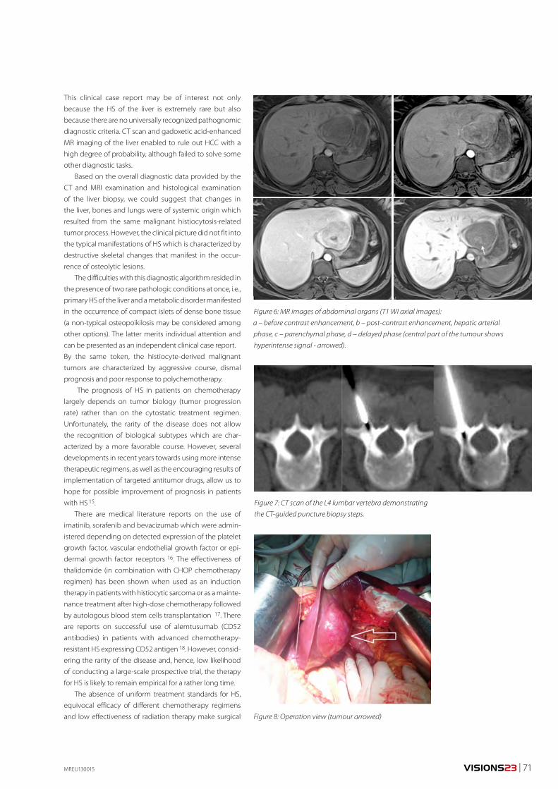
VISIONS23 | 71
This clinical case report may be of interest not only
because the HS of the liver is extremely rare but also
because there are no universally recognized pathognomic
diagnostic criteria. CT scan and gadoxetic acid-enhanced
MR imaging of the liver enabled to rule out HCC with a
high degree of probability, although failed to solve some
other diagnostic tasks.
Based on the overall diagnostic data provided by the
CT and MRI examination and histological examination
of the liver biopsy, we could suggest that changes in
the liver, bones and lungs were of systemic origin which
resulted from the same malignant histiocytosis-related
tumor process. However, the clinical picture did not fit into
the typical manifestations of HS which is characterized by
destructive skeletal changes that manifest in the occur-
rence of osteolytic lesions.
The difficulties with this diagnostic algorithm resided in
the presence of two rare pathologic conditions at once, i.e.,
primary HS of the liver and a metabolic disorder manifested
in the occurrence of compact islets of dense bone tissue
(a non-typical osteopoikilosis may be considered among
other options). The latter merits individual attention and
can be presented as an independent clinical case report.
By the same token, the histiocyte-derived malignant
tumors are characterized by aggressive course, dismal
prognosis and poor response to polychemotherapy.
The prognosis of HS in patients on chemotherapy
largely depends on tumor biology (tumor progression
rate) rather than on the cytostatic treatment regimen.
Unfortunately, the rarity of the disease does not allow
the recognition of biological subtypes which are char-
acterized by a more favorable course. However, several
developments in recent years towards using more intense
therapeutic regimens, as well as the encouraging results of
implementation of targeted antitumor drugs, allow us to
hope for possible improvement of prognosis in patients
with HS 15.
There are medical literature reports on the use of
imatinib, sorafenib and bevacizumab which were admin-
istered depending on detected expression of the platelet
growth factor, vascular endothelial growth factor or epi-
dermal growth factor receptors 16. The effectiveness of
thalidomide (in combination with CHOP chemotherapy
regimen) has been shown when used as an induction
therapy in patients with histiocytic sarcoma or as a mainte-
nance treatment after high-dose chemotherapy followed
by autologous blood stem cells transplantation 17. There
are reports on successful use of alemtusumab (CD52
antibodies) in patients with advanced chemotherapy-
resistant HS expressing CD52 antigen 18. However, consid-
ering the rarity of the disease and, hence, low likelihood
of conducting a large-scale prospective trial, the therapy
for HS is likely to remain empirical for a rather long time.
The absence of uniform treatment standards for HS,
equivocal efficacy of different chemotherapy regimens
and low effectiveness of radiation therapy make surgical
Figure 6: MR images of abdominal organs (Т1 WI axial images):
а – before contrast enhancement, b – post-contrast enhancement, hepatic arterial
phase, c – parenchymal phase, d – delayed phase (central part of the tumour shows
hyperintense signal - arrowed).
Figure 7: CT scan of the L4 lumbar vertebra demonstrating
the CT-guided puncture biopsy steps.
MREU130015
Figure 8: Operation view (tumour arrowed)
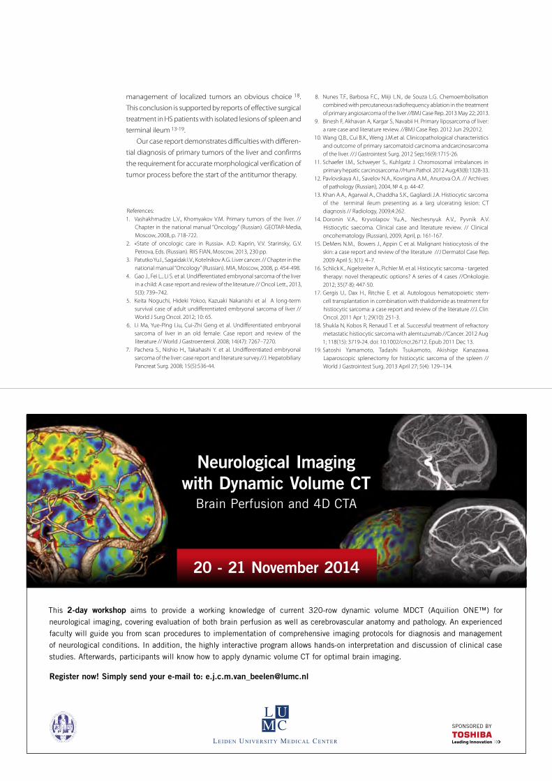
References:1. Vashakhmadze L.V., Khomyakov V.M. Primary tumors of the liver. //
Chapter in the national manual “Oncology” (Russian). GEOTAR-Media, Moscow, 2008, p. 718-722.
2. «State of oncologic care in Russia». A.D. Kaprin, V.V. Starinsky, G.V. Petrova, Eds. (Russian). RIIS FIAN, Moscow, 2013, 230 pp.
3. Patutko Yu.I., Sagaidak I.V., Kotelnikov A.G. Liver cancer. // Chapter in the national manual “Oncology” (Russian). MIA, Moscow, 2008, p. 454-498.
4. Gao J., Fei L., Li S. et al. Undifferentiated embryonal sarcoma of the liver in a child: A case report and review of the literature // Oncol Lett., 2013, 5(3): 739–742.
5. Keita Noguchi, Hideki Yokoo, Kazuaki Nakanishi et al A long-term survival case of adult undifferentiated embryonal sarcoma of liver //World J Surg Oncol. 2012; 10: 65.
6. Li Ma, Yue-Ping Liu, Cui-Zhi Geng et al. Undifferentiated embryonal sarcoma of liver in an old female: Case report and review of the literature // World J Gastroenterol. 2008; 14(47): 7267–7270.
7. Pachera S., Nishio H., Takahashi Y. et al. Undifferentiated embryonal sarcoma of the liver: case report and literature survey.//J. Hepatobiliary Pancreat Surg. 2008; 15(5):536-44.
management of localized tumors an obvious choice 18.
This conclusion is supported by reports of effective surgical
treatment in HS patients with isolated lesions of spleen and
terminal ileum 13-19.
Our case report demonstrates difficulties with differen-
tial diagnosis of primary tumors of the liver and confirms
the requirement for accurate morphological verification of
tumor process before the start of the antitumor therapy.
8. Nunes T.F., Barbosa F.C., Miiji L.N., de Souza L.G. Chemoembolisation combined with percutaneous radiofrequency ablation in the treatment of primary angiosarcoma of the liver //BMJ Case Rep. 2013 May 22; 2013.
9. Binesh F, Akhavan A, Kargar S, Navabii H. Primary liposarcoma of liver: a rare case and literature review. //BMJ Case Rep. 2012 Jun 29;2012.
10. Wang Q.B., Cui B.K., Weng J.M.et al. Clinicopathological characteristics and outcome of primary sarcomatoid carcinoma andcarcinosarcoma of the liver. //J Gastrointest Surg. 2012 Sep;16(9):1715-26.
11. Schaefer I.M., Schweyer S., Kuhlgatz J. Chromosomal imbalances in primary hepatic carcinosarcoma //Hum Pathol. 2012 Aug;43(8):1328-33.
12. Pavlovskaya A.I., Savelov N.A., Kovrigina A.M., Anurova O.A. // Archives of pathology (Russian), 2004, № 4, p. 44-47.
13. Khan A.A., Agarwal A., Chaddha S.K., Gagliardi J.A. Histiocytic sarcoma of the terminal ileum presenting as a larg ulcerating lesion: CT diagnosis // Radiology, 2009;4:262.
14. Doronin V.A., Kryvolapov Yu.A., Nechesnyuk A.V., Pyvnik A.V. Histiocytic saecoma. Clinical case and literature review. // Clinical oncohematology (Russian), 2009, April, p. 161-167.
15. DeMers N.M., Bowers J., Appin C et al. Malignant histiocytosis of the skin: a case report and review of the literature //J Dermatol Case Rep. 2009 April 5; 3(1): 4–7.
16. Schlick K., Aigelsreiter A., Pichler M. et al. Histiocytic sarcoma - targeted therapy: novel therapeutic options? A series of 4 cases //Onkologie. 2012; 35(7-8): 447-50.
17. Gergis U., Dax H., Ritchie E. et al. Autologous hematopoietic stem-cell transplantation in combination with thalidomide as treatment for histiocytic sarcoma: a case report and review of the literature //J. Clin Oncol. 2011 Apr 1; 29(10): 251-3.
18. Shukla N, Kobos R, Renaud T. et al. Successful treatment of refractory metastatic histiocytic sarcoma with alemtuzumab //Cancer. 2012 Aug 1; 118(15): 3719-24. doi: 10.1002/cncr.26712. Epub 2011 Dec 13.
19. Satoshi Yamamoto, Tadashi Tsukamoto, Akishige Kanazawa. Laparoscopic splenectomy for histiocytic sarcoma of the spleen //World J Gastrointest Surg. 2013 April 27; 5(4): 129–134.
SPONSORED BY
Neurological Imagingwith Dynamic Volume CT
Brain Perfusion and 4D CTA
This 2-day workshop aims to provide a working knowledge of current 320-row dynamic volume MDCT (Aquilion ONE™) for
neurological imaging, covering evaluation of both brain perfusion as well as cerebrovascular anatomy and pathology. An experienced
faculty will guide you from scan procedures to implementation of comprehensive imaging protocols for diagnosis and management
of neurological conditions. In addition, the highly interactive program allows hands-on interpretation and discussion of clinical case
studies. Afterwards, participants will know how to apply dynamic volume CT for optimal brain imaging.
Register now! Simply send your e-mail to: [email protected]
20 - 21 November 2014
VISIONS22 | 25www.toshiba-medical.euULTRASOUND CT MRI X-RAY SERVICESFind out more at
toshiba-medical.eu/xario200
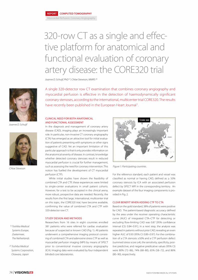
74 | VISIONS23 ©2014 TOSHIBA MEDICAL SYSTEMS
CLINICAL NEED FOR BOTH ANATOMICAL AND FUNCTIONAL ASSESSMENTIn the diagnosis and management of coronary artery
disease (CAD), imaging plays an increasingly important
role. In particular, non-invasive CT coronary angiography
(CTA) has emerged as an attractive tool for initial evalua-
tion of patients presenting with symptoms or other signs
suggestive of CAD. Yet an important limitation of this
particular approach is that it only provides information on
the anatomical severity of disease. In contrast, knowledge
whether detected coronary stenoses result in reduced
myocardial perfusion is crucial for further management,
such as assessing the need for coronary intervention. This
notion has fuelled the development of CT myocardial
perfusion (CTP).
While initial studies have shown the feasibility of
combined CTA and CTP, these experiences were limited
to single-center evaluations in small patient cohorts.
However, for a test to be accepted in the clinical arena,
more robust, prospective data are needed. Recently, the
results from the first large, international, multicenter trial
on this topic, the CORE320 trial, have become available,
confirming the value of combined CTA and CTP with
320-detector row CT.
STUDY DESIGN AND METHODSResearchers from 16 sites in eight countries enrolled
381 patients who were referred for cardiac evaluation
because of suspected or known CAD (Fig. 1). All patients
underwent a comprehensive imaging protocol consist-
ing of combined CTA and adenosine stress CTP as well as
myocardial perfusion imaging (MPI) by means of SPECT
prior to conventional invasive coronary angiography
(ICA). Imaging data were evaluated by four independent
blinded core laboratories.
320-row CT as a single and effec-tive platform for anatomical and functional evaluation of coronary artery disease: the CORE320 trial
REPORT
A single 320-detector row CT examination that combines coronary angiography and myocardial perfusion is effective in the detection of haemodynamically significant coronary stenoses, according to the international, multicenter trial CORE320. The results have recently been published in the European Heart Journal1.
Figure 1: Participating countries.
Myocardial Perfusion, Coronary Angiography
COMPUTED TOMOGRAPHY
Joanne D. Schuijf
Chloe Steveson
2) Toshiba Medical
Systems Corporation,
Otawara, Japan
1) Toshiba Medical
Systems Europe,
Zoetermeer,
The Netherlands
Joanne D. Schuijf, PhD 1); Chloe Steveson, MMRS 2)
For the reference standard, each patient and vessel was
classified as normal or having CAD, defined as ≥ 50%
coronary stenosis by ICA with an associated perfusion
defect by SPECT MPI in the corresponding territory. An
example dataset of the four imaging components is pro-
vided in Fig. 2.
CLEAR BENEFIT WHEN ADDING CTP TO CTABased on the gold standard, 38% of patients were positive
for CAD. The patient-based diagnostic accuracy defined
by the area under the receiver operating characteristic
curve (AUC) of integrated CTA–CTP for detecting or
excluding flow-limiting CAD was 0.87 [95% confidence
interval (CI): 0.84–0.91]. In a next step, the analysis was
repeated in patients without prior CAD, revealing an even
higher AUC of 0.93 (95% CI: 0.89–0.97). For the combina-
tion of a CTA stenosis ≥50% and a CTP perfusion defect
(summed stress score ≥4), the sensitivity, specificity, posi-
tive predictive, and negative predicative values (95% CI)
were 80% (72–86), 74% (68–80), 65% (58–72), and 86%
(80–90), respectively.
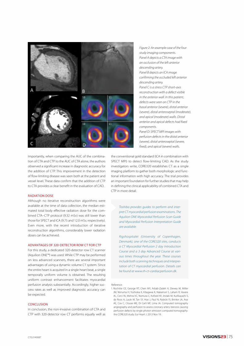
VISIONS23 | 75
Figure 2: An example case of the four
study imaging components.
Panel A depicts a CTA image with
an occlusion of the left anterior
descending artery.
Panel B depicts an ICA image
confirming the occluded left anterior
descending artery.
Panel C is a stress CTP short-axis
reconstruction with a defect visible
in the anterior wall. In this patient,
defects were seen on CTP in the
basal anterior (severe), distal anterior
(severe), distal anteroseptal (moderate),
and apical (moderate) walls. Distal
anterior and apical defects had fixed
components.
Panel D: SPECT MPI images with
perfusion defects in the distal anterior
(severe), distal anteroseptal (severe,
fixed), and apical (severe) walls.
Reference:1. Rochitte CE, George RT, Chen MY, Arbab-Zadeh A, Dewey M, Miller
JM, Niinuma H, Yoshioka K, Kitagawa K, Nakamori S, Laham R, Vavere AL, Cerci RJ, Mehra VC, Nomura C, Kofoed KF, Jinzaki M, Kuribayashi S, de Roos A, Laule M, Tan SY, Hoe J, Paul N, Rybicki FJ, Brinker JA, Arai AE, Cox C, Clouse ME, Di Carli MF, Lima JA. Computed tomography angiography and perfusion to assess coronary artery stenosis causing perfusion defects by single photon emission computed tomography: the CORE320 study. Eur Heart J. 2013 Nov 19.
Importantly, when comparing the AUC of the combina-
tion of CTA and CTP to the AUC of CTA alone, the authors
observed a significant increase in diagnostic accuracy for
the addition of CTP. This improvement in the detection
of flow-limiting disease was seen both at the patient and
vessel level. These data confirm that the addition of CTP
to CTA provides a clear benefit in the evaluation of CAD.
RADIATION DOSE Although no iterative reconstruction algorithms were
available at the time of data collection, the median esti-
mated total body effective radiation dose for the com-
bined CTA–CTP protocol (9.32 mSv) was still lower than
those for SPECT and ICA (9.75 and 12.0 mSv, respectively).
Even more, with the recent introduction of iterative
reconstruction algorithms, considerably lower radiation
doses can be achieved.
ADVANTAGES OF 320-DETECTOR ROW CT FOR CTPFor this study, a dedicated 320-detector row CT scanner
(Aquilion ONE™) was used. While CTP may be performed
on less advanced scanners, there are several important
advantages of using a dynamic volume CT system. Since
the entire heart is acquired in a single heart beat, a single
temporally uniform volume is obtained. The resulting
uniform contrast enhancement facilitates myocardial
perfusion analysis substantially. Accordingly, higher suc-
cess rates as well as improved diagnostic accuracy can
be expected.
CONCLUSIONIn conclusion, the non-invasive combination of CTA and
CTP with 320-detector row CT performs equally well as
the conventional gold standard (ICA in combination with
SPECT MPI) to detect flow-limiting CAD. As the study
investigators write, CORE320 establishes CT as a single
imaging platform to gather both morphologic and func-
tional information with high accuracy. The trial provides
an important foundation for further studies that may help
in defining the clinical applicability of combined CTA and
CTP in more detail.
Toshiba provides guides to perform and inter-
pret CT myocardial perfusion examinations. The
Aquilion ONE Myocardial Perfusion Scan Guide
and Myocardial Perfusion Interpretation Guide
are available.
Rigshospitalet (University of Copenhagen,
Denmark), one of the CORE320 sites, conducts
a CT Myocardial Perfusion 2 day Introduction
Course and a 3 day Advanced Course at vari-
ous times throughout the year. These courses
include both scanning techniques and interpre-
tation of CT myocardial perfusion. Details can
be found at www.rh-ct-cardiacperfusion.dk.
CTEU140087
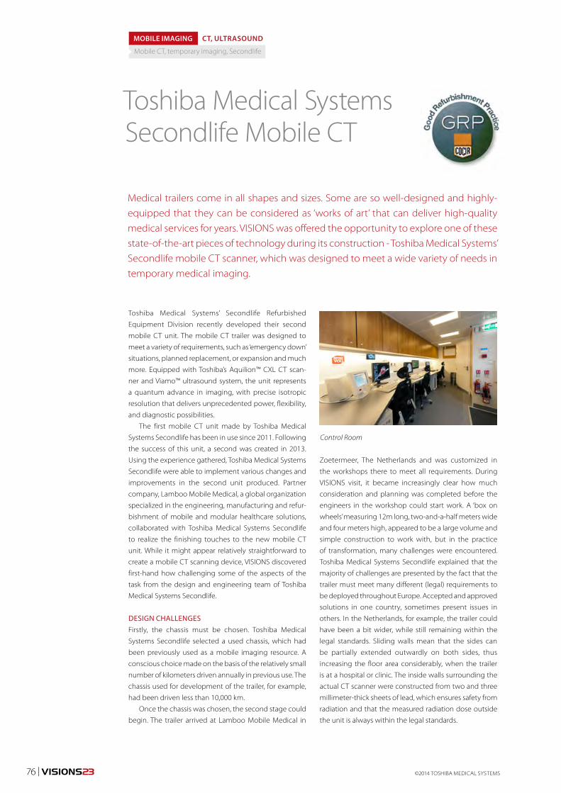
©2014 TOSHIBA MEDICAL SYSTEMS76 | VISIONS23
Toshiba Medical Systems’ Secondlife Refurbished
Equipment Division recently developed their second
mobile CT unit. The mobile CT trailer was designed to
meet a variety of requirements, such as ‘emergency down’
situations, planned replacement, or expansion and much
more. Equipped with Toshiba’s Aquilion™ CXL CT scan-
ner and Viamo™ ultrasound system, the unit represents
a quantum advance in imaging, with precise isotropic
resolution that delivers unprecedented power, flexibility,
and diagnostic possibilities.
The first mobile CT unit made by Toshiba Medical
Systems Secondlife has been in use since 2011. Following
the success of this unit, a second was created in 2013.
Using the experience gathered, Toshiba Medical Systems
Secondlife were able to implement various changes and
improvements in the second unit produced. Partner
company, Lamboo Mobile Medical, a global organization
specialized in the engineering, manufacturing and refur-
bishment of mobile and modular healthcare solutions,
collaborated with Toshiba Medical Systems Secondlife
to realize the finishing touches to the new mobile CT
unit. While it might appear relatively straightforward to
create a mobile CT scanning device, VISIONS discovered
first-hand how challenging some of the aspects of the
task from the design and engineering team of Toshiba
Medical Systems Secondlife.
DESIGN CHALLENGESFirstly, the chassis must be chosen. Toshiba Medical
Systems Secondlife selected a used chassis, which had
been previously used as a mobile imaging resource. A
conscious choice made on the basis of the relatively small
number of kilometers driven annually in previous use. The
chassis used for development of the trailer, for example,
had been driven less than 10,000 km.
Once the chassis was chosen, the second stage could
begin. The trailer arrived at Lamboo Mobile Medical in
Toshiba Medical SystemsSecondlife Mobile CT
Medical trailers come in all shapes and sizes. Some are so well-designed and highly-equipped that they can be considered as ‘works of art’ that can deliver high-quality medical services for years. VISIONS was offered the opportunity to explore one of these state-of-the-art pieces of technology during its construction - Toshiba Medical Systems’ Secondlife mobile CT scanner, which was designed to meet a wide variety of needs in temporary medical imaging.
MOBILE IMAGING CT, ULTRASOUND
Mobile CT, temporary imaging, Secondlife
Zoetermeer, The Netherlands and was customized in
the workshops there to meet all requirements. During
VISIONS visit, it became increasingly clear how much
consideration and planning was completed before the
engineers in the workshop could start work. A ‘box on
wheels’ measuring 12m long, two-and-a-half meters wide
and four meters high, appeared to be a large volume and
simple construction to work with, but in the practice
of transformation, many challenges were encountered.
Toshiba Medical Systems Secondlife explained that the
majority of challenges are presented by the fact that the
trailer must meet many different (legal) requirements to
be deployed throughout Europe. Accepted and approved
solutions in one country, sometimes present issues in
others. In the Netherlands, for example, the trailer could
have been a bit wider, while still remaining within the
legal standards. Sliding walls mean that the sides can
be partially extended outwardly on both sides, thus
increasing the floor area considerably, when the trailer
is at a hospital or clinic. The inside walls surrounding the
actual CT scanner were constructed from two and three
millimeter-thick sheets of lead, which ensures safety from
radiation and that the measured radiation dose outside
the unit is always within the legal standards.
Control Room
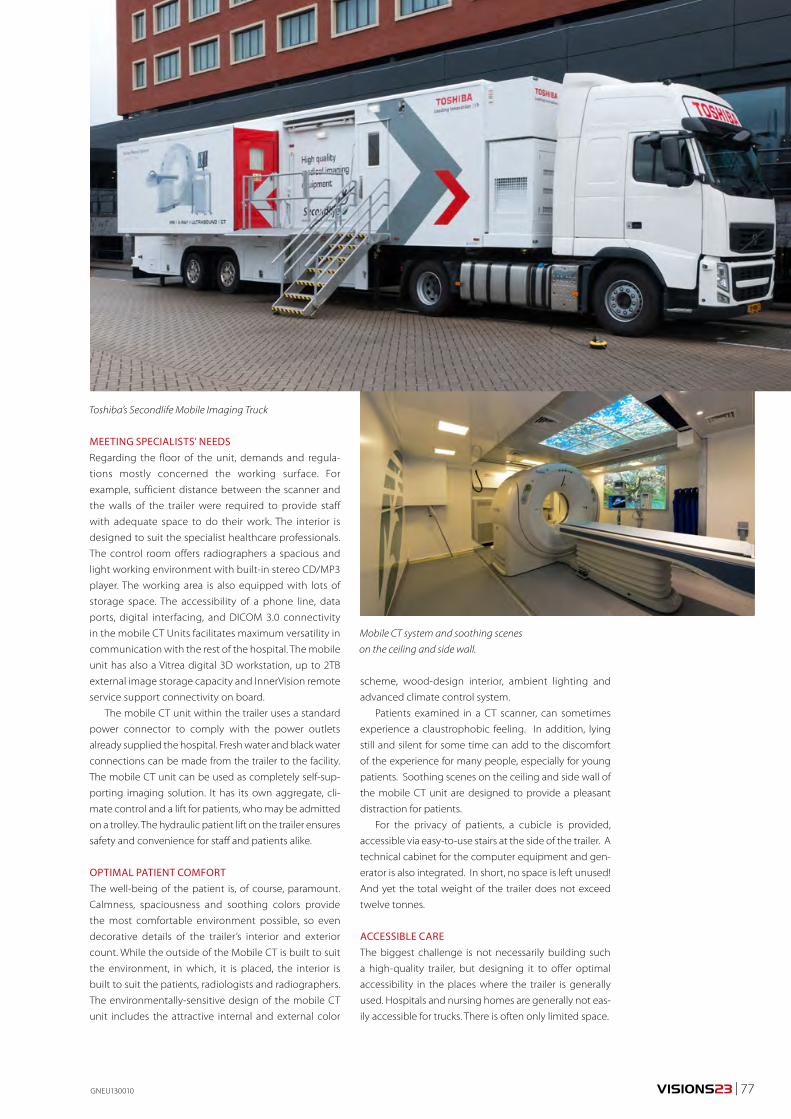
VISIONS23 | 77
MEETING SPECIALISTS’ NEEDSRegarding the floor of the unit, demands and regula-
tions mostly concerned the working surface. For
example, sufficient distance between the scanner and
the walls of the trailer were required to provide staff
with adequate space to do their work. The interior is
designed to suit the specialist healthcare professionals.
The control room offers radiographers a spacious and
light working environment with built-in stereo CD/MP3
player. The working area is also equipped with lots of
storage space. The accessibility of a phone line, data
ports, digital interfacing, and DICOM 3.0 connectivity
in the mobile CT Units facilitates maximum versatility in
communication with the rest of the hospital. The mobile
unit has also a Vitrea digital 3D workstation, up to 2TB
external image storage capacity and InnerVision remote
service support connectivity on board.
The mobile CT unit within the trailer uses a standard
power connector to comply with the power outlets
already supplied the hospital. Fresh water and black water
connections can be made from the trailer to the facility.
The mobile CT unit can be used as completely self-sup-
porting imaging solution. It has its own aggregate, cli-
mate control and a lift for patients, who may be admitted
on a trolley. The hydraulic patient lift on the trailer ensures
safety and convenience for staff and patients alike.
OPTIMAL PATIENT COMFORTThe well-being of the patient is, of course, paramount.
Calmness, spaciousness and soothing colors provide
the most comfortable environment possible, so even
decorative details of the trailer’s interior and exterior
count. While the outside of the Mobile CT is built to suit
the environment, in which, it is placed, the interior is
built to suit the patients, radiologists and radiographers.
The environmentally-sensitive design of the mobile CT
unit includes the attractive internal and external color
GNEU130010
scheme, wood-design interior, ambient lighting and
advanced climate control system.
Patients examined in a CT scanner, can sometimes
experience a claustrophobic feeling. In addition, lying
still and silent for some time can add to the discomfort
of the experience for many people, especially for young
patients. Soothing scenes on the ceiling and side wall of
the mobile CT unit are designed to provide a pleasant
distraction for patients.
For the privacy of patients, a cubicle is provided,
accessible via easy-to-use stairs at the side of the trailer. A
technical cabinet for the computer equipment and gen-
erator is also integrated. In short, no space is left unused!
And yet the total weight of the trailer does not exceed
twelve tonnes.
ACCESSIBLE CAREThe biggest challenge is not necessarily building such
a high-quality trailer, but designing it to offer optimal
accessibility in the places where the trailer is generally
used. Hospitals and nursing homes are generally not eas-
ily accessible for trucks. There is often only limited space.
Mobile CT system and soothing scenes
on the ceiling and side wall.
Toshiba’s Secondlife Mobile Imaging Truck
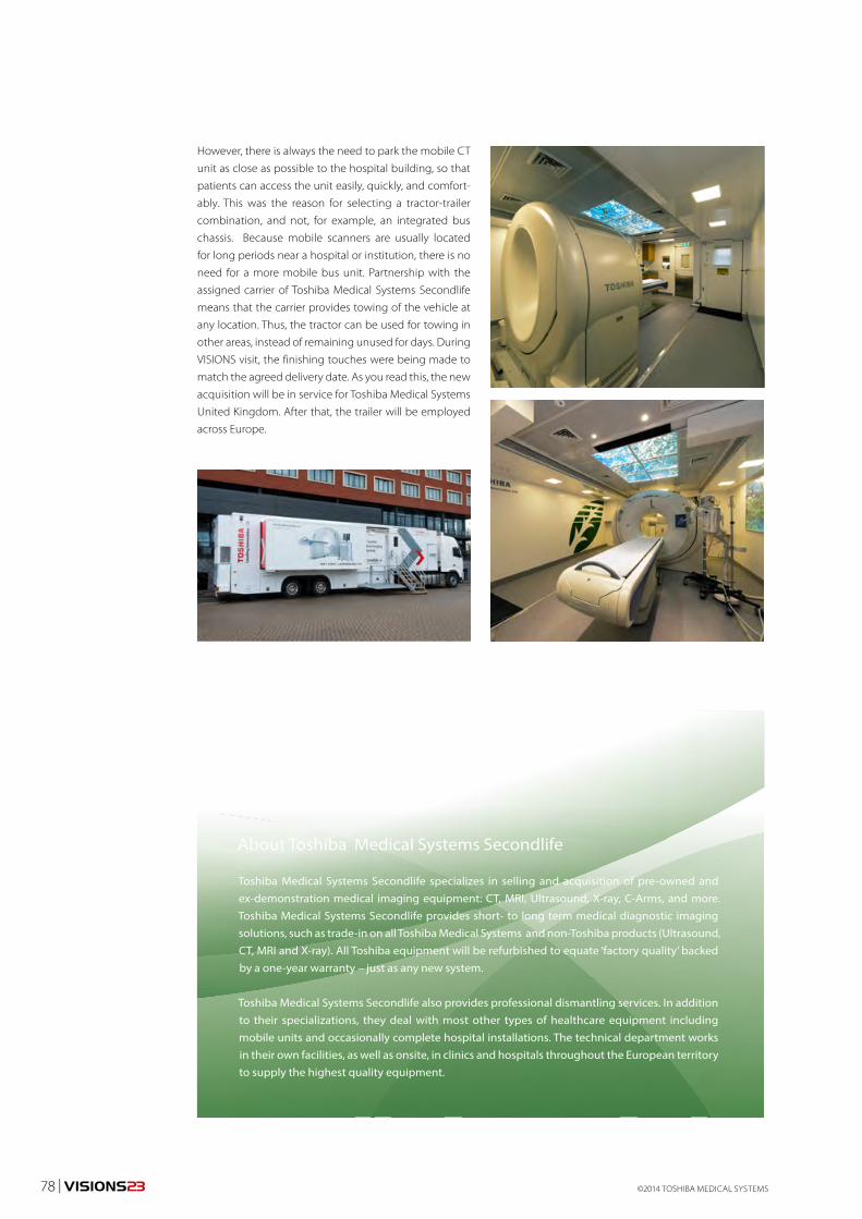
©2014 TOSHIBA MEDICAL SYSTEMS78 | VISIONS23
However, there is always the need to park the mobile CT
unit as close as possible to the hospital building, so that
patients can access the unit easily, quickly, and comfort-
ably. This was the reason for selecting a tractor-trailer
combination, and not, for example, an integrated bus
chassis. Because mobile scanners are usually located
for long periods near a hospital or institution, there is no
need for a more mobile bus unit. Partnership with the
assigned carrier of Toshiba Medical Systems Secondlife
means that the carrier provides towing of the vehicle at
any location. Thus, the tractor can be used for towing in
other areas, instead of remaining unused for days. During
VISIONS visit, the finishing touches were being made to
match the agreed delivery date. As you read this, the new
acquisition will be in service for Toshiba Medical Systems
United Kingdom. After that, the trailer will be employed
across Europe.
About Toshiba Medical Systems Secondlife
Toshiba Medical Systems Secondlife specializes in selling and acquisition of pre-owned and ex-demonstration medical imaging equipment: CT, MRI, Ultrasound, X-ray, C-Arms, and more. Toshiba Medical Systems Secondlife provides short- to long term medical diagnostic imaging solutions, such as trade-in on all Toshiba Medical Systems and non-Toshiba products (Ultrasound, CT, MRI and X-ray). All Toshiba equipment will be refurbished to equate ‘factory quality’ backed by a one-year warranty – just as any new system.
Toshiba Medical Systems Secondlife also provides professional dismantling services. In addition to their specializations, they deal with most other types of healthcare equipment including mobile units and occasionally complete hospital installations. The technical department works in their own facilities, as well as onsite, in clinics and hospitals throughout the European territory to supply the highest quality equipment.
Imprint
Publisher:TOSHIBA Medical Systems Europe B.V.,Zilverstraat 1NL-2718 RP ZoetermeerTel.: +31 79 368 92 22Fax: +31 79 368 94 44Web: www.toshiba-medical.euEmail: [email protected]
Editor-in-chief: Jack Hoogendoorn ([email protected])
Modality coordinators:CT: Roy IrwanUL: Joerg Schlegel XR: Jaco Terlouw
“Creating New Standards in Ultrasound Cardiology” & “Customer Focus: Rigshospitalet, Copenhagen” by The Creative Practice (www.thecreativepractice.com)
Design & Layout:Boerma Reclame (www.boermareclame.com)
Printing: Schefferdrukkerij (www.schefferdrukkerij.nl)
Subscription Service:www.toshiba-medical.eu/visions
© 2014 by TOSHIBA Medical Systems EuropeAll rights reserved
Caspar Berger
AdmissionFree
Museum Beelden aan Zee
Harteveltstraat 1
2586 EL Den Haag
T: +31 70 358 58 57
www.beeldenaanzee.nl
This voucher entitles free admission for one person to Museum Beelden aan Zee.
Valid until June 1st 2014
Hidden in the dunes of Scheveningen, like
a pearl in the sand, you will find museum
Beelden aan Zee, a truly unique museum in the
Netherlands that focuses on modern and con-
temporary international sculpture. Man - the
human image - is the leitmotiv of the collection
of Beelden aan Zee. The collection holds work
from sculptors as: Karel Appel, Atelier van
Lieshout, Fernando Botero, Cesar, Tony Cragg,
Igor Mitoraj, Jan Meefout and Ossip Zadkine.
As from January 31st 2014 the following expo-
sition can be seen at museum Beelden aan Zee:
• Caspar Berger - Bone.
• Yubi Kirindongo - Rebel in Art & Soul.
• George Minne - Voorbode van de Moderne
Kunst & Nick Ervinck - GNI-RI.
www.beeldenaanzee.nl
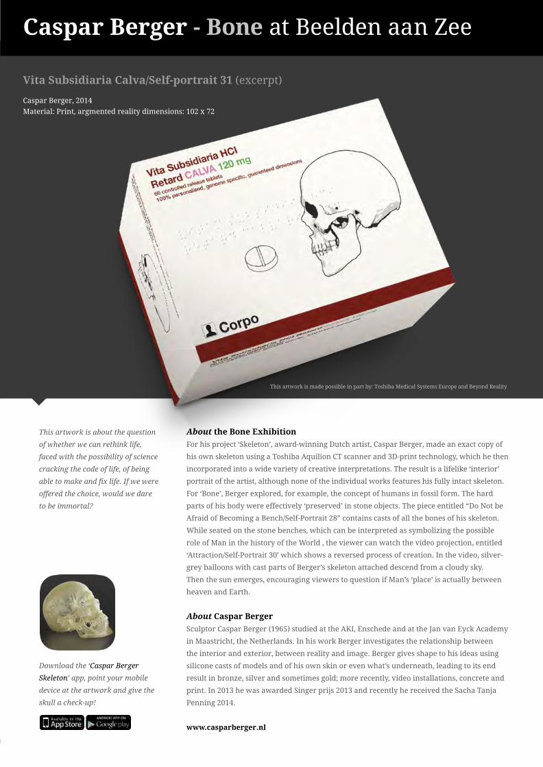
Spot Fluoro - Reducing Dose in Interventional Procedures
Superiority of Lung Subtraction Above Dual Energy
Creating New Standards in Ultrasound Cardiology
Vantage ELAN Perfectly Combines Performance and Affordability 23
X-RAY
COMPUTED TOMOGRAPHY
ULTRASOUND
MAGNETIC RESONANCE
ISSN 1617-2876
VISIONSFEBRUARY 2014
Caspar Berger Caspar Berger - Bone at Beelden aan Zee
Vita Subsidiaria Calva/Self-portrait 31 (excerpt)
Caspar Berger, 2014
Material: Print, argmented reality dimensions: 102 x 72
This artwork is about the question
of whether we can rethink life,
faced with the possibility of science
cracking the code of life, of being
able to make and fix life. If we were
offered the choice, would we dare
to be immortal?
Download the ‘Caspar Berger
Skeleton’ app, point your mobile
device at the artwork and give the
skull a check-up!
About the Bone Exhibition
For his project ‘Skeleton’, award-winning Dutch artist, Caspar Berger, made an exact copy of
his own skeleton using a Toshiba Aquilion CT scanner and 3D-print technology, which he then
incorporated into a wide variety of creative interpretations. The result is a lifelike ‘interior’
portrait of the artist, although none of the individual works features his fully intact skeleton.
For ‘Bone’, Berger explored, for example, the concept of humans in fossil form. The hard
parts of his body were effectively ‘preserved’ in stone objects. The piece entitled “Do Not be
Afraid of Becoming a Bench/Self-Portrait 28” contains casts of all the bones of his skeleton.
While seated on the stone benches, which can be interpreted as symbolizing the possible
role of Man in the history of the World , the viewer can watch the video projection, entitled
‘Attraction/Self-Portrait 30’ which shows a reversed process of creation. In the video, silver-
grey balloons with cast parts of Berger’s skeleton attached descend from a cloudy sky.
Then the sun emerges, encouraging viewers to question if Man’s ‘place’ is actually between
heaven and Earth.
About Caspar Berger
Sculptor Caspar Berger (1965) studied at the AKI, Enschede and at the Jan van Eyck Academy
in Maastricht, the Netherlands. In his work Berger investigates the relationship between
the interior and exterior, between reality and image. Berger gives shape to his ideas using
silicone casts of models and of his own skin or even what’s underneath, leading to its end
result in bronze, silver and sometimes gold; more recently, video installations, concrete and
print. In 2013 he was awarded Singer prijs 2013 and recently he received the Sacha Tanja
Penning 2014.
www.casparberger.nl
AdmissionFree
Museum Beelden aan Zee
Harteveltstraat 1
2586 EL Den Haag
T: +31 70 358 58 57
www.beeldenaanzee.nl
This voucher entitles free admission for one person to Museum Beelden aan Zee.
Valid until June 1st 2014
Hidden in the dunes of Scheveningen, like
a pearl in the sand, you will find museum
Beelden aan Zee, a truly unique museum in the
Netherlands that focuses on modern and con-
temporary international sculpture. Man - the
human image - is the leitmotiv of the collection
of Beelden aan Zee. The collection holds work
from sculptors as: Karel Appel, Atelier van
Lieshout, Fernando Botero, Cesar, Tony Cragg,
Igor Mitoraj, Jan Meefout and Ossip Zadkine.
As from January 31st 2014 the following expo-
sition can be seen at museum Beelden aan Zee:
• Caspar Berger - Bone.
• Yubi Kirindongo - Rebel in Art & Soul.
• George Minne - Voorbode van de Moderne
Kunst & Nick Ervinck - GNI-RI.
www.beeldenaanzee.nl
This artwork is made possible in part by: Toshiba Medical Systems Europe and Beyond Reality