Topical Anti-infl ammatory Potential of Pumpkin (Cucurbita ... 1168.pdf · 1Laboratório de...
9
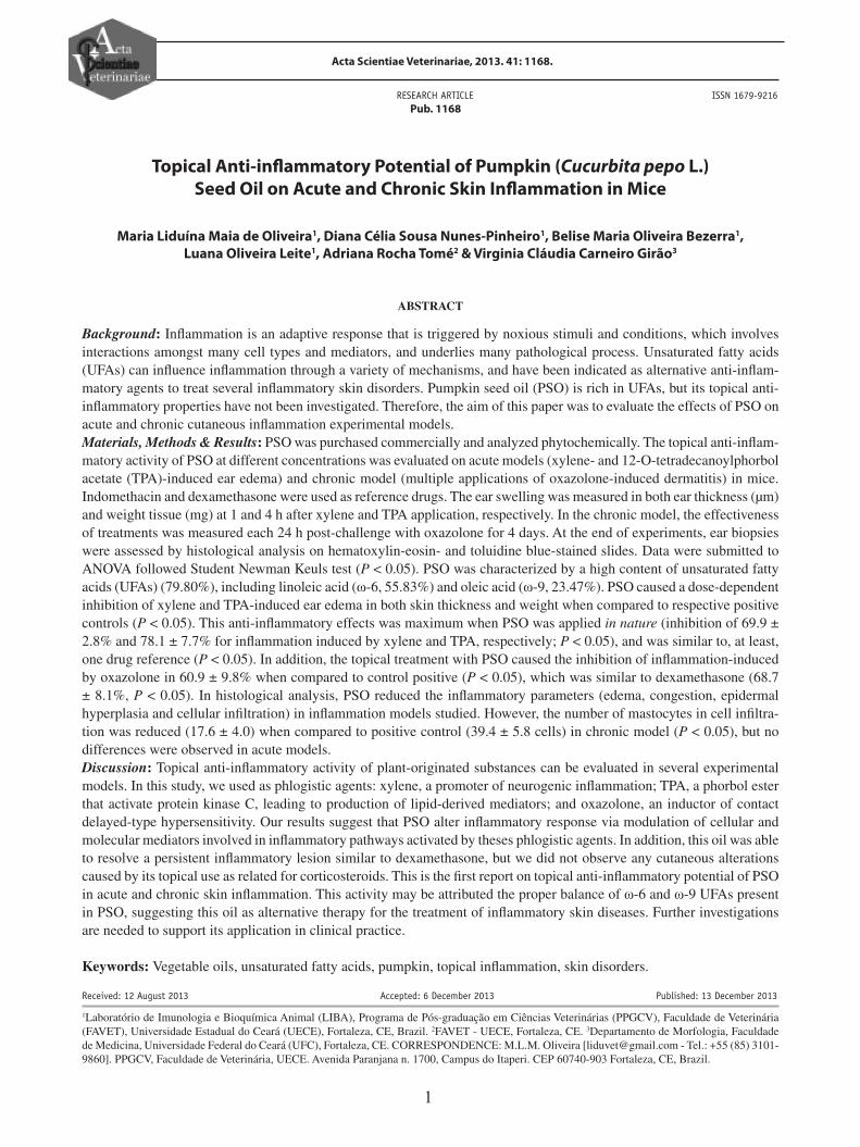
Acta Scientiae Veterinariae, 2013. 41: 1168. RESEARCH ARTICLE Pub. 1168 ISSN 1679-9216 1 Received: 12 August 2013 Accepted: 6 December 2013 Published: 13 December 2013 1 Laboratório de Imunologia e Bioquímica Animal (LIBA), Programa de Pós-graduação em Ciências Veterinárias (PPGCV), Faculdade de Veterinária (FAVET), Universidade Estadual do Ceará (UECE), Fortaleza, CE, Brazil. 2 FAVET - UECE, Fortaleza, CE. 3 Departamento de Morfologia, Faculdade de Medicina, Universidade Federal do Ceará (UFC), Fortaleza, CE. CORRESPONDENCE: M.L.M. Oliveira [[email protected] - Tel.: +55 (85) 3101- 9860]. PPGCV, Faculdade de Veterinária, UECE. Avenida Paranjana n. 1700, Campus do Itaperi. CEP 60740-903 Fortaleza, CE, Brazil. Topical Anti-inflammatory Potential of Pumpkin (Cucurbita pepo L.) Seed Oil on Acute and Chronic Skin Inflammation in Mice Maria Liduína Maia de Oliveira 1 , Diana Célia Sousa Nunes-Pinheiro 1 , Belise Maria Oliveira Bezerra 1 , Luana Oliveira Leite 1 , Adriana Rocha Tomé 2 & Virginia Cláudia Carneiro Girão 3 ABSTRACT Background: Inflammation is an adaptive response that is triggered by noxious stimuli and conditions, which involves interactions amongst many cell types and mediators, and underlies many pathological process. Unsaturated fatty acids (UFAs) can influence inflammation through a variety of mechanisms, and have been indicated as alternative anti-inflam- matory agents to treat several inflammatory skin disorders. Pumpkin seed oil (PSO) is rich in UFAs, but its topical anti- inflammatory properties have not been investigated. Therefore, the aim of this paper was to evaluate the effects of PSO on acute and chronic cutaneous inflammation experimental models. Materials, Methods & Results: PSO was purchased commercially and analyzed phytochemically. The topical anti-inflam- matory activity of PSO at different concentrations was evaluated on acute models (xylene- and 12-O-tetradecanoylphorbol acetate (TPA)-induced ear edema) and chronic model (multiple applications of oxazolone-induced dermatitis) in mice. Indomethacin and dexamethasone were used as reference drugs. The ear swelling was measured in both ear thickness (μm) and weight tissue (mg) at 1 and 4 h after xylene and TPA application, respectively. In the chronic model, the effectiveness of treatments was measured each 24 h post-challenge with oxazolone for 4 days. At the end of experiments, ear biopsies were assessed by histological analysis on hematoxylin-eosin- and toluidine blue-stained slides. Data were submitted to ANOVA followed Student Newman Keuls test (P < 0.05). PSO was characterized by a high content of unsaturated fatty acids (UFAs) (79.80%), including linoleic acid (ω-6, 55.83%) and oleic acid (ω-9, 23.47%). PSO caused a dose-dependent inhibition of xylene and TPA-induced ear edema in both skin thickness and weight when compared to respective positive controls (P < 0.05). This anti-inflammatory effects was maximum when PSO was applied in nature (inhibition of 69.9 ± 2.8% and 78.1 ± 7.7% for inflammation induced by xylene and TPA, respectively; P < 0.05), and was similar to, at least, one drug reference (P < 0.05). In addition, the topical treatment with PSO caused the inhibition of inflammation-induced by oxazolone in 60.9 ± 9.8% when compared to control positive (P < 0.05), which was similar to dexamethasone (68.7 ± 8.1%, P < 0.05). In histological analysis, PSO reduced the inflammatory parameters (edema, congestion, epidermal hyperplasia and cellular infiltration) in inflammation models studied. However, the number of mastocytes in cell infiltra- tion was reduced (17.6 ± 4.0) when compared to positive control (39.4 ± 5.8 cells) in chronic model (P < 0.05), but no differences were observed in acute models. Discussion: Topical anti-inflammatory activity of plant-originated substances can be evaluated in several experimental models. In this study, we used as phlogistic agents: xylene, a promoter of neurogenic inflammation; TPA, a phorbol ester that activate protein kinase C, leading to production of lipid-derived mediators; and oxazolone, an inductor of contact delayed-type hypersensitivity. Our results suggest that PSO alter inflammatory response via modulation of cellular and molecular mediators involved in inflammatory pathways activated by theses phlogistic agents. In addition, this oil was able to resolve a persistent inflammatory lesion similar to dexamethasone, but we did not observe any cutaneous alterations caused by its topical use as related for corticosteroids. This is the first report on topical anti-inflammatory potential of PSO in acute and chronic skin inflammation. This activity may be attributed the proper balance of ω-6 and ω-9 UFAs present in PSO, suggesting this oil as alternative therapy for the treatment of inflammatory skin diseases. Further investigations are needed to support its application in clinical practice. Keywords: Vegetable oils, unsaturated fatty acids, pumpkin, topical inflammation, skin disorders.
Transcript of Topical Anti-infl ammatory Potential of Pumpkin (Cucurbita ... 1168.pdf · 1Laboratório de...
Acta Scientiae Veterinariae, 2013. 41: 1168.
RESEARCH ARTICLE Pub. 1168
ISSN 1679-9216
1
Received: 12 August 2013 Accepted: 6 December 2013 Published: 13 December 2013
1Laboratório de Imunologia e Bioquímica Animal (LIBA), Programa de Pós-graduação em Ciências Veterinárias (PPGCV), Faculdade de Veterinária (FAVET), Universidade Estadual do Ceará (UECE), Fortaleza, CE, Brazil. 2FAVET - UECE, Fortaleza, CE. 3Departamento de Morfologia, Faculdade de Medicina, Universidade Federal do Ceará (UFC), Fortaleza, CE. CORRESPONDENCE: M.L.M. Oliveira [[email protected] - Tel.: +55 (85) 3101-9860]. PPGCV, Faculdade de Veterinária, UECE. Avenida Paranjana n. 1700, Campus do Itaperi. CEP 60740-903 Fortaleza, CE, Brazil.
Topical Anti-infl ammatory Potential of Pumpkin (Cucurbita pepo L.)
Seed Oil on Acute and Chronic Skin Infl ammation in Mice
Maria Liduína Maia de Oliveira1, Diana Célia Sousa Nunes-Pinheiro1, Belise Maria Oliveira Bezerra1,
Luana Oliveira Leite1, Adriana Rocha Tomé2 & Virginia Cláudia Carneiro Girão3
ABSTRACT
Background: Infl ammation is an adaptive response that is triggered by noxious stimuli and conditions, which involves interactions amongst many cell types and mediators, and underlies many pathological process. Unsaturated fatty acids (UFAs) can infl uence infl ammation through a variety of mechanisms, and have been indicated as alternative anti-infl am-matory agents to treat several infl ammatory skin disorders. Pumpkin seed oil (PSO) is rich in UFAs, but its topical anti-infl ammatory properties have not been investigated. Therefore, the aim of this paper was to evaluate the effects of PSO on acute and chronic cutaneous infl ammation experimental models.Materials, Methods & Results: PSO was purchased commercially and analyzed phytochemically. The topical anti-infl am-matory activity of PSO at different concentrations was evaluated on acute models (xylene- and 12-O-tetradecanoylphorbol acetate (TPA)-induced ear edema) and chronic model (multiple applications of oxazolone-induced dermatitis) in mice. Indomethacin and dexamethasone were used as reference drugs. The ear swelling was measured in both ear thickness (µm) and weight tissue (mg) at 1 and 4 h after xylene and TPA application, respectively. In the chronic model, the effectiveness of treatments was measured each 24 h post-challenge with oxazolone for 4 days. At the end of experiments, ear biopsies were assessed by histological analysis on hematoxylin-eosin- and toluidine blue-stained slides. Data were submitted to ANOVA followed Student Newman Keuls test (P < 0.05). PSO was characterized by a high content of unsaturated fatty acids (UFAs) (79.80%), including linoleic acid (ω-6, 55.83%) and oleic acid (ω-9, 23.47%). PSO caused a dose-dependent inhibition of xylene and TPA-induced ear edema in both skin thickness and weight when compared to respective positive controls (P < 0.05). This anti-infl ammatory effects was maximum when PSO was applied in nature (inhibition of 69.9 ± 2.8% and 78.1 ± 7.7% for infl ammation induced by xylene and TPA, respectively; P < 0.05), and was similar to, at least, one drug reference (P < 0.05). In addition, the topical treatment with PSO caused the inhibition of infl ammation-induced by oxazolone in 60.9 ± 9.8% when compared to control positive (P < 0.05), which was similar to dexamethasone (68.7 ± 8.1%, P < 0.05). In histological analysis, PSO reduced the infl ammatory parameters (edema, congestion, epidermal hyperplasia and cellular infi ltration) in infl ammation models studied. However, the number of mastocytes in cell infi ltra-tion was reduced (17.6 ± 4.0) when compared to positive control (39.4 ± 5.8 cells) in chronic model (P < 0.05), but no differences were observed in acute models.Discussion: Topical anti-infl ammatory activity of plant-originated substances can be evaluated in several experimental models. In this study, we used as phlogistic agents: xylene, a promoter of neurogenic infl ammation; TPA, a phorbol ester that activate protein kinase C, leading to production of lipid-derived mediators; and oxazolone, an inductor of contact delayed-type hypersensitivity. Our results suggest that PSO alter infl ammatory response via modulation of cellular and molecular mediators involved in infl ammatory pathways activated by theses phlogistic agents. In addition, this oil was able to resolve a persistent infl ammatory lesion similar to dexamethasone, but we did not observe any cutaneous alterations caused by its topical use as related for corticosteroids. This is the fi rst report on topical anti-infl ammatory potential of PSO in acute and chronic skin infl ammation. This activity may be attributed the proper balance of ω-6 and ω-9 UFAs present in PSO, suggesting this oil as alternative therapy for the treatment of infl ammatory skin diseases. Further investigations are needed to support its application in clinical practice.
Keywords: Vegetable oils, unsaturated fatty acids, pumpkin, topical infl ammation, skin disorders.

2
M.L.M. Oliveira, D.C.S. Nunes-Pinheiro, B.M.O. Bezerra, et al. 2013. Topical Anti-infl ammatory Potential of Pumpkin (Cucurbita pepo L.) Seed Oil on Acute and Chronic Skin Infl ammation in Mice. Acta Scientiae Veterinariae. 41: 1168.
INTRODUCTION
Infl ammation is a normal defense mechanism that protects the host from infection and other insults. It involves interactions amongst many cell types and chemical mediators for restoring homeostasis [13]. These mediators include lipid-derived mediators, peptide mediators, enzymes, and adhesion molecules, depending upon the cell type involved and the nature of the injurious stimulus [3,10,15]. However, when produced in an unregulated fashion, they can cause damage to host tissues, leading to disease [3].
Infl ammatory skin disorders can be treated with some success by pharmaceutical agents, such as corticosteroids and non-steroidal anti-infl ammatory drugs (NSAIDs). However, they can frequently cause a set of undesirable side effects [31]. Because of these risks, alternative bioactive molecules are being intensively investigated. In this scenario, fatty acids are highlighted as important effectors and regulators molecules in the immune-infl ammatory response [18].
Unsaturated fatty acids (UFAs) are found in sig-nifi cant quantities in vegetable oils. Pumpkin (Cucurbita pepo L.; Cucurbitaceae) seed oil (PSO) is extraordinarily rich in UFAs, which represent about 84% of the total fatty acids. It is also rich in minerals and antioxidants as tocopherols and carotenoids [20]. Despite several veg-etable oils rich in UFAs have been related as promising alternative anti-infl ammatory agents on skin disorders [17,19,22], information is not available about the anti-infl ammatory properties of PSO.
The aim of the present study was to investigate the effects of PSO on acute and chronic cutaneous infl ammation models in mice, in order to verify its topical anti-infl ammatory potential.
MATERIALS AND METHODS
Phytochemical analysis
The oil from the seeds of pumpkin (PSO1) was purchased commercially and used for transesterifi ca-tion reactions [5]. The recovered fatty acid methyl esters were analyzed by gas chromatography coupled to mass spectrometry, using a CGC AGILENT 68650 series GC system under the following conditions-col-umn: Methylpolysiloxane DB-23 AGILENT capillary column (60 m x 0.25 mm); carrier gas: He (1 mL/min); injector temperature: 250°C; detector temperature: 280°C; column temperature: 110°C/5 min, 110-215°C
at 5°C/min and then 215°C/24 min; mass spectrum: electronic impact 70 eV. The identifi cation of the con-stituents was performed by a computer-based library search, retention indices and visual interpretation of the mass spectra [1,2].
Animals
Female Swiss mice weighing 25-30 g, obtained from Central Animal House of the Federal University of Ceará were used for this study. Mice were housed in standard polypropylene cages under controlled conditions of temperature (23-25°C), relative humidity (40-45%) and 12 h light/dark cycle, with free access to commercial pellet diet and water.
Xylene and 12-O-tetradecanoyl-phorbol-13-acetate (TPA)-induced acute ear edema
Ear edema was induced in mice (n = 7/group) by topical application on the inner and outer surfaces of the right ear of the following phlogistic agents: xylene2
(20 µL/ear) and 12-O-tetradecanoyl-phorbol-13-ace-tate (TPA3) 2.5 µg/ear in acetone2. Immediately after the application of each phlogistic agent, the right ears were topically treated with PSO at 100% (in nature), 50% and 25% (v/v) in acetone (20 µL/ear), vehicle (acetone, 20 µL/ear, positive control), indomethacin4 (0.5 mg/ear) and dexamethasone5 (0.05 mg/ear). The left ear was considered as contralateral control and received only vehicle or saline solution (negative control, 20 µL/ear). The ear edema was evaluated 1 h after xylene and 4 h after TPA application [8,14,19].
Ear edema measurement
Ear thickness was measured before and after induction of the infl ammatory response using a digital micrometer. The micrometer was applied near the tip of the ear just distal to the cartilaginous ridges and the thickness was recorded in µm [22]. To evaluate the ear weight, animals were euthanized, and then both ears biopsies were removed and individually weighed [19]. Edema was expressed as right ear thickness variation and as the weight difference between the right and left ears. The inhibition edema percentage was calculated as weight reduction in comparison to positive control.
Oxazolone-induced chronic dermatitis
Contact dermatitis induction was performed ac-cording to Ginner et al. [8]. Briefl y, mice (n = 7/group) were sensitized by topical application of 2% solution (w/v) oxazolone3 in acetone (50 µL) on the shaved abdo-
3
M.L.M. Oliveira, D.C.S. Nunes-Pinheiro, B.M.O. Bezerra, et al. 2013. Topical Anti-infl ammatory Potential of Pumpkin (Cucurbita pepo L.) Seed Oil on Acute and Chronic Skin Infl ammation in Mice. Acta Scientiae Veterinariae. 41: 1168.
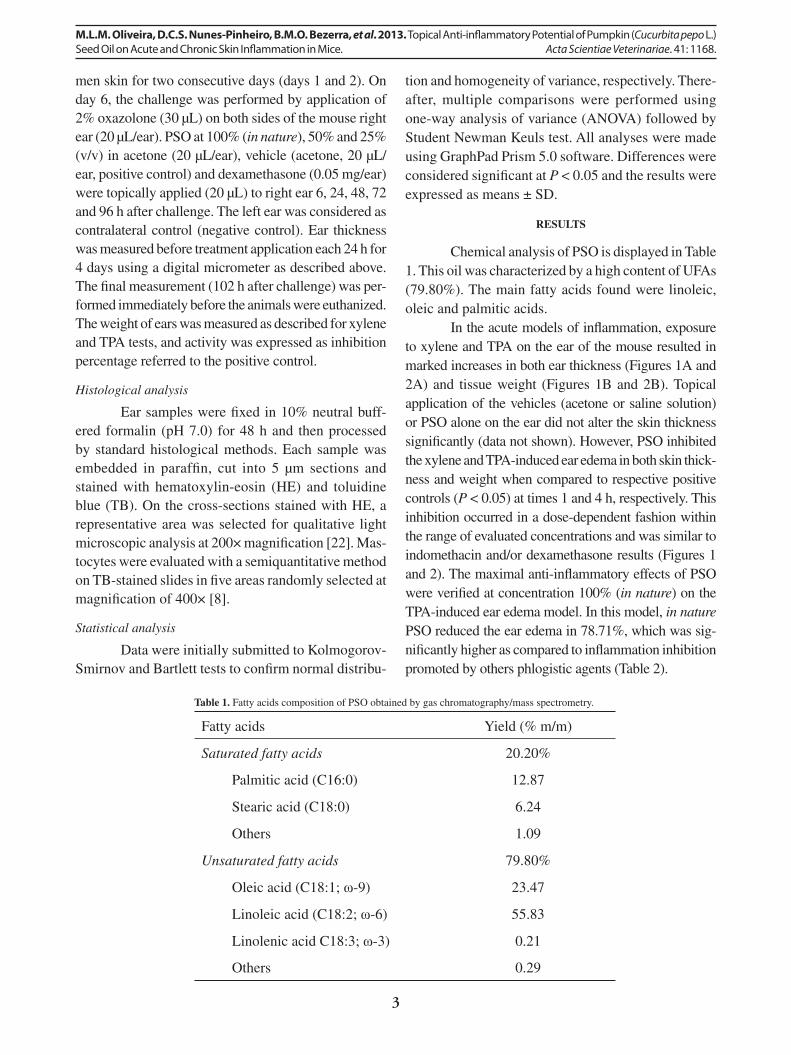
men skin for two consecutive days (days 1 and 2). On day 6, the challenge was performed by application of 2% oxazolone (30 µL) on both sides of the mouse right ear (20 µL/ear). PSO at 100% (in nature), 50% and 25% (v/v) in acetone (20 µL/ear), vehicle (acetone, 20 µL/ear, positive control) and dexamethasone (0.05 mg/ear) were topically applied (20 µL) to right ear 6, 24, 48, 72 and 96 h after challenge. The left ear was considered as contralateral control (negative control). Ear thickness was measured before treatment application each 24 h for 4 days using a digital micrometer as described above. The fi nal measurement (102 h after challenge) was per-formed immediately before the animals were euthanized. The weight of ears was measured as described for xylene and TPA tests, and activity was expressed as inhibition percentage referred to the positive control.
Histological analysis
Ear samples were fi xed in 10% neutral buff-ered formalin (pH 7.0) for 48 h and then processed by standard histological methods. Each sample was embedded in paraffi n, cut into 5 µm sections and stained with hematoxylin-eosin (HE) and toluidine blue (TB). On the cross-sections stained with HE, a representative area was selected for qualitative light microscopic analysis at 200× magnifi cation [22]. Mas-tocytes were evaluated with a semiquantitative method on TB-stained slides in fi ve areas randomly selected at magnifi cation of 400× [8].
Statistical analysis
Data were initially submitted to Kolmogorov-Smirnov and Bartlett tests to confi rm normal distribu-
tion and homogeneity of variance, respectively. There-after, multiple comparisons were performed using one-way analysis of variance (ANOVA) followed by Student Newman Keuls test. All analyses were made using GraphPad Prism 5.0 software. Differences were considered signifi cant at P < 0.05 and the results were expressed as means ± SD.
RESULTS
Chemical analysis of PSO is displayed in Table 1. This oil was characterized by a high content of UFAs (79.80%). The main fatty acids found were linoleic, oleic and palmitic acids.
In the acute models of infl ammation, exposure to xylene and TPA on the ear of the mouse resulted in marked increases in both ear thickness (Figures 1A and 2A) and tissue weight (Figures 1B and 2B). Topical application of the vehicles (acetone or saline solution) or PSO alone on the ear did not alter the skin thickness signifi cantly (data not shown). However, PSO inhibited the xylene and TPA-induced ear edema in both skin thick-ness and weight when compared to respective positive controls (P < 0.05) at times 1 and 4 h, respectively. This inhibition occurred in a dose-dependent fashion within the range of evaluated concentrations and was similar to indomethacin and/or dexamethasone results (Figures 1 and 2). The maximal anti-infl ammatory effects of PSO were verifi ed at concentration 100% (in nature) on the TPA-induced ear edema model. In this model, in nature PSO reduced the ear edema in 78.71%, which was sig-nifi cantly higher as compared to infl ammation inhibition promoted by others phlogistic agents (Table 2).
Table 1. Fatty acids composition of PSO obtained by gas chromatography/mass spectrometry.
Fatty acids Yield (% m/m)
Saturated fatty acids 20.20%
Palmitic acid (C16:0) 12.87
Stearic acid (C18:0) 6.24
Others 1.09
Unsaturated fatty acids 79.80%
Oleic acid (C18:1; ω-9) 23.47
Linoleic acid (C18:2; ω-6) 55.83
Linolenic acid C18:3; ω-3) 0.21
Others 0.29

4
M.L.M. Oliveira, D.C.S. Nunes-Pinheiro, B.M.O. Bezerra, et al. 2013. Topical Anti-infl ammatory Potential of Pumpkin (Cucurbita pepo L.) Seed Oil on Acute and Chronic Skin Infl ammation in Mice. Acta Scientiae Veterinariae. 41: 1168.
Table 2. Topical anti-infl ammatory effect of pumpkin seed oil (PSO) and reference drugs in acute and chronic models of infl ammation at the end of the experimental period.
Treatment groups
Infl ammation inhibition (%)
Phlogistic agent
Xylene TPA Oxazolone
PSO 100% (in nature) 69.61 ± 2.79a 78.71 ± 7.70b 60.86 ± 9.81c
PSO 50% 56.54 ± 4.81a 70.89 ± 5.79b 32.81 ± 7.32c
PSO 25% 49.57 ± 7.52a 69.02 ± 6.12b 28.90 ± 6.90c
Indomethacin 51.00 ± 12.08a 84.11 ± 5.86b -Dexamethasone 65.07 ± 8.98a 88.44 ± 6.60b 68.69 ± 8.10a
Results are expressed as means ± SD (n = 7) of ears weight reduction in relation to the positive control of each model. Different superscripts letters within the same line indicate statistically signifi cant difference among different infl ammation models (P < 0.05).
Figure 1. Topical activity of pumpkin seed oil (PSO), indomethacin (Indo) and dexamethasone (Dexa) on xylene (Xyl)-induced ear edema in mice. Ear edema was measured at 1 h after induction of infl ammation in both ear thickness (A) and tissue weight (B). The positive control only received acetone topically after the challenge with xylene. The bars represent the mean ± SD for seven animals. Different small letters indicate statistically signifi cant differences among groups (P < 0.05).
Figure 2. Topical activity of pumpkin seed oil (PSO), indomethacin (Indo) and dexamethasone (Dexa) on TPA-induced ear edema in mice. Ear edema was measured at 4 h after induction of infl ammation in both ear thickness (A) and tissue weight (B). The positive control only received acetone topically after the challenge with TPA. The bars represent the mean ± SD for seven animals. Different small letters indicate statistically signifi cant differences among groups (P < 0.05).
5
M.L.M. Oliveira, D.C.S. Nunes-Pinheiro, B.M.O. Bezerra, et al. 2013. Topical Anti-infl ammatory Potential of Pumpkin (Cucurbita pepo L.) Seed Oil on Acute and Chronic Skin Infl ammation in Mice. Acta Scientiae Veterinariae. 41: 1168.
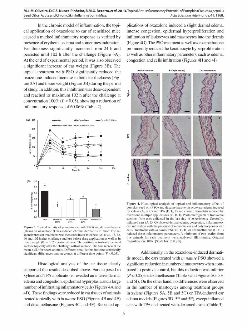
In the chronic model of infl ammation, the topi-cal application of oxazolone to ear of sensitized mice caused a marked infl ammatory response as verifi ed by presence of erythema, edema and sometimes induration. Ear thickness signifi cantly increased from 24 h and persisted until 102 h after the challenge (Figure 3A). At the end of experimental period, it was also observed a signifi cant increase of ear weight (Figure 3B). The topical treatment with PSO signifi cantly reduced the oxazolone-induced increase in both ear thickness (Fig-ure 3A) and tissue weight (Figure 3B) during the period of study. In addition, this inhibition was dose-dependent and reached its maximum 102 h after the challenge at concentration 100% (P < 0.05), showing a reduction of infl ammatory response of 60.86% (Table 2).
Histological analysis of the ear tissue clearly supported the results described above. Ears exposed to xylene and TPA applications revealed an intense dermal edema and congestion, epidermal hyperplasia and a large number of infi ltrating infl ammatory cells (Figures 4A and 4D). These fi ndings were reduced in ear tissues of animals treated topically with in nature PSO (Figures 4B and 4E) and dexamethasone (Figures 4C and 4F). Repeated ap-
plications of oxazolone induced a slight dermal edema, intense congestion, epidermal hyperproliferation and infi ltration of leukocytes and mastocytes into the dermis (Figure 4G). The PSO treatment as well as dexamethasone prominently reduced the keratinocyte hyperproliferation as well as other infl ammatory parameters, such as edema, congestion and cells infi ltration (Figures 4H and 4I).
Figure 3. Topical activity of pumpkin seed oil (PSO) and dexamethasone (Dexa) on oxazolone (Oxa)-induced chronic dermatitis in mice. The re-sponsiveness of treatments was measured in ear thickness (A) at 24, 48, 72, 96 and 102 h after challenge and just before drug application as well as in tissue weight (B) at 102 h post-challenge. The positive control only received acetone topically after the challenge with oxazolone. The bars represent the mean ± SD for seven animals. Different small letters indicate statistically signifi cant differences among groups at different time points (P < 0.05).
Figure 4. Histological analysis of topical anti-infl ammatory effect of pumpkin seed oil (PSO) and dexamethasone on acute ear edema induced by xylene (A, B, C) and TPA (D, E, F) and chronic dermatitis induced by oxazolone multiple applications (G, H, I). Photomicrograph of transverse sections from ears collected in the last day of experiments. Generally, infl amed ears (A, D, G) showed dermal edema, congestion, infl ammatory cell infi ltration with the presence of mononuclear and polymorphonuclear cells. Treatment with in nature PSO (B, E, H) or dexamethasone (C, F, I) reduced these infl ammatory parameters. A minimum of two section from fi ve animals for each treatment were analyzed. HE staining. Original magnifi cation: 100x. [Scale bar: 200 µm].
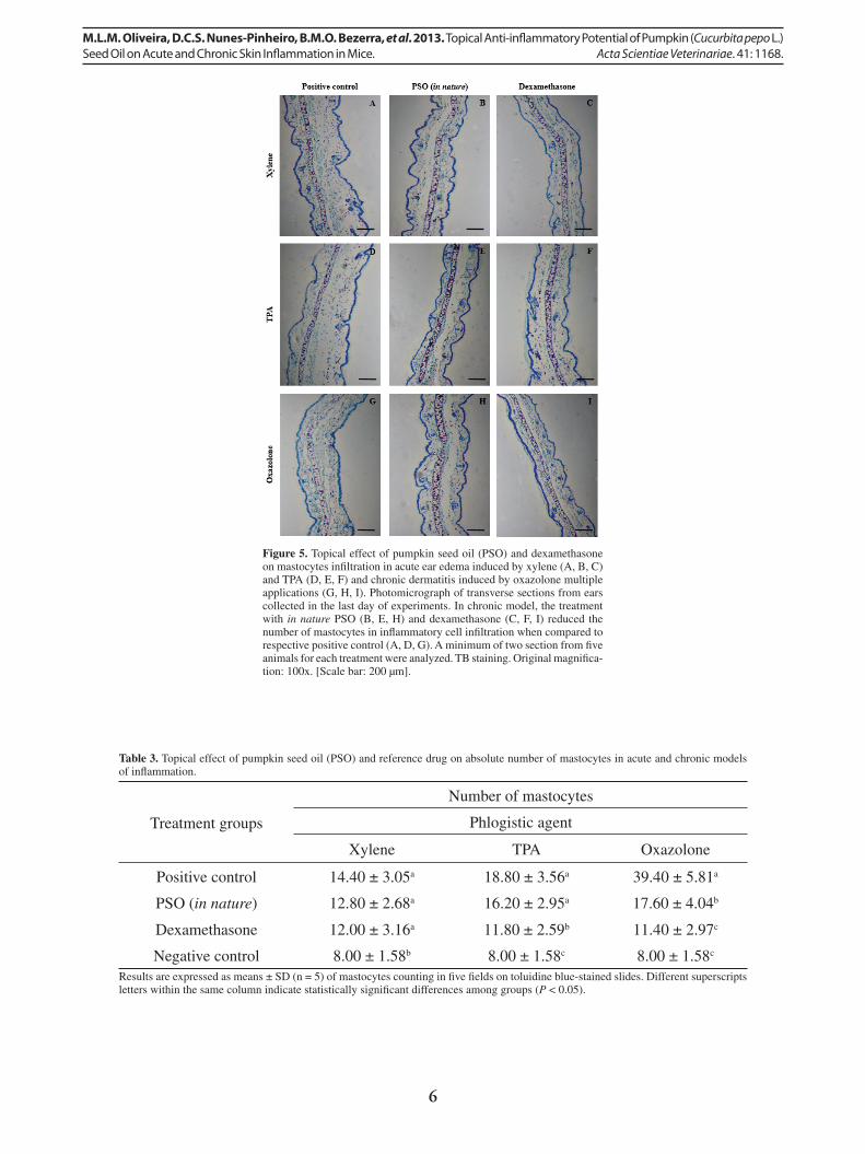
Additionally, in the oxazolone-induced dermati-tis model, the ears treated with in nature PSO showed a signifi cant reduction in number of mastocytes when com-pared to positive control, but this reduction was inferior (P < 0.05) to dexamethasone (Table 3 and Figures 5G, 5H and 5I). On the other hand, no differences were observed in the number of mastocytes among treatment groups in xylene (Figures 5A, 5B and 5C) or TPA-induced ear edema models (Figures 5D, 5E and 5F), except infl amed ears with TPA and treated with dexamethasone (Table 3).
6
M.L.M. Oliveira, D.C.S. Nunes-Pinheiro, B.M.O. Bezerra, et al. 2013. Topical Anti-infl ammatory Potential of Pumpkin (Cucurbita pepo L.) Seed Oil on Acute and Chronic Skin Infl ammation in Mice. Acta Scientiae Veterinariae. 41: 1168.
Figure 5. Topical effect of pumpkin seed oil (PSO) and dexamethasone on mastocytes infi ltration in acute ear edema induced by xylene (A, B, C) and TPA (D, E, F) and chronic dermatitis induced by oxazolone multiple applications (G, H, I). Photomicrograph of transverse sections from ears collected in the last day of experiments. In chronic model, the treatment with in nature PSO (B, E, H) and dexamethasone (C, F, I) reduced the number of mastocytes in infl ammatory cell infi ltration when compared to respective positive control (A, D, G). A minimum of two section from fi ve animals for each treatment were analyzed. TB staining. Original magnifi ca-tion: 100x. [Scale bar: 200 µm].
Table 3. Topical effect of pumpkin seed oil (PSO) and reference drug on absolute number of mastocytes in acute and chronic models of infl ammation.
Treatment groups
Number of mastocytes
Phlogistic agent
Xylene TPA Oxazolone
Positive control 14.40 ± 3.05a 18.80 ± 3.56a 39.40 ± 5.81a
PSO (in nature) 12.80 ± 2.68a 16.20 ± 2.95a 17.60 ± 4.04b
Dexamethasone 12.00 ± 3.16a 11.80 ± 2.59b 11.40 ± 2.97c
Negative control 8.00 ± 1.58b 8.00 ± 1.58c 8.00 ± 1.58c
Results are expressed as means ± SD (n = 5) of mastocytes counting in fi ve fi elds on toluidine blue-stained slides. Different superscripts letters within the same column indicate statistically signifi cant differences among groups (P < 0.05).

7
M.L.M. Oliveira, D.C.S. Nunes-Pinheiro, B.M.O. Bezerra, et al. 2013. Topical Anti-infl ammatory Potential of Pumpkin (Cucurbita pepo L.) Seed Oil on Acute and Chronic Skin Infl ammation in Mice. Acta Scientiae Veterinariae. 41: 1168.
DISCUSSION
Models of topical infl ammation induced by different phlogistic agents has been extensively used as pharmacological tools for the investigation of several substances with anti-infl ammatory potential in alter-native medicine. These compounds include products of plant origin as vegetable oils, which can be useful in the treatment of infl ammatory skin disorders [7]. In the present study, we have examined the pharma-cological properties of PSO in acute and chronic skin infl ammation in order to demonstrate its topical anti-infl ammatory potential.
Firstly, we studied the anti-infl ammatory activ-ity of PSO in two models of topical acute edema. The xylene is a phlogistic agent that act on target cells in the periphery such as mast cells, immune cells, and vascular smooth muscle via VR1 receptors, and pro-mote neurogenic infl ammation, which is characterized by redness and warmth, swelling, and hypersensitivity. This infl ammatory response result from the release of neuropeptides from primary sensory nerve terminals [15,21]. Another phlogistic agent, TPA is a phorbol ester able to activate protein kinase C, which activates other enzymatic cascades in turn, such as phospholi-pase A
2, leading to release of arachidonic acid (AA).
This signaling pathway stimulates local infl ammation with vascular permeability, edema formation and polymorphonuclear leukocytes infi ltration as conse-quence of the production of infl ammatory eicosanoids by cyclooxygenase (COX) and lipoxygenase (LOX) enzymes [6,7]. Many drug classes, such as corticoste-roids and NSAIDs, can modulate the acute ear edema response in varying degrees, depending on the nature of edematogen and involved mediators [7,21]. In the present work, we observed that the topical application of PSO was able of reducing infl ammation in xylene and TPA-induced acute ear edema in a dose-dependent fashion, and this effect was similar to, at least, one drug reference (Figures 1 and 2). Moreover, PSO reduced the infl ammatory lesion and cellular infi ltration in both acute ear edema models (Figures 4B and 4E). Taken together, these results suggest that this oil alter infl ammatory response via modulation of cellular and molecular mediators involved in infl ammatory path-ways activated by theses phlogistic agents.
A third model used in this study was the chronic skin infl ammation induced by repeated ex-posure to oxazolone. Skin infl ammation is persistent
in this model, which induces a contact delayed-type hypersensitivity resembling human contact dermati-tis. This response is characterized by a sustained ear swelling, marked polymorphonuclear, macrophage, mastocytes and T lymphocytes infi ltration, and abun-dant pro-infl ammatory mediators [16]. Increased skin thickening is often the fi rst hallmark of events that oc-cur during cutaneous infl ammation, including dermal edema, infl ammatory cells infi ltration and prolifera-tion of epidermal keratinocytes [12]. Here we verifi ed that the topical application of PSO were active both in the early stage and throughout the infl ammatory process and decreased the ear thickness in model of oxazolone-induced chronic dermatitis. This reduction was dose-dependent and similar to dexamethasone results. Corticosteroids are well known to have potent anti-infl ammatory effects, however its topical use can cause intense skin atrophy, one of the serious side effects limiting their uses for chronic skin diseases [23]. Thus, our fi ndings support the ability of PSO to resolve a persistent infl ammatory lesion, with an ef-fi cacy comparable to dexamethasone. In addition, we did not observe any cutaneous alterations when PSO alone was applied to mice skin.
With an extent of activity comparable to refer-ence drug, PSO reduced two important events related to the skin infl ammatory response induced by oxazolone: the migration of neutrophils and the number of masto-cytes, as determined by HE and TB staining, respec-tively. Indeed, the accumulation of polymorphonuclear cells and mastocytes plays a critical role in cutaneous infl ammatory diseases such as contact dermatitis, and is related to the pathological mechanism of disease [16]. Neutrophils recruitment is an essential factor in the acute infl ammatory process, acting as fi rst-line defense cells in the initiation and resolution phases of this process [11]. Another immune cell, the mastocytes act as important sentinels in the skin, helping to limit or even prevent the damage that results from injurious agent due to release of various biochemical mediators [25]. Under condition in which uncontrolled infi ltration of these cells occurs, they can become the main aggressor factor and have pathological role [11,25]. Thus, our results directly illustrate the inhibitory effects of PSO on neutrophils and mastocytes within the target tissue, providing fur-ther evidence that PSO ameliorates oxazolone-induced contact dermatitis and indicating the therapeutic effect of this oil for chronic skin disorders.

8
M.L.M. Oliveira, D.C.S. Nunes-Pinheiro, B.M.O. Bezerra, et al. 2013. Topical Anti-infl ammatory Potential of Pumpkin (Cucurbita pepo L.) Seed Oil on Acute and Chronic Skin Infl ammation in Mice. Acta Scientiae Veterinariae. 41: 1168.
With respect to this study, for the fi rst time, we demonstrate the topical anti-infl ammatory potential of PSO in acute and chronic skin infl ammation. Al-together, we believed that the results evidenced here may be attributed the promising ratio of ω-6 and ω-9 UFAs (2:1) present in PSO, namely linoleic and oleic acids (Table 1).
Fatty acids are a diverse set of molecules that act as cell membranes constituents, gene expression regulators, signal transduction modifi ers and cell pro-liferation, generating products that modulate a variety of biological mechanisms, including those linked to homeostasis, immune responsiveness, and infl am-mation [9]. These bioactive molecules have shown to be potential anti-infl ammatory agents due, at least in part, to their ability to inhibit enzymes that modulate the production of AA metabolites, or produce anti-infl ammatory eicosanoids [4]. In this context, linoleic and oleic acids have been reported to modulate the skin immune-infl ammatory responses [4,10,18]. These pre-vious fi ndings are consistent with our results, which we suggest that the proper balance of these UFAs present in PSO can have a great importance in modulation of different infl ammatory signaling according to injuri-ous stimulus.
In recent years, plant-derived natural products have become known for its benefi cial effects on in-fl ammatory response [12,14,24]. Amongst the natural products as rich source of UFAs, the vegetable oils have been indicated as alternative therapy for treat skin infl ammatory process [17,18]. Previously, we reported that Caryocar coriaceum oil (ω-6:ω-9 / 1:24), inhibited the xylene-induced ear edema, but this inhibition was twice less effective compared to the current study [19]. In other study, this same oil demonstrated signifi cant topical antiedematous effect against croton oil, but did not present signifi cant reduction on capsaicin-induced ear neurogenic infl ammation [22]. Here we verifi ed that PSO showed topical anti-infl ammatory activity
similar to reference drugs that inhibit the production of eicosanoids via modulation of phospholipase A2, COX and LOX enzymes [7,23], suggesting that this fi nding may be focused for future studies aiming to unravel mechanism of action of PSO.
CONCLUSION
In summary, this study represents the fi rst report that PSO acts as a topical anti-infl ammatory agent, and it is effective against acute and chronic skin infl ammatory processes. The anti-infl ammatory activ-ity of PSO may be attributed to promising proportions of ω-6 and ω-9 UFAs present in it, due to either their individual activity or the synergistic effect of these bioactive molecules. Together, these fi nding suggest that PSO may be an important alternative therapy for the treatment of infl ammatory skin diseases, such as psoriasis, contact dermatitis and atopic dermatitis. Nev-ertheless, further investigations need to be performed to determine the precise mechanism of action and support its application in clinical practice.
SOURCES AND MANUFACTURERS1Vital Âtman, Uchoa, SP, Brazil.2Dinâmica Química, Diadema, SP, Brazil.3Sigma Chemical Co., St. Louis, MO, USA.4Merck Sharp & Dohme, Cramlington, Northumberland, UK.5EMS, Hortolândia, SP, Brazil.
Acknowledgements. The authors would like to thank the Vital Âtman for providing the pumpkin seed oil and phytochemi-cal analysis, and Laboratório de Manipulação de Oócitos e Folículos Pré-Antrais (LAMOFOPA/UECE) for support with histological photomicrographs.
Ethical approval. Experimental protocols were approved by the Ethics Committee for Use of Animals of the State University of Ceará (UECE), under number 103401806-49.
Declaration of interest. The authors declare no confl icts of interest.
REFERENCES
1 Adams R.P. 1989. Identifi cation of essential oils by ion trap mass spectroscopy. San Diego: Academic Press, 456p.2 Alencar W.J., Craveiro A.A. & Matos F.J.A. 1984. Kovats indices as preselection routine in mass spectra library
search of volatiles. Journal of Natural Products. 47(5): 890-892.3 Calder P.C. 2013. Omega-3 polyunsaturated fatty acids and infl ammatory processes: nutrition or pharmacology? Brit-
ish Journal of Clinical Pharmacology. 75(3): 645-662.4 Cardoso C.R., Favoreto Jr. S., Oliveira L.L., Vancim J.O., Barban G.B., Ferraz D.B. & Silva J.S. 2011. Oleic acid
modulation of the immune response in wound healing: a new approach for skin repair. Immunobiology. 216(3): 409-415.

9
M.L.M. Oliveira, D.C.S. Nunes-Pinheiro, B.M.O. Bezerra, et al. 2013. Topical Anti-infl ammatory Potential of Pumpkin (Cucurbita pepo L.) Seed Oil on Acute and Chronic Skin Infl ammation in Mice. Acta Scientiae Veterinariae. 41: 1168.
www.ufrgs.br/actavet1168
5 Firestone D. 2009. Offi cial methods and recommended practices of the American Oil Chemists’ Society. 6th edn. Champaign: American Oil Chemists’ Society, 1200p.
6 Furstenberger G., Csuk-Glanzer B.I., Marks F. & Keppler D. 1994. Phorbol ester-induced leukotriene biosynthesis and tumor promotion in mouse epidermis. Carcinogenesis. 15(12): 2823-2827.
7 Gábor M. 2000. Mouse ear infl ammation models and their pharmacological applications. Budapest: Akadémiai Kiadó, 336p.8 Giner R.M., Villalba M.L., Recio M.C., Mánez S., Cerdá-Nicolás M. & Ríos J.L. 2000. Anti-infl ammatory glyco-
terpenoids from Scrophularia auriculata. European Journal of Pharmacology. 389(2-3): 243-252.9 Kalish B.T., Fallon E.M. & Puder M. 2012. A tutorial on fatty acid biology. Journal of Parenteral and Enteral Nutrition.
36(4): 380-388.10 Kendall A.C. & Nicolaou A. 2013. Bioactive lipid mediators in skin infl ammation and immunity. Progress and Lipid
Research. 52(1): 141-164.11 Kolaczkowska E. & Kubes P. 2013. Neutrophil recruitment and function in health and infl ammation. Nature Reviews
Immunology. 13(3): 159-175.12 Lee D.Y., Choi G., Yoon T., Cheon M.S., Choo B.K. & Kim H.K. 2009. Anti-infl ammatory activity of Chrysanthe-
mum indicum extract in acute and chronic cutaneous infl ammation. Journal of Ethnopharmacology. 123(1): 149-154.13 Medzhitov R. 2008. Origin and physiological roles of infl ammation. Nature. 454(7203): 428-435.14 Monteiro M.V.B., Leite A.K.R.M., Bertini L.M., Morais S.M. & Nunes-Pinheiro D.C.S. 2007. Topical anti-
infl ammatory, gastroprotective and antioxidant effects of the essential oil of Lippia sidoides Cham. leaves. Journal of Ethnopharmacology. 111(2): 378-382.
15 More S.V., Kumar H., Kim I.S., Song S.Y. & Choi D.K. 2013. Cellular and molecular mediators of neuroinfl amma-tion in the pathogenesis of Parkinson’s disease. Mediators of Infl ammation. 2013(952375): 1-12.
16 Nosbaum A., Vocanson M., Rozieres A., Hennino A. & Nicolas J.F. 2009. Allergic and irritant contact dermatitis. European Journal of Dermatology. 19(4): 325-332.
17 Oliveira A.P., Franco E.D., Barreto R.R., Cordeiro D.P., Melo R.G., Aquino C.M.F., Silva A.A.R., Medeiros P.L., Silva T.G., Goes A.J.S. & Maia M.B.S. 2013. Effect of semisolid formulation of Persea americana Mill (avocado) oil on wound healing in rats. Evidence-Based Complementary and Alternative Medicine. 2013(472382): 1-8.
18 Oliveira M.L.M. & Nunes-Pinheiro D.C.S. 2013. Biomarcadores celulares e moleculares envolvidos na resposta imune-infl amatória modulada por ácidos graxos insaturados. Acta Veterinaria Brasilica. 7(2): 113-124.
19 Oliveira M.L.M., Nunes-Pinheiro D.C.S., Tomé A.R., Mota E.F., Lima-Verde I.A., Pinheiro F.G.M., Campello C.C. & Morais S.M. 2010. In vivo topical anti-infl ammatory and wound healing activities of the fi xed oil of Caryocar coriaceum Wittm. seeds. Journal of Ethnopharmacology. 129(2): 214-219.
20 Procida G., Stancher B., Cateni F. & Zacchigna M. 2013. Chemical composition and functional characterization of commercial pumpkin seed oil. Journal of the Science of Food Agriculture. 93(5): 1035-1041.
21 Richardson J.D. & Vasko M.R. 2002. Cellular mechanisms of neurogenic infl ammation. The Journal of Pharmacol-ogy and Experimental Therapeutics. 302(3): 839-845.
22 Saraiva R.A., Araruna M.K.A., Oliveira R.C., Menezes K.D.P., Leite G.O., Kerntopf M.R., Costa J.G.M., Rocha J.B.T., Tomé A.R., Campos A.R. & Menezes I.R.A. 2011. Topical anti-infl ammatory effect of Caryocar coriaceum Wittm. (Caryocaraceae) fruit pulp fi xed oil on mice ear edema induced by different irritant agents. Journal of Ethnop-harmacology. 136(3): 504-510.
23 Schoepe S., Schacke H., May E. & Asadullah K. 2006. Glucocorticoid therapy-induced skin atrophy. Experimental Dermatology. 15(6): 406-420.
24 Vasconcelos A.K.P., Tomé A.R., Pereira B.S. & Nunes-Pinheiro D.C.S. 2007. Avaliação dos unguentos à base de extratos hexânico ou etanólico das folhas de Momordica charantia L. sobre as lesões cutâneas experimentais em coel-hos. Acta Scientiae Veterinariae. 35(1): 59-65.
25 Voehringer D. 2013. Protective and pathological roles of mast cells and basophils. Nature Reviews Immunology. 13(5): 362-375.