Tongue coating microbiome as a potential biomarker for ... · tongue-coating microbiomes of...
14
RESEARCH ARTICLE Tongue coating microbiome as a potential biomarker for gastritis including precancerous cascade Jiaxing Cui 1 , Hongfei Cui 1,3 , Mingran Yang 1 , Shiyu Du 2 , Junfeng Li 1 , Yingxue Li 1 , Liyang Liu 1 , Xuegong Zhang 1,4& , Shao Li 1,4& 1 MOE Key Laboratory of Bioinformatics and TCM-X center/Bioinformatics Division, BNRist/Department of Automation, Tsinghua University, Beijing 100084, China 2 China-Japan Friendship Hospital, Beijing 100029, China 3 Institute for Artificial Intelligence and Department of Computer Science and Technology, Tsinghua University, Beijing 100084, China 4 School of Life Sciences and Center for Synthetic and Systems Biology, Tsinghua University, Beijing 100084, China & Correspondence: [email protected] (X. Zhang), [email protected] (S. Li) Received September 18, 2018 Accepted October 23, 2018 ABSTRACT The development of gastritis is associated with an increased risk of gastric cancer. Current invasive gas- tritis diagnostic methods are not suitable for monitoring progress. In this work based on 78 gastritis patients and 50 healthy individuals, we observed that the variation of tongue-coating microbiota was associated with the occurrence and development of gastritis. Twenty-one microbial species were identified for differentiating tongue-coating microbiomes of gastritis and healthy individuals. Pathways such as microbial metabolism in diverse environments, biosynthesis of antibiotics and bacterial chemotaxis were up-regulated in gastritis patients. The abundance of Campylobacter concisus was found associated with the gastric precancerous cascade. Furthermore, Campylobacter concisus could be detected in tongue coating and gastric fluid in a validation cohort containing 38 gastritis patients. These observations provided biological evidence of tongue diagnosis in traditional Chinese medicine, and indicated that tongue-coating microbiome could be a potential non-invasive biomarker, which might be suitable for long-term monitoring of gastritis. KEYWORDS gastritis, tongue coating, metagenomics, Campylobacter concisus, non-invasive biomarker INTRODUCTION Gastritis, which is a worldwide problem, is defined as an inflamed condition of the gastric mucosa (Price, 1991; Stolte and Meining, 2001; Owen, 2003; Rugge et al., 2007). Gas- tritis development is a multistep and multifactorial process (Guo et al., 2017). The stage of gastritis can be divided into superficial gastritis, atrophic gastritis, intestinal metaplasia and dysplasia (Correa, 1992; Correa and Piazuelo, 2012). Long-term studies have confirmed that the development of gastritis increases the risk of gastric cancer (Sipponen et al., 1985; Filipe et al., 1994; Miehlke et al., 1998; Meining et al., 2002; Ohata et al., 2004; Song et al., 2015). Gastritis diag- nosis in clinical practice relies primarily on endoscopy and histological examination (Dixon et al., 1996; Rugge et al., 2007), which are invasive procedures that cannot be done frequently. Hence, monitoring and controlling at regular intervals with non-invasive methods are highly demanded in the prevention and treatment of gastritis. Therefore, it is important to find biomarkers associated with the occurrence and development of gastritis. Bacteria were shown to contribute to gastritis when Barry Marshall and Robin Warren found in 1984 that Helicobacter Jiaxing Cui, Hongfei Cui—These authors contributed equally to this work. Electronic supplementary material The online version of this article (https://doi.org/10.1007/s13238-018-0596-6) contains sup- plementary material, which is available to authorized users. © The Author(s) 2018 Protein Cell https://doi.org/10.1007/s13238-018-0596-6 Protein & Cell Protein & Cell
Transcript of Tongue coating microbiome as a potential biomarker for ... · tongue-coating microbiomes of...

RESEARCH ARTICLE
Tongue coating microbiome as a potentialbiomarker for gastritis including precancerouscascade
Jiaxing Cui1, Hongfei Cui1,3, Mingran Yang1, Shiyu Du2, Junfeng Li1, Yingxue Li1, Liyang Liu1,Xuegong Zhang1,4& , Shao Li1,4&
1 MOE Key Laboratory of Bioinformatics and TCM-X center/Bioinformatics Division, BNRist/Department of Automation,Tsinghua University, Beijing 100084, China
2 China-Japan Friendship Hospital, Beijing 100029, China3 Institute for Artificial Intelligence and Department of Computer Science and Technology, Tsinghua University, Beijing 100084,China
4 School of Life Sciences and Center for Synthetic and Systems Biology, Tsinghua University, Beijing 100084, China& Correspondence: [email protected] (X. Zhang), [email protected] (S. Li)
Received September 18, 2018 Accepted October 23, 2018
ABSTRACT
The development of gastritis is associated with anincreased risk of gastric cancer. Current invasive gas-tritis diagnostic methods are not suitable for monitoringprogress. In this work based on 78 gastritis patients and50 healthy individuals, we observed that the variation oftongue-coating microbiota was associated with theoccurrence and development of gastritis. Twenty-onemicrobial species were identified for differentiatingtongue-coating microbiomes of gastritis and healthyindividuals. Pathways such as microbial metabolism indiverse environments, biosynthesis of antibiotics andbacterial chemotaxis were up-regulated in gastritispatients. The abundance of Campylobacter concisuswas found associated with the gastric precancerouscascade. Furthermore, Campylobacter concisus couldbe detected in tongue coating and gastric fluid in avalidation cohort containing 38 gastritis patients. Theseobservations provided biological evidence of tonguediagnosis in traditional Chinese medicine, and indicatedthat tongue-coating microbiome could be a potential
non-invasive biomarker, which might be suitable forlong-term monitoring of gastritis.
KEYWORDS gastritis, tongue coating, metagenomics,Campylobacter concisus, non-invasive biomarker
INTRODUCTION
Gastritis, which is a worldwide problem, is defined as aninflamed condition of the gastric mucosa (Price, 1991; Stolteand Meining, 2001; Owen, 2003; Rugge et al., 2007). Gas-tritis development is a multistep and multifactorial process(Guo et al., 2017). The stage of gastritis can be divided intosuperficial gastritis, atrophic gastritis, intestinal metaplasiaand dysplasia (Correa, 1992; Correa and Piazuelo, 2012).Long-term studies have confirmed that the development ofgastritis increases the risk of gastric cancer (Sipponen et al.,1985; Filipe et al., 1994; Miehlke et al., 1998; Meining et al.,2002; Ohata et al., 2004; Song et al., 2015). Gastritis diag-nosis in clinical practice relies primarily on endoscopy andhistological examination (Dixon et al., 1996; Rugge et al.,2007), which are invasive procedures that cannot be donefrequently. Hence, monitoring and controlling at regularintervals with non-invasive methods are highly demanded inthe prevention and treatment of gastritis. Therefore, it isimportant to find biomarkers associated with the occurrenceand development of gastritis.
Bacteria were shown to contribute to gastritis when BarryMarshall and Robin Warren found in 1984 that Helicobacter
Jiaxing Cui, Hongfei Cui—These authors contributed equally to thiswork.
Electronic supplementary material The online version of thisarticle (https://doi.org/10.1007/s13238-018-0596-6) contains sup-
plementary material, which is available to authorized users.
© The Author(s) 2018
Protein Cellhttps://doi.org/10.1007/s13238-018-0596-6 Protein&Cell
Protein
&Cell

pylori (HP) in the stomach played an important role in chronicgastritis (Marshall and Warren, 1984). However, it wasreported that approximately 25% of chronic gastritis patientswere not infected by Helicobacter pylori, suggesting thatsome other bacteria or factors, which are capable of causinginflammation, are involved (Jonkers et al., 1997). Since then,a series of studies based on different cohorts in stomachhave confirmed the role of bacteria other than Helicobacterpylori in gastric lesions (Sjostedt et al., 1985; Sahay et al.,1995; Li et al., 2009; Hakalehto et al., 2011; Aviles-Jimenezet al., 2014; Eun et al., 2014; Schulz et al., 2016; Ferreiraet al., 2017; Sohn et al., 2017). The Firmicutes phylum andthe Streptococcus genus were found to exhibit increasedabundance in gastritis patients compared to normal controls(Li et al., 2009). Analyses of gastric mucosa microbialchanges of superficial gastritis (SG), atrophic gastritis (AG),intestinal metaplasia (IM) and gastric cancer (GC) found thatgastric microbial composition and interaction shifted, indi-cating that gastritis development is associated with shiftinghuman microbiota (Aviles-Jimenez et al., 2014; Eun et al.,2014; Coker et al., 2017).
Among diseases in the digestive tract, gastritis status maybe reflected in the coating of tongue, which is the initial partof the digestive tract. On the one hand, food and microbestransferred into the stomach could remain in the residue onthe tongue coating. On the other hand, the gastroe-sophageal reflux of gastritis patients could bring materialsfrom the stomach to the tongue coating. The associationbetween difference in tongue coating microbiota and theoccurrence of gastritis has been explored in several studies.Our previous work focused on the tongue coating of gastritispatients and initially found 123 and 258 species-level OTUsenriched in the tongue coating of gastritis with Cold Syn-drome and that with Hot Syndrome, respectively. Moreover,tongue-coating images of healthy controls and gastritispatients were distinguishable (Jiang et al., 2012). Sun andcolleagues confirmed that microbial components of tonguecoating in chronic gastritis patients were different from thosein normal controls based on 16S ribosomal RNA denaturedgradient gel electrophoresis (Sun et al., 2013). Ye et al.found that Bacillus was present only in the yellow tonguecoating of chronic erosive gastritis patients by using IlluminaMiseq sequencing of the V4–V5 region of the 16S ribosomalRNA gene (Ye et al., 2016). Furthermore, for traditionalChinese medicine (TCM), tongue appearance is a majorindicator of physical status including that of the stomach. Ourprevious work also confirmed that tongue images could beused to classify gastritis patients and healthy volunteers andto classify gastritis patients with different TCM subtypes(Kanawong et al., 2012). However, as far as we know, thereare no systematical analyses based on metagenomicsequencing to reveal the tongue coating microbiome varia-tion associated with the occurrence and development ofgastritis. This study focused on whether the gastric conditionis reflected in the tongue-coating microbiome, which might
be objective, non-invasive and suitable for long-termmonitoring.
In this study, we collected the tongue coating samples of78 gastritis patients and 50 healthy subjects. Metagenomicanalysis revealed that the variation in tongue-coatingmicrobiota was associated with the occurrence and devel-opment of gastritis. A network including 21 tongue-coatingspecies that differentiated the tongue-coating microbiomesof gastritis patients and healthy controls was identified.Pathways such as microbial metabolism in diverse environ-ments, biosynthesis of antibiotics and bacterial chemotaxiswere up-regulated in gastritis patients. Furthermore, theabundance of Campylobacter concisus was detected to beassociated with the precancerous cascade of gastritis. Totest whether Campylobacter concisus existed in the stom-ach, we detected the Campylobacter concisus in tonguecoating and gastric fluid in a validation cohort of 38 gastritispatients by quantitative polymerase chain reaction (qPCR).The study showed that tongue coating microbiota couldpotentially be a non-invasive biomarker, which might beobjective and suitable for long-term monitoring.
RESULTS
Patient and healthy control characteristics
In an exploratory cohort, 78 gastritis patients received anendoscopic examination were enrolled. Gastritis patientswere divided into 3 groups according to gastric precancerouscascade, including superficial gastritis, atrophic gastritis andintestinal metaplasia, based on histopathology. Fifty healthyvolunteers with no complaints of stomach discomfort wererecruited as normal controls (Tables 1 and S1). Tongueimages and tongue-coating samples were collected from allparticipants. For patients, the clinical symptoms were alsorecorded (Table S2). In a validation cohort, 15 superficialgastritis, 7 atrophic gastritis and 16 intestinal metaplasiapatients were recruited. Metagenomic sequencing wasconducted in the exploratory cohort to determine the tongue-coating microbiota associated with gastritis. Campylobacterconcisus was tested in tongue coating and gastric fluid in thevalidation cohort by qPCR (Fig. 1).
Phylogenetic and gene profiles of tongue-coatingmicrobiomes in controls and patients
Metagenomic sequencing was performed on tongue-coatingsamples, generating approximately 24–211 million reads persample. We denoted this database as Meta-Tongue data-base. Using a pipeline similar to the Human MicrobiomeProject (HMP), 176 species were obtained from all tonguecoating samples. These species were classified into 12phyla, 21 classes, 35 orders, 56 families and 87 genera(Fig. 2A and Table S3). Variations of microbial composition atthe phylum level between individuals could be seen
RESEARCH ARTICLE Jiaxing Cui et al.
© The Author(s) 2018
Protein
&Cell

(Fig. 2B). De novo sequence assembly and gene clusteringon tongue coating samples identified ∼1.9 M non-redundantputative genes. The genes were annotated to 5,377 KOsand classified according to functions (Fig. 2C and Table S4).
Comparing the taxonomy of normal controls and gastritispatients, we found that 149 species were shared in normalcontrols and gastritis patients, and 17 species wereobserved only in normal controls, whereas 10 species wereobserved in gastritis patients. Considering the stage ofpatients, 8 species, 1 species and 1 species were observedonly in superficial gastritis, atrophic gastritis and IM patients,respectively. Comparing the microbial constitution of HP-positive and HP-negative patients, 143 species were sharedin both groups, whereas 14 species and 6 species existedonly in HP-positive and HP-negative patients, respectively(Fig. S1). Considering the abundance of microbes at thephylum level, Proteobacteria, Firmicutes, Fusobacteria,Actinobacteria and Bacteroidetes dominated the tonguecoating microbiota. Among which, Fusobacteria was signifi-cantly higher in patients (P = 0.003, Wilcoxon rank-sum testfollowed by FDR correction), whereas others had not sig-nificant differences between two groups (Fig. S2).
Tongue-coating species as a potential biomarkerassociated with gastritis
In terms of alpha diversity, the species richnesses in gastritispatients were significantly lower than those of normal con-trols (P = 0.01, Wilcoxon rank-sum test), indicating a lowernumber of species in gastritis patients. The Shannonindexes were significantly higher than those of normal con-trols (P = 0.0005, Wilcoxon rank-sum test), indicating a moreuniform species distribution in gastritis patients (Fig. 3A).The alpha diversities of HP-positive and HP-negativepatients did not show a significant difference (Fig. S3). Interms of beta diversity, the Jaccard distances betweengastritis patients were higher than those between normalcontrols, indicating that patients’ samples were highly dis-similar (Fig. 3A). There was no difference of Jaccard dis-tances between HP-positive patients and those between HP-negative patients (Fig. S4). After Wilcoxon rank-sum testfollowed by FDR correction, no species were found signifi-cantly different between HP-positive and HP-negativepatients. After comparing patients with different spicy pref-erences (23 prefer spicy food, 32 do not eat spicy food, 23
Figure 1. Graphic summary of the study design.
Table 1. Demographic characteristics of healthy controls and gastritis patients.
Demographicvariable
Characteristics Normalcontrols
Gastritis patients
Total Superficialgastritis
Atrophicgastritis
Intestinalmetaplasia
Sample size 50 78 44 11 23
Age Mean ± SD 44 ± 15.6 48 ± 13.5 46 ± 14.5 47 ± 12.3 55 ± 11.5
Sex Male/Female 23/27 31/47 16/28 3/8 10/13
HP Positive/Negative
NA 23/55 16/28 2/9 5/18
Tongue coating microbiome as a biomarker for gastritis RESEARCH ARTICLE
© The Author(s) 2018
Protein
&Cell

no obvious preference), no species were found to be sig-nificantly different among three groups (P > 0.5, Wilcoxonrank-sum test followed by FDR correction).
The Wilcoxon rank-sum test followed by FDR correctionwas used to characterize the significance of differences intongue-coating microbiota abundance between the normalcontrols and gastritis patients. Finally, 21 species were foundto be significantly different in abundance between the twogroups (P<0.05): 11 species including Veillonella parvula,Corynebacterium matruchotii, Kingella oralis, Atopobiumrimae, Aggregatibacter aphrophilus, Streptococcus san-guinis, Acinetobacter lwoffii, Prevotella amnii, Prevotellabivia, Cardiobacterium hominis and Oribacterium sinus weredecreased in gastritis patients, whereas 10 species includingStreptococcus infantis, Treponema vincentii, Leptotrichiaunclassified, Campylobacter rectus, Campylobacter showae,
Capnocytophaga gingivalis, Leptotrichia buccalis, Campy-lobacter concisus, Selenomonas flueggei and Leptotrichiahofstadii were increased in gastritis group (Fig. 3B). These21 species were defined as tongue-coating species associ-ated with gastritis. To explore whether these tongue-coatingspecies can distinguish gastritis patients from normal con-trols, hierarchical clustering of the abundance of these spe-cies in tongue coating samples from gastritis patients andnormal controls was performed (Fig. S5). The clusteringresult showed that the gastritis patients group and normalcontrols group can be mainly separated, indicating thattongue-coating microbes may be potential biomarkers forgastritis.
A correlation network was constructed to assessthe potential relationship between these 21 tongue-coating species (Fig. 3C). Controls-enriched species were
Figure 2. Taxonomy and gene profiles of the tongue-coating samples. (A) The relative abundance of species identified in
tongue-coating samples, ordered by the taxonomy tree. (B) Relative abundance of major phyla across tongue-coating samples.
(C) Gene counts annotated to different KO functions.
RESEARCH ARTICLE Jiaxing Cui et al.
© The Author(s) 2018
Protein
&Cell

significantly decreased in gastritis patients’ tongue coating,while patients-enriched species were significantly increasedin gastritis patients’ tongue coating. Patients-enriched spe-cies had a stronger correlation with each other than controls-enriched species (P = 0.005, Wilcoxon rank-sum test),suggesting that patients-enriched species affected the hostby interacting with each other and playing similar roles.Furthermore, the abundances of some species were alsocorrelated with symptoms. Cardiobacterium hominis washigher in patients with bitter taste, and Selenomonas flueg-gei was higher in patients with dry mouth (Fig. 3D).
After exploring the potential functions of these tongue-coating species in gastritis by literature mining, we foundsome species enriched in patients that had the potential toinduce inflammation and immune response in the host(Fig. 3E). Campylobacter concisus preferentially attached tocell-cell junctions, which led to damage of epithelial barrierfunctions (Man et al., 2010a, b). Campylobacter concisuscould induce expression of cytokines and chemokines suchas tumor necrosis factor (TNF), interleukin 1 beta (IL1B),interleukin 10 (IL10), C-C motif chemokine ligand 2 (CCL2),C-X-C motif chemokine ligand 1 (CXCL1), C-X-C motifchemokine ligand 2 (CXCL2), C-X-C motif chemokine ligand9 (CXCL9), C-X-C motif chemokine ligand 10 (CXCL10), theassembly of inflammasome interferon gamma inducibleprotein 16 (IFI16) and activate the key inflammatory path-ways involving nuclear factor kappa B (NF-kB), signaltransducer and activator of transcription (STAT), cAMPresponsive element binding protein 1 (CREB1) and inter-feron regulatory factor signaling (Man et al., 2010a, b;Kaakoush et al., 2015). Members of the Campylobacterconcisus secretome including flagellin B (FlaB), ATP syn-thase F1 alpha subunit and outer membrane protein 18(OMP18) were able to stimulate an immunoreaction (Kovachet al., 2011). Furthermore, the GroEL-like protein, whichcould induce the secretion of interleukin 6 (IL6) and inter-leukin 8 (IL8), could be secreted by Campylobacter rectus(Hinode et al., 1998). These findings showed the potentialfunctions of species that had a predicted gastritisassociation.
Differential abundance of tongue-coating microbialgenes between controls and patients
To explore the variation in tongue-coating microbial functionsin gastritis samples, genes of tongue-coating microbes wereanalyzed. After comparing the genes annotated to KyotoEncyclopedia of Genes and Genomes (KEGG) betweennormal controls and gastritis patients by Wilcoxon rank-sumtest followed by FDR correction, we found that 878 geneswere significantly different between normal controls andgastritis patients (P < 0.05). Among these, 519 genes weresignificantly increased in gastritis patients (Table S5),including DedD protein, threonine aldolase and N-acetyl-muramoyl-L-alanine amidase. Moreover, 359 genes
including adenosylcobinamide hydrolasewere, NADP-de-pendent alcohol dehydrogenase, putative metalloproteaseand glyoxylate reductase were significantly decreased ingastritis patients (Table S6).
In order to reveal the functions that up-regulated genesplayed, enrichment analysis was conducted by Fisher’sexact test followed by FDR correction. Finally, 519 up-reg-ulated genes enriched in 28 pathways (P < 0.05) (Fig. 4).Pathways such as metabolic pathways, microbial metabo-lism in diverse environments, biosynthesis of secondarymetabolites, biosynthesis of antibiotics, flagellar assembly,bacterial chemotaxis, ABC transporters, carbon metabolismand biosynthesis of amino acids were found to be enrichedin up-regulated genes, indicating a variation of microbialfunctions in gastritis patients.
Campylobacter concisus is associated with gastritisstages, and can be detected in gastric fluid
To assess the differences of patients in different stages ofgastric precancerous cascade, the abundances of tongue-coating microbes were compared in four groups includingnormal controls, superficial gastritis, atrophic gastritis andintestinal metaplasia. This division indicated the develop-ment from normal status to the transformation from gastritisto precancerous lesions. Finally, the abundance of Campy-lobacter concisus, together with the abundances of theclass, order, family, and genus it belongs to, were found tohave clear association with gastritis stages (P = 0.02,Cochran-Armitage test). The abundance of Campylobacterconcisus in superficial gastritis, atrophic gastritis andintestinal metaplasia was higher than that in normal controls.In addition, the abundance of Campylobacter concisusincreased during precancerous cascade (Fig. 5). TheCochran-Armitage test for trend showed that there was asignificant association between the abundance of Campy-lobacter concisus and the precancerous cascade (P = 0.02),which was more significant when using only the first threestages (normal controls, superficial gastritis and atrophicgastritis, P = 0.004).
In the intestinal metaplasia group, the samples clearlyclustered into two subclasses according to the abundanceof Campylobacter concisus. We termed the subclass withhigh abundance as the “high abundance group” (HAG) andanother subclass as the “low abundance group” (LAG).The HAG samples showed a distinct increasing trendtogether with the health and other two gastritis stagesgroups (Cochran-Armitage test for trend, P = 3.8 × 10⁻⁵).This indicated that high heterogeneity exists in the intesti-nal metaplasia stage and there may be subgroups withspecific characteristics such as TCM or western medicinephenotypes, which requires further studies in largesamples.
After analysis of the potential relationships betweenCampylobacter concisus and clinical symptoms, we found
Tongue coating microbiome as a biomarker for gastritis RESEARCH ARTICLE
© The Author(s) 2018
Protein
&Cell

RESEARCH ARTICLE Jiaxing Cui et al.
© The Author(s) 2018
Protein
&Cell
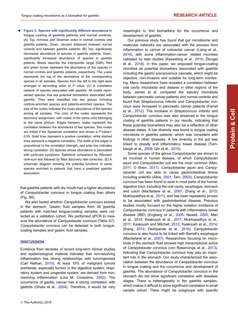
that gastritis patients with dry mouth had a higher abundanceof Campylobacter concisus in tongue coating than others(Fig. S6).
We also tested whether Campylobacter concisus existedin the stomach. Gastric fluid samples from 38 gastritispatients with matched tongue-coating samples were col-lected as a validation cohort. We performed qPCR to mea-sure the abundance of Campylobacter concisus (Table S7).Campylobacter concisus can be detected in both tongue-coating samples and gastric fluid samples.
DISCUSSION
Evidence from decades of recent long-term clinical studiesand epidemiological material indicates that non-resolvinginflammation has strong relationships with tumorigenesis(Carl Nathan, 2010). At least 15% of malignant tumorsworldwide, especially tumors in the digestive system, respi-ratory system and urogenital system, are derived from non-resolving inflammation (Lisa M. Coussens, 2002). Theoccurrence of gastric cancer has a strong correlation withgastritis (Ohata et al., 2004). Therefore, it would be very
meaningful to find biomarkers for the occurrence anddevelopment of gastritis.
Our previous study has found that gut microbiome andmolecular networks are associated with the process frominflammation to cancer of colorectal cancer (Liang et al.,2014), with some inflammation-cancer related microbesvalidated by later studies (Kesselring et al., 2016; Zitvogelet al., 2018). In this paper, we proposed tongue-coatingmicrobes as potential biomarkers associated with gastritisincluding the gastric precancerous cascade, which might beobjective, non-invasive and suitable for long-term monitor-ing. Many researchers have revealed a correlation betweenoral cavity microbiota and disease in other regions of thebody. James et al. compared the salivary microbiotabetween pancreatic cancer patients and normal controls andfound that Streptococcus infantis and Campylobacter con-cisus were increased in pancreatic cancer patients (Farrellet al., 2012). The increase in Streptococcus infantis andCampylobacter concisus was also observed in the tonguecoating of gastritis patients in our results, indicating thatthese potential biomarkers may also be a reflection of otherdisease states. A low diversity was found in tongue coatingmicrobiota in gastritis patients, which was consistent withfindings in other diseases. A low diversity in the gut waslinked to obesity and inflammatory bowel disease (Turn-baugh et al., 2008; Qin et al., 2010).
Some species of the genus Campylobacter are shown tobe involved in human disease, of which Campylobacterjejuni and Campylobacter coli are the most common (Man,2011; O Brien, 2017). Campylobacter jejuni and Campy-lobacter coli are able to cause gastrointestinal illnessincluding enteritis (Allos, 2001; Tam, 2003). Campylobacterconcisus has been found to exist in most parts of the humandigestive tract, including the oral cavity, esophagus, stomachand colon (Macfarlane et al., 2007; Zhang et al., 2010;Mukhopadhya et al., 2011), and has been repeatedly provento be associated with gastrointestinal disease. Previousstudies mostly focused on the higher isolation incidence ofCampylobacter concisus in patients with inflammatory boweldisease (IBD) (Engberg et al., 2005; Newell, 2005; Manet al., 2010; Kaakoush et al., 2011; Mukhopadhya et al.,2011; Kaakoush and Mitchell, 2012; Kaakoush et al., 2014;Zhang, 2014; Deshpande et al., 2016). Campylobacterconcisus is also found to be linked with Barrett’s esophagus(Macfarlane et al., 2007). Researchers focusing on micro-biota in the stomach fluid showed high transcriptional activeof Campylobacter concisus (von Rosenvinge et al., 2013),indicating that Campylobacter concisus may play an impor-tant role in the stomach. Our study characterized the asso-ciation between the abundance of Campylobacter concisusin tongue coating and the occurrence and development ofgastritis. The abundance of Campylobacter concisus in thestomach did not show significant correlation with diseasesstages. There is heterogeneity in the gastritis samples,which makes it difficult to show significant correlation in smallsample cohort. There might be subgroups with specific
Figure 3. Species with significantly different abundance in
tongue coating of gastritis patients and normal controls.
(A) Top, richness and Shannon index in normal controls and
gastritis patients. Down, Jaccard distances between normal
controls and between gastritis patients. (B) Top, significantly
decreased abundance of species in gastritis patients. Down,
significantly increased abundance of species in gastritis
patients. Boxes describe the interquartile range (IQR). Red
and green boxes represent the abundance of the species in
normal controls and gastritis patients, respectively. The y-axis
represents the log of the abundance of the corresponding
species in all samples. Species from the left to the right were
arranged in ascending order of P value. (C) A correlation
network of species associated with gastritis. All nodes repre-
sented species that are potential biomarkers associated with
gastritis. They were classified into two groups including
controls-enriched species and patients-enriched species. The
size of the nodes indicates the mean abundance of the species
among all samples. The color of the nodes represents the
taxonomy assignment, with nodes of the same color belonging
to the same phylum. Edges between nodes represent the
correlation between the abundance of two species. Two nodes
are linked if the Spearman correlation test shows a P-value<
0.05. Solid lines represent a positive correlation, while dashed
lines represent a negative correlation. The width of the edges is
proportional to the correlation strength, and wide line indicates
strong correlation. (D) Species whose abundance is associated
with particular symptoms. Statistical comparison by Wilcoxon
rank-sum test followed by false discovery rate correction. (E) A
schematic diagram showing the potential functions of some
species enriched in patients that have a predicted gastritis
association.
b
Tongue coating microbiome as a biomarker for gastritis RESEARCH ARTICLE
© The Author(s) 2018
Protein
&Cell

characteristics such as TCM or western medicine pheno-types, which require further studies in large samples.
There are cheaper and convenient ways for gastritisdiagnosis such as endoscopy and histological examinationin clinical practice, however, they are invasive. Biomarkersfrom tongue coating microbiome based on tongue diagnosiscould provide beneficial complement for gastritis diagnosis,from the non-invasive, individualized and long-term moni-toring aspects. Furthermore, in traditional Chinese medicine,tongue coating can reflect the health status of human body.Our work provided the biological evidence of tongue diag-nosis. We found that species associated with gastritis hadpotential correlation with symptoms including dry mouth andbitter taste. In addition, Campylobacter concisus andCampylobacter rectus were recorded to upregulate genessuch as immune factors, cytokines and CCL2 (Kaakoushet al., 2015), which were associated with the Hot Syndromeof gastritis in TCM (Li et al., 2007; Li et al., 2013). Especially,TNF which can be induced by Campylobacter concisus isassociated with bitter taste (Feng et al., 2015), which is animportant Damp Heat phenotype related to gastritis in tra-ditional Chinese medicine. These findings showed a slighthint that tongue coating species such as Campylobacterconcisusmay have potential association with Hot Syndrome,
which includes status such as “Shang-huo”, Damp Heat, Yin-deficiency, and so on in TCM.
In conclusion, we have demonstrated the variation oftongue-coating microbiota in gastritis patients by metage-nomic sequencing, identified potential biomarkers for gas-tritis including precancerous cascade. Our work takes a steptoward a potential non-invasive biomarker for gastritis, whichmight be objective and suitable for long-term monitoring.Furthermore, before using tongue-tongue microbiomes asbiomarker for larger samples, more studies are required toreveal the influence of food intake and the geographicalareas of people. It has been known that Helicobacter pylori isan important factor in gastritis and has been found associ-ated with some gastritis sub-types as well as gastric cancerin many studies (McColl, 2010). Gastritis is actually an“umbrella term” for a number of diseases including thoseinduced by Helicobacter pylori and by other factors (Suganoet al., 2015; Suzuki and Mori, 2015). Patients without Heli-cobacter pylori infection may also have gastritis includingsuperficial, atrophic or even gastric cancer (Genta andSonnenberg, 2015; Pogoriler et al., 2015; Horiuchi et al.,2016; Overby et al., 2017). In future research on largersamples, taking precise Helicobacter pylori information intoconsideration in the study of gastritis sub-types and stages
Figure 4. Up-regulated tongue-coating microbial genes and enriched pathways. Blue nodes represent KEGG KOs, and yellow
nodes represent KEGG pathways. The edge between two nodes indicates that the gene was in the pathway. The size of the yellow
node is proportional to the connection degree, i.e., pathways with a higher number of up-regulated genes exhibit larger nodes.
RESEARCH ARTICLE Jiaxing Cui et al.
© The Author(s) 2018
Protein
&Cell

may reveal more specific biomarkers for the diagnose ofgastritis and gastric cancer.
MATERIALS AND METHODS
Sample collection
For the exploratory cohort, 99 patients who received an endoscopic
examination in Beijing Dongzhimen TCM Hospital, Beijing Xiyuan
TCM Hospital and China-Japan Friendship Hospital were recruited.
Endoscopic examinations were performed, and the clinical symp-
toms were recorded. Biopsy specimens were selected from the
antrum. The histological assessment was done by two experienced
pathologists following clinical guidelines according to “the updated
Sydney System” (Dixon et al., 1996). The inclusion criteria were a
confirmed diagnosis of gastritis according to histological examina-
tion. Autoimmune gastritis patients were excluded. The Helicobacter
pylori infection for all patients was identified by pathological exami-
nation. Healthy volunteers were recruited from Tsinghua University
during their annual physical examination in the hospital. Seventy-six
healthy people who reported no complaints of stomach discomfort in
the past 5 years and also confirmed no gastritis in the examination
were enrolled. The exclusion criteria for both patients and healthy
volunteers consisted of the use of glucocorticoids and antibiotics for
the past 3 months. Tongue images and tongue coating samples
were collected from all participants. After quality control of metage-
nomic sequencing, 167 samples including 93 patients and 74 con-
trols passed the acceptance criteria. Samples with contamination
and patients without histopathological results were then filtered.
Finally, 78 patients and 50 controls were retained for further analysis.
Patients were divided into different stages of gastric precancerous
cascade according to the histopathological results. This study mainly
focused on people living in Beijing.
For the validation cohort, 38 patients who received an endo-
scopic examination in the China-Japan Friendship Hospital were
recruited with the same inclusion and exclusion criteria for the
exploration cohort. Samples were different from that in exploratory
cohort. Tongue-coating and gastric fluid samples were collected.
Histological examination results and clinical symptoms were
recorded.
Specimen collection, DNA extraction and preservation
Tongue-coating swabs were used to collect tongue-coating samples
of participants before consumption of breakfast and water. The
tongue was scraped from the root to the tip 30 times by simultane-
ously rolling the swab, the swab was placed into an RNase-free
Eppendorf tube with 1 mL of phosphate-buffered saline (PBS), and
the swab was agitated in order to wash out the tongue coating. The
above step was repeated twice with a new swab and new Eppendorf
Figure 5. The abundance of Campylobacter concisus during the precancerous cascade. Boxes describe the interquartile range
(IQR). The y-axis represents the relative abundance of the corresponding species in all samples. Statistical comparison by Wilcoxon
rank-sum test.
Tongue coating microbiome as a biomarker for gastritis RESEARCH ARTICLE
© The Author(s) 2018
Protein
&Cell

tube to ensure that the tongue coating was sufficiently collected.
After collection of tongue coating, the tubes were centrifuged at
5,000 ×g for 5 min. Supernatant and sediment were preserved
separately in sterile tubes and stored at −80 °C until analysis. DNA
of microbiota in the samples was extracted following the user guide
of a modified version of the MO-BIO PowerSoil DNA Isolation Kit
(MO-BIO Laboratories, Inc., Carlsbad, CA, USA).
Library construction and sequencing
The sequencing library was constructed using a modified version of
the NEBNext Ultra DNA library Prep Kit (New England Biolabs,
Ipswich, MA, USA) protocol. We constructed barcoded, paired-end
libraries with an insert size of ∼500 bp for each sample. Four sam-
ples were designed to be sequenced in each lane. Samples were
randomly assigned to different sequence lanes. Extracted DNA (200
ng) was used for library construction for each sample. Samples were
sequenced in two batches. Samples in the first batch (172 samples)
were sent to BGI Shenzhen for sequencing. After quality determi-
nation, libraries passing quality control were sequenced with the
Illumina HiSeq 2000 platform. The read length was set to 90 bp.
Samples in the second batch (3 samples) were sequenced in Tsin-
ghua University with the Illumina HiSeq 2500 platform. The read
length was set to 100 bp.
Quality control, assembly and gene prediction
A modified version of fastx_barcode_splitter in the FASTX-Toolkit
(http://hannonlab.cshl.edu/fastx_toolkit/) was used to decode the
barcoded samples of each lane with the parameter “—mismatches
2”. We used the pipeline “assemble_revise_predict_genes_with_
hg19_screen” in the “runMOCAT.pl” of the MOCAT V1.3 (http://vm-
lux.embl.de/∼kultima/MOCAT/) (Kultima et al., 2012) toolkit for
quality control, assembly and gene (ORF) prediction. The configured
file of MOCAT was similar to that used by Li et al. (2014).
Sequences after quality control were clean reads of high quality
without human reads. To ensure better reliability, only samples with
more than 15 M clean reads were used for subsequent analysis. A
total of 167 samples passed the entire quality control (Table S8).
Taxonomy profiling
We used MetaPhlAn Version 1.7.8 (http://huttenhower.sph.harvard.
edu/metaphlan) (Segata et al., 2012) to calculate the taxonomy
abundance table for each sample. MetaPhlAn aligns short reads
directly to the customized prokaryote database, which was con-
structed by extracting clade-specific marker genes from the NCBI
genome database with the phylogenetic information of the NCBI
taxonomic tree. Abundances were calculated based on read counts
of corresponding marker genes normalized by gene length and
sequencing depth. We used clean reads as inputs, and the
parameters were “–bt2_ps very-sensitive, -t ‘rel_ab’”.
Construction and annotation of Integrated Tongue Gene Catalogue
(ITGC)
We first constructed a “Meta-Tongue Gene Catalogue” (MTGC) for
the Meta-Tongue dataset. We merged all the ORFs from 167 high
quality samples and did a redundancy removal according to their
sequence similarity using CD-HIT (Li and Godzik, 2006; Fu et al.,
2012) v4.6.1 (http://weizhongli-lab.org/cd-hit/). The parameters we
used in CD-HIT were “-G 0 -M 0 -T 4 -c 0.95 -aS 0.9 -n 8 -B 1”. To
construct a more comprehensive catalogue, we collected HMP
dorsum samples (Huttenhower et al., 2012) and used a same
pipeline as above to build an “HMP Tongue Gene Catalogue”
(HTGC). We then merged MTGC and HTGC using CD-HIT with
same parameters, and got the final Integrated Tongue Gene Cata-
logue (ITGC).
We mapped ITGC genes to NCBI nr database (created in
2012.2.28 by NCBI) with blastp (Altschul et al., 1990) in the
Blast 2.2.29+ toolkit. We used “-evalue 1e-5 -outfmt 6” in our
study. The outputs from blastp were processed with MEGAN
(Huson et al., 2007) v5.7.1 (http://ab.inf.uni-tuebingen.de/
software/megan5/) with parameters “maxMatches=100 minS-
core=50.0 maxExpected=0.01 topPercent=10.0 minSupport=1
minComplexity=0.0 useMinimalCoverageHeuristic=false paired=
false useIdentityFilter=false”.
Gene and function profiling
For each sample, we mapped its clean paired-end reads to ITGC
genes, calculated gene abundances and function abundances
according to the mapping results. We used SOAPaligner v2.2.1
(http://soap.genomics.org.cn/soapaligner.html) with parameters “-r 2
-M 4 -l 30 -v 9” in our study. The ITGC gene relative abundances
were calculated in a similar way with that in the work of Qin et al.
(2012).
Statistical analysis
To determine the differential abundance of species and genes
between controls and patients, the Wilcoxon rank-sum test was
used. P-values were adjusted with false discovery rate. The corre-
lation between species was analyzed using Spearman’s correlation.
The comparison of alpha diversity index and Jaccard distance was
tested by Wilcoxon rank-sum test. The increasing trend of the
abundance of Campylobacter concisus with gastritis stages was
tested by Cochran-Armitage test. Samples in HAG were labeled as 1
and others were labeled as 0. Health and the three gastritis stages
were labeled as 0, 1, 2, 3 separately.
Detection of Campylobacter concisus by qPCR
Abundances of Campylobacter concisus in tongue coating and
gastric fluid were assessed using quantitative PCR (qPCR).
Universal 16S rDNA was used as internal reference for the esti-
mation of all the microbes. The abundance of Campylobacter con-
cisus was estimated using the proportion of Campylobacter concisus
in all the microbes. qPCR was performed with TransStart Top Green
Qpcr SuperMix.
ETHICS APPROVAL AND CONSENT TO PARTICIPATION
This study was approved by the Medical Ethical Committee of the
Beijing Dongzhimen TCM Hospital and complied with the standards
RESEARCH ARTICLE Jiaxing Cui et al.
© The Author(s) 2018
Protein
&Cell

indicated by the Declaration of Helsinki. All participants signed the
informed consent.
AVAILABILITY OF DATA AND MATERIAL
The Meta-Tongue metagenomics sequencing data have been
deposited in BIGD database (http://bigd.big.ac.cn) and can be
found under accession PRJCA000663. Gene and function abun-
dance tables can be found in ftp://166.111.5.235:8082/Publications/
Meta-Tongue.
ACKNOWLEDGEMENTS
We thank Yongan Ye and Zhihong Li in Beijing Dongzhimen TCM
Hospital and Zhenhua Li in Beijing Xiyuan TCM Hospital for
providing help with sample collection. We also acknowledge Lai
Wei in Sun Yat-sen University and Jingren Zhang and Juanjuan
Wang in Tsinghua University for their helpful advices on experi-
ments, and thank Geng Tian and Jidong Lang in Tsinghua University
and Ting Chen, Rui Jiang, Congmin Zhu, Peng Zhang and Lianshuo
Li in our Division for their suggestions. We also thank Jianhuo Fang
in Tsinghua University and Huiying Li, Xujun Liang, Aidi Tan and
Liang Ruan in our Division for helping for sample collection. This
study was supported by the National Natural Science Foundation of
China (Grant Nos. 81630103, 91729301, 91229201 and 81225025)
and the Project of Tsinghua-Fuzhou Insititute for Data Technology
(TFIDT2018001) to S. Li and grants 61673231 and 61721003 to X.
Zhang.
ABBREVIATIONS
AG, atrophic gastritis; CCL2, C-C motif chemokine ligand 2;
CREB1, cAMP responsive element binding protein 1; CXCL1, C-X-
C motif chemokine ligand 1; CXCL2, C-X-C motif chemokine ligand
2; CXCL9, C-X-C motif chemokine ligand 9; CXCL10, C-X-C motif
chemokine ligand 10; FlaB, flagellin B; GC, gastric cancer; HAG,
high abundance group; HMP, Human Microbiome Project; HP,
helicobacter pylori; HTGC, HMP Tongue Gene Catalogue; IBD,
inflammatory bowel disease; IFI16, interferon gamma inducible
protein 16; IL1B, interleukin 1 beta; IL6, interleukin 6; IL8,
interleukin 8; IL10, interleukin 10; IM, intestinal metaplasia; IQR,
interquartile range; ITGC, Integrated Tongue Gene Catalogue;
KEGG, Kyoto Encyclopedia of Genes and Genomes; LAG, low
abundance group; MTGC, Meta-Tongue Gene Catalogue; NF-kB,
nuclear factor kappa B; OMP18, outer membrane protein 18; PBS,
phosphate-buffered saline; qPCR, quantitative polymerase chain
reaction; SG, superficial gastritis; STAT, signal transducer and
activator of transcription; TCM, traditional Chinese medicine; TNF,
tumor necrosis factor
COMPLIANCE WITH ETHICS GUIDELINES
Jiaxing Cui, Hongfei Cui, Mingran Yang, Shiyu Du, Junfeng Li,
Yingxue Li, Liyang Liu, Xuegong Zhang and Shao Li declare that
they have no conflict of interest. All procedures followed were in
accordance with the ethical standards of the responsible committee
on human experimentation (institutional and national) and with the
Helsinki Declaration of 1975, as revised in 2000 (5). Informed
consent was obtained from all patients for being included in the
study.
AUTHOR CONTRIBUTIONS
J. Cui and H. Cui performed the bioinformatics analyses, and
prepared figures and tables. M. Yang performed the qPCR exper-
iment. J. Cui, M. Yang, L. Liu and S. Du collected the samples.
H. Cui, J. Li and Y. Li processed the sequence data. S. Li and X.
Zhang designed the project. S. Li conceived the study. J. Cui wrote
early drafts, and all authors contributed to the revision of the
manuscript.
OPEN ACCESS
This article is distributed under the terms of the Creative Commons
Attribution 4.0 International License (http://creativecommons.org/
licenses/by/4.0/), which permits unrestricted use, distribution, and
reproduction in any medium, provided you give appropriate credit to
the original author(s) and the source, provide a link to the Creative
Commons license, and indicate if changes were made.
REFERENCES
Allos BM (2001) Campylobacter jejuni Infections: update on emerg-
ing issues and trends. Clin Infect Dis 32:1201–1206Altschul SF, Gish W, Miller W, Myers EW, Lipman DJ (1990) Basic
local alignment search tool. J Mol Biol 215:403–410Aviles-Jimenez F, Vazquez-Jimenez F, Medrano-Guzman R, Man-
tilla A, Torres J (2014) Stomach microbiota composition varies
between patients with non-atrophic gastritis and patients with
intestinal type of gastric cancer. Sci Rep 4:4202
Carl Nathan AD (2010) Nonresolving inflammation. Cell. 19:871–882
Coker OO, Dai Z, Nie Y, Zhao G, Cao L, Nakatsu G, Wu WK, Wong
SH, Chen Z, Sung JJY et al (2017) Mucosal microbiome
dysbiosis in gastric carcinogenesis. Gut:2017–314281Correa P (1992) Human gastric carcinogenesis: a multistep and
multifactorial process—1st American-cancer-society award lec-
ture on cancer-epidemiology and prevention. Cancer Res
52:6735–6740Correa P, Piazuelo MB (2012) The gastric precancerous cascade.
J Digest Dis 13:2–9Coussens LM, Werb Z (2002) Inflammation and cancer. Nature.
420:860
Deshpande NP, Wilkins MR, Castaño-Rodríguez N, Bainbridge E,
Sodhi N, Riordan SM, Mitchell HM, Kaakoush NO (2016)
Campylobacter concisus pathotypes induce distinct global
responses in intestinal epithelial cells. Sci Rep 6:34288
Dixon MF, Genta RM, Yardley JH, Correa P (1996) Classification
and grading of gastritis: The updated sydney system. Am J Surg
Pathol 20:1161–1181
Tongue coating microbiome as a biomarker for gastritis RESEARCH ARTICLE
© The Author(s) 2018
Protein
&Cell
Engberg J, Bang DD, Aabenhus R, Aarestrup FM, Fussing V,
Gerner-Smidt P (2005) Campylobacter concisus: an evaluation of
certain phenotypic and genotypic characteristics. Clin Microbiol
Infect 11:288–295Eun CS, Kim BK, Han DS, Kim SY, Kim KM, Choi BY, Song KS, Kim
YS, Kim JF (2014) Differences in gastric mucosal microbiota
profiling in patients with chronic gastritis, intestinal metaplasia,
and gastric cancer using pyrosequencing methods. Helicobacter
19:407–416Farrell JJ, Zhang L, Zhou H, Chia D, Elashoff D, Akin D, Paster BJ,
Joshipura K, Wong DTW (2012) Variations of oral microbiota are
associated with pancreatic diseases including pancreatic cancer.
Gut 61:582–588Feng P, Jyotaki M, Kim A, Chai J, Simon N, Zhou M, Bachmanov AA,
Huang L, Wang H (2015) Regulation of bitter taste responses by
tumor necrosis factor. Brain Behav Immunol 49:32–42Ferreira RM, Pereira-Marques J, Pinto-Ribeiro I, Costa JL, Carneiro
F, Machado JC, Figueiredo C (2017). Gastric microbial commu-
nity profiling reveals a dysbiotic cancer-associated microbiota.
Gut:2017–314205Filipe MI, Munoz N, Matko I, Kato I, Pompe-Kirn V, Jutersek A,
Teuchmann S, Benz M, Prijon T (1994) Intestinal metaplasia
types and the risk of gastric cancer: a cohort study in Slovenia. Int
J Cancer 57:324–329Fu L, Niu B, Zhu Z, Wu S, Li W (2012) CD-HIT: accelerated for
clustering the next-generation sequencing data. Bioinformatics
28:3150–3152Genta RM, Sonnenberg A (2015) Helicobacter-negative gastritis: a
distinct entity unrelated to Helicobacter pylori infection. Aliment
Pharmacol Ther 41:218–226Guo Y, Nie Q, MacLean AL, Li Y, Lei J, Li S (2017) Multiscale
modeling of inflammation-induced tumorigenesis reveals com-
peting oncogenic and oncoprotective roles for inflammation.
Cancer Res 77:6429–6441Hakalehto E, Vilpponen-Salmela T, Kinnunen K, von Wright A (2011)
Lactic acid bacteria enriched from human gastric biopsies. ISRN
Gastroenterol 2011:1–4Hinode D, Yoshioka M, Tanabe S, Miki O, Masuda K, Nakamura R
(1998) The GroEL-like protein from Campylobacter rectus:
immunological characterization and interleukin-6 and -8 induction
in human gingival fibroblast. FEMS Microbiol Lett 167:1–6Horiuchi Y, Fujisaki J, Yamamoto N, Shimizu T, Miyamoto Y, Tomida
H, Taniguchi C, Morishige K, Omae M, Ishiyama A et al (2016)
Biological behavior of the intramucosal Helicobacter pylori-
negative undifferentiated-type early gastric cancer: comparison
with Helicobacter pylori-positive early gastric cancer. Gastric
Cancer 19:160–165Huson DH, Auch AF, Qi J, Schuster SC (2007) MEGAN analysis of
metagenomic data. Genome Res 17:377–386Huttenhower C, Gevers D, Knight R, Abubucker S, Badger JH,
Chinwalla AT, Creasy HH, Earl AM, FitzGerald MG, Fulton RS
et al (2012) Structure, function and diversity of the healthy human
microbiome. Nature 486:207–214Jiang B, Liang X, Chen Y, Ma T, Liu L, Li J, Jiang R, Chen T, Zhang
X, Li S (2012) Integrating next-generation sequencing and
traditional tongue diagnosis to determine tongue coating micro-
biome. Sci Rep 2:936
Jonkers D, Gisbertz I, de Bruine A, Bot F, Arends JW, Stobberingh E,
Schouten H, Stockbrugger R (1997) Helicobacter pylori and non-
Helicobacter pylori bacterial flora in gastric mucosal and tumour
specimens of patients with primary gastric lymphoma. Eur J Clin
Invest 27:885–892Kaakoush NO, Mitchell HM (2012) Campylobacter concisus: a new
player in intestinal disease. Front Cell Infect MI:2
Kaakoush NO, Deshpande NP, Wilkins MR, Tan CG, Burgos-
Portugal JA, Raftery MJ, Day AS, Lemberg DA, Mitchell H (2011)
The pathogenic potential of Campylobacter concisus strains
associated with chronic intestinal diseases. PLoS ONE 6:e29045
Kaakoush NO, Mitchell HM, Man SM (2014) Role of emerging
campylobacter species in inflammatory bowel diseases. Inflamm
Bowel Dis 20:2189–2197Kaakoush NO, Deshpande NP, Man SM, Burgos-Portugal JA,
Khattak FA, Raftery MJ, Wilkins MR, Mitchell HM (2015)
Transcriptomic and proteomic analyses reveal key innate
immune signatures in the host response to the gastrointestinal
pathogen Campylobacter concisus. Infect Immunol 83:832–845Kanawong R, Obafemi-Ajayi T, Ma T, Xu D, Li S, Duan Y (2012)
Automated tongue feature extraction for ZHENG classification in
traditional chinese medicine. Evid-Based Complement Altern
2012:1–14Kesselring R, Glaesner J, Hiergeist A, Naschberger E, Neumann H,
Brunner SM, Wege AK, Seebauer C, Köhl G, Merkl S et al (2016)
IRAK-M expression in tumor cells supports colorectal cancer
progression through reduction of antimicrobial defense and
stabilization of STAT3. Cancer Cell 29:684–696Kovach Z, Kaakoush NO, Lamb S, Zhang L, Raftery MJ, Mitchell H
(2011) Immunoreactive proteins of Campylobacter concisus, an
emergent intestinal pathogen. FEMS Immunol Med Microbiol
63:387–396Kultima JR, Sunagawa S, Li J, Chen W, Chen H, Mende DR,
Arumugam M, Pan Q, Liu B, Qin J et al (2012) MOCAT: a
metagenomics assembly and gene prediction toolkit. PLoS ONE
7:e47656
Li W, Godzik A (2006) Cd-hit: a fast program for clustering and
comparing large sets of protein or nucleotide sequences.
Bioinformatics 22:1658–1659Li S, Zhang ZQ, Wu LJ, Zhang XG, Li YD, Wang YY (2007)
Understanding ZHENG in traditional Chinese medicine in the
context of neuro-endocrine-immune network. IET Syst Biol 1:51–60
Li X, Wong GL, To K, Wong VW, Lai LH, Chow DK, Lau JY, Sung JJ,
Ding C (2009) Bacterial microbiota profiling in gastritis without
Helicobacter pylori infection or non-steroidal anti-inflammatory
drug use. PLoS ONE 4:e7985
Li R, Ma T, Gu J, Liang X, Li S (2013) Imbalanced network
biomarkers for traditional Chinese medicine Syndrome in gastritis
patients. Sci Rep 3:1543
Li J, Jia H, Cai X, Zhong H, Feng Q, Sunagawa S, Arumugam M,
Kultima JR, Prifti E, Nielsen Tet al (2014) An integrated catalog of
reference genes in the human gut microbiome. Nat Biotechnol
32:834–841Liang X, Li H, Tian G, Li S (2014) Dynamic microbe and molecule
networks in a mouse model of colitis-associated colorectal
cancer. Sci Rep 4:4985
RESEARCH ARTICLE Jiaxing Cui et al.
© The Author(s) 2018
Protein
&Cell

Macfarlane S, Furrie E, Macfarlane GT, Dillon JF (2007) Microbial
colonization of the upper gastrointestinal tract in patients with
Barrett’s esophagus. Clin Infect Dis 45:29–38Man SM (2011) The clinical importance of emerging Campylobacter
species. Nat Rev Gastroenterol Hepatol 8:669–685Man SM, Kaakoush NO, Leach ST, Nahidi L, Lu HK, Norman J, Day
AS, Zhang L, Mitchell HM (2010a) Host attachment, invasion,
and stimulation of proinflammatory cytokines by Campylobacter
concisus and other non-Campylobacter jejuni Campylobacter
Species. J Infect Dis 202:1855–1865Man SM, Zhang L, Day AS, Leach ST, Lemberg DA, Mitchell H
(2010b) Campylobacter concisus and other Campylobacter
species in children with newly diagnosed Crohnʼs disease.
Inflamm Bowel Dis 16:1008–1016Marshall BJ, Warren JR (1984) Unidentified curved bacilli in the
stomach of patients with gastritis and peptic ulceration. Lancet
1:1311–1315McColl KE (2010) Clinical practice. Helicobacter pylori infection. Eur
J Pediatr 362:1597–1604Meining A, Riedl B, Stolte M (2002) Features of gastritis predispos-
ing to gastric adenoma and early gastric cancer. J Clin Pathol
55:770–773Miehlke S, Hackelsberger A, Meining A, Hatz R, Lehn N, Malfer-
theiner P, Stolte M, Bayerdorffer E (1998) Severe expression of
corpus gastritis is characteristic in gastric cancer patients infected
with Helicobacter pylori. Br J Cancer 78:263–266Mukhopadhya I, Thomson JM, Hansen R, Berry SH, El-Omar EM,
Hold GL (2011) Detection of Campylobacter concisus and other
Campylobacter species in colonic biopsies from adults with
ulcerative colitis. PLoS ONE 6:e24190
Newell DG (2005) Campylobacter concisus: an emerging pathogen?
Eur J Gastroenterol Hepatol 17:1013–1014O Brien SJ (2017) The consequences of Campylobacter infection.
Curr Opin Gastroen 33:14–20Ohata H, Kitauchi S, Yoshimura N, Mugitani K, Iwane M, Nakamura
H, Yoshikawa A, Yanaoka K, Arii K, Tamai H et al (2004)
Progression of chronic atrophic gastritis associated with Heli-
cobacter pylori infection increases risk of gastric cancer. Int J
Cancer 109:138–143Overby A, Murayama SY, Michimae H, Suzuki H, Suzuki M,
Serizawa H, Tamura R, Nakamura S, Takahashi S, Nakamura
M (2017) Prevalence of gastric non-Helicobacter pylori-Heli-
cobacters in Japanese patients with gastric disease. Digestion
95:61–66Owen DA (2003) Gastritis and carditis. Mod Pathol 16:325–341Pogoriler J, Kamin D, Goldsmith JD (2015) Pediatric non-Helicobac-
ter pylori atrophic gastritis: a case series. Am J Surg Pathol
39:786–792Price AB (1991) The Sydney system: histological division. J Gas-
troenterol Hepatol 6:209–222Qin J, Li R, Raes J, Arumugam M, Burgdorf KS, Manichanh C,
Nielsen T, Pons N, Levenez F, Yamada T et al (2010) A human
gut microbial gene catalogue established by metagenomic
sequencing. Nature 464:59–65Qin J, Li Y, Cai Z, Li S, Zhu J, Zhang F, Liang S, Zhang W, Guan Y,
Shen D et al (2012) A metagenome-wide association study of gut
microbiota in type 2 diabetes. Nature 490:55–60
Rugge M, Meggio A, Pennelli G, Piscioli F, Giacomelli L, De Pretis G,
Graham DY (2007) Gastritis staging in clinical practice: the OLGA
staging system. Gut 56:631–636Sahay P, West AP, Birkenhead D, Hawkey PM (1995) Campylobac-
ter jejuni in the stomach. J Med Microbiol 43:75–77Schulz C, Schütte K, Malfertheiner P (2016) Helicobacter pylori and
other gastric microbiota in gastroduodenal pathologies. Digest
Dis 34:210–216Segata N, Waldron L, Ballarini A, Narasimhan V, Jousson O,
Huttenhower C (2012) Metagenomic microbial community profil-
ing using unique clade-specific marker genes. Nat Methods
9:811–814Sipponen P, Kekki M, Haapakoski J, Ihamaki T, Siurala M (1985)
Gastric cancer risk in chronic atrophic gastritis: statistical calcu-
lations of cross-sectional data. Int J Cancer 35:173–177Sjostedt S, Heimdahl A, Kager L, Nord CE (1985) Microbial
colonization of the oropharynx, esophagus and stomach in
patients with gastric diseases. Eur J Clin Microbiol 4:49–51Sohn SH, Kim N, Jo HJ, Kim J, Park JH, Nam RH, Seok YJ, Kim YR,
Lee DH (2017) Analysis of gastric body microbiota by pyrose-
quencing: possible role of bacteria other than helicobacter pylori
in the gastric carcinogenesis. J Cancer Prev 22:115–125Song H, Ekheden IG, Zheng Z, Ericsson J, Nyren O, Ye W (2015)
Incidence of gastric cancer among patients with gastric precan-
cerous lesions: observational cohort study in a low risk Western
population. BMJ-Brit Med J 351:3867
Stolte M, Meining A (2001) The updated Sydney system: classifi-
cation and grading of gastritis as the basis of diagnosis and
treatment. Can J Gastroenterol 15:591–598Sugano K, Tack J, Kuipers EJ, Graham DY, El-Omar EM, Miura S,
Haruma K, Asaka M, Uemura N, Malfertheiner P (2015) Kyoto
global consensus report on Helicobacter pylori gastritis. Gut
64:1353–1367Sun ZM, Zhao J, Qian P, Wang YQ, Zhang WF, Guo CR, Pang XY,
Wang SC, Li FF, Li Q (2013) Metabolic markers and microeco-
logical characteristics of tongue coating in patients with chronic
gastritis. BMC Complement Altern Med 13:227
Suzuki H, Mori H (2015) Helicobacter pylori: Helicobacter pylori
gastritis: a novel distinct disease entity. Nat Rev Gastroenterol
Hepatol 12:556–557Tam C (2003) Campylobacter coli: an important foodborne patho-
gen. J Infect 47:28–32Turnbaugh PJ, Hamady M, Yatsunenko T, Cantarel BL, Duncan A,
Ley RE, Sogin ML, Jones WJ, Roe BA, Affourtit JP et al (2008) A
core gut microbiome in obese and lean twins. Nature 457:480–484
von Rosenvinge EC, Song Y, White JR, Maddox C, Blanchard T,
Fricke WF (2013) Immune status, antibiotic medication and pH
are associated with changes in the stomach fluid microbiota.
ISME J 7:1354–1366Ye J, Cai X, Yang J, Sun X, Hu C, Xia J, Shen J, Su K, Yan H, Xu Y
et al (2016) Bacillus as a potential diagnostic marker for yellow
tongue coating. SCI REP-UK:6
Zhang L (2014) Campylobacter concisus and inflammatory bowel
disease. World J Gastroenterol 20:1259
Zhang L, Budiman V, Day AS, Mitchell H, Lemberg DA, Riordan SM,
Grimm M, Leach ST, Ismail Y (2010) Isolation and detection of
Tongue coating microbiome as a biomarker for gastritis RESEARCH ARTICLE
© The Author(s) 2018
Protein
&Cell
Campylobacter concisus from saliva of healthy individuals and
patients with inflammatory bowel disease. J Clin Microbiol
48:2965–2967
Zitvogel L, Ma Y, Raoult D, Kroemer G, Gajewski TF (2018) The
microbiome in cancer immunotherapy: Diagnostic tools and
therapeutic strategies. Science 359:1366–1370
RESEARCH ARTICLE Jiaxing Cui et al.
© The Author(s) 2018
Protein
&Cell


![Cockatiel ([i]Nymphicus hollandicus[i]) gut microbiomes ...Cockatiel (Nymphicus hollandicus) gut microbiomes, bacterial inhabitantsof a worldwide distributed pet Author names and affiliations](https://static.fdocuments.in/doc/165x107/611080c4f40c465cea143cab/cockatiel-inymphicus-hollandicusi-gut-microbiomes-cockatiel-nymphicus.jpg)