Tolerance and Autoimmunity -...
57
Tolerance and Autoimmunity Lecture 19 April 20, 2009 Dr. Raveche Pathways : Deletion/Anergy Central v.s. Peripheral Tolerance Factors Involved in Induction of Tolerance Define mechanisms that lead to Autoimmunity
Transcript of Tolerance and Autoimmunity -...

Tolerance and AutoimmunityLecture 19 April 20, 2009 Dr. Raveche
Pathways : Deletion/Anergy
Central v.s. Peripheral Tolerance
Factors Involved in Induction of Tolerance
Define mechanisms that lead toAutoimmunity

+
+ +
Autoimmunity
-
--Tolerance
Immunity
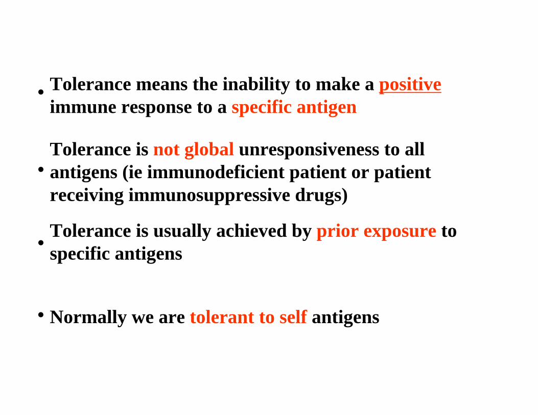
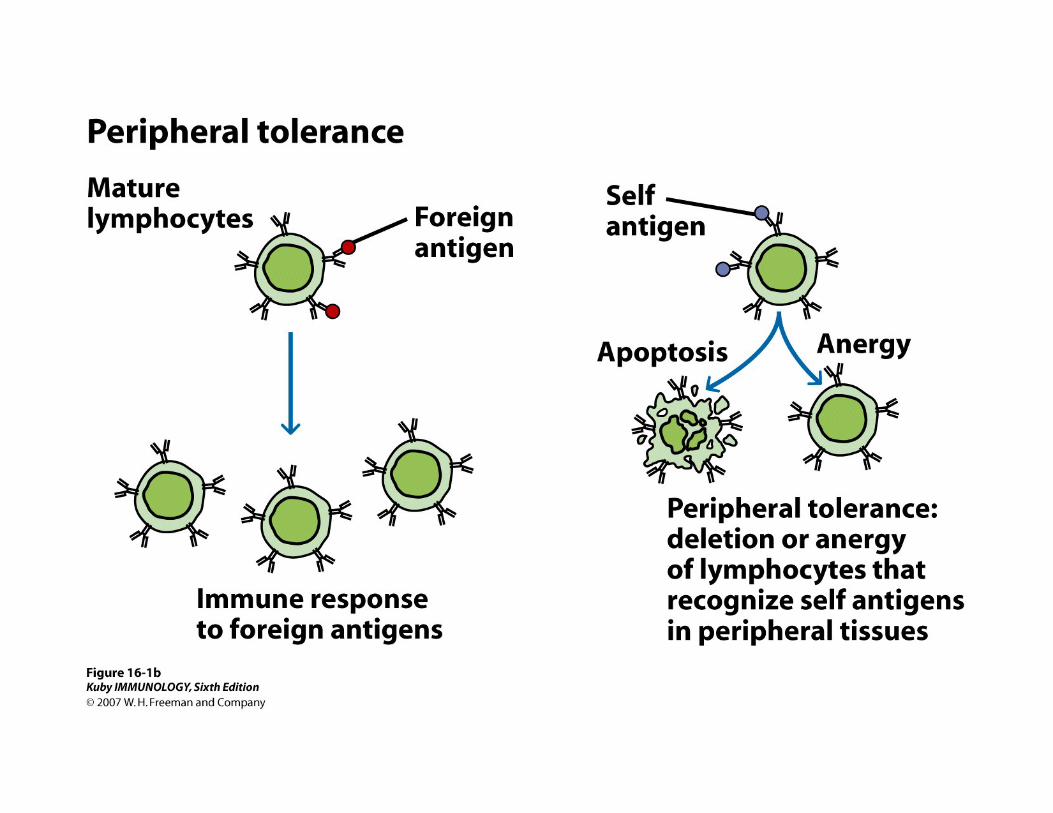
Tolerance means the inability to make a positive immune response to a specific antigen
•
Tolerance is not global unresponsiveness to all antigens (ie immunodeficient patient or patient receiving immunosuppressive drugs)
•
Tolerance is usually achieved by prior exposure to specific antigens•
Normally we are tolerant to self antigens
•


Central Tolerance is Maintained by
Clonal Deletion--removal of antigen reactive cells
Main mechanism is apoptosis: programmed cell death
Consequence of immature self-reactive lymphocytes recognizing self-antigen

B cells reactivate RAG and undergo 2nd V rearrangement
Anergic B cells with downregulated mIg
Central T l
1
2
3

• Normal mice do not have detectable numbers of B cells reactive to hen egg lysozyme (naïve B cells have different BCRs with no exposure to Ag, no clonality)
2.Single transgenic >90% of anti-HEL transgenic mice have mIgM reactive with HEL (no Ag injection required these are naïve B cells)
3. Double transgenic: All B (except those that undergo receptor editing) react to HEL, however HEL Ag is also present in periphery--anergy occurs by downregulation of membrane IgM expression
= Mean Fluorescence Intensity, green shaded area are cells with no reactivity to HEL

Immature B cells Undergo Receptor Edit
Immature B cells that encounter Ag in bone marrow undergo apoptosis UNLESS
• RAG genes are reactivated
• Additional light chain VJ recombination
• New light chain produced
• Different Ig receptor which does not react with Agpresent in bone marrow

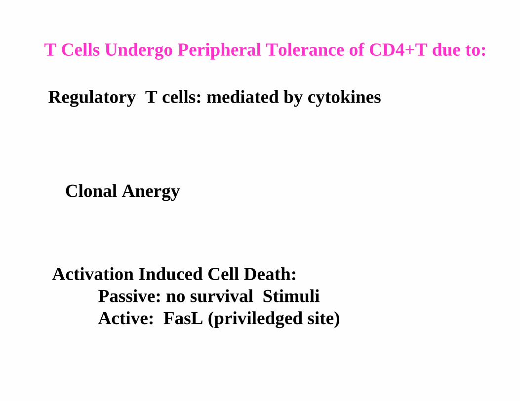
T Cells Undergo Peripheral Tolerance of CD4+T due to:
Regulatory T cells: mediated by cytokines
Activation Induced Cell Death: Passive: no survival StimuliActive: FasL (priviledged site)
Clonal Anergy

Thymic Selection deletes HEL reactive T cells (HEL now self-antigen)

T reg CD4+, CD25+, FOXP3+
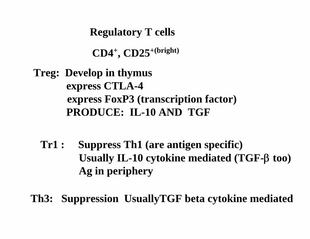
Th3: Suppression UsuallyTGF beta cytokine mediated
Regulatory T cells
Treg: Develop in thymus express CTLA-4express FoxP3 (transcription factor)PRODUCE: IL-10 AND TGF
Tr1 : Suppress Th1 (are antigen specific)Usually IL-10 cytokine mediated (TGF-β too)Ag in periphery
CD4+, CD25+(bright)

Downloaded from: StudentConsult (on 7 April 2006 10:43 PM)© 2005 Elsevier

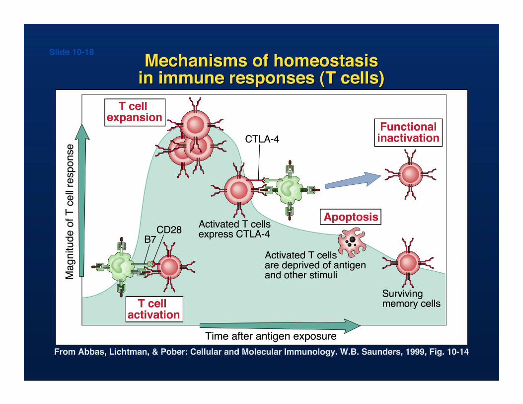
B7 Costimulatory Pathways• B7-2 on APC (constitutive), B7-1 appears later• Receptors…
• CD28 (low affinity-constitutive on T cells, surface expression • CTLA-4 CD152 (high affinity cytoplasmic and upon
stimulation becomes surfaceCD28 mediates:
• T cell proliferation• Induction of bcl-xl• Increase in CD40L (CD154• Differentiation of CD8+CTL• Cytokine production
CTLA-4 (CD152• Induces apoptosis• KO mice have LPD

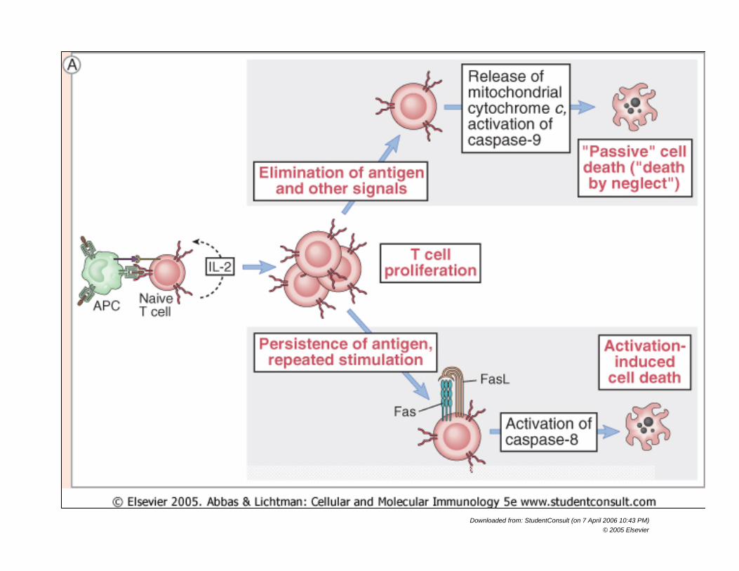
T Cells Undergo Peripheral Tolerance of CD4+T due to:
Activation-Induced Cell Death
Apoptosis: Activation of cysteine proteases, caspases
Not NecrosisTriggered by ligand binding to receptors (Fas, TNFR)Characterized by DNA cleavage
Nuclear fragmentationplasma membrane blebbingphagocytosis of apoptotic bodies
Prevented by inhibitors of caspases (FLIP)activation of Bcl family
Downloaded from: StudentConsult (on 7 April 2006 10:43 PM)© 2005 Elsevier

Factors Involved in Tolerance
Nature of antigen- soluble antigens are better tolerogens
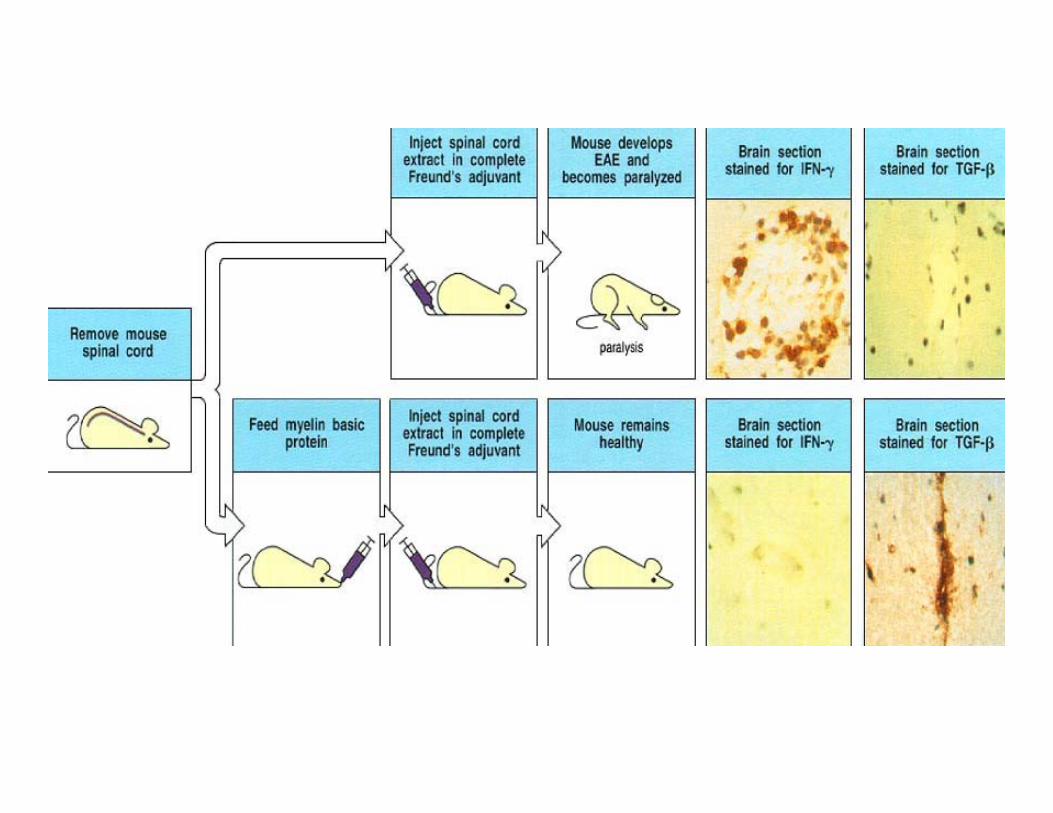
Route of antigen administration--oral good for tolerance
Dose of antigen
Inefficient antigen presentation leads to T cell tolerance
Tolerance is easier to achieve in newborns


Autoimmunity
Describe Mechanisms responsible for autoimmune damage
Name autoimmune diseases and major self-antigen
Autoimmunity is a breakdown in toleranceImmune hyperactivity, self recognitionNormally, tolerant to self antigens

Autoimmune Diseases can by Systemic or Organ-Specific

Overall Mechanism• Autoimmune diseases results from breakdown of
self-tolerance in B cells, or T cells, or both.
• Genetic, hormonal and environmental factors or infectious agents may contribute to the development of autoimmune diseases.
• Damage may be due to immune complexes, circulating autoantibodies, and/or autoreactive T
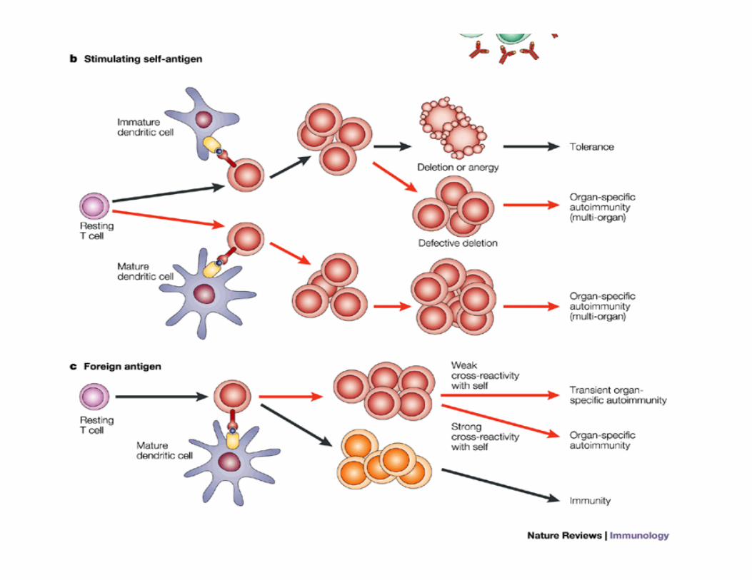
• Once initiated, autoimmune reactions may injure tissues and cause the release and alteration of other tissue antigens resulting in activation of lymphocytes specific for these other antigens.
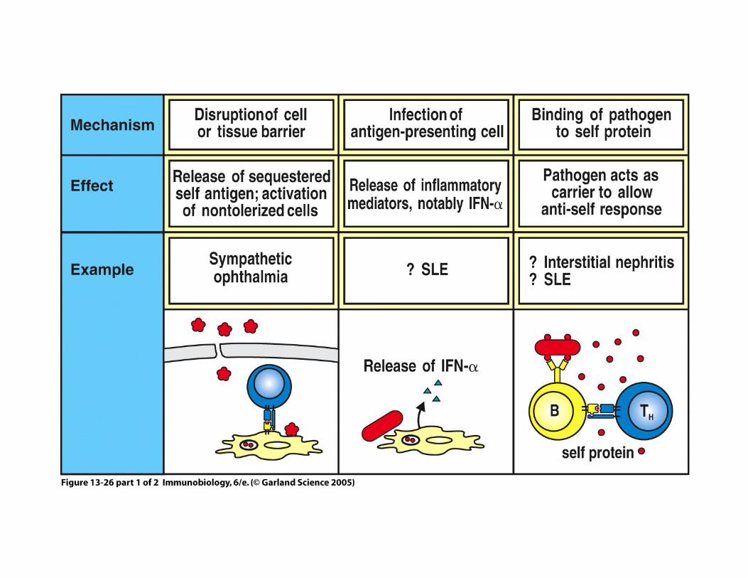
Figure 13-26 part 1 of 2
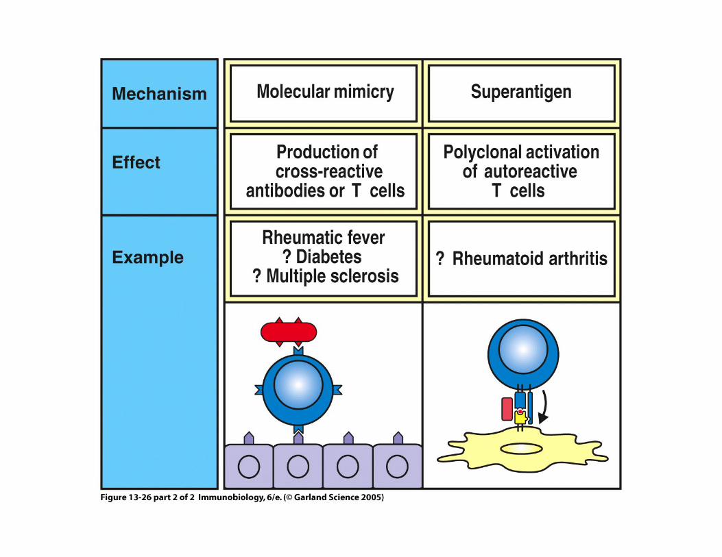
Figure 13-26 part 2 of 2

Type IV


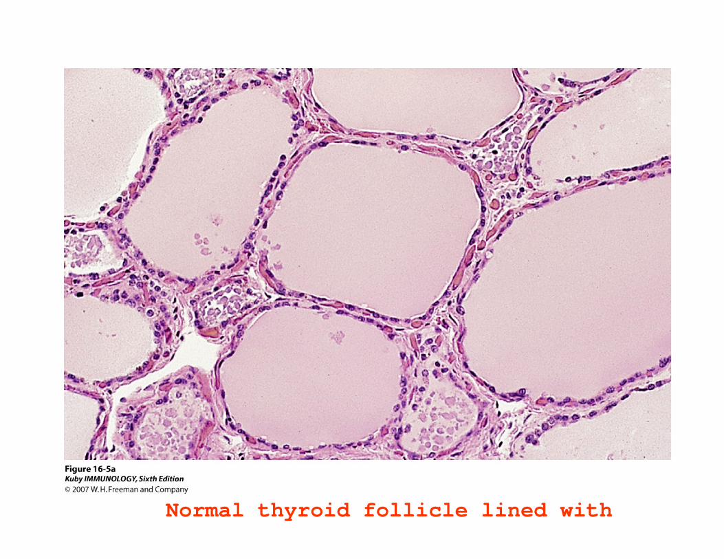
Normal thyroid follicle lined with

Thyroid follicle in Hashimoto’s thyroiditis with l h t i filt ti

Mechanisms ofAb deposition
Effector mechanismsof tissue injury
Abbas 18-1B
Type III hypersensitivity


Possible Causes of Immune Complex Deposition
• Size of complex - small complexes are not always phagocytosed & can be deposited in vessels.
• Charge - cationic antigens bind to negatively charged components of the basement membranes of blood vessels and kidney glomeruli.
• Sites of high hydrostatic pressure (kidney).• Following activation of inflammatory cells and mast cells - cytokines
& vasoactive mediators are released leading to increased adhesion of leukocytes to endothelium, increased vascular permeability and enlarged interendothelial spaces allowing deposition of complexes.

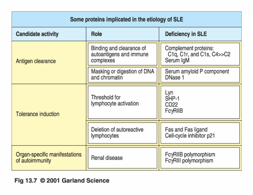
Pathologic features of antibody-mediated glomerulonephritis
Abbas 18-3Goodpasture’s Systemic Lupus Erythematosus (SLE)

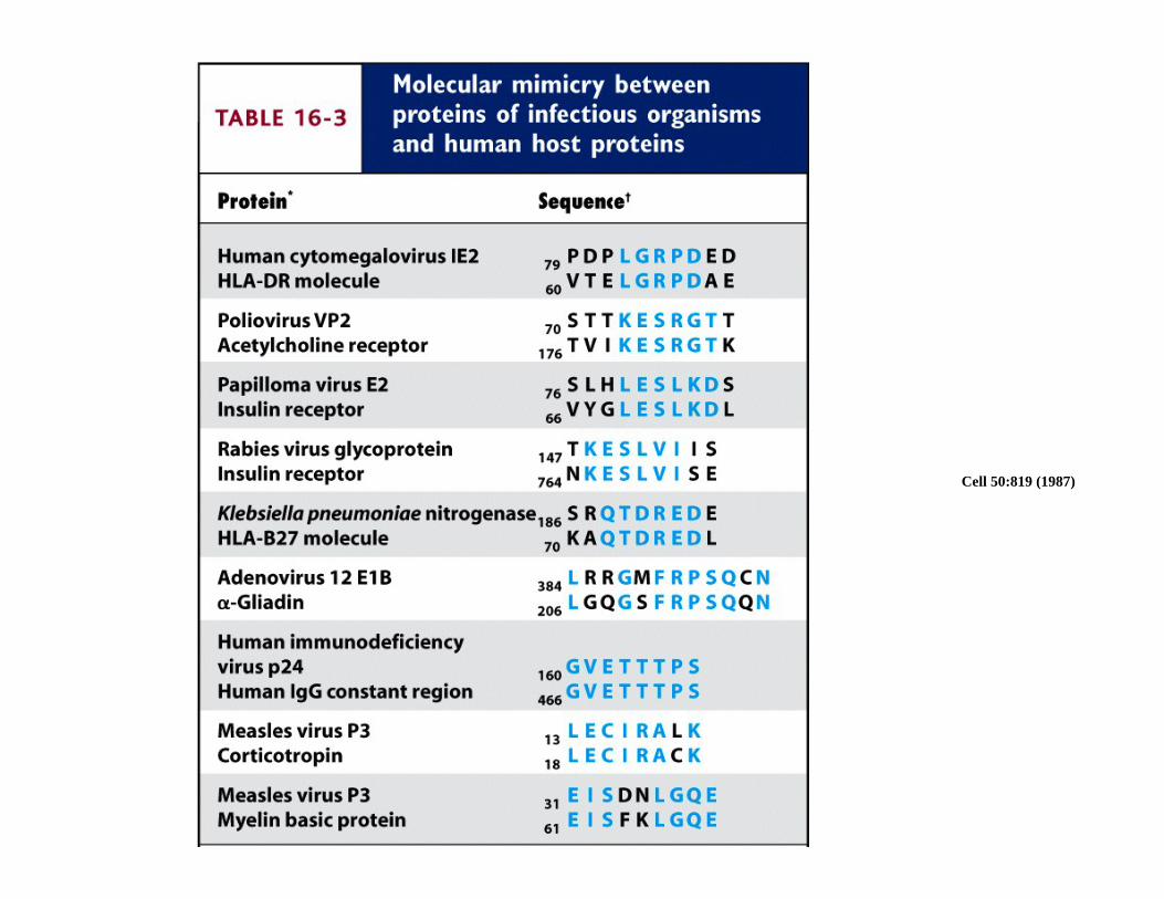
Cell 50:819 (1987)


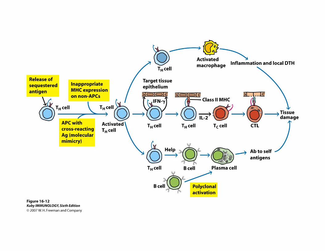
Mechanism of Autoimmunity
Failure of Central ToleranceThymic Selection is Flawed
Peripheral Tolerance DefectiveFailure of activation induced Cell death --AICD
Hyper Immune responseCytokine ImbalanceAbnormal expression of co-stimulatory molecules
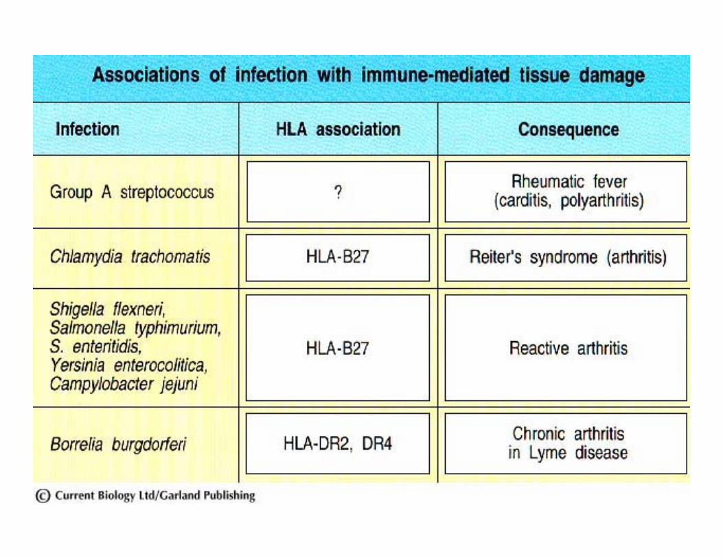
Cross-Reactivity


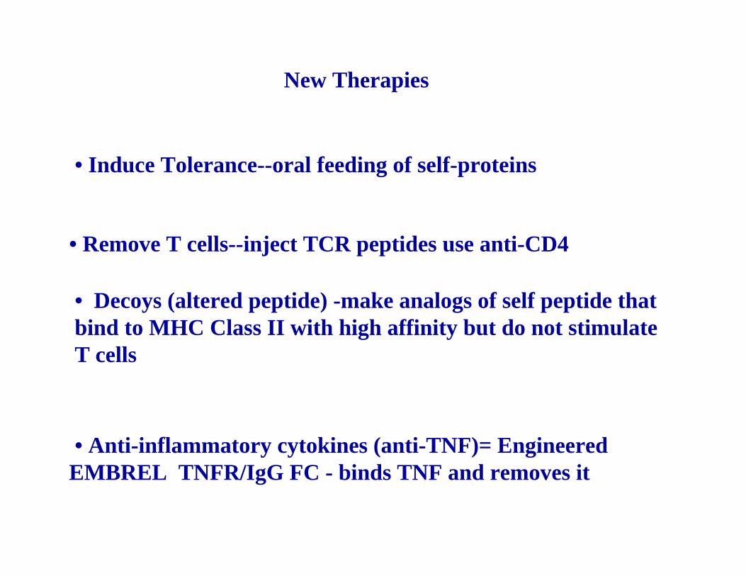
New Therapies
• Decoys (altered peptide) -make analogs of self peptide that bind to MHC Class II with high affinity but do not stimulate T cells
• Anti-inflammatory cytokines (anti-TNF)= Engineered EMBREL TNFR/IgG FC - binds TNF and removes it
• Remove T cells--inject TCR peptides use anti-CD4
• Induce Tolerance--oral feeding of self-proteins


![RESEARCH Open Access...the treatment of autoimmunity [7]. Therefore, vaccine platforms are needed that induce robust tolerance and are efficacious in inflammatory environments. Vaccine](https://static.fdocuments.in/doc/165x107/60b9140531f82a5c661f0a3f/research-open-access-the-treatment-of-autoimmunity-7-therefore-vaccine-platforms.jpg)