Toktam Faghihi Pharm - clinicalpharmacy.ir otitis... · tympanosclerosis (scars), atrophic areas,...
63
Toktam Faghihi Pharm.D
Transcript of Toktam Faghihi Pharm - clinicalpharmacy.ir otitis... · tympanosclerosis (scars), atrophic areas,...
Toktam Faghihi
Pharm.D
Acute otitis media (AOM), also called suppurative
otitis media, is one of the most frequent diagnoses for
children seeking acute medical care.
Acute otitis media (AOM)
Middle ear effusion (MEE)
Otitis media with effusion (OME)
Acute otitis media (AOM): acute bacterial infection of middle ear fluid.
Middle ear effusion (MEE): fluid in the middle ear cavity.
MEE occurs in both otitis media with effusion (OME) and AOM.
Otitis media with effusion (OME): middle ear fluid that is not infected.
OME is also called serous, secretory, or nonsuppurative otitis media.
OME frequently precedes the development of AOM or follows its resolution.
The distinction between OME and AOM may be difficult, since they are part of a continuous spectrum
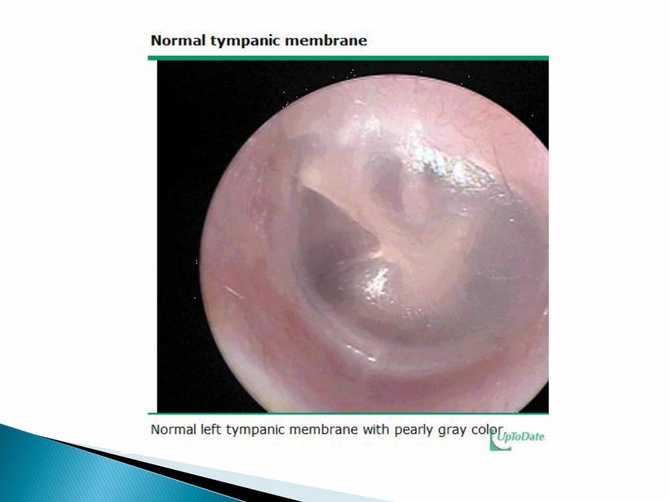
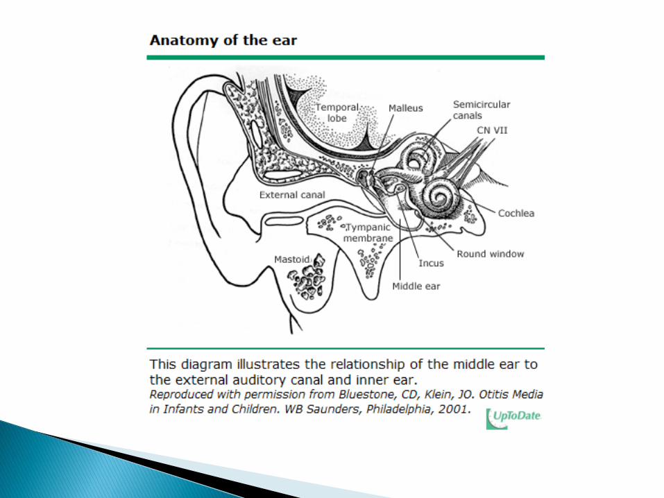
The normal middle ear is aerated, and the tympanic
membrane is slightly convex, translucent, mobile, and
intact
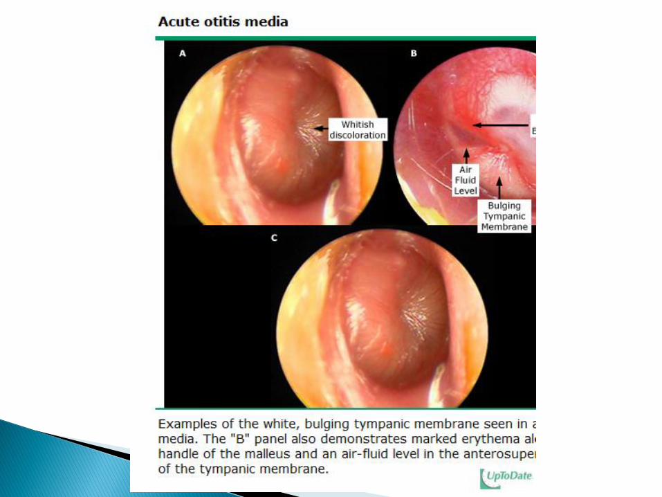
In AOM: the middle ear is fluid-filled, and the tympanic
membrane is usually bulging, erythematous, cloudy, and
immobile.
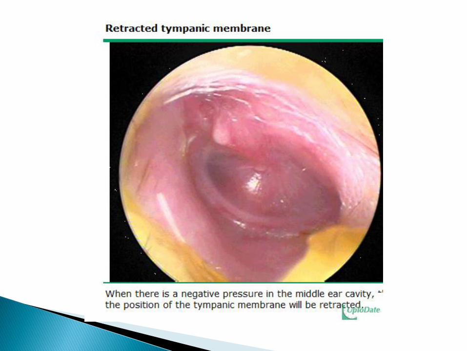
Increased pressure in the middle ear may lead to central
ischemia, necrosis, and perforation of the tympanic membrane.
Accurate diagnosis of AOM requires systematic evaluation of the tympanic membrane for position, translucency, mobility, color, and other findings (eg, fluid level, perforation).
Systematic assessment of the tympanic membrane is facilitated by the use of the COMPLETES mnemonic:
Color (eg, gray, white, pale yellow, amber, pink, red, blue)
Other conditions (eg, fluid level, bubbles, perforation, otorrhea, bullae, tympanosclerosis (scars), atrophic areas, retraction pockets, cholesteatoma)
Mobility
Position (eg, neutral, retracted, full, or bulging)
Lighting (a halogen light source and fully charged battery should be used)
Entire surface (the four quadrants of the tympanic membrane should be examined)
Translucency
External auditory canal and auricle (eg, deformed, displaced, inflamed, foreign body)
Seal (a good seal requires an airtight pneumatic system and a speculum that is large enough to prevent air leak)
The clinical diagnosis of AOM requires:
MEEand
acute history, signs and symptoms of middle ear inflammation
A diagnosis of AOM also can be established if there is acute purulent otorrhea and otitis externa has been excluded.
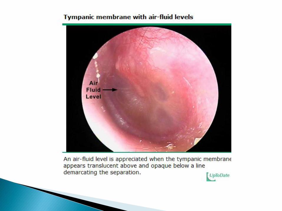
MEE can be confirmed by one or both of the following findings on otoscopy:
Bubbles or an air-fluid level
Two or more of the following:
Abnormal color (white, yellow, amber, or blue)
Opacity (involving part or all of the tympanic membrane) not due to scarring
Impairment of mobility
MEE is necessary but not sufficient for a diagnosis of AOM; there also must be evidence of acute inflammation.
If a child has MEE but no evidence of acute inflammation, he or she has OME.
Signs of acute inflammation are necessary to differentiate AOM from OME.
Otoscopic signs: Distinct fullness or bulging of the tympanic membrane: the
best and most reproducible sign Marked redness
Non-otoscopic symptoms:
Ear pain (Otalgia)
Unaccustomed tugging or rubbing of the ear
Otorrhea or ear discharge or swelling about the ear (which may indicate disease of the mastoid) are specific physical findings, if present.
Nonspecific symptoms and signs include fever, irritability, headache, apathy, anorexia, vomiting, and diarrhea.
Young children, especially infants, are more likely to present with nonspecific than specific symptoms
Non-otoscopic symptoms must be accompanied by
abnormal otoscopic findings as described above to
make a diagnosis of AOM.
As an example, a child who complains of ear pain may
be diagnosed with AOM if he or she also has a white or
yellow tympanic membrane with marked decrease in
mobility
The diagnosis of AOM requires evidence of an acute
history, signs and symptoms of middle ear
inflammation (distinct erythema of the tympanic
membrane or otalgia),
AND
the presence of middle ear effusion (eg, tympanic
membrane bulging, decreased or absent tympanic
membrane mobility, presence of an air-fluid level, or
otorrhea)
The main consideration in the differential diagnosis of AOM is OME.
Otitis media with effusion — MEE, with decreased mobility and opacification or cloudiness of the tympanic membrane, occurs in both AOM and OME.
However, careful evaluation of the position, color, and other findings of the tympanic membrane can help to distinguish AOM from OME.
In AOM, the tympanic membrane is usually bulging; in OME, it is usually retracted or in the neutral position.
In AOM, the tympanic membrane is typically red, white, or pale yellow; in OME, it is typically amber or blue.
,….
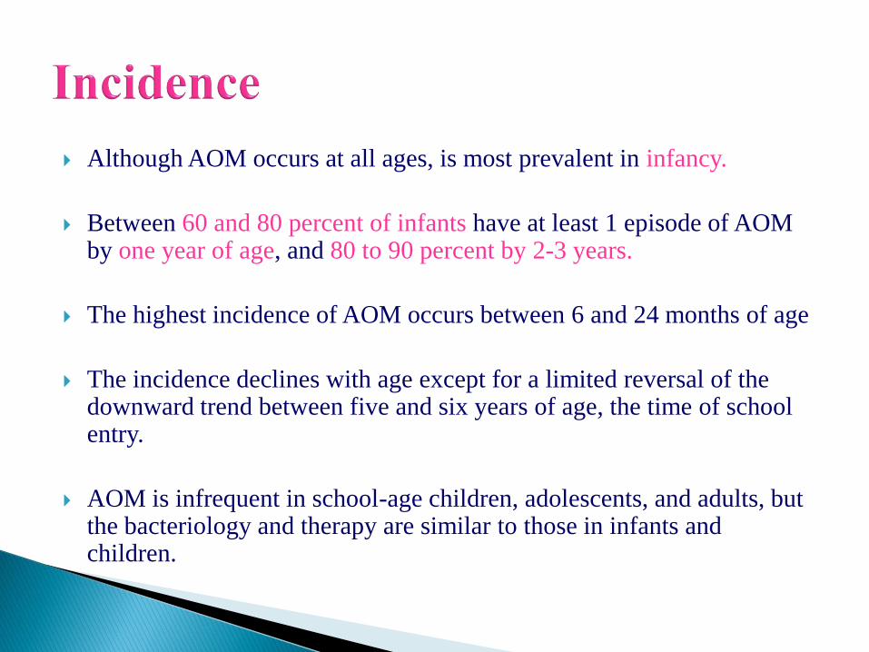
Although AOM occurs at all ages, is most prevalent in infancy.
Between 60 and 80 percent of infants have at least 1 episode of AOM by one year of age, and 80 to 90 percent by 2-3 years.
The highest incidence of AOM occurs between 6 and 24 months of age
The incidence declines with age except for a limited reversal of the downward trend between five and six years of age, the time of school entry.
AOM is infrequent in school-age children, adolescents, and adults, but the bacteriology and therapy are similar to those in infants and children.
young age,
day care attendance,
not having been breast fed (Breast feeding for at least three
months)
exposure to tobacco smoke,
pacifier use,
ethnicity (Native Americans and Alaskan and Canadian
Eskimos)
family history
Others: season, underlying diseases (allergic rhinitis)
Age
The occurrence of disease early in life is probably a result
of a number of factors, including immature anatomy,
physiology, and immunologic responses.
Some of these factors are identifiable (eg, the change in
skull configuration and vectors of the eustachian tube,
development of antibodies following exposure to bacterial
pathogens)
The patient has an antecedent event (usually an upper respiratory tract
viral infection or allergy).
The event results in congestion of the respiratory mucosa of the nose, nasopharynx and eustachian tube.
Congestion of the mucosa in the eustachian tube obstructs the narrowest portion of the tube, the isthmus.
Obstruction of the isthmus causes negative pressure followed by accumulation of secretions produced by the mucosa of the middle ear.
Viruses and bacteria that colonize the upper respiratory tract can reach the middle ear via aspiration, reflux, or insufflation.
Microbial growth in the middle ear secretions may result in suppuration with clinical signs of AOM.
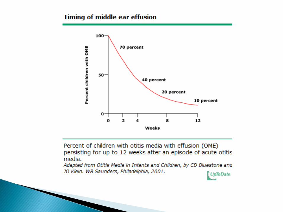
I. Fluid may persist for weeks to months after the onset of signs of AOM despite treatment with appropriate antimicrobial agents.
Whenever fluid fills the middle ear space, there is some loss of hearing that may lead to problems of development of speech, language, and cognitive abilities in the child.
II. Extension of the suppurative process to adjacent structures may lead to complications such as mastoiditis, labyrinthitis, meningitis, and lateral sinus thrombosis,cavernous sinus thrombosis, subdural empyema, and carotid artery thrombosis.
III. Tympanic membrane perforation:
because the pressure of the middle ear abscess on the membrane leads to central ischemia, necrosis, and perforation.
AOM caused by group A streptococci (GAS) is associated with higher rates of tympanic membrane perforation than AOM caused by other pathogens
Persistent or fluctuating hearing loss is present in most patients with middle ear effusion.
The median loss is 25 dB, which is equivalent to putting plugs in the patient's ears.
Some studies have noted that children with prolonged time spent with middle ear effusion have lower scores on tests of speech, language, and cognitive abilities.
In addition to hearing impairment, patients with otitis media may suffer vestibular, balance, and motor dysfunctions
Children in developing areas — Lack of access to
medical care and local environmental factors lead to
severe suppurative episodes of otitis media in children
living in developing areas.
The prevalence of perforated eardrums in children aged
4 to 12 months living in an aboriginal settlement in
Australia was 25 percent.
Three species of bacteria account for most of the
bacterial isolates from middle ear fluid:
Streptococcus pneumoniae
Haemophilus influenzae
Moraxella catarrhalis
The most common viral pathogens include respiratory
syncytial virus, rhinoviruses, influenza viruses, and
adenoviruses.
Although the specificity of ear-related findings in the
diagnosis of otitis media is poor, some clinical features
do correlate with a particular organism
Fever ≥38ºC and earache were reported more
frequently in patients with pneumococcus (44 percent
with pneumococcus compared with 25 percent with H.
influenzae and 26 percent with Moraxella)
Eye symptoms, primarily conjunctivitis, were identified
more often in patients with H. influenzae (54 percent
with H. influenzae compared with 15 percent with
pneumococcus and 18 percent with Moraxella)
There was no difference in otologic findings among the
three pathogens.
The systemic and local signs and symptoms of AOM usually resolve in 24 to 72 hours with appropriate antimicrobial therapy, and somewhat more slowly in children who are not treated.
However, persistence of middle ear effusion (MEE)after the resolution of acute symptoms is common.
MEE is associated with conductive hearing loss.
Prolonged presence of middle ear fluid may lead to the
mistaken diagnosis of AOM in patients with a
subsequent illness resulting from other causes.
Pain remedies
Guidelines issued in 2004 by the American Academy of
Pediatrics (AAP) and the American Academy of Family
Physicians (AAFP) state that the management of AOM
should include an assessment of pain and treatment if
pain is present
Ibuprofen (10 mg/kg three times per day)
Acetaminophen (10 mg/kg three times per day)
An oral decongestant, such as pseudoephedrine, may
relieve nasal congestion, and antihistamines may help
patients with known or suspected nasal allergy.
However, the efficacy of antihistamines and
decongestants in treating AOM has not been proven.
In addition, treatment with antihistamines may prolong
the duration of middle ear effusion.
The American Academy of Pediatrics (AAP)
recommends that over-the-counter cough and cold
medications should not be given to infants and children
younger than two years of age, due to the risk of life-
threatening side effects
Antibiotic Therapy VERSUS Observation
The AAP/AAFP 2004 guideline suggests that observation
without use of antibacterial therapy is an option for selected
children with uncomplicated AOM based upon diagnostic
certainty, age, illness severity, and assurance of follow-up
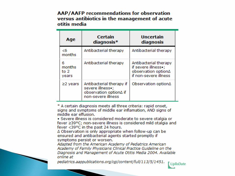
Antibacterial therapy should be administered to any child younger than the age of six months, regardless of the degree of diagnostic certainty.
For children ages six months to two years, antibacterial therapy is recommended when the diagnosis of AOM is certain or if the diagnosis is uncertain but illness is severe (moderate to severe otalgia or fever ≥39ºC in the previous 24 hours). Observation is an option for children in whom the diagnosis is not certain and illness is not severe.
For children older than two years, antibacterial therapy is recommended if the diagnosis is certain and illness is severe. Observation is an option when the diagnosis is certain but illness is not severe, and in patients with an uncertain diagnosis.
The AAP/AAFP 2004 guidelines underline the difference in approach to therapy of the child younger or older than two years of age.
The older child has fewer episodes of middle ear infection and less severe disease than the infant age group.
The differences in disease incidence and severity due to age likely occur because of changes in the anatomy and physiology of the middle ear system and the increase in immune protection against middle ear pathogens.
Children older than two years who have bilateral disease or otorrhea are best managed with antimicrobial therapy.
For those who are normal hosts and who have mild symptoms and signs of unilateral AOM, the watchful waiting option with appropriate follow-up may be appropriate
Clinical and microbiologic efficacy
Acceptability (taste, texture) of the oral preparation
Absence of side effects and toxicity
Convenience of the dosing schedule
Cost
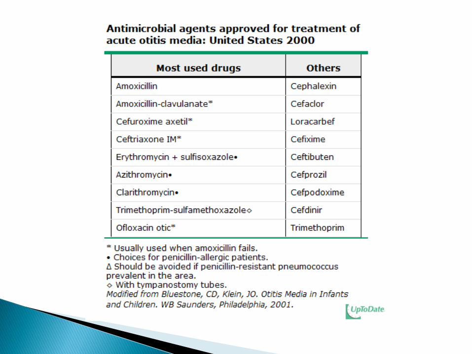
Seventeen antimicrobial drugs (16 oral and one parenteral preparation) have been approved by the US Food and Drug Administration (FDA) for the treatment of AOM.
In addition, two otic preparations (eg, ofloxacin otic and ciprofloxacin-dexamethasone otic) also are available for treatment of AOM with otorrhea in children with tympanostomy tubes in place or tympanic membrane perforation.
First-line therapy
A 2001 meta-analysis concluded there is no evidence to
support any particular antibiotic regimen versus another for
treatment of AOM.
Nevertheless, amoxicillin remains the drug of choice
because it is effective, safe, relatively inexpensive, and has
a narrow microbiologic spectrum.
The AAP/AAFP guideline recommends a dose of
amoxicillin of 80 to 90 mg/kg per day.
For heavier children, we suggest a maximum dose of 3
g/day, although diarrhea is a potential adverse effect at
higher doses.
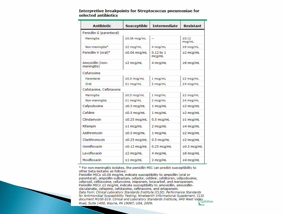
Doubling the dose from 40 to 80 mg/kg per day provide
activity against most intermediate strains of S. pneumoniae,
including many of the resistant strains.
Only S. pneumoniae that are highly resistant to penicillin
will not respond to this regimen.
As a result, more than 80 percent of children with
pneumococcal AOM would respond to high-dose
amoxicillin treatment.

Despite the increasing importance of H. influenzae,
including beta-lactamase-producing strains, high-dose
amoxicillin remains the preferred choice for initial
therapy.
Amoxicillin should not be used as first-line therapy in
children who are at high risk for AOM caused by an
amoxicillin-resistant otopathogen:
1. Children who were treated with antibiotics in the previous 30 days,
particularly beta-lactam antibiotics.
2. Children with concurrent purulent conjunctivitis (otitis-conjunctivitis
syndrome usually is caused by nontypeable H. influenzae, which is
frequently resistant to beta-lactam antibiotics)
3. Children receiving amoxicillin for chemoprophylaxis of recurrent
AOM (or urinary tract infection)

Children in the above categories should start therapy
with an agent with activity against beta-lactamase-
producing nontypeable H. influenzae, as well as S.
pneumoniae, such as amoxicillin-clavulanate
Penicillin allergy — Acceptable alternatives to penicillin in patients with allergy to penicillin depend upon the type of the previous hypersensitivity reaction
Non-type 1 reactions — In patients who report penicillin allergy but who did not experience a type 1 hypersensitivity reaction (urticaria or anaphylaxis), we suggest one of the following:
Cefdinir (14 mg/kg per day in 1 or 2 doses; maximum dose 600 mg/day)
Cefpodoxime (10 mg/kg per day once daily; maximum dose 800 mg/day)
Cefuroxime (cefuroxime axetil suspension: 30 mg/kg per day in two divided doses, maximum dose 1 g/day; cefuroxime tablets: 250 mg every 12 hours)
However, these oral agents do not achieve sufficient concentration in the middle ear to eradicate penicillin-resistant S. pneumoniae.
A single intramuscular dose of ceftriaxone (50 mg/kg)
provides high concentrations in the middle ear for more
than 48 hours and may be considered an alternative for
children with AOM and history of non-type 1 penicillin
allergy.
If clinical signs improve within 48 hours following
administration of ceftriaxone, no further therapy is
necessary.
If clinical signs persist, a second dose is administered
and, if necessary, a third dose.
Type 1 reactions
Macrolide antibiotics can be used for patients who have had a type 1 hypersensitivity reaction to amoxicillin or other beta-lactam antimicrobial agents.
However, macrolide resistance is common (approximately 35 percent) among isolates of S. pneumoniae (lack of activity against approximately one-third of pneumococcal isolates)
Macrolides lack of activity against most Haemophilus influenzae isolates.
Available macrolide drugs approved for AOM include:
Erythromycin plus sulfisoxazole, Clarithromycin Azithromycin
Erythromycin plus sulfisoxazole (50 to 150 mg/kg per day of the erythromycin component divided into 4 doses; maximum dose 2g erythromycin or 6 g sulfisoxazole/day) may be the most effective of these regimens but is rejected often by patients based upon taste and frequency of dosing
Five days of azithromycin (10 mg/kg per day
[maximum dose 500 mg/day] as a single dose on day
one and 5 mg/kg per day [maximum dose 250 mg/day]
for days two through five) or
clarithromycin (15 mg/kg per day divided into 2 doses;
maximum dose 1 g/day) can be used,
but resistant pneumococcal isolates cannot be
overcome by increasing the dose of macrolides, unlike
the scenario with beta-lactam drugs.
Trimethoprim-sulfamethoxazole (TMP-SMX) should
be used with caution because of:
1) The presence of resistant pneumococci and may be
useful only in regions where pneumococcal resistance
to this combination is not a concern.
2) In addition, TMP-SMX should not be used if group A
streptococcus (GAS, S. pyogenes) is suspected (eg,
when there is an associated tympanic membrane
perforation.
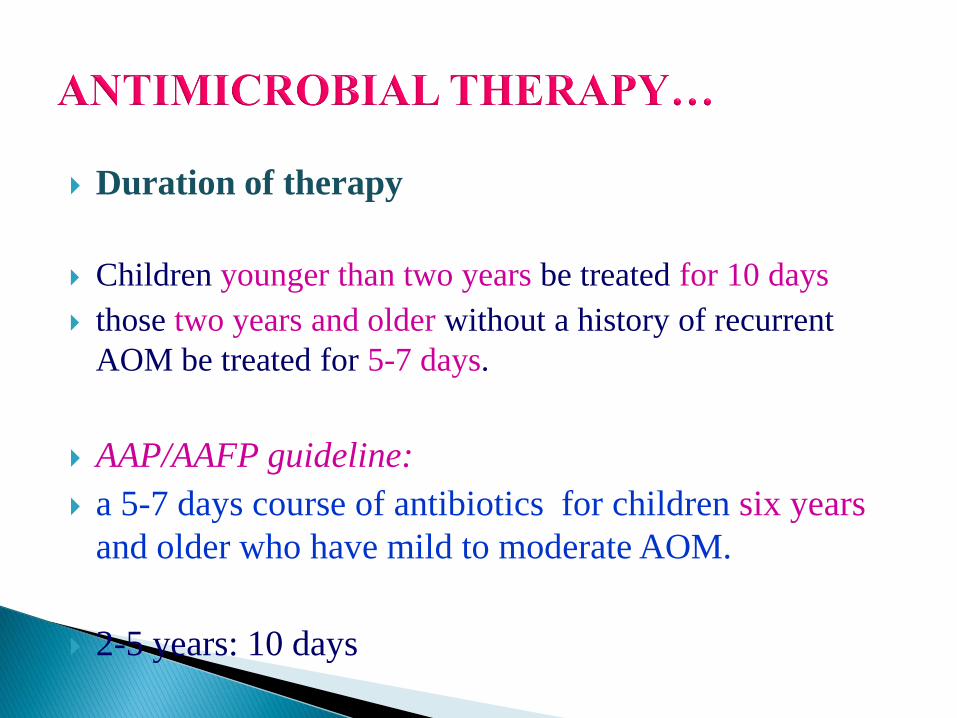
Duration of therapy
Children younger than two years be treated for 10 days
those two years and older without a history of recurrent
AOM be treated for 5-7 days.
AAP/AAFP guideline:
a 5-7 days course of antibiotics for children six years
and older who have mild to moderate AOM.
2-5 years: 10 days
Treatment failure
Recurrent AOM
Tympanic membrane perforation
FOLLOW-UP
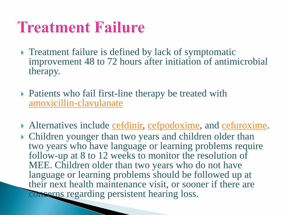
Treatment failure is defined by lack of symptomatic improvement 48 to 72 hours after initiation of antimicrobial therapy.
Patients who fail first-line therapy be treated with amoxicillin-clavulanate
Alternatives include cefdinir, cefpodoxime, and cefuroxime. Children younger than two years and children older than
two years who have language or learning problems require follow-up at 8 to 12 weeks to monitor the resolution of MEE. Children older than two years who do not have language or learning problems should be followed up at their next health maintenance visit, or sooner if there are concerns regarding persistent hearing loss.
Require follow-up at 8 to 12 weeks to monitor the resolution of MEE:
Children ≤ 2 yrs children > 2 yrs who have language or learning problems
Children older than two years who do not have language or learning problems should be followed up at their next health maintenance visit, or sooner if there are concerns regarding persistent hearing loss.