Today’s guest speaker
67
Transcript of Today’s guest speaker
Today’s guest speaker
2
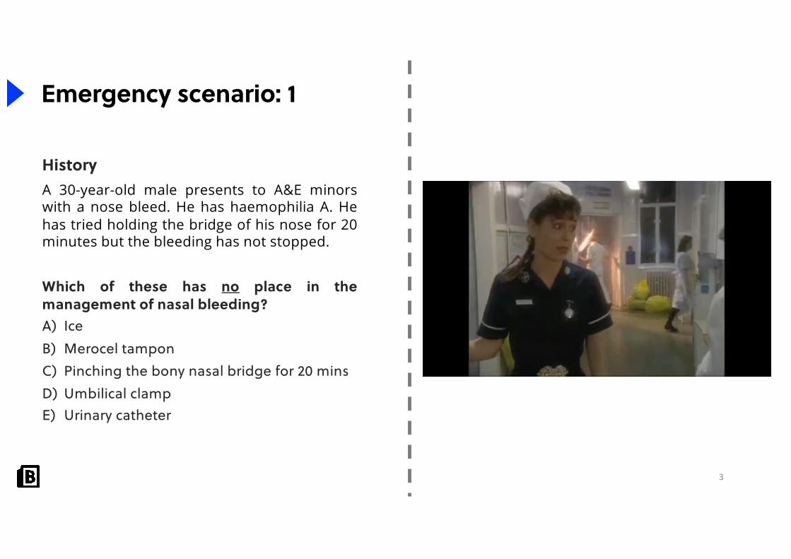
HistoryA 30-year-old male presents to A&E minorswith a nose bleed. He has haemophilia A. Hehas tried holding the bridge of his nose for 20minutes but the bleeding has not stopped.
3
Emergency scenario: 1
PLEASE INSERT IMAGE HERE (if appropriate)
Which of these has no place in themanagement of nasal bleeding?A) IceB) Merocel tamponC) Pinching the bony nasal bridge for 20 minsD) Umbilical clampE) Urinary catheter
• Common ENT emergency
• Common condition – 10% of the population
• Epistaxis may be fatal
4
Epistaxis (Nosebleeds)
5
Epistaxis
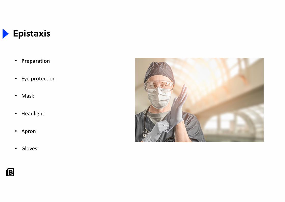
• Preparation
• Eye protection
• Mask
• Headlight
• Apron
• Gloves
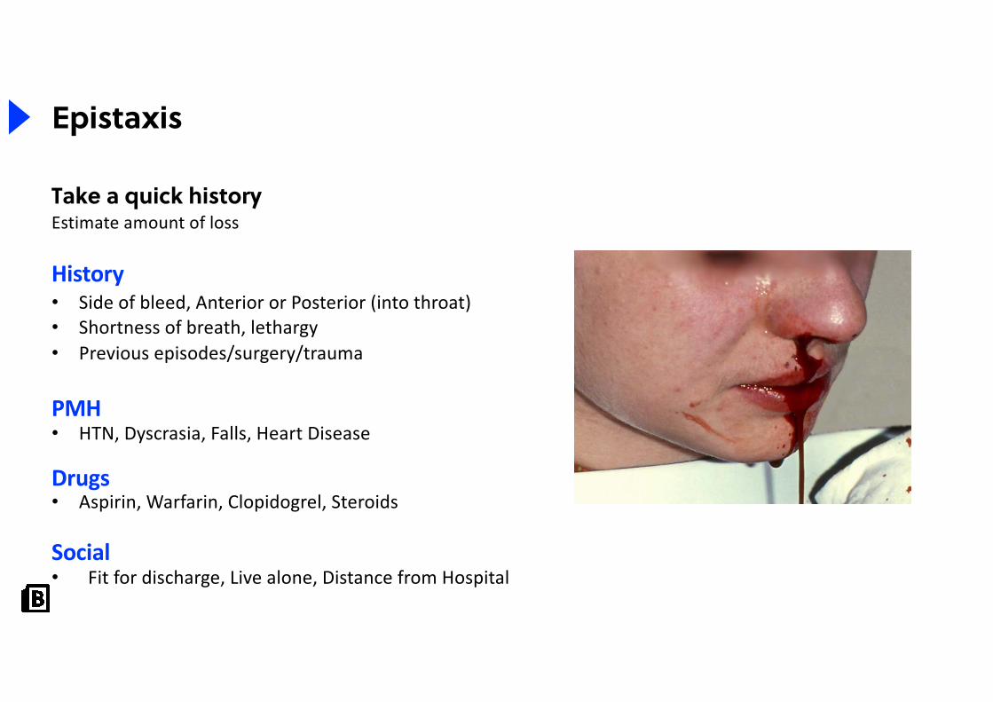
Take a quick historyEstimate amount of loss
History• Side of bleed, Anterior or Posterior (into throat)• Shortness of breath, lethargy• Previous episodes/surgery/trauma
PMH• HTN, Dyscrasia, Falls, Heart Disease
Drugs• Aspirin, Warfarin, Clopidogrel, Steroids
Social• Fit for discharge, Live alone, Distance from Hospital 6
Epistaxis
your name
Epistaxis Management
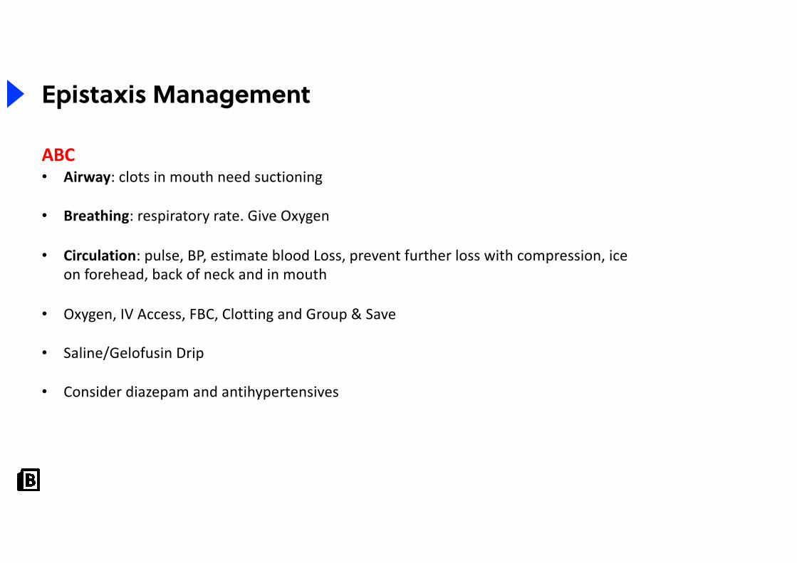
ABC• Airway: clots in mouth need suctioning
• Breathing: respiratory rate. Give Oxygen
• Circulation: pulse, BP, estimate blood Loss, prevent further loss with compression, ice on forehead, back of neck and in mouth
• Oxygen, IV Access, FBC, Clotting and Group & Save
• Saline/Gelofusin Drip
• Consider diazepam and antihypertensives
your name
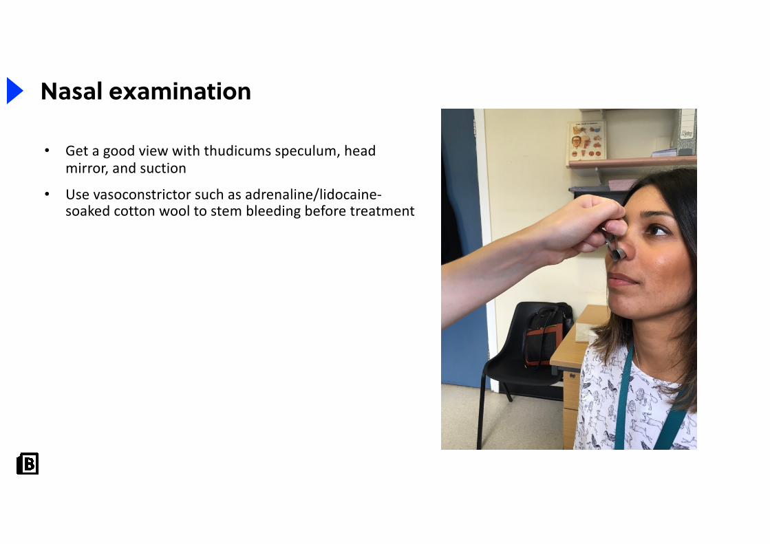
Nasal examination
• Get a good view with thudicums speculum, head mirror, and suction
• Use vasoconstrictor such as adrenaline/lidocaine-soaked cotton wool to stem bleeding before treatment

your name
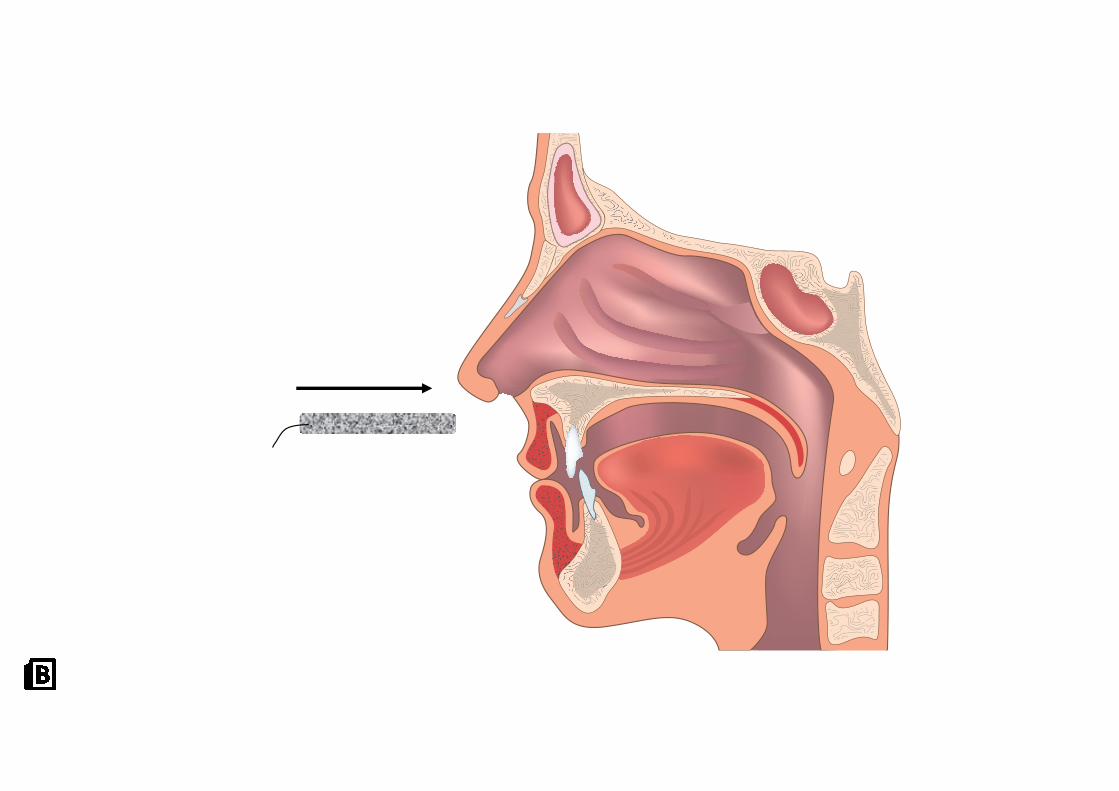
Silver Nitrate Cautery
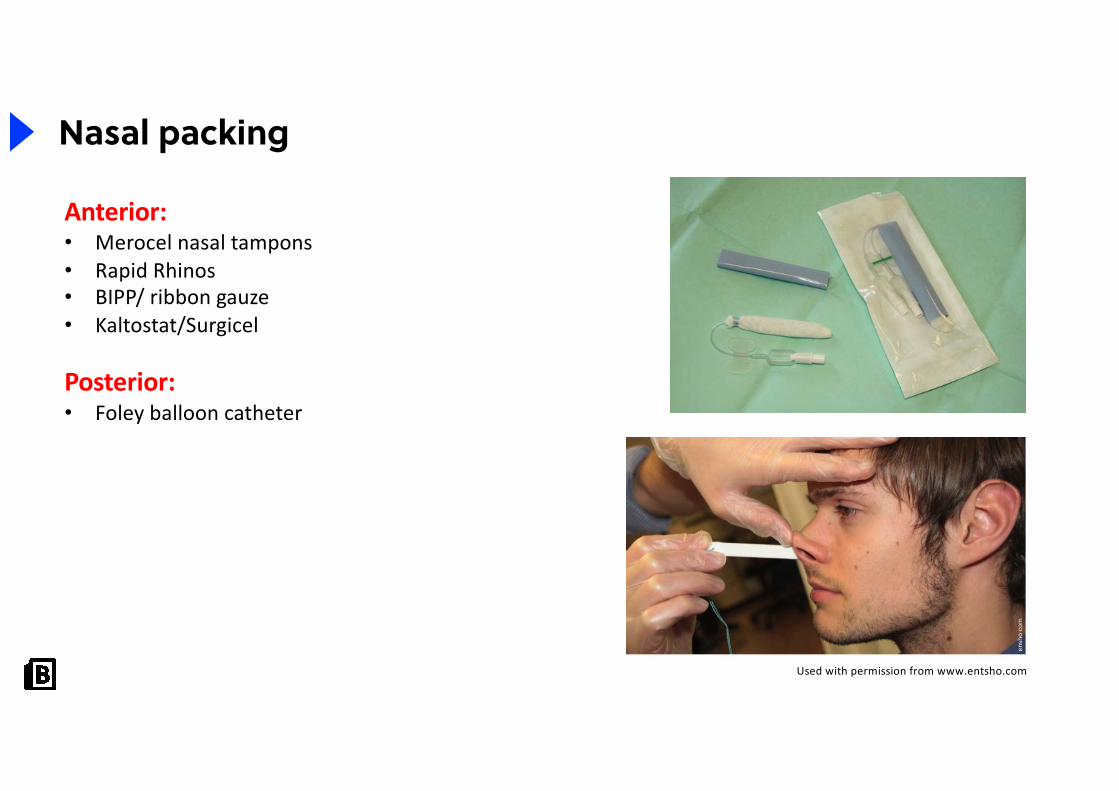
Nasal packing
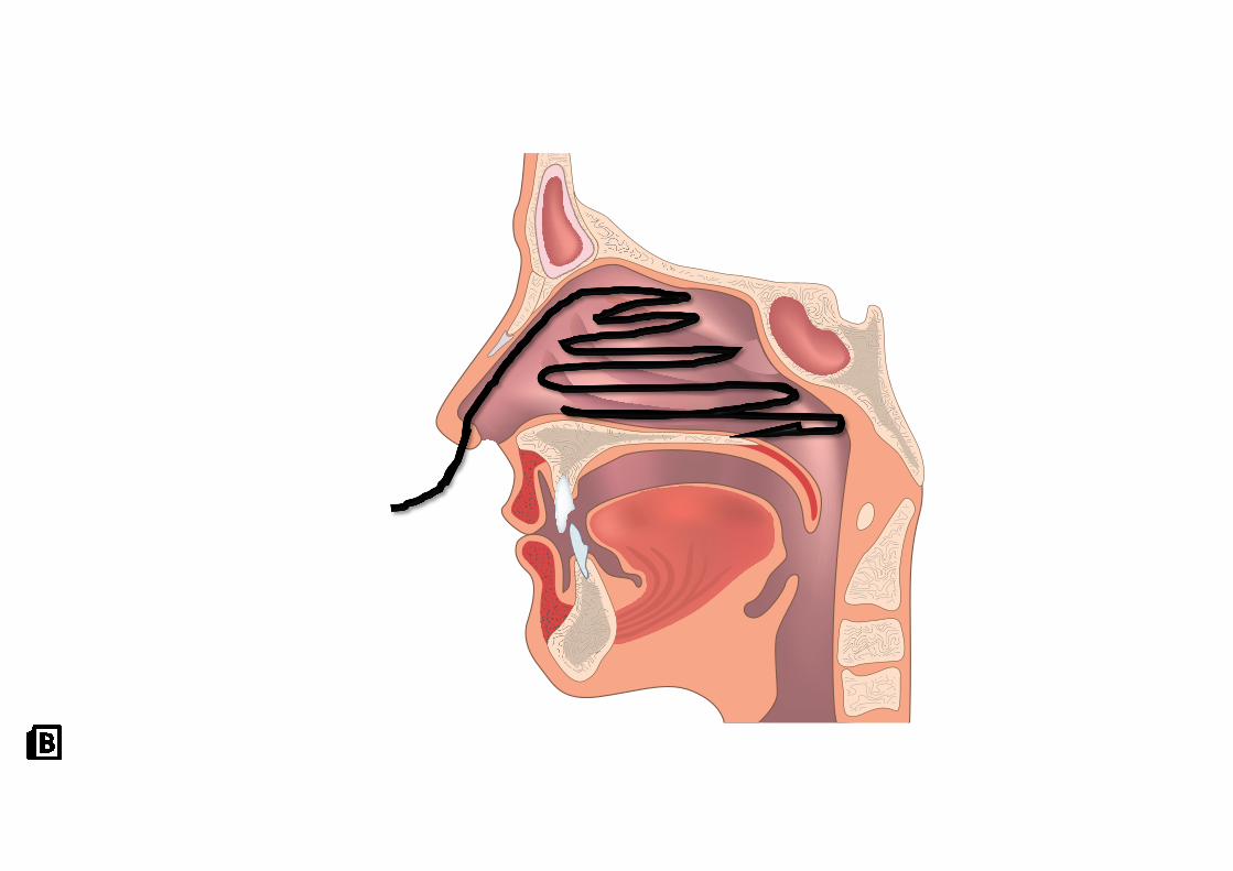
Anterior:• Merocel nasal tampons• Rapid Rhinos• BIPP/ ribbon gauze• Kaltostat/Surgicel
Posterior:• Foley balloon catheter
Used with permission from www.entsho.com
your name
your name
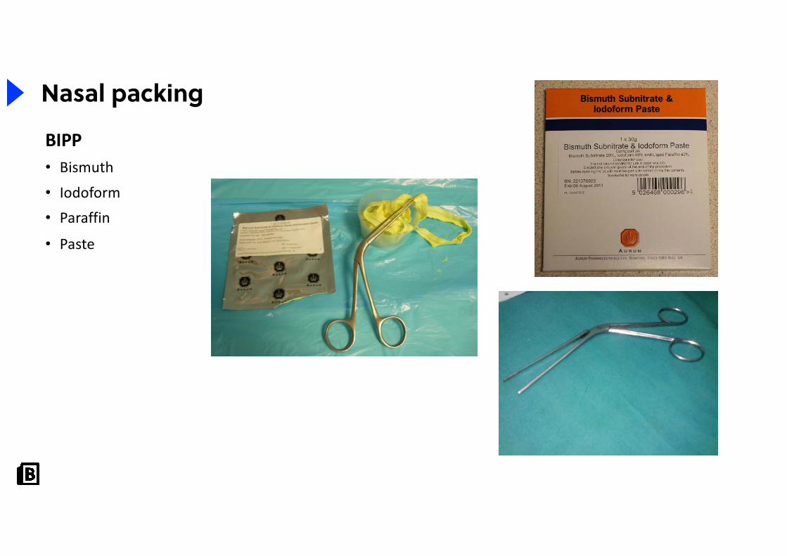
BIPP• Bismuth
• Iodoform
• Paraffin
• Paste
Nasal packing
your name
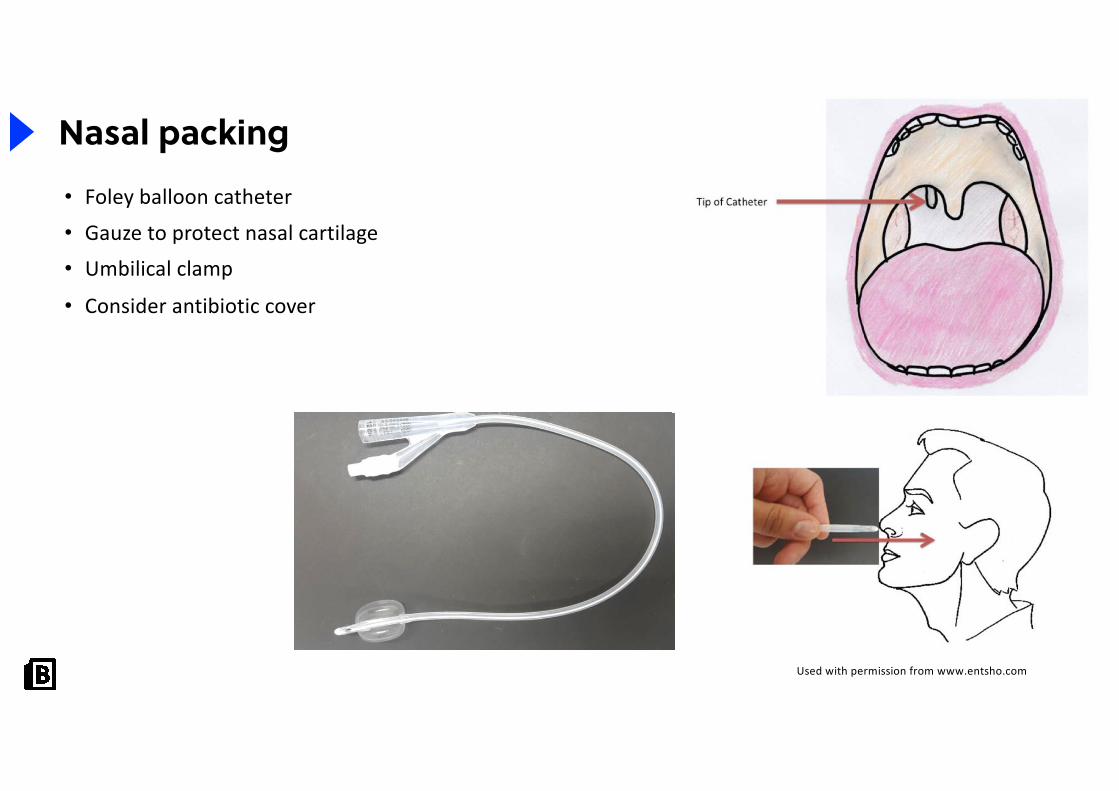
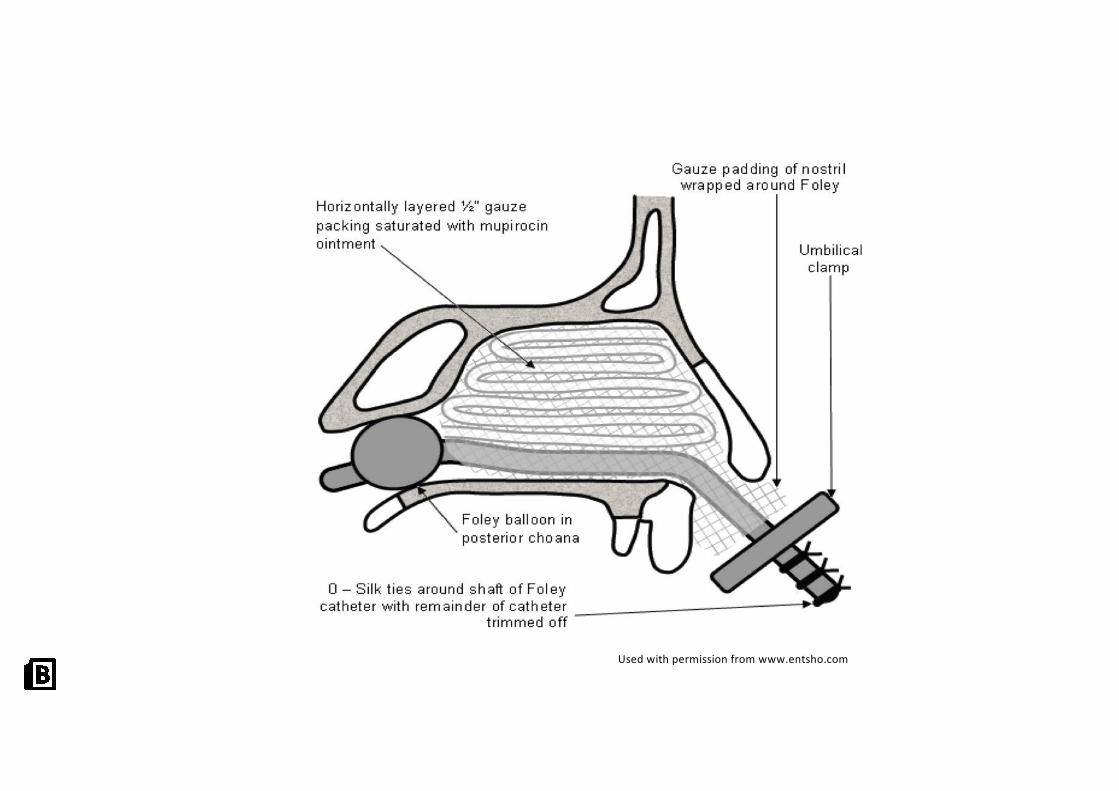
Nasal packing• Foley balloon catheter
• Gauze to protect nasal cartilage
• Umbilical clamp
• Consider antibiotic cover
Used with permission from www.entsho.com
Used with permission from www.entsho.com
your name
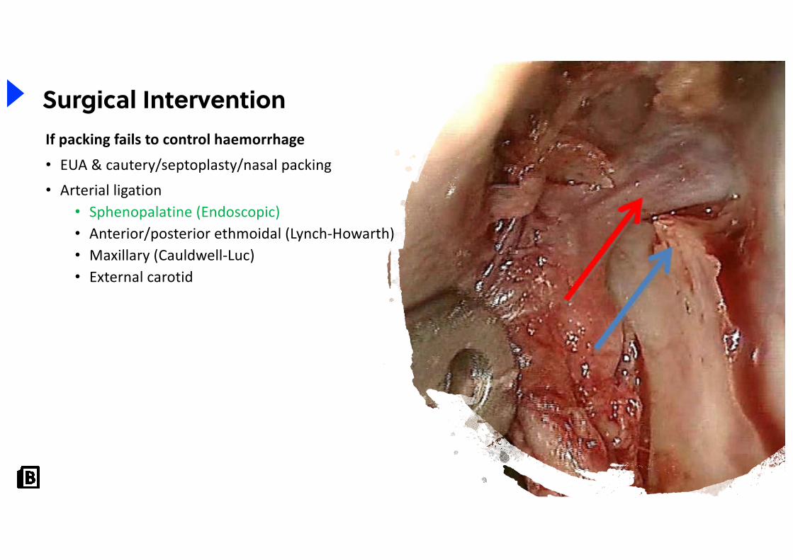
Surgical InterventionIf packing fails to control haemorrhage
• EUA & cautery/septoplasty/nasal packing
• Arterial ligation• Sphenopalatine (Endoscopic)• Anterior/posterior ethmoidal (Lynch-Howarth) • Maxillary (Cauldwell-Luc) • External carotid
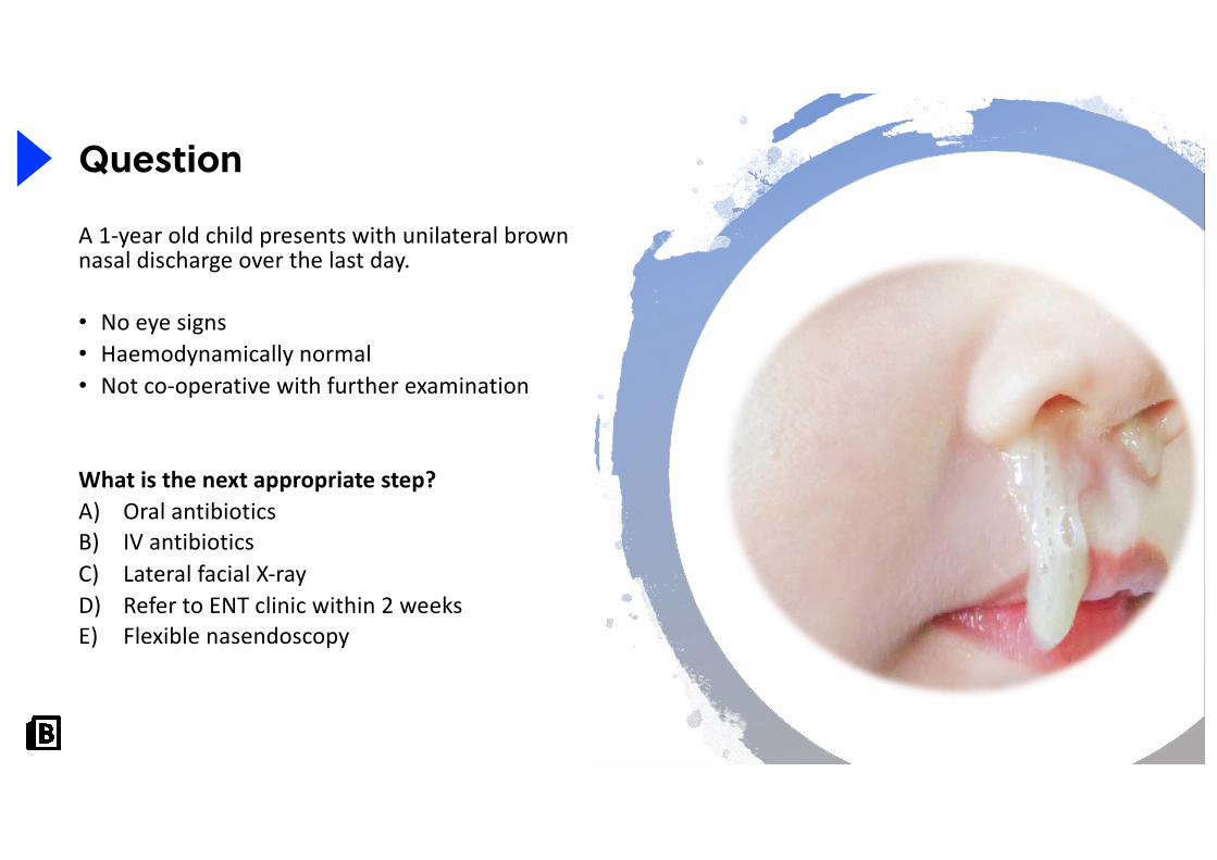
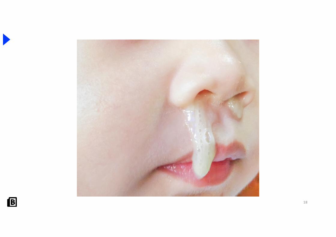
A 1-year old child presents with unilateral brown nasal discharge over the last day.
• No eye signs• Haemodynamically normal• Not co-operative with further examination
What is the next appropriate step?A) Oral antibioticsB) IV antibioticsC) Lateral facial X-rayD) Refer to ENT clinic within 2 weeksE) Flexible nasendoscopy
17
Question
18

19
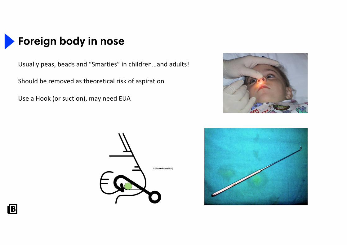
Foreign body in nose
Usually peas, beads and “Smarties” in children…and adults!
Should be removed as theoretical risk of aspiration
Use a Hook (or suction), may need EUA
your name
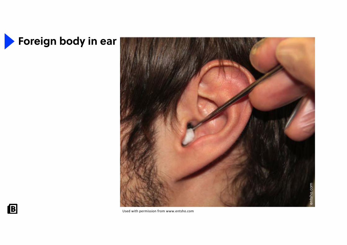
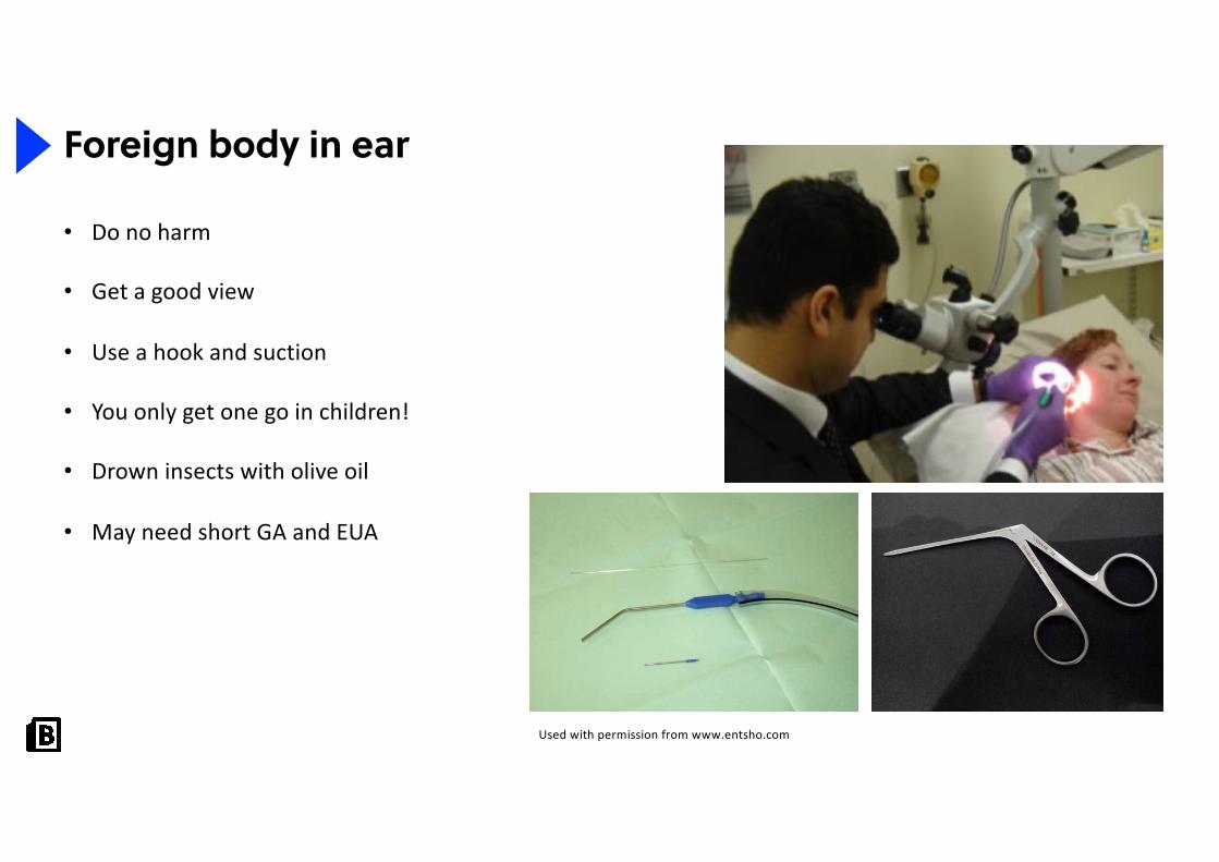
Foreign body in ear
Used with permission from www.entsho.com
your name
Foreign body in ear
• Do no harm
• Get a good view
• Use a hook and suction
• You only get one go in children!
• Drown insects with olive oil
• May need short GA and EUA
Used with permission from www.entsho.com
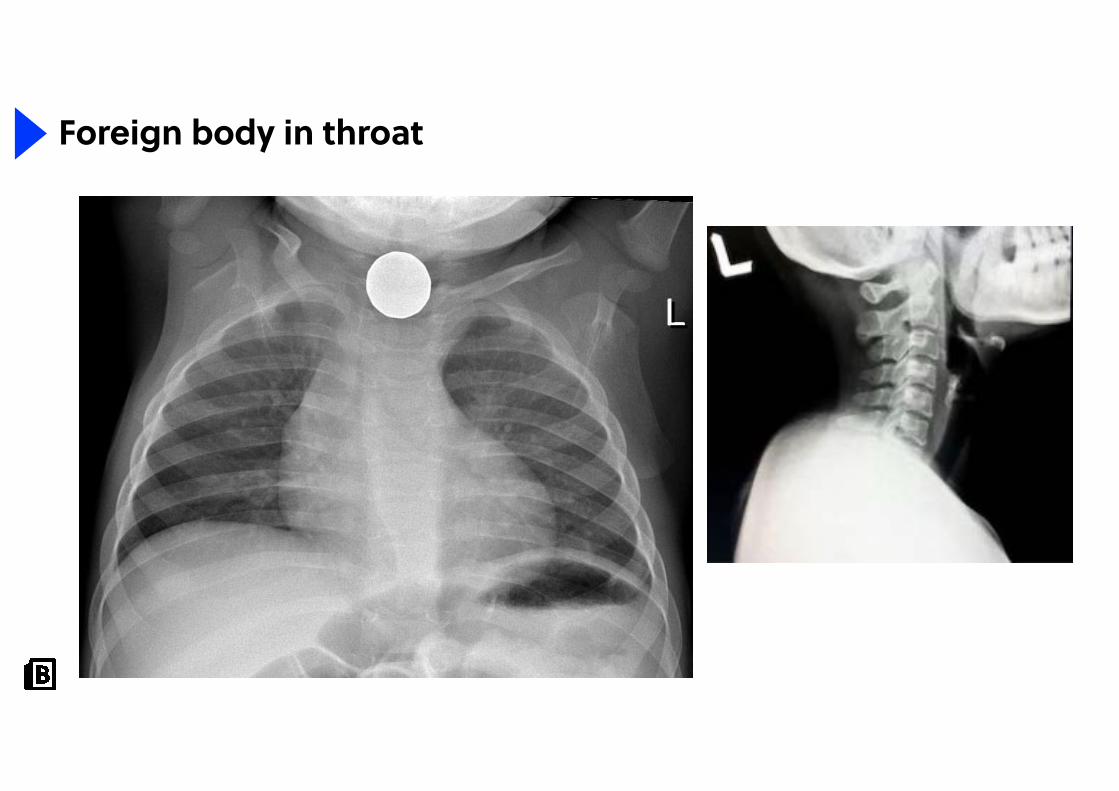
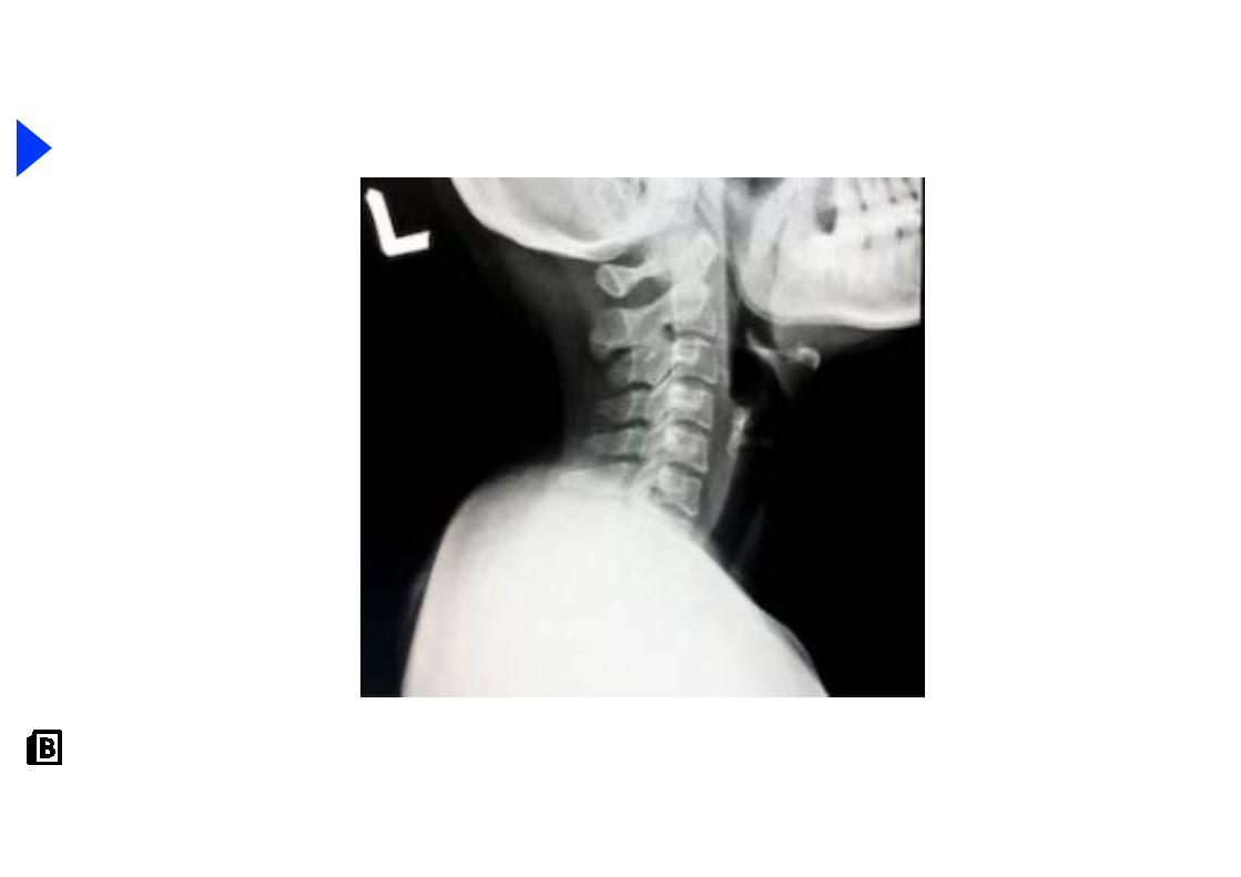
Foreign body in throat
Foreign body in throat
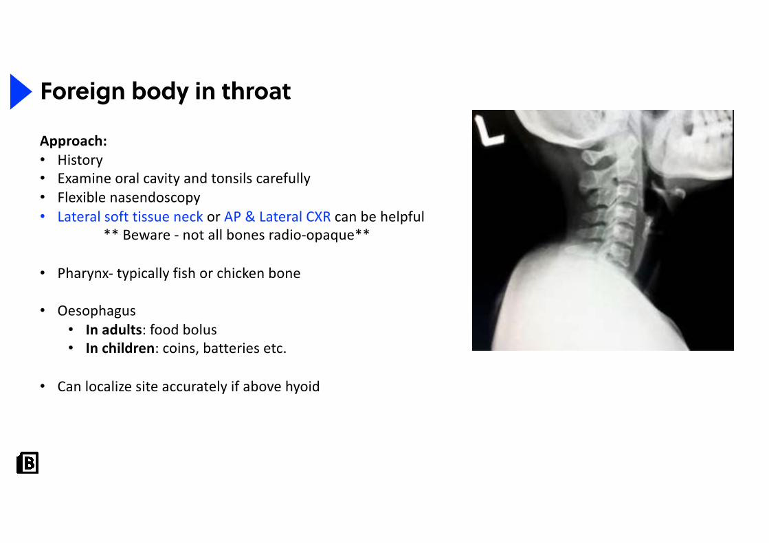
Approach:• History• Examine oral cavity and tonsils carefully• Flexible nasendoscopy• Lateral soft tissue neck or AP & Lateral CXR can be helpful
** Beware - not all bones radio-opaque**
• Pharynx- typically fish or chicken bone
• Oesophagus• In adults: food bolus• In children: coins, batteries etc.
• Can localize site accurately if above hyoid
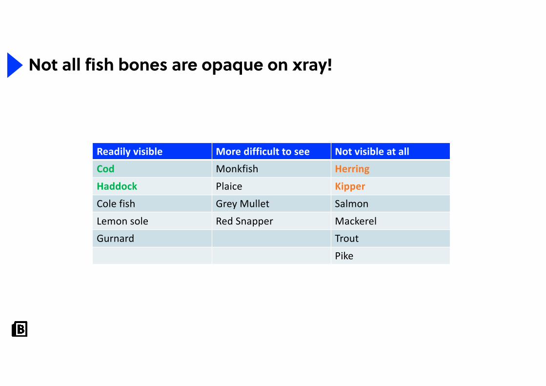
Readily visible More difficult to see Not visible at allCod Monkfish Herring
Haddock Plaice Kipper
Cole fish Grey Mullet Salmon
Lemon sole Red Snapper MackerelGurnard Trout
Pike
Not all fish bones are opaque on xray!
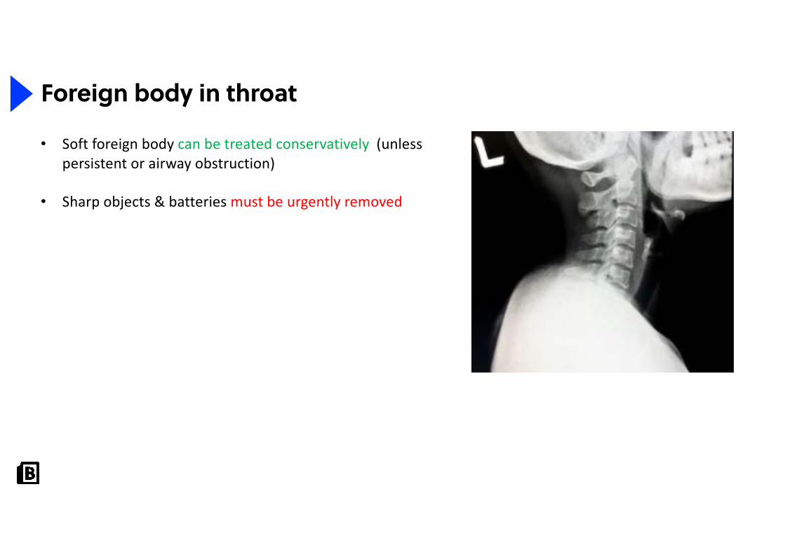
Foreign body in throat
• Soft foreign body can be treated conservatively (unless persistent or airway obstruction)
• Sharp objects & batteries must be urgently removed
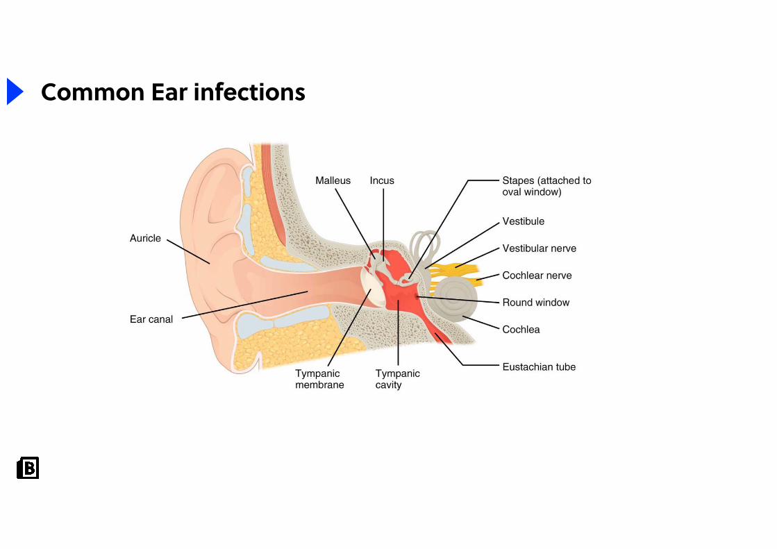
Common Ear infections
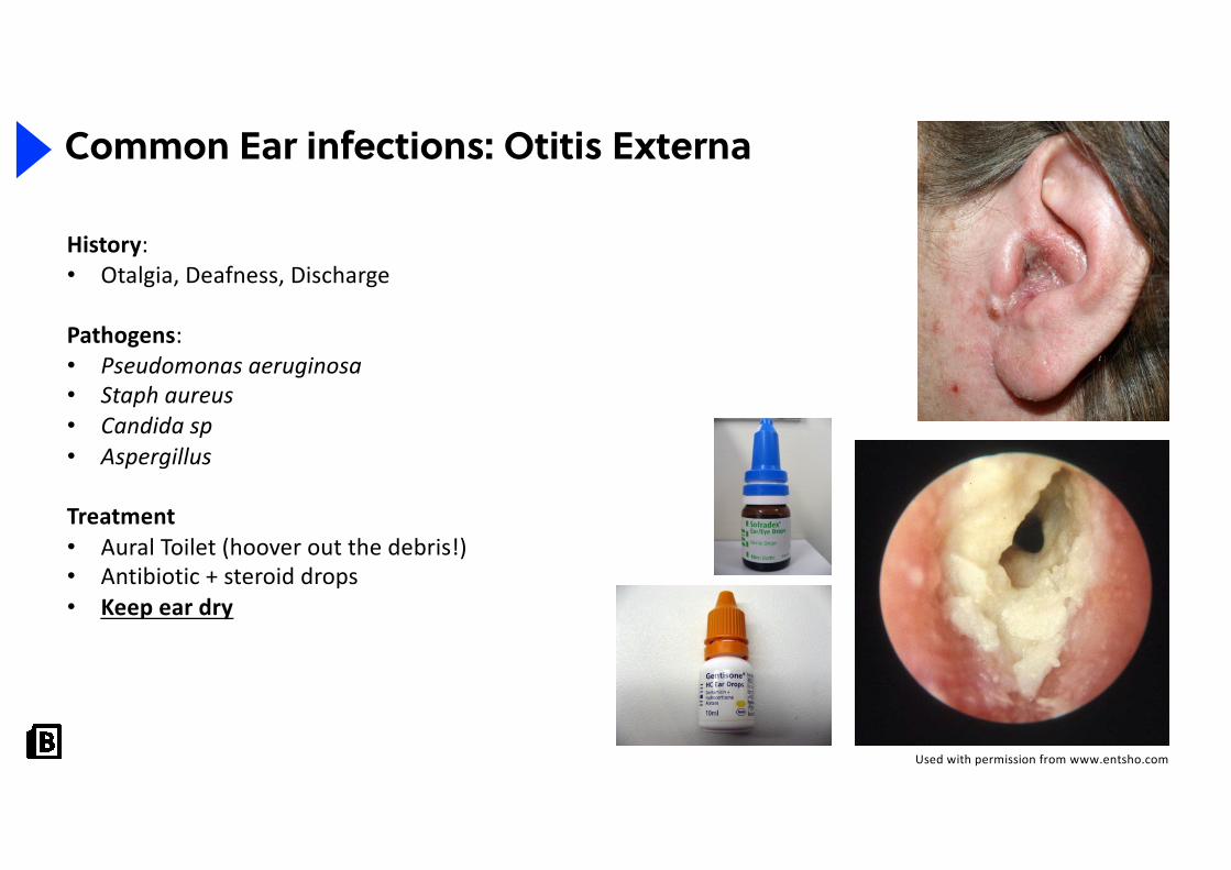
Common Ear infections: Otitis Externa
History:• Otalgia, Deafness, Discharge
Pathogens:• Pseudomonas aeruginosa• Staph aureus • Candida sp• Aspergillus
Treatment• Aural Toilet (hoover out the debris!)• Antibiotic + steroid drops• Keep ear dry
Used with permission from www.entsho.com
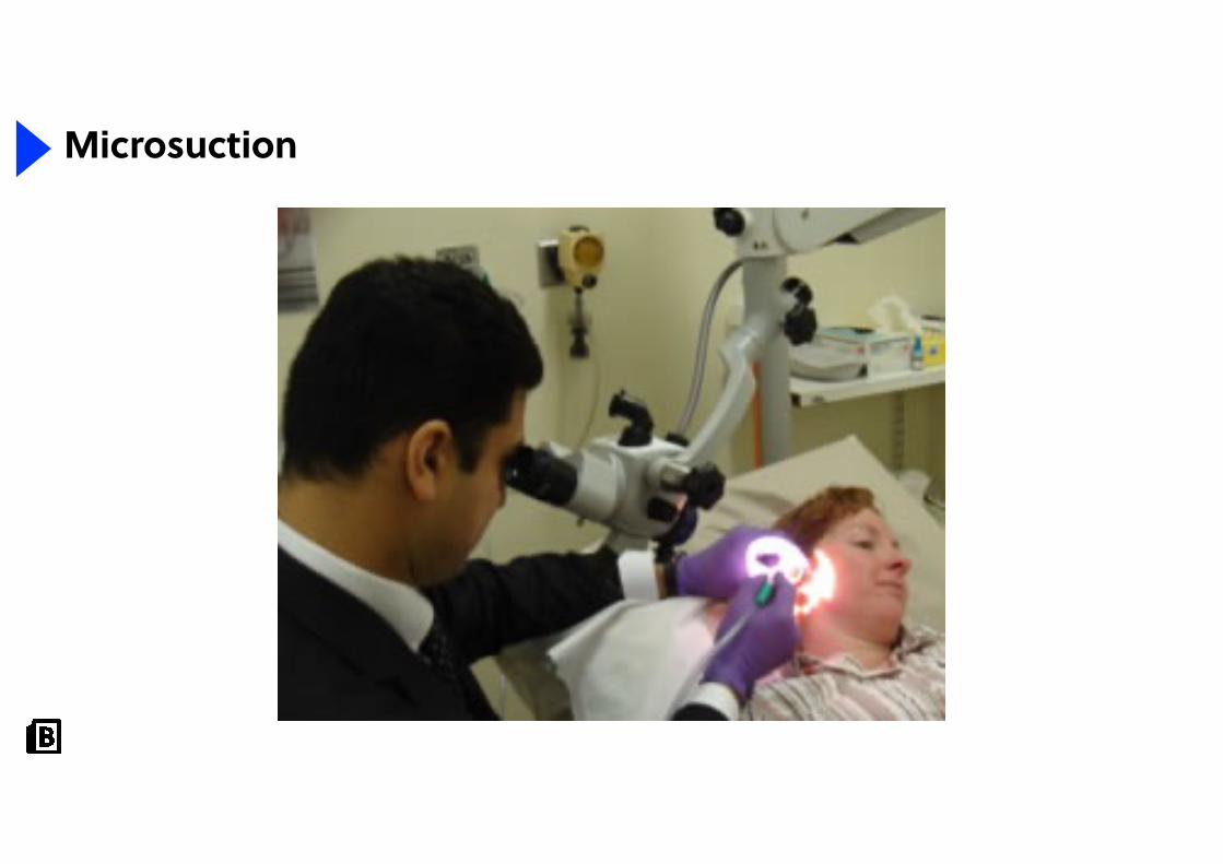
Microsuction
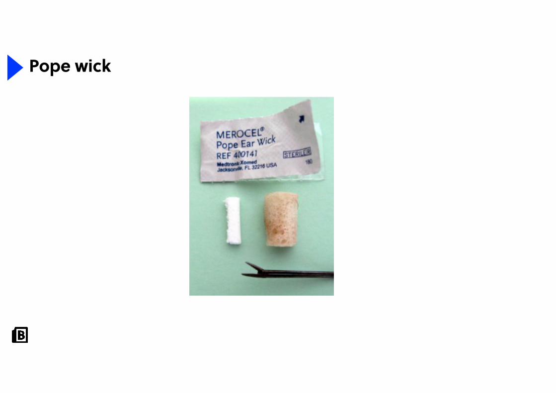
Pope wick
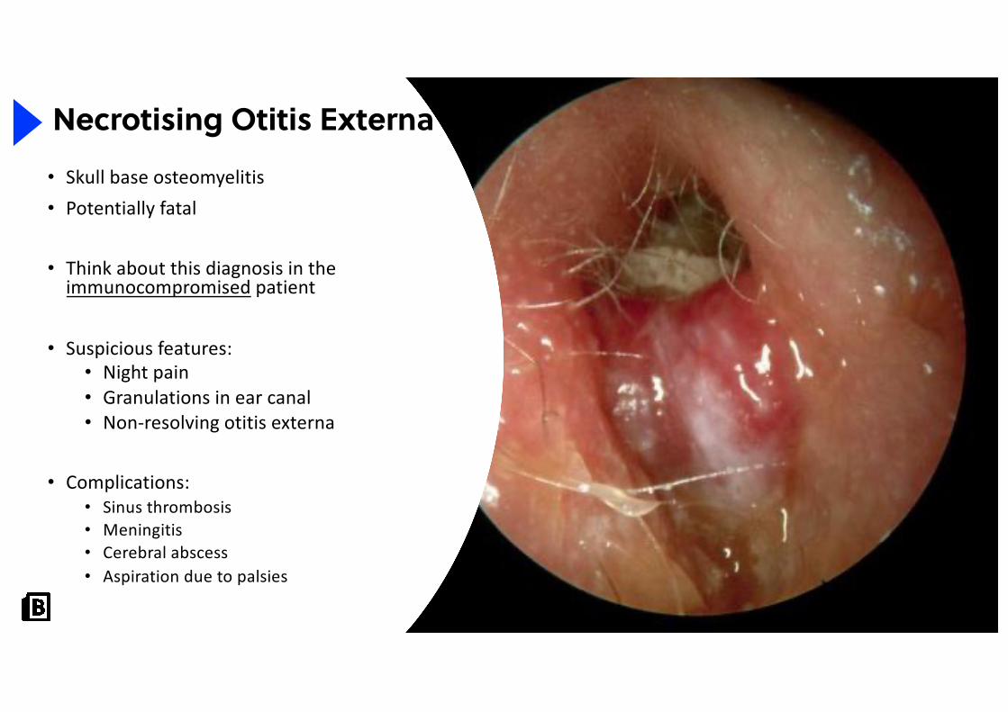
• Skull base osteomyelitis• Potentially fatal
• Think about this diagnosis in theimmunocompromised patient
• Suspicious features:• Night pain• Granulations in ear canal• Non-resolving otitis externa
• Complications: • Sinus thrombosis• Meningitis• Cerebral abscess• Aspiration due to palsies
Necrotising Otitis Externa

Necrotising Otitis Externa
• Admit
• CT
• Inflammatory markers
• MDT Management• Microbiology/ID/OPAT: IV antibiotics until resolution of pain and inflammatory markers• Diabetic control• Additional therapy based on palsies
• SLT• Physio• Ophthalmology
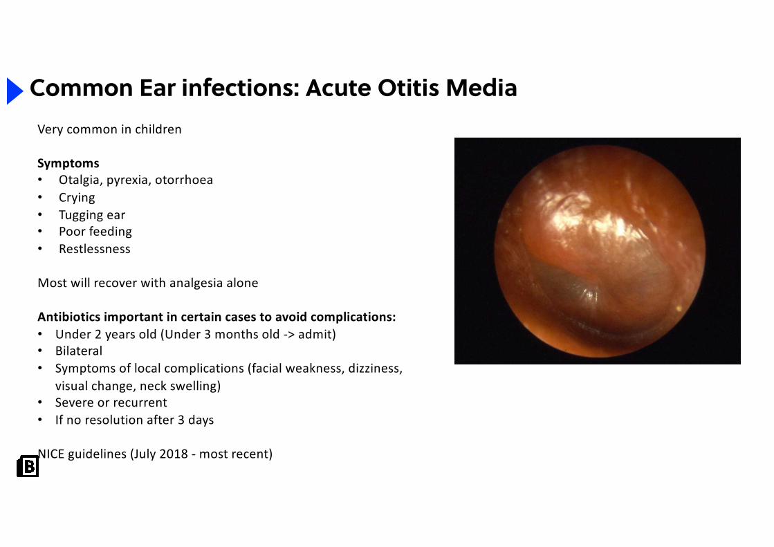
Common Ear infections: Acute Otitis MediaVery common in children
Symptoms• Otalgia, pyrexia, otorrhoea• Crying• Tugging ear• Poor feeding• Restlessness
Most will recover with analgesia alone
Antibiotics important in certain cases to avoid complications:• Under 2 years old (Under 3 months old -> admit)• Bilateral• Symptoms of local complications (facial weakness, dizziness,
visual change, neck swelling)• Severe or recurrent • If no resolution after 3 days
NICE guidelines (July 2018 - most recent)
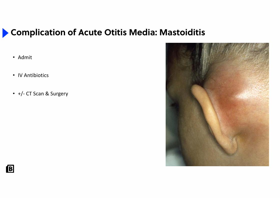
• Admit
• IV Antibiotics
• +/- CT Scan & Surgery
Complication of Acute Otitis Media: Mastoiditis
36
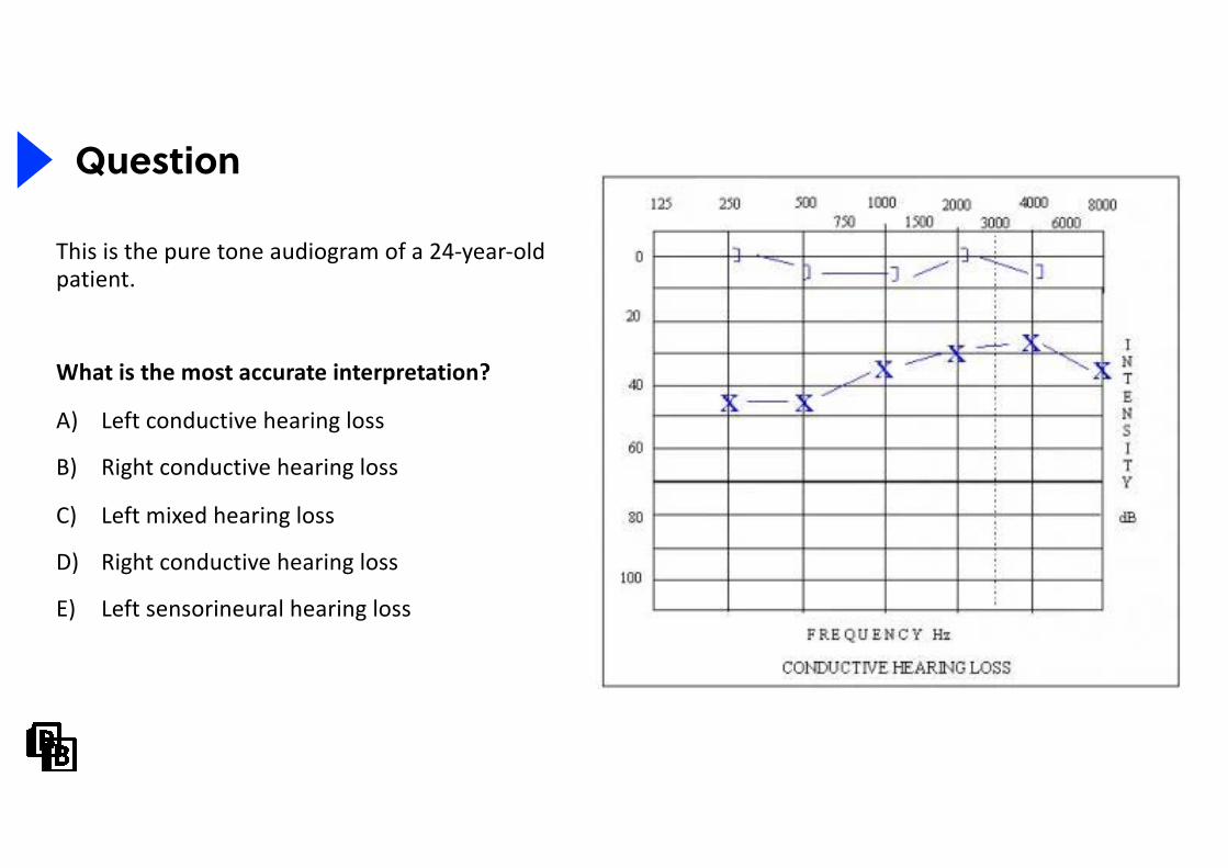
Question
This is the pure tone audiogram of a 24-year-old patient.
What is the most accurate interpretation?
A) Left conductive hearing loss
B) Right conductive hearing loss
C) Left mixed hearing loss
D) Right conductive hearing loss
E) Left sensorineural hearing loss
37
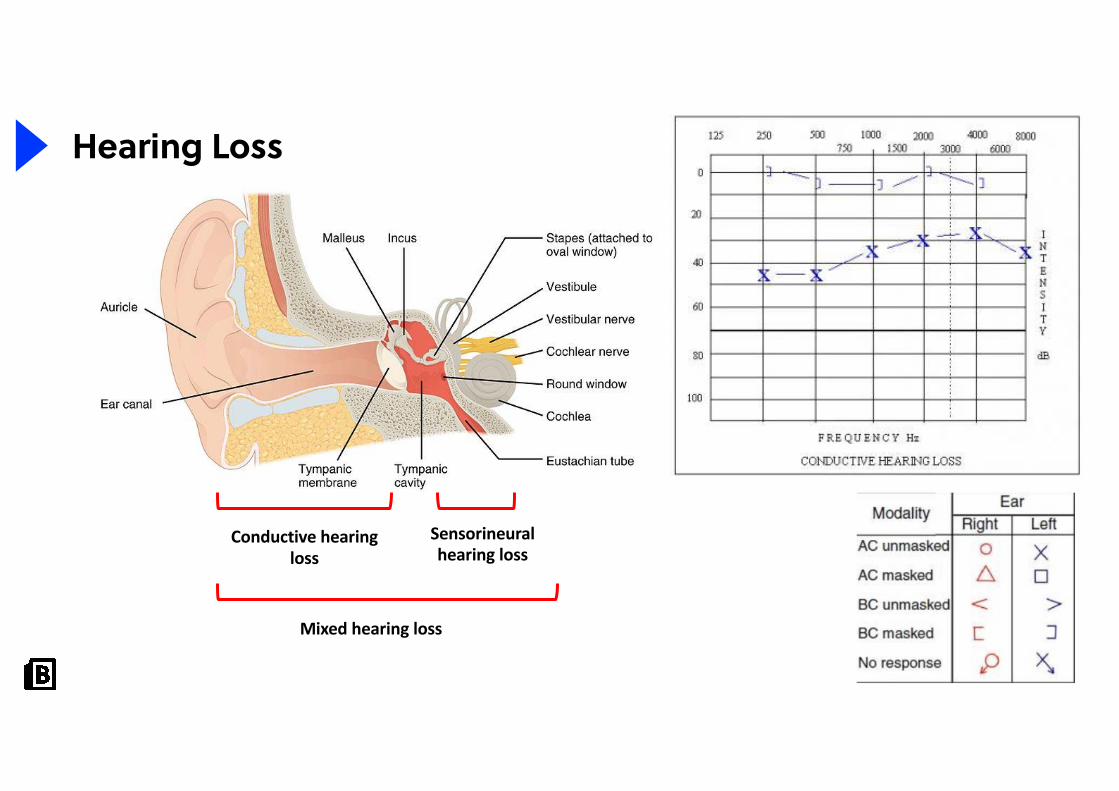
Hearing Loss
Conductive hearing loss
Sensorineural hearing loss
Mixed hearing loss
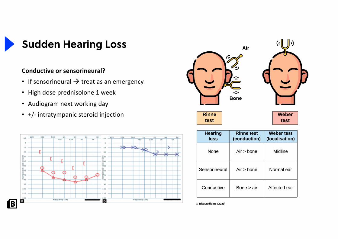
Conductive or sensorineural?
• If sensorineural à treat as an emergency
• High dose prednisolone 1 week
• Audiogram next working day
• +/- intratympanic steroid injection
Sudden Hearing Loss
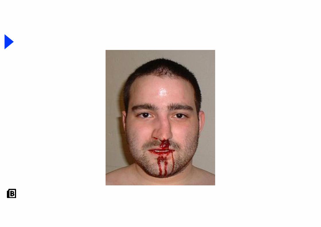
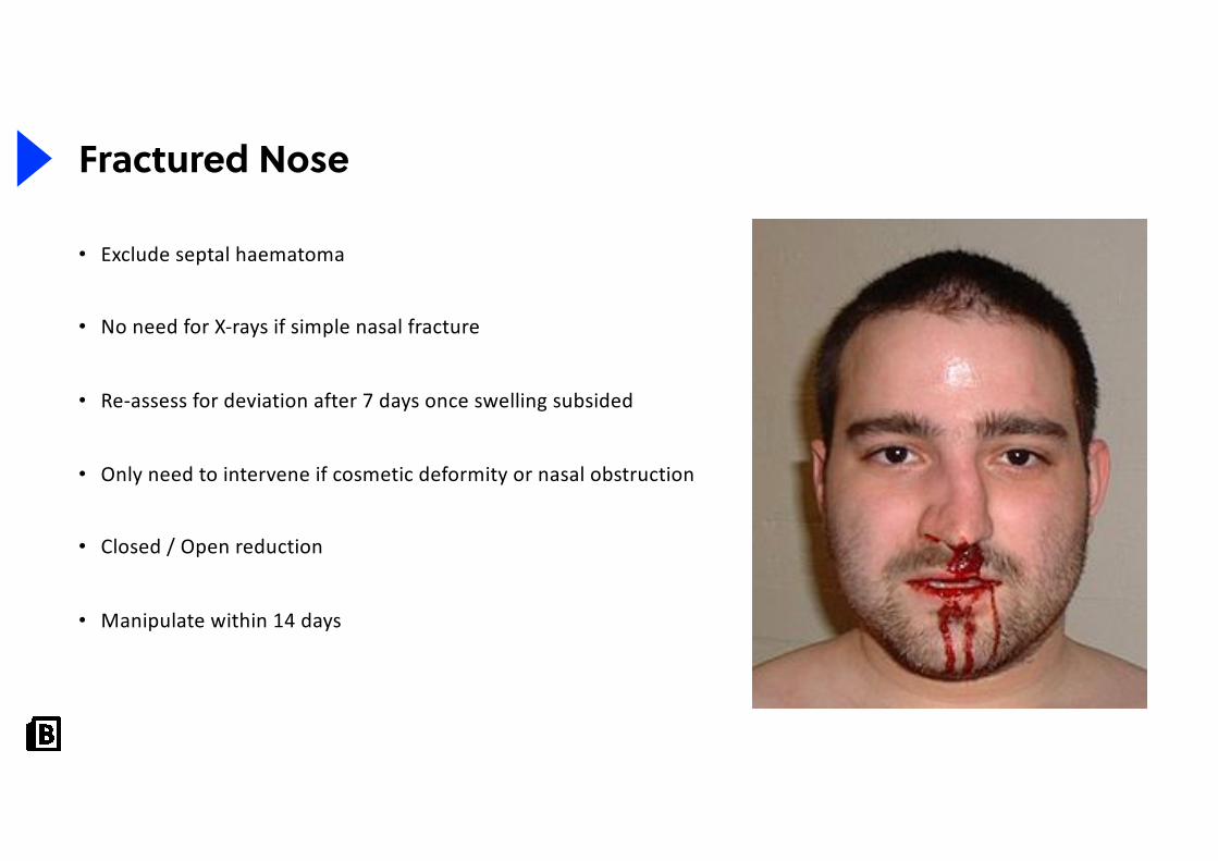
Fractured Nose
• Exclude septal haematoma
• No need for X-rays if simple nasal fracture
• Re-assess for deviation after 7 days once swelling subsided
• Only need to intervene if cosmetic deformity or nasal obstruction
• Closed / Open reduction
• Manipulate within 14 days
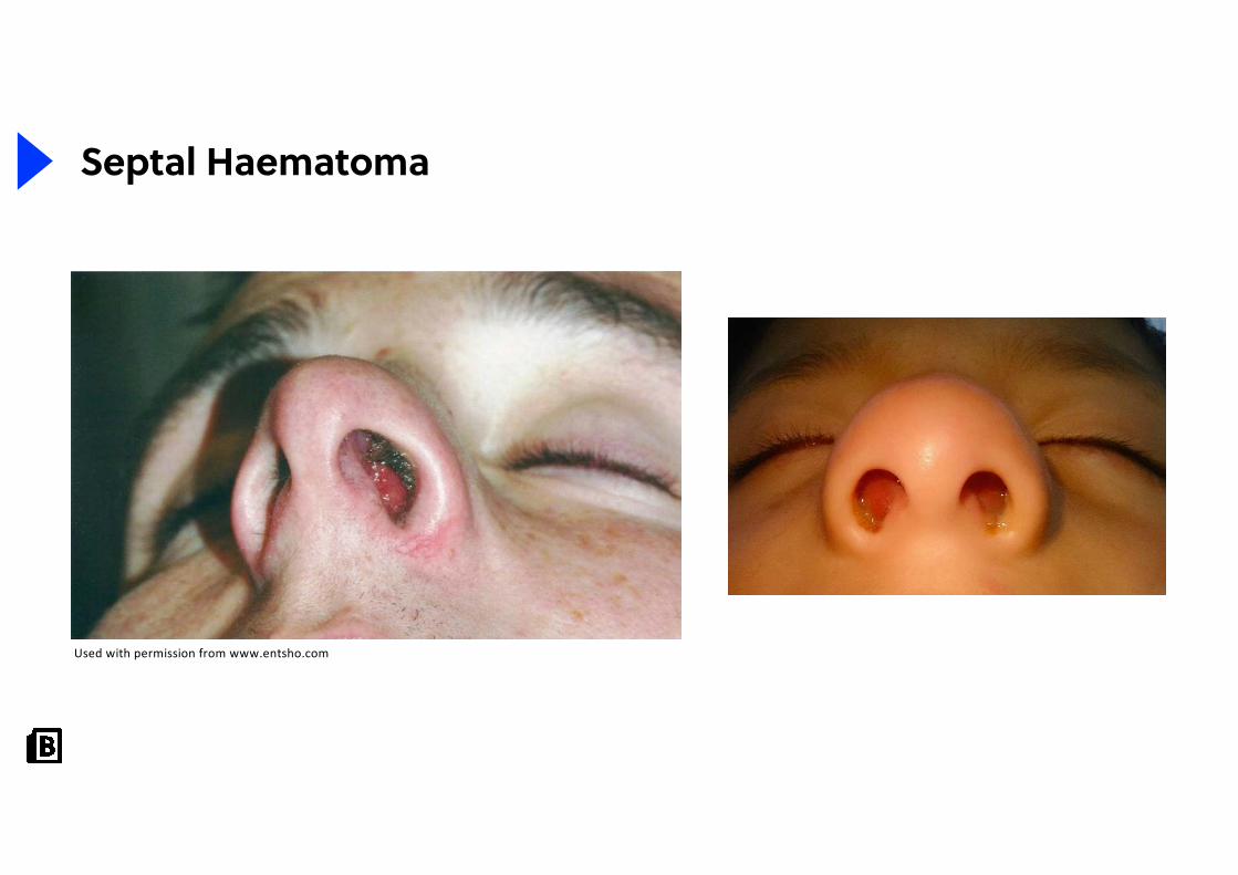
Septal Haematoma
Used with permission from www.entsho.com
42
Question
It’s Saturday evening. You could be outside having a BBQ but you’ve chosen to log onto a seminarinstead.
Can you take any more of this?
A) My brain is fried, please stop. Why am I even geeking it up on a Saturday evening in the first place?B) Skip straight to the top decile questionC) Bring it on. Let’s have some airway emergenciesD) Skip straight to info on why ENT is a great career choiceE) Please come back another day with more ENT topics

“Doctor, I’ve had a sore throat for days”
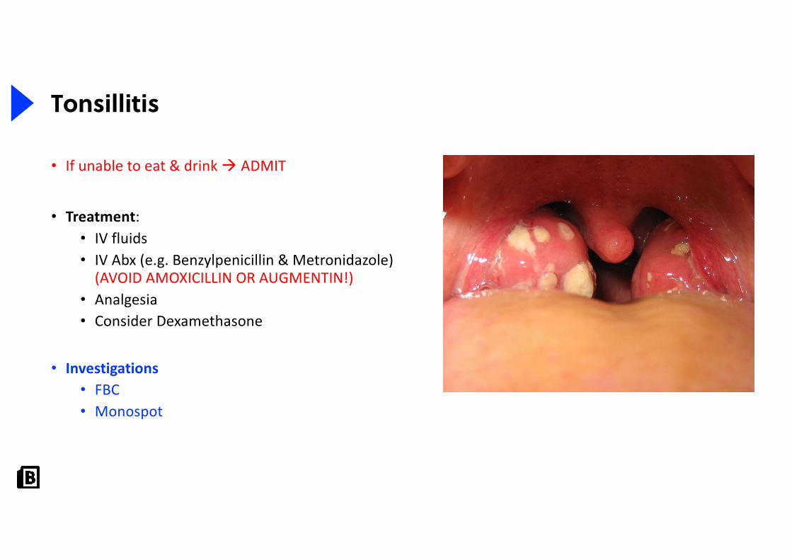
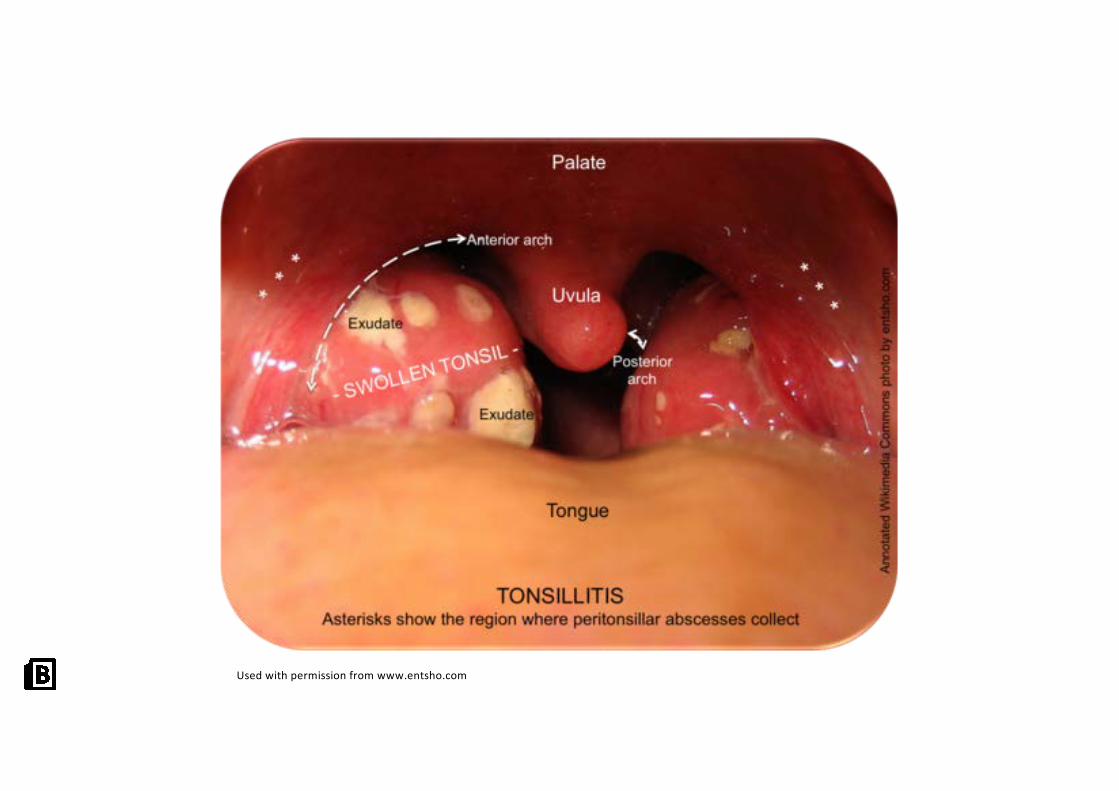
Tonsillitis
• If unable to eat & drink à ADMIT
• Treatment:• IV fluids• IV Abx (e.g. Benzylpenicillin & Metronidazole)
(AVOID AMOXICILLIN OR AUGMENTIN!)• Analgesia• Consider Dexamethasone
• Investigations• FBC• Monospot
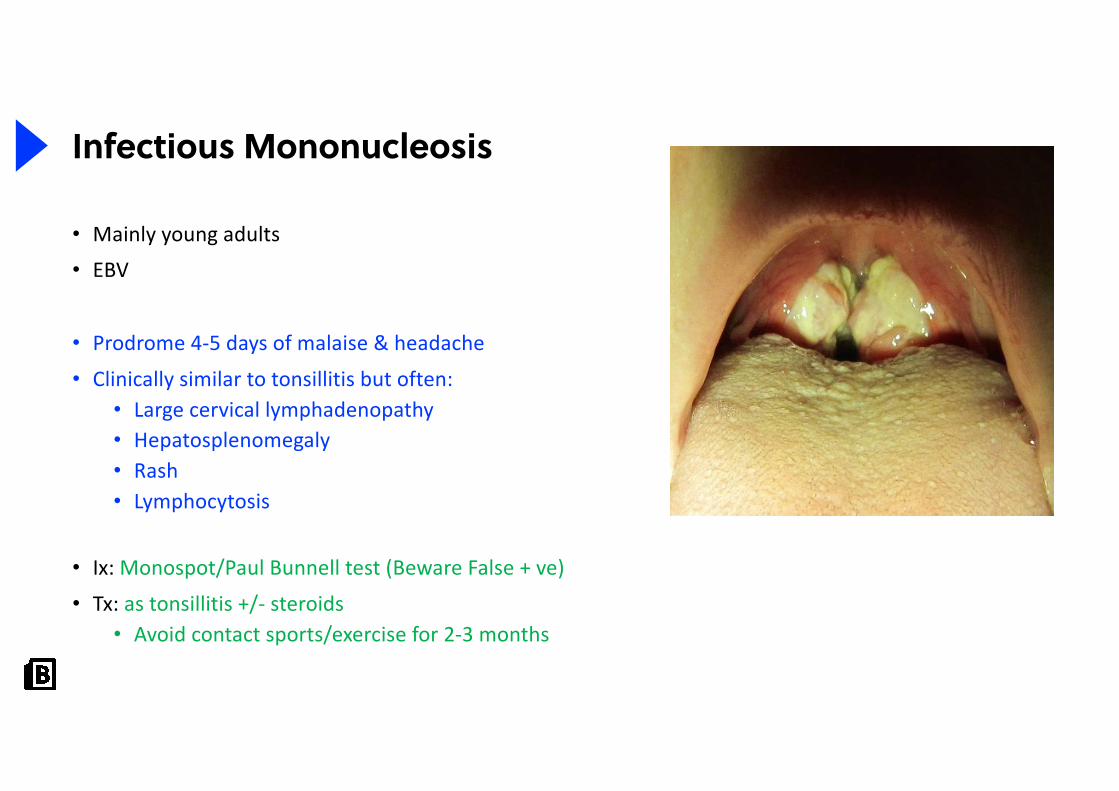
Infectious Mononucleosis
• Mainly young adults
• EBV
• Prodrome 4-5 days of malaise & headache
• Clinically similar to tonsillitis but often:• Large cervical lymphadenopathy• Hepatosplenomegaly• Rash• Lymphocytosis
• Ix: Monospot/Paul Bunnell test (Beware False + ve)
• Tx: as tonsillitis +/- steroids• Avoid contact sports/exercise for 2-3 months
Used with permission from www.entsho.com
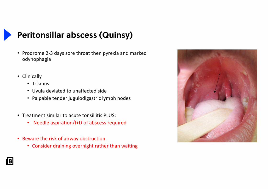
Peritonsillar abscess (Quinsy)
• Prodrome 2-3 days sore throat then pyrexia and marked odynophagia
• Clinically • Trismus • Uvula deviated to unaffected side• Palpable tender jugulodigastric lymph nodes
• Treatment similar to acute tonsillitis PLUS:• Needle aspiration/I+D of abscess required
• Beware the risk of airway obstruction• Consider draining overnight rather than waiting
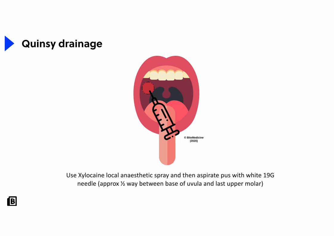
Use Xylocaine local anaesthetic spray and then aspirate pus with white 19G needle (approx ½ way between base of uvula and last upper molar)
Quinsy drainage
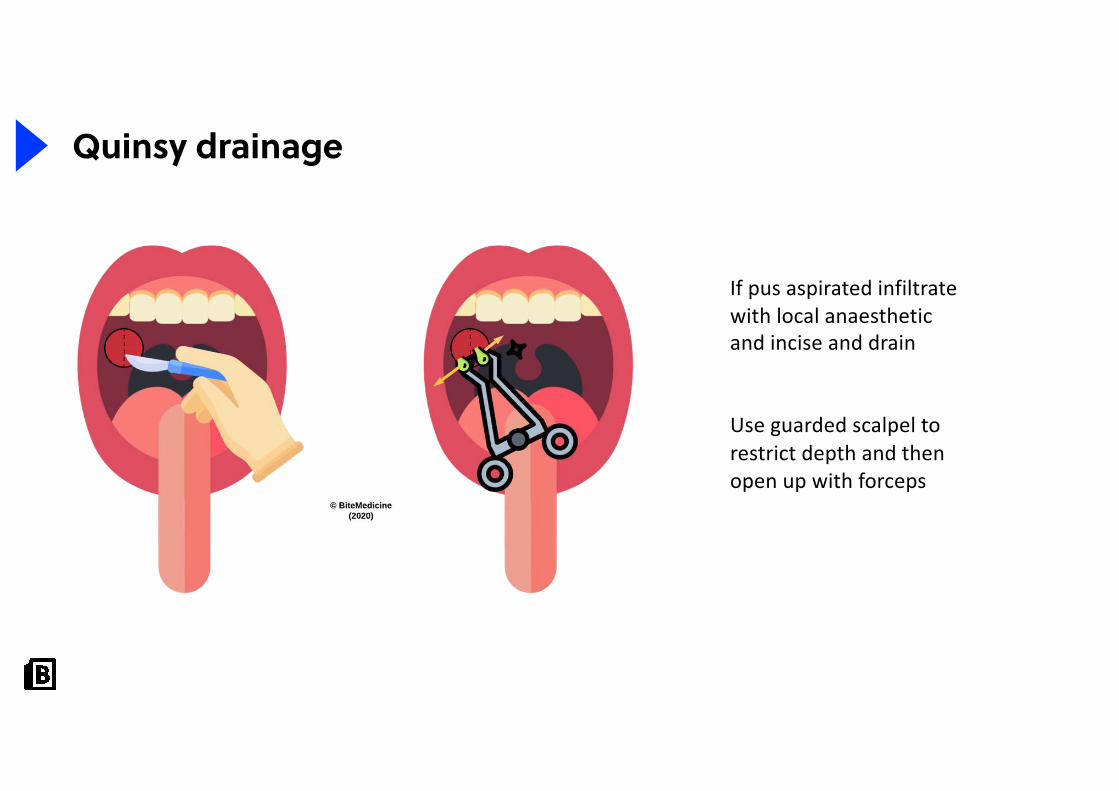
If pus aspirated infiltrate with local anaesthetic and incise and drain
Use guarded scalpel to restrict depth and then open up with forceps
Quinsy drainage
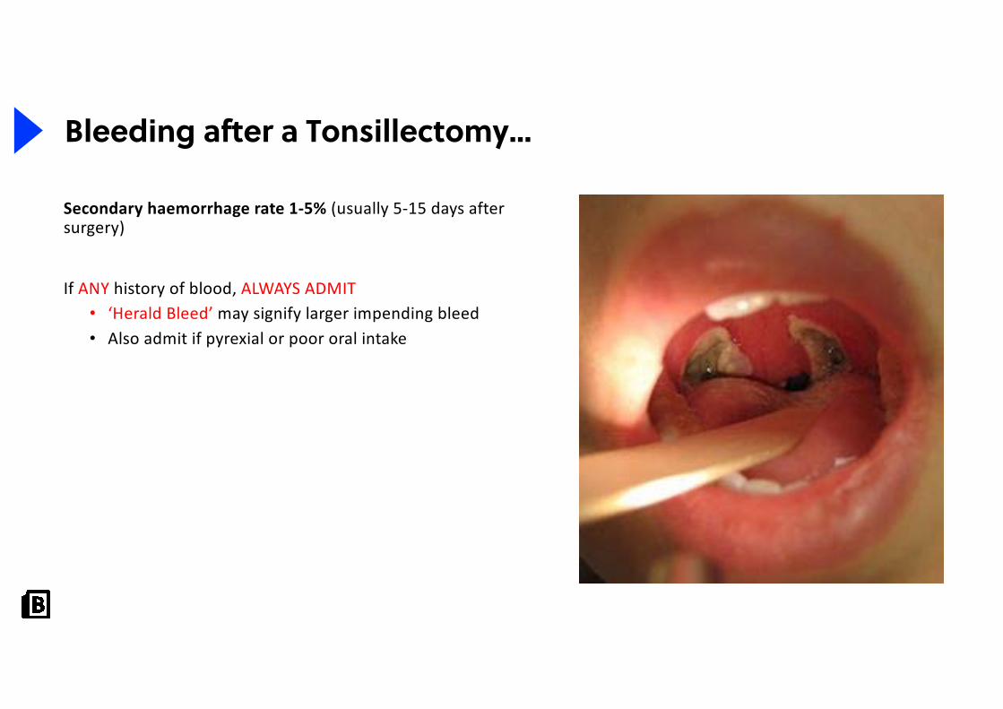
Bleeding after a Tonsillectomy…
Secondary haemorrhage rate 1-5% (usually 5-15 days after surgery)
If ANY history of blood, ALWAYS ADMIT• ‘Herald Bleed’ may signify larger impending bleed• Also admit if pyrexial or poor oral intake
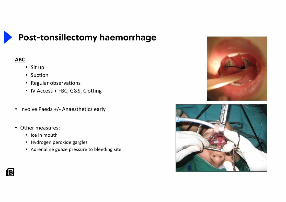
Post-tonsillectomy haemorrhage
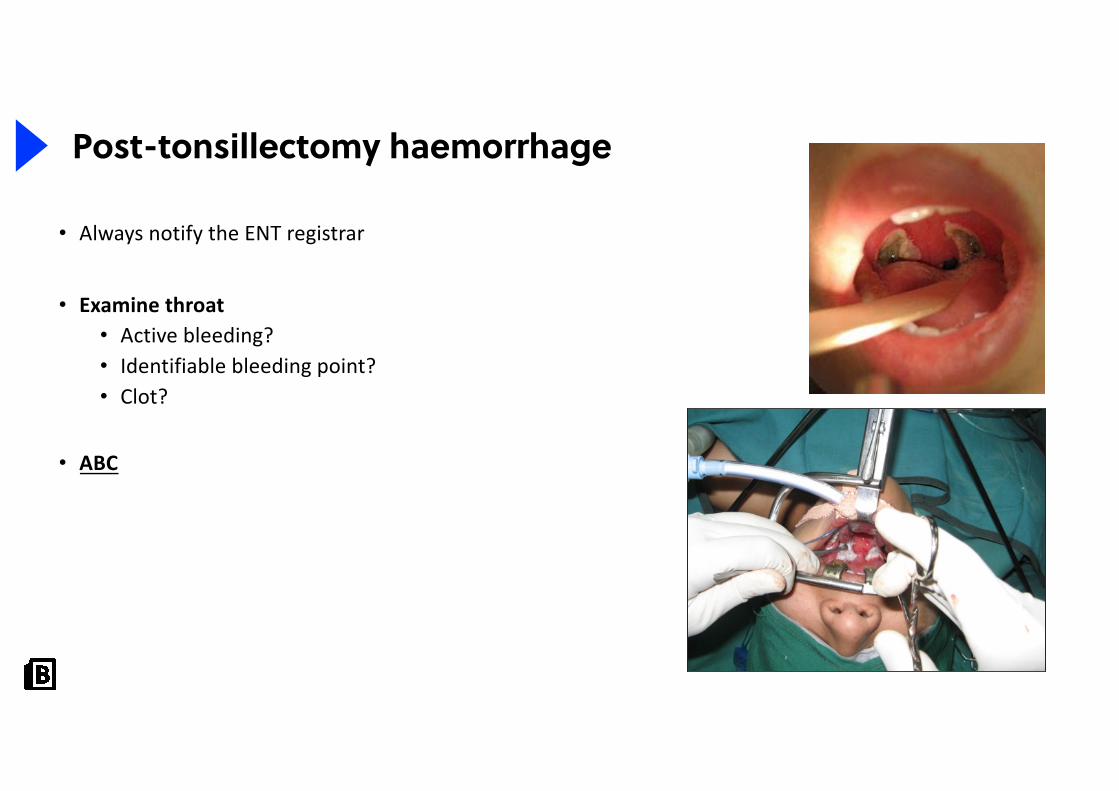
• Always notify the ENT registrar
• Examine throat• Active bleeding?• Identifiable bleeding point?• Clot?
• ABC
Post-tonsillectomy haemorrhage
ABC• Sit up• Suction• Regular observations• IV Access + FBC, G&S, Clotting
• Involve Paeds +/- Anaesthetics early
• Other measures:• Ice in mouth• Hydrogen peroxide gargles• Adrenaline guaze pressure to bleeding site
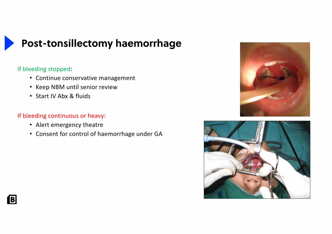
Post-tonsillectomy haemorrhage
If bleeding stopped:• Continue conservative management• Keep NBM until senior review• Start IV Abx & fluids
If bleeding continuous or heavy:• Alert emergency theatre• Consent for control of haemorrhage under GA
Stridor
• Classically a bovine-like inspiratory noise associated with laryngeal obstruction
• More common in children because of the relatively small diameter of the airways
• DIAGNOSIS TAKES SECOND PLACE TO MANAGEMENT

Acute airway obstruction
• Rapid airway assessment • Including flexible nasendoscopy (if appropriate)
• Contact senior colleague and senior anaesthetist
• Jaw thrust, head extension, simple airways
• Humidified Oxygen/Heliox
• IV steroids
• Adrenaline nebs
• Inform theatres à ? Intubate ? Surgical airway

Children• Croup• Acute epiglottitis• Foreign body• Laryngeal papilloma• Congenital abnormalities• Laryngomalacia
Adults• Laryngeal neoplasms• Bilateral vocal cord palsy• Croup• Epiglottitis• Trauma• Foreign body• Stenosis
Causes of airway obstruction
An Introduction to ENT
Reasons for a tracheostomy
• Real upper airway obstruction• Supraglottic• Glottic• Subglottic
• Impending upper airway obstruction
Also:
• Prolonged ventilation
• Bronchial toilet
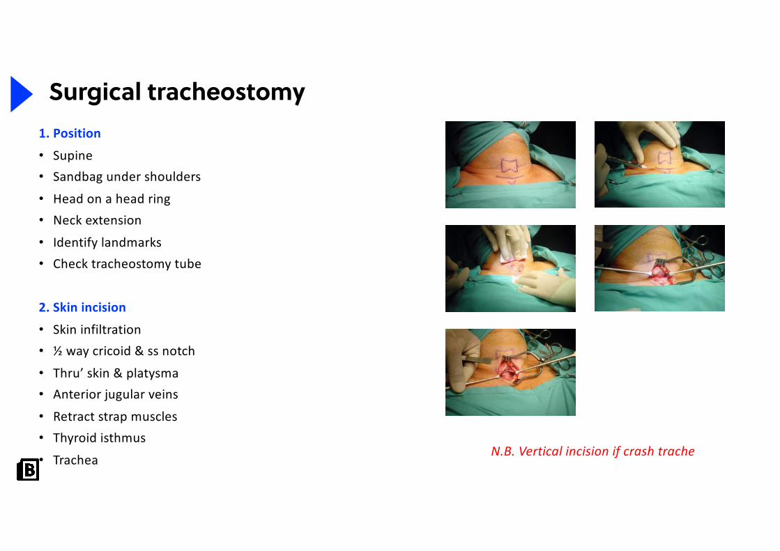
N.B. Vertical incision if crash trache
Surgical tracheostomy1. Position
• Supine• Sandbag under shoulders
• Head on a head ring• Neck extension
• Identify landmarks• Check tracheostomy tube
2. Skin incision
• Skin infiltration• ½ way cricoid & ss notch
• Thru’ skin & platysma• Anterior jugular veins
• Retract strap muscles• Thyroid isthmus
• Trachea
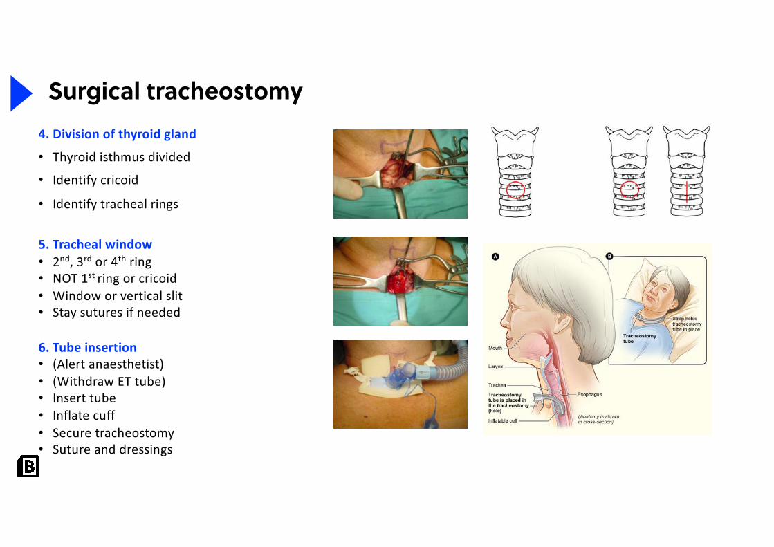
Surgical tracheostomy4. Division of thyroid gland
• Thyroid isthmus divided
• Identify cricoid
• Identify tracheal rings
5. Tracheal window• 2nd, 3rd or 4th ring• NOT 1st ring or cricoid• Window or vertical slit• Stay sutures if needed
6. Tube insertion• (Alert anaesthetist)• (Withdraw ET tube)• Insert tube• Inflate cuff• Secure tracheostomy• Suture and dressings
60
Top-decile question
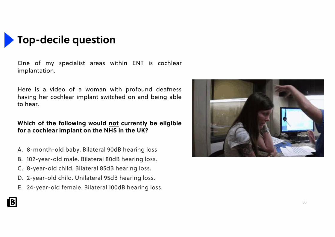
One of my specialist areas within ENT is cochlearimplantation.
Here is a video of a woman with profound deafnesshaving her cochlear implant switched on and being ableto hear.
Which of the following would not currently be eligiblefor a cochlear implant on the NHS in the UK?
A. 8-month-old baby. Bilateral 90dB hearing lossB. 102-year-old male. Bilateral 80dB hearing loss.C. 8-year-old child. Bilateral 85dB hearing loss.D. 2-year-old child. Unilateral 95dB hearing loss.E. 24-year-old female. Bilateral 100dB hearing loss.
61
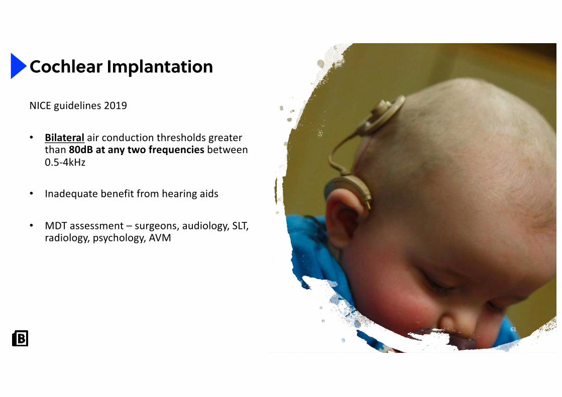
Cochlear Implantation
NICE guidelines 2019
• Bilateral air conduction thresholds greater than 80dB at any two frequencies between 0.5-4kHz
• Inadequate benefit from hearing aids
• MDT assessment – surgeons, audiology, SLT, radiology, psychology, AVM
Why ENT?
• Not all snot & wax!• One of medicine’s well-hidden best careers
• Patients young & old
• Wide variety of cases
• Make people better quickly!• Gadgets
• Challenging surgery, emotional impact
• Look at the Consultants
References
• Slide 1: Video source: Cardiac arrest (BBC1, 1994) via YouTube• Slide 5: https://www.shutterstock.com/image-photo/female-doctor-nurse-wearing-scrubs-protective-
1668187618• Slide 6: I, Welleschik / CC BY-SA (http://creativecommons.org/licenses/by-sa/3.0/).
https://commons.wikimedia.org/wiki/File:Epistaxis1.jpg.
• Slide 9: Therapeutic Intranasal Drug Delivery Needleless treatment options for medical problems. http://www.intranasal.net/Epistaxis/default.htm
• Slide 9: Arzol silver nitrate. BBODO / CC BY-SA (https://creativecommons.org/licenses/by-sa/3.0). https://commons.wikimedia.org/wiki/File:Caustic_pencil.png
• Slide 10: www.entsho.com
• Slide 11 and 13: https://www.shutterstock.com/image-vector/anatomy-nose-throat-human-organ-structure-123867553
• Slide 12: Exposing & Bonding of Brackets to Teeth Explanation & Warnings. http://www.exodontia.info/Expose_and_Bonding_of_Teeth.html
• Slide 12: Sarindam7 / CC BY-SA (https://creativecommons.org/licenses/by-sa/4.0). https://commons.wikimedia.org/wiki/File:Tilly%27s_Nasal_Dressing_Forceps_ENT_Instrument_Medical1.jpg
• Slide 14 and 15: www.entsho.com
• Slide 16: https://vula.uct.ac.za/access/content/group/ba5fb1bd-be95-48e5-81be-586fbaeba29d/Sphenopalatine%20artery%20_SPA_%20ligation.pdf
References: Part 2
• Slide 17 and 18: https://www.shutterstock.com/image-photo/image-mucus-nose-young-boy-235618357• Slide 19: Part of Mr Manjaly’s collection• Slide 20: LITFL - https://litfl.com/nasal-foreign-body/ - non-commercial use (non-profit)• Slide 20: Sarindam7 / CC BY-SA (https://creativecommons.org/licenses/by-sa/4.0).
https://commons.wikimedia.org/wiki/File:ENT_Instruments_Foreign_Body_Hook.jpg• Slide 21 and 22: www.ENTSho.com• Slide 22: http://www.tocan.de/shop / CC BY-SA (https://creativecommons.org/licenses/by-sa/4.0).
https://commons.wikimedia.org/wiki/File:Fremdk%C3%B6rperzange_nach_Hartmann,_8_cm,_fein.jpg• Slide 22: BBODO / CC BY-SA (https://creativecommons.org/licenses/by-sa/3.0).
https://commons.wikimedia.org/wiki/File:Caustic_pencil.png• Slide 23: Samir at English Wikipedia / CC BY (https://creativecommons.org/licenses/by/3.0)
https://commons.wikimedia.org/wiki/File:Foreign_body_aspiration.jpg• Slide 23, 24, 26, 27: https://www.shutterstock.com/image-photo/zooming-closeup-lateral-view-plain-film-
795694396• Slide 28: OpenStax / CC BY (https://creativecommons.org/licenses/by/4.0).
https://commons.wikimedia.org/wiki/File:1404_The_Structures_of_the_Ear.jpg• Slide 29: CNX OpenStax / CC BY (https://creativecommons.org/licenses/by/4.0).
https://commons.wikimedia.org/wiki/File:OSC_Microbio_21_02_folliculit_(cropped).jpg• Slide 29: www.ENTSho.com• Slide 30: Part of Mr Manjaly’s collection
References: Part 3
• Slide 31: Otitis externa Inflammation of the outer ear Mr James W Fairley BSc MBBS FRCS MS Consultant ENT Surgeon. https://entkent.com/otitis-externa/
• Slide 32: Kirse Bock, Therese Ovesen. Optimised diagnosis and treatment of necrotizing external otitis is warranted. Published in Danish medical bulletin 2011.
• Slide 34: B. Welleschik / CC BY-SA (http://creativecommons.org/licenses/by-sa/3.0/). https://commons.wikimedia.org/wiki/File:Otitis_media_entdifferenziert2.jpg
• Slide 35: B. Welleschik / CC BY-SA (http://creativecommons.org/licenses/by-sa/3.0/). https://commons.wikimedia.org/wiki/File:Mastoiditis1.jpg
• Slide 36: Part of Mr Manjaly’s collection
• Slide 37: OpenStax / CC BY (https://creativecommons.org/licenses/by/4.0). https://commons.wikimedia.org/wiki/File:1404_The_Structures_of_the_Ear.jpg
• Slide 38: Graph is part of Mr Manjaly’s collection
• Slides 39 and 40: Rls / CC BY-SA (http://creativecommons.org/licenses/by-sa/3.0/). https://commons.wikimedia.org/wiki/File:BrokenNose.jpg
• Slide 41: www.ENTSho.com
• Slide 41: Afrodriguezg / CC BY-SA (https://creativecommons.org/licenses/by-sa/4.0). https://commons.wikimedia.org/wiki/File:Nasal_Septal_Hematoma.jpg
• Slides 43 and 44: Michaelbladon at English Wikipedia / Public domain. https://commons.wikimedia.org/wiki/File:Tonsillitis.jpg
References: Part 4
• Slide 45: James Heilman, MD / CC BY-SA (https://creativecommons.org/licenses/by-sa/3.0). https://commons.wikimedia.org/wiki/File:Mononucleosis.JPG
• Slide 46: www.ENTSho.com• Slide 47: James Heilman,MD / CC BY-SA (https://creativecommons.org/licenses/by-sa/3.0).
https://commons.wikimedia.org/wiki/File:PeritonsilarAbsess.jpg• Slides 50, 51, 52, and 53: James Heilman, MD / CC BY-SA (https://creativecommons.org/licenses/by-
sa/3.0). https://commons.wikimedia.org/wiki/File:Tonsillectomy09.jpg• Slides 51, 52, 53, 56, 58 and 59: part of Mr Manjaly’s personal collection• Slide 59: National Heart Lung and Blood Institute (NIH) / Public domain.
https://commons.wikimedia.org/wiki/File:Tracheostomy_NIH.jpg• Slide 60: Sloan Churman. 29 years old and hearing myself for the 1st time!
https://www.youtube.com/watch?v=LsOo3jzkhYA• Slide 61: Bjorn Knetsch from The Netherlands / CC BY (https://creativecommons.org/licenses/by/2.0).
https://commons.wikimedia.org/wiki/File:Infant_with_cochlear_implant.jpg
All other images and diagrams are the property of BiteMedicine and are not suitable for reproduction or redistribution.
• You will now receive a Certificate of Attendance for attendance at each webinar! Simply fill out the feedback form.
• Want to get involved? Contact us at [email protected] to receive your information pack.
• Stay up-to-date!• Website: www.bitemedicine.com• Facebook: www.facebook.com/biteemedicine• Instagram: @bitemedicine• Email: [email protected]
67
Instagram:
Masterpass series textbooks:


![GUEST SPEAKER IN GRADE FIVE CARTOON ART!rbkia.org/Newsletter/november2014/17thNOV2014.pdf · GUEST SPEAKER IN GRADE FIVE – CARTOON ART! O n ] ]th November, ^ \ ] `, we had a guest](https://static.fdocuments.in/doc/165x107/5ab286447f8b9ad9788d42c5/guest-speaker-in-grade-five-cartoon-artrbkiaorgnewsletternovember2014-speaker.jpg)