
Today 4/17/06 1.Stress, Adaptation, and Regulation of Homeostasis 2.Hypothalamus, pituitary axis...
45
Today 4/17/06 1. Stress, Adaptation, and Regulation of Homeostasis 2. Hypothalamus, pituitary axis 3. Diabetes and aging
-
date post
21-Dec-2015 -
Category
Documents
-
view
219 -
download
2
Transcript of Today 4/17/06 1.Stress, Adaptation, and Regulation of Homeostasis 2.Hypothalamus, pituitary axis...

Today 4/17/06
1. Stress, Adaptation, and Regulation of Homeostasis
2. Hypothalamus, pituitary axis
3. Diabetes and aging

Stress, Adaptation, Homeostasis

Continually Changing Environment
Challenges Steady Statenecessary for maintenance of optimal
body function (homeostasis)
Homeostasis is maintained by a complex of
neuroendocrine adjustments
Neuroendocrine Adjustmentsfocus on the
Hypthalamic-Pituitary-Adrenal (HPA) Axis

Physiological Responses to StressBlood PressureSpeed of ConductionAccelerated Cardiac RhythmRedistribution of Blood
from most active to less active organs
CVS
Respiratory Speed & Volume of Pulmonary Respiration
Metabolic Breakdown of GlycogenBlood GlucoseBreakdown of LipidsBlood Lipids
Gastro-Intestinal function

Physiological Responses to Stress (cont.)
HPA axis:CRHACTHCortisolDHEAEpinephrineNorepinephrine
GHRHGnRHGHFSHLH
Hormonal

With age there is:• Breakdown of self-organizing systems
(dynamic instability)• Declining capacity to adapt to the environment
With consequence• Failure of adaptation• Increased pathology
or
• Evolution, progress, creativity, hormesis=the stimulating or beneficial effect of small doses of a toxic substance that at
higher doses has an inhibitory or adverse effect.
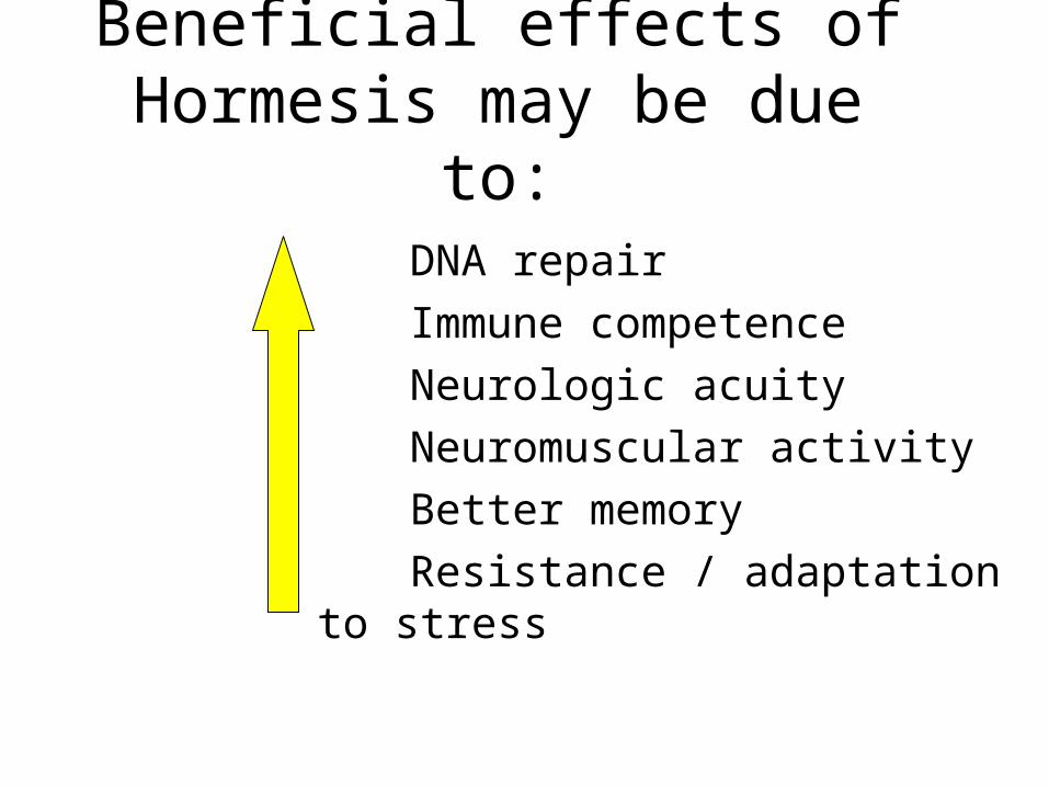
Beneficial effects of Hormesis may be due to:
DNA repair
Immune competence
Neurologic acuity
Neuromuscular activity
Better memory
Resistance / adaptation to stress

• High energy consumption
• Active growth & development
• Active reproductive function
Several lines of investigations have shown that manipulation of the genome will result in changes of the phenome. These changes involve alteration of
the endocrine signaling with a shift
• Reduce energy consumption• Arrest of growth, development, reproductive function• High resistance to stress
From To

Among invertebrates, the most used models have been the fly (Drosophila melanogaster) and the nematode (C. elegans)
Suppression of the receptor for insulin/IGF hormone will produce a mutant nematode that will live 6x longer than corresponding controls and be more resistant to all stress, but they will not grow, undergo development, or reproduce.
C. Elegans 2 week lifespanhermaphrodite19,000 genes
959 cells

IN FLIES (Drosophila melanogaster):
Genetic ManipulationInactivation of IGF-1
receptor analog
Decreased growthDelayed maturation
Shift of metabolismfrom aerobicto anaerobic
Greater resistanceto stress
Increased longevityDecreased mortality

IN MAMMALS (Rodents):
Genetic ManipulationInactivation of IGF-1, I, GH,PL, & TSH receptor analog
Increased longevity 18-40% Delayed aging & mortality
Decreased growthDelayed maturation
Most functions normal
Shift of metabolismfrom aerobic to anaerobic
Decreased free radical accumulation
Greater resistanceto stress

• If response to stress is severe & prolonged it may represent a major risk for the “diseases of adaptation”
(e.g. cardiovascular, cognitive, emotional, metabolic diseases)
& shorten the lifespan
• If the response to stress is moderate & of short duration, it may stimulate hormesis:
– the functions of alertness, vigilance & motivation– a greater availability & utilization of metabolic energy– favor DNA repair – improve protein folding (chaperone stimulation)– prevent/decrease free radical accumulation– promote survival and may delay aging

CHAPERONES
Prevent production of Inactive protein
Protein fragmentsProtein aggregates
Intracellular peptides that helpother proteins to fold
WITHOUT CHAPERONES
Miss a fold, prompt a disease Amyloidosis Lung, blood, liver diseasesDiabetes, cancer, infections
Severe stress?

Stress Proteins or Heat Shock Proteins (HSP)
Theyare synthesized in response
To a sudden rise in temperatureOr other types of stress

ON FLIES, WORMS, RODENTS:
LONGEVITY is associated With stimulation (up-regulation)Of genes involved in response to stress including those of HSP
HSPs act as chaperones and promote greater tolerance/resistance to stress (thermic and others)
Hence, increased longevity and hormesis may depend onIncreased HSPs and their actions as chaperones

Interventions to prevent or treat deleterious effects of stress
According Grandmother Pharmacologic/Genetic Psychotherapy
•Good nutrition•Regular exercise•Good habits •Regular medical visits•Good education in youthand continuing into oldage•Avoiding isolation, livingwith family and in community
•Hypnotics & sedatives•Tranquilizers &Anti-anxiety drugs•Hormones •others
•Psychiatric counseling•Meditation•Yoga •Continuing interactionwith family & community

Coping Skills to Withstand Stress
Knowledgee.g. years
of education
Inner Resourcese.g. beliefs,assumptions
Spiritualitye.g. religious
beliefs
Social supporte.g. interpersonal
relations

Risk Factors (Allostatic Load)Endangering Health and Shortening Life Span
Elevated Physiologic Indices (at risk) Systolic blood pressure: 148 mm Hg
Diastolic blood pressure: 83 mm Hg
Waist-hip ration: 0.94
Tot al cholesterol-High Density Lipoprotein ratio: 5.9
Tot al glycosylated hemoglobin level: 7.1%
Urinary cortisol level: 25.7 mg/g creatinine
Urinary epinephrine level: 5 mg/ g creatinine
Urinary norepinephrine level: 48 mg/g creatinine
Lowered Physiologic Indices (at risk) HDL cholesterol level: 1.45 mmol/ L
DHEA (Dehydroepiandrosterone) level: 2.5 mol/ L
Table 10.9

Questions
• How does the body respond to stress?
• What are some ways to prevent deleterious effects of stress?
• What did we learn from the flies, worms, rodents examples?
• How is the response to stress different with aging?

Hypothalmo-Pituitary-Thyroid System

Table 13.3Major Actions of Thyroid
Hormones• Calorigenesis
• Metabolism
• Brain maturation
• Behavior
• Growth & development

Figure 13-3
CNS
HYPOTHALAMUS
TRH
PITUITARY
TSH
THYROID GLAND
T3 T4 rT3
TARGET CELLS
T4 T3
INTRACELLULAR (NUCLEAR) BINDING
METABOLIC RESPONSE
CLEARANCE
FREE &BOUND
(-)
(-)

Table 13-2: Some MORPHOLOGIC Changes in the Thyroid Gland with Aging
FOLLICLES:- Are distended
- Change in color- Epithelium flattened w/
reduced secretion
Fewer mitoses
Increased connective tissue;
Fibrosis
Atherosclerotic changes

Table 13-2 (con’t.): Some SECRETORY Changes in the Thyroid Gland with Aging
Simultaneously decreased secretion and metabolic
clearance of T4 with resulting essentially normal levels
Failure of up-regulation of
T3 nuclear receptors
peripheral conversion of T4 to T3
TSH levels in 10% of the elderly, associated
in antithyroid antibodies, present even in the absence of
manifestations of hypothyroidism
circulating T3 levels but generally within
the normal (lower) range

Table 13-1: Some Critical Aspects of Thyroid Hormone Regulation
1. Major source of circulating T3 from peripheral deiodination of T4 (NOT from thyroid gland secretion)
2. The negative feedback at the pituitary anterior lobe is mainly through T4 (taken from circulation & converted into T3)
3. The peripheral deiodination of T4 depends on the physiological state of the organism. It allows an autonomy of response of the tissues to the hormones.
4. Deiodination can convert T4 (a less biologically active hormone) to T3 (a more active hormone). This conversion depends on the activity of the various deiodinating enzymes.

Table 13-5 In the Elderly, Thermoregulatory Insufficiency
Results from:
Decreased heat production,Decreased body mass,
Reduced muscle activity,Less efficient shivering,
Reduced sweating response, Less efficient vasomotor responses,Decline in temperature perception.

Table 13-6 Autoimmune Diseases of the Thyroid Gland
Characteristics Graves’ Disease Hashimoto’s Thyroiditis
Thyroid Status Hyperthyroid Hypothyroid
TSH Generally undetectable Normal to elevated
T4, T3 (serum) Above normal Below normal
Antibodies(ABs) Stimulatory ABs compete with TSH at receptor sites
Loss of TSH control over thyroid function
Some ABs block TSH actions
Autoantibodies against thyroglobulin, T3, T4, thyroid destroy thyroid microsomal and nuclear components
Generally present Generally present
Lymphocytic Invasion Limited Marked
Female:Male Ratio As high as 10:1 As high as 10:1

Questions
• What does thyroid hormone do?
• What are the morphological changes with aging?
• What are the secretory changes with aging?

Diabetes

Pancreas endocrine functions
• B cells: insulin (stores glucose)
• A cells: glucagon (mobilizes glucose)
• D cells: Somatostatin (regulatory function)
• F cells: pancreatic polypeptide (regulatory function)
• Don’t forget—the pancreas also has exocrine functions, secreting enzymes needed in digestion


Insulin vs. Glucagon
Insulin• Anabolic (building) hormone• Increases glucose transport to
muscles and adipose for use• Stores excess glucose in liver
and muscles as glycogen• Lowers blood glucose• Inhibits gluconeogenesis
(endogenous glucose production)
• Promotes growth overall
Glucagon• Catabolic (breaking down)
hormone• Breaks down glycogen to
increase blood glucose level• Promotes gluconeogenesis

Insulin’s function in detail
• Insulin is stimulated to be secreted by high blood glucose levels (after a meal).– Glucose binds to GLUT 2
receptor on B cell.– Ultimately causes the
exocytosis of insulin from B cells.
• Insulin binds to target cell receptors and this complex is taken into the cell.
• Insulin now stimulates GLUT 4 to bring glucose into the cells.
• Glucose levels in the blood now decline

Pancreas changes with aging
• Atrophy• Increased incidence of tumor• Presence of amyloid material and
lipofuscin granules (signs of abnormal cell metabolism)
• But these changes can’t account for the degree of metabolic change we see in elderly individuals. There must be a change in sensitivity to insulin in the body!
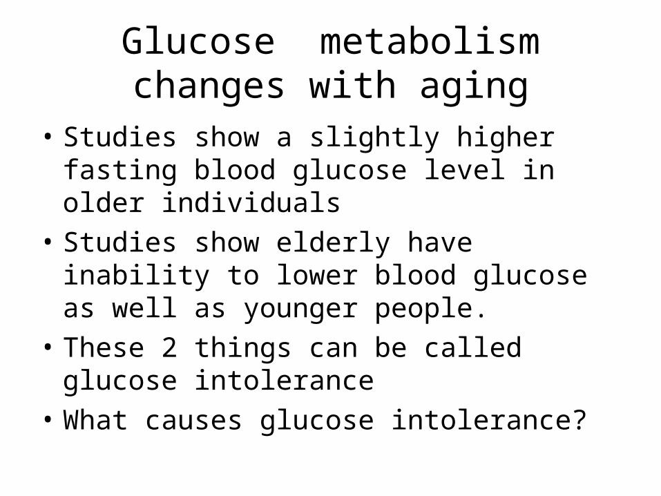
Glucose metabolism changes with aging
• Studies show a slightly higher fasting blood glucose level in older individuals
• Studies show elderly have inability to lower blood glucose as well as younger people.
• These 2 things can be called glucose intolerance
• What causes glucose intolerance?

What is responsible for glucose intolerance with aging?
• The pancreas: Insulin secretion may be depressed
• The peripheral tissue receptors may be resistant to insulin
• The liver may not be responding properly to insulin
It is widely believed that the glucose intolerance is due to insulin resistance at the peripheral tissues.

Insulin resistance: an explanation for glucose intolerance in elderly
• Insulin resistance: failure of insulin to stimulate glucose uptake by peripheral tissue.– No problem with insulin secretion, metabolism in the
elderly
• Resistance due to – receptor problem?– Post-receptor pathway problem?
Due to defect in the signaling pathway once insulin has attached to its receptor.

Why else do elderly have glucose intolerance?
• Loss of hepatic sensitivity to insulin and reduced glycogenesis
• Increased glucagon levels (thus opposing insulin’s effects)
• Changes in diet/exercise• Impaired glucose uptake in muscles and loss of muscle
mass• Increase in adipose tissue (obesity) which may
contribute to impaired uptake in adipose tissue. – Cell enlargement reduces the numbers of receptors
concentration of receptors on cell surface– remember that there may be an overall decrease in number of
insulin receptors.

Diabetes type 2
• Insulin resistance that meets criteria for significantly impaired glucose tolerance, as measured by fasting and glucose tolerance tests. – Glucose of 126 mg/dL or higher after an overnight fast
on more than one occasion. (Fasting test)– After 75 g oral glucose, diagnostic values are 200
mg/dL or more 2 hours after the oral glucose. (Tolerance test)
• Insulin secretory capacity is partially preserved (contrast to Diabetes type 1 in which B cells are destroyed)

Prevalence/Risk factors
• 1999 study showed it affects 7% of US population; 16-20% of adults over age 65.
• Risk factors:– Age– Reduced physical activity– Obesity: adipocytes secrete factors that modulate
insulin activity in a negative way– Ethnicity differences
• Need to screen high risk individuals because sx show up late

Pathogenesis
Caused by genetic and environmental influences• Impaired insulin sensitivity at peripheral cells• Impaired insulin secretion• Increased liver production of glucose because
liver not responsive to insulin’s inhibitory effects on gluconeogenesis
• Often insulin secretion become impaired after a period of insulin insensitivity, causing B cells to work too hard and thus fail

Consequences of Type 2 diabetes
• Microvascular changes– Infections/Gangrene– Blindness– Atherosclerosis (due to changes in arterial wall)
• Macrovascular changes– Stroke– Heart Disease
• Nephropathy (Kidney disease)• Neuropathies (gut motility slowed, sensation
changes in feet)

Theories of Complications
1. High levels of glucose lead to formation of Advanced Glycosylation End products (AGEs). They cross-link proteins and accelerate atherosclerosis, kidney damage, artery wall damage
2. Excess Glucose is metabolized through a different pathway, the sorbitol pathway which forms free radicals
3. Excess glucose activates Protein Kinase C and alters transcription/translation and thus causes damage

Treatment
• Lifestyle modification! – Diet– Weight loss– Exercise
• Pharmacologic– Reduce insulin resistance– Stimulate insulin secretion– Give insulin

Questions
• What is diabetes type 2?
• What is glucose intolerance?
• What is the proposed mechanism for glucose intolerance in the elderly?
• What are complications of diabetes?
• How does insulin work?


