![Apoptosis as anticancer mechanism: function and ... · apoptosis [2]. Usually, the balance between the pro-apoptotic and anti-apoptotic protein regulators is a Review critical key](https://static.fdocuments.in/doc/165x107/5e7309828c15867a030037eb/apoptosis-as-anticancer-mechanism-function-and-apoptosis-2-usually-the.jpg)
TO DIE OR TO DIFFERENTIATE: APOPTOTIC AND NON …own ability to ubiquitinate itself. Once the...
165
TO DIE OR TO DIFFERENTIATE: APOPTOTIC AND NON-APOPTOTIC ROLES OF DEATH MOLECULES IN DROSOPHILA MELANOGASTER Thesis by Jun Ryul Huh In Partial Fulfillment of the Requirements for the Degree of Doctor of Philosophy California Institute of Technology Pasadena, California 2005 (Defended May 17, 2005)
Transcript of TO DIE OR TO DIFFERENTIATE: APOPTOTIC AND NON …own ability to ubiquitinate itself. Once the...

TO DIE OR TO DIFFERENTIATE: APOPTOTIC AND NON-APOPTOTIC
ROLES OF DEATH MOLECULES IN DROSOPHILA MELANOGASTER
Thesis by
Jun Ryul Huh
In Partial Fulfillment of the Requirements
for the Degree of
Doctor of Philosophy
California Institute of Technology
Pasadena, California
2005
(Defended May 17, 2005)

ii
© 2005
Jun R. Huh
All Rights Reserved

iii
ACKNOWLEDGMENTS
Upon looking back at when I started at Caltech, I realize how much I was able to learn
during the course of my graduate studies. I believe I became an independent scientist
with the ability to pose interesting questions, find efficient ways to tackle them, and try to
solve them. Needless to say, none of this would have ever been possible without my
mentor, Bruce Hay. His office was always open day and night, to give me advice and
help. I was fascinated by his great dedication, energy, and enthusiasm for science. It is
these qualities that I have tried to emulate. If I have accomplished anything during the
past years, it was all thanks to his guidance and support.
I am grateful to my committee members David Anderson, Bill Dunphy, Paul Sternberg,
and Kai Zinn for their support and advice throughout my years at Caltech.
I would like to thank the Hay lab and the people in administration in the biology
department, as well as those in the Caltech EM and the animal facility. I would also like
to thank my friends in biology (dubbed as ‘Bio-yung-sa’). Lastly, I would like to
dedicate my thesis to my parents. Without their belief and encouragement, I simply
could not have dealt with the difficult days.

iv
ABSTRACT
Virtually every cell, if not all, are ready to die under stressful conditions or by
necessity during animal development. In Drosophila, three pro-apoptotic proteins,
Rpr/Hid/Grim, had been found to induce cell death by preventing the function of the cell
death inhibitor, DIAP1. However, the mechanistic details of this process were largely
unknown. We have found that Rpr/Hid/Grim induce DIAP1 destabilization through
ubiquitination and general translational inhibition. Moreover, from the in-vitro and in-
vivo studies, we also found that ubiquitination of DIAP1 by Hid is dependant on DIAP1’s
own ability to ubiquitinate itself. Once the life-or-death decision is made, cells can
efficiently start apoptosis by quickly removing pre-existing death inhibitors using these
mechanisms.
In addition to the canonical roles of death machinery, we have also studied their
roles in non-apoptotic developmental processes. In the testis, germline stem cells
ultimately give rise to 64 individual sperms. Spermatocytes, and later, spermatids,
develop within a single membranous structure, or syncytium. Formation of free-
swimming sperms requires the encapsulation of each spermatid by an independent
plasma membrane and the elimination of most of the sperm cytoplasm. We demonstrated
that at least three independent caspase activation pathways are likely to be involved in
these processes with different spatial and temporal activation patterns, and that a global
inhibition of caspase activity results in male sterility.
External stresses such as radiation and heat shock were known to induce large
amounts of cell death (up to 60% of the total cell population) in proliferative tissues like
Drosophila larval imaginal discs. Interestingly, larvae exposed to such stress ultimately

v
develop into normal adult flies. This is facilitated by the compensatory proliferation of
cells that neighbor the dying cells. In order to study the mechanistic basis for this process,
we uncoupled cell death from death activation by expressing Hid in the presence of P35,
a viral inhibitor of effecter caspases. Interestingly, neighboring cells of clones expressing
Hid underwent compensatory proliferation, which was no longer observed when we
blocked the activation of initiator caspase, Dronc. Our observations indicate that non-
apoptotic Dronc activity is required for the generation of a non-autonomous proliferation
signal.

vi
TABLE OF CONTENTS
Acknowledgments iii
Abstract iv
Chapter 1: Introduction: A fly’s-eye view of death; an insight from 1 Drosophila apoptosis
Chapter 2: Apoptosis inducers Hid, Rpr, and Grim negatively regulate 48
levels of the caspase inhibitor DIAP1 by distinct mechanisms.
Chapter 3: Multiple apoptotic caspase cascades are required in non-apoptotic 82
roles for Drosophila spermatid individualization.
Chapter 4: Compensatory proliferation induced by cell death in the 123
Drosophila wing disc requires activity of the apical cell death
caspase Dronc in a non-apoptotic role.
Chapter 5: Summary: Future Directions 146

1
CHAPTER 1
Introduction: A fly’s-eye view of death; an insight from Drosophila apoptosis
Jun R. Huh and Bruce A. Hay
(In preparation)

2
Programmed cell death is essential during animal development. It regulates tissue
homeostasis by removing superfluous or damaged cells. For past 15 years, studies from
worms, fruit flies, and mice have provided us with lots of information about how a
myriad of cell death players are regulated, interact with each other, and respond to
external stimuli in order to achieve "death" in controlled a way. The importance of this
mechanism can be inferred from the fact that many pathological conditions, including
cancers, are usually found to have uncontrolled components in the apoptotic pathway.
Many apoptotic players have non-apoptotic roles during animal development making the
apoptosis world much more complicated than previously envisioned.
Introduction
Programmed cell death (PCD) or apoptosis is required for sculpting structures, deleting
unneeded structures, controlling cell numbers by eliminating superfluous or harmful cells,
and producing differentiated cells during animal development [1]. Many external and
internal stimuli can lead to PCD including DNA damage, binding of death ligands to
death receptors, withdrawal of growth factors, and transcriptional up-regulation of death
activators. Caspases, a family of cysteine proteases, become a converging point for the
various upstream events of PCD. If a cell decides to kill itself either by triggering the
internal death program or by responding to external stimuli, a set of proteases called
upstream caspases are activated. These caspases have a long polypeptide stretch in the N
terminus that the other caspases lack. Upstream caspases can be activated as long as they
are brought into close proximity to each other via adaptor molecules. The proximity

3
sometimes leads to their cross-cleavages [2, 3]. The active versions of upstream caspases,
which usually exist as dimmers, cause cleavage of downstream caspases with shorter or
no N terminal polypeptide stretch. Since the activity of downstream caspases is believed
to be responsible for most morphological features of PCD, these caspases are thought to
be as executioner caspases. In accordance with this idea, downstream caspases are able
to cleave many substrates including nuclear lamins and inhibitors of DNA nucleases,
which leads to DNA laddering [4], one of the well-known apoptotic hallmarks.
Because PCD results in devastating effects on cells and it cannot be easily undone, a fine
orchestration of various players, both positive and negative regulators of apoptosis, is
absolutely essential. If some part of this regulation becomes out of control, too much or
too little cell death tends to occur, most of which ultimately lead to detrimental outcomes
like neurodegenerative diseases or cancer in animals. Understanding how PCD occurs
and is regulated, therefore, is of utmost importance not only to the basic scientists
studying animal development, but also to the medical personnel fighting against such
diseases. Most information about PCD, if not all, has been acquired by studying animal
model organisms such as worms, fruit flies, and mice (Figure 1). In this review, we first
start by explaining the similarities and the differences of PCD among these three different
model organisms. We then turn to Drosophila melanogaster, the fruit fly system, to
highlight dynamic interactions among a myriad of cell death regulators and explain how
they fit into the complicated, but elaborate, regulatory network of PCD.

4
Figure 1. The core apoptosis machine compared in Caenorhabditis elegans,
Drosophila melanogaster, and mammals.
(a) In C. elegans the adaptor protein CED-4 promotes the activation of the caspase CED-3. CED-4 activity
is inhibited by the Bcl-2 family member, CED-9. Various stimuli promote death by inducing tissue-specific
expression of EGL-1, which disrupts CED-9 function. (b) In D. melanogaster the adaptor protein Ark
(homologous to CED-4 in worms and Apaf-1 in mammals) promotes activation of the apical caspase Dronc
in many cells that should normally live. This activation might be regulated by the pro-and anti-apoptotic
multidomain Bcl-2 family members Debcl and Buffy, but this is largely speculative (indicated by the
question mark associated with the arrow). DIAP1, an inhibitor of apoptosis protein (IAP), inhibits Dronc
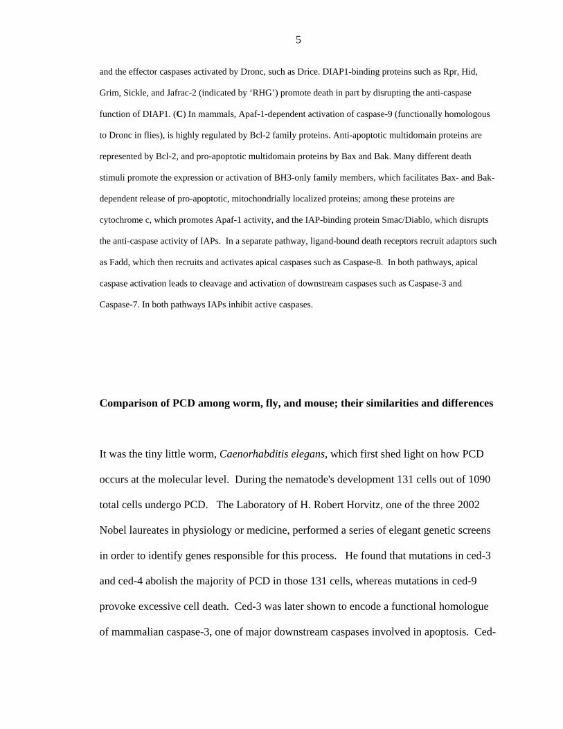
5
and the effector caspases activated by Dronc, such as Drice. DIAP1-binding proteins such as Rpr, Hid,
Grim, Sickle, and Jafrac-2 (indicated by ‘RHG’) promote death in part by disrupting the anti-caspase
function of DIAP1. (C) In mammals, Apaf-1-dependent activation of caspase-9 (functionally homologous
to Dronc in flies), is highly regulated by Bcl-2 family proteins. Anti-apoptotic multidomain proteins are
represented by Bcl-2, and pro-apoptotic multidomain proteins by Bax and Bak. Many different death
stimuli promote the expression or activation of BH3-only family members, which facilitates Bax- and Bak-
dependent release of pro-apoptotic, mitochondrially localized proteins; among these proteins are
cytochrome c, which promotes Apaf-1 activity, and the IAP-binding protein Smac/Diablo, which disrupts
the anti-caspase activity of IAPs. In a separate pathway, ligand-bound death receptors recruit adaptors such
as Fadd, which then recruits and activates apical caspases such as Caspase-8. In both pathways, apical
caspase activation leads to cleavage and activation of downstream caspases such as Caspase-3 and
Caspase-7. In both pathways IAPs inhibit active caspases.
Comparison of PCD among worm, fly, and mouse; their similarities and differences
It was the tiny little worm, Caenorhabditis elegans, which first shed light on how PCD
occurs at the molecular level. During the nematode's development 131 cells out of 1090
total cells undergo PCD. The Laboratory of H. Robert Horvitz, one of the three 2002
Nobel laureates in physiology or medicine, performed a series of elegant genetic screens
in order to identify genes responsible for this process. He found that mutations in ced-3
and ced-4 abolish the majority of PCD in those 131 cells, whereas mutations in ced-9
provoke excessive cell death. Ced-3 was later shown to encode a functional homologue
of mammalian caspase-3, one of major downstream caspases involved in apoptosis. Ced-

6
4 was found to be an adaptor molecule whose function is required for Ced-3 activation
[5]. Likewise, Apaf-1, the mammalian homologue of ced-4, activates caspase-9, which
ultimately activates caspase-3. In summary, ced-3 and ced-4 work as positive regulators
for PCD. However, cell death cannot be regulated if the animal only possesses an
activation mechanism. Thus, negative regulators of PCD are also needed for controlling
cell death. Ced-9, a mammalian anti-apoptotic bcl-2 protein homologue, inhibits the
function of Ced-4 by directly binding to it [6]. When cells decide to die the upstream
regulator Egl-1 becomes transcriptionally up-regulated and negatively regulates Ced-9,
ultimately leading to the activation of ced-3 with the help of ced-4 [5]. In worms,
however, endogenous caspase inhibitors, which are able to directly block the function of
ced-3, have not been identified. This observation suggests that most important decision
for PCD in C. Elegans seems to be made at the upstream level by regulating induction of
the pro-apoptotic gene egl-1.
In contrast to the strategy utilized in worms, ‘to die or not to die’ decisions in flies are
made at the level of downstream players, caspases. Several studies from fruit fly cell
lines clearly show that the caspase Dronc is in a continuously activated mode even
without any upstream input. In other words, unlike the worm's ced-4, where it is under a
continuous inhibition by Ced-9, the function of the fly Apaf-1 homologue, dArk does not
seem to be inhibited in a normal situation. Instead, inhibition of caspase activity in non-
dying cells is achieved through the function of DIAP1, a Drosophila inhibitor of
apoptosis (IAP) protein. IAPs were originally identified as cell death inhibitors from
several insect viruses [7]. The importance of the physiological role of DIAP1 is most well

7
exemplified by the observation that virtually every single cell in homozygous DIAP1
mutant embryos or larval tissues lacking DIAP1 undergoes PCD due to the excessive
caspase activation [8, 9]. Obviously it seems like there is continuous caspase activation
and concomitant inhibition by DIAP1 in Drosophila. When this delicate balance is
disrupted, likely due to the increased activation of caspases or less inhibition by DIAP1,
the daunting outcome of PCD can be easily envisioned. For the fruit fly system, the latter
seems to be a major trigger for PCD. When cells are destined to die, transcriptional or
post-transcriptional up-regulation of cell death activators like Rpr/Hid/Grim, and possibly
Sickle and Jafrac-2, easily tip the balance toward less caspase inhibition by preventing
DIAP1’s ability as a caspase inhibitor. Thus cells in a sense, at least for Drosophila, are
likely to die not because they choose to die, but because they choose not to live. In other
words, the default pathway for the fruit fly cells is to kill themselves, but they simply
survive as long as DIAP1-mediated inhibition prevails.
Like many other biological pathways, the mammalian pathway for PCD is more complex
and can regulate and execute PCD in a multitude of different ways. However, the core
features of PCD are well preserved. Mammalian cells harbor upstream cell death
activators such as BH3-only or bcl2 proteins, adaptor molecules like Apaf-1 or FADD,
and upstream and downstream caspases. They appear to use variants of both strategies
found in worms and fruit fly PCD. Firstly, like in worms, BH3-only molecules,
mammalian counterparts of Egl-1, can induce cell death by disrupting the function of
anti-apoptotic Bcl-2 family proteins, which prevent the activity of pro-apoptotic Bcl-2
family proteins. Once activated, pro-apoptotic Bcl-2 proteins result in the release of
Cytochrome-c from the mitochondria, which binds to Apaf-1 and ultimately leads to the

8
activation of caspase-9. Secondly, like in flies, caspase inhibition by the mammalian IAP
family can be relieved by cytoplasmic Smac/Diablo, a mammalian counterpart of fly
Rpr/Hid/Grim, which is localized in the mitochondria of non-dying cells. Thus, it
appears that both the activation by pro-apoptotic players through mitochondria and the
inhibition of an IAP-dependent-caspase-inhibition are utilized in mammalian PCD.
Contrary to this approach, worms and flies seem to mainly utilize only one of these
mechanisms. Besides these two strategies mammals are also equipped with a third
method to cause PCD, the receptor mediated death pathway that allows the animal to
quickly respond to rapid environmental changes more efficiently. The best example of
this can be taken from the mammalian immune system. When Fas ligands in immune
cells impinge onto the infected cells by interacting with Fas receptors on their membranes,
Caspase-8 is recruited and activated via the adaptor molecule Fadd, resulting in PCD.
Curiously, in a fruit fly system, structural homologues of these players seem to be used in
a functionally distinct pathway, i.e., innate immune response pathway, which will be
described in the later part of this review.
Now we will change gears and turn to Drosophila system in order to outline various
players in PCD, summarize what we know and what we don’t know at this point, and
provide some insights on how we can tackle some of the unanswered, but fundamental,
questions.
Various players in Drosophila PCD

9
Diap1 as a peace keeper
The most eminent player in a world of Drosophila PCD is DIAP1 (Figure 2). DIAP1 is
responsible for the survival of most, if not all, fly cells. DIAP1 was firstly isolated
among the cellular IAPs by a genetic screen for cell death suppressors [10]. When over-
expressed, DIAP1 is sufficient to inhibit Rpr/Hid/Grim/Caspase mediated cell death.
DIAP1’s physiological significance can be validated from several lines of evidence,
mainly performed by us and several other groups [8, 11, 12]. The first line of evidence is
that mutant DIAP1 cells undergo apoptosis. Secondly, major cell death activators like
Rpr/Hid/Grim in Drosophila seem to exercise their function specifically by blocking
DIAP1’s role as a cell death inhibitor. Lastly DIAP1 can directly bind and inhibit
caspases. In the absence of DIAP1, cells die due to the excessive activation of
downstream caspase Drice [13]. In accordance with this, over-expression of P35 (a
suicide inhibitor for caspases derived from the Autographa californica
nucleopolyhedrovirus [14]) can rescue cell death caused by loss of DIAP1 in fly eyes.
The role of another IAP protein, DIAP2, is less clear. Ectopic expression of DIAP2 in fly
eyes block Rpr and Grim induced cell death [10], and inhibition of its expression in the
S2 cell manifests increased sensitivity to certain types of apoptosis [15]. The insect
hormone Ecdysone was reported to induce transcriptional up-regulation of DIAP2,
suggesting its role during salivary glands apoptosis [16]. However, as the DIAP2 mutant
is not available its physiological function remains a mystery.

10
Figure 2. Regulation of cell death in Drosophila melanogaster.
A more detailed illustration of several cell death pathways. The TNF-family ligand Eiger binds the
receptor Wengen in physiological contexts that remain to be identified. This leads through ill-defined
steps (the two arrows) to activation of Misshapen (Msn), the c-Jun N-terminal kinase (JNK) kinase
kinase kinase (JNKKKK). Msn phosphorylates and activates dTAK1 (the JNKKK), which promotes
activation of Hemipterous (Hep; the JNKK). Hep phosphorylates and activates JNK. In a second
pathway leading to JNK activation, binding of Reaper (Rpr) to DIAP1 results in stabilization of the
tumor-necrosis factor-associated factor 1 (Traf1); this leads to the activation of the apoptosis signal-
regulating kinase 1 (Ask1) and to JNK activation. JNK activation promotes cell death in some, but by
no means all, contexts. Members of the RHG family of DIAP1-binding proteins (pink cloud) are
regulated through multiple pathways. Rpr expression is activated in most, if not all, dying cells in the

11
embryo and is also induced by various stimuli. Jafrac2 is released from the endoplasmic reticulum in
response to UV irradiation (ER stress). Hid is negatively regulated by the EGF
receptor/Ras/MAPKinase pathway through phosphorylation (EGF signaling). The bantam miRNA
negatively regulates Hid translation, whereas Hippo/Salvador/Warts stimulate Hid expression. All
RHG family members bind to DIAP1 and inhibit its anti-apoptotic activities. In addition, at least Rpr
and Grim also have DIAP1-independent pro-apoptotic activities, one of which is the general inhibition
of translation. The mir-14 micro RNA inhibits cell death and fat storage through unknown mechanisms.
There is one major question that still need to be answered; what makes Dronc, an
upstream caspase, continuously processed (or activated) in S2 cells, and why is the
removal of DIAP1 sufficient to cause PCD in flies? Unlike mammalian Apaf-1, dArk
does not require Cyt-c to induce caspase activation [15, 17]. It has been suggested that
either the dArk-Dronc complex is self-sufficient for its activation or some other unknown
activation mechanism exists. In any rate, a genetic screen to find potential suppressors of
small eye phenotype, caused by expression of Diap1 double strand RNA (dsRNA), would
provide some clue for this question. Alternatively, performing mass spectrometry
analysis using dArk as tagged baits would be useful to identify dArk interacting proteins,
some of which might lead us to better understanding about how dArk-Dronc is activated.
Rpr/Hid/Grim as an axis of evil
A glimpse of molecular mechanism of PCD in Drosophila came from the discovery that a
small genomic deletion region (H99) covering the Rpr/Hid/Grim genes removes the

12
majority of cell death during embryogenesis [18]. This was striking, as it clearly
suggested that cell death itself is regulated by genes. Over-expression of any of these
genes also caused PCD in many tissues, including fly eyes. In dying cells Rpr and Grim
are up-regulated, whereas Hid was shown to exist in both dying and non-dying cells. Rpr
is induced by hormone Ecdysone, P53/DNA damage, or Hox proteins following cell
death signals [19]. The Rpr mutant has an enlarged central nerve system without affecting
most developmental cell death [20]. Unfortunately little is known about Grim. Hid,
however, is negatively regulated at the transcriptional or the post-transcriptional level by
the Erk pathway, which functions not only for cell death, but for cell survival pathway
[21, 22]. The Hid mutant has defects in normally occurring cell death during pupal eye
development [23] and manifests a semi-lethal phenotype. Even though H99 abrogates
most PCD and makes the embryo lethal, no single gene seems to be absolutely essential
for normally occurring PCD. Thus, Rpr/Hid/Grim might either work together to mediate
cell death or have some redundant function. Curiously Rpr/Hid/Grim do not have high
structural homology with each other. They do however have limited homology at the
short N-terminal region. It was this region that was shown to bind and inhibit Diap1 (but
other regions also have a binding capability). Interestingly, a mammalian counterpart of
Rpr/Hid/Grim, named Smac/Diablo, also has limited homology in this N-terminal region.
Based on this structural information, two more death players, Sickle and Jafrac2, have
been found [24-27]. Sickle shows a very similar transcription pattern to that of Rpr.
Jafrac2 is released into cytosol from the endoplsmic reticulum following an induction of
apoptosis. Both genes can induce PCD in over-expression contexts, but verifying their
physiological roles has yet to be accomplished because of the lack of mutants.

13
Drosophila also seems to have an extrinsic cell death pathway involving the tumor
necrosis factor (TNF) ligands, Eiger [28, 29], and its receptor Wengen [30]. Eiger is able
to induce PCD in a JNK (Jun amino terminal kinase) pathway dependent manner. Since
Eiger mutants, however, have no obvious defects either for PCD or for immune response,
its physiological role is still a mystery. One hypothesis to explain this is that Eiger might
be required only for cell death under certain stressful conditions. A recent observation
lends credence to this idea since eiger mutants live longer after bacterial infections
without affecting bacterial proliferation, suggesting its role in pathogen induced animal
death [31].
Caspases as rogue executioners
In Drosophila, seven caspases exist [3]. Dredd, Dronc, and Dream/Strica are thought to
act as upstream caspases because they have long pro-domains, whereas Drice, Dcp-1,
Decay, and Daydream/Damm, which lack a pro domain or have a relatively short one, act
as downstream caspases. Regardless of the size of the pro-domain the majority of them
can kill cells when they are over-expressed in fly tissues or cell lines. Their endogenous
roles, however, are mostly unknown due mainly to the lack of corresponding mutants.
Dredd originally was assumed to play a role in PCD [32], but identification of dredd
mutant assigned its role as a signal transducer in an innate immune pathway [33], which
will be discussed more in the later part of this review. Studies from a Dcp-1 mutant by
Herman Stellar lab, initially suggested its role in tracheal development and in

14
developmental PCD because the mutants had larval lethality and melanoma formation
due to the lack of PCD [34]. Another suggested function was the dumping of nurse cell
contents into maturing oocytes during oogenesis [35]. These phenotypes, however, were
found to be caused by the additional loss of the neighboring gene, pita. Mutants that are
only lacking dcp-1 are viable and manifest normal nurse cell death [36]. These mutants
do however appear to affect starvation-induced germline cell death, suggesting Dcp-1’s
role in a stress response PCD pathway.
For Dream/Strica [37], Drice [38], Decay [39], and Daydream/Damm [40], loss-of-
function mutants are not available so their definitive roles in PCD and development are
still unclear. Are they absolutely required for certain types of cell death? Or do they
have somewhat overlapping functions? Without mutants, we cannot answer either of
these questions for sure. But in terms of what they can do, the function of Dronc and
Drice, although mostly well studied, are shedding light on the other genes functions.
Dronc is a counterpart of mammalian caspase-9, and over-expression of its inactive form
works as a dominant-negative because it blocks Rpr/Hid/Grim induced cell death in fly
eyes [41, 42]. Using this Dronc dominant negative form, Dronc's physiological role has
been indirectly pursued. Dronc also cleaves and leads to the activation of downstream
caspases, Dcp-1 and Drice [42]. Ecdysone and its receptor complex were shown to
directly bind to the dronc promoter and induce its transcriptional up-regulation [43, 44],
which implies that the pro-apoptotic role of Dronc is in hormone dependent death.
Recently, three groups have independently reported Dronc mutant phenotypes [45-47].
In accordance with previous data, Dronc is responsible for the majority of the occurring

15
PCD during animal development or by external stress. Specifically Dronc mutants lack
cell death induced by Hid over-expression in fly eyes and by irradiation of wing discs.
Dronc is also required for salivary gland histolysis [46]. Hemocyte cell culture from
Dronc mutants showed resistance against several different death stimulating reagents [45].
However, a couple of things are worthy of note. Dronc mutants still have a few cells
undergoing PCD during embryogenesis, and small portions of Dronc mutants (<10%)
survived up to adulthood [47], both of which are different from those in H99 deletion
mutants. These observations clearly support an idea that either Dronc independent
caspase activation mechanisms exist or Rpr/Hid/Grim can achieve cell death through a
non-caspase activation cascade. Generation of the Dream/Strica mutants, the last
upstream caspase whose function remains at large, will probably clarify if either of these
is correct.
Based on the structural homology to the mammalian caspase-3 and its temporal and
spatial activation pattern in cells undergoing PCD [13], Drice is regarded as one of the
major downstream caspases for PCD. Removal of Drice (and Dronc) by RNAi
completely blocks stress-induced cell death in S2 cells, suggesting their essential roles in
PCD [15, 48]. Once activated by Dronc, Drice can also cleave the boundary between the
pro-domain and large subunit of Dronc, which could potentially makes it less sensitive to
the DIAP1 mediated inhibition [49] comprising a positive feedback loop [50]. A recent
study also suggested that cleavage of the Drice pro-domain, and thereby an exposure of
an IAP binding motif in the large subunit, is essential for Drice’s binding ability to
DIAP1 [51]. Drice is also transcriptionally activated by ecdysone [52], which presages

16
its role in hormone-dependent cell death. Again the definite role of Drice will be
elucidated once Drice mutant becomes available. One interesting question to ask is if the
Drice mutant is embryonic lethal like H99. We don’t know the answer yet, but Drice
might not be absolutely required for PCD like Dronc. The fly has three more
downstream caspases including Dcp-1, which has the highest structural homology to
Drice. Even though Dcp-1 mutants develop quite normally other than having some minor
defects in stress induced cell death, it could still well be that Dcp-1 and Drice have
redundant roles. Specifically the level of Dcp-1 protein or its activity might be increased
when Drice function is inhibited. Answers to all of these questions will be available once
the Drice and Dcp-1 double mutant is generated.
A battle between DIAP1 versus Rpr/Hid/Grim and caspases.
As mentioned earlier, the endogenous role of DIAP1 keeps caspases from being activated,
either by direct inhibition or post-translational modification. Once pro-apoptotic players
such as Rpr/Hid/Grim bind to DIAP1, however, it is no longer able to inhibit caspase
activity (Figure 3). For the past several years, many groups including ours have studied
dynamic interactions among these molecules in order to address how DIAP1 is regulated
by Rpr/Hid/Grim and caspases. It would not be unreasonable to postulate that DIAP1 is
actively regulated through multiple mechanisms since it plays a key role for regulating
PCD during animal development. Indeed, several transcriptional and post-transcriptional
regulation mechanisms have been unveiled.

17
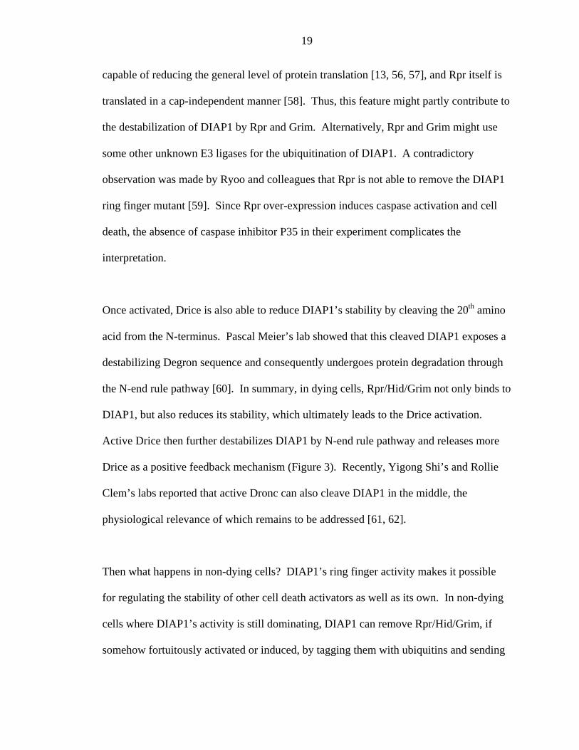
Figure 3. Dynamic interactions between cell death activators and DIAP1 in
Drosophila melanogaster.

18
Because different regions of DIAP1 are involved in these regulations, it is helpful to
explain its structure before delving into more details. DIAP1 consists of two N-terminal
BIR (Baculovirus IAP repeat) domains and one C-terminal Ring finger domain. The BIR
is a functional domain that interacts with other proteins and has been found in many other
IAP family proteins including metazoan cell death inhibitors and yeast cell cycle
regulators [7]. Ring fingers have been found in many diverse proteins with many
different functions, some of which confer ubiquitin E3 ligase activity. Genetic,
biochemical, and structural studies have shown that Rpr/Hid/Grim, Dronc, and Drice can
bind to one or both BIR domains [53]. Specifically, Dronc and the N-terminal peptides
of Rpr/Hid/Grim bind to the same conserved groove of DIAP1’s BIR2 domain [49]. In a
normal situation, this region is occupied by Dronc (and possibly this binding induces
Dronc ubiquitination [54]) so that no Dronc activation occurs. Upon the induction of
PCD, Rpr/Hid/Grim can invade the interface of the Diap1-Dronc complex and liberate
Dronc by sequestering DIAP1. Now Dronc undergoes its activation with the help of
dArk, which ultimately cleaves Drice and Dcp-1 [42], leading to their subsequent
activation. Rpr/Hid/Grim, then, decrease DIAP1's half life by tagging it with ubiquitins
for protein degradation. DIAP1’s ring finger activity is required for Hid mediated DIAP1
ubiquitination. We know this because Hid is no longer able to decrease the stability of
th6 allele, a Diap1 ring finger mutant with a C412Y mutation [13, 55]. Rpr and Grim,
however, are still able to decrease Th6 protein level, which suggests that they don’t need
DIAP1’s ring finger activity [[13, 55] and unpublished data]. Additionally the DIAP1
ring mutants that work as suppressors for Hid are mild enhancers for Rpr and Grim
induced death [12]. Intriguingly several groups have reported that Rpr and Grim are
19
capable of reducing the general level of protein translation [13, 56, 57], and Rpr itself is
translated in a cap-independent manner [58]. Thus, this feature might partly contribute to
the destabilization of DIAP1 by Rpr and Grim. Alternatively, Rpr and Grim might use
some other unknown E3 ligases for the ubiquitination of DIAP1. A contradictory
observation was made by Ryoo and colleagues that Rpr is not able to remove the DIAP1
ring finger mutant [59]. Since Rpr over-expression induces caspase activation and cell
death, the absence of caspase inhibitor P35 in their experiment complicates the
interpretation.
Once activated, Drice is also able to reduce DIAP1’s stability by cleaving the 20th amino
acid from the N-terminus. Pascal Meier’s lab showed that this cleaved DIAP1 exposes a
destabilizing Degron sequence and consequently undergoes protein degradation through
the N-end rule pathway [60]. In summary, in dying cells, Rpr/Hid/Grim not only binds to
DIAP1, but also reduces its stability, which ultimately leads to the Drice activation.
Active Drice then further destabilizes DIAP1 by N-end rule pathway and releases more
Drice as a positive feedback mechanism (Figure 3). Recently, Yigong Shi’s and Rollie
Clem’s labs reported that active Dronc can also cleave DIAP1 in the middle, the
physiological relevance of which remains to be addressed [61, 62].
Then what happens in non-dying cells? DIAP1’s ring finger activity makes it possible
for regulating the stability of other cell death activators as well as its own. In non-dying
cells where DIAP1’s activity is still dominating, DIAP1 can remove Rpr/Hid/Grim, if
somehow fortuitously activated or induced, by tagging them with ubiquitins and sending

20
them to the proteasome for proteasome mediated degradation [63] (Figure 3). Dronc is
also negatively regulated by DIAP1, but whether or not ubiquitinated Dronc also
undergoes degradation is not clear [49, 54]. Another example can be taken from the JNK
pathway. DIAP1 over-expression has been shown to down-regulate dTraf1, an upstream
JNK activator, by ubiquitination dependent proteasome degradation pathway [64]. At
any rate, the data suggests the role of DIAP1 as a double-edged sword. In non-dying
cells, ring finger activity of DIAP1 prevents unwanted death by active inhibition or
degradation of cell death activators. In dying cells, however, the same activity ensures
accelerated PCD by actively degrading DIAP1 itself with the help of cell death activators.
Then how does DIAP1’s ubiquitination occur? Genetic studies from several different
labs led to the identification of various players in this pathway such as Uba-1 (E1
activation enzyme), UbcD1 (E2 ubiquitin conjugation enzyme), SkpA (a component of
SCF-type E3 ligase), Fat facet (de-ubiquitination enzyme), and Morgue (a novel F box
gene) [59, 65, 66]. Morgue encodes a unique protein that contains both a F box and an
inactive E2 conjugation domain, suggesting its role as an adaptor molecule. Morgue loss
of function partially suppresses Rpr/Hid/Grim induced cell death by possibly stabilizing
DIAP1. However, it is still largely unknown which components, among these, form a
complex with DIAP1 under physiological condition. Are these molecules always in the
same complex with DIAP1 or could some of them be only mustered to this complex
when cells are undergoing apoptosis? These questions could be easily solved through
biochemical and proteomics approaches. One can pull down DIAP1 associating
complexes from S2 cell, either in the presence or absence of cell death stimuli and

21
perform the mass-spectrophotometric analysis. It would be interesting to know which
components are involved in maintaining DIAP1’s own stability and which are involved in
degrading cell death molecules. The other questions are: what is the role of de-
ubiquitination enzymes and what is the function of other Ubiquitin- like molecules such
as Sumo in the regulation of DIAP1’s protein stability? Additionally, DIAP1 was
reported to be regulated by phosphorylation as well. A fly ortholog of a mammalian Mst
kinase, Hippo, can phosphorylate and destabilize DIAP1 [67] presumably by affecting its
auto-ubiquitination efficiency. But how this event occurs awaits more extensive analysis.
In the end, it is not so simple; exceptional rules of engagement
We have discussed the role of DIAP1 and Rpr/Hid/Grim, which are the most well-studied
cell death inhibitor and activators, respectively, in Drosophila PCD, and their mutual and
dynamic interactions. Mutants removing DIAP1 or Rpr/Hid/Grim altogether (H99) show
embryonic lethality, which strongly indicate that both cell death inhibitor and activators
are essential for animal development. Just like too much cell death causes catastrophic
effects, too little cell death also causes deleterious effects like cancer. One of the classic
examples comes from an old finding that bcl-2 once had been known as a tumor inducing
oncogene. Bcl-2 when mutated actually resulted in cancer, not because bcl-2 mutants
activated cell proliferation, but because they blocked PCD [68]. Besides, several
mammalian IAP homologues were found to be up-regulated in cancer cell lines. There is
no cancer in fruit flies. Recently, however, salvador and hippo mutants, whose proteins

22
might be involved in the same pathway, were shown to induce extra-proliferation and
diminish cell death altogether [67, 69-72]. Interestingly, an increase of DIAP1’s
transcription and its protein stability were, as suggested, partly responsible for the
salvador and hippo mutants mediated phenotypes. Moreover, a human ortholog of
salvador is also found to be mutated in several cancer cell lines. After all, having more
DIAP1 is not always a good thing.
Other exceptional cases can be found in Drosophila oogenesis. Maturing oocytes require
a huge influx of cytoplamic materials from neighboring nurse cells. To do this, nurse
cells undergo apoptosis-like-morphological-changes. Intriguingly Lynn Cooley’s lab
found that the H99 germline clone, which removes Rpr/Hid/Grim, didn’t affect PCD in
nurse cells, which strongly suggests the presence of novel upstream apoptotic molecules
[73]. Jafrac-2 and Sickle are good candidates for these upstream apoptotic molecules.
Denise Montell’s lab also reported that certain combinations of DIAP1 mutant alleles or
its mutant clones didn’t give rise to excessive cell death in oogenesis [74]. Instead, there
are other problems including egg chamber polarity defects, extra nurse cells, follicle cell
defects, and border cell migration defects. So it seems clear that non-conventional death
pathways play important roles during Drosophila oogenesis. But it should be noted that
rpr, grim, hid, and diap1 are expressed in the Drosophila ovary [73]. If they are
dispensable for developmental PCD, then why do they need to be present? As we discuss
later, they may play non-apoptotic roles during oocyte development. Otherwise, they
could be responsible for the stress-induced death during oogenesis, where Dcp-1 plays an
important role [36].

23
Other cell death inhibitors: Deterin and dBruce
Deterin possesses a single BIR domain and lacks a ring finger motif. When over-
expressed in cell culture, Deterin inhibits cell death induced by Rpr expression and
cytotoxic challenges [75]. Survivin, a mammalian homologue of Deterin, was reported to
be involved in cytokinesis as well. Since a Deterin mutant is not available, its role in
apoptosis remains unclear.
dBruce is a 530 KDa protein containing a single BIR domain and an E2 ubiquitin
conjugation domain. When over-expressed in fly eyes, it is sufficient to block Rpr and
Grim but not Hid induced cell death. dBruce seems to act as endogenous cell death
inhibitor since removing one copy aggravates Rpr or Grim dependent cell death. Its
mutant is viable but shows a male sterile phenotype [76]. Hyper-condensation of sperm
nuclei was observed in the dBruce mutant, and it was hypothesized that this phenotype
was due to the increased level of caspase activation [77]. Since no supporting data was
provided in the same paper to show that the dBruce mutant has more caspase activity or
that its sterility can be rescued by co-expression of caspase inhibitors, more extensive
characterization of dBruce mutants is required to understand how dBruce functions to
prevent sterility, which might have nothing to do with caspase activity. How does
dBruce inhibit Rpr and Grim induced death? Since dBruce still suppresses lysine
negative Rpr and Grim, its inhibition might not be from the simple ubiquitination of Rpr
and Grim [76]. Moreover, dBruce failed to suppress Dronc and Dream/Strica induced
death. Contrary to the fly data, the proposed functions of mouse Bruce were direct

24
inhibition and ubiquitination mediated proteasomal degradation of caspase-9 and Smac
[78, 79]. The mouse Bruce mutant manifests an embryonic lethal phenotype without any
dramatic evidence of increased cell death. But in accordance with fly data, mutant Bruce
deficient cells were shown to be more susceptible to apoptosis.
Bcl-2 family in Drosophila; Are they hidden weapons of mass destruction?
Structurally fly Bcl-2 family proteins Debcl and Buffy (among many names referring to
these two Bcl-2 proteins, we decided to go by FlyBase nomenclature) belong to the pro-
apoptotic group [80]. Indeed, several groups previously reported that Debcl over-
expression can kill cells [81-83]. However, a RNAi-based study during embryogenesis,
showed that Buffy seems to work as a cell death suppressor [84]. To futher support this
finding, over-expression of Buffy partially suppressed Rpr/Hid/Grim induced cell death
in fly eyes and reduced excessive apoptosis in DIAP1 mutant embryos. More
interestingly, Buffy can completely suppress Debcl induced cell death in fly wings. All
the previous pieces of evidence strengthen the idea that Buffy is a cell death inhibitor
[84]. But the questions remain as to their endogenous roles. It is a prominent task in the
fly cell death field to generate Debcl and Buffy mutants and characterize their phenotype.
As discussed earlier, there seems to be a constitutive driving force in fly PCD, which
allows unleashed dArk dependent Dronc activation. Simple removal of DIAP1, a fly life-
saver, is enough to induce apoptosis without any upstream death activation signal. The
question is if Debcl and Buffy might act at this point, rendering Dronc continually active.

25
One simple way to test this hypothesis right away would be to knock down Debcl in S2
cell by RNAi and see if this abrogates DIAP1 loss mediated Dronc activation. If Debcl is
required for Dronc activation, Debcl RNAi might prevent cells from undergoing
apoptosis even in the absence of DIAP1. As Kumar's group noted, Buffy over-expression
didn’t suppress Dronc induced death in fly eyes, which makes sense if Debcl and Buffy
are upstream regulators of Dronc.
In mammalian systems, Bcl-2 family proteins affect apoptosis through mitochondria by
regulating the release of cell death stimulating molecules such as Cytochrome C (cyt-C).
In flies, however, the involvement of mitochondria and cyt-C in PCD is not clear.
Addition of cyt-C was reported to induce caspase activation in fly embryo extracts [85].
Also, the appearance of altered antigenic moiety of cyt-C correlated with PCD in flies
and Drosophila cell lines [86]. However, the release of cyt-C into cytosol from
mitochondria upon cell death activation was not detected, and more importantly, removal
of two different forms of cyt-C does not affect PCD [15, 17], which is a strong argument
that cyt-C plays no crucial roles in Drosophila PCD. Even though Debcl was previously
shown to localize in mitochondria [83], how they induce apoptosis and the role of
mitochondria remain a mystery. In order to solve this puzzle, it would be intriguing to
study the potential involvement of fly homologues of AIF and Endo G in Debcl mediated
death, as their mammalian counterparts act as death activators and were shown to localize
in mitochondria. Overall, fly Bcl-2 proteins are likely to be involved in Drosophila PCD,
but their physiological identities are still at large.

26
microRNA and cell death
microRNA (miRNA) are small non-coding RNAs generally 21 to 23 nucleotides, which
are originally encoded as 60 to 80 nucleotides stem loop intermediate (or pre-miRNA)
and subsequently cleaved by Dicer [87-89]. Estimates are that approximately 1% of
predicted animal genes encode miRNAs. The role of miRNA is presumed to negatively
regulate certain target genes at the post-transcriptional level, which depends on their
partial complementary sequence homologies. Using the awesome power of fly genetics,
we and the Cohen lab individually identified two miRNAs, mir-14 and bantam, as cell
death inhibitors [90, 91].
Dominant modifier screening allowed us to find a small region harboring the potential
miRNA mir-14. Eye-specific expression of mir-14 acted as a strong suppressor against
Rpr/Hid/Grim or Dronc dependent cell death, and a removal of mir-14 enhanced their cell
death phenotype. mir-14 mutant flies showed pupal lethality and manifested obesity,
potentially caused by increased levels of triacylglycerol. The mutant of mir-14 shows
elevated levels of Drice strongly suggesting that mir-14 is an endogenous cell death
inhibitor. However, as of yet, the target genes for mir-14 are unidentified. Knowing the
target genes for mir-14 is crucial in understanding how mir-14 acts in both fat regulation
and apoptosis. Bantam plays dual roles, i.e., it inhibits cell death by negatively regulating
the cell death activator Hid and promotes cell growth by an unknown mechanism [91].

27
The question is if there are more miRNAs regulating PCD in Drosophila. Computational
approaches to identify miRNA targets provide promising answers to this question [92].
mir-2 and mir-13 are likely to function as cell death inhibitors against Rpr, Grim, and
Sickle induced death, based on target gene prediction and cell reporter analysis. But their
roles in flies need to be validated by making transgenic flies or mutants. Sooner or later,
we may end up having a lot more players in the fly death world, many of which would be
supplied from the miRNA world.
Apostasy of apoptotic players; their non-apoptotic roles during Drosophila
development
As discussed so far, various players in apoptosis are under tight control in order to kill
cells efficiently at specific times. Both negative and positive feedback loops, which
function between many cell death activator and inhibitor molecules, contribute to the
elegant regulation of PCD. Since apoptotic molecules exist in every cell, however, it
would not be surprising to find that the PCD pathway is being used for other
developmental processes. Indeed it would be wasteful for cells to only use cell death
machinery for PCD events. In this review, as we decided to focus on a fruit fly system,
we will only cover a few cases from Drosophila to exemplify the importance of apoptotic
machinery in non-cell death contexts. In the mammalian system, however, more
examples for the role of apoptotic machinery can be easily found including differentiation
of immune cells and certain types of neurons.

28
Capsase in an innate immune response
The adaptive immune response has been thought to play a key role in the fight against an
immense number of pathogens by utilizing specific antibody and antigen interactions.
The importance of the innate immune response, however, can be appreciated from the
fact that the adaptive immune response only offers weak protection in the absence of an
innate immune system [93]. Since the fruit fly doesn’t have an adaptive immunity it can
be a useful genetic system to study innate immunity. Indeed, several labs have performed
elegant genetic screens and successfully identified quite a few molecules involved in the
innate immune response in Drosophila. The Toll-dependent pathway has been suggested
to be involved in gram-positive bacterial and fungal-induced immune response [94, 95],
whereas Imd (immune deficiency) pathway is involved in immune response to gram-
negative bacterial infection [96, 97]. At least two death molecules such as an adaptor
molecule dFadd and an upstream caspase Dredd have been found as essential components
of Imd pathway [98-100]. Imd encodes a protein with a death domain. Its mammalian
homologue, RIP (receptor interacting protein), was known to play an important role in
NF-kB activation pathway and apoptosis [101]. Likewise, Imd relays signals through
Tak1/Relish (a fly NF-kB homologue) and dFadd/Dredd, ultimately leading to the
transcriptional induction of antimicrobial peptides [99, 100, 102]. Imd over-expression
alone is sufficient for inducing antimicrobial genes, and its activity can be blocked by co-
expression of the caspase inhibitor P35 [101]. Moreover, Dredd mutants abolish an
immune response against gram-negative bacterial infection.

29
Caspases usually exert their function by cleaving certain protein substrates after aspartic
acids. Dredd was found to activate Relish through endo-proteolytic cleavage [103]. The
cleaved N-terminal fragment of Relish, separated from its inhibitory C-terminal fragment,
can translocate into a nucleus and subsequently induce transcriptional up-regulation of
many downstream genes. If immune response induces caspase activation, the question is
why immune-active cells don’t undergo PCD. One possibility is that the normal function
of Dredd has nothing to do with PCD. In other words, activated Dredd might be
incapable of activating downstream caspases like Drice or Dcp-1. The other possibility is
that Dredd activity is continuously regulated so that its activity is maintained only enough
to induce immune response not PCD. One interesting observation to support this
hypothesis came from a S2 cell study in O’Farrell’s lab. They suggested that Dnr1
(defense repressor 1), which has ring finger activity, works as a Dredd inhibiting protein,
of which the protein level is also positively regulated by Dredd itself [104]. Thus, once
activated by Imd and dFadd, Dredd induces Relish activation but, at the same time, its
excessive activation can be suppressed by concomitant up-regulation of Dnr1. However,
removing Dnr1 didn’t result in excessive cell death. Instead, a constitutive activation of
immune pathway occurred, which might suggest, if the latter hypothesis is right, the
presence of another Dredd inhibitor to block apoptosis. Whether or not Dnr1 directly
inhibits Dredd, however, needs to be addressed. Biochemical study with purified Dredd
and Dnr1 protein and genetic studies using Dnr1 mutant flies will give us some clue.
Another interesting question is if the over-expression of DNR-1 abrogates Dredd
mediated Relish processing. But the most interesting experiment would be to see if
DNR-1 can also block other caspases’ activities. This can be easily tested by making fly

30
lines over-expressing DNR-1 and crossing them to flies over-expressing various death
activators.
Caspase in spermatogenesis
During fly spermatogenesis, a cleaved form of Drice (or Dcp-1) is detected immuno-
histochemically in sperm cysts undergoing sperm individualization; a process where 64
sperms in one cytoplasm are separated from each other and a removal of cytoplasm
between them takes place. (It should be noted that a cleaved (and presumably activated)
form of Drice possesses high structural similarity to that of Dcp-1. So we cannot
definitely define which caspase activation is responsible for testis staining.) Addition or
expression of ectopic caspase inhibitors was reported to disrupt a coordinated movement
of the actin cone in the individualization complex [77] and was later proven to cause
sperm individualization defects [105]. Moreover, Hid and a cleaved form of Dronc are
localized around the actin cones and hid/dfadd/dredd mutants harbor partial defects in the
sperm individualization process [105] (Figure 4). Even though it is quite interesting to
know that the function of the apoptotic machinery is important for the sperm-
individualization process, many questions still remain unanswered. First, why are sperm
cells, possessing high levels of caspase activity, not dying? Is this similar to the immune
system? Are there sperm specific caspase inhibitors responsible for the sperm cell
survival? Alternatively, are well-known cell death inhibitors like DIAP1 also being
utilized during sperm development? Previously it was reported that the dBruce mutant is
male sterile [76] and has hyper-condensed nuclei based on its DNA staining [77]. But as

31
stated earlier, a role for dBruce during sperm development is not clear. Second, what are
the caspase substrates? It seems clear that reducing caspase activity results in sperm
individualization defects. But deciding what they are actually doing awaits the finding of
the caspase substrates. Many actin regulating proteins are known to be involved in the
sperm individualization process [106]. Thirdly how is Drice (or Dcp-1) activated?
Unlike PCD in other tissues, Drice (or Dcp-1) activation in sperm doesn’t require any
known upstream cell death activators [105]. Interestingly, the Dronc mutant manifests
the same level of DEVDase (a caspase-3 like) activity as wild type, which clearly
suggests the existence of Dronc independent downstream caspase activation mechanism
[45]. Characterization of mutant Driceless, which lacks Drice (or Dcp-1) activation [105],
will be interesting to unveil a sperm specific activation mechanism for downstream
caspases.
Compensatory proliferation
When massive cell death occurs in an un-patterned and homogeneous tissue like fly wing
discs, extra proliferation in neighboring non-dying cells compensates for the loss of cells
in order to maintain homeostasis of tissue size. A recent study showed that the function
of the caspase Dronc in dying cells, is required for inducing compensatory proliferation
in neighboring cells [107]. How Dronc mediates this extra proliferation and what Dronc
substrates are, however, remain to be addressed.

32
Border cell migration
During oogenesis, a cluster of specialized follicle cells and abutting nurse cells, or border
cell, migrates to the border between nurse cells and the developing oocyte. DIAP1 was
found to act as a suppressor for the border cell migration defects caused by over-
expression of dominant negative Rac, a member of the Rho family GTPase [74]. DIAP1
seems to play a role in a border cell migration by regulating dArk dependent Dronc
activation or possibly by interacting with Rac and an actin-binding protein, Profilin. At
any rate, an involvement of Dronc in a border-cell migration provides a good example for
its non-apoptotic roles during animal development (Figure 4). What is not clear is how
Dronc exert its effects at this point. It could cleave some cytoskeleton regulating proteins
to activate or inhibit their function, or Dronc could use unknown proteins as substrates to
generate signals required for the border-cell migration. It would certainly be interesting
to check these cytoskeleton regulation proteins in Dronc mutants.

33
Figure 4. Non-apoptotic roles of apoptotic caspases in Drosophila melanogaster. (a) During the late stages of spermatogenesis spermatids, which develop within a syncytium, must become
separated from each other. This individualization involves the activity of many caspase cascades, including
those that culminate in the activation of Dronc and Dcp-1, Dredd, and Drice. The mechanisms by which
these cascades are activated, and their targets, are unknown. (b) During the migration of somatic follicle
cells known as border cells, Profilin and Rac interact with DIAP1, which regulates Dronc activity. Dronc
participates in, but is not absolutely required for border-cell migration. When cells in the fly wing disc die
as a result of stress (induced by heat or X-ray irradiation), they are replaced by neighboring cells, which
undergo compensatory proliferation. This helps to maintain a constant tissue size. Dronc activity is required
for compensatory proliferation, although where and how Dronc acts are unknown.
34
Autophagy; Non-apoptotic cell death in Drosophila
Relatively little was known about the mechanism of non-apoptotic cell death in
Drosophila. Autophagic cell death, characterized by its internalization of cytoplasmic
compartments and the subsequent appearance of vacuoles inside the dying cells, was
known to play an important role in salivary gland destruction during pupal development
in Drosophila [108]. Unlike apoptotic cell death, where macrophage mediated corpse
engulfment takes place, autophagic vacules are ultimately targeted to the lysosome inside
dying cells for degradation. This autophagic cell death was known to occur in many
other organisms including human. In flies, the steroid ecdysone signal regulates
autophagic cell death in salivary glands [16], and a recent study showed that both P35-
inhibitable effector caspases and upstream caspase Dronc are required for this process
[109] because either expression of caspase inhibitor P35 or of dominant negative Dronc
is sufficient to block salivary gland cell death. Rpr and Hid might also have important
roles in this type of death since, with a reduction of Rpr and Hid, larval salivary glands
persist much longer than those of a wild type [9]. However, Rpr mutants were reported
to show no dramatic alteration [20]. DIAP1 prevents premature death, as RNAi-mediated
loss of DIAP1 leads to the necrotic death in the larval tissue [9]. Moreover, it was
previously known that pro-apoptotic molecules like Rpr, Hid, dArk, Dronc, and Drice are
induced prior to the salivary gland autophagic death [108]. So it seems that many
conventional apoptotic molecules play important roles in autophagic cell death. Why
then does autophagic death, not apoptotic death, occur in salivary glands even in presence
of these same apoptotic molecules? Caspase substrates might be different in salivary

35
glands, so the outcome is different. The alternative possibility is that an involvement of
some other signals might favor the autophagic death. Two recent studies showed that
TOR (target of rapamysin) and PI3 kinase signaling pathway negatively regulate a
starvation-induced, or stress-induced, autophagy in Drosophila fat body [110, 111]. It
remains to be tested if these signals also play any role in programmed, or developmental,
autophagic death in salivary glands.
Conclusions
It is not clear, at this point, whether different systems came about to develop distinct
mechanisms for regulating PCD over many years or whether we are just looking at
different parts of the whole picture in spite of their overall similarity. At any rate, the
Drosophila community has provided valuable insights into the PCD world due to its
collaborative atmosphere as well as its system’s genetic feasibility. The great potential
of RNAi-based screens in Drosophila cell lines allow faster and more efficient genome
wide cell death screening. Indeed, using this approach, Norbert Perrimon’s group
screened for genes affecting ATP metabolism in cells, which found genes potentially
important for cell death and proliferation [112]. More specific screens are also doable.
Identifying regulators involved in Dronc auto-procession can be easily done by
performing an epistatic test to find genes, the removal of which inhibits DIAP1-loss
mediated cell death. The vast availability of fly insertion lines, more powerful due to the
recent advent of new piggyBac insertions by Exelixis and of more extensive and well-

36
characterized P element insertions by Genexel, will also give rise to many cell death
mutants in the Drosophila field, which will ultimately lead us to the comprehensive
understanding of the apoptotic and non-apoptotic roles of various apoptotic players. At
the end of day, apoptotic machinery is likely to be found to be crucial in many different
biological processes. And there are going to be a lot more players. Are we going to have
more or less unified cell death models in the future, regardless of the different system
models? No one knows the answer. But one thing is clear: the more we understand how
it works, the better will be the tool that we will have to fight against many pathological
conditions caused by abnormal apoptosis, from cancer to AIDS, since apoptosis matters
from animal development and homeostasis to our everyday life.

37
Reference List
1. Jacobson, M.D., Weil, M., and Raff, M.C. (1997). Programmed cell death in
animal development. Cell 88, 347-354.
2. Degterev, A., Boyce, M., and Yuan, J. (2003). A decade of caspases. Oncogene
22, 8543-8567.
3. Vernooy, S.Y., Copeland, J., Ghaboosi, N., Griffin, E.E., Yoo, S.J., and Hay, B.A.
(2000). Cell death regulation in Drosophila: conservation of mechanism and
unique insights. J Cell Biol 150, F69-76.
4. Nagata, S. (2000). Apoptotic DNA fragmentation. Exp. Cell Res. 256, 12-18.
5. Hengartner, M.O. (2000). The biochemistry of apoptosis. Nature 407, 770-776.
6. Hengartner, M.O., and Horvitz, H.R. (1994). C. elegans cell survival gene ced-9
encodes a functional homolog of the mammalian proto-oncogene bcl-2. Cell 76,
665-676.
7. Hay, B.A. (2000). Understanding IAP function and regulation: a view from
Drosophila. Cell Death Differ 7, 1045-1056.
8. Wang, S.L., Hawkins, C.J., Yoo, S.J., Muller, H.A., and Hay, B.A. (1999). The
Drosophila caspase inhibitor DIAP1 is essential for cell survival and is negatively
regulated by HID. Cell 98, 453-463.
9. Yin, V.P., and Thummel, C.S. (2004). A balance between the diap1 death
inhibitor and reaper and hid death inducers controls steroid-triggered cell death in
Drosophila. Proc Natl Acad Sci U S A. 101, 8022-8027.
10. Hay, B.A., Wassarman, D.A., and Rubin, G.M. (1995). Drosophila homologs of
baculovirus inhibitor of apoptosis proteins function to block cell death. Cell 83,
1253-1262.
11. Goyal, L., McCall, K., Agapite, J., Hartwieg, E., and Steller, H. (2000). Induction
of apoptosis by Drosophila reaper, hid and grim through inhibition of IAP
function. Embo J 19, 589-597.
12. Lisi, S., Mazzon, I., and White, K. (2000). Diverse domains of THREAD/DIAP1
are required to inhibit apoptosis induced by REAPER and HID in Drosophila.
Genetics 154, 669-678.

38
13. Yoo, S.J., Huh, J.R., Muro, I., Yu, H., Wang, L., Wang, S.L., Feldman, R.M.R.,
Clem, R.J., Muller, H.-A.J., and Hay, B.A. (2002). Apoptosis inducers Hid, Rpr
and Grim negatively regulate levels of the caspase inhibitor DIAP1 by distinct
mechanisms. Nature Cell Biol. 4, 416-424.
14. Hay, B.A., Wolff, T., and Rubin, G.M. (1994). Expression of baculovirus P35
prevents cell death in Drosophila. Development 120, 2121-2129.
15. Zimmermann, K.C., Ricci, J.E., Droin, N.M., and Green, D.R. (2002). The role of
ARK in stress-induced apoptosis in Drosophila cells. J Cell Biol 156, 1077-1087.
16. Jiang, C., Lamblin, A.F., Steller, H., and Thummel, C.S. (2000). A steroid-
triggered transcriptional hierarchy controls salivary gland cell death during
Drosophila metamorphosis. Mol Cell 5, 445-455.
17. Dorstyn, L., Mills, K., Lazebnik, Y., and Kumar, S. (2005). The two cytochrome
c species, DC3 and DC4, are not required for caspase activation and apoptosis in
Drosophila cells. J Cell Biol. 167, 405-410.
18. White, K., Grether, M.E., Abrams, J.M., Young, L., Farrell, K., and Steller, H.
(1994). Genetic control of programmed cell death in Drosophila. Science 264,
677-683.
19. Huh, J., and Hay, B. (2002). Apoptosis: sculpture of a fly's head. Nature 418, 926-
928.
20. Peterson, C., Carney, G.E., Taylor, B.J., and White, K. (2002). Reaper is required
for neuroblast apoptosis during Drosophila development. Development 128,
1467-1476.
21. Bergmann, A., Agapite, J., McCall, K., and Steller, H. (1998). The Drosophila
gene hid is a direct molecular target of Ras-dependent survival signaling. Cell 95,
331-341.
22. Kurada, P., and White, K. (1998). Ras promotes cell survival in Drosophila by
downregulating hid expression. Cell 95, 319-329.
23. Yu, S.Y., Yoo, S.J., Yang, L., Zapata, C., Srinivasan, A., Hay, B.A., and Baker,
N.E. (2002). A pathway of signals regulating effector and initiator caspases in the
developing Drosophila eye. Development 129, 3269-3278.

39
24. Wing, J.P., Karres, J.S., Ogdahl, J.L., Zhou, L., Schwartz, L.M., and Nambu, J.R.
(2002). Drosophila Sickle Is a Novel Grim-Reaper Cell Death Activator. Curr
Biol 12, 131-135.
25. Christich, A., Kauppila, S., Chen, P., Sogame, N., Ho, S.I., and Abrams, J.M.
(2002). The Damage-Responsive Drosophila Gene sickle Encodes a Novel IAP
Binding Protein Similar to but Distinct from reaper, grim, and hid. Curr Biol 12,
137-140.
26. Srinivasula, S.M., Datta, P., Kobayashi, M., Wu, J.W., Fujioka, M., Hegde, R.,
Zhang, Z., Mukattash, R., Fernandes-Alnemri, T., Shi, Y., Jaynes, J.B., and
Alnemri, E.S. (2002). sickle, a novel Drosophila death gene in the reaper/hid/grim
region, encodes an IAP-inhibitory protein. Curr Biol 12, 125-130.
27. Tenev, T., Zachariou, A., Wilson, R., Paul, A., and Meier, P. (2002). Jafrac2 is an
IAP antagonist that promotes cell death by liberating Dronc from DIAP1. Embo J
21, 5118-5129.
28. Igaki, T., Kanda, H., Yamamoto-Goto, Y., Kanuka, H., Kuranaga, E., Aigaki, T.,
and Miura, M. (2002). Eiger, a TNF superfamily ligand that triggers the
Drosophila JNK pathway. Embo J 21, 3009-3018.
29. Moreno, E., Yan, M., and Basler, K. (2002). Evolution of TNF Signaling
Mechanisms. JNK-Dependent Apoptosis Triggered by Eiger, the Drosophila
Homolog of the TNF Superfamily. Curr Biol 12, 1263.
30. Kanda, H., Igaki, T., Kanuka, H., Yagi, T., and Miura, M. (2002). Wengen, a
member of the Drosophila tumor necrosis factor receptor superfamily, is required
for Eiger signaling. J Biol Chem 277, 28372-28375.
31. Brandt, S.M., Dionne, M.S., Khush, R.S., Pham, L.N., Vigdal, T.J., and Schneider,
D.S. (2004). Secreted Bacterial Effectors and Host-Produced Eiger/TNF Drive
Death in aSalmonella-Infected Fruit Fly. PLoS Biol 2, e418.
32. Chen, P., Rodriguez, A., Erskine, R., Thach, T., and Abrams, J.M. (1998). Dredd,
a novel effector of the apoptosis activators reaper, grim, and hid in Drosophila.
Dev Biol 201, 202-216.

40
33. Leulier, F., Rodriguez, A., Khush, R.S., Abrams, J.M., and Lemaitre, B. (2000).
The Drosophila caspase Dredd is required to resist gram-negative bacterial
infection. EMBO Reports 1, 353-358.
34. Song, Z., McCall, K., and Steller, H. (1997). DCP-1, a Drosophila cell death
protease essential for development [published erratum appears in Science 1997
Jul 11;277(5323):167]. Science 275, 536-540.
35. McCall, K., and Steller, H. (1998). Requirement for DCP-1 caspase during
Drosophila oogenesis. Science 279, 230-234.
36. Laundrie, B., Peterson, J., Baum, J., Chang, J., Fileppo, D., Thompson, S., and
McCall, K. (2003). Germline cell death is inhibited by P-element insertions
disrupting the dcp-1/pita nested gene pair in Drosophila. Genetics 165, 1881-1888.
37. Doumanis, J., Quinn, L., Richardson, H., and Kumar, S. (2001). STRICA, a novel
Drosophila melanogaster caspase with an unusual serine/threonine-rich
prodomain, interacts with DIAP1 and DIAP2. Cell Death Differ 8, 387-394.
38. Fraser, A.G., and Evan, G.I. (1997). Identification of a Drosophila melanogaster
ICE/CED-3-related protease, drICE. Embo J 16, 2805-2813.
39. Dorstyn, L., Read, S.H., Quinn, L.M., Richardson, H., and Kumar, S. (1999).
DECAY, a novel drosophila caspase related to mammalian caspase-3 and
caspase-7 [In Process Citation]. J Biol Chem 274, 30778-30783 [MEDLINE
record in process].
40. Harvey, N.L., Daish, T., Mills, K., Dorstyn, L., Quinn, L.M., Read, S.H.,
Richardson, H., and Kumar, S. (2001). Characterization of the Drosophila caspase,
DAMM. J Biol Chem 276, 25342-25350.
41. Meier, P., Silke, J., Leevers, S.J., and Evan, G.I. (2000). The Drosophila caspase
DRONC is regulated by DIAP1. Embo J 19, 598-611.
42. Hawkins, C.J., Yoo, S.J., Peterson, E.P., Wang, S.L., Vernooy, S.Y., and Hay,
B.A. (2000). The Drosophila caspase DRONC cleaves following glutamate or
aspartate and is regulated by DIAP1, HID, and GRIM. J Biol Chem 275, 27084-
27093.

41
43. Dorstyn, L., Colussi, P.A., Quinn, L.M., Richardson, H., and Kumar, S. (1999).
DRONC, an ecdysone-inducible Drosophila caspase. Proc Natl Acad Sci U S A
96, 4307-4312.
44. Cakouros, D., Daish, T., and Kumar, S. (2004). Ecdysone receptor directly binds
the promoter of the Drosophila caspase dronc, regulating its expression in specific
tissues. J Cell Biol. 165, 631-640.
45. Chew, S.K., Akdemir, F., Chen, P., Lu, W.J., Mills, K., Daish, T., Kumar, S.,
Rodriguez, A., and Abrams, J.M. (2004). The apical caspase dronc governs
programmed and unprogrammed cell death in Drosophila. Dev Cell. 7, 897-907.
46. Daish, T., Mills, K., and Kumar, S. (2004). Drosophila caspase DRONC is
required for specific developmental cell death pathways and stress-induced
apoptosis. Dev Cell. 7, 909-915.
47. Xu, D., Li, Y., Arcaro, M., Lackey, M., and Bergmann, A. (2005). The CARD-
carrying caspase Dronc is essential for most, but not all, developmental cell death
in Drosophila. Development 132, 2125-2134.
48. Igaki, T., Yamamoto-Goto, Y., Tokushige, N., Kanda, H., and Miura, M. (2002).
Down-regulation of DIAP1 triggers a novel Drosophila cell death pathway
mediated by Dark and DRONC. J Biol Chem 277, 23103-23106.
49. Chai, J., Yan, N., Huh, J.R., Wu, J.-W., Li, W., Hay, B.A., and Shi, Y. (2003).
Molecular mechanisms of Reaper/Grim/Hid-mediated suppression of DIAP1-
dependent Dronc ubiquitination. Nature Structural Biology 10, 892-898.
50. Muro, I., Monser, K., and Clem, R. (2004). Mechanism of Dronc activation in
Drosophila cells. J Cell Sci. 117, 5035-5041.
51. Tenev, T., Zachariou, A., Wilson, R., Ditzel, M., and Meier, P. (2005). IAPs are
functionally non-equivalent and regulate effector caspases through distinct
mechanisms. Nat Cell Biol 7, 70-77.
52. Kilpatrick, Z.E., Cakouros, D., and Kumar, S. (2005). Ecdysone-mediated up-
regulation of the effector caspase DRICE is required for hormone-dependent
apoptosis in Drosophila cells. J Biol Chem 280, 11981-11986.

42
53. Zachariou, A., Tenev, T., Goyal, L., Agapite, J., Steller, H., and Meier, P. (2003).
IAP-antagonists exhibit non-redundant modes of action through differential
DIAP1 binding. Embo J 22, 6642-6652.
54. Wilson, R., Goyal, L., Ditzel, M., Zachariou, A., Baker, D.A., Agapite, J., Steller,
H., and Meier, P. (2002). The DIAP1 RING finger mediates ubiquitination of
Dronc and is indispensable for regulating apoptosis. Nature Cell Biology 4, 445-
450.
55. Yokokura, T., Dresnek, D., Huseinovic, N., Lisi, S., Abdelwahid, E., Bangs, P.,
and White, K. (2004). Dissection of DIAP1 functional domains via a mutant
replacement strategy. J Biol Chem 279, 52603-52612.
56. Holley, C.L., Olson, M.R., Colon-Ramos, D.A., and Kornbluth, S. (2002). Reaper
eliminates IAP proteins through stimulated IAP degradation and generalized
translational inhibition. Nature Cell Biol. 4, 439-444.
57. Tait, S.W., Werner, A.B., de Vries, E., and Borst, J. (2004). Mechanism of action
of Drosophila Reaper in mammalian cells: Reaper globally inhibits protein
synthesis and induces apoptosis independent of mitochondrial permeability. Cell
Death Differ 11, 800-811.
58. Hernandez, G., Vazquez-Pianzola, P., Sierra, J.M., and Rivera-Pomar, R. (2004).
Internal ribosome entry site drives cap-independent translation of reaper and heat
shock protein 70 mRNAs in Drosophila embryos. Rna 10, 1783-1797.
59. Ryoo, H.D., Bergmann, A., Gonen, H., Ciechanover, A., and Steller, H. (2002).
Regulation of Drosophila IAP1 degradation and apoptosis by reaper and ubcD1.
[erratum appears in Nat Cell Biol 2002 Jul;4(7):546.]. Nature Cell Biology 4,
432-438.
60. Ditzel, M., Wilson, R., Tenev, T., Zachariou, A., Paul, A., Deas, E., and Meier, P.
(2003). Degradation of DIAP1 by the N-end rule pathway is essential for
regulating apoptosis. Nature Cell Biol. 5, 467-473.
61. Yan, N., Wu, J., Chai, J., Li, W., and Shi, Y. (2004). Molecular mechanisms of
DrICE inhibition by DIAP1 and removal of inhibition by Reaper, Hid and Grim.
Nat Struct Mol Biol. 11, 420-428.

43
62. Muro, I., Means, J.C., and Clem, R.J. (2005). Cleavage of the apoptosis inhibitor
DIAP1 by the apical caspase DRONC in both normal and apoptotic Drosophila
cells. J Biol Chem.
63. Olson, M.R., Holley, C.L., Yoo, S.J., Huh, J.R., Hay, B.A., and Kornbluth, S.
(2002). Reaper is regulated by IAP-mediated ubiquitination. (Submitted).
64. Kuranaga, E., Kanuka, H., Igaki, T., Sawamoto, K., Ichijo, H., Okano, H., and
Miura, M. (2002). Reaper-mediated inhibition of DIAP1-induced DTRAF1
degradation results in activation of JNK in Drosophila. Nat Cell Biol 4, 705-710.
65. Wing, J.P., Schreader, B.A., Yokokura, T., Wang, Y., Andrews, P.S., Huseinovic,
N., Dong, C.K., Ogdahl, J.L., Schwartz, L.M., White, K., and Nambu, J.R. (2002).
Drosophila Morgue is an F box/ubiquitin conjugase domain protein important for
grim-reaper mediated apoptosis. Nat Cell Biol 4, 451-456.
66. Hays, R., Wickline, L., and Cagan, R. (2002). Morgue mediates apoptosis in the
Drosophila melanogaster retina by promoting degradation of DIAP1. Nature Cell
Biology 4, 425-431.
67. Harvey, K.F., Pfleger, C.M., and Hariharan, I.K. (2003). The Drosophila Mst
ortholog, hippo, restricts growth and cell proliferation and promotes apoptosis.
Cell 114, 457-467.
68. Vaux, D.L., Weissman, I.L., and Kim, S.K. (1992). Prevention of programmed
cell death in Caenorhabditis elegans by human bcl-2. Science 258, 1955-1957.
69. Tapon, N., Harvey, K.F., Bell, D.W., Wahrer, D.C.R., Schiripo, T.A., Haber, D.A.,
and Hariharan, I.K. (2002). Salvador promotes both cell cycle exit and apoptosis
in Drosophila and is mutated in human cancer cell lines. Cell 110, 467-478.
70. Wu, S., Huang, J., Dong, J., and Pan, D. (2003). hippo encodes a Ste-20 family
protein kinase that restricts cell proliferation and promotes apoptosis in
conjunction with salvador and warts. Cell 114, 445-456.
71. Udan, R.S., Kango-Singh, M., Nolo, R., Tao, C., and Halder, G. (2003). Hippo
promotes proliferation arrest and apoptosis in the Salvador/Warts pathway. Nat
Cell Biol 5, 914-920.
72. Pantalacci, S., Tapon, N., and Leopold, P. (2003). The Salvador partner Hippo
promotes apoptosis and cell-cycle exit in Drosophila. Nat Cell Biol 5, 921-927.

44
73. Foley, K., and Cooley, L. (1998). Apoptosis in late stage Drosophila nurse cells
does not require genes within the H99 deficiency. Development 125, 1075-1082.
74. Geisbrecht, E., and Montell, D. (2004). A role for Drosophila IAP1-mediated
caspase inhibition in Rac-dependent cell migration. Cell 118, 111-125.
75. Jones, G., Jones, D., Zhou, L., Steller, H., and Chu, Y. (2000). Deterin, a new
inhibitor of apoptosis from Drosophila melanogaster. J Biol Chem 275, 22157-
22165.
76. Vernooy, S.Y., Chow, V., Su, J., Verbrugghe, K., Yang, J., Cole, S., Olson, M.R.,
and Hay, B.A. (2002). Drosophila Bruce can potently suppress Rpr- and Grim-
dependent but not Hid-dependent cell death. Curr Biol 12, 1164-1168.
77. Arama, E., Agapite, J., and Steller, H. (2003). Caspase activity and a specific
cytochrome c are required for sperm differentiation in Drosophila. Developmental
Cell 4, 687-697.
78. Hao, Y., Sekine, K., Kawabata, A., Nakamura, H., Ishioka, T., Ohata, H.,
Katayama, R., Hashimoto, C., Zhang, X., Noda, T., Tsuruo, T., and Naito, M.
(2004). Apollon ubiquitinates SMAC and caspase-9, and has an essential
cytoprotection function. Nat Cell Biol 6, 849-860.
79. Bartke, T., Pohl, C., Pyrowolakis, G., and Jentsch, S. (2004). Dual role of
BRUCE as an antiapoptotic IAP and a chimeric E2/E3 ubiquitin ligase. Mol Cell
14, 801-811.
80. Igaki, T., and Miura, M. (2004). Role of Bcl-2 family members in invertebrates.
Biochim Biophys Acta. 1644, 73-81.
81. Colussi, P.A., Quinn, L.M., Huang, D.C., Coombe, M., Read, S.H., Richardson,
H., and Kumar, S. (2000). Debcl, a proapoptotic Bcl-2 homologue, is a
component of the Drosophila melanogaster cell death machinery. J Cell Biol 148,
703-714.
82. Brachmann, C.B., Jassim, O.W., Wachsmuth, B.D., and Cagan, R.L. (2000). The
Drosophila Bcl-2 family member dBorg-1 functions in the apoptotic response to
UV-irradiation. Current Biol. 10, 547-550.

45
83. Igaki, T., Kanuka, H., Inohara, N., Sawamoto, K., Nunez, G., Okano, H., and
Miura, M. (2000). Drob-1, a Drosophila member of the Bcl-2/CED-9 family that
promotes cell death. Proc Natl Acad Sci U S A 97, 662-667.
84. Quinn, L., Coombe, M., Mills, K., Daish, T., Colussi, P., Kumar, S., and
Richardson, H. (2003). Buffy, a Drosophila bcl-2 protein, has anti-apoptotic and
cell cycle inhibitory functions. EMBO J. 22, 3568-3579.
85. Kanuka, H., Sawamoto, K., Inohara, N., Matsuno, K., Okano, H., and Miura, M.
(1999). Control of the cell death pathway by Dapaf-1, a Drosophila Apaf-1/CED-
4- related caspase activator. Mol Cell 4, 757-769.
86. Varkey, J., Chen, P., Jemmerson, R., and Abrams, J.M. (1999). Altered
cytochrome c display precedes apoptotic cell death in Drosophila. J Cell Biol 144,
701-710.
87. Lai, E.C. (2003). microRNAs: runts of the genome assert themselves. Curr Biol
13, R925-936.
88. Bartel, D.P. (2004). MicroRNAs: genomics, biogenesis, mechanism, and function.
Cell 116, 281-297.
89. Xu, P., Guo, M., and Hay, B.A. (2004). MicroRNAs and the regulation of cell
death. Trends Genet 20, 617-624.
90. Xu, P., Vernooy, S.Y., Guo, M., and Hay, B.A. (2003). The Drosophila
microRNA Mir-14 suppresses cell death and is required for normal fat
metabolism. Curr Biol 13, 790-795.
91. Brennecke, J., Hipfner, D.R., Stark, A., Russell, R.B., and Cohen, S.M. (2003).
bantam encodes a developmentally regulated microRNA that controls cell
proliferation and regulates the proapoptotic gene hid in Drosophila. Cell 113, 25-
36.
92. Enright, A.J., John, B., Gaul, U., Tuschl, T., Sander, C., and Marks, D.S. (2003).
MicroRNA targets in Drosophila. Genome Biology 5, R1.
93. Beutler, B. (2004). Innate immunity: an overview. Mol Immunol 40, 845-859.
94. Lemaitre, B., Nicolas, E., Michaut, L., Reichhart, J.M., and Hoffmann, J.A.
(1996). The dorsoventral regulatory gene cassette spatzle/Toll/cactus controls the
potent antifungal response in Drosophila adults. Cell 86, 973-983.
46
95. Michel, T., Reichhart, J.M., Hoffmann, J.A., and Royet, J. (2001). Drosophila
Toll is activated by Gram-positive bacteria through a circulating peptidoglycan
recognition protein. Nature 414, 756-759.
96. Brennan, C.A., and Anderson, K.V. (2004). Drosophila: the genetics of innate
immune recognition and response. Annu Rev Immunol 22, 457-483.
97. Hoffmann, J.A., and Reichhart, J.M. (2002). Drosophila innate immunity: an
evolutionary perspective. Nat Immunol 3, 121-126.
98. Leulier, F., Rodriguez, A., Khush, R.S., Abrams, J.M., and Lemaitre, B. (2000).
The Drosophila caspase Dredd is required to resist gram-negative bacterial
infection. EMBO Rep 1, 353-358.
99. Leulier, F., Vidal, S., Saigo, K., Ueda, R., and Lemaitre, B. (2002). Inducible
expression of double-stranded RNA reveals a role for dFADD in the regulation of
the antibacterial response in Drosophila adults. Curr Biol 12, 996-1000.
100. Naitza, S., Rosse, C., Kappler, C., Georgel, P., Belvin, M., Gubb, D., Camonis, J.,
Hoffmann, J.A., and Reichhart, J.M. (2002). The Drosophila immune defense
against gram-negative infection requires the death protein dFADD. Immunity 17,
575-581.
101. Georgel, P., Naitza, S., Kappler, C., Ferrandon, D., Zachary, D., Swimmer, C.,
Kopczynski, C., Duyk, G., Reichhart, J.M., and Hoffmann, J.A. (2001).
Drosophila immune deficiency (IMD) is a death domain protein that activates
antibacterial defense and can promote apoptosis. Dev Cell 1, 503-514.
102. Vidal, S., Khush, R.S., Leulier, F., Tzou, P., Nakamura, M., and Lemaitre, B.
(2001). Mutations in the Drosophila dTAK1 gene reveal a conserved function for
MAPKKKs in the control of rel/NF-kappaB-dependent innate immune responses.
Genes Dev 15, 1900-1912.
103. Stoven, S., Ando, I., Kadalayil, L., Engstrom, Y., and Hultmark, D. (2000).
Activation of the Drosophila NF-kappaB factor Relish by rapid endoproteolytic
cleavage. EMBO Rep 1, 347-352.
104. Foley, E., and O'Farrell, P.H. (2004). Functional dissection of an innate immune
response by a genome-wide RNAi screen. PLoS Biol 2, E203.

47
105. Huh, J.R., Vernooy, S.Y., Yu, H., Yan, N., Shi, Y., Guo, M., and Hay, B.A.
(2004). Multiple apoptotic caspase caspase cascades are required in nonapoptotic
roles for Drosophila spermatid individualization. PLOS Biology 2, 43-53.
106. Rogat, A.D., and Miller, K.G. (2002). A role for Myosin VI in actin dynamics at
sites of membrane remodeling during Drosophila spermatogenesis. J. Cell Sci.
115, 4855-4865.
107. Huh, J.R., Guo, M., and Hay, B.A. (2004). Compensatory proliferation induced
by cell death in the Drosophila wing disc requires activity of the apical cell death
caspase Dronc in a nonapoptotic role. Curr Biol 14, 1262-1266.
108. Baehrecke, E.H. (2003). Autophagic programmed cell death in Drosophila. Cell
Death Differ 10, 940-945.
109. Martin, D.N., and Baehrecke, E.H. (2004). Caspases function in autophagic
programmed cell death in Drosophila. Development 131, 275-284.
110. Rusten, T.E., Lindmo, K., Juhasz, G., Sass, M., Seglen, P.O., Brech, A., and
Stenmark, H. (2004). Programmed autophagy in the Drosophila fat body is
induced by ecdysone through regulation of the PI3K pathway. Dev Cell 7, 179-
192.
111. Scott, R.C., Schuldiner, O., and Neufeld, T.P. (2004). Role and regulation of
starvation-induced autophagy in the Drosophila fat body. Dev Cell 7, 167-178.
112. Boutros, M., Kiger, A.A., Armknecht, S., Kerr, K., Hild, M., Koch, B., Haas, S.A.,
Consortium, H.F.A., Paro, R., and Perrimon, N. (2004). Genome-wide RNAi
analysis of growth and viability in Drosophila cells. Science 303, 832-835.
48
CHAPTER 2
Apoptosis inducers Hid, Rpr, and Grim negatively regulate levels of the caspase
inhibitor DIAP1 by distinct mechanisms.
Soon Ji Yoo* , Jun R. Huh*, Israel Muro, Hong Yu, Lijuan Wang, Susan L. Wang, R.
M. Renny Feldman, Rollie J. Clem, H.-Arno J. Müller, and Bruce A. Hay
* These two authors contributed equally to this work.
Published in Nature Cell Biology 4, 416-424, May 2002
49
Abstract
Inhibitor of apoptosis (IAP) proteins suppress apoptosis and inhibit caspases.
Several IAPs have also been shown to be E3 ubiquitin-protein ligases, promoting
auto-ubiquitination and ubiquitination of bound caspases. Regulators of IAP auto-
ubiquitination, and thus IAP levels, are not known. Here we show that Head
involution defective (Hid), Reaper (Rpr), and Grim downregulate DIAP1 protein
levels. Hid stimulates DIAP1 polyubiquitination and degradation. In contrast to Hid,
Rpr and Grim are able to downregulate DIAP1 through a mechanism that does not
require DIAP1 function as a ubiquitin-protein ligase. Observations with Grim suggest
that one mechanism by which these proteins bring about a relative decrease in DIAP1
levels is to promote a general suppression of protein translation. Together, these
observations define two mechanisms by which DIAP1 ubiquitination controls cell
death: 1) Increased ubiquitination promotes degradation directly, and 2) a decrease in
global protein synthesis leads to a differential loss of short lived proteins such as
DIAP1. Loss of DIAP1 is often sufficient to promote effector caspase activation.
Therefore, these mechanisms are likely to provide an important proapoptotic function.

50
Apoptosis is an evolutionarily conserved process by which organisms eliminate cells
that are damaged or unwanted (reviewed in1,2). Central components of the machinery
that carries out this process are caspases, a family of aspartate-specific, cysteine-
dependent proteases3,4. Most if not all cells express caspases sufficient to carry out
apoptosis5. Because proteolysis is irreversible, and caspases can engage in amplifying
cascades of proteolysis, caspase activation and activity must be tightly regulated. The
only known cellular caspase inhibitors are members of the IAP family6, 7. Many of
the death- inhibiting IAPs, including the essential Drosophila IAP, DIAP1, have a C-
terminal RING domain. In many proteins, including several IAPs, this domain is
necessary and sometimes sufficient to mediate E3 ubiquitin-protein ligase activity,
catalyzing the transfer of ubiquitin to E3-bound substrates8. This activity can promote
autoubiquitination and subsequent IAP degradation, which presumably promotes
apoptosis9. It can also promote the ubiquitination of bound caspases, presumably
thereby inhibiting apoptosis10,11. An important unanswered question is how IAP E3
activity is regulated. Evidence that such regulation occurs and is important comes
from the observation in thymocytes that some death stimuli lead to a decrease in IAP
protein levels through a pathway that requires they be functional as E3s9. These
observations point towards the existence of molecules that promote cell death by
stimulating IAP ubiquitination. The Drosophila apoptosis inducer and IAP-
interacting protein Head involution defective (Hid) is an interesting candidate to
consider because versions of the Drosophila IAP DIAP1 that lack an intact RING

51
domain are better able to protect against Hid-dependent death then are wild type
versions12,13.
Here we show that in fact expression of Hid leads to a dramatic decrease in
DIAP1 levels in vivo, and that this is associated with a corresponding increase in
caspase activation. This activity requires DIAP1 ubiquitin-protein ligase activity, and
in vitro assays demonstrate that Hid directly promotes DIAP1 ubiquitination and
degradation. Expression of two other Drosophila apoptosis inducers, Reaper (Rpr)
and Grim also leads to the in vivo loss of DIAP1. Interestingly, this activity does not
require DIAP1's function as a ubiquitin-protein ligase, indicating that these proteins
are able to regulate DIAP1 levels through a different mechanism. In vitro
observations with Grim described in this report, and with Rpr by Holley et al.
(personal communication), show that these proteins can bring about a general
inhibition of protein translation. Finally, we show that DIAP1 has a short half-life,
that of the DIAP1-inhibitable apical caspase Dronc is much longer, and loss of
DIAP1 is in many cells sufficient to activate effector caspases. Together, these
observations define two mechanisms by which IAP protein levels can be decreased
with respect to those of apoptosis inducers.
Results
To characterize the relationship between IAP function and caspase activation in vivo
we generated an antibody that recognizes cleaved, and therefore active, versions of
the effector caspase Drice14. This antibody specifically recognized dying cells.
Labeling was present in many cells throughout the wildtype stage 14 embryo (Fig. 1a),

52
but was essentially absent from embryos homozygous for a chromosomal deletion
(H99) that lack normally occurring cell death15(Fig. 1b). Anti-active Drice labeling
(Fig. 1d) also correlated well with that of DNA fragmentation (Fig. 1c), which
requires caspase activation in Drosophila16,17.
DIAP1 functions in the early embryo to inhibit caspase activity. DIAP1 inhibits
the activity of multiple Drosophila caspases18,19,20,21, and is required for the
survival of many cells in the fly12,19,22,13. Loss of DIAP1 in the embryo is
associated with a large increase in DEVDase caspase activity, but which cells caspase
activation occurs in is unknown19. We stained embryos with anti-active Drice that
were homozygous for a loss-of-function allele of DIAP1, th109 23,13. Essentially all
cells activated Drice (Fig 1f,h). Initially, embryos could be identified that stained for
active Drice (Fig. 1f), but that lacked significant DNA fragmentation, as visualized
with TUNEL staining (Fig 1e). Ultimately, however, cells became reactive with both
probes (Fig. 1g,h). We also generated homozygous mutant DIAP1 embryos in which
the baculovirus caspase inhibitor p35 is ubiquitously expressed during embryogenesis
(Fig. 1j). In these embryos Drice activation still occurred throughout the embryo at
the extended germband stage (stage 10) (Fig. 1l). However, through stage 11 DNA
fragmentation was always largely absent (Fig. 1i,k). In addition, while embryos
homozygous for amorphic DIAP1 alleles undergo morphogenetic arrest during
germband extension (stage 8)19, DIAP1 mutant embryos that expressed p35
underwent normal morphogenesis until stage 11 (Fig. 1i,k). After this time maternal

53
p35 was lost, and massive cell death resumed (data not shown). Together these
observations make several points. Loss of DIAP1 in the early embryo is sufficient to
result in effector caspase activation in most if not all cells, and this activity is required
for nuclease activation. Drice activation is itself largely insensitive to the presence of
p35. Since Drice requires cleavage by other proteases for activation, it is likely that
loss of DIAP1 initially leads to activation of a p35-insensitive protease. Dronc, a
DIAP1-inhibitable but p35-insensitive caspase present during late oogenesis and
throughout embryogenesis, and implicated in multiple death pathways, is a good
candidate to be such an activator 20,21,24,25. Consistent with this model, removal of
Dronc in S2 cells using RNAi prevented cell death induced by down regulation of
DIAP1 (I. Muro and R. J. Clem, manuscript submitted). Finally, the fact that DIAP1
mutant embryos rescued from death by p35 expression continued morphologically
normal-looking development for several more hours is consistent with the hypothesis
that DIAP1's primary antiapoptotic function, at least during early embryonic
development, is to inhibit caspase activity.
Hid, Rpr, and Grim downregulate DIAP1 in vivo through distinct, post-
transcriptional mechanisms. Because loss of DIAP1 is sufficient to promote cell
death-inducing caspase activity in many cells it is important to understand how its
activity is regulated. Rpr, Hid, and Grim are essential apoptosis inducers in
Drosophila26. One mechanism by which they act involves binding to DIAP1,
thereby directly suppressing its ability to inhibit caspase activity19,22,27. A number
of observations argue that these proteins also have other proapoptotic activities, but
the relationship of these to the regulation of DIAP1 function is

54
unknown28,22,29,30,31,32. Control of IAP stability has recently emerged as an
interesting possibility based on the twin observations that IAPs can autoubiquitinate,
thus promoting their degradation, and that some death stimuli lead to an increase in
IAP ubiquitination9. Genetic evidence argues that Hid has a greater ability to promote
apoptosis in the presence of wildtype DIAP1 than in the presence of RING mutated
forms, consistent with the possibility that Hid promotes death by stimulating DIAP1
auto-ubiquitination and degradation12,13. To examine this possibility directly we
characterized wing discs expressing Hid and p35 in the posterior wing compartment
under the control of the engrailed promoter (Fig. 2a and Fig. 2b, respectively, and Fig.
2 c merge). Coexpression of Hid and p35 led to activation of Drice in the posterior
wing compartment, where Hid is expressed (Fig. 2g). Interestingly, these cells
showed a dramatic downregulation of DIAP1 levels (Fig. 2h. and 2i). Importantly,
this was not associated with an increase in Drice activity, as evidenced by the fact that
wing discs expressing Hid and p35 showed low levels of DNA fragmentation (Fig.
2f), similar to those of wing discs expressing p35 alone (Fig. 2e).
We also examined wing discs expressing Rpr and p35 under engrailed control.
Drice became activated in the posterior wing compartment (Fig. 2j), and this was
associated with a dramatic loss of DIAP1 protein (Fig. 2k,l). Because larvae
expressing Grim and p35 under engrailed control do not survive through late third
instar larval stages, we analyzed Grim's effects on DIAP1 levels in embryos.
Coexpression of Hid and p35 under engrailed control led, as in the wing disc, to a loss
of DIAP1 in the cells that expressed Hid (Fig.3 a,b. compare to c). Similar results
were obtained when Grim or Rpr was coexpressed with p35. DIAP1 levels were

55
dramatically lowered in a segmentally repeated pattern corresponding to the domain
of engrailed expression (Fig. 3d, Fig. 3e, respectively). In both the wing disc and the
embryo, expression of Rpr or Grim leads to a greater decrease in DIAP1 levels then
does expression of Hid (Fig. 2 and Fig. 3). These differences may simply reflect
differences in the strength of transgene expression. Alternatively, they may point
towards differences in the mechanisms by which Hid, and Rpr and Grim promote loss
of DIAP1, or mechanisms by which their ability to mediate this loss is regulated.
To determine if expression of Hid, Rpr, or Grim in combination with p35 was
promoting a decrease in DIAP1 transcript levels we examined DIAP1 expression
levels in embryos of various genotypes. DIAP1 was ubiquitously expressed in
wildtype embryos, probably in all cells (data not shown). However, superimposed on
this general pattern were domains in which the DIAP1 transcript was increased.
These could be observed when in situ hybridizations were developed for a very short
period of time (Fig. 4). At stage 10 (Fig. 4d) segmental modulation of DIAP1 is
apparent. This repeated pattern overlaps with the parasegmental boundaries (arrows
in Fig. 4f,h,j,l). It also overlaps with the domains of engrailed expression (Fig.
4e,g,i,k). Importantly, levels of DIAP1 transcript within the engrailed domain, in
which Hid, Rpr, or Grim were expressed, were similar in wildtype (Fig. 4e,f), Hid-
expressing (Fig. 4g,h), Grim-expressing (Fig. 4i,j), or Rpr-expressing (Fig. k,l)
embryos. These observations do not rule out the possibility that Hid, Rpr, and Grim
act as transcriptional regulators in some contexts. However, they do suggest that
transcriptional downregulation of DIAP1 is not a dominant mechanism by which they
promote the loss of DIAP1 protein.

56
Together, the above observations demonstrate that expression of Hid, Rpr, or
Grim leads to a post-transcriptional loss of DIAP1. The th6 mutant form of DIAP1
alters an essential cysteine residue in the DIAP1 RING domain13 and lacks ubiquitin-
protein ligase activity (Fig. 6b). To provide an in vivo test of whether the ability of
Hid, Rpr, and Grim to reduce DIAP1 levels requires DIAP1 ubiquitin-protein ligase
activity we followed the fate of the th6 protein in tissues that expressed p35 and one
of these proteins. th6 homozygotes die early during embryogenesis. As discussed
below, wildtype DIAP1 has robust auto-ubiquitination activity and a short half-life
(approximately 45 min in S2 cells). Thus, we reasoned that much of the DIAP1
protein present in th6 heterozygotes was of the mutant form since it lacked wildtype
DIAP1's ability to undergo autoubiquitination. Wing discs from th6 heterozygotes
that expressed p35 and Rpr or Hid under engrailed control showed strong Drice
activation in the posterior wing compartment (Fig. 5a, 5d, respectively). In the case of
Rpr-expressing discs this was associated with a strong decrease in DIAP1 protein
levels, similar to that seen in wildtype wing discs (Fig. 5b). In contrast, there was no
appreciable difference in DIAP1 levels between the anterior and posterior wing
compartments in Hid-expressing wing discs from th6 heterozygotes (Fig. 5e). We
carried out similar experiments in th6 embryos expressing Grim and p35 under
engrailed control. As with Rpr in the wing disc, heterozygosity for th6 did not
appreciably diminish the ability of Grim to promote the disappearance of DIAP1
(data not shown). Based on these observations we conclude that Hid, but not Rpr and
Grim, promotes the loss of DIAP1 through a mechanism that requires an intact RING
domain.

57
Hid stimulates DIAP1 polyubiquitination. We introduced DIAP1 into a
ubiquitination assay containing E1, E2 (Ubc5), and ubiquitin, and characterized the
DIAP1 products by Western blotting with a DIAP1 monoclonal antibody. DIAP1 was
shifted into a ladder of high molecular weight species in a time-dependent manner
(Fig. 6a). DIAP1 was similarly upshifted in the presence of Drosophila embryo
extract containing the proteosome inhibitor lactacystin (Fig. 6b). In contrast, th6
DIAP1 remained unchanged when incubated in the same system (Fig. 6b). Together,
these observations show that DIAP1 has E3 ubiquitin-protein ligase activity, resulting
in autoubiquitination, and that this requires an intact DIAP1 RING domain. The
failure of th6 DIAP1 to be modified in the presence of embryo extract is consistent
with, but does not prove, that in this more complex system the laddering seen with
wildtype DIAP1 was the result of autoubiquitination rather than ubiquitination by
some other ligase. Addition of recombinant Rpr and Grim to this reaction did not
result in appreciable stimulation of DIAP1 polyubiquitination as compared with
reactions containing GST as a control (Fig. 6c). In contrast, addition of Hid led to a
marked stimulation of DIAP1 polyubiquitination, as indicated by the increase in high
molecular weight DIAP1 bands at 10 and 30 minutes, compared with reactions
containing GST (Fig. 6d). Importantly, however, though Hid bound th6 DIAP1 as
well as wildtype DIAP1 (Fig. 7c), it was unable to stimulate th6 DIAP1
polyubiquitination (Fig. 6e). This suggests that Hid promotes DIAP1
autoubiquitination rather than ubiquitination by some other E3. However, because th6
DIAP1 is a mutant protein, in which the RING structure has been disrupted, we
cannot rule out the possibility that it is simply unable to interact with a second E3 that

58
mediates Hid-dependent stimulation of DIAP1 ubiquitination. A definitive
demonstration that Hid promotes DIAP1 autoubiquitination will require the
reconstitution of this reaction using purified proteins. The failure of th6 DIAP1 to
change in abundance in the presence of Hid also argues against the possibility that
Hid stimulation of the loss of unmodified DIAP1, such as that seen in Fig. 6c,d,
occurs by activation a DIAP1-cleaving protease. This point is also suggested, but with
wildtype DIAP1, by the results of ubiquitination assays containing wildtype DIAP1,
GST, and Hid in embryo extract, in which methylated ubiquitin was added to the
reaction. Methylated ubiquitin is unable to form polyubiquitin chains with substrate
bound ubiquitin33. Thus substrate proteins can only be monoubiquitinated, making it
more straightforward to follow protein fate with SDS-PAGE. DIAP1, in the presence
of embryo extract, GST, and methylated ubiquitin was monoubiquitinated in a time-
dependent manner. As expected, substitution of Hid for GST stimulated DIAP1
monoubiquitination. However, it did not lead to a large decrease in the levels of the
unmodified form that might suggest proteolytic cleavage (Fig. 6f). It is difficult to
quantify stimulation of polyubiquitination by following the fate of the high molecular
weight products generated. Instead, we quantified Hid's ability to stimulate DIAP1
polyubiquitination by following the disappearance of unmodified DIAP1 over time
(Fig. 6g). There was a time-dependent loss of unmodified DIAP1 in all assays,
reflecting its movement into higher molecular weight ubiquitinated forms (Fig. 6c,d,
and g). Hid, but not GST, Rpr, or Grim, stimulated this loss such that by 30 min there
was a greater than fivefold decrease in unmodified DIAP1 as compared to assays
containing equivalent concentrations of the other proteins (Fig. 6g).

59
Together the above observations (as well as observations presented in Fig. 7)
argue that Hid promotes DIAP1 ubiquitination, perhaps by stimulating
autoubiquitination, and that this subsequently leads to its degradation via the
proteosome. Loss of DIAP1, as shown in Fig. 1 for the DIAP1 mutant embryos, is
sufficient to promote effector caspase activation. Thus, Hid combines two anti-DIAP1
proapoptotic functions: it directly inhibits DIAP1's activity as a caspase inhibitor, and
it promotes its degradation. Both of these activities should contribute to the observed
increase in caspase activation in Hid-expressing cells (Fig. 2, 5).
Grim, but not Hid, can generally suppress translation. Rpr and Grim also promote
the disappearance of DIAP1 in vivo. However, they did not appreciably stimulate
DIAP1 polyubiquitination in a Drosophila extract. This lack of activity may reflect
limitations of the assay used or the adoption of unphysiological conformations by Rpr
and Grim. Also, factors necessary for the ability of Rpr and Grim to stimulate DIAP1
ubiquitination might not be functional in the extract as prepared. This possibility is
reflected in the observation that Hid did not significantly promote DIAP1
polyubiquitination in a purified reaction consisting of E1, E2, DIAP1, and ubiquitin,
suggesting that other factors may be required (S.J. Yoo and B.A. Hay, unpublished).
Alternatively, Rpr and Grim might regulate a distinct step of ubiquitination
stimulation that is not captured in our assay. For example, given DIAP1's robust
ubiquitination in the embryo extract used, it is possible that proteins that stimulate
ubiquitination by inhibiting inhibitors of ubiquitination would not be recognized as
such. Notwithstanding these possibilities, several points can be made. Hid did
function in the Drosophila embryo extract system. In addition, in vivo Rpr and Grim,

60
but not Hid, promoted the loss of th6 DIAP1, which is not functional as a ubiquitin
ligase. Finally, while heterozygosity for th6 resulted in strong suppression of Hid-
dependent cell death in the fly eye, consistent with a model in which Hid promotes
apoptosis by stimulating DIAP1 degradation, heterozygosity for th6 did not lead to
suppression of Rpr- or Grim-dependent cell death13. Together, these observations
argue that Rpr and Grim are able to regulate DIAP1 levels through post-
transcriptional mechanisms distinct from those used by Hid. Inhibition of protein
synthesis is often sufficient to induce apoptosis (cf 34 and references therein). To
determine if Rpr and Grim suppress DIAP1 translation we carried out experiments in
a rabbit reticulocyte lysate translation system. We focused our analysis on Grim. 35S
labeled versions of wildtype DIAP1, th6 DIAP1, or firefly luciferase were translated
alone or in the presence of recombinant Hid or Grim (Fig. 7a). The appearance of
wildtype DIAP1 was largely eliminated by cotranslational addition of Hid (Fig. 7a).
In contrast, the appearance of th6 DIAP1 or luciferase was decreased only very
modestly. Thus, cotranslational addition of Hid specifically prevented the appearance
of DIAP1, and this required that DIAP1 be functional as a ubiquitin-protein ligase. In
contrast, cotranslational incubation of translation reactions with Grim led to a large,
general decrease in the appearance of all three proteins. (Fig. 7a). Hid and Grim were
also added to translation reactions following termination of translation. Post-
translational addition of Hid resulted in a decrease in the levels of wildtype DIAP1,
but not those of th6 DIAP1 or luciferase, consistent with the idea that Hid was
promoting DIAP1 ubiquitination and degradation (Fig. 7b). Hid was, however, still
able to bind th6 DIAP1 (Fig. 7c). In contrast, post-translational addition of Grim had

61
no effect on the abundance of wildtype DIAP1, th6 DIAP1 or luciferase, suggesting
that Grim was suppressing translation directly. We tested this hypothesis further in a
Drosophila embryo extract translation system derived from 0- to 2- hour old
embryos35. Translation reactions were carried out in the presence of GST, Hid, or
Grim, and 35S labeled translation products derived from endogenous mRNAs
visualized following SDS-PAGE. Similar to our observations with the reticulocyte
lysate system, the presence of Grim, but not Hid, led to a significant decrease in total
protein translation (Fig. 7d). The mechanism underlying this phenomenon is unlikely
to involve caspase cleavage of translation factors36 since Grim suppressed translation
in reticulocyte lysates and Drosophila extracts in the presence of the potent caspase
inhibitor zVAD. Finally, if Rpr and Grim promote apoptosis by inhibiting translation,
then inhibition of protein synthesis in Drosophila might be expected to result in cell
death. Consistent with this hypothesis, S2 cells exposed to cycloheximide died
rapidly, adopting an apoptotic morphology (see Supplementary Fig.1).
These results, in conjunction with those of Holley et al. using Rpr40, argue that
Grim and Rpr can promote a general suppression of translation. Our experiments
involved overexpression in vivo and the use of recombinant protein in extracts. Thus,
we cannot rule out the possibility that Grim- and/or Rpr-dependent effects on
translation reflect interactions with cellular components that would not normally
occur. However, the hypothesis is imminently testable. Translational control targets
can be identified. In addition, it will ultimately be important to demonstrate 1) that
endogenous levels of Rpr or Grim in identified cells fated to die, in the presence of
caspase inhibitors, are sufficient to promote translational suppression, and 2) that

62
elimination of these genes leads to a loss of translational suppression in these same
cells.
Discussion
IAPs inhibit active caspases directly, and in addition can promote their ubiquitination
and degradation. Thus, they constitute a last line of defense against death-inducing
caspase activity. The importance of this role is made clear from our characterization
of the DIAP1 loss-of-function phenotype. Loss of DIAP1 is sufficient to promote
effector caspase activation in most if not all cells in the early embryo, and this activity
is necessary for apoptotic nuclease activation. Thus regulation of the relative levels of
IAPs and the caspases they inhibit is one mechanism by which cells can control
whether to live or die. Here we described two mechanisms by which DIAP1
ubiquitination regulates this balance. Hid stimulates DIAP1 ubiquitination and
degradation directly. Grim (this work) and Rpr40 can promote a general suppression
of protein translation. DIAP1 has a short half-life (about 40 min) due to its activity as
a ubiquitin-protein ligase, but that of an important target, the apical caspase Dronc, is
much longer (about 3 hours) (see supplemental materials). Thus, all other things
being equal, inhibition of protein synthesis will lead to an imbalance in the relative
levels of apoptosis inducers and inhibitors such as IAPs. In some cases, such as the
early fly embryo, this alone will be sufficient to promote unrestrained caspase
activation, while in others it may constitute a mechanism for sensitizing cells to other
caspase-activating pathways. As mechanistic insight is gained into the workings of
this pathway it will be interesting to see if translational inhibition can be harnessed as

63
a therapeutic tool. Finally, mutations that alter the RING domain stabilize IAPs (this
work,40 and9). If these proteins retain the ability to inhibit caspases they will act as
better death inhibitors, thus inappropriately promoting cell survival. However, such
mutated IAPs will also be unable to promote the ubiquitination and degradation of
bound apoptosis inducers, such as caspases, and perhaps proteins such as Rpr, Hid,
Grim, or their mammalian counterparts, thus promoting cell death. Which outcome
dominates will likely depend on cell type and context. In either case, it will be
interesting to see if C-terminal mutations in IAPs are associated with dysregulation of
cell death in human disease.
References
1. Wyllie, A. H., Kerr, J. F. & Currie, A. R. Cell death: the significance of
apoptosis. Int Rev Cytol 68, 251-306. (1980).
2. Raff, M. C. Social controls on cell survival and cell death. Nature 356, 397-
400. (1992).
3. Alnemri, E. S. et al. Human ICE/CED-3 protease nomenclature. Cell 87, 171.
(1996).
4. Thornberry, N. A. & Lazebnik, Y. Caspases: enemies within. Science 281,
1312-1316. (1998).
5. Weil, M. et al. Constitutive expression of the machinery for programmed cell
death. J Cell Biol 133, 1053-1059. (1996).
6. Miller, L. K. An exegesis of IAPs: salvation and surprises from BIR motifs.
Trends Cell Biol 9, 323-328. (1999).

64
7. Hay, B. A. Understanding IAP function and regulation: a view from
Drosophila. Cell Death Differ 7, 1045-1056. (2000).
8. Pickart, C. M. Mechanisms Underlying Ubiquitination. Annu Rev Biochem 70,
503-533. (2001).
9. Yang, Y., Fang, S., Jensen, J. P., Weissman, A. M. & Ashwell, J. D. Ubiquitin
protein ligase activity of IAPs and their degradation in proteasomes in
response to apoptotic stimuli. Science 288, 874-877. (2000).
10. Huang, H. et al. The inhibitor of apoptosis, cIAP2, functions as a ubiquitin-
protein ligase and promotes in vitro monoubiquitination of caspases 3 and 7. J
Biol Chem 275, 26661-26664. (2000).
11. Suzuki, Y., Nakabayashi, Y. & Takahashi, R. Ubiquitin-protein ligase activity
of X-linked inhibitor of apoptosis protein promotes proteasomal degradation
of caspase-3 and enhances its anti-apoptotic effect in Fas-induced cell death.
Proc Natl Acad Sci U S A 98, 8662-8667. (2001).
12. Hay, B. A., Wassarman, D. A. & Rubin, G. M. Drosophila homologs of
baculovirus inhibitor of apoptosis proteins function to block cell death. Cell
83, 1253-1262. (1995).
13. Lisi, S., Mazzon, I. & White, K. Diverse domains of THREAD/DIAP1 are
required to inhibit apoptosis induced by REAPER and HID in Drosophila.
Genetics 154, 669-678. (2000).
14. Fraser, A. G. & Evan, G. I. Identification of a Drosophila melanogaster
ICE/CED-3-related protease, drICE. Embo J 16, 2805-2813. (1997).

65
15. White, K. et al. Genetic control of programmed cell death in Drosophila.
Science 264, 677-683. (1994).
16. Hay, B. A., Wolff, T. & Rubin, G. M. Expression of baculovirus P35 prevents
cell death in Drosophila. Development 120, 2121-2129. (1994).
17. Yokoyama, H. et al. A novel activation mechanism of caspase-activated
DNase from Drosophila melanogaster. J Biol Chem 275, 12978-12986. (2000).
18. Hawkins, C. J., Wang, S. L. & Hay, B. A. A cloning method to identify
caspases and their regulators in yeast: identification of Drosophila IAP1 as an
inhibitor of the Drosophila caspase DCP-1. Proc Natl Acad Sci U S A 96,
2885-2890. (1999).
19. Wang, S. L., Hawkins, C. J., Yoo, S. J., Muller, H. A. & Hay, B. A. The
Drosophila caspase inhibitor DIAP1 is essential for cell survival and is
negatively regulated by HID. Cell 98, 453-463. (1999).
20. Meier, P., Silke, J., Leevers, S. J. & Evan, G. I. The Drosophila caspase
DRONC is regulated by DIAP1. Embo J 19, 598-611. (2000).
21. Hawkins, C. J. et al. The Drosophila caspase DRONC cleaves following
glutamate or aspartate and is regulated by DIAP1, HID, and GRIM. J Biol
Chem 275, 27084-27093. (2000).
22. Goyal, L., McCall, K., Agapite, J., Hartwieg, E. & Steller, H. Induction of
apoptosis by Drosophila reaper, hid and grim through inhibition of IAP
function. Embo J 19, 589-597. (2000).

66
23. Moore, L. A., Broihier, H. T., Van Doren, M., Lunsford, L. B. & Lehmann, R.
Identification of genes controlling germ cell migration and embryonic gonad
formation in Drosophila. Development 125, 667-678. (1998).
24. Quinn, L. M. et al. An essential role for the caspase dronc in developmentally
programmed cell death in Drosophila. J Biol Chem 275, 40416-40424. (2000).
25. Dorstyn, L., Colussi, P. A., Quinn, L. M., Richardson, H. & Kumar, S.
DRONC, an ecdysone-inducible Drosophila caspase. Proc Natl Acad Sci U S
A 96, 4307-4312. (1999).
26. Vernooy, S. Y. et al. Cell death regulation in Drosophila: conservation of
mechanism and unique insights. J Cell Biol 150, F69-76. (2000).
27. Wu, J. W., Cocina, A. E., Chai, J., Hay, B. A. & Shi, Y. Structural analysis of
a functional DIAP1 fragment bound to grim and hid peptides. Mol Cell 8, 95-
104. (2001).
28. Vucic, D., Kaiser, W. J. & Miller, L. K. Inhibitor of apoptosis proteins
physically interact with and block apoptosis induced by Drosophila proteins
HID and GRIM. Mol Cell Biol 18, 3300-3309. (1998).
29. Wright, C. W. & Clem, R. J. Sequence requirements for hid binding and
apoptosis regulation in the anti-apoptotic baculovirus protein Op-IAP: hid
binds Op-IAP in a manner similar to Smac binding of XIAP. J Biol Chem 20,
20. (2001).
30. Chen, P., Lee, P., Otto, L. & Abrams, J. Apoptotic activity of REAPER is
distinct from signaling by the tumor necrosis factor receptor 1 death domain. J
Biol Chem 271, 25735-25737. (1996).

67
31. Wing, J. P., Zhou, L., Schwartz, L. M. & Nambu, J. R. Distinct cell killing
properties of the Drosophila reaper, head involution defective, and grim genes.
Cell Death Differ 5, 930-939. (1998).
32. Wing, J. P., Schwartz, L. M. & Nambu, J. R. The RHG motifs of Drosophila
Reaper and Grim are important for their distinct cell death-inducing abilities.
Mech Dev 102, 193-203. (2001).
33. Hershko, A. & Heller, H. Occurrence of a polyubiquitin structure in ubiquitin-
protein conjugates. Biochem Biophys Res Commun 128, 1079-1086. (1985).
34. Fulda, S., Meyer, E. & Debatin, K. M. Metabolic inhibitors sensitize for CD95
(APO-1/Fas)-induced apoptosis by down-regulating Fas-associated death
domain-like interleukin 1- converting enzyme inhibitory protein expression.
Cancer Res 60, 3947-3956. (2000).
35. Tuschl, T., Zamore, P. D., Lehmann, R., Bartel, D. P. & Sharp, P. A. Targeted
mRNA degradation by double-stranded RNA in vitro. Genes Dev 13, 3191-
3197. (1999).
36. Clemens, M. J., Bushell, M., Jeffrey, I. W., Pain, V. M. & Morley, S. J.
Translation initiation factor modifications and the regulation of protein
synthesis in apoptotic cells. Cell Death Differ 7, 603-615. (2000).
37. Han, K. & Manley, J. L. Functional domains of the Drosophila Engrailed
protein. Embo J 12, 2723-2733. (1993).
38. Muller, H. A. & Wieschaus, E. armadillo, bazooka, and stardust are critical
for early stages in formation of the zonula adherens and maintenance of the

68
polarized blastoderm epithelium in Drosophila. J Cell Biol 134, 149-163.
(1996).
39. Tautz, D. & Pfeifle, C. A non-radioactive in situ hybridization method for the
localization of specific RNAs in Drosophila embryos reveals translational
control of the segmentation gene hunchback. Chromosoma 98, 81-85. (1989).
40. Holley, C. L., Olson, R. R., Colon-Ramos, D. A., & Kornbluth, S. Reaper-
mediated elimination of IAP proteins through stimulated IAP degradation
and generalized translational inhibition. Nature Cell Biol. (in press).
Acknowledgements
We thank members of the Deshaies lab for their assistance with initial ubiquitination
assays and Dr. Sally Kornbluth for providing the Reaper peptide and for sharing
unpublished observations. We also thank Phillip D. Zamore and members of his lab
for providing the Drosophila embryo translation extract and translation protocols. We
also thank Gerald M. Rubin and Elaine Kwan for the production of the anti-DIAP1
monoclonal antibody used. This work was supported in part by a grant from the
Deutsche Forschungsgemeinschaft (MU1168/4-1) to H.-A.J.M. and grants from the
Burroughs Wellcome Fund (New Investigator awards in the Pharmacological
Sciences), the Ellison Medical Foundation, and NIH grant GM057422-01 to B.A.H.
Methods

69
Fly Strains
The following fly stocks were used: Oregon R. UAS::p35 (on third). UAS::p35
UAS::hid (onX). UAS::grim/UAS::grim (on2),UAS::p35/UAS::p35 (on3).
UAS::rpr/UAS::rpr (on2); UAS::p35/UAS::p35 (on3). en::Gal4/en::Gal4 37.
th109.07/TM3 [ftz::lacZ]. th5/TM3[ftz::lacZ]. th6/TM3[ftz::lacZ].
mat67G4/mat67G4 ; th5 mat15G4/Tm3[ftz::lacZ]. th5 UAS::p35/TM3 [ftz::lacZ].
en::Gal4 and th6 were generous gifts from S. Crews and K. White, respectively.
mat67G4 and mat15G4 are VP16-fusions with Gal4 driven by the maternal alpha
tubulin promoter (D. St. Johnston and Jean-Paul Vincent, personal communication).
Antibody generation
Anti-active Drice-specific antibodies were raised in rabbits using a synthetic
octapeptide corresponding to residues surrounding the cleavage site of Drice
(QRSQTETD) conjugated with Keyhole Limpet Hemocyanin (KLH) as the
immunogen (Covance Research Products Inc). Active-DRICE specific antibodies
were purified by sequential protein affinity purification methods. Antisera were firstly
applied to a column bound with full length inactive Drice (DriceC211A) to eliminate
antibodies reactive with uncleaved Drice. The flow-through was applied to a DRICE
p21 subunit (residues 81-230) affinity column. Bound proteins were eluted using 100
mM glycine, pH 2.5. These antibodies detect the large fragment of active DRICE,
but do not recognize full-length DRICE or the closely related caspase DCP-1 (Huh
and Hay, unpublished). Anti-HID antibody were produced in rabbits using full length
Hid with a C-terminal His-6 tag as the immunogen (Covance Research Products Inc.).

70
Anti-Dronc antibodies were produced in rabbits using a C-terminally His-6 tagged
version of the Dronc p20 subunit as the immunogen. An anti-DIAP1 monoclonal
antibody was generated in mouse using a GST-DIAP1 fusion protein as the
immunogen.
Fixation, imunolabeling and in situ hybridization of embryos
Embryos were collected and staged as described 19. For immunolabeling embryos
were fixed with modified Stefanini’s Fixative 38. For in situ hybridization embryos
were fixed with 8% para-formaldehyde in phosphate-buffered saline. Immunolabeling
and TUNEL assays were essentially performed as described 19. In situ hybridization
with Digoxygenin labeled DIAP1 antisense mRNA was performed after 39.
Antibodies were used at the following concentrations: rabbit anti-Drice (1:5000);
rabbit anti-Hid (1:1000); mouse anti p35 1:10; mouse anti DIAP1 (1:200); rabbit anti
ß-Gal (Cappel; 1:1500); mouse anti ß-Gal (Promega; 1:1500); mouse anti Engrailed
(DSHB Iowa; 1:10). All secondary antibodies (Cy2, Cy3, Cy5, or horse-radish
peroxidase conjugated) used were from Dianova (Jackson, USA). The TUNEL kit
was from Roche. Embryos or wing discs were mounted in Vectashield mounting
medium (Vector) and viewed with a Leica TCS-NT confocal microscope.
Preparation of Recombinant Drosophila proteins
DIAP1D20E was prepared as described previously (Wang et al., 2000) from GST-
TEV-DIAP1D20E followed by TEV cleavage. pET23a-Hid-His6 and pET23a-Grim-
His6 were purified under denaturing conditions. They were expressed in
71
BL21(DE3)pLysS. The pellet was dissolved in buffer S (6M Guanidine-HCl,
100mM NaH2PO4, 10 mM Tris-HCl pH 8.0, 10mM β ME, 500 mM NaCl) followed
by sonication and centrifugation. The supernatant was mixed with Ni-
agarose(Qiagen) at room temperature for an hour. The resin was washed with buffer
WA (8M urea, 100mM NaH2PO4, 10 mM Tris-HCl pH 8.0, 10mM β mercaptoethanol,
500 mM NaCl) four times and subsequently washed with buffer WB (buffer WA but
pH.6.3). The protein was eluted with buffer EL (buffer WA but pH4.5), and each
protein was renatured by stepwise dialysis from 8M urea to 0M urea buffer. Proteins
were then dialyzed into buffer UD (20 mM Tris, pH 7.5, 100 mM NaCl, 1 mM DTT,
10 % Glycerol. A full length Reaper peptide was provided by Dr. Sally Kornbluth and
introduced into buffer UD prior to use.
Embryo extract preparation and ubiquitination assays
Drosophila embryo extract was made from 2- to 5- hour-old embryos, 25oC. Embryos
were dechorionated with 50% bleach, rinsed, suspended in an equal volume of buffer
EX (20 mM Tris pH 7.5,100 mM NaCl, 5 mM ATP, 2.5 mM MgCl2, 1 mM DTT,
0.25 M Sucrose), and homogenized. The supernatant was collected after
centrifugation at 12,000 xg. The extract was adjusted to 10 g/ul with buffer EX. For
ubiquitination assays with Drosophila embryo extract GST, Hid, Reaper, or Grim
were preincubated with 1 ul of embryo extract at room temperature for 20 min.
Subsequently DIAP1D20E and His-ubiquitin (Calbiochem, 2 ug total) were added.
The reaction was carried out in a final volume of 15 ul of buffer UR (25 mM Tris,
pH7.5, 0.5 mM DTT, 2 mM ATP, 5 mM MgCl2). GST, Hid, Rpr, or Grim were

72
present at final concentrations of 1 uM, and DIAP1 at 0.5 uM. The reaction was
carried out at 37 oC for various times. Reactions were stopped by adding SDS sample
buffer. DIAP1 was visualized using a mouse anti-DIAP1 antibody following SDS-
PAGE and Western blotting. NIH image software was used to quantify DIAP1D20E
band intensity. Ubiquitination reactions with purified proteins utilized E1 and E2
(GST-UbcH5A) purchased from Affiniti Research products. DIAP1 (0.5uM, 400 ng)
was incubated with 100 ng of E1 and 300 ng of E2, and 2 ug of His-ubiquitin in
buffer UR. Products were characterized as above.
In vitro translation reactions
Rabbit reticulocyte lysate translation reactions were carried out according to the
manufacturers instructions (Promega). Recombinant Hid and Grim were used at a
final concentration of 7 nM and 70 nM, respectively. For co-translational reactions
proteins were pre-incubated with reticulocyte lysate at 30 oC for 10 min prior to
addition of RNA template. For post-translational assays, RNA templates were
translated at 30 oC for 1 hr. Reactions were terminated by a 5 min incubation with 0.8
ug/ul RNase A (Qiagen), followed by addition of recombinant proteins. Translation
products were resolved by 12 % SDS-PAGE. Drosophila extract translation reactions
were carried out as described previously35, in the presence of recombinant GST, Hid,
or Grim (80ng). Proteins were preincubated with embryo extract (provided by Phillip
Zamore, U. Mass. Medical School) at 25 oC for 10 min prior to addition of amino
acid mix and 35S methionine. All translation reaction reactions included 100 uM of
the general caspase inhibitor zVAD.

73
Hid binding assay
Reticulocyte lysate translated DIAP1, th6 DIAP1, or luciferase were bound to 10 ul
of Affigel resin (Biorad) prebound with Hid or GST in binding buffer (HEPES, pH
7.5, 25mM NaCl, 250mM 20mM mercaptoethanol, 0.1% Triton X-100, containing
the CompleteTM EDTA-free protease inhibitor (Boehringer Mannheim)). Samples
were processed for SDS-PAGE following multiple washes with binding buffer.
DIAP1 and Dronc half-life measurements, and cell-killing assay
6 x 10⁶ S2 cells were seeded in TC-100 media containing 10% fetal bovine serum
(Invitrogen) overnight. The following day, cycloheximide was added at 100 ug/ml.
Carrier (ethanol) was added alone to a separate well as a control. Cells were then
harvested at 20 min intervals up to 140 min, centrifuged at 14,000 rpm for 2 min, and
lysed in SDS-PAGE loading buffer. Lysate was run on SDS-PAGE and
immunoblotted. Blots were developed for DIAP1 using monoclonal anti-DIAP1,
anti-mouse secondary antibody, and SuperSignal chemiluminescent substrate (Pierce).
Blots for Dronc were developed similarly using rabbit anti-Dronc and anti-rabbit
secondary antibody. After measuring band intensity by densitometry, the results were
graphed and half-life was determined by best fit analysis. For measuring cell killing
in response to cycloheximide, S2 cells were treated with 100 ug/ml cycloheximide or
0.1% ethanol as a carrier control. At 3, 6, and 12 hours after treatment, viability was
determined by comparing the number of intact cells to the number of intact cells at
time zero. Dying cells exhibited typical apoptotic morphology.

74
Figure 1. Loss of DIAP1 leads to Drice activation in most, if not all cells. Confocal
images of embryos of various genotypes are shown. a, Pattern of anti-active Drice-positive cells in
wildtype embryo is shown in green. b, Active Drice-positive cells are absent from homozygous H99
embryos. c, Pattern of TUNEL positive cells in stage 10 embryo is shown in green. These embryos
also express lacZ under the control of the ftz promoter. Anti-beta-galactosidase staining is shown in
blue. d, Embryo in c stained with anti-active Drice, in red. e, Homozygous th109 embryo at stage 8
stained for TUNEL in green. f, Embryo in e stained for active Drice, in red. g, th109 embryo at stage
10 stained for TUNEL. h, Embryo in g stained for active Drice. i, TUNEL staining of a stage 10
embryo with genotype mat67G4/mat67G4; mat15G4, th5/th5, UAS::p35. This embryo is mutant for
DIAP1 but survives due to p35 expression. j, Embryo in i stained for p35. k, Embryo with same
genotype as in j at stage 11 stained for TUNEL. l, Embryo in j stained with anti-active Drice. Anti-
active Drice antibodies specifically recognize dying cells (a,b). Anti-active Drice labeling (d) shows

75
good correspondence with TUNEL staining (c). Drice (f) becomes activated before TUNEL labeling
appears (e). Ubiquitous expression of p35 (j) rescues stage 10 (i) and 11 (k) embryos from cell death,
as indicated by the essential lack of TUNEL labeling, in green. However, ubiquitous expression of p35
does not prevent the activation of Drice (l).
Figure 2. Expression of Hid or Rpr results in a loss of DIAP1 in Drosophila wing
discs. Confocal images of wing discs from third instar larvae of various genotypes. Posterior is to the
right. a, Wing disc from third instar larvae of genotype UAS::Hid, UAS::p35; engrailed Gal4
(en::Gal4), stained with anti-Hid, in green. b, Wing disc in a stained with anti-p35, in red. c, Merge of
a and b. d, Wing disc of genotype UAS::p35; en::Gal4 stained with anti-DIAP1, in red. e, UAS-p35;
enGal4 wing disc stained for TUNEL, in green. f, UAS::Hid, UAS::p35; en::Gal4 wing disc stained for
TUNEL, in green. g,UAS::Hid, UAS::p35; en::Gal4 wing disc stained with anti-Hid, in green. h, Wing
disc in g stained with anti-DIAP1, in red. i, merge of g and h. j, Wing disc of genotype UAS::Rpr,
UAS::p35; en::Gal4 stained with anti-active Drice, in green. k, Wing disc in j stained with anti-DIAP1,
in red. l, Merge of j and k. The engrailed promoter drives expression of Hid and p35 in the posterior
wing compartment (a-c). In the presence of engrailed-driven p35 DIAP1 is expressed uniformly
throughout the wing disc (d). Levels of TUNEL labeling in wing discs expressing Hid and p35 (f) are

76
similar to those of discs expressing p35 alone (e). This, as well as the fact that Hid-expressing wing
discs are morphologically normal, despite the fact that they have been expressing Hid for quite some
time, argues that p35 effectively suppresses Hid-dependent effector caspase activity. Expression of Hid
under engrailed control results in Drice activation in the posterior wing compartment (g). This is
associated with a corresponding decrease in DIAP1 in these same cells (h,i). Expression of rpr under
engrailed control also leads to Drice activation in the posterior wing compartment (j). This is also
associated with a corresponding decrease in DIAP1 levels (k,l).
Figure 3. Expression of Hid, Rpr, or Grim results in a loss of DIAP1 in the
embryo. Confocal images of the ventral (a-c) or lateral (d-f) surfaces of embryos at stage 10 are
shown. Anti-DIAP1 labeling is shown in red and anti-Hid labeling in green. a, Embryo with the
genotype UAS::Hid, UAS::p35; en::Gal4 stained with anti-Hid. b, Embryo in a stained with anti-
DIAP1. c, wild type embryo stained with anti-DIAP1 antibody. d, Embryo of genotype
UAS::Grim/en::Gal4; UAS::p35 stained with anti-DIAP1. e, Embryo of genotype UAS::Rpr/en::Gal4;
UAS::p35 stained with anti-DIAP1. f, Embryo of genotype en::Gal4; UAS::p35 stained with anti-
DIAP1. DIAP1 expression is decreased in the engrailed expression domain, in which Hid is expressed
(a-c). Embryos expressing p35 under engrailed control express DIAP1 ubiquitously (f). Embryos
expressing Grim and p35 (d), or Rpr and p35 (e), under engrailed control, show a large decrease in
DIAP1 in the engrailed expression domain.

77
Figure 4. Expression of Hid, Rpr, or Grim does not influence DIAP1 transcript
levels. Whole mount in situ hybridizations of embryos of various genotypes with a digoxygenin-
labeled DIAP1 antisense mRNA, in blue (a-l). Anti-engrailed staining is shown in brown (e), as is anti-
p35 staining (g,i,k). In wild type embryos, DIAP1 is dynamically expressed during early development.
DIAP1 message is first detected in a uniform distribution in cleavage stage embryos (data not shown).
a, During cellularization (stage 5), DIAP1 levels are increased in the dorsal blastoderm and in seven
dorsoventral stripes. b, During gastrulation (stage 8), DIAP1 is expressed at low-uniform levels with
moderately higher expression in the anterior midgut primordium, the posterior midgut, and the
neuroblasts. c, In the extended germband embryo (stage 9), high levels of DIAP1 transcripts persist in
the neuroblasts. Higher levels of transcript are also present in the proctodeum and anterior midut. d, At
stage 10, DIAP1 shows prominent segmental expression modulation. Stronger expression is now also
seen in the proctodeum at the anlagen of the Malpighian tubules. The segmentally modulated

78
expression of DIAP1 observed in wild type stage 10 embryos overlaps with the engrailed expression
domain (immunolabeling with En antibodies seen in brown) (e), and with the position of the
parasegmental grooves in stage 11 embryos (arrows in f). This pattern of DIAP1 expression remains
unimpaired in embryos expressing Hid (g,h), Grim (i,j), or Rpr (k,l) together with p35 in the engrailed
domain (immunolabeling with anti-p35 seen in brown). Embryos are shown as midsagittal optical
sections at stage 10 (e,g,i,k) or as sagittal sections at stage 11 (f,h,j,l). The genotypes of the embryos
are: a-f: wildtype. g,h: UAS::hid,UAS::p35; en::Gal4. i,j: UAS::grim /en:.Gal4; UAS::p35. k,l:
UAS::rpr/en::Gal4 ; UAS::p35.
Figure 5. Hid-dependent downregulation of DIAP1 requires that DIAP1 be
competent as a ubiquitin-protein ligase, but Rpr-dependent downregulation of
DIAP1 does not. Confocal images of wing discs of various genotypes stained with anti-active
Drice and anti-DIAP1. Posterior is to the right. a, Wing disc of genotype UAS::Rpr/en::Gal4;
UAS::p35/th6 stained with anti-active Drice, in green. b, Wing disc from a stained with anti-DIAP1, in
red. c, Merge of a and b. d, Wing disc of genotype UAS::Hid, UAS::p35; en::Gal4; th6/+ stained with
anti-active Drice, in green. e, Wing disc from d stained with anti-DIAP1, in red. f, Merge of d and e.
Engrailed-driven expression of Rpr and p35 in th6 heterozygote wing discs leads to Drice activation
and a corresponding decrease in DIAP1 levels in the posterior wing compartment (a-c). Engrailed-
driven expression of Hid and p35 in th6 heterozygote wing discs also leads to Drice activation in the
posterior wing compartment. However, DIAP1 levels remain relatively constant throughout the wing
disc (d-f).

79
Figure 6. Hid stimulates DIAP1 polyubiquitination. Western blots are shown of wildtype
DIAP1 and th6 DIAP1in combination with other factors, probed with an anti-DIAP1 antibody. a,
DIAP1 underwent time-dependent poly-ubiquitination in a reaction consisting of purified E1, E2,
DIAP1, and ubiquitin. Poly-ubiquitinated species of DIAP1 were upshifted, as indicated (Diap1-Ub(n))
b, Wildtype DIAP1 was also poly-ubiquitinated in Drosophila embryo extracts supplemented with the
proteosome inhibitor lactacystin (compare time 0 with 30 min), but th6 DIAP1 was not. c, DIAP1
underwent time-dependent poly-ubiquitination in the presence of embryo extract, lactacystin, and GST
as a control protein. Substitution of Rpr or Grim for GST in this assay did not appreciably stimulate
DIAP1 poly-ubiquitination further. d, In contrast, substitution of Hid for GST resulted in a strong
stimulation of DIAP1 poly-ubiquitination, as indicated by the increased abundance of high molecular
weight DIAP1 immunoreactive species at 10 and 30 min. e, Polybiquitination of th6 DIAP1 in embryo
extracts was not stimulated by Hid, nor was degradation of the unmodified DIAP1. f, DIAP1
underwent time-dependent mono-ubiquitination (DIAP1-Ubm) when embryo extracts containing GST
were supplemented with methyl-ubiquitin. Substitution of Hid for GST in a similar reaction resulted in
a stimulation of DIAP1 monoubiquitination. Hid did not stimulate a dramatic loss of unmodified

80
DIAP1, arguing against the possibility that stimulates the degradation of wildtype, unmodified DIAP1.
g, Quantification of the time-dependent loss of unmodified DIAP1 in experiments similar to those
shown in c and d for wildtype DIAP1. The zero time point was normalized to 100%. Hid promotes
roughly a fivefold decrease in the levels of unmodified DIAP1 by 30 minutes of incubation (n = five
independent experiments). Rpr and Grim did not appreciably stimulate loss of unmodified DIAP1 in
comparison to assays that contained GST (n = 3 independent experiments).
Figure 7. Hid induces a posttranslational loss of DIAP1, while Grim causes a
general suppression of translation. Wildtype DIAP1, th6 DIAP1, or luciferase were
translated in a reticulocyte lysate system in the presence of 35S methionine, either alone or in
combination with Hid or Grim. Hid or Grim were also added to some reactions following translation
termination. The products were visualized following SDS-PAGE a, Hid suppressed the appearance of
wildtype DIAP1 (DIAP1WT), but not th6 DIAP1 or luciferase, when added co-translationally. In
contrast, co-translational addition of Grim led to a decrease in the appearance of all three proteins. b,
Translation reactions for wildtype DIAP1, th6 DIAP1, or luciferase were either left untreated or
incubated with Hid or Grim. Post-translational addition of Hid led to a decrease in the levels of
wildtype DIAP1, but not th6 DIAP1 or luciferase. Post-translational addition of Grim had no effect on
the stability of any of the three proteins. c, In vitro translated versions of wildtype DIAP1, th6 DIAP1,
or luciferase were assayed for their ability to bind Hid-bound beads. Wildtype DIAP1 and th6 DIAP1
bound Hid beads equally well, but did not bind GST-beads. d, Drosophila translation extract
supplemented with 35S methionine and either GST, Hid, or Grim. Endogenous mRNAs drive the
synthesis of multiple proteins. Grim, but not Hid, suppresses protein synthesis in this system.

81
Supplement. Fig. 1. DIAP1 and Dronc half-life in S2 cells. The disappearance of DIAP1
and Dronc were monitored over time in S2 cells following Western blotting with DIAP1- and Dronc-
specific antibodies. a, DIAP1 has a half life of about 40 min. b, Dronc has a half life of about 3.1 hours.
c, Cyclohedimide kills S2 cells.

82
CHAPTER 3
Multiple apoptotic caspase cascades are required in non-apoptotic roles for
Drosophila spermatid individualization.
Jun R. Huh, Stephanie Y. Vernooy, Hong Yu, Nieng Yan2, Yigong Shi2, Ming Guo1,
and Bruce A. Hay
Published in PLoS Biology 2, 43-53, Jan 2004

83
Abstract
Spermatazoa are generated and mature within a germline syncytium. Differentiation
of haploid syncytial spermatids into single motile sperm requires the encapsulation of
each spermatid by an independent plasma membrane and the elimination of most
sperm cytoplasm, a process known as individualization. Apoptosis is mediated by
caspase family proteases. Many apoptotic cell deaths in Drosophila utilize
Reaper/Hid/Grim family pro-apoptotic proteins. These proteins promote cell death, at
least in part, by disrupting interactions between the caspase inhibitor Diap1 and the
apical caspase Dronc, which is continually activated in many viable cells through
interactions with Ark, the Drosophila homolog of the mammalian death-activating
adaptor Apaf-1. This leads to unrestrained activity of Dronc and other DIAP1-
inhibitable caspases activated by Dronc. Here we demonstrate that Ark- and Hid-
dependent activation of Dronc occurs at sites of spermatid individualization, and that
all three proteins are required for this process. dFADD, the Drosophila homolog of
mammalian FADD, an adaptor that mediates recruitment of apical caspases to ligand-
bound death receptors, and its target caspase Dredd are also required. A third
apoptotic caspase, Drice, is activated throughout the length of individualizing
spermatids in a process that requires the product of the driceless locus, which also
participates in individualization. Our results demonstrate that multiple caspases and
caspase regulators, likely acting at distinct points in time and space, are required for
spermatid individualization, a non-apoptotic process.

84
Introduction
Most if not all cells have the potential to carry out the apoptotic cell-death program
(Jacobson et al. 1997). Key players in this process are caspase family proteases.
Apical caspases are activated through interactions with adapter molecules in response
to death signals arising from cellular compartments such as the mitochondria and
plasma membrane death receptors. These caspases transduce death signals by
cleaving and activating effector caspases. Effector caspases then cleave and alter the
function of a number of cellular proteins, leading to the morphological and
biochemical events associated with apoptosis (Kumar and Doumanis 2000).
Proteolysis is an irreversible protein modification. Therefore, caspase activation is
normally kept under tight control in viable cells. However, in Drosophila the
apoptotic effector caspase Drice is cleaved and activated throughout the length of
elongated spermatids, and testis-specific expression of the baculovirus caspase
inhibitor p35 results in male sterility, despite the fact that apoptosis is not an obligate
step in spermatogenesis (Arama et al. 2003). These observations demonstrate that
caspase activity is important for male fertility but leave a number of questions
unanswered: What events in spermatid differentiation are caspases required for?
Which caspases mediate this requirement? How are they activated, and where do they
act? And how do these cells avoid apoptosis?
Spermatid development in Drosophila takes place within a syncytium (cyst), in which
64 haploid spermatid nuclei descended from a diploid primary spermatagonial cell are
connected by abundant cytoplasmic bridges (reviewed in Fuller, 1993; Lindsley and

85
Tokuyasu 1980). In mammals similar bridges facilitate the sharing of haploid gene
products between spermatids, thereby allowing spermatid development to be directed
by the products of both sets of parental chromosomes (Erickson, 1973; Braun, et al.
1989). It is presumed that intercellular bridges play a similar role in Drosophila.
Ultimately, these bridges must be eliminated in a process known as individualization
in order to form freely swimming sperm. At the end of male meiosis each cyst
contains 64 haploid spermatids, each approximately 2 mm long, encapsulated by two
somatic cyst cells. The 64 nuclei are located at the basal end of the testis, near the
seminal vesicle, and the flagellar tails extend apically, throughout the length of the
testis. Individualization in Drosophila initiates when an actin-based structure, known
as an investment cone assembles around each spermatid nucleus (Tokuyasu et al.
1972). These assemble into a macroscopic structure known as the individualization
complex (Fabrizio et al. 1998), which moves along the length of the cyst towards the
sperm tails. The individualization complex is the site at which the cyst membrane is
remodeled to enclose each sperm. Cytoplasm and organelles are extruded from
between the sperm tails and pushed ahead of the individualization complex, forming a
visible bulge, known as the cystic bulge. When the cystic bulge reaches the sperm
tails it is detached and becomes known as the waste bag (Tokuyasu et al. 1972). A
similar process, involving encapsulation of syncytial spermatids within individual
plasma membranes and elimination of excess cytoplasm, also occurs during
mammalian spermatogenesis (de Krester and Kerr 1994). The importance of
cytoplasm elimination for human fertility is suggested by the fact that many
conditions or treatments resulting in infertility disrupt this process (Russell 1991;

86
Keating et al. 1997; Akbarsha et al. 2000). Cytoplasm elimination during
spermatogenesis may also represent a strategy by which male gametes eliminate
cytoplasmic parasites, thereby preventing their transmission to the zygote (Randerson
and Hurst 2001).
Results
Caspase activity is required for spermatid individualization.
To determine if caspase activity is required for spermatid individualization we
examined cysts from flies in which caspase activity in the male germline was
inhibited. We generated flies that expressed the broad specificity Drosophila caspase
inhibitor DIAP1 or the baculovirus caspase inhibitor p35 under the control of the
male germline-specific β2-tubulin promoter (β2tub-DIAP1 and β2tub-p35 flies,
respectively) (Hay 2000; Santel et al. 2000). Cysts undergoing individualization
contain activated versions of the effector caspase Drice, as visualized with an anti-
active Drice-specific antibody (Arama et al. 2003). Testis from wildtype animals
always contained active Drice-positive cysts with prominent cystic bulges and waste
bags (Figure 1A). In contrast, while elongated cysts from β2tub-DIAP1 and β2tub-
p35 flies remained active Drice positive, cystic bulges and waste bags were largely
absent, and were reduced in size when present (Figure 1B and 1C). In addition, the
normally coordinated tailward movement of investment cones in active Drice-positive
wildtype cysts (Figure 1D) was dramatically disrupted in β2tub-DIAP1 males (Figure
1E). Cysts from β2tub-p35 males showed more mild defects in investment cone
movement (Figure 1F). These phenotypes, in conjunction with related observations

87
by Arama et al. (Arama et al. 2003) suggest, but do not prove, that caspase inhibition
results in defects in individualization. To further test this hypothesis we examined
spermatids for individualization defects directly, using transmission electron
microscopy (EM). In cysts from wildtype animals in which individualization had
occurred, spermatid tails consisted largely of a flagellar axoneme and major and
minor mitochondrial derivatives, all of which was tightly surrounded by a unit plasma
membrane (Figure 1G and 1J). In contrast, many cysts from β2tub-DIAP1 and β2tub-
p35 flies’ spermatids failed to separate from each other and contained excess
cytoplasm, often including an enlarged minor mitochondrial derivative (Figure 1H
and 1K) and (Figure 1I and 1L), respectively. Phenotypes similar to those seen in
cysts from β2tub-DIAP1 and β2tub-p35 flies were also observed in cysts from flies in
which levels of the caspase Drosophila caspase-1 (Dcp-1) were decreased specifically
in the male germline using RNA interference (β2-tubulin-Dcp-1-RNAi flies.
(Supplement Figure 1). Short prodomain caspases such as Dcp-1 and Drice are
activated in response to cleavage by upstream, signal-transducing caspases (Hawkins
et al., 2000; Hay, 2000; Meier et al., 2000; Shi, 2002). Together, these observations
demonstrate that caspase activity is required for individualization and suggest that
Dcp-1 (but perhaps not Drice; see discussion below) is an important downstream
target caspase.
Ark and Dronc participate in spermatid individualization.
What are the pathways that lead to caspase activation during individualization? Cell
death in many contexts in the fly requires the activity of the Drosophila Apaf-1

88
homolog, Ark, which promotes activation of the apical caspase Dronc (Dorstyn et al.
2002; Igaki et al. 2002; Muro et al. 2002; Zimmermann et al. 2002). Dronc in turn can
cleave and activate the downstream caspases Dcp-1 and Drice (Hawkins et al. 2000;
Meier et al. 2000; Muro et al. 2002). Genetic and biochemical evidence implicates all
three of these caspases as apoptosis inducers (Kumar and Doumanis 2000). Animals
homozygous for a hypomorphic Ark allele (ArkCD4) showed a high level of male
sterility (Rodriguez et al. 1999), despite the fact that cell death is not an obligate step
in spermatogenesis (Fuller, 1993). This suggested to us that Ark-dependent Dronc
activity might be important. To test this hypothesis we decreased Ark levels
specifically in the male germline by expressing double-stranded RNA homologous to
Ark under the control of the β2-tubulin promoter (β2tub-Ark-RNAi flies)(Figure 2I).
To decrease levels of active Dronc we generated flies that expressed a dominant
negative version of Dronc (Dn-Dronc) under the control of the β2-tubulin promoter
(β2tub-Dn-Dronc flies). Similar versions of Dronc are potent suppressors of Dronc-
dependent cell death in other contexts (Hawkins et al., 2000; Meier et al., 2000).
Drice was still activated in elongated cysts from β2tub-Ark-RNAi and β2tub-Dn-
Dronc males (Figure 2A and 2C). However, as with active Drice-positive cysts from
β2tub-Diap1 and β2tub-p35 flies, cystic bulges and waste bags were largely absent,
and coordinated investment cone movement was disrupted (Figure 2B and 2D).
Examination of β2tub-Ark-RNAi and β2tub-Dn-Dronc spermatids using EM showed
that inhibition of Ark (Figure 2E, 2G, 2H) and Dronc (Figure 2F) function resulted in
individualization failure in many cysts. In addition, many single spermatid units that

89
were surrounded by a unit plasma membrane still contained large fingers of excess
cytoplasm (Figure 2E and 2G) (See discussion below).
Ark-dependent activation of Dronc occurs at sites of individualization and
requires the apoptosis inducer Hid.
To determine where active Dronc is localized, and thus where Dronc is likely to be
functioning during individualization, we generated an antibody that recognized
versions of Dronc that had undergone autoactivation-associated cleavage at glutamate
352 (TQTE) (Supplement Figure 2). In contrast to active Drice, which appeared
uniformly throughout the cyst, just as the individualization complex began its apical
movement away from the spermatid nuclei (Figure 3A and 3B), active Dronc showed
a dynamic pattern of localization. It was initially observed in a punctate pattern just
apical to the juxta-nuclear individualization complex (Figure 3C, arrowhead). The
individualization complex moved through this region (Figure 3C, arrow), and active
Dronc then trailed the individualization complex for the remainder of its apical
movement through the cyst (Figure 3D). As expected, Dronc activation required Ark
and was eliminated in testes in which Ark levels were decreased (Figure 3E), or in
which access of wildtype Dronc to Ark was inhibited by expression of inactive Dronc,
Dn-Dronc (Figure 3F).
How is Dronc activation during individualization regulated? Dronc undergoes
continuous Ark-dependent activation in many viable cells (Rodriguez et al. 2002;
Dorstyn et al. 2002; Igaki et al. 2002; Muro et al. 2002; Zimmermann et al. 2002).
DIAP1 promotes the survival of these cells by ubiquitylating Dronc (Wilson et al.

90
2002; Chai et al. 2003) and inhibiting the activity of caspases activated by Dronc
(Hawkins et al. 1999; Wang et al. 1999). Reaper/Hid/Grim family proteins promote
Dronc activity and apoptosis by disrupting DIAP1/caspase interactions, thereby
preventing DIAP1-dependent ubiquitylation of Dronc and inhibition of caspases
activated by Dronc (Wang et al. 1999; Lisi et al. 2000; Goyal et al. 2000; Wilson et al.
2002; Chai et al. 2003). To determine if Reaper/Hid/Grim family proteins played a
similar role during individualization we examined available mutants for these genes.
Cysts from flies lacking reaper (XR38/H99) (Peterson et al. 2002) showed normal
investment cone movement. In contrast, the coordinated movement of investment
cones was disrupted in hid05014/H99 cysts (Figure 4D, compare with 4C), indicating a
requirement for Hid in spermatogenesis. In addition, Hid protein was enriched in the
cystic bulge region of wildtype cysts (Figure 4A), but not those from animals that
lacked Hid (hid05014/H99) (Figure 4B). These observations suggested that Hid
participates in Dronc activation and/or stabilization, and thereby spermatid
individualization. Several observations support this hypothesis. First, cysts from two
different hid allelic combinations, hidA329/hidA329 (data not shown) and hid05014/H99,
showed defects in individualization similar to those observed in β2tub-Diap1 or
β2tub-p35 males, demonstrating a requirement for Hid in this process (Figure 4E and
4F). Second, localized, active Dronc was eliminated in hid05014/H99 (and
hidA329/hidA329, data not shown) flies, consistent with the idea that Hid promotes
individualization, at least in part, by promoting Dronc activity (Figure 4D, compare
with C). Hid, by virtue of its ability to disrupt IAP-caspase interactions, may also

91
regulate the activation of other caspase cascades during spermatid individualization
(see below).
Spermatid individualization utilizes multiple pathways of caspase activation.
Together the above observations demonstrate that components of a canonical
apoptosis-inducing pathway involving Ark, Dronc, and Hid are required for spermatid
individualization. However, it is important to note that the individualization defects
observed in testis from β2tub-Ark-RNAi and β2tub-Dn-Dronc males (Figure 2) were
less severe than those seen in β2tub-Diap1, β2tub-p35, or hid05014/H99 males (Figure
1; Figure 4). These differences may reflect incomplete inactivation of Ark and Dronc.
Alternatively, they may reflect roles for Ark- and Dronc-independent caspase
activities. Dredd is an interesting candidate to mediate such an activity since it is an
apical caspase that can promote cell death in some contexts (Chen et al. 1998; Hu and
Yang 2000). Its activation is stimulated through interactions with dFadd, the
Drosophila homolog of mammalian FADD, an adaptor that mediates recruitment of
apical caspases to ligand-bound death receptors, thereby promoting caspase activation
(Hu and Yang, 2000). Elongated cysts from dFaddf02804/dFaddf02804 and
DreddB118/DreddB118 males (both are genetic null mutations) contained active Drice
but often-showed uncoordinated investment cone movement (data not shown). At the
EM level, elongated cysts from testis of single dFaddf02804/dFaddf02804 and
DreddB118/DreddB118 males showed a range of phenotypes. About 50% of cysts from
DreddB118/DreddB118 males and almost all cysts from dFaddf02804/dFaddf02804 males
(>90%) displayed defects in individualization similar to those of β2tub-Diap1 and

92
β2tub-p35 flies (Figure 4G, and Figure 4H, respectively). In other cysts
individualization occurred apparently normally (data not shown). Together these
observations argue that Fadd and Dredd participate in individualization. The fact that
loss of dFadd resulted in phenotypes more severe than those due to loss of Dredd
suggests that dFadd has functions in individualization independent of promoting
Dredd activation.
Finally, we noted that Drice activation was insensitive to inhibition (but perhaps not
complete elimination) of Hid, Ark, and Dronc; to complete loss of Hid, Dredd, or
Fadd; and to expression of the potent general caspase inhibitors DIAP1, p35 or p49
(Figures 1-4, and data not shown). This, together with the observation that Dronc and
Drice were activated in distinct spatial and temporal patterns (Figure 3A-D), suggests
that Drice activation occurs through an unknown Ark- and Dronc-, dFadd- and
Dredd-independent mechanism. It has been proposed that Drice activation in
spermatids is essential for fertility, and that Drice activation is mediated by an
isoform of cytochrome-c, cytochrome-c-d (cyt-c-d), based on the observation that
males homozygous for a P element insertion (bln1) in the cyt-c-d gene were sterile
and lacked active Drice staining in testis (Arama et al., 2003). However, as illustrated
in Figure 5, the region surrounding the bln1 insertion contains multiple transcription
units. In addition, cysts from bln1 males showed multiple defects in spermatogenesis
prior to individualization, including failure to carry out polyglycylation of axonemal
microtubules (Figure 5C, 5E) and aberrant development of the major and
mitochondrial derivatives (Figure 5F-H). These observations leave it unclear if cyt-c-

93
d is in any direct sense required for Drice activation, or if Drice is required for
fertility. We serendipitously identified a line of flies carrying an X chromosome
mutation (driceless) in which Drice activation during spermatid individualization was
completely eliminated (Figure 6A) (see Methods for details). Testes from these flies
contained large cystic bulges in which individualization complexes were present as a
coordinated front, as in wildtype (Figure 6B). In contrast to bln1 males, driceless
males were fertile and investment cones moved apically. As expected from this
phenotype, some cysts from driceless males underwent individualization normally
(~50%) (Figure 6C). However, in others individualization failed completely (figure
6D and 6E).
The above observations indicate that Driceless promotes individualization, but leave
the role of Drice (which we have thus far been unable to effectively inactivate with
RNAi) unclear. Interestingly, cysts from driceless males also showed reduced levels
of localized active Dronc staining (data not shown), raising the possibility that
Driceless has at least some of its effects on individualization through regulation of
Dronc activity. We do not favor a simple linear model in which Driceless mediates its
effects on individualization only by promoting Dronc-dependent activation of Drice.
This is because removal of Hid, or inhibition of Ark or Dronc, each of which
inhibited individualization, had no significant effect on Drice activation. An attractive
alternative possibility is that Driceless-dependent activation of Drice promotes
individualization, at least in part by indirectly facilitating local activation of Dronc,
and perhaps by other caspases such as Dredd (see discussion below) that are
94
themselves activated through distinct pathways. Positive feedback pathways that
perform a similar caspase-activating function have been described in a number of
apoptotic contexts (Adams 2003). Drice can cleave DIAP1 near its N-terminus. This
promotes DIAP1 degradation through the N-end rule ubiquitylation pathway (Ditzel
et al 2003), providing one possible mechanism by which active Drice facilitate the
activation of other caspases. Characterization of driceless should provide insight into
the functional relationships between these caspases in spermatogenesis.
Discussion
Altogether, our observations demonstrate that multiple caspases and caspase
regulators, acting at distinct points in space and time, are utilized to promote
spermatid individualization. In one pathway, whose mechanism of activation is
unknown, active Drice appears throughout elongated spermatids just as
individualization begins. Driceless, which promotes individualization, is required for
Drice activation. But whether active Drice mediates the requirement for Driceless is
unknown. In a second pathway Hid, concentrated through unknown mechanisms in
the cystic bulge, promotes the local Ark-dependent activation of the apical caspase
Dronc, presumably at least in part through disruption of complexes between Dronc
and DIAP1. As discussed above, active Drice may facilitate this activation.
Components of a second pathway for apical caspase activation, dFadd and Dredd, are
also important for individualization. These proteins bind each other (Hu and Yang,
2000; Horng and Medzhitov, 2001), and dFadd expression promotes Dredd activation
(Hu and Yang, 2000). Adaptors such as dFadd mediate recruitment of apical caspases
95
to ligand-bound death receptors, thereby promoting caspase activation. Interestingly,
dFadd and Dredd are absolutely required for the innate immune response to gram-
negative bacterial infection (Hultmark, 2003). In this pathway dFadd-dependent
activation of Dredd promotes cleavage and activation of the transcription factor
Relish. Dredd activation is mediated by homophilic Death Domain (DD) interactions
between dFadd and IMD that occur downstream of PGRP-LC receptor binding to
bacterial cell-wall components (Hultmark, 2003). Homophilic DD interactions also
mediate binding of dFadd to the adaptor dMyd88, a component of the Toll receptor-
dependent immune response to fungal infection (Horng and Medzhitov, 2001). It will
be interesting to determine if these or other receptor pathways mediate the
requirements for dFadd and Dredd during spermatid individualization.
How do caspases contribute to spermatid individualization? Testis from flies mutant
for any one of the above pathways (Ark, Dronc, and Hid; dFadd and Dredd;
Driceless) contained cysts in which individualization failed to occur. Interestingly,
however, other cysts in the same testis, or from testes of sibling males, carried out
individualization apparently normally. Thus, these flies were fertile, though in some
cases at a very reduced frequency (β2tub-Ark-RNAi and dFaddf02804/dFaddf02804; hid
mutants have defects in external genetalia that prevent mating). These observations
suggest that no one of these caspase pathways is absolutely required for
individualization. The stochastic nature of the defects observed -complete failure of
individualization in some mutant cysts and apparently normal individualization in
others- may reflect a requirement for a threshold level of caspase activity, which can
be achieved through multiple pathways, or as a result of positive feedback/crosstalk

96
between pathways in order for a cyst to initiate individualization. Consistent with
these possibilities, double mutants between components of two distinct caspase
cascades, Ark and Dredd, each of which show significant fertility as single mutants
(ArkCD4/ArkCD4, 70% fertile n = 20; DreddB118/Dredd B118, 79% fertile n = 24), are
almost completely sterile (8% fertile, n = 12) in combination.
Caspase activity may also participate more directly in processes that mediate
encapsulation or cytoplasm elimination. Several observations suggest a role for
caspases in at least the latter process. First, in contrast to the situation in wildtype
cysts, active Drice was not effectively swept up into the stunted cystic bulges formed
in the presence of caspase inhibitors such as p35 (Figure 7), or in other contexts in
which caspase activity was inhibited (β2tub-DIAP1; β2-tubulin-Dcp-1-RNAi; β2tub-
Ark-RNAi; β2tub-Dn-Dronc; hid05014/H99; dFaddf02804/dFaddf02804; data not shown).
Second, spermatids in cysts with decreased levels of Ark often contained large fingers
of excess cytoplasm despite the fact that in some cysts membrane encapsulation
occurred apparently normally (Figure 2). Together these observations are interesting
because they also suggest that the processes of investment cone movement and
spermatid encapsulation can be separated from that of cytoplasm elimination.
Investment cones carry out a daunting task. They move apically within a cyst for
more than 2 mm, sieving and sweeping an ever-increasing body of cytoplasmic
organelles, components of the nuclear membrane, nucleoplasm, and bulk cytoplasm
in front of them. Little is known about how investment cones function other than that
movement is actin-based and that a number of actin-binding proteins are located in or

97
around these structures (Hicks et al., 1999; Noguchi and Miller, 2003). It is tempting
to speculate that spermatid caspase activity functions, at least in part, to free
organelles from preexisting attachments, thus facilitating their apical transport. In this
way caspase activity would provide a permissive environment for investment cone
movement and cytoplasm removal. More active roles in promoting membrane
remodeling or investment cone-dependent force generation or movement based on
spatially restricted cleavage of cytoskeletal components or other proteins can also be
imagined. The identification of caspase substrates will be important to understanding
how caspases regulate this process.
What is the relationship of our observations in Drosophila to spermatid
differentiation in mammals? During step 18 of murine spermatid differentiation, a
lobe of cytoplasm accumulates around the spermatid head. It then separates from the
spermatid body and is ultimately phagocytosed by the associated Sertoli cell (de
Krester and Kerr 1994). Separation of this mass, known as the residual body, removes
a large volume of spermatid cytoplasm. It also brings about the encapsulation of each
spermatid within a single plasma membrane since the cytoplasmic bridges linking
spermatids are between the membrane compartments defined by the residual bodies.
Finally, it severs the connection between the spermatid and the Sertoli cell that
supported and anchored it, thereby freeing the now individualized spermatozoa to
enter the seminiferous tubule. Residual bodies show several features commonly
associated with apoptosis: their plasma membrane binds Annexin V and they are
phagocytosed by Sertoli cells (Blanco-Rodriguez and Martinez-Garcia 1999), which
also phagocytose apoptotic germ cells (c.f. Shiratsuchi et al. 1997, and references

98
therein). In addition, residual body cytoplasm is condensed, and contains elevated
levels of caspase-1 (Blanco-Rodriguez and Martinez-Garcia 1999) and the pro-
apoptotic bcl-2 family member bak (Krajewski et al. 1996). These observations
suggest that, as in Drosophila, local activation of apoptotic caspase cascades within
late stage spermatids promotes their individualization and elimination of excess
cytoplasm. Mice lacking the pro-apoptotic proteins Apaf-1 or the bcl-2 family
member bax are infertile and have dramatic defects in spermatogenesis (Knudson et
al. 1995; Honarpour et al. 2000; Russell et al. 2002). However, these phenotypes are
thought to be an indirect consequence of a failure in an earlier, normally occurring
postnatal spermatagonial cell death. A test of the importance of caspase activity in
mammalian spermatid differentiation will be most directly achieved by determining
the consequences of caspase inhibition specifically in these cells.
Finally, how is it that elongated spermatids avoid apoptosis in the presence of
activated apoptotic caspases for prolonged periods of time? Perhaps the caspase
substrates are different from those targeted during apoptosis. But if so, then what is
the basis for the selective targeting? If the targets are the same as those activated
during apoptosis then how is the caspase cascade kept from promoting an apoptotic
cell fate? Tight control over the subcellular site of caspase activation (or stabilization
of the active caspase), such as we observed with Dronc, provides one possible
solution. Others may also exist. In particular, it is important to recognize that while
active-caspase-specific antibodies recognize caspases that are in the cleaved and
therefore activated conformation, these caspases may be kept inactive through
interactions with other proteins or as a result of post-translational modification.

99
Drosophila is a powerful system in which to isolate male-sterile mutants (c.f.
Castrillon et al. 1993; Fuller 1993; Fabrizio et al. 1998). It is likely that an exploration
of the relationship between the genes identified by these mutations and the apoptotic
regulators described here will provide insight into these questions.
Methods
Fly strains and constructs
All crosses and stocks were maintained at 250C. The following fly stocks were used:
w1118, ArkCD4/Cyo (Rodriguez et al. 1999), H99/TM3 (White et al. 1994),
hid05014/TM3 (Grether et al. 1995), dFaddf02804/TM6B (Naitza et al. 2002),
DreddB118/FM7 (Leulier et al. 2000), GMR-DroncF118E (Chai et al. 2003), and
bln1/Cyo (Castrillon et al. 1993). Dronc F118E contains a mutation that prevents
interaction between Dronc and DIAP1. Thus, DroncF118E has enhanced activity in
vivo (Chai et al. 2003). The P element vector pβ2Tub contains sequences from the
β2-tubulin locus (85D) sufficient to direct testis germline-specific expression. It was
generated by removing an Xho1-EcoR1 promoter fragment from pGMR (Hay et al.
1994) and by introducing in its place a 340 bp fragment from the 2 -tubulin locus
(Santel et al. 2000), amplified by PCR with the primers 5' gcg ctc gag atc ctc tat tgc
ttc caa ggc acc and 5' gcg gaa ttc agt tag ggc ccc ttt ttc aca ccg. Coding region
fragments for dominant negative Dronc (Dn-Dronc) (Hawkins et al., 2000) and
DIAP1C422Y (which results in stabilization of DIAP1 by blocking its ability to auto-
ubiquitinate (Yoo et al., 2002)) were introduced into pβ2Tub to produce pTub-Dn-
Dronc and pTub-DIAP1, respectively. A vector to express double stranded RNA for

100
Ark was generated as follows. A 900 bp fragment of Ark genomic DNA
corresponding to the first exon and intron was amplified using primers 5' gcg gaa ttc
ccg aag agg cat cgc gag cat ata cg and 5' cgc aga tct ata agg ggt gag tgc tcc cag cgg
ctc. This was introduced into pβ2Tub using EcoRI and BglII. A second fragment,
corresponding to the first exon, but in reverse orientation, was amplified using
primers 5' gcg gcg gcc gc gct aac gca ggg tcc ttc gga ggc and 5' cgc agg cct aag agg
cat cgc gag cat ata cgc. This was introduced into the intermediate described above
using NotI and StuI, generating pTub-Ark-RNAi. A similar strategy was used to
generate pTub-Dcp-1-RNAi. A 540 bp fragment of Dcp-1 genomic DNA
corresponding to the first exon and intron was amplified using primers 5' ctg ccg gaa
ttc ttc gac ata ccc tcg ctg and 5' cgc gga aga tct gtt gcg cca gga gaa gta g. A second
fragment, corresponding to the first exon, but in reverse orientation, was amplified
using primers 5' aag gaa aaa a gcg gcc gc cgg aat ggt cga gta gga gaa g and 5' cgc gga
agg cct ttg aaa acc tgg gat tc. Germ line transformants of pTub-Dn-Dronc, pTub-
DIAP1, and pTub-Ark-RNAi were created using standard procedures. Testis
characterized in this paper carried multiple copies of the relevant 2 -tubulin
expression transgene. These were: β2tub-DIAP1, β2tub-p35 and β2tub-Dn-Dronc
(four copies); β2tub-Ark-RNAi (three copies); β2tub-Dcp-1-RNAi (six copies).
Isolation of the driceless mutant
We stained testis from pucE69/TM6B males (Martin-Blanco et al. 1998) with active
Drice antibodies. These males lacked active Drice staining but fully elongated

101
axonemes were present, as visualized by staining with AXO49 antibody. The
mutation was mapped to the X chromosome using standard procedures.
Immunocytochemistry
Conditions for immunocytochemistry and confocal microscopy were as described in
(Yoo et al. 2002). Palloidin-Alexafluor488 (Molecular Probes Inc.) was used at 1:40
to label filamentous actin; toto-3 was used for DNA labeling at 1:10,000 (Molecular
Probes Inc.). Antibodies were used at the following concentrations; purified rabbit
anti-active Drice (1:50) (Yoo et al. 2002): purified rabbit anti-Dronc (1:100; this
paper) ; mouse anti-DIAP1 (1:400) (Yoo et al. 2002); mouse anti-Axo49 (1:5,000)
(Bressac et al. 1995); rabbit anti-Hid (1:1,000) (Yoo et al. 2002); and purified rabbit
anti-active Dronc peptide (1:50) and anti-Dcp-1 (1:100) (this paper). Anti-Dcp-1
antibodies were produced in rabbits and purified using a C-terminally 6x His tagged
version of the Dcp-1 p20 subunit as the immunogen. Anti-Dronc antibodies were
raised against the c-terminal fragment of the Dronc large subunit (amino acid residues
336-352; EPVYTAQEEKWPDTQTE), and anti-active Dronc-specific antibodies
were raised in rabbits using a synthetic nonapeptide corresponding to residues just N-
terminal to the Dronc autoactivation cleavage site E352 (EKWPDTQTE), both of
which were conjugated with Keyhole Limpet Hemocyanin (KLH) as the immunogen
(Covance Research Products Inc). Active-Dronc specific antibodies were purified by
sequential protein affinity purification. Antisera were first applied to a column bound
with full-length, inactive Dronc (DroncC318A) to eliminate antibodies reactive with
uncleaved Dronc. The flow-through was applied to a Dronc large subunit (residues 1-

102
352) affinity column. Bound proteins were eluted using 100 mM glycine, pH 2.5.
These antibodies detect the large fragment of active Dronc (cleaved after E352), but
do not recognize full length Dronc (supplement figure 1). Anti-Dronc antibodies
were purified using full length inactive Dronc (DroncC318A). Western analysis to
demonstrate binding specificity was carried out with 100 ng of full-length
Dronc(C318A) and Dronc(1-352). These were detected using purified anti-Dronc
peptide (1:100) or purified anti-active Dronc peptide (1:100) antibodies.
Male fertility tests
Individual male flies were placed with 4 week old virgin females in vials for three
days at 250C. They were then transfered to fresh vials with four new females and
allowed to mate for another three days. Males were scored as sterile if they failed to
produce progeny by the eighth day.
Western blotting of adult testis
Testes extracts were prepared in 50 ul cell lysis buffer (20 mM HEPES-KOH (pH
7.6), 150 mM NaCl, 10% glycerol, 1% Triton X-100, 2 mM EDTA, 1x protease
inhibitor cocktail (Roche), and 1 mM DTT) from 30~50 adults of the appropriate
genotype. 70 ug of total protein was used for Western analysis using rabbit anti-Ark
(1:1,000) (generously provided by Lai Wang and Xiaodong Wang) or purified rabbit
anti-Dcp-1 (1:100). Filters were stripped using Restore Western blot stripping buffer
(Pierce) and reprobed with rabbit anti-full length Drice (1:1,000) (Dorstyn et al. 2002)
as a loading control.

103
Electron microscopy
Testes were dissected from adult 2- to 4-day-old males raised at 250C and prepared
for electron microscopy as described (Tokuyasu et al., 1972). Thin sections were
observed and photographed using a Philips 201 transmission electron microscopy at
80 kV accelerating voltage. Elongated cysts in which spermatids should have been
undergoing, or have undergone, individualization were identified by their central
position in the testis as well as the stage of differentiation of major and mitochondrial
derivatives (Tokuyasu et al., 1972). At least 2 to 3 testes of each genotype were
examined.
Acknowledgments
We thank Jules Hoffmann, Bruno Lemaitre, Marco Di Fruscio, Kristen White, John
Abrams, Alfonso Martinez-Arias, and Takashi Adachi-Yamada for fly stocks; M.E.
Bre for AXO-49; and Lai Wang and Xiaodong Wang for anti-Ark antibodies. We
also thank Pat Koen and Jean Edens for assistance with electron microscopy, and
David Anderson for the use of his Confocal microscope. This work was supported by
grants to BAH from the Ellison Medical Foundation, The Burroughs Wellcome Fund
(new Investigators award), and an NIH grant R01 GM057422.
References
Adams, JM (2003) Ways of dying: multiple pathways to apoptosis. Genes and Dev
17: 2481-2495.

104
Akbarsha, MA, Latha, PN, and Murugaian, P (2000) Retention of cytoplasmic droplet
by rat cauda epididymal spermatozoa after treatment with cytotoxic and
xenobiotic agents. J Reprod Fertil 120: 385-90.
Arama, E, Agapite, J, and Steller, H (2003) Caspase activity and a specific
cytochrome c are required for sperm differentiation in Drosophila. Dev Cell 4:
687-697.
Blanco-Rodriguez, J, and Martinez-Garcia, C (1999) Apoptosis is physiologically
restricted to a specialized cytoplasmic compartment in rat spermatids. Biology
of Reproduction 61: 1541-1547.
Braun, RE, Behringer, RR, Peschon, JJ, Brinster, RL, and Palmiter, RD (1989)
Genetically haploid spermatids are phenotypically diploid. Nature 337: 373-
376.
Bressac, C, Bre, MH, Darmanaden-Delorme, J, Laurent, M, Levilliers, N, and Fleury,
A (1995) A massive new posttranslational modification occurs on axonemal
tubulin at the final step of spermatogenesis in Drosophila. Eur. J. Cell Biol.
67: 346-355.
Castrillon, DH, Gonczy, P, Alexander, S, Rawson, R, Eberhart, CG, Viswanathan, S,
DiNardo, S, and Wasserman, SA (1993) Toward a molecular genetic analysis

105
of spermatogenesis in Drosophila melanogaster: characterization of male-
sterile mutants generated by single P element mutagenesis. Genetics 135: 489-
505.
Chai, J, Yan, N, Huh, JR, Wu, J-W, Li, W, Hay, BA, and Shi, Y (2003) Molecular
mechanisms of Reaper/Grim/Hid-mediated suppression of DIAP1-dependent
Dronc ubiquitination. Nature Structural Biology 10: 892-898.
Chen, P, Rodriguez, A, Erskine, R, Thach, T, and Abrams, JM (1998). Dredd, a novel
effector of the apoptosis activators reaper, grim, and hid in Drosophila. Dev
Biol 201: 202-16.
de Krester, DM, and Kerr, JB (1994) The cytology of testis. In The Physiology of
Reproduction, E. Knobil and J. Neill, eds. (New York: Raven press Ltd.), pp.
1177-1290.
Ditzel, M, Wilson, R, Tenev, T, Zachariou, A, Paul, A, Deas, E, and Meier, P (2003)
Degradation of DIAP1 by the N-end rule pathway is essential for regulating
apoptosis. Nature Cell Biol 5: 467-473.
Dorstyn, L, Read, S, Cakouros, D, Huh, JR, Hay, BA, and Kumar, S (2002) The role
of cytochrome c in caspase activation in Drosophila melanogaster cells. J Cell
Biol 156: 1089-98.

106
Erickson, RP (1973). Haploid gene expression versus meiotic drive: the relevance of
intercellular bridges during spermatogenesis. Nat. New Biol. 243: 210-212.
Fabrizio, JJ, Hime, G, Lemmon, SK, and Bazinet, C (1998). Genetic dissection of
sperm individualization in Drosophila melanogaster. Development 125: 1833-
43.
Fuller, M (1993) Spermatogenesis, A. Martinez-Arias and M. Bate, eds. (New York:
Cold Spring Harbor Press).
Goyal, L, McCall, K, Agapite, J, Hartweig, E, and Steller H (2000) induction of
apoptosis by Drosophila reaper, hid and grim through inhibition of IAP
function. EMBO J. 19: 589-597.
Grether, ME, Abrams, J M, Agapite, J, White, K, and Steller, H (1995) The head
involution defective gene of Drosophila melanogaster functions in
programmed cell death. Genes Dev 9: 1694-708.
Hawkins, CJ, Wang, SL, and Hay, BA (1999) A cloning method to identify caspases
and their regulators in yeast: identification of Drosophila IAP1 as an inhibitor
of the Drosophila caspase DCP-1. PNAS 96: 2885-2890.
107
Hawkins, CJ, Yoo, SJ, Peterson, EP, Wang, SL, Vernooy, SY, and Hay, BA (2000)
The Drosophila caspase DRONC cleaves following glutamate or aspartate
and is regulated by DIAP1, HID, and GRIM. J Biol Chem 275: 27084-93.
Hay, BA (2000). Understanding IAP function and regulation: a view from Drosophila.
Cell Death Differ 7: 1045-56.
Hay, BA, Wolff, T, and Rubin, GM (1994) Expression of baculovirus P35 prevents
cell death in Drosophila. Development 120: 2121-9.
Hicks, JL, Deng, WM, Rogat, AD, Miller, KG, and Bownes, M (1999) Class VI
unconventional myosin is required for spermatogenesis in Drosophila. Mol.
Biol. Cell 10: 4341-4353.
Honarpour, N, Du, C, Richardson, JA, Hammer, RE, Wang, X, and Herz, J (2000)
Adult Apaf-1-deficient mice exhibit male infertility. Dev Biol. 218: 248-258.
Horng, T, and Medzhitov, R (2001) Drosophila MyD88 is an adaptor in the Toll
signaling pathway. PNAS 98: 12654-12658.
Hu, SM, and Yang, SL (2000) dFADD, a novel death domain-containing adaptor
protein for the Drosophila caspase Dredd. J. Biol. Chem. 275: 30761-30764.

108
Hultmark, D (2003) Drosophila immunity: paths and patterns. Curr. Opinion
Immunol. 15: 12-19.
Igaki, T, Yamamoto-Goto, Y, Tokushige, N, Kanda, H, and Miura, M (2002) Down-
regulation of DIAP1 triggers a novel Drosophila cell death pathway mediated
by Dark and DRONC. J Biol Chem 277: 23103-6.
Jacobson, MD, Weil, M, and Raff, MC (1997) Programmed cell death in animal
development. Cell 88: 347-54.
Keating, J, Grundy, CE, Fivey, PS, Elliott, M, and Robinson, J (1997) Investigation
of the association between the presence of cytoplasmic residues on the human
sperm midpiece and defective sperm function. Journal of Reproduction and
Fertility 110: 71-77.
Knudson, CM, Tung, KS, Tourtellotte, WG, Brown, GA, and Korsmeyer, SJ (1995)
Bax-deficient mice with lymphoid hyperplasia and male germ cell death.
Science 270: 96-99.
Krajewski, S, Krajewska, M, and Reed, JC (1996) Immunohistochemical analysis of
in vivo patterns of Bak expression, a proapoptotic member of the Bcl-2
protein family. Cancer Research 56: 2849-2855.
109
Kumar, S, and Doumanis, J (2000) The fly caspases. Cell Death Differ 7: 1039-44.
Leulier, F, Rodriguez, A, Khush, RS, Abrams, JM, and Lemaitre, B (2000) The
Drosophila caspase Dredd is required to resist gram-negative bacterial
infection. EMBO Reports 1: 353-358.
Lindsley, DI, and Tokuyasu, KT (1980) Spermatogenesis. In Genetics and Biology of
Drosophila, M. Ashburner and T. R. Wright, eds. (New York: Academic
Press), pp. 225-294.
Lisi, S, Mazzon, L, and White, K (2000) Diverse domains of THREAD/DIAP1 are
required to inhibit apoptosis induced by REAPER and HID in Drosophila.
Genetics 154: 669-678.
Meier, P, Silke, J, Leevers, SJ, and Evan, GI (2000) The Drosophila caspase DRONC
is regulated by DIAP1. Embo J 19: 598-611.
Muro, I, Hay, BA, and Clem, RJ (2002) The Drosophila DIAP1 protein is required to
prevent accumulation of a continuously generated, processed form of the
apical caspase DRONC. J. Biol. Chem 277: 49644-49650.
Naitza, S, Rosse, C, Kappler, C, Georgel, P, Belvin, M, Gubb, D, Camonis, J,
Hoffman, JA, and Reichhart, JM (2002) The Drosophila immune defense

110
against gram-negative infection requires the death protein dFADD. Immunity
17: 575-581.
Noguchi, T, and Miller, KG (2003) A role for actin dynamics in individualiztion
during spermatogenesis in Drosophila melanogaster. Development 130: 1805-
1816.
Peterson, C, Carney, GE, Taylor, BJ, and White, K (2002) reaper is required for
neuroblast apoptosis during Drosophila development. Development 128:
1467-1476.
Randerson, JP, and Hurst, LD (2001) The uncertain evolution of the sexes. Trends
Ecol. and Evol. 16: 571-579.
Rodriguez, A, Oliver, H, Zou, H, Chen, P, Wang, X, and Abrams, JM (1999) Dark is
a Drosophila homologue of Apaf-1/CED-4 and functions in an evolutionarily
conserved death pathway. Nat Cell Biol 1: 272-9.
Russell, LD (1991) The perils of sperm release - "let my children go." International
Journal of Andrology 14: 307-311.
Russell, LD, Chiarini-Garcia, H, Korsmeyer, SJ, and Knudson, CM (2002) Bax-

111
dependent spermatogonia apoptosis is required for testicular development and
spermatogenesis. Biol Reprod 66: 950-958.
Santel, A, Kaufmann, J, Hyland, R, and Renkawitz-Pohl, R (2000) The initiator
element of the Drosophila beta2 tubulin gene core promoter contributes to
gene expression in vivo but is not required for male germ-cell specific
expression. Nucleic Acids Res 28: 1439-46.
Shi, Y (2002) Mechanisms of Caspase Activation and Inhibition during Apoptosis.
Mol Cell 9: 459-70.
Shiratsuchi, A, Umeda, M, Ohba, Y, and Nakanishi, Y (1997) Recognition of
phosphatidylserine on the surface of apoptotic spermatogenic cells and
subsequent phagocytosis by Sertoli cells of the rat. J Biol Chem 272: 2354-8.
Tokuyasu, K, Peacock, WJ, and Hardy, RW (1972) Dynamics of spermiogenesis in
Drosophila melanogaster. I. Individualization process. Z. Zellforsch. 124:
479-506.
White, K, Grether, ME, Abrams, JM, Young, L, Farrell, K, and Steller, H (1994)
Genetic control of programmed cell death in Drosophila. Science 264: 677-83.

112
Wilson, R, Goyal, L, Ditzel, M, Zachariou, A, Baker, D A, Agapite, J, Steller, H, and
Meier, P (2002). The DIAP1 RING finger mediates ubiquitination of Dronc
and is indispensable for regulating apoptosis. Nature Cell Biology 4: 445-50.
Yoo, SJ, Huh, JR, Muro, I, Yu, H, Wang, L, Wang, SL, Feldman, RMR, Clem, RJ,
Muller, H-AJ, and Hay, BA (2002) Apoptosis inducers Hid, Rpr and Grim
negatively regulate levels of the caspase inhibitor DIAP1 by distinct
mechanisms. Nature Cell Biol. 4: 416-424.
Wang, SL, Hawkins, CJ, Yoo, SJ, Muller, H-A, and Hay, BA (1999) The Drosophila
caspase inhibitor DIAP1 is essential for cell survival and is negatively
regulated by Hid. Cell 98: 453-463.
Zimmermann, KC, Ricci, JE, Droin, NM, and Green, DR (2002) The role of ARK in
stress-induced apoptosis in Drosophila cells. J Cell Biol 156: 1077-87.

113
Figure 1. Caspase activity is required for spermatid individualization. (A-C) Testis of
different genotypes were visualized with antibodies specific for activated Drice (green). (A) wildtype
testis. Active Drice is present in multiple elongated cysts. Cystic bulges (cb) and waste bags (wb) are
indicated by arrows. (B,C) Testes from β2tub-DIAP1 and β2tub-p35 males, respectively. Active Drice
is present in elongated cysts, but cystic bulges and waste bags are reduced in number and size. (D-F)
Phalloidin-stained investment cones from testes of different genotypes (red). Spermatid axonemes in
panels (D-F) are highlighted by the AXO49 antibody, which recognizes polyglycylated tubulin
(Bressac et al., 1995) (blue). (D) In wild type testes investment cones move as a coordinated group.
(E,F) Coordinated investment cone movement is disrupted in cysts from β2tub-DIAP1 and β2tub-p35
males, respectively. (G-L) EM sections of elongated cysts of different genotypes. (G) A cyst from a
wildtype male that has undergone individualization. The boxed region is shown at higher
magnification in (J), along with the locations of the major mitochondrial derivative (mj), minor

114
mitochondrial derivative (mi), and axoneme (ax). A single spermatid unit is outlined with the dashed
line. (H,I) In cysts from β2tub-DIAP1 and β2tub-p35 males, respectively, many spermatid units are
present in a common cytoplasm that contains organelles, often including an enlarged minor
mitochondrial derivative. Boxed regions of β2tub-DIAP1 and β2tub-p35 cysts shown in (H) and (I)
are shown at higher magnification in (K) and (L), respectively. Several examples of multiple
spermatids present in a common cytoplasm are outlined by the dashed line in (K) and (L). Scale bar for
EM micrographs: black bar = 1 um.

115
Figure 2. Ark and Dronc are required for spermatid individualization. (A,C) Testis from
β2tub-Ark-RNAi and β2tub-Dn-Dronc males, respectively. Active Drice-positive cysts are present,
but cystic bulges and waste bags are largely absent. (B,D) Investment cone movements in testis from
β2tub-Ark-RNAi and β2tub-Dn-Dronc, respectively, are uncoordinated. (E,G,H) EM images of an
elongated cyst from a β2tub-Ark-RNAi male. Some individualization failures are observed (E,G,H),
two of which are highlighted by the dashed lines in (G) and (H). In addition, many spermatids that
have apparently undergone individualization still contain large amounts of excess cytoplasm (E,G). (F)
EM image of a cyst from a β2tub-Dn-Dronc male. A large region in which individualization did not
occur is outlined. (I) Western blot from wildtype (Wt) and β2tub-Ark-RNAi (DArki) testis probed
with anti-Ark and Anti-Drice antibodies. Ark, but not Drice levels are greatly reduced in β2tub-Ark-
RNAi testis.

116
Figure 3. Dronc activation occurs in association with individualization complexes
and is Ark-dependent. (A,B) Wildtype testis stained for active Drice (green), filamentous actin
(Phalloidin: red), and DNA (TOTO-3: blue). (A) Active Drice is present throughout the length of cysts
undergoing individualization. (B) Higher magnification of the testis in a. Arrowhead points to a cyst in
which the individualization complex has assembled around the spermatid nuclei, but Drice activation
has not occurred. Arrow points to a neighboring cyst in which the individualization complex has just
begun to move away from the spermatid nuclei. Active Drice is now present throughout the length of
this cyst, indicating that Drice activation within a cyst occurs rapidly and globally. (C) Active Dronc
(green) is initially present in a punctate pattern, apical to the individualization complex (red) at the
base of the testis (arrowheads). The individualization complex then moves through the region
containing active Dronc (arrow). (D) Subsequently, active Dronc is found associated with the trailing
edge of the individualization complex as it moves apical within the cyst. A higher magnification view
of active Dronc staining in the leftmost cyst is shown in the inset. (E,F) Active Dronc is eliminated in
cysts from β2tub-Ark-RNAi and β2tub-Dn-Dronc testis, respectively.
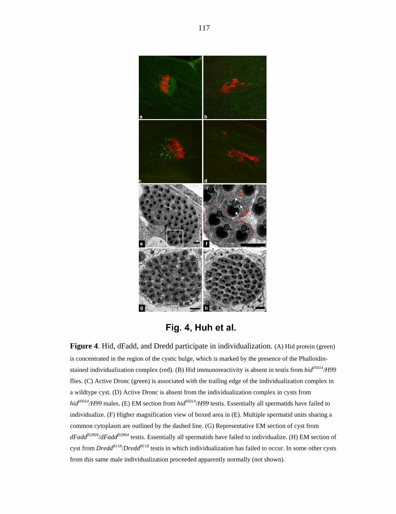
117
Figure 4. Hid, dFadd, and Dredd participate in individualization. (A) Hid protein (green)
is concentrated in the region of the cystic bulge, which is marked by the presence of the Phalloidin-
stained individualization complex (red). (B) Hid immunoreactivity is absent in testis from hid05014/H99
flies. (C) Active Dronc (green) is associated with the trailing edge of the individualization complex in
a wildtype cyst. (D) Active Dronc is absent from the individualization complex in cysts from
hid05014/H99 males. (E) EM section from hid05014/H99 testis. Essentially all spermatids have failed to
individualize. (F) Higher magnification view of boxed area in (E). Multiple spermatid units sharing a
common cytoplasm are outlined by the dashed line. (G) Representative EM section of cyst from
dFaddf02804/dFaddf02804 testis. Essentially all spermatids have failed to individualize. (H) EM section of
cyst from DreddB118/DreddB118 testis in which individualization has failed to occur. In some other cysts
from this same male individualization proceeded apparently normally (not shown).

118
Figure 5. The bln1 P element insertion, which inhibits cyto-c-d expression, results in
pleiotropic defects in spermatogenesis. (A) Genomic organization of the cyt-c-d region. Upper
half of the panel illustrates the structure of the region, as described by Arama et al(Arama et al. 2003).
The lower half of the panel indicates the relative locations of several other genes in the region, as
annotated by the Berkeley Drosophila Genome Project as of August 2002
(http://flybase.bio.indiana.edu/search/). The bln1 P element is inserted within the cyt-c-d l transcription
unit. This P element is also inserted within the transcription unit of a second gene CR31808-RA
(RE70695). Both of these genes, and the bln1 P element, reside within the intron of a third gene,
CG31782. (B,D) Wildtype and bln1 testis, respectively, stained with anti-active Drice antibodies.
Active-Drice immunoreactivity is eliminated in bln1 testis, as described (Arama et al. 2003). (C,E)
Wildtype and bln1 testis, respectively, stained with AXO49 antibodies (blue), which recognize
polyglycylated tubulin present in axonemal microtubules, and phalloidin (red). Polyglycylation occurs

119
prior to individualization (Bressac et al. 1995). Axonemes of elongated cysts from wildtype flies stain
with AXO49 (C) while those from bln1 males do not (E). (F-I) EM micrographs of cysts of different
developmental stages from wildtype (F,G) and bln1 (H) testis. (F) Wildtype cyst prior to
individualization. Note the structures of the major and minor mitochondrial derivatives, in particular,
the fact that the major mitochondrial derivative is increased in size and is electron dense. (G) Wildtype
cyst following individualization. (H) Representative example of the most mature cysts found in bln1
testis. Note the dramatically increased cell size, and the lack of differentiation of the major and
mitochondrial derivatives, as compared to wild type.
Figure 6. driceless males lack active Drice staining and show defects in
individualization.

120
(A) Testis from driceless male stained with active Drice. Active Drice staining is eliminated. (B)
Elongated cysts from driceless male. AXO49 staining (blue) outlines the location of three cystic bulges.
Individualization complexes (arrows) are marked with phalloidin (red). (C) Example of a cyst from a
driceless male in which individualization has proceeded normally. (D) Example of a cyst from a
driceless male in which individualization has failed to occur. (E) Boxed area in (D) shown at higher
magnification highlights. A region in which individualization has failed is outlined with a dashed line.
Figure 7. Active Drice is eliminated from the cytoplasm of wildtype spermatids
following passage of the individualization complex, but not from spermatids in which
caspase activity has been inhibited. (A) Cystic bulge from a wild type cyst stained with active
Drice (red). The cystic bulge (arrowhead) is moving to the left. Active Drice staining is absent in areas
of the spermatid bundle that the individualization complex has passed through, and in which excess
cytoplasm has been eliminated (arrow). (B) Cystic bulge from a β2tub-p35 cyst. The cystic bulge
(arrowhead) is decreased in size, and active Drice is present in areas of the spermatid bundle that the
individualization complex has moved through (arrows). These observations suggest that caspase
inhibition results in at least a partial failure to eliminate excess cytoplasm, but that this is not
necessarily associated with lack of movement of the individualization complex.

121
Supplement Figure 1. Inhibition of Dcp-1 prevents spermtaid individualization.
(A) EM section from β2tub-Dcp-1-RNAi testis. Individualization has failed to occur throughout the
cyst. (B) The boxed area in M is shown at a higher magnification. Spermatid units sharing a common
cytoplasm are outlined by the dashed line. (C) Western blot from wildtype (Wt) and β2tub-Dcp-1-
RNAi (Dcpi) testis probed with anti-Dcp-1 and Anti-Drice antibodies. Dcp-1, but not Drice levels are
greatly reduced in β2tub-Dcp-1-RNAi testis.
Supplement Figure 2. Antibodies specific for active Dronc. (A) Third instar eye imaginal
disc from GMR-DRONCF118E larvae stained with purified anti-DRONC peptide antiserum (green). All

122
cells posterior to the morphogenetic furrow are labeled with this antiserum, as expected based on the
pattern of GMR-dependent gene expression (Hay et al., 1994). Eye discs from wildtype larvae showed
only very low, uniform levels of staining (data not shown). The inset shows a Western blot probed
with purified anti-DRONC peptide antiserum. The first lane was loaded with full length DRONC
mutated in its active site (DRONCC318A). The second lane was loaded with a version of DRONC
consisting of only residues 1-352. This protein terminates following glutamate 352, the DRONC
autoactivation cleavage site, and is equivalent to the large subunit of cleaved and active DRONC. The
anti-Dronc antibodies react well with both proteins. (B) Third instar eye imaginal disc from GMR-
DRONCF118E larvae stained with anti-active DRONC antiserum extensively purified to select for
antibodies that react only with versions of DRONC that have been cleaved at glutamate 352, as in the
methods. Only cells in the most posterior region of the eye disc, which are presumably undergoing
apoptosis, react with these purified antibodies. The inset shows a Western blot similar to that in panel
(A), which was probed with the purified active Dronc-specific antibodies. These antibodies react with
the glutamate 352-cleaved version of DRONC, but not with full length DRONC.

123
CHAPTER 4
Compensatory proliferation induced by cell death in the Drosophila wing disc requires
activity of the apical cell death caspase Dronc in a non-apoptotic role.
Jun R. Huh, Ming Guo2, and Bruce A. Hay
Published in Current Biology 14, 1262-1266, July 2004

124
Summary
Achieving proper organ size requires a balance between proliferation and cell death. For
example, at least 40 to 60 percent of cells in the Drosophila wing disc can be lost, yet these
discs go on to give rise to normal-looking adult wings as a result of compensatory
proliferation [1-3]. The signals that drive this proliferation are unknown. One intriguing
possibility is that they derive, at least in part, from the dying cells. To explore this hypothesis
we activated cell death signaling in specific populations of cells in the developing wing but
prevented these cells from dying through expression of the baculovirus p35 protein, which
inhibits the activity of effector caspases that mediates apoptosis [4]. This allowed us to
uncouple the activation steps of apoptosis from death itself. Here we report that stimulation
of cell death signaling in the wing disc -in the absence of cell death- results in increased
proliferation and ectopic expression of wingless, a known mitogen in the wing. Activation of
the apical cell death caspase Dronc is necessary and sufficient to drive both of these
processes. Our results demonstrate an unanticipated function of an apical cell-death caspase,
the non-autonomous induction of proliferation. This activity is likely to contribute to tissue
homeostasis by promoting local compensatory proliferation in response to cell death. We
speculate that dying cells may communicate cell fate or behavior instructions to their
neighbors in other contexts as well.
Results and Discussion
Expression of the cell death activator head involution defective (Hid) promotes caspase
activation and cell death in many contexts in the fly [5], including the wing [4,6]. We

125
characterized wing discs expressing Hid and p35 under the control of the engrailed promoter,
which drives expression in the posterior wing compartment. As noted previously [4], cells
expressing Hid and p35 show decreased levels of the IAP family caspase inhibitor DIAP1
and high levels of activation of the cell death effector caspase Drice. However, these cells do
not die because expression of p35 inhibits the activity (but not the activation) of effector
caspases such as Drice [4]. We monitored cell proliferation in wing discs using anti-
phosphohistone H3 antibodies, which label cells in M phase. Wing discs that express p35
alone in the posterior compartment showed a general low level of proliferation in both
anterior and posterior compartments (Figure 1A,B). In contrast, wing discs that express Hid
and p35 under engrailed control consistently showed a 2- to 4-fold increase in proliferation in
the posterior compartment (Figure 1C,D; Figure S1).
How are HID-dependent cell death signals transduced to promote cell proliferation? Multiple
IAPs, including DIAP1, can participate in various signal transduction pathways, in addition
to functioning as caspase inhibitors [7-9]. To determine if the HID-dependent increase in
proliferation required the loss of DIAP1 we decreased DIAP1 levels in the absence of other
apical death signals using RNA interference (RNAi) in the presence of p35. Double-stranded
RNA corresponding to DIAP1 (DIAP1-RNAi) and p35 were expressed under the control of
the patched promoter, which drives expression in a stripe at the anterior-posterior
compartment border. Expression of p35 alone did not cause a decrease in DIAP1 [4] (data
not shown) or result in increased levels of proliferation within the domain of patched
expression (Figure 2A,B). Expression of DIAP1 RNAi and p35 under patched control
resulted in a dramatic decrease in DIAP1 protein levels (Figure 2C,D), but again there was

126
no increase in proliferation (Figure 2E,F). These observations (as well as observations
presented in Figure 3 and Figure 4) argue that HID and/or molecules activated by HID, but
not the loss of DIAP1 (which may however facilitate the activation or activity of these other
molecules), are required to induce cell proliferation.
Dronc is an apical caspase that mediates HID-dependent cell death, as well as that initiated
by other death activators [10-13]. Dronc and several mammalian apical cell death caspases
have also been shown to participate in several non-apoptotic processes [14,15]. To explore
roles for Dronc in mediating Hid-induced proliferation in the wing we expressed a dominant
negative version of DRONC (DRONCC318S) [10,15] along with Hid and p35 under engrailed
control (Figure 3A). DIAP1 levels were still decreased (Figure 3B), as with expression of
Hid and p35 [4]. However, increased cell proliferation in the posterior compartment was no
longer observed (Figure 3C,D; Figure S1). These results demonstrate that Dronc activity is
required to mediate Hid-dependent proliferation. Further support for this conclusion comes
from an examination of adult wings. Wings from flies expressing p35 under engrailed control
appeared normal (Figure 3E), while wings from flies expressing Hid and p35 under engrailed
control had an expanded posterior compartment, consistent with the observation of increased
proliferation in this domain during larval development (Figure 3G,H). In contrast, wings
from flies that expressed DroncC318S as well Hid and p35 under engrailed control were
normal in appearance (Figure 3F). We showed that activation of Dronc, in the absence of cell
death, was sufficient to drive cell proliferation by following the fate of cells in wing discs
from flies that expressed Dronc and p35 under patched or engrailed control. Dronc-
expressing cells did not die, as evidenced by a lack of increased TUNEL staining in these

127
cells (data not shown), and DIAP1 levels were if anything slightly increased in the regions
with elevated Dronc (Figure 4A,B). These regions did, however, show increased cell
proliferation (Figure 4C-F).
Hid promotes cell death, at least in part by disrupting interactions between caspases and
DIAP1 [16,17] and through stimulation of DIAP1 degradation [4]. Loss of DIAP1 results in
cell death [16,18-23], and where examined this is mediated by Ark-dependent activation of
Dronc, which cleaves and activates the downstream effector caspases Dcp-1 and Drice
[10,11,20-23]. Together these observations raise a question as to why expression of Hid or
Dronc in the presence of p35, but not loss of DIAP1 in the presence of p35, resulted in
increased proliferation. One possibility is that Hid promotes Dronc activation or activity
through pathways other than simple elimination of the Dronc inhibitor DIAP1. In the context
of such a model Hid-dependent activation of Dronc would simply be more robust than that
due to the loss of DIAP1 in otherwise healthy cells. Alternatively, Hid may have activities
that cooperate with Dronc (but are not sufficient by themselves (see Figure 3C,D)) to
promote proliferation. Regardless of the mechanisms by which Dronc is activated, our
observations demonstrate that Dronc activity is both necessary and sufficient to stimulate cell
proliferation in response to a signal that would normally induce cell death. Loss of DIAP1 is
not necessary, as shown in Figures 3 and 4. However, it is worth noting that loss of DIAP1 is
likely to be sufficient in wildtype discs, in which Dronc activation leads to activation of
downstream caspases, which may themselves promote further Dronc activation and
ultimately apoptosis. In our system we have simply dampened any such feedback pathways
through expression of p35.

128
It has been known for many years that cell death in the larval wing disc leads to
compensatory proliferation [1-3]. We showed that activation of a Dronc-dependent death
signal, even in the absence of death itself, also leads to proliferation. Based on these
observations we argue that dying cells provide, through a Dronc-dependent pathway, at least
one component of the signals that stimulate compensatory proliferation in healthy neighbors.
Further support for this hypothesis comes from examination of wing discs carrying clones of
tissue expressing p35 alone (Figure 5A,C,E), or Hid and p35 (Figure 5B,D,F), in an
otherwise wildtype background. Discs with clones expressing Hid and p35 showed increased
proliferation as compared to discs expressing p35 alone. Importantly, most of this
proliferation occurred in cells outside the clones. Cells that express Hid and activate Dronc
normally die. Therefore, an important prediction of this model is that apical death signals
within the dying cell lead to the generation of signals that stimulate the proliferation of cells
that neighbor dying cells. The secreted protein Wingless is a candidate to mediate at least
some component of such a signal because Wingless can drive proliferation in the wing disc
(c.f. [24] and references therein). In fact we observed a Dronc-dependent increase in wingless
expression in regions of the wing disc expressing Hid and p35, but not p35 alone (Figure S2).
These results are consistent with the hypothesis that wingless contributes to cell proliferation
induced in response to death signaling. However, several points should be noted: 1) Wingless
expression does not occur uniformly in areas expressing Hid and p35 (Figure S2). 2) Our
results do not address the question of which cells express wingless -cells with high level
Dronc activation or their neighbors. 3) The effects of wingless on proliferation in the wing
are complex. Proliferation is stimulated in some contexts [24], but inhibited in others [25]. 4)

129
Roles for other pathways remain to be explored. Experiments that follow the consequences of
manipulating wingless expression will be required to test this hypothesis directly.
How does Dronc activity stimulate proliferation? In general caspases recognize tetrapeptide
substrates in which the C-terminal P1 residue is an aspartate. Dronc is exceptional in this
regard in that it also cleaves well after glutamate. The only known Dronc targets are itself
and the effector caspases Dcp-1 and Drice [10,11]. Dcp-1 and Drice are unlikely to mediate
Dronc-dependent effects on proliferation since they are inhibited by p35 in our experiments.
Screens with combinatorial peptide libraries indicate that while Dronc has distinct overall
target sequence preferences, many different tetrapeptide substrates are acceptable [10]. In
addition, caspases interact with their substrates only very transiently. These facts make target
identification through bioinformatic or protein purification approaches problematic. Other
approaches, such as genetic screens for enhancers and suppressors of phenotypes associated
with ectopic DRONC expression in contexts in which death is inhibited by p35 (Huh and
Hay, unpublished), may provide insight into this question.
Concluding remarks
All together our results demonstrate that activation of the apical caspase Dronc, which is
important for many cell deaths in the fly, is necessary and sufficient to induce proliferation in
cells of the developing wing in response to a cell death stimulus such as Hid expression.
Caspase activity has also been shown to stimulate proliferation in B and T cells in mammals
[14,26]. In these cells low levels of caspase activity are thought to function cell-
autonomously to cleave and modify the functions of key cell-cycle regulators. In contrast,

130
our results argue that Dronc activity, in its role as an apoptosis inducer, plays two distinct
roles that link cell death and proliferation, but in different cells. In its primary role Dronc
transduces death signals in the cells in which it is activated. At the same time it promotes the
production by these cells of a signal that drives the proliferation of nearby cells. Together
these two Dronc functions provide a mechanism that couples ectopic cell death with the
compensatory proliferation that is required to maintain tissue size homeostasis and allow
normal development.
Paradoxically, Dronc's role as a component of such a homeostatic mechanism may also
contribute to deregulation of tissue growth in some contexts. For example, it has recently
been reported that cells expressing high levels of dMyc, the Drosophila homolog of the myc
oncogene, are super-competitors. They survive and proliferate at the expense of neighboring
cells, which are eliminated by apoptosis [6,27]. This death is mediated at least in part by
increased expression of Hid [6]. As demonstrated above, expression of Hid and the
subsequent activation of Dronc in dying cells lead to the generation of signals that drive the
proliferation of neighbors. Therefore, upregulation of dMyc may induce a positive feedback
cycle in which cells with increased dMyc levels promote the death of neighbors, which then
sends a signal back to the dMyc-overexpressing cells that further stimulates their
proliferation. To focus on one specific version of such a model, it will be interesting to
determine if Dronc activation in dying cells results in changes in the levels of dMyc
expression in neighboring cells.

131
Finally, we note that activation of Dronc-dependent cell death leads in many contexts to a
decrease in total cell number (c.f. [10-12]). Thus it is certainly not the case that activation of
Dronc in dying cells provides a compensatory proliferation signal to neighbors in all contexts.
Instead, it seems likely that cells in the wing disc, and probably those of other tissues going
through a period of unpatterned growth, are primed to respond to Dronc activation with the
production of signals that induces compensatory cell proliferation in neighbors that are
themselves receptive to these signals. Given our observations, it is interesting to consider the
possibility that apical caspases such as Dronc may function to regulate cell fate non-
autonomously in other contexts as well. For example, one can easily imagine a situation in
which activation of Dronc-dependent death in one group of cells results in the transmission
of a signal to surrounding cells that informs them that a particular developmental event has
been completed and/or that provides instructions on what to do next. This idea is speculative,
but it is eminently testable by following the consequences of up- or down-regulation of
Dronc activity in specific genetic backgrounds, as described in this report.

132
Figure 1. Activation of Hid-dependent cell death signaling, but in the absence of cell death,
induces cell proliferation. Confocal images of wing discs from third instar larvae of various
genotypes. Posterior is to the right. (A) Wing disc from a third instar larvae of genotype UAS::p35,
engrailed Gal4 (en::Gal4), stained with anti-phosphohistone H3 (red) and anti-p35 (green). (B) The same wing
disc with anti-phosphohistone H3 staining in white. (C) Wing disc from a third instar of genotype UAS::Hid,
UAS::p35, engrailed Gal4 (en::Gal4), stained with anti-Hid (green) and anti-phosphohistone H3 (red). (D) The
same wing disc with anti-phosphohistone H3 staining in white. In panels (B) and (D) the Anterior-Posterior
compartment boundary is indicated by the white line. Anti-phosphohistone H3 staining, which labels cells in M
phase of the cell cycle, is increased in the posterior compartment of Hid- and p35-expressing discs (C,D), but
not those that express p35 alone (A,B).

133
Figure 2. Loss of DIAP1, in the presence of p35, is not sufficient to promote cell
proliferation in the wing disc. Confocal images of wing discs from third instar larvae of
various genotypes. Posterior is to the right. (A) Wing disc from a third instar larvae of genotype
UAS::p35, patched Gal4 (ptc::Gal4), stained with anti-p35 (green) and anti-phosphohistone H3 (red). (B) The
same wing disc with anti-phosphohistone H3 staining shown in white. (C) Wing disc of genotype UAS::DIAP1-
RNAi, UAS::p35, ptc:Gal4 stained with anti-DIAP1(red) and anti-p35 (green). (D) The same wing disc as in
(C); only the anti-DIAP1 staining is shown. (E) Wing disc of genotype UAS::DIAP1-RNAi, UAS::p35,
ptc::Gal4 stained with anti-p35 (green) and anti-phosphohistone H3 (red). (F) The wing disc in (E) with anti-
phosphohistone H3 shown in white. In panels (B) and (F) the boundaries of the ptc::Gal4 expression domain are
shown as a white line. Panels (A) and (B) show the pattern of proliferation in wing discs expressing p35 alone
under patched control. Panels (C) and (D) show that expression of UAS::DIAP1-RNAi, UAS::p35 under
patched control leads to the loss of DIAP1 (arrow) within the patched expression domain. Panels (E) and (F)
show that expression of UAS::DIAP1-RNAi, UAS::p35 under patched control does not lead to an increase in
proliferation within the ptc::Gal4 expression domain (arrowhead). Note that the wing discs in panels (C,D) are

134
different from those in panels (E,F) because both anti-DIAP1 and anti-phospho-histone H3 are mouse
monoclonal antibodies.
Figure 3. Dronc activity is necessary for Hid-dependent stimulation of proliferation in the
wing disc. Confocal images of wing discs from third instar larvae (A-D) and adult wings (E-
H) of various genotypes. (A, B) Wing disc of genotype UAS::DroncC318S, UAS::Hid, UAS::p35,eng ::Gal4
stained with anti-Dronc (green) (A) and anti-DIAP1 (red) (B). (C) Wing disc of genotype UAS::DroncC318S,
UAS::Hid, UAS::p35,eng ::Gal4 stained with anti-Hid (green) and anti-phosphohistone H3 (red). (D) The wing
disc in (C) with anti-phosphohistone H3 in white. The Anterior-Posterior compartment boundary is indicated by
the white line. Wing discs that express Hid and p35 under engrailed control show a dramatic decrease in DIAP1
levels in the posterior compartment of the wing disc [4]. Coexpression of DroncC318S with Hid and p35 under
engrailed control (A) still led to a dramatic decrease in DIAP1 levels in the posterior compartment (arrow) (B).
Wing disc of genotype UAS::DroncC318S, UAS::Hid, UAS::p35, eng::Gal4 showed no increase in proliferation

135
in the posterior compartment relative to that present in the anterior compartment (C,D). The wing disc in panels
(A) and (B) is different from the one shown in panels (C) and (D). Anti-Dronc staining is used to mark the A-P
compartment boundary in (A) and (B). Anti-Hid staining is used to mark the boundary in (C) and (D). (E) Adult
wing of genotype UAS::p35,eng ::Gal4. (F) Adult wing of genotype UAS::DroncC318S, UAS::Hid, UAS::p35,
eng::Gal4. (G,H) Adult wings of genotype UAS::Hid, UAS::p35,eng ::Gal4. Expression of p35 alone in the
posterior wing compartment results in the formation of a normal adult wing (E). Expression of Hid and P35 in
the posterior compartment resulted in an expansion of this compartment (G,H). This expansion is associated
with the presence of extra tissue folds (several of which are indicated with the arrowhead). Co-expression of the
dominant negative form of Dronc, Dronc C318S, with Hid and p35 in the posterior wing compartment suppressed
the overgrowth phenotypes associated with Hid expression (F).
Figure 4. Dronc activation is sufficient to stimulate proliferation in the wing disc. Confocal
images of wing discs from third instar larvae of various genotypes. Dronc expression was
visualized with GFP (green) (see methods for details). (A) Wing disc of genotype UAS::Dronc,

136
UAS::p35; ptc::Gal4 stained with anti-DIAP1 (red). (B) Disc in (A) with only anti-DIAP1 staining shown. (C)
Wing disc of genotype UAS::Dronc, UAS::p35; ptc::Gal4 stained with anti-phosphohistone H3 (red). (D) Disc
in (C) with anti-phosphohistone H3 shown in white. The ptc-expressing domain lies within the white lines. (E)
Wing disc of genotype UAS::Dronc, UAS::p35; en::Gal4 stained with anti-phosphohistone H3 (red). (F) Disc in
(E) with anti-phosphohistone H3 shown in white. The A-P compartment border is indicated by the white line.
Figure 5. Activation of Hid-dependent cell death signaling in clones is associated with
increased total disc proliferation. Single section confocal images of discs from third instar
larvae of various genotypes. Wing discs are stained with anti-phosphohistone-H3 (white or green) and
anti-p35 (red). (A,C,E) HS::FLP; UAS::p35; Act5C<CD2<Gal4, or (B,D,F) HS::FLP; UAS::p35, UAS::HidAla5;
Act5C<CD2<Gal4.

137
Supplementary Materials
Figure S1. Wing discs in which Hid is expressed in the posterior compartment show a Dronc-
dependent increase in phosphohistone-H3 labeling, indicative of increased proliferation. The
ratio of phosphohistone-H3 positive cells in the posterior versus anterior compartment (P/A ratio) was
determined for discs of three different genotypes: UAS::p35, en::Gal4 (n=4), UAS::Hid, UAS::p35, en::Gal4
(n=3), and UAS::DroncC318S; UAS::Hid, UAS::p35, en::Gal4 (n=3).

138
Figure S2. Wingless levels are regulated by cell-death signaling. Confocal images of wing
discs from third instar larvae of various genotypes. All discs are stained with anti-wingless
(red). (A) Wing disc of genotype UAS::p35, en::Gal4. (B) Wing disc of genotype UAS::Hid, UAS::p35,
en::Gal4. (C) Wing disc of genotype UAS::DroncC318S; UAS::Hid, UAS::p35, en::Gal4. (D) Wing disc of
genotype UAS::DIAP1-RNAi; UAS::p35, ptc::Gal4. (E) Wing disc of genotype UAS::Dronc, UAS::p35,
ptc::Gal4. Wingless levels in the posterior compartment are increased in the presence of Hid and p35 (B), but
not p35 alone (A). This increase requires Dronc activity (C). Loss of DIAP1 alone, in the presence of p35, does
not result in an increase in wingless levels (D), while expression of Dronc with p35 does (E).

139
Supplemental Experimental Procedures
Fly strains and constructs
All crosses and stocks were maintained at 25 0C. The following fly stocks were used: w1118,
En-Gal4, UAS-Hid, UAS-P35 [4], UAS-Dronc C318S [10], UAS-HidAla5 (in which six MAPK
phosphorylation sites in Hid are mutated to alanine) [S1], and UAS-Dronc #80 [12]. ptc-Ga4,
Act5C<CD2<Gal4, and HS-FLP were from the Bloomington Stock Center. In the UAS-
Dronc #80 construct GFP is present as a C-terminal fusion to Dronc [12]. To generate double
stranded RNA corresponding to the coding region of DIAP1 we amplified a 605 bp fragment
of DIAP1 using primers 5' EcoR1-gaa cag cac gct ctc tgg cta ag and 5' Xho1-BamH1-ttt gag
gac ttg ggt gcg cat tgg. This fragment was ligated into the EcoR1 and Xho1 sites of the
SympUAST-w vector [S2], generating UAS-DIAP1-RNAi. Germ line transformants were
created using standard procedures.
Generation of clones
Larvae of genotype HS::FLP; UAS::p35; Act5C<CD2<Gal4, or HS::FLP; UAS::p35,
UAS::HidAla5; Act5C<CD2<Gal4 were heat shocked for 15 minutes at 37 oC. Discs from
third instar larvae were processed as described below.
Immunocytochemistry
Conditions for immunocytochemistry and confocal microscopy were as described in [4,16].
Antibodies were used at the following concentrations: purified rabbit anti-Dronc (1:50) [15],

140
mouse anti-DIAP1 (1:400) [4], mouse anti-P35 (1:100) [4], rabbit anti-P35 (1:1,000)
(Biocarta); rabbit anti-Hid (1:1,000) [4], mouse anti-Wingless (1:30) (Developmental Studies
Hybridoma Bank), and mouse anti-Phospho Histone (1:50) (Cell Siginaling Technology, Inc).
Supplementary References
S1. Bergmann, A., Tugentman, M., Shilo, B-Z., and Steller, H. (2002). Regulation of cell
number by MAPK-dependent control of apoptosis: a mechanism for trophic survival
signaling. Dev Cell 2, 159-170.
S2. Giordano, E., Rendina, R., Peluso, I., and Furia, M. (2002). RNAi Triggered by
Symmetrically Transcribed Transgenes in Drosophila melanogaster. Genetics 160, 637-648
Acknowledgments
We thank Sharad Kumar, Helena Richardson, Stephen Crews, Andreas Bergmann, and Ennio
Giordano for fly stocks and vectors, and Melvin Simon for use of his Confocal microscope.
The Wingless antibodies developed by Stephen M. Cohen were obtained from the
Developmental Studies Hybridoma Bank developed under the auspices of the NICHD and
maintained by The University of Iowa, Department of Biological Sciences, Iowa City, IA
52242.

141
References
1. Haynie, J.L., and Bryant, P.J. (1977). The effects of X-rays on the proliferation dynamics
of cells in the imaginal wing disc of Drosophila melanogaster. Wilhelm Roux's Archives 183,
85-100.
2. James, A.A., and Bryant, P.J. (1981). A quantitative study of cell death and mitotic
inhibition in g-irradiated imaginal wing discs of Drosophila melanogaster. Radiat. Res. 87,
552-564.
3. Milan, M., Campuzano, S., and Garcia-Bellido, A. (1997). Developmental parameters of
cell death in the wing disc of Drosophila. Proc. Natl. Acad. Sci. USA 94, 5691-5696.
4. Yoo, S.J. Huh, J.R., Muro, I., Yu, H., Wang, S.L., Feldman, R.M.R., Clem, R.J., Muller,
H.-A.J., and Hay, B.A. (2002). Apoptosis inducers Hid, Rpr and Grim negatively regulate
levels of the caspase inhibitor DIAP1 by distinct mechanisms. Nature Cell Biol. 4, 416-424.
5. Hay, B.A. (2000). Understanding IAP function and regulation: a view from Drosophila.
Cell Death Differ 7, 1045-1056.
6. de la Cova, C., Abril, M., Bellosta, P., Gallant, P., and Johnston, L.A. (2004). Drosophila
myc regulates organ size by inducing cell competition. Cell 117, 107-116.

142
7. Salvesen, G.S. and Duckett, C.S. (2002). IAP proteins: blocking the road to death's door.
Nature Reviews Cell Biology 3, 401-410.
8. Kuranaga, E., Kanuka, H., Igaki, T., Sawamoto, K., Ichijo, H., Okano, H., and Miura, M..
(2002). Reaper-mediated inhibition of DIAP1-induced DTRAF1 degradation results in
activation of JNK in Drosophila. Nat Cell Biol 4, 705-710.
9. Lewis, J., Burstein, E., Reffey, S.B., Bratton, S.B., Roberts, A.B., and Duckett, C.S. (2004).
Uncoupling of the signaling and caspase-inhibitory properties of X-linked inhibitor of
apoptosis. J. Biol. Chem. 279, 9023-9029.
10. Hawkins, C.J., Yoo, S.J., Peterson, E.P., Wang, S.L., Vernooy, S.Y., and Hay, B.A.
(2000). The Drosophila caspase DRONC cleaves following glutamate or aspartate and is
regulated by DIAP1, HID, and GRIM. J Biol Chem 275, 27084-27093.
11. Meier, P., Silke, J., Leevers, S.J., and Evan, G.I. (2000). The Drosophila caspase
DRONC is regulated by DIAP1. Embo J 19, 598-611.
12. Quinn, L.M., Dorstyn, L., Mills, K., Colussi, P.A., Chen, P., Coombe, M., Abrams, J.,
and Kumar, S. (2000). An essential role for the caspase dronc in developmentally
programmed cell death in Drosophila. J Biol Chem 275, 40416-40424.

143
13. Martin, D.N. and Baehrecke, E.H. (2004). Caspases function in autophagic programmed
cell death in Drosophila. Development 131, 275-284.
14. Schwerk, C. and Schulze-Osthoff, K. (2003). Non-apoptotic functions of caspases in
cellular proliferation and differentiation. Biochemical Pharmacology 66, 1453-1458.
15. Huh, J.R., Vernooy, S.Y., Yu, H., Yan, N., Shi, Y., Guo, M., and Hay, B.A. (2004).
Multiple apoptotic caspase cascades are required in nonapoptotic roles for Drosophila
spermatid individualization. PLOS Biology 2, 43-53.
16. Wang, S.L., Hawkins, C.J., Yoo, S.J., Muller, H.A., and Hay, B.A. (1999). The
Drosophila caspase inhibitor DIAP1 is essential for cell survival and is negatively regulated
by HID. Cell 98, 453-463.
17. Wu, J.W., Cocina, A.E., Chai, J., Hay, B.A., and Shi, Y. (2001). Structural analysis of a
functional DIAP1 fragment bound to grim and hid peptides. Mol Cell 8, 95-104.
18. Hay, B.A., Wassarman, D.A., and Rubin, G.M. (1995). Drosophila homologs of
baculovirus inhibitor of apoptosis proteins function to block cell death. Cell 83, 1253-1262.
19. Goyal, L., McCall, K., Agapite, J., Hartwieg, E., and Steller, H. (2000). Induction of
apoptosis by Drosophila reaper, hid and grim through inhibition of IAP function. Embo J 19,
589-597.
144
20. Igaki, T., Yamamoto-Goto, Y., Tokushige, N., Kanda, H., and Miura, M. (2002). Down-
regulation of DIAP1 triggers a novel Drosophila cell death pathway mediated by Dark and
DRONC. J Biol Chem 277, 23103-23106.
21. Muro, I., Hay, B.A., and Clem, R.J. (2002). The Drosophila DIAP1 protein is required to
prevent accumulation of a continuously generated, processed form of the apical caspase
DRONC. J. Biol. Chem 277, 49644-49650.
22. Zimmermann, K.C., Ricci, J.E., Droin, N.M., and Green, D.R. (2002). The role of ARK
in stress-induced apoptosis in Drosophila cells. J Cell Biol 156, 1077-1087.
23. Rodriguez, A., Chen, P., Oliver, H. and Abrams, J.M. (2002). Unrestrained caspase-
dependent cell death caused by loss of Diap1 function requires the Drosophila Apaf-1
homolog, Dark. Embo J 21, 2189-2197.
24. Giraldez, A.J. and Cohen, S.M. (2003). Wingless and Notch signaling provide cell
survival cues and control cell proliferation during wing development. Development 130,
6533-6543.
25. Johnston, L.A. and Sanders, A.L. (2003). Wingless promotes cell survival but constrains
sgrowth during Drosophila wing development. Nat Cell Biol 5, 827-833.

145
26. Olson, N.E., Graves, J.D., Shu, G.L., Ryan, E.J. and Clark, E.A. (2003). Caspase activity
is required for stimulated B lymphocytes to enter the cell cycle. J. Immunol. 170, 6065-6072.
27. Moreno, E. and Basler, K. (2004). dMyc transforms cells into super-competitors. Cell
117, 117-129.

146
CHAPTER 5
Summary: Future Directions

147
Apoptosis is an irreversible process, which brings about cell death without
causing any unwanted effects on neighboring cells or tissues [1]. Since hyper- or
hypo-activation of death machinery often leads to the pathological outcomes in the
entire body, its fine regulation must be ensured. The second chapter of this thesis
illustrated one of these regulation mechanisms in Drosophila apoptosis. Drosophila
is a very useful system for studying apoptosis, not only for its genetic feasibility, but
also for its relative simplicity in cell death regulation. Unlike the mammalian system
where both intrinsic and extrinsic death pathway play roles, the majority of apoptosis
during Drosophila development is dependent on three death-activator proteins
(Rpr/Hid/Grim) and one inhibitor protein (DIAP1). Several labs including ours have
shown that Rpr/Hid/Grim cause cells to die by inhibiting the function of DIAP1.
However, it was not known what the consequences of that inhibition were. Does
Rpr/Hid/Grim simply sequester DIAP1 so that caspases can be activated? If so, could
this reaction be reversible? Or could Rpr/Hid/Grim inactivate DIAP1 permanently,
so that once the death decision is made, there no way to undo it? We have shown that
the binding of Rpr/Hid/Grim to DIAP1 reduces its protein level either by general
translational inhibition, or ubiquitin dependent proteasome pathway. Among these,
Hid induced DIAP1 ubiquitination is rendered through DIAP1’s own ability as a E3
ligase. Recently, activated caspases such as Dronc and Drice were shown to cleave
DIAP1 and presumably make it inactive also [2-4]. Along with these findings, our
studies suggest a feed-forward mechanism in apoptosis. Once cells are doomed to die,
Rpr/Hid/Grim is up-regulated or activated to bind and remove DIAP1 so that caspases

148
are released from DIAP1 and become activated. These active caspases then cleave
and inactivate the remaining DIAP1 in an accelerated manner.
Caspases exist as zymogens in every cell. Once death-activation signals come
along, a cleavage event occurs between their large and small subunits, and they
become activated. In order to study death-activation events, we developed antibodies
that recognize only active (cleaved) caspases, without detecting inactive zymogens.
These antibodies led us to find the developmental contexts, where caspases are
playing non-apoptotic roles such as those in sperm development, in addition to their
conventional roles in apoptosis. The third chapter describes that at least three
different caspase activation mechanisms are utilized during Drosophila
spermatogenesis in order to remove excessive cytoplasm in sperm and separate them
from one another. In addition, the fourth chapter showed another example of how
caspases play non-apoptotic roles. We showed that the activity of the caspase Dronc
is both sufficient and necessary to bring about compensatory proliferation under
stressful conditions during the development of un-patterned tissue like the Drosophila
wing. Many caspases, adaptor molecules, and death activators/inhibitors comprise a
delicate cell-death regulatory network that allows a very fine regulation of caspase
activity to be achieved. Since these death molecules exist in every cell to make them
ready for any un-expected sudden death, it would be wasteful if cells are utilizing
these molecules only for cell death, which occurs just once in the entire course of
their life-cycle. Our findings, non-apoptotic roles of cell death machinery in both
spermatogenesis and compensatory proliferation, illustrate caspases’ non-
conventional, but important, roles during animal development. Once we have a better

149
understanding of how death machinery is regulated, then we may be able to apply our
knowledge to non-death and clinically important processes.
Finally I would like to discuss unanswered questions originating from these
studies and possible ways to solve them. For the regulation of DIAP1 by Rpr and
Grim, the most intriguing question I have is about the identity of E3 ligase involved
in this process. Unlike Hid, Rpr/Grim can still induce a reduction of DIAP1 even in
the absence of its E3 ligase activity. This could be due to their ability to reduce
general translation, but they also could be using some other E3 ligase to degrade
DIAP1. A series of genetic screens performed in our lab identified several genes
whose ectopic expression enhances Rpr and Grim dependent death, without affecting
Hid dependent death (unpublished data). Interestingly, one of them turned out to
encode potential E3 ligases. It would be very interesting, therefore, to check the level
of DIAP1 in the mutant backgrounds of this gene upon the induction of Rpr or Grim.
If any of the genes detected in the screens act as an E3 ligase, whose activity is
required for Rpr/Grim induced DIAP1 reduction, a removal of the ligase might
stabilize DIAP1 and alleviate Rpr/Grim induced death phenotype. The other
approach would be to make a stable S2 cell line with inducible rpr or grim genes. Rpr
or Grim would then be expressed in the presence of chemical inhibitors against
caspases to keep cells alive. Reduced DIAP1 level could then be measured by
western analysis. Alternatively GFP or luciferase could be tagged to DIAP1 for
efficient analysis of DIAP1’s protein level. Now a RNAi-based screen, using a
dsRNA library that encompasses the majority of fly genes, can be performed to
identify genes whose activity is required for Rpr- or Grim- induced DIAP1 removal.

150
This approach renders it possible to identify not only a responsible E3, but also other
components in the DIAP1 regulation pathway by Rpr or Grim.
For the non-apoptotic roles of cell death machinery in sperm differentiation
and compensatory proliferation, the following questions remain to be answered. First,
how do sperm avoid cell death even in the presence of caspase activation? Second
what are the mechanisms of caspase activation? Are the same canonical death
activation mechanisms being used in non-apoptotic situations? If not, are there any
specific death activators only dedicated to the non-apoptotic roles of death
machinery? Third, how do caspases exert their activity? In other words, how does
the activity of caspases bring about membrane re-organization and cytoskeleton
removal during spermatogenesis, as well as induce proliferative signals to the
neighboring tissues during compensatory proliferation? Fourth, do caspases create
proliferative signals in other developmental and pathological contexts as well?
How do sperm avoid cell death given that a high level of caspase activation
occurs during spermatogenesis? Their activation might be physically separated from
the nucleus, like that of Dronc, so that no cell death need happen. But the question of
how caspase activation occurs only in a restricted area still remains. Alternatively,
caspase targets whose activity is required to kill cells once activated by casapses
might be absent in testes. If this is the case, unlike other somatic cells, sperm cells
are not able to die even in the presence of caspase activation. (But we showed in
Figure 1 that we can kill sperm cysts by an ectopic expression of downstream
caspases.) Or cells also might prevent death with the help of cell death inhibitors.
The question then becomes what is the nature of these inhibitors. Are they the same

151
death inhibitors found in somatic cells? Is DIAP1 required for the sperm cell survival
as well? Our preliminary data suggest it might not be. When the DIAP1 level is
reduced using RNAi technique in testes, neither an increase of caspase activation nor
cell death was observed (unpublished data). However, a more definite answer can
only be acquired by generating DIAP1-null sperm cysts by using a mitotic
recombination technique. Specifically mutant cysts can be marked with GFP by
MARCM technique [5]. As mentioned earlier, DIAP1 is absolutely required for most
of the fly cell survival in different tissues. If that is the case for spermatogenesis as
well, no GFP-positive clones (DIAP1 minus cysts) would appear. If GFP-positive
cyst clones exist, however, that means DIAP1 is not necessary for the sperm cyst
survival, which could be consistent with our RNAi data. Besides, it was recently
reported that DIAP1 is dispensable for oocyte cell survival [6]. Novel death
inhibitors other than DIAP1 might, therefore, be important during germline
development. How then do we identify these novel inhibitors? There are many
possible ways to tackle this question, but I would like to suggest using genetic
approaches. Specifically a dominant modifier screen [7] would be a good choice. In
chapter 3, we described the development of a testis-specific driver. When an active
version of Drice was expressed under this driver, flies became completely sterile
(Figure 1), presumably due to the excessive cell death. One can easily design a
screen to identify suppressor lines for this sterility by mobilizing testis-specific
drivers over the entire genome and crossing individual lines to the line expressing
active Drice in testis. This could potentially identify novel inhibitors as well as
DIAP1. What would the characteristics of these novel testis-specific inhibitors be?

152
We found that DEVDase (a downstream caspase) activity manifests a bimordial
distribution in extracts generated from pools of numerous fly testes (Figure 2). This
data clearly suggest the idea that caspases remain active only for a relatively short
period of time in spite of their active (cleaved) conformation. Thus, I would expect
that caspase inhibitors in sperm cysts might act like suicide inhibitors, which can
directly bind to caspases’ active sites and inhibit them. This hypothesis is supported
by evidence that the expression of viral suicide caspase inhibitors like P35 are known
to induce increased immuno-staining for active (or cleaved) caspases, similar to that
of the testis, even though they have lost their activity [8].
We have shown that Dronc activation during spermatogenesis is dependent on
the presence of the conventional cell death activator, Hid. However for Drice/Dcp-1
and Fadd/Dredd, it is still not known how they are activated. Mapping and cloning of
driceless in which no Drice/Dcp-1 activation occurs would be very helpful to identify
an upstream activation mechanism for these genes. Using meiotic recombination
mapping techniques and genomic rescues with publicly available genomic duplication
lines, we were able to narrow its potential region down to 0.37 Mb on X chromosome
(unpublished data and Figure 3). We are in the process of identifying the responsible
gene or genes among the 40 candidates. Hopefully this will lead us to a better
understanding how Dirce/Dcp-1 is regulated. Fadd and Dredd were previously shown
to be involved in the innate immune response of flies. We have also checked other
components in the immune response pathway for their potential roles during
spermatogenesis and found that PGRP-LC, Ird5, and Relish mutants have some
sperm individualization defects (unpublished data). Therefore, the same Dredd
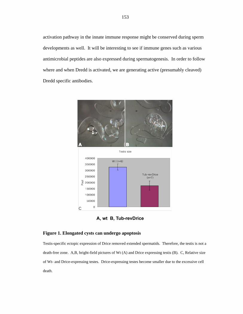
153
activation pathway in the innate immune response might be conserved during sperm
developments as well. It will be interesting to see if immune genes such as various
antimicrobial peptides are also expressed during spermatogenesis. In order to follow
where and when Dredd is activated, we are generating active (presumably cleaved)
Dredd specific antibodies.
Figure 1. Elongated cysts can undergo apoptosis
Testis-specific ectopic expression of Drice removed extended spermatids. Therefore, the testis is not a
death-free zone. A,B, bright-field pictures of Wt (A) and Drice expressing testis (B). C, Relative size
of Wt- and Drice-expressing testes. Drice-expressing testes become smaller due to the excessive cell
death.

154
Figure 2. DEVDase activity in testis extracts manifested a binomial distribution
pattern, which suggests a presence of suicide caspase inhibitors.

155
.
Figure 3. Mapped locus of driceless.

156
Identifying caspases’ substrates is not a trivial task due to their transient
interaction with caspases. However, understanding about how caspases function is
dependent on finding their targets. Proteomic approaches like mass-
spectrophotometric analysis and 2D gel electrophoresis can be entailed for this
purpose. Genome wide screening can also be an effective way to identify substrates.
Previously one of the actin binding proteins, Gelsolin, was identified as a caspase
substrate by incubating pooled genes in the presence or absence of caspases and by
comparing resulting band cleavage (or migration) patterns [9]. Similar approaches
can be taken. Testes extracts can be prepared from wild type and caspase mutant's
testes. Various antibodies against candidate target genes, such as actin regulator
proteins or membrane remodeling factors are used to detect any differently processed
protein bands. Once potential caspases targets are identified, un-cleavable variants of
these proteins can be expressed in testes to determine if they phenocopy caspase
mutants. Due to the technical difficulty of making protein extracts from wing discs, it
is better to use indirect approaches to find Dronc targets in the compensatory
proliferation pathway. As discussed in chapter 4, over-expression of Dronc and P35
induces extra proliferation. Using a genetic screening, therefore, to identify
suppressor lines that abrogate this ectopic proliferation would provide some clues on
how Dronc brings about compensatory proliferation.
Since Dronc and Drice mutants recently became available [10-12 and
unpublished data] and both of them are not embryonic lethal, it would be interesting
to study their response to the external stress during the larval stage. If Dronc is
involved in the generation of proliferative signals in tissues other than the wing discs,

157
we might be able to find those contexts by comparing these mutants to wild type flies.
Since the final outcome of death is blocked in Drice mutants, paradoxically more
proliferation would ensue upon the induction of the death pathway (also refer to
chapter 4) due to the hyper Dronc activation. Dronc mutants, however, should
behave in similar ways to wild type. One other interesting experiment would be to
determine the regeneration potentials in these mutants. The ability of Drosophila
imaginal disc to regenerate after its partial incision is well known [13, 14]. The
question is whether or not caspases play any roles during this process. If they do,
Drice mutants might beget higher tissue regeneration efficiency thanks to the
increased proliferation. On the contrary, Dronc mutants might completely lack this
potential. These are obviously doable experiments with intriguing clinical
implications.

158
References 1. Jacobson, M.D., Weil, M., and Raff, M.C. (1997). Programmed cell death in
animal development. Cell 88, 347-354.
2. Muro, I., Means, J.C., and Clem, R.J. (2005). Cleavage of the apoptosis
inhibitor DIAP1 by the apical caspase DRONC in both normal and apoptotic
Drosophila cells. J Biol Chem 280, 18683-18688.
3. Chai, J., Yan, N., Huh, J.R., Wu, J.-W., Li, W., Hay, B.A., and Shi, Y. (2003).
Molecular mechanisms of Reaper/Grim/Hid-mediated suppression of DIAP1-
dependent Dronc ubiquitination. Nature Structural Biology 10, 892-898.
4. Ditzel, M., Wilson, R., Tenev, T., Zachariou, A., Paul, A., Deas, E., and Meier,
P. (2003). Degradation of DIAP1 by the N-end rule pathway is essential for
regulating apoptosis. Nature Cell Biol. 5, 467-473.
5. Lee, T., and Luo, L. (2001). Mosaic analysis with a repressible cell marker
(MARCM) for Drosophila neural development. TINS 24, 251-254.
6. Geisbrecht, E., and Montell, D. (2004). A role for Drosophila IAP1-mediated
caspase inhibition in Rac-dependent cell migration. Cell 118, 111-125.
7. Hay, B.A., Huh, J.R., and Guo, M. (2004). The genetics of cell death:
approaches, insights and opportunities in Drosophila. Nat Rev Genet 5, 911-
922.
8. Yoo, S.J., Huh, J.R., Muro, I., Yu, H., Wang, L., Wang, S.L., Feldman,
R.M.R., Clem, R.J., Muller, H.-A.J., and Hay, B.A. (2002). Apoptosis
inducers Hid, Rpr and Grim negatively regulate levels of the caspase inhibitor
DIAP1 by distinct mechanisms. Nature Cell Biol. 4, 416-424.
9. Kothakota, S., Azuma, T., Reinhard, C., Klippel, A., Tang, J., Chu, K.,
McGarry, T.J., Kirschner, M.W., Koths, K., Kwiatkowski, D.J., and Williams,
L.T. (1997). Caspase-3-generated fragment of gelsolin: effector of
morphological change in apoptosis. Science 278, 294-298.
10. Xu, D., Li, Y., Arcaro, M., Lackey, M., and Bergmann, A. (2005). The
CARD-carrying caspase Dronc is essential for most, but not all,
developmental cell death in Drosophila. Development 132, 2125-2134.

159
11. Daish, T., Mills, K., and Kumar, S. (2004). Drosophila caspase DRONC is
required for specific developmental cell death pathways and stress-induced
apoptosis. Dev Cell. 7, 909-915.
12. Chew, S.K., Akdemir, F., Chen, P., Lu, W.J., Mills, K., Daish, T., Kumar, S.,
Rodriguez, A., and Abrams, J.M. (2004). The apical caspase dronc governs
programmed and unprogrammed cell death in Drosophila. Dev Cell. 7, 897-
907.
13. Haynie, J.L., and Bryant, P.J. (1977). The effects of X-rays on the
proliferation dynamics of cells in the imaginal wing disc of Drosophila
melanogaster. Wilhelm Roux's Archives 183, 85-100.
14. Bryant, P.J. (1975). Pattern formation in the imaginal wing disc of Drosophila
melanogaster: fate map, regeneration and duplication. J Exp Zool 193, 49-77.