
TMJ Disc Displacement Without Reduction Management_A Systematic Review
16
http://jdr.sagepub.com/ Journal of Dental Research http://jdr.sagepub.com/content/93/7_suppl/37S The online version of this article can be found at: DOI: 10.1177/0022034514528333 2014 93: 37S originally published online 21 March 2014 J DENT RES M. Al-Baghdadi, J. Durham, V. Araujo-Soares, S. Robalino, L. Errington and J. Steele TMJ Disc Displacement without Reduction Management: A Systematic Review Published by: http://www.sagepublications.com On behalf of: International and American Associations for Dental Research can be found at: Journal of Dental Research Additional services and information for http://jdr.sagepub.com/cgi/alerts Email Alerts: http://jdr.sagepub.com/subscriptions Subscriptions: http://www.sagepub.com/journalsReprints.nav Reprints: http://www.sagepub.com/journalsPermissions.nav Permissions: What is This? - Mar 21, 2014 OnlineFirst Version of Record - Apr 7, 2014 OnlineFirst Version of Record - May 30, 2014 OnlineFirst Version of Record - Jun 19, 2014 Version of Record >> by guest on November 9, 2014 For personal use only. No other uses without permission. jdr.sagepub.com Downloaded from © International & American Associations for Dental Research by guest on November 9, 2014 For personal use only. No other uses without permission. jdr.sagepub.com Downloaded from © International & American Associations for Dental Research
-
Upload
arcelino-farias-neto -
Category
Documents
-
view
21 -
download
0
description
TMJ Disc Displacement Without Reduction Management_A Systematic Review
Transcript of TMJ Disc Displacement Without Reduction Management_A Systematic Review

http://jdr.sagepub.com/Journal of Dental Research
http://jdr.sagepub.com/content/93/7_suppl/37SThe online version of this article can be found at:
DOI: 10.1177/0022034514528333
2014 93: 37S originally published online 21 March 2014J DENT RESM. Al-Baghdadi, J. Durham, V. Araujo-Soares, S. Robalino, L. Errington and J. Steele
TMJ Disc Displacement without Reduction Management: A Systematic Review
Published by:
http://www.sagepublications.com
On behalf of:
International and American Associations for Dental Research
can be found at:Journal of Dental ResearchAdditional services and information for
http://jdr.sagepub.com/cgi/alertsEmail Alerts:
http://jdr.sagepub.com/subscriptionsSubscriptions:
http://www.sagepub.com/journalsReprints.navReprints:
http://www.sagepub.com/journalsPermissions.navPermissions:
What is This?
- Mar 21, 2014OnlineFirst Version of Record
- Apr 7, 2014OnlineFirst Version of Record
- May 30, 2014OnlineFirst Version of Record
- Jun 19, 2014Version of Record >>
by guest on November 9, 2014 For personal use only. No other uses without permission.jdr.sagepub.comDownloaded from
© International & American Associations for Dental Research
by guest on November 9, 2014 For personal use only. No other uses without permission.jdr.sagepub.comDownloaded from
© International & American Associations for Dental Research
37S
JDR Clinical Research Supplementvol. 93 • issue 7 • suppl no. 1
DOI: 10.1177/0022034514528333. 1Department of Oral and Maxillofacial Surgery, School of Dental Sciences, Newcastle University, UK; 2Institute of Health and Society, Newcastle University, UK; 3Walton Library, Newcastle University, UK; and 4Department of Restorative Dentistry, School of Dental Sciences, Newcastle University, Newcastle upon Tyne, UK; *corresponding author, [email protected] or [email protected]
Protocol Registration Number: PROSPERO 2012, CRD42012003153. Available from: http://www.crd.york.ac.uk/PROSPERO/display_record.asp?ID=CRD42012003153.
A supplemental appendix to this article is published electronically only at http://jdr.sagepub.com/supplemental.
© International & American Associations for Dental Research
M. Al-Baghdadi1,2*, J. Durham1,2, V. Araujo-Soares2, S. Robalino2, L. Errington3, and J. Steele2,4
TMJ Disc Displacement without Reduction Management: A Systematic Review
cLinicAL REViEw
ABSTRACT: Various interventions have been used for the management of patients with temporomandibular joint (TMJ) disc displacement without reduc-tion (DDwoR), but their clinical effec-tiveness remains unclear. This system-atic review investigated the effects of these interventions and is reported in accordance with Preferred Reporting Items for Systematic Reviews and Meta-Analyses (PRISMA) guidelines. Electronic and manual searches up to November 1, 2013, were conducted for English-language, peer-reviewed, pub-lications of randomized clinical trials comparing any form of conservative or surgical interventions for patients with clinical and/or radiologic diag-nosis of acute or chronic DDwoR. Two primary outcomes (TMJ pain inten-sity and maximum mouth opening) and a number of secondary outcomes were examined. Two reviewers per-formed data extraction and risk of bias assessment. Data collection and analysis were performed according to Cochrane recommendations. Twenty studies involving 1,305 patients were included. Data analysis involved 21 comparisons between a variety of inter-ventions, either between interventions, or between intervention and placebo
or no intervention. Meta-analysis on homogenous groups was conducted in 4 comparisons. In most comparisons made, there were no statistically signif-icant differences between interventions relative to primary outcomes at short- or long-term follow-up (p > .05). In a separate analysis, however, the major-ity of reviewed interventions reported significantly improved primary out-come measures from their baseline lev-els over time (p < .05). Evidence lev-els, however, are currently insufficient for definitive conclusions, because the included studies were too heteroge-neous and at an unclear to high risk of bias. In view of the comparable therapeutic effects, paucity of high-quality evidence, and the greater risks and costs associated with more com-plex interventions, patients with symp-tomatic DDwoR should be initially treated by the simplest and least inva-sive intervention.
Key Words: temporomandibular joint surgery, internal derangement, closed lock, meta-analysis, disc disorder, TMD.
Introduction
Temporomandibular joint (TMJ) disc displacement without
reduction (DDwoR) is a specific temporomandibular disorder (TMD) that can cause TMJ pain and limited mouth opening (painful locking), sometimes called a “closed lock” (Okeson, 2007). DDwoR can be acute or chronic depending on the duration of locking (Sembronio et al., 2008; Saitoa et al., 2010). Its incidence among TMD patients is estimated at 2% to 8% (Manfredini et al., 2011; Poveda-Roda et al., 2012).
Various interventions have been suggested for DDwoR, but to date, the most efficacious/effective approach is still unclear, which may result in management being based more on experience than evidence (Durham et al., 2007). The aim of this systematic review, therefore, was to investigate the effects of different conservative and surgical interventions used in the management of TMJ DDwoR.
MethodsProtocol and RegistrationThis systematic review was conducted
in accordance with the Cochrane Collaboration (Higgins and Green, 2011) and the Centre for Reviews and Dissemination (Akers et al., 2009) guidance, and is reported according to the Preferred Reporting Items for
by guest on November 9, 2014 For personal use only. No other uses without permission.jdr.sagepub.comDownloaded from
© International & American Associations for Dental Research

38S
JDR Clinical Research Supplement July 2014
Systematic Reviews and Meta-Analyses (PRISMA) statement (Moher et al., 2009). All the methods of data collection/analysis and inclusion/exclusion criteria were pre-specified and documented in the review protocol (Al-Baghdadi et al., 2012).
Criteria for Studies to be Considered (PICOS, Appendix 1)
•Participants: Any age, and gender with clinical and/or radiologic diagnosis of acute or chronic DDwoR.
•Interventions: Any form of conservative or surgical interventions.
•Comparators/Control: Any alternative intervention, placebo, or no treatment.
•Outcomes: Primary outcomes: TMJ pain intensity and unassisted/active maximum mouth opening (MMO). Secondary outcomes: other mandibular movements, mandibular function or patient’s quality of life, therapy cost, operation/admission duration in surgical trials, and adverse events. The outcomes were evaluated over both short-term (≤ 3 mo) and long-term (> 3 mo) follow-up periods.
•Studies: Randomized and quasi-randomized clinical trials (RCTs and q-RCTs).
Search Strategy (Appendix 2)Four databases – CENTRAL,
MEDLINE, EMBASE, and Scopus – were electronically searched up to November 1, 2013. Other sources were manually searched: citation search and reference lists of included studies, reference lists of relevant review articles and textbook chapters, and 7 journals highly likely to contain studies relevant to the review topic.
Data Collection and AnalysisSelection of Studies
Eligible studies were selected according to the inclusion/exclusion criteria. Irrelevant reports were identified by their title/abstract and were excluded by the first reviewer (MA). The full texts of all potentially eligible studies were retrieved and independently examined in duplicate by two reviewers (MA, JD) to establish eligibility. Throughout the review process, disagreements were resolved by consensus or, when necessary, by a
third reviewer ( JS). Studies excluded at this stage were identified and reasons for exclusion recorded.
Data Extraction and Management
A standardized, pre-piloted, extraction form based on Cochrane recommendations was used. Eligible studies’ data were extracted and recorded by the first reviewer (MA). The second reviewer ( JD), blinded to the authors’ names, institutions, and journal, crosschecked the validity of all data extracted. Authors of included studies were contacted to clarify study design and/or request missing data as required.
Risk-of-Bias Assessment
The methodologic quality of included studies was assessed independently and in duplicate by two reviewers (MA, JD ‘blinded’) using the Cochrane risk of bias tool (Higgins et al., 2011). Each domain in the tool was allocated one of the following judgments: low, unclear, or high risk of bias. Sample size calculation was also examined.
Data AnalysisThe planned data analysis for this review
was performed according to Cochrane statistical guidelines (Higgins and Green, 2011) with the Review Manager Software (version 5.2) (RevMan, 2012) to compare the effects of different interventions (i.e., between-group statistical differences). For dichotomous data, the estimates of effect of an intervention were expressed as risk ratios (RR) together with 95% confidence intervals (CI). For continuous data, mean differences (MD) with 95% CI were used. Clinical and statistical heterogeneities were assessed across the studies prior to pooling. Clinical heterogeneity was determined by examination of each study’s clinical characteristics for any diversity/variation in, for example, technique/delivery of interventions, severity/chronicity of condition, and treatment outcomes. Statistical heterogeneity was assessed by chi-square and I2 statistics (Higgins and Thompson, 2002). A significant p value < .05 for chi-square test and an I2 statistic > 50% were considered substantial heterogeneity (Deeks et al., 2011). Pooling of clinically
and statistically homogeneous trials was done by a fixed-effect model if there were 2 studies pooled and by a random-effects model if more than 2 studies were pooled. When there was substantial heterogeneity between studies, meta-analysis was not undertaken and the data were integrated into a narrative analysis of the findings. A test for funnel plot asymmetry to assess publication bias (Egger et al., 1997) was planned, but was not performed because of insufficient numbers of studies pooled in the meta-analyses. Where possible, a subgroup analysis based on chronicity of the locking condition (acute or chronic: according to duration of locking threshold for chronic lock where disc recapture much less likely estimated at 4 wk) was conducted. Studies, without soft-tissue imaging confirming the DDwoR clinical diagnosis, were excluded in a sensitivity analysis to identify any effect on primary outcomes in the meta-analysis.
Additional data analysis was also performed for examination of the changes from baseline in primary outcomes for each individual intervention at short- and long-term follow-ups (i.e., within-group statistical difference from baseline). This separate analysis was performed to help readers interpret the potential clinical significance of improvement from baseline for each intervention.
ResultsSearchThe search strategy identified a total of
3,333 records from all databases. Of these, the full texts of 172 potentially eligible papers were retrieved and examined. Fig. 1 illustrates the screening process.
Description of StudiesTwenty studies met the inclusion
criteria (Lundh et al., 1992; Petersson et al., 1994; Linde et al., 1995; Fridrich et al., 1996; Schiffman et al., 1996; Goudot et al., 2000; Holmlund et al., 2001; Minakuchi et al., 2001; Yuasa and Kurita, 2001; Maloney et al., 2002; Peroz et al., 2004; Yoshida et al., 2005; Ismail et al., 2007; Politi et al., 2007; Schiffman et al., 2007; Diracoglu et al., 2009; Haketa et al., 2010; Yoshida et al., 2011; Craane et al., 2012; Sahlstrom et al., 2013).
by guest on November 9, 2014 For personal use only. No other uses without permission.jdr.sagepub.comDownloaded from
© International & American Associations for Dental Research

39S
JDR Clinical Research Supplementvol. 93 • issue 7 • suppl no. 1
Summary characteristics of included studies are available in Appendix 3. The list of excluded studies and reasons for exclusion is available upon request.
Risk of BiasNone of the included studies was
at low risk of bias across all domains (Appendix 4). Eight were assessed as unclear overall risk of bias because of insufficient information in the trial report and/or from the contacted authors, or because it was not possible to make a definite judgment in at least one domain of the bias assessment tool. The remaining studies were assessed as high
overall risk of bias. Of the 20 studies included, 7 presented a priori sample size calculation, and 8 had inadequate statistical power (< 80%) (Appendix 3).
Effects of InterventionsThe reviewed interventions varied
widely in invasiveness. For the purpose of this review, the interventions were grouped into 3 modalities, based on their level of invasiveness (Appendix 5):
(1) non-invasive (conservative), including education, self-management, splint therapy, physiotherapy, and their combinations;
(2) minimally invasive, including arthrocentesis; or
(3) invasive (surgical), including arthroscopic and open joint surgeries.
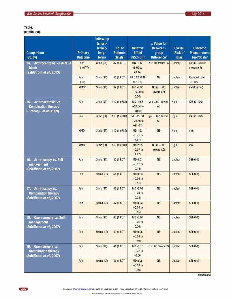
Twenty-one comparisons were made among interventions. Data for the 21 comparisons (between-group statistical analysis) are presented in the text, with the primary outcomes described at short- and long-term follow-up time points in the Table. Data examining within-group differences from baseline for primary outcomes (within-group statistical analysis) at short- and long-term follow-ups are tabulated and presented in
Figure 1.Study flow diagram.
by guest on November 9, 2014 For personal use only. No other uses without permission.jdr.sagepub.comDownloaded from
© International & American Associations for Dental Research

40S
JDR Clinical Research Supplement July 2014
Table.Summary of Findings for the Primary Outcomes (pain at jaw function and unassisted/active maximum mouth opening)
Comparison(Study)
Primary Outcome
Follow-up (short-term & long-term)
No. of Patients(Trials)
Relative Effect
(95% CI)a
p Value for Between-
group Differenceb
Overall Risk of
Bias
Outcome Measurement
Tool/Scalec
1. MM vs. No treatment(Yoshida et al., 2011)
MMO 10 min (ST) 148 (1 RCT) RR 16.67
(5.44 to
51.06)
p < .0001 favors
MM
High MMO > 38 mm
2. Jaw exercises vs. Education only
(Craane et al., 2012)
Paind 3 mo (ST) 42 (1 RCT) MD 3.81
(–6.15 to
13.77)
NS Unclear VAS (0-100)
Paind 13 mo (LT) 42 (1 RCT) MD 0.62
(–5.46 to
6.70)
NS Unclear VAS (0-100)
MMO 3 mo (ST) 45 (1 RCT) MD -3.10
(–6.96 to
0.76)
NS Unclear aMMO (mm)
MMO 13 mo (LT) 42 (1 RCT) MD -3.80
(–7.68 to
0.08)
NS (p = .05
toward Educ)
Unclear aMMO (mm)
3. Self-management vs. Education only
(Minakuchi et al., 2001)
Pain 2 mo (ST) 44 (1 RCT) MD -4.40
(–19.54 to
10.74)
NS Unclear VAS (0-100) on
chewing
MMO 2 mo (ST) 44 (1 RCT) MD -1.40
(–6.90 to
4.10)
NS Unclear aMMO (mm)
4. Self-management vs. No treatment
(Yuasa and Kurita, 2001)
Pain &
MMO
1 mo (ST) 60 (1 RCT) RR 1.80
(1.00 to
3.23)
NS (p = .05
toward SM)
Unclear No. improved
patients for: VAS
pain & MMO
Subgroup
analysis
1 mo (ST) 15 Acute RR 1.05
(0.57 to
1.94)
NS
45 Chronic RR 2.51
(1.06 to
5.95)
p < .05 favors SM
5. Self-management vs. Splint
(Haketa et al., 2010)
Pain 2 mo (ST) 44 (1 RCT) MD -15.20
(–31.55 to
1.15)
NS (p = .07
toward SM)
Unclear VAS (0-100)
MMO 2 mo (ST) 44 (1 RCT) MD 6.00
(2.67 to
9.33)
p < .001 favors
SM
Unclear MMO with pain
(mm)
6. Splint vs. Control(Lundh et al., 1992)
Pain 12 mo (LT) 51 (1 RCT) RR 0.49
(0.26 to
0.92)
p < .05 favors
Control
High No. reduced pain
7. Splint vs. TENS(Linde et al., 1995)
Pain 6 wk (ST) 31 (1 RCT) RR 8.53
(1.21 to
60.33)
p < .05 favors
Splint
High Reduction in
pain ≥ 50%
MMO 6 wk (ST) 31 (1 RCT) MD -0.16
(–4.07 to
3.75)
NS High Change from
baseline (mm)
(continued)
by guest on November 9, 2014 For personal use only. No other uses without permission.jdr.sagepub.comDownloaded from
© International & American Associations for Dental Research

41S
JDR Clinical Research Supplementvol. 93 • issue 7 • suppl no. 1
Table. (continued)
8. Combination therapye vs. Education only
(Minakuchi et al., 2001)
Pain 2 mo (ST) 46 (1 RCT) MD -2.80
(–16.12 to
10.52)
NS Unclear VAS (0-100) on
chewing
MMO 2 mo (ST) 46 (1 RCT) MD 1.40
(–3.94 to
6.74)
NS Unclear aMMO (mm)
9. Combination therapy vs. Self-management
(Minakuchi et al., 2001; Schiffman et al., 2007)
Pain 2-3 mo (ST) 97 (2 RCTs) SMD 0.22
(–0.19 to
0.62)
NS Unclear VAS & SSI
Pain 60 mo (LT) 50 (1 RCT) MD 0.00
(–0.13 to
0.13)
NS Unclear SSI (0-1)
MMO 2 mo (ST) 48 (1 RCT) MD 2.80
(–2.95 to
8.55)
NS Unclear aMMO (mm)
10. Jaw exercise + splint vs. Splintf
(Maloney et al., 2002; Ismail et al., 2007)
Pain 1-3 mo (ST) 50 (2 RCTs) MD 0.90
(–12.28 to
14.07)
NS High VAS & NRS
(0-100)
MMO 1-3 mo (ST) 50 (2 RCTs) MD 4.67
(1.80 to 7.55)
p < .01 favors
Ex+Sp
High aMMO (mm)
11. Active PEMF vs. Placebo PEMF
(Peroz et al., 2004)
Paind 6 wk (ST) 31 (1 RCT) MD 0.23
(–17.96 to
18.42)
NS Low VAS (0-100)
Paind 4 mo (LT) 30 (1 RCT) MD 19.49
(0.97 to
38.01)
p < .05 favors
placebo
Unclear VAS (0-100)
MMOd 6 wk (ST) 31 (1 RCT) MD -2.47
(–8.23 to
3.29)
NS Low aMMO (mm)
MMO 4 mo (LT) 30 (1 RCT) MD -1.00
(–6.09 to
4.09)
NS Unclear aMMO (mm)
12. Active iontophoresis vs. Placebo iontophoresisg
(Schiffman et al., 1996)
Pain 1 wk (ST) 18 (1 RCT) MD -0.03
(–0.21 to
0.15)
NS Unclear SSI (0-1)
MMO 1 wk (ST) 18 (1 RCT) MD 1.90
(–5.70 to
9.50)
NS Unclear aMMO (mm)
13. Arthrocentesis vs. Arthrography only
(Petersson et al., 1994)
Painh 2 mo (ST) 33 (1 RCT) MD -16.02
(–34.79 to
2.75)
NS (p = .09
toward AC)
High VAS (0-100) after
chewing
MMO 2 mo (ST) 33 (1 RCT) MD -3.00
(–9.54 to
3.54)
NS High mm
Comparison(Study)
Primary Outcome
Follow-up (short-term & long-term)
No. of Patients(Trials)
Relative Effect
(95% CI)a
p Value for Between-
group Differenceb
Overall Risk of
Bias
Outcome Measurement
Tool/Scalec
(continued)
by guest on November 9, 2014 For personal use only. No other uses without permission.jdr.sagepub.comDownloaded from
© International & American Associations for Dental Research
42S
JDR Clinical Research Supplement July 2014
14. Arthrocentesis vs. ATN LA block
(Sahlstrom et al., 2013)
Paind
(no ITT)
3 mo (ST) 37 (1 RCT) MD 24.60
(6.06 to
43.14)
p < .01 favors LA Unclear VAS (0-100) at
movements
Pain
(ITT)
3 mo (ST) 45 (1 RCT) RR 0.72 (0.46
to 1.14)
NS Unclear Reduced pain
≥ 30%
MMOd 3 mo (ST) 37 (1 RCT) MD -4.90
(–10.00 to
0.20)
NS (p = .06
toward LA)
Unclear aMMO (mm)
15. Arthrocentesis vs. Combination therapy
(Diracoglu et al., 2009)
Pain 3 mo (ST) 110 (1 qRCT) MD -19.3
(–28.54 to
–10.06)
p < .0001 favors
AC
High VAS (0-100)
Pain 6 mo (LT) 110 (1 qRCT) MD -28.80
(–36.56 to
–21.04)
p < .0001 favors
AC
High VAS (0-100)
MMO 3 mo (ST) 110 (1 qRCT) MD 1.93
(–0.75 to
4.61)
NS High mm
MMO 6 mo (LT) 110 (1 qRCT) MD 2.35
(–0.07 to
4.77)
NS (p = .06
toward AC)
High mm
16. Arthroscopy vs. Self-management
(Schiffman et al., 2007)
Pain 3 mo (ST) 50 (1 RCT) MD 0.01
(–0.12 to
0.14)
NS Unclear SSI (0-1)
Pain 60 mo (LT) 51 (1 RCT) MD 0.03
(–0.09 to
0.15)
NS Unclear SSI (0-1)
17. Arthroscopy vs. Combination therapy
(Schiffman et al., 2007)
Pain 3 mo (ST) 43 (1 RCT) MD -0.08
(–0.24 to
0.08)
NS Unclear SSI (0-1)
Pain 60 mo (LT) 47 (1 RCT) MD 0.03
(–0.09 to
0.15)
NS Unclear SSI (0-1)
18. Open surgery vs. Self-management
(Schiffman et al., 2007)
Pain 3 mo (ST) 48 (1 RCT) MD -0.07
(–0.20 to
0.06)
NS Unclear SSI (0-1)
Pain 60 mo (LT) 50 (1 RCT) MD 0.05
(–0.09 to
0.19)
NS Unclear SSI (0-1)
19. Open surgery vs. Combination therapy
(Schiffman et al., 2007)
Pain 3 mo (ST) 41 (1 RCT) MD -0.16
(–0.32 to
–0.00)
p < .05 favors OS Unclear SSI (0-1)
Pain 60 mo (LT) 46 (1 RCT) MD 0.05
(–0.09 to
0.19)
NS Unclear SSI (0-1)
Comparison(Study)
Primary Outcome
Follow-up (short-term & long-term)
No. of Patients(Trials)
Relative Effect
(95% CI)a
p Value for Between-
group Differenceb
Overall Risk of
Bias
Outcome Measurement
Tool/Scalec
(continued)
Table. (continued)
by guest on November 9, 2014 For personal use only. No other uses without permission.jdr.sagepub.comDownloaded from
© International & American Associations for Dental Research

43S
JDR Clinical Research Supplementvol. 93 • issue 7 • suppl no. 1
20. Arthroscopy vs. Arthrocentesis
(Fridrich et al., 1996; Goudot et al., 2000)
Pain 12 mo (LT) 62 (1 RCT) MD 10.00
(–1.20 to
21.20)
NS (p = .08
toward AC)
High VAS (0-100)
MMO 6-24 mo (LT) 81 (2 RCTs) MD 5.13
(3.20 to
7.06)
p < .0001 favors
AS
High mm
21. Open surgery vs. Arthroscopy
(Holmlund et al., 2001; Politi et al., 2007; Schiffman et al., 2007)
Pain 3 mo (ST) 42 (1 RCT) MD -0.08
(–0.23 to
0.07)
NS Unclear SSI (0-1)
Pain 12 mo (LT) 81 (3 RCTs) SMD -0.50
(–0.95 to
–0.06)
p < .05 favors OS High VAS & SSI
Sensitivity
analysis
12 mo (LT) 61 (2 RCTs) SMD -0.43
(–0.93 to
0.08)
NS High VAS & SSI
MMO 12 mo (LT) 40 (2 RCTs) RR 1.07
(0.76 to
1.49)
NS High MMO > 35 mm
Abbreviations: AC, arthrocentesis; aMMO, active (unassisted) maximum mouth opening; AS, arthroscopy; ATN LA block, auriculotemporal nerve local anesthesia block; CI, confidence interval; Educ, education; Ex+Sp, exercises plus splint; ITT, intention-to-treat analysis; LT, long-term; MD, mean difference; min, minutes; MM, mandibular manipulation; mm, millimeters; MMO, maximum mouth opening; mo, months; No., number of patients; NRS, numerical rating scale; NS, non-significant; OS, open surgery; PEMF, pulsed electromagnetic fields; qRCT, quasi-randomized clinical trial; RCT, randomized clinical trial; RR, risk ratio; SM, self-management; SMD, standardized mean difference; SSI, symptoms severity index; ST, short-term; TENS, transcutaneous electric nerve stimulation; VAS, visual analog scale; wk, weeks.aThe risk ratio (RR) is the ratio of the chance of experiencing a particular event that occurs with use of the intervention that occurs with the use of control. The mean difference (MD) is the difference in the values of means between 2 groups in a clinical trial. It estimates the amount by which an intervention changes the outcome on average compared with the control. It can be used as a summary statistic in meta-analysis when outcome measurements in all studies are made on the same scale. The standardized mean difference (SMD) is used as a summary statistic in meta-analysis when the studies all assess the same outcome but measure it on different scales. It expresses the size of the intervention effect in each study relative to its variance (SD). Further details about the statistical analysis used to measure the relative effects of interventions in clinical trials are available in the Cochrane handbook for systematic reviews of interventions, which is accessible online at: http://handbook.cochrane.org/.bStatistical significance (p value < .05) for between-group statistical differences.c For uniformity, data were analyzed and presented by rescaling pain scales (VAS and NRS) on 0-10 cm (Goudot et al., 2000; Holmlund et al., 2001; Maloney et al., 2002; Politi et al., 2007; Diracoglu et al., 2009) to a 0-100 mm scale.dUnpublished statistical data provided by the contacted authors (personal e-mail communication).eCombination therapy of splint plus jaw exercises (± self-care/education/medication ± cognitive behavioral therapy [CBT]) conservative interventions.fIn Maloney et al. (2002), Therabite device + splint group and wooden tongue depressors (WTDs) + splint group were merged as one group: jaw exercises plus splint.gIn Schiffman et al. (1996), three groups were compared (active iontophoresis by dexamethasone + lidocaine, control iontophoresis by lidocaine only, and placebo iontophoresis by normal saline). In this review, however, only the comparison between active and placebo iontophoresis was considered and reported.hEstimated from Fig. 2 in the published trial.
Comparison(Study)
Primary Outcome
Follow-up (short-term & long-term)
No. of Patients(Trials)
Relative Effect
(95% CI)a
p Value for Between-
group Differenceb
Overall Risk of
Bias
Outcome Measurement
Tool/Scalec
Appendix 6 to allow readers to assess the potential clinical significance of the differences. Data on all secondary outcomes are available upon request.
Comparisons of Non-invasive Interventions
Mandibular Manipulation vs. Control
Mandibular manipulation (MM) was compared against the control in 2 studies
with the main difference being the delivery of manipulation: by clinicians (Yoshida et al., 2005) or by patients (Yoshida et al., 2011). No extractable numerical data were available from the former study, but the authors reported that 172 out of 204 (84%) patients in the MM group showed reduced pain and increased opening at 1 wk. Of 172 improvers, 170 had ‘acute’ (≤ 4 wk) and 2 had ‘chronic’ (> 4 wk)
DDwoR. In Yoshida et al. (2011), the number of patients with MMO > 38 mm was significantly greater 10 min after self-MM, and these ‘improvers’ also had a short duration of locking (mean = 35 days) (Table, Comparison 1).
Jaw Exercises vs. Education
Craane et al. (2012) compared jaw manipulation by physiotherapists
Table. (continued)
by guest on November 9, 2014 For personal use only. No other uses without permission.jdr.sagepub.comDownloaded from
© International & American Associations for Dental Research

44S
JDR Clinical Research Supplement July 2014
with education in DDwoR with/without limited opening. Jaw exercises demonstrated no additional effect over education alone on all measured outcomes over the short or long term (Table, Comparison 2).
Self-management vs. Control
Two studies compared self-management (self-exercises + self-care/medication) with no active treatment over the short term (Minakuchi et al., 2001; Yuasa and Kurita, 2001). No statistically significant differences in all measured outcomes between self-management and education were demonstrated by Minakuchi et al. (2001) (Table, Comparison 3). In Yuasa and Kurita (2001), a greater number of patients experienced decreased pain and increased opening in the self-management group, but the difference was not statistically significant. In a subgroup-analysis, however, self-management demonstrated a statistically significant difference over no treatment with ‘chronic’ (> 4 wk) DDwoR (Table, Comparison 4).
Self-management vs. Splint
Haketa et al. (2010) compared self-management involving self-exercises (+ self-care/NSAIDs) with splint (+ self-care/NSAIDs). Although there was greater reduction in pain intensity in the self-management group over the short term, the difference was not statistically significant. For MMO, however, self-management demonstrated a statistically significant difference in effect over splint (Table, Comparison 5).
Splint vs. Control
Lundh et al. (1992) made this comparison on patients diagnosed by arthrography and given information and pain medication as needed. The number of patients with reduced pain was significantly greater in untreated individuals than in those treated with splints over the long term (Table, Comparison 6).
Splint vs. Transcutaneous Electric Nerve Stimulation
In Linde et al. (1995), the number of patients with ≥ 50% pain reduction was significantly greater in the splint group
than in the transcutaneous electric nerve stimulation (TENS) group, but there was no statistically significant difference between the interventions on MMO over the short term (Table, Comparison 7). TENS caused mild transient hypersensitivity pre-auricular skin reaction.
Combination Therapy vs. Education
Minakuchi et al. (2001) compared the short-term effect of combined splint plus exercises (+ self-care/medication/education) with education only, with no statistically significant differences in effect between the interventions on all measured outcomes (Table, Comparison 8).
Combination Therapy vs. Self-management
Two studies compared combination therapy including splint plus exercises (+ self-care/medication/education ± cognitive behavioral therapy [CBT]) with self-management (self-care/medication/education ± self-exercises) (Minakuchi et al., 2001; Schiffman et al., 2007), with no statistically significant differences between the effects of the interventions on all measured outcomes over the longest follow-up (Table, Comparison 9). Pooling the data demonstrated no statistically significant differences between the short-term effects of the interventions on pain intensity [standardized mean differences (SMD) = 0.22; 95% CI, -0.19 to 0.62; p = .29] (Fig. 2).
Combination of Splint Plus Jaw Exercises vs. Splint
Two studies made this comparison on patients with “disc displacement” or osteoarthritis with the main difference being the delivery of jaw exercises: by clinicians (Ismail et al., 2007) or by patients using either a mechanical device (Therabite) or wooden tongue depressors (WTDs) (Maloney et al., 2002). Pooling the data showed no statistically significant difference in effects between the combined splint + exercises vs. splint alone on pain over the short term (MD = 0.90; 95%CI, -12.28 to 14.07; p = .89). For MMO, however, the meta-analysis showed a statistically significant difference in effect in favor of
the combined treatment (MD = 4.67 mm; 95%CI, 1.80 to 7.55; p = .001) (Fig. 3 and Table, Comparison 10).
Active Pulsed Electromagnetic Fields (PEMF) vs. Placebo PEMF
In Peroz et al. (2004), active PEMF did not demonstrate an additional effect over placebo on all measured outcomes in DDwoR patients over both the short and longer terms (Table, Comparison 11).
Active Iontophoresis vs. Placebo Iontophoresis
In Schiffman et al. (1996), active iontophoresis by dexamethasone + lidocaine demonstrated greater short-term effects over placebo iontophoresis by normal saline on all measured outcomes, but the differences were not statistically significant (Table, Comparison 12). Iontophoresis caused 2 types of mild transient adverse events (skin erythema and dizziness).
Comparisons of Minimally Invasive vs. Non-invasive Interventions
Arthrocentesis vs. Control
Two studies evaluated the short-term effect of arthrocentesis with a control group: diagnostic arthrography (Petersson et al., 1994) and auriculotemporal nerve (ATN) block as sham treatment (Sahlstrom et al., 2013). In both, arthrocentesis did not demonstrate a statistically significant effect over the control groups on all measured outcomes (Table, Comparisons 13 & 14). Pooling the data to evaluate the overall effect of arthrocentesis was not possible because of clinical (incomparable ‘controls’) and statistical (chi-square < .05; I2 > 50%) heterogeneity.
Arthrocentesis vs. Combination Therapy
Diracoglu et al. (2009) compared arthrocentesis with a combination of splint plus self-care/self-exercises in patients with ‘acute’ DDwoR (≤ 4 wk). In this q-RCT, arthrocentesis demonstrated a statistically significant difference in effect over the combined treatment on pain over both the short
by guest on November 9, 2014 For personal use only. No other uses without permission.jdr.sagepub.comDownloaded from
© International & American Associations for Dental Research

45S
JDR Clinical Research Supplementvol. 93 • issue 7 • suppl no. 1
and longer terms, but there was no statistically significant difference between the interventions on MMO (Table, Comparison 15).
Comparisons of Invasive vs. Non-invasive Interventions
Arthroscopy vs. Conservative Treatments
Schiffman et al. (2007) compared arthroscopic surgery with 2
conservative treatment strategies: self-management (self-care/medication/education) and combination of splint plus exercises (+ self-care/medication/education + CBT). Arthroscopy did not demonstrate statistically significant differences in effect over conservative interventions on all measured outcomes over the short or long term (Table, Comparisons 16 & 17).
Open Surgery vs. Conservative Treatments
Schiffman et al. (2007) also compared open surgery with the same conservative interventions: self-management and combination therapy. Open surgery did not demonstrate statistically significant differences in effect over self-management on all measured outcomes over the short or long term (Table, Comparison 18). When compared with
Figure 2.Forest plot of pooled data regarding pain outcome for combination therapy vs. self-management. Guidance for interpreting forest plots can be found in Lewis and Clarke (2001).
Figure 3.Forest plot of pooled data regarding pain and mandibular movements outcomes for combination of splint plus jaw exercises vs. splint only.
by guest on November 9, 2014 For personal use only. No other uses without permission.jdr.sagepub.comDownloaded from
© International & American Associations for Dental Research

46S
JDR Clinical Research Supplement July 2014
the combination therapy, open surgery demonstrated a statistically significant difference in effect on pain over the short term, but not over the long term (Table, Comparison 19).
Comparison of Invasive vs. Minimally Invasive Interventions
Arthroscopy vs. Arthrocentesis
Two studies made this comparison on patients with disc displacement with/without reduction (Fridrich et al., 1996; Goudot et al., 2000). In Goudot et al. (2000), no statistically significant difference in effects between the interventions on pain over the long term was demonstrated. For MMO, pooling the data resulted in a statistically significant difference in favor of arthroscopy over the long term (MD = 5.13 mm; 95%CI, 3.20 to 7.06; p < .0001) (Fig. 4 and Table, Comparison 20). Four surgical complications were reported by Goudot et al. (2000): 2 intra-operative complications in the arthrocentesis group (2 severe reversible bradycardias) and 2 post-operative complications in the arthroscopic group (transient frontal palsy and prolonged cervico-facial edema).
Comparison of Invasive Interventions
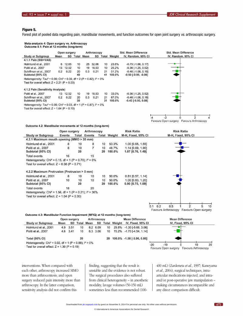
Open Surgery vs. Arthroscopy
Three studies made this comparison with no statistically significant differences between the effects of the 2 surgeries on all measured outcomes over the longest follow-up (Holmlund et al., 2001; Politi et al., 2007; Schiffman et al., 2007).
When combined in meta-analysis, a significant overall effect for open surgery over arthroscopy on reducing the pain intensity over the long term was demonstrated (SMD = -0.50; 95%CI, -0.95 to -0.06; p = .03). However, sensitivity analysis by excluding the study without confirmatory diagnostic imaging (Holmlund et al., 2001) showed no statistically significant difference between the surgical procedures (SMD = -0.43; 95%CI, -0.93 to 0.08; p = .10). Furthermore, pooling the data from 2 studies (Holmlund et al., 2001; Politi et al., 2007) showed no statistically significant difference between the long-term effects of surgeries on the number of patients with MMO > 35 mm (RR = 1.07; 95%CI, 0.76 to 1.49; p = .71) (Fig. 5 and Table, Comparison 21). Open surgery caused one transient motor nerve injury (Schiffman et al., 2007) and several transient sensory nerve injuries (Holmlund et al., 2001; Politi et al., 2007).
DiscussionSummary of Main FindingsThere was high clinical heterogeneity
among the studies included, which was unsurprising given the differing interventions used, and the considerable variations in techniques applied and combinations and/or delivery of interventions. In most comparisons, therefore, there was only 1 trial, and only 4 meta-analyses could be performed on trials of homogenous comparable groups.
In this review, analysis was conducted between and within groups. When the interventions were compared with each
other (between groups), the least invasive conservative interventions, including patient education and self-management, seemed to exert effects comparable with those of more ‘active’ (combined splint plus physiotherapy) or ‘invasive’ (TMJ surgery) treatment approaches. Splints as a solitary treatment approach, however, seemed to have no additional effect over other active interventions or no treatment, although as an adjunct to others, they may help to alleviate symptoms.
Among the physiotherapeutic interventions, early mandibular manipulation seemed to exert an immediate effect, increasing MMO in patients with ‘acute’ DDwoR. Jaw ‘stretching’ exercises, either alone or in combination with others, also increased MMO, but their effects were inconsistent between studies, while the electro-physical modalities had, in general, no significant effect over placebo treatment or splints and could be associated with transient adverse events.
Minimally invasive arthrocentesis and invasive arthroscopic and open joint surgical interventions did not, in general, demonstrate significant differences in effects over non-invasive conservative interventions and could be associated with complications. Nevertheless, in one study, arthrocentesis reduced pain intensity more than did conservative treatment in ‘acute’ DDwoR (Diracoglu et al., 2009). That study, however, used quasi-randomization based on alternate allocation to intervention groups, and, if excluded from this review, arthrocentesis has not been proven to have additional effects over conservative
Figure 4.Forest plot of pooled data regarding maximum mouth opening outcome for arthroscopy vs. arthrocentesis.
by guest on November 9, 2014 For personal use only. No other uses without permission.jdr.sagepub.comDownloaded from
© International & American Associations for Dental Research
47S
JDR Clinical Research Supplementvol. 93 • issue 7 • suppl no. 1
interventions. When compared with each other, arthroscopy increased MMO more than arthrocentesis, and open surgery reduced pain intensity more than arthroscopy. In the latter comparison, sensitivity analysis did not confirm this
finding, suggesting that the result is unstable and the evidence is not robust. The surgical procedures also suffered from clinical heterogeneity – in anesthetic modality, lavage volumes (50-150 mL) sometimes less than recommended (100-
400 mL) (Zardeneta et al., 1997; Kaneyama et al., 2004), surgical techniques, intra-articular medications injected, and intra- and/or post-operative jaw manipulation – making circumstances incomparable and any direct comparison difficult.
Figure 5.Forest plot of pooled data regarding pain, mandibular movements, and function outcomes for open joint surgery vs. arthroscopic surgery.
by guest on November 9, 2014 For personal use only. No other uses without permission.jdr.sagepub.comDownloaded from
© International & American Associations for Dental Research
48S
JDR Clinical Research Supplement July 2014
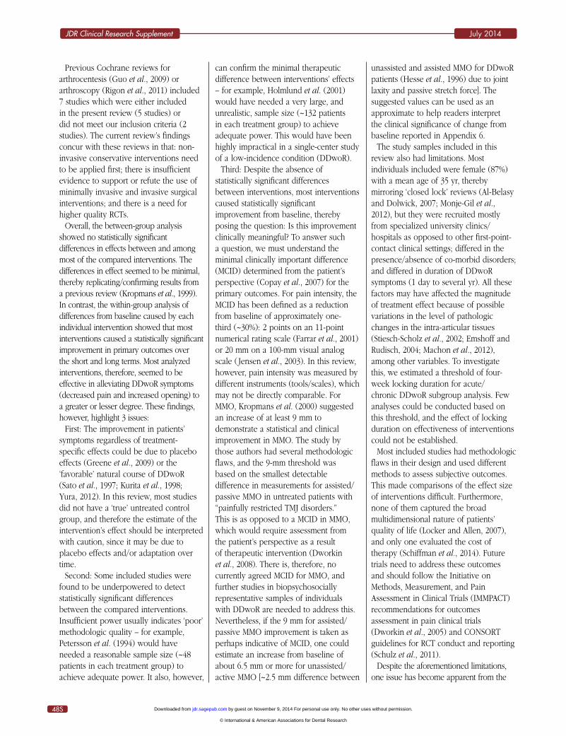
Previous Cochrane reviews for arthrocentesis (Guo et al., 2009) or arthroscopy (Rigon et al., 2011) included 7 studies which were either included in the present review (5 studies) or did not meet our inclusion criteria (2 studies). The current review’s findings concur with these reviews in that: non-invasive conservative interventions need to be applied first; there is insufficient evidence to support or refute the use of minimally invasive and invasive surgical interventions; and there is a need for higher quality RCTs.
Overall, the between-group analysis showed no statistically significant differences in effects between and among most of the compared interventions. The differences in effect seemed to be minimal, thereby replicating/confirming results from a previous review (Kropmans et al., 1999). In contrast, the within-group analysis of differences from baseline caused by each individual intervention showed that most interventions caused a statistically significant improvement in primary outcomes over the short and long terms. Most analyzed interventions, therefore, seemed to be effective in alleviating DDwoR symptoms (decreased pain and increased opening) to a greater or lesser degree. These findings, however, highlight 3 issues:
First: The improvement in patients’ symptoms regardless of treatment-specific effects could be due to placebo effects (Greene et al., 2009) or the ‘favorable’ natural course of DDwoR (Sato et al., 1997; Kurita et al., 1998; Yura, 2012). In this review, most studies did not have a ‘true’ untreated control group, and therefore the estimate of the intervention’s effect should be interpreted with caution, since it may be due to placebo effects and/or adaptation over time.
Second: Some included studies were found to be underpowered to detect statistically significant differences between the compared interventions. Insufficient power usually indicates ‘poor’ methodologic quality – for example, Petersson et al. (1994) would have needed a reasonable sample size (~48 patients in each treatment group) to achieve adequate power. It also, however,
can confirm the minimal therapeutic difference between interventions’ effects – for example, Holmlund et al. (2001) would have needed a very large, and unrealistic, sample size (~132 patients in each treatment group) to achieve adequate power. This would have been highly impractical in a single-center study of a low-incidence condition (DDwoR).
Third: Despite the absence of statistically significant differences between interventions, most interventions caused statistically significant improvement from baseline, thereby posing the question: Is this improvement clinically meaningful? To answer such a question, we must understand the minimal clinically important difference (MCID) determined from the patient’s perspective (Copay et al., 2007) for the primary outcomes. For pain intensity, the MCID has been defined as a reduction from baseline of approximately one-third (~30%): 2 points on an 11-point numerical rating scale (Farrar et al., 2001) or 20 mm on a 100-mm visual analog scale ( Jensen et al., 2003). In this review, however, pain intensity was measured by different instruments (tools/scales), which may not be directly comparable. For MMO, Kropmans et al. (2000) suggested an increase of at least 9 mm to demonstrate a statistical and clinical improvement in MMO. The study by those authors had several methodologic flaws, and the 9-mm threshold was based on the smallest detectable difference in measurements for assisted/passive MMO in untreated patients with “painfully restricted TMJ disorders.” This is as opposed to a MCID in MMO, which would require assessment from the patient’s perspective as a result of therapeutic intervention (Dworkin et al., 2008). There is, therefore, no currently agreed MCID for MMO, and further studies in biopsychosocially representative samples of individuals with DDwoR are needed to address this. Nevertheless, if the 9 mm for assisted/passive MMO improvement is taken as perhaps indicative of MCID, one could estimate an increase from baseline of about 6.5 mm or more for unassisted/active MMO [~2.5 mm difference between
unassisted and assisted MMO for DDwoR patients (Hesse et al., 1996) due to joint laxity and passive stretch force]. The suggested values can be used as an approximate to help readers interpret the clinical significance of change from baseline reported in Appendix 6.
The study samples included in this review also had limitations. Most individuals included were female (87%) with a mean age of 35 yr, thereby mirroring ‘closed lock’ reviews (Al-Belasy and Dolwick, 2007; Monje-Gil et al., 2012), but they were recruited mostly from specialized university clinics/hospitals as opposed to other first-point-contact clinical settings; differed in the presence/absence of co-morbid disorders; and differed in duration of DDwoR symptoms (1 day to several yr). All these factors may have affected the magnitude of treatment effect because of possible variations in the level of pathologic changes in the intra-articular tissues (Stiesch-Scholz et al., 2002; Emshoff and Rudisch, 2004; Machon et al., 2012), among other variables. To investigate this, we estimated a threshold of four-week locking duration for acute/chronic DDwoR subgroup analysis. Few analyses could be conducted based on this threshold, and the effect of locking duration on effectiveness of interventions could not be established.
Most included studies had methodologic flaws in their design and used different methods to assess subjective outcomes. This made comparisons of the effect size of interventions difficult. Furthermore, none of them captured the broad multidimensional nature of patients’ quality of life (Locker and Allen, 2007), and only one evaluated the cost of therapy (Schiffman et al., 2014). Future trials need to address these outcomes and should follow the Initiative on Methods, Measurement, and Pain Assessment in Clinical Trials (IMMPACT) recommendations for outcomes assessment in pain clinical trials (Dworkin et al., 2005) and CONSORT guidelines for RCT conduct and reporting (Schulz et al., 2011).
Despite the aforementioned limitations, one issue has become apparent from the
by guest on November 9, 2014 For personal use only. No other uses without permission.jdr.sagepub.comDownloaded from
© International & American Associations for Dental Research

49S
JDR Clinical Research Supplementvol. 93 • issue 7 • suppl no. 1
results of this review: Most interventions appear to alleviate DDwoR symptoms, with no significant differences between non-invasive conservative interventions and minimally invasive or invasive surgical interventions. Given the paucity of evidence and the difficulty in interpreting the minimal clinically important difference, this finding suggests that patients with DDwoR probably should be initially managed with the most minimal and least invasive intervention. Escalation to more invasive treatment should occur only in the face of objective clinical need. This, however, should be interpreted in the context of a review based mostly on single studies of unclear to high risk of bias. Future well-conducted research may change or confirm this.
ConclusionImplications for PracticeThe comparable therapeutic effects of
reviewed interventions suggest using the simplest, least costly, and least invasive interventions for the initial management of DDwoR. Of the variety of non-invasive conservative interventions reviewed, the least invasive were patient education, self-management, and early mandibular manipulation. Currently, there is insufficient evidence to support or refute the use of minimally invasive and invasive surgical interventions for DDwoR. However, there may well be specific clinical cases where a surgical intervention may help, but the body of evidence does not give a clear indication of when this may be.
Implications for ResearchThere is weak evidence to support
the initial use of simple, minimal, non-invasive conservative interventions, particularly patient education, self-management, and early mandibular manipulation, for DDwoR. Future research needs to examine these interventions specifically to provide more robust evidence of their efficacy or lack of it. The evidence for the effectiveness of minimally invasive surgical intervention through arthrocentesis and lavage is contradictory. Given its less invasive nature, future high-quality
pragmatic RCTs are required to compare the effects of arthrocentesis with those of conservative interventions.
Detailed descriptions about recommended research design are available upon request.
Acknowledgments
The review authors thank Prof. Frank Lobbezoo (ACTA Amsterdam, Netherlands) and Dr. Stephen Davies (Manchester Dental School, UK) for their critical review of the study protocol. The review authors also thank the responding authors of included studies and the four anonymous manuscript reviewers for their constructive and helpful critique. This study is funded by the higher committee for education development in Iraq (HCED) and was undertaken as a part of a postgraduate PhD clinical program in the Department of Oral and Maxillofacial Surgery, School of Dental Sciences, Newcastle University, UK. The authors declare no potential conflicts of interest with respect to the authorship and/or publication of this article.
ReferencesAkers J, Aguiar-Ibáñez R, Sari AB, Beynon
S, Booth A, Burch J, et al. (2009). Systematic Reviews Centre for Reviews and Dissemination’s (CRD) guidance for undertaking reviews in health care/ 3rd ed. York, UK: York Publishing Services Ltd.
Al-Baghdadi M, Durham J, Araujo-Soares V, Robalino S, Errington L, Steele J (2012). Interventions for the management of temporomandibular joint disc displacement without reduction (a systematic review protocol). PROSPERO:CRD42012003153 Accessed on 2/21/2014 at: http://www .crd.york.ac.uk/PROSPERO/display_record .asp?ID=CRD42012003153.
Al-Belasy FA, Dolwick MF (2007). Arthrocentesis for the treatment of temporomandibular joint closed lock: a review article. Int J Oral Maxillofac Surg 36:773-782.
Copay AG, Subach BR, Glassman SD, Polly DW Jr, Schuler TC (2007). Understanding the minimum clinically important difference: a review of concepts and methods. Spine J 7:541-546.
Craane B, Dijkstra PU, Stappaerts K, De Laat A (2012). Randomized controlled trial on physical therapy for TMJ closed lock. J Dent Res 91:364-369.
Deeks JJ, Higgins JPT, Altman DG (2011). Chapter 9: Analysing data and undertaking meta-analyses. In: Cochrane Handbook for Systematic Reviews of Interventions Version 5.1.0 (updated March 2011). Higgins JP, Green S, editors. The Cochrane Collaboration, 2011. Accessed on 2/21/2014 at: www.cochrane-handbook.org.
Diracoglu D, Saral IB, Keklik B, Kurt H, Emekli U, Ozcakar L, et al. (2009). Arthrocentesis versus nonsurgical methods in the treatment of temporomandibular disc displacement without reduction. Oral Surg Oral Med Oral Pathol Oral Radiol Endod 108:3-8.
Durham J, Exley C, Wassell R, Steele JG (2007). ‘Management is a black art’—professional ideologies with respect to temporomandibular disorders. Br Dent J 202:E29.
Dworkin RH, Turk DC, Farrar JT, Haythornthwaite JA, Jensen MP, Katz NP, et al. (2005). Core outcome measures for chronic pain clinical trials: IMMPACT recommendations. Pain 113:9-19.
Dworkin RH, Turk DC, Wyrwich KW, Beaton D, Cleeland CS, Farrar JT, et al. (2008). Interpreting the clinical importance of treatment outcomes in chronic pain clinical trials: IMMPACT recommendations. J Pain 9:105-121.
Egger M, Davey Smith G, Schneider M, Minder C (1997). Bias in meta-analysis detected by a simple, graphical test. BMJ 315:629-634.
Emshoff R, Rudisch A (2004). Determining predictor variables for treatment outcomes of arthrocentesis and hydraulic distention of the temporomandibular joint. J Oral Maxillofac Surg 62:816-823.
Farrar JT, Young JP Jr, LaMoreaux L, Werth JL, Poole RM (2001). Clinical importance of changes in chronic pain intensity measured on an 11-point numerical pain rating scale. Pain 94:149-158.
Fridrich KL, Wise JM, Zeitler DL (1996). Prospective comparison of arthroscopy and arthrocentesis for temporomandibular joint disorders. J Oral Maxillofac Surg 54:816-821.
Goudot P, Jaquinet AR, Hugonnet S, Haefliger W, Richter M (2000). Improvement of pain and function after arthroscopy and arthrocentesis of the temporomandibular joint: a comparative study. J Craniomaxillofac Surg 28:39-43.
Greene CS, Goddard G, Macaluso GM, Mauro G (2009). Topical review: placebo responses and therapeutic responses. How are they related? J Orofac Pain 23:93-107.
Guo C, Shi Z, Revington P (2009). Arthrocentesis and lavage for treating temporomandibular joint disorders. Cochrane Database Syst Rev 4:CD004973.
by guest on November 9, 2014 For personal use only. No other uses without permission.jdr.sagepub.comDownloaded from
© International & American Associations for Dental Research

50S
JDR Clinical Research Supplement July 2014
Haketa T, Kino K, Sugisaki M, Takaoka M, Ohta T (2010). Randomized clinical trial of treatment for TMJ disc displacement. J Dent Res 89:1259-1263.
Hesse JR, Naeije M, Hansson TL (1996). Craniomandibular stiffness in myogenous and arthrogenous CMD patients, and control subjects: a clinical and experimental investigation. J Oral Rehabil 23:379-385.
Higgins JP, Green S (2011). Cochrane Handbook for Systematic Reviews of Interventions Version 5.1.0 [updated March 2011]: The Cochrane Collaboration, 2011. Accessed on 2/21/2014 at: www.cochrane-handbook.org.
Higgins JP, Thompson SG (2002). Quantifying heterogeneity in a meta-analysis. Stat Med 21:1539-1558.
Higgins JP, Altman DG, Gotzsche PC, Juni P, Moher D, Oxman AD, et al. (2011). The Cochrane Collaboration’s tool for assessing risk of bias in randomised trials. BMJ 343:d5928.
Holmlund AB, Axelsson S, Gynther GW (2001). A comparison of discectomy and arthroscopic lysis and lavage for the treatment of chronic closed lock of the temporomandibular joint: a randomized outcome study. J Oral Maxillofac Surg 59:972-977.
Ismail F, Demling A, Heßling K, Fink M, Stiesch-Scholz M (2007). Short-term efficacy of physical therapy compared to splint therapy in treatment of arthrogenous TMD. J Oral Rehabil 34:807-813.
Jensen MP, Chen C, Brugger AM (2003). Interpretation of visual analog scale ratings and change scores: a reanalysis of two clinical trials of postoperative pain. J Pain 4:407-414.
Kaneyama K, Segami N, Nishimura M, Sato J, Fujimura K, Yoshimura H (2004). The ideal lavage volume for removing bradykinin, interleukin-6, and protein from the temporomandibular joint by arthrocentesis. J Oral Maxillofac Surg 62:657-661.
Kropmans T, Dijkstra P, Stegenga B, Stewart R, de Bont L (2000). Smallest detectable difference of maximal mouth opening in patients with painfully restricted temporomandibular joint function. Eur J Oral Sci 108:9-13.
Kropmans TJ, Dijkstra PU, Stegenga B, de Bont LG (1999). Therapeutic outcome assessment in permanent temporomandibular joint disc displacement. J Oral Rehabil 26:357-363.
Kurita K, Westesson PL, Yuasa H, Toyama M, Machida J, Ogi N (1998). Natural course of untreated symptomatic temporomandibular joint disc displacement without reduction. J Dent Res 77:361-365.
Lewis S, Clarke M (2001). Forest plots: trying to see the wood and the trees. BMJ 322:1479-1480.
Linde C, Isacsson G, Jonsson BG (1995). Outcome of 6-week treatment with transcutaneous electric nerve stimulation compared with splint on symptomatic temporomandibular joint disk displacement without reduction. Acta Odontol Scand 53:92-98.
Locker D, Allen F (2007). What do measures of ‘oral health-related quality of life’ measure? Community Dent Oral Epidemiol 35:401-411.
Lundh H, Westesson PL, Eriksson L, Brooks SL (1992). Temporomandibular joint disk displacement without reduction. Treatment with flat occlusal splint versus no treatment. Oral Surg Oral Med Oral Path Oral Radiol Endod 73:655-658.
Machon V, Sedy J, Klima K, Hirjak D, Foltan R (2012). Arthroscopic lysis and lavage in patients with temporomandibular anterior disc displacement without reduction. Int J Oral Maxillofac Surg 41:109-113.
Maloney GE, Mehta N, Forgione AG, Zawawi KH, Al-Badawi EA, Driscoll SE (2002). Effect of a passive jaw motion device on pain and range of motion in TMD patients not responding to flat plane intraoral appliances. Cranio 20:55-65.
Manfredini D, Guarda-Nardini L, Winocur E, Piccotti F, Ahlberg J, Lobbezoo F (2011). Research diagnostic criteria for temporomandibular disorders: a systematic review of axis I epidemiologic findings. Oral Surg Oral Med Oral Pathol Oral Radiol Endod 112:453-462.
Minakuchi H, Kuboki T, Matsuka Y, Maekawa K, Yatani H, Yamashita A (2001). Randomized controlled evaluation of non-surgical treatments for temporomandibular joint anterior disk displacement without reduction. J Dent Res 80:924-928.
Moher D, Liberati A, Tetzlaff J, Altman DG, Group P (2009). Preferred reporting items for systematic reviews and meta-analyses: the PRISMA statement. BMJ 339:b2535. Accessed on 2/21/2014 at: http://www.prisma-statement.org/.
Monje-Gil F, Nitzan D, Gonzalez-Garcia R (2012). Temporomandibular joint arthrocentesis. Review of the literature. Med Oral Patol Oral Cir Bucal 17:e575-581.
Okeson JP (2007). Joint intracapsular disorders: diagnostic and nonsurgical management considerations. Dent Clin North Am 51:85-103, vi.
Peroz I, Chun YH, Karageorgi G, Schwerin C, Bernhardt O, Roulet JF, et al. (2004). A multicenter clinical trial on the use of pulsed electromagnetic fields in the treatment of temporomandibular disorders. J Prosthet Dent 91:180-187.
Petersson A, Eriksson L, Lundh H (1994). No short-term difference in outcome after temporomandibular joint arthrography alone or with immediate lavage. Oral Surg Oral Med Oral Pathol Oral Radiol Endod 77:322-326.
Politi M, Sembronio S, Robiony M, Costa F, Toro C, Undt G (2007). High condylectomy and disc repositioning compared to arthroscopic lysis, lavage, and capsular stretch for the treatment of chronic closed lock of the temporomandibular joint. Oral Surg Oral Med Oral Pathol Oral Radiol Endod 103:27-33.
Poveda-Roda R, Bagan JV, Sanchis JM, Carbonell E (2012). Temporomandibular disorders. A case-control study. Med Oral Patol Oral Cir Bucal 17:e794-800.
RevMan (2012). Review Manager (RevMan). 5.2. Copenhagen, Denmark: The Nordic Cochrane Centre, The Cochrane Collaboration.
Rigon M, Pereira LM, Bortoluzzi MC, Loguercio AD, Ramos AL, Cardoso JR (2011). Arthroscopy for temporomandibular disorders. Cochrane Database Syst Rev 5:CD006385.
Sahlstrom LE, Ekberg EC, List T, Petersson A, Eriksson L (2013). Lavage treatment of painful jaw movements at disc displacement without reduction. A randomized controlled trial in a short-term perspective. Int J Oral Maxillofac Surg 42:356-363.
Saitoa T, Yamadaa H, Nakaokaa K, Horiea A, Mishimab A, Nomurac Y, et al. (2010). Risk factors for the poor clinical outcome of visually guided temporomandibular joint irrigation in patients with chronic closed lock. Asian J Oral Maxillofac Surg 22:133-137.
Sato S, Kawamura H, Nagasaka H, Motegi K (1997). The natural course of anterior disc displacement without reduction in the temporomandibular joint: follow-up at 6, 12, and 18 months. J Oral Maxillofac Surg 55:234-238.
Schiffman EL, Braun BL, Lindgren BR (1996). Temporomandibular joint iontophoresis: a double-blind randomized clinical trial. J Orofac Pain 10:157-165.
Schiffman EL, Löök JO, Hodges JS, Swift JQ, Decker KL, Hathaway KM, et al. (2007). Randomized effectiveness study of four therapeutic strategies for TMJ closed lock. J Dent Res 86:58-63; erratum in J Dent Res 92:98, 2013.
Schiffman EL, Velly AM, Löök JO, Hodges JS, Swift JQ, Decker KL, et al. (2014). Effects of four treatment strategies for temporomandibular joint closed lock. Int J Oral Maxillofac Surg 43:217-226.
Schulz KF, Altman DG, Moher D, Group C (2011). CONSORT 2010 statement: updated
by guest on November 9, 2014 For personal use only. No other uses without permission.jdr.sagepub.comDownloaded from
© International & American Associations for Dental Research

51S
JDR Clinical Research Supplementvol. 93 • issue 7 • suppl no. 1
guidelines for reporting parallel group randomised trials. Int J Surg 9:672-677.
Sembronio S, Albiero AM, Toro C, Robiony M, Politi M (2008). Is there a role for arthrocentesis in recapturing the displaced disc in patients with closed lock of the temporomandibular joint? Oral Surg Oral Med Oral Pathol Oral Radiol Endod 105:274-280.
Stiesch-Scholz M, Tschernitschek H, Rossbach A (2002). Early begin of splint therapy improves treatment outcome in patients with temporomandibular joint disk displacement without reduction. Clin Oral Investig 6:119-123.
Yoshida H, Fukumura Y, Suzuki S, Fujita S, Kenzo O, Yoshikado R, et al. (2005). Simple
manipulation therapy for temporomandibular joint internal derangement with closed lock. Asian J Oral Maxillofac Surg 17:256-260.
Yoshida H, Sakata T, Hayashi T, Shirao K, Oshiro N, Morita S (2011). Evaluation of mandibular condylar movement exercise for patients with internal derangement of the temporomandibular joint on initial presentation. Br J Oral Maxillofac Surg 49:310- 313.
Yuasa H, Kurita K (2001). Randomized clinical trial of primary treatment for temporomandibular joint disk displacement without reduction and without osseous
changes: a combination of NSAIDs and mouth-opening exercise versus no treatment. Oral Surg Oral Med Oral Pathol Oral Radiol Endod 91:671- 675.
Yura S (2012). Natural course of acute closed lock of the temporomandibular joint. Br J Oral Maxillofac Surg 50:646- 649.
Zardeneta G, Milam SB, Schmitz JP (1997). Elution of proteins by continuous temporomandibular joint arthrocentesis. J Oral Maxillofac Surg 55:709- 716.
by guest on November 9, 2014 For personal use only. No other uses without permission.jdr.sagepub.comDownloaded from
© International & American Associations for Dental Research


![C_White Quality Management_A Financial Perspective[1]](https://static.fdocuments.in/doc/165x107/56d6bd991a28ab30168e9a77/cwhite-quality-managementa-financial-perspective1.jpg)

