
TLC March 27, 2013 - CMNRP March 27, 2013 Shawn Hollinger ... fetal membranes Intrauterine growth...
7
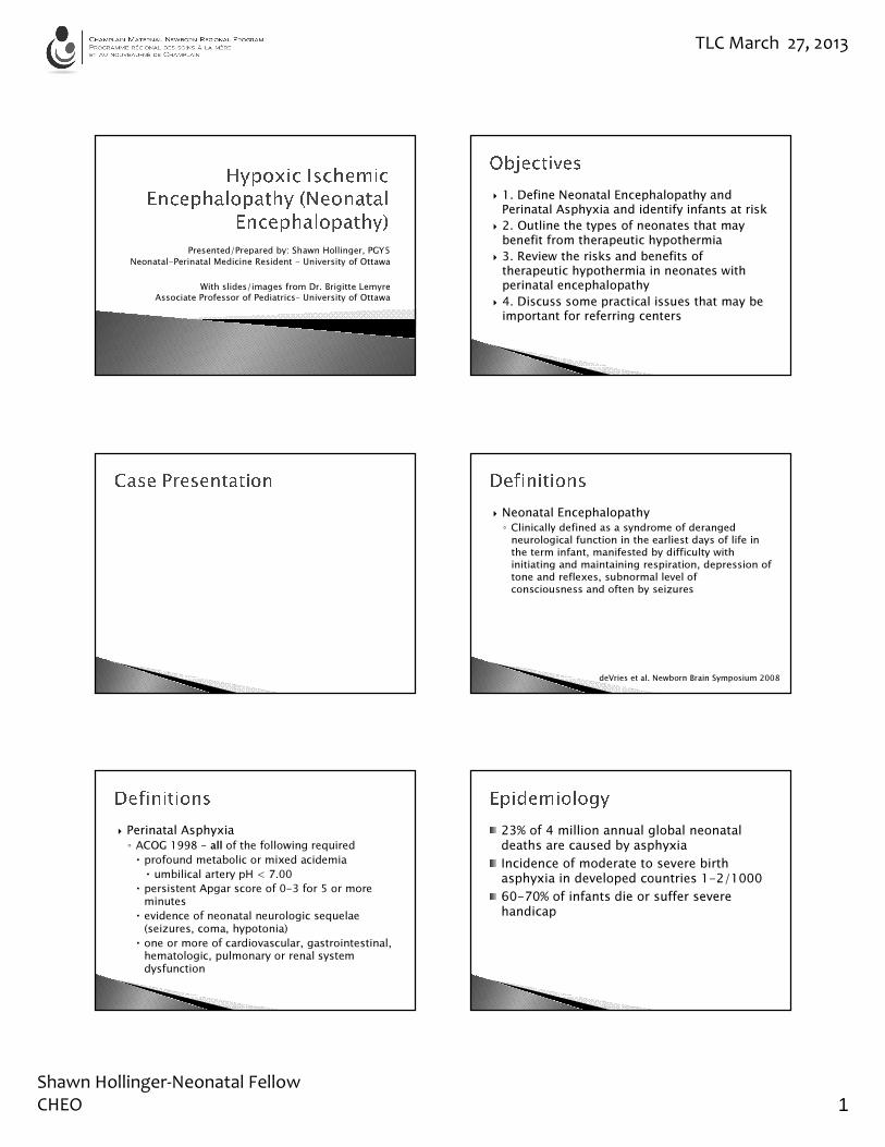
TLC March 27, 2013 Shawn Hollinger-Neonatal Fellow CHEO 1 Presented/Prepared by: Shawn Hollinger, PGY5 Neonatal-Perinatal Medicine Resident - University of Ottawa With slides/images from Dr. Brigitte Lemyre Associate Professor of Pediatrics– University of Ottawa 1. Define Neonatal Encephalopathy and Perinatal Asphyxia and identify infants at risk 2. Outline the types of neonates that may benefit from therapeutic hypothermia 3. Review the risks and benefits of therapeutic hypothermia in neonates with perinatal encephalopathy 4. Discuss some practical issues that may be important for referring centers Neonatal Encephalopathy ◦ Clinically defined as a syndrome of deranged neurological function in the earliest days of life in the term infant, manifested by difficulty with initiating and maintaining respiration, depression of tone and reflexes, subnormal level of consciousness and often by seizures deVries et al. Newborn Brain Symposium 2008 Perinatal Asphyxia ◦ ACOG 1998 - all of the following required profound metabolic or mixed acidemia umbilical artery pH < 7.00 persistent Apgar score of 0-3 for 5 or more minutes evidence of neonatal neurologic sequelae (seizures, coma, hypotonia) one or more of cardiovascular, gastrointestinal, hematologic, pulmonary or renal system dysfunction 23% of 4 million annual global neonatal deaths are caused by asphyxia Incidence of moderate to severe birth asphyxia in developed countries 1-2/1000 60-70% of infants die or suffer severe handicap
Transcript of TLC March 27, 2013 - CMNRP March 27, 2013 Shawn Hollinger ... fetal membranes Intrauterine growth...
TLC March 27, 2013
Shawn Hollinger-Neonatal Fellow
CHEO 1
Presented/Prepared by: Shawn Hollinger, PGY5
Neonatal-Perinatal Medicine Resident - University of Ottawa
With slides/images from Dr. Brigitte Lemyre
Associate Professor of Pediatrics– University of Ottawa
� 1. Define Neonatal Encephalopathy and Perinatal Asphyxia and identify infants at risk
� 2. Outline the types of neonates that may benefit from therapeutic hypothermia
� 3. Review the risks and benefits of therapeutic hypothermia in neonates with perinatal encephalopathy
� 4. Discuss some practical issues that may be important for referring centers
� Neonatal Encephalopathy◦ Clinically defined as a syndrome of deranged
neurological function in the earliest days of life in
the term infant, manifested by difficulty with
initiating and maintaining respiration, depression of
tone and reflexes, subnormal level of
consciousness and often by seizures
deVries et al. Newborn Brain Symposium 2008
� Perinatal Asphyxia
◦ ACOG 1998 - all of the following required
� profound metabolic or mixed acidemia
� umbilical artery pH < 7.00
� persistent Apgar score of 0-3 for 5 or more minutes
� evidence of neonatal neurologic sequelae(seizures, coma, hypotonia)
� one or more of cardiovascular, gastrointestinal, hematologic, pulmonary or renal system dysfunction
23% of 4 million annual global neonatal deaths are caused by asphyxia
Incidence of moderate to severe birth asphyxia in developed countries 1-2/1000
60-70% of infants die or suffer severe handicap

TLC March 27, 2013
Shawn Hollinger-Neonatal Fellow
CHEO 1
Prepartum Intrapartum Neonatal
Maternal Obstetric
Toxemia (eclampsia) Abruptio Placentae Abnormal Presentation Prematurity
Diabetes Mellitus Prolapse of Umbilical cord
Precipitate delivery Respiratory Distress
Drug addiction Placenta previa Prolonged labour Cardiopulmonary anomalies
Cardiovascular Disease Hydramnios Difficult forceps delivery
Infectious disease
Infectious disease Premature rupture of fetal membranes
Intrauterine growth retardation
Hemolytic disease
Isoimmunization Multiple pregnancy Prolonged pregnancy Septic or viremic shock
� Conditions That Predispose the Fetus and Newborn Infant to Asphyxia
� Initial injury
� Reperfusion
� Secondary injury
Primary damage
Vulnerable area
� Neurological Signs◦ Seizures
◦ Abnormal Respiratory Pattern
◦ Hyperalertness, Obtundation or Coma
◦ Jitteriness
◦ Posturing
◦ Impaired suck, swallow or gag
◦ Abnormal pupillary responses
◦ Abnormal tone
� Lungs: PPHN, surfactant disruption, MAS
� Kidneys: oliguria, ATN
� Heart: Tricuspid regurgitation, myocardial necrosis, shock
� Metabolic: acidosis, hypoglycemia/Na/Ca
� GI: NEC, hepatic dysfunction
� Hematology: DIC, thrombocytopenia
� Adrenal insufficiency
� Acidosis
� Lactate
� Hypoglycemia
� Coagulopathy
� Increased Urea & Creatinine (often later)
� Increased AST, ALT & GGT (often later)
� Other signs depending on the cause
� Sarnat and Sarnat – Arch Neurology 1976
◦ 21 infants > 36 wks, fetal distress or Apgar 5 or less at 1 or 5 minutes
◦ serial neurologic exams and EEGs in hospital and at follow-up
◦ defined normal as within 1 month of 50th percentile on Denver, absence of hemiparesis or focal neurologic deficits, normal muscle tone and reflexes and normal EEG

TLC March 27, 2013
Shawn Hollinger-Neonatal Fellow
CHEO 1
� Hyperalert
� Normal tome, overactive reflexes
� Strong Moro, normal or weak suck
� Generalized sympathetic function increased: mydriasis, tachycardia
� No seizures, normal EEG
� Lethargic or obtunded
� Hypotonic
� Weak or absent suck, weak Moro
� Generalized parasympathetic function increase: miosis, bradycardia
� Common seizures, abnormal EEG
� Stuporous
� Flaccid, intermittent decerebrate posturing
� Absent suck, gag, Moro
� Depressed autonomic function: variable pupils, variable heart rate
� Very abnormal EEG: burst suppression, isoelectric
� Supportive
� Homeostasis
� Therapeutic Hypothermia
� Ensure physiologic, oxygen and acid-base balance (Aim for normal pCO2)
� Correct caloric, fluid and electrolytes disturbances� Maintain blood volume and homeostasis� Treat infection� Monitor for hyperbilirubinemia� Treat seizures
Klinger G. Arch Dis Child Fetal Neonatal Ed 2005

TLC March 27, 2013
Shawn Hollinger-Neonatal Fellow
CHEO 1
� Lowers the metabolic rate
� Essentially ‘resting’ the brain
� Decreases the seizure threshold
Primary damage
Vulnerable area
Author/Author/studystudy
YearYear # of # of patientspatients
Method Method of of coolingcooling
aEEGaEEG Withdraw Withdraw reportedreported
Follow upFollow up
GunnGunn 19981998 3131 head head nono nono 18 mo18 mo
GluckmanGluckman 20052005 234234 headhead yesyes nono 18 mo18 mo
ShankaranShankaran 20052005 208208 bodybody nono yesyes 1818--22 mo22 mo
AzzopardiAzzopardi 20092009 325325 bodybody yesyes yesyes 1818--22 mo22 mo
SimbrunerSimbruner** 20102010 129129 bodybody yesyes nono 1818--21 mo21 mo
ZhouZhou 20102010 194194 headhead nono nono 18 mo18 mo
Jacobs*Jacobs* 20112011 221221 bodybody nono yesyes 24 24 monthsmonths
Total number of infants enrolled: 1214* Stopped early due to lack of clinical equipoise
Tagin et al Arch Pediatr Adoles Med 2012
Pooled risk ratio favors therapyRR 0.76 (0.69-0.84); NNT 7 (5-10)
RR 0.67 (0.56-0.81) NNT 6
RR 0.83 (0.74-0.92)NNT 7

TLC March 27, 2013
Shawn Hollinger-Neonatal Fellow
CHEO 1
� University of Ottawa Criteria:◦ Cord pH <7.0 or base deficit > 16
◦ OR
◦ Cord (or baby gas within an hour of birth) pH 7.01
– 7.15 or base deficit 10 to 15.9 AND
� History of acute perinatal event (cord prolapse,
placental abruption, etc) AND
� Apgar <6 at 10 minutes or at least 10 minutes PPV
� Evidence of moderate to severe encephalopathy◦ Clinical seizures OR
◦ At least one sign in 3 or more categories on
physical exam
CategoryCategory ModerateModerateEncephalopathyEncephalopathy
Severe EncephalopathySevere Encephalopathy
1. Level of1. Level ofconsciousnessconsciousness
LethargicLethargic Stupor/comaStupor/coma
2. Spontaneous2. Spontaneousactivityactivity
Decreased activityDecreased activity No activityNo activity
3. Posture3. Posture Distal flexion, full Distal flexion, full extensionextension
DecerebrateDecerebrate (arms extended (arms extended and internally rotated, legs and internally rotated, legs extended with feet in forced extended with feet in forced plantar flexion)plantar flexion)
4. Tone4. Tone HypotoniaHypotonia (focal, (focal, general)general)
FlaccidFlaccid
5. 5. Primitive Primitive reflexesreflexes
SuckSuckMoroMoro
WeakWeakIncompleteIncomplete
AbsentAbsentAbsentAbsent
6. 6. Autonomic Autonomic systemsystem
PupilsPupilsHeart rateHeart rate
RespirationsRespirations
ConstrictedConstrictedBradycardiaBradycardiaPeriodic breathingPeriodic breathing
Skew deviation/dilated/nonSkew deviation/dilated/non--reactive to lightreactive to lightVariable HR Variable HR ApneaApnea
� Presence of known chromosomal anomaly� Presence of major congenital anomalies� Severe intrauterine growth restriction (weight <2000g)
� Infants in severe condition for which no additional intensive therapy will be offered after discussion with parents by attending neonatologist (i.e. refractory hypotension, refractory acidosis, comatose with absent brain-stem reflexes etc.)
� Congenital ano-rectal anomaly based on visual inspection
� Evidence of head trauma or intracranial hemorrhage
� Initiated in October 2009
� Whole body cooling using Blanketroll 3
� Aim for 33-34 °C rectal x 72 hours
� Rewarming over 12h
� aEEG for neuro monitoring
� Passive cooling prior to admission
� aEEG can be used as adjunct in borderline cases◦ Clear biochemical criteria
◦ Unclear physical and neuro exam (or evolving)
� Sarkar J Perinatol 2008

TLC March 27, 2013
Shawn Hollinger-Neonatal Fellow
CHEO 1
� Bradycardia (80-100 bpm)� Leukopenia (WBC < 5 x 10 9/L)� Coagulopathy� Hypoglycemia� Hypokalemia� Shivering � Seizures if rewarmed too fast
◦ 38 term AGA infants with HIE◦ EEG within 48 hours and neuro f/u > 1 year
� normal EEG n=14� normal outcome (13), minor sequelae (1)
� extremely abnormal EEG n=10� death (5), major sequelae (4), normal (1)
� intermediate EEG n=14� improved background < 7 days good (4/5)
prognosis
� Selton and Andre, Neuropediatrics 1997
aEEG is intended to be used clinically to complement the neurologic exam, conventional EEG and neuroimaging
aEEG within first 6 hours of life predictive of outcomes at 24 months (non-cooled)
Speed of recovery and severity of the abnormality of aEEG is prognosticallyvaluable
Shah DK et al. Pediatrics 2008
� MRI is routinely done on day 4-7 of life
� Routine MRI images may show signs of significant damages
� DWI may pick up damage that does not show on the routine imaging
� Look for damage particularily in the watershed areas and basal ganglia system
Volpe JJ. 2001

TLC March 27, 2013
Shawn Hollinger-Neonatal Fellow
CHEO 1
� Think about the diagnosis! (remember the time-window)
� When in doubt – call transport team to discuss potential candidate
� Discuss with transport team initiating passive cooling
� Qualify for the Neonatal Follow Up Clinic (NNFU)
� Followed at key developmental ages
� Multidisciplinary team that can identify areas of developmental concern and refer to appropriate services
� If severe/seizures, may also follow up with neurology
� Recognize the children that are at risk
� Advocate that they attend their follow up appointments
� Utilize developmental tools
� Follow growth curves including weight and head circumference
� Possible other therapies/adjuncts
� Many risk factors for Neonatal encephalopathy
� If you think a baby might benefit from therapeutic hypothermia, call your referral center to discuss (without delay!)
� Hypothermia has some side effects, but is effective in cases of moderate to severe encephalopathy
� Follow up in the community and early recognition of developmental concerns is important
� Volpe JJ. Neurology of the newborn.
� De Vries L et al. Long term outcomes after HIE. Arch Dis Child Fetal Neonat Ed 2010;95:F220
� Perlman M. HIE prediction of outcome. J Pediatr2011;158:e51


![User Equipage Costs (Hollinger).ppt [Read-Only]](https://static.fdocuments.in/doc/165x107/623c024ed8ef4029da21a607/user-equipage-costs-hollingerppt-read-only.jpg)


