Title: Thyroid disruption in zebrafish ( Danio rerio ... · mM NaCl, 0.17 mM KCl, 0.33 mM CaCl2,...
29
1 Title: Thyroid disruption in zebrafish (Danio rerio) larvae: different molecular response patterns lead to impaired eye development and visual functions Authors: *Baumann, Lisa 1 ; Ros, Albert 1 ; Rehberger, Kristina 1 ; Neuhauss, Stephan CF 2 ; Segner, Helmut 1 *corresponding author 1 University of Berne, Vetsuisse Faculty, Centre for Fish and Wildlife Health, Länggassstrasse122, CH-3012 Berne, Switzerland 2 University of Zurich, Institute of Molecular Life Sciences, Winterthurerstrasse 190, CH-8057 Zurich, Switzerland E-Mail: [email protected]; [email protected]; [email protected]; [email protected]; [email protected]
Transcript of Title: Thyroid disruption in zebrafish ( Danio rerio ... · mM NaCl, 0.17 mM KCl, 0.33 mM CaCl2,...
1
Title: Thyroid disruption in zebrafish (Danio rerio) larvae: different molecular response
patterns lead to impaired eye development and visual functions
Authors: *Baumann, Lisa1; Ros, Albert1; Rehberger, Kristina1; Neuhauss, Stephan CF2;
Segner, Helmut1
*corresponding author
1University of Berne, Vetsuisse Faculty, Centre for Fish and Wildlife Health,
Länggassstrasse122, CH-3012 Berne, Switzerland
2University of Zurich, Institute of Molecular Life Sciences, Winterthurerstrasse 190, CH-8057
Zurich, Switzerland
E-Mail: [email protected]; [email protected];
2
Abstract:
The vertebrate thyroid system is important for multiple developmental processes, including
eye development. Thus, its environmentally induced disruption may impact important fitness-
related parameters like visual capacities and behaviour. The present study investigated the
relation between molecular effects of thyroid disruption and morphological and physiological
changes of eye development in zebrafish (Danio rerio). Two test compounds representing
different molecular modes of thyroid disruption were used: propylthiouracil (PTU), which is
an enzyme-inhibitor of thyroid hormone synthesis, and tetrabromobisphenol A (TBBPA),
which interacts with the thyroid hormone receptors. Both chemicals significantly altered
transcript levels of thyroid system-related genes (TR, TR, TPO, TSH, DIO1, DIO2 and
DIO3) in a compound-specific way. Despite these different molecular response patterns, both
treatments resulted in similar pathological alterations of the eyes such as reduced size, RPE
cell diameter and pigmentation, which were concentration-dependent. The morphological
changes translated into impaired visual performance of the larvae: the optokinetic response
was significantly and concentration-dependently decreased in both treatments, together with a
significant increase of light preference of PTU-treated larvae. In addition, swimming activity
was impacted. This study provides first evidence that different modes of molecular action of
the thyroid disruptors can be associated with uniform apical responses. Furthermore, this
study is the first to show that pathological eye development, as it can be induced by exposure
to thyroid disruptors, indeed translates into impaired visual capacities of zebrafish early life
stages.
Keywords: zebrafish, thyroid disruption, eye development, behaviour
Abbreviations: PTU, propylthiouracil; TBBPA, tetrabromobisphenol A; EDC, endocrine
disrupting compound; MoA, mode of action; DMSO, dimethylsulfoxide; dpf, days post
fertilization; TR, thyroid receptor; DIO, deiodinase; TPO, thyroperoxidase; TSH, thyroid-
stimulating hormone; PFA, paraformaldehyde; OKR, optokinetic response; T3,
triiodothyronine; T4, thyroxine; RPE, retinal pigment epithelium
3
1. Introduction
Environmental contaminants with endocrine disrupting properties (endocrine disrupting
compounds, EDCs) affect wildlife and humans by interfering with endocrine homeostasis and
hormonal processes, e.g. oestrogen or thyroid signalling. The possible adverse outcomes of
EDC exposure have become a major issue in ecotoxicology and human toxicology, including
the scientific challenges to understand the molecular and physiological modes of action
(MoA) of EDCs as well as their ecological consequences. An important endocrine system that
is targeted by a variety of environmental contaminants is the thyroid hormone system. Field
surveys, epidemiological studies as well as laboratory experiments on birds (Brouwer et al.,
1998), amphibians (Carr and Patiño, 2011), fish (Brar et al., 2010; Brown et al., 2004) or
humans (Patrick, 2009) provide ample evidence that exposure to environmental pollutants can
be associated with disruption of thyroid hormone synthesis, metabolism and / or transport and
thus can lead to altered thyroid hormone status and thyroid hormone signalling. Thyroid-
disrupting effects have been reported for, e.g. polychlorinated biphenyls, polyaromatic
hydrocarbons, polybrominated diphenyl ethers, organochlorine pesticides as well as different
metals (Brown et al., 2004; Yu et al., 2013). Those environmental chemicals appear to
interfere with the thyroid system of vertebrates by two principal molecular mechanisms:
a) Disrupting the thyroid hormone homeostasis, which includes effects on enzymes
responsible for iodine uptake and thyroid hormone synthesis, and transport in the
blood, on cellular uptake degradation and excretion = thyroid gland function
disruptors (Thienpont et al., 2011).
b) Ligand binding to the thyroid receptors in the peripheral target tissues and
subsequent activation of thyroid signalling; this MoA does not necessarily involve
changes in endogenous hormone levels but in thyroid signalling = thyroid hormone
action disruptors (Boas et al., 2012).
Besides these basic molecular changes associated to thyroid disruptors, the physiological and
fitness consequences of environmental exposure remain less investigated. It is known that the
thyroid system plays an important role in key physiological processes such as growth,
development and reproduction, therefore, its environmentally induced disruption may impact
4
a wide variety of health and fitness-related parameters such as larval survival, behaviour,
fertility or metamorphosis (Power et al., 2001).
An important part of fish development is the differentiation of the eyes and craniofacial
structures, as these are essential for predator avoidance and food finding/intake. Thus,
malformations of the eyes and retinal cells might ultimately translate in a reduction of their
individual fitness. The eye and craniofacial development of vertebrates is a complex process
that is partially regulated by thyroid hormones (Bohnsack and Kahana, 2013; Darras et al.,
2015). Knock-down of specific thyroid-regulating genes as well as treatment with different
thyroid disruptors resulted in reduced eye size, malformations of eyes and head, and
disruption of retinal pigment cell layers of zebrafish larvae (Heijlen et al., 2014; Reider and
Connaughton, 2014). It has been reported that cone photoreceptor development seems to be
under direct regulation of thyroid hormones (Raine and Hawryshyn, 2009; Suzuki et al.,
2013), and it has been described, that thyroid receptors are expressed in the outer nuclear
layer of the retina, which contains the developing photoreceptors (Bertrand et al., 2007). The
exact pathways still need further investigation, even though a recent study could show that
different genes involved in phototransduction are changed in their expression after
knockdown of deiodinase enzymes in zebrafish, which regulate intracellular thyroid hormone
levels (Bagci et al., 2015). However, many factors involved in the complex thyroidal
regulation of eye development in fish remain unclear and need to be clarified and
investigated. One particularly relevant but yet unanswered question is whether the phenotypic
outcome (altered eye development) differs with the molecular MoA of the thyroid disruptors.
While the role of thyroid hormones in vertebrate eye development is well proven, and
although a number of studies have shown that disruption of the thyroid hormone system
affects eye morphology of developing fish, we lack clear proof that this translates into
impaired functionality of the optic system. How does the disrupted eye development due to
thyroid disruption result in direct physiological impairment of the developing fish? For
understanding the impact of thyroid disruptors on fish we have to show which specific
developmental processes are disrupted, but additionally it needs to be shown what the
implications are for functional capacities of the organism. Thus, the central hypotheses to be
tested in the present study are (i) that different molecular initiating events of thyroid
disruption lead to the same dysfunctional eye development and morphology of developing
zebrafish larvae and (ii) that the disruption of morphological eye development translates into
functional impairment of visual performance of the exposed fish.
5
To verify or reject our hypotheses, we treated zebrafish embryos with two thyroid disrupting
compounds that display different molecular actions on the thyroid system – inhibition of
thyroid hormone synthesis with propylthiouracil (PTU) versus interaction with the thyroid
receptors by tetrabromobisphenol A (TBBPA). Even though both substances seem to have
multiple effects on the thyroid system, there is clear evidence that the basic MoAs differ,
which is essential for our further investigations on disrupted eye development. PTU is a
thiouracil-derived drug that is widely used to inhibit elevated thyroid hormone production in
humans. It inhibits iodine and thyroperoxidase (TPO) from their normal interactions with
thyroglobulin to form thyroid hormones. Thus, exposure to PTU results in lowered thyroid
hormone levels (Van der Ven et al., 2006a). Consequently, the chemical often serves as
reference substance in exposure experiments on the thyroid system. Recent studies revealed
the adverse effect of PTU on the zebrafish thyroid system (Schmidt and Braunbeck, 2011)
and embryonic development (Jomaa, 2014). Also effects of PTU on fish eye development
have been reported (Macaulay et al., 2015; Raine and Hawryshyn, 2009), which makes it an
interesting candidate for the current project. Due to its presence in the environment and biota
(Environment Canada, 2013; Kitamura et al., 2002), TBBPA was chosen as exposure
chemical in the present study as representative for a thyroid action disruptor. It is a
brominated flame retardant and derivative of bisphenol A with different thyroid disrupting
capacities. It is able to interact with the thyroid receptors either as agonist, or as antagonist,
depending on the concentration and the endogenous TH levels of the organism (Crofton,
2008; Kitamura et al., 2002; Zhang et al., 2014). But also interaction with binding proteins
and hepatic clearance were reported (Boas et al., 2012). Previous studies have shown that
TBBPA competes with T4 in its binding to the plasma-transporter protein, transthyretin
(Hamers, 2006). TBBPA is acutely toxic in high concentrations and disrupts development and
reproduction of zebrafish at low concentrations (Kuiper et al., 2006). Moreover, it impairs
thyroid hormone-dependent metamorphosis in amphibian models (Fini et al., 2007; Jagnytsch
et al., 2006). In zebrafish changes in gene expression of thyroid-related genes have been
reported (W. Chan and K. Chan, 2012).
6
2. Materials & Methods:
2.1. Test chemicals
Tetrabromobisphenol A (TBBPA, CAS 79-94-7) and Propylthiouracil (PTU, CAS 51-52-5)
were purchased from Sigma Aldrich. Exposure concentrations were 0, 100, 200, 300 and 400
g/L for TBBPA (resp. 0, 0.18, 0.37, 0.55, 0.73 M) and 0, 50, 100 and 250 mg/L for PTU
(resp. 0, 0.29, 0.59, 1.47 mM). These concentrations were chosen based on range-finding tests
to determine treatments that were sub-lethal. For TBBPA we selected concentrations that
were lower than 1.09 mg/L, the 96h median effective concentration for zebrafish embryo
treatments (W. Chan and K. Chan, 2012). Moreover, the range of concentrations we used
(100-400 g/L for TBBPA) have been shown to be well supported for larvae up to 8 days post
fertilization, but showing clearly observable malformations at 400 g/L (Wu et al., 2015).
Treatment concentrations of PTU have been reported to result in depigmentation and clear
effects on the thyroid at levels above 50 mg/L (Schmidt and Braunbeck, 2011; Van der Ven et
al., 2006b). TBBPA was dissolved in DMSO with a maximal final concentration in the
exposure medium of 0.04%. Both chemicals were applied in E3 embryo medium (zfin.org, 5
mM NaCl, 0.17 mM KCl, 0.33 mM CaCl2, 0.33 mM MgSO4).
2.2. Zebrafish maintenance and exposure
A breeding group of 250 adult wildtype (AB strain) zebrafish (Danio rerio) was kept in a 210
L recirculating mass-spawning breeding tank for egg production (Fleuren&Nooijen,
Netherlands). Water parameters were constantly held at 27°C, pH 7.8 - 8.0 and day/night
cycle of 14/10 hours. Freshly fertilized eggs were collected in the morning, washed in E3
medium and then distributed into pre-incubated (= plates were filled with the solutions 1 day
before start of the test. For the start of the test, solutions were renewed) 24-well plates
containing the exposure solutions latest 2 hours after fertilization (1 egg per well, 2 ml
solution per well). Plates were incubated at 28°C under a day/night cycle of 14/10 hours.
Exposure solutions were renewed daily with freshly prepared solutions. The exposure
experiments were stopped at 5 days post-fertilization (dpf) by euthanizing the larvae in 300
mg/L MS-222 (Sigma). The described set-up applies for all further experiments and was
performed in at least 3 independent experiments (replicates). For measuring the effect on
mRNA expression we carried out 6 replicates for TBBPA, and 5 replicates for PTU. For
7
measuring the effect on OKR we carried out 3 replicates for each chemical. For light-dark-
preference we carried out 3 replicates for PTU and 4 for TBBPA. For morphology and
histology 3 replicates each.
2.3. qRT-PCR
24 euthanized larvae (5 dpf) of one exposure plate were quickly frozen in a 2 ml reaction tube
on dry ice and stored at -80°C for further processing. To isolate RNA a stainless steel bead (5
mm diameter, Qiagen) and 1 ml TRI Reagent solution (Sigma) were added to each tube, and
larvae were homogenized in a Tissue Lyser (Qiagen) at highest frequency for 2 min.
Subsequently, total RNA was isolated by the acid guanidinium thiocyanate-phenol-
chloroform extraction according to the manufacturer’s instructions. RNA concentration and
quality was checked with a spectrophotometer NanoDrop (ND1000 Thermo Scientific).
Isolation typically resulted in about 20 µl of 900 ng/µl RNA with 260nm/280nm ratio ranging
between 2.0 and 2.2. When the purity ratio (260nm/230nm) was lower than 1.8, LiCl was
added, the precipitate was restituted with pure water, and the concentration and purity was
measured again using the NanoDrop. Afterwards DNAse digestion and cDNA synthesis with
1 g RNA per reaction were performed with Promega GoScript™ Reverse Transcriptase, as
described in the instructions.
qRT-PCR was run using a standardized protocol based on Promega GoTaq® qPCR Master
Mix which uses Bryt™ Green dye to detect DNA formed during the PCR amplification. 96-
well fast qPCR Plates (ThermoFisher AB-1900) were loaded with 7.5 l Master Mix, 1.0 l
of one of the forward and reverse primers (Microsynth, 20 times diluted), 1.0 l H2O and 1.5
l of a cDNA sample. Plates were set-up to contain for each cDNA samples: 1) the primers
for the reference gene in triplicates; 2) all of the target gene primers in triplicates, 3) a blank
consisting of 1.5 l H2O instead of cDNA to check for possible contaminations in duplicates.
The qPCR was run on an Applied Biosystems 7500 Fast Real-Time PCR System, allowing a
5 min. period at 95 °C to denature the anti-Taq DNA polymerase antibodies in the GoTaq®
qPCR Master Mix. The qRT-PCR amplification was then allowed to run for 40 cycles (3 sec.
95 °C / 30 sec. 60 °C). A dissociation phase (melting curve) was added to check for non-
specific binding or multiple binding with the primers. Ct values were extracted using
standardized settings: baseline was set to the average of normalized Rn values at cycle 3 to 7,
and the log Ct values were extracted from the resulting ΔRn values at a threshold value of 0.2
8
(Applied Biosystems Detection Software version 1.3.1). 18S was used as reference gene as it
gave stable Ct values over treatments. Other potential reference genes elfa, Ribo11, G6P, and
TATA were down-regulated in the higher treatment groups and thus not used (supplemental
data). Efficiency of target and reference gene primers were compared and showed good linear
relationship (r2 > 0.9). Fold induction was calculated using the 2^-∆∆Ct method.
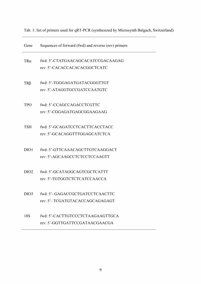
The expression of thyroid receptor alpha and beta (TR and TR), thyroperoxidase (TPO),
thyroid-stimulating hormone (TSH) as well as, deiodinase type 1, 2 and 3 (DIO1, DIO2 and
DIO3) were analysed using the following primers (5’-3’ direction) (Q. Chen et al., 2012;
Cheng et al., 2015; Pinto et al., 2013):
9
Tab. 1: list of primers used for qRT-PCR (synthesized by MicrosynthBalgach, Switzerland)
Gene Sequences of forward (fwd) and reverse (rev) primers
TR fwd: 5’-CTATGAACAGCACATCCGACAAGAG
rev: 5’-CACACCACACACGGCTCATC
TR fwd: 5’-TGGGAGATGATACGGGTTGT
rev: 5’-ATAGGTGCCGATCCAATGTC
TPO fwd: 5’-CCAGCCAGACCTCGTTC
rev: 5’-CGGAGATGAGCGGAAGAAG
TSH fwd: 5’-GCAGATCCTCACTTCACCTACC
rev: 5’-GCACAGGTTTGGAGCATCTCA
DIO1 fwd: 5’-GTTCAAACAGCTTGTCAAGGACT
rev: 5’-AGCAAGCCTCTCCTCCAAGTT
DIO2 fwd: 5’-GCATAGGCAGTCGCTCATTT
rev: 5’-TGTGGTCTCTCATCCAACCA
DIO3 fwd: 5’- GAGACCGCTGATCCTCAACTTC
rev: 5’- TCGATGTACACCAGCAGAGAGT
18S fwd: 5’-CACTTGTCCCTCTAAGAAGTTGCA
rev: 5’-GGTTGATTCCGATAACGAACGA
10
2.4. Size measurements and histology
For measurements of total body length and eye size, 24 euthanized larvae (5 dpf) from one
exposure plate were transferred into 4 % PFA and stored at 4 °C. 10-11 larvae per treatment
(from 3 independent replicates respectively) were measured using a stereomicroscope (Nikon
SMZ745T) with camera (the imaging source, dfk72buc02), using NIS elements software to
estimate the sizes with µm precision. Total length was measured as the length of the line
between mouth and tip of the tail. When larvae were curved correction for this by drawing a
path through the middle of the body was done. Eye size was measured at the longest diameter
of the eye. Relative eye length was calculated as the ratio of eye length over total length
expressed as a percentage.
For histological analysis of the eyes 24 euthanized larvae (5 dpf) from one exposure plate
were transferred into 4 % PFA and fixed over night at 4 °C (3 independent replicates
respectively). Subsequently, they were transferred into an agarose mold as described before
(Sabaliauskas et al., 2006) and aligned in the same orientation. The agarose block containing
the larvae was then processed in an embedding machine (Medite TPC 15Trio) in an ascending
series of ethanol, followed by xylol and finally paraffin. 3 m thick paraffin slides (coronal
sections) were produced with a rotating microtome (Microm HM340E). Slides were stained
with haematoxylin/eosin (using a Leica ST5020 and CV5030) and then analysed under a light
microscope (Olympus BX51) with camera (Olympus DP27). The associated software
(CellSense, Olympus) was used to analyse the taken pictures by measuring the diameter of the
different retinal cell layers of the larvae. If possible, at least 3 slides of each of the 2 eyes of
each zebrafish larva were analysed. The cell diameter was measured at 8 different locations in
the retina. Moreover, the pigmentation of the pigment cell layer was quantified at these 8
locations by measuring the grey values (0 = black, 250 = white).
2.5. Optokinetic response (OKR)
The OKR, a compensatory ocular motor reflex triggered by large scale movements in the
surround, was assessed as described before (Huber-Reggi et al., 2014). In summary, single
larvae (5 dpf) were taken from the exposure plates (24-well plate, 1 larva per well in 2 ml)
and placed dorsal-up in a 35 mm Petridish containing 3 % pre-warmed (28°C)
methylcellulose prepared in E3 medium. We carried out 3 independent replicates on different
days. The larva was placed in the centre of a paper drum on which a computer-generated
11
visual stimulus (vertical black-and-white stripes rotating around the fish) was projected via a
wide-angle conversion lens and a mirror from a LCD projector (PLV-Z3000; Sanyo).
Computer-generated gratings stimulated the larva binocularly and the eye position was
automatically tracked over time. The response of the eyes to different contrasts, velocities and
frequencies of the moving stripes was assessed by recording of an infrared-sensitive CCD
camera (Guppy F-038B NIR; Allied Vision Technologies). The term contrast describes the
distinction between black and white moving stripes, which changed in the range of 0-100%.
The term angular velocity is defined as the speed in which the stripes were rotating around the
larvae (degrees per second). Spatial frequency describes the frequency of direction changes of
the stripes and is defined as cycles per degree. The resulting eye velocity (degree per second)
was calculated in real time. Data were analysed by custom-developed software written in
MATLAB (MathWorks).
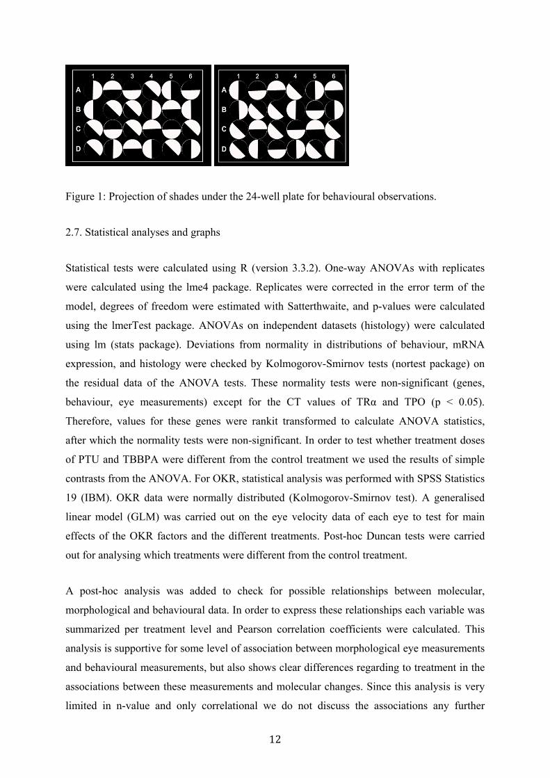
2.6. Swimming activity and light-dark preference
In addition to measuring the OKR, it was measured whether changes in light would result in
changes in phototactic swimming activity (X. Chen and Engert, 2014). To do so larvae (5 dpf)
were tested in their treatment plates. In total for PTU 3 independent experiments were carried
out (275 larvae), and for TBBPA 4 replicates were performed (449 larvae). The 24-well plate
(1 larva per well in 2 ml solution) was placed on top of a horizontally mounted LCD screen
(Fujitsu Scenicview, brightness: 0.03 cd/cm2 or about 5000 lux per well [1.9 cm2 3 mm
distance]), and under each well a half-light circle (see fig. 1) was projected using a
PowerPoint presentation (Microsoft). The circles were inverted after 2 minutes and this was
repeated 3 times, resulting in 4 periods (for example: left dark – right dark – left dark – right
dark). A camera (Nikon D90) was mounted one meter above the screen and from this point
every 30 seconds a picture was taken. Ambient light was blocked by placing a black paper
shield around the screen and camera. From a total of 16 pictures the following parameters
were taken: (i) background preference: did the larva prefer the light or dark part of the well.
For background preference only the data of the second minute (3rd and 4th picture) per period
was analysed to allow the larva to swim to the preferred part in the well; (ii) general
movement: a change in position from one to the other picture. This latter measurement was
taken to test for possible changes of general activity due to impaired vision.
12
Figure 1: Projection of shades under the 24-well plate for behavioural observations.
2.7. Statistical analyses and graphs
Statistical tests were calculated using R (version 3.3.2). One-way ANOVAs with replicates
were calculated using the lme4 package. Replicates were corrected in the error term of the
model, degrees of freedom were estimated with Satterthwaite, and p-values were calculated
using the lmerTest package. ANOVAs on independent datasets (histology) were calculated
using lm (stats package). Deviations from normality in distributions of behaviour, mRNA
expression, and histology were checked by Kolmogorov-Smirnov tests (nortest package) on
the residual data of the ANOVA tests. These normality tests were non-significant (genes,
behaviour, eye measurements) except for the CT values of TRα and TPO (p < 0.05).
Therefore, values for these genes were rankit transformed to calculate ANOVA statistics,
after which the normality tests were non-significant. In order to test whether treatment doses
of PTU and TBBPA were different from the control treatment we used the results of simple
contrasts from the ANOVA. For OKR, statistical analysis was performed with SPSS Statistics
19 (IBM). OKR data were normally distributed (Kolmogorov-Smirnov test). A generalised
linear model (GLM) was carried out on the eye velocity data of each eye to test for main
effects of the OKR factors and the different treatments. Post-hoc Duncan tests were carried
out for analysing which treatments were different from the control treatment.
A post-hoc analysis was added to check for possible relationships between molecular,
morphological and behavioural data. In order to express these relationships each variable was
summarized per treatment level and Pearson correlation coefficients were calculated. This
analysis is supportive for some level of association between morphological eye measurements
and behavioural measurements, but also shows clear differences regarding to treatment in the
associations between these measurements and molecular changes. Since this analysis is very
limited in n-value and only correlational we do not discuss the associations any further

13
because in order to test the causality of such relationships experimental manipulations of the
variables are necessary, which was not the scope of this manuscript. A correlation matrix
summarizing the results is provided as supplemental data.
All statistical analyses were two-tailed with the level of significance (alpha) set at 0.05.
Graphs were made with R, using the graphics package.
14
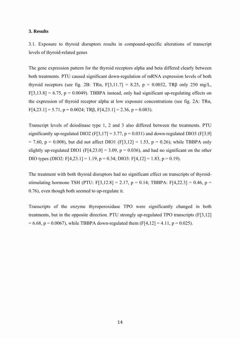
3. Results
3.1. Exposure to thyroid disruptors results in compound-specific alterations of transcript
levels of thyroid-related genes
The gene expression pattern for the thyroid receptors alpha and beta differed clearly between
both treatments. PTU caused significant down-regulation of mRNA expression levels of both
thyroid receptors (see fig. 2B: TRα, F[3,11.7] = 8.25, p = 0.0032, TRβ only 250 mg/L,
F[3,13.8] = 6.75, p = 0.0049). TBBPA instead, only had significant up-regulating effects on
the expression of thyroid receptor alpha at low exposure concentrations (see fig. 2A: TRα,
F[4,23.1] = 5.71, p = 0.0024; TRβ, F[4,23.1] = 2.36, p = 0.083).
Transcript levels of deiodinase type 1, 2 and 3 also differed between the treatments. PTU
significantly up-regulated DIO2 (F[3,17] = 3.77, p = 0.031) and down-regulated DIO3 (F[3,9]
= 7.60, p = 0.008), but did not affect DIO1 (F[3,12] = 1.53, p = 0.26); while TBBPA only
slightly up-regulated DIO1 (F[4,23.0] = 3.09, p = 0.036), and had no significant on the other
DIO types (DIO2: F[4,23.1] = 1.19, p = 0.34; DIO3: F[4,12] = 1.83, p = 0.19).
The treatment with both thyroid disruptors had no significant effect on transcripts of thyroid-
stimulating hormone TSH (PTU: F[3,12.8] = 2.17, p = 0.14; TBBPA: F[4,22.3] = 0.46, p =
0.76), even though both seemed to up-regulate it.
Transcripts of the enzyme thyroperoxidase TPO were significantly changed in both
treatments, but in the opposite direction. PTU strongly up-regulated TPO transcripts (F[3,12]
= 6.68, p = 0.0067), while TBBPA down-regulated them (F[4,12] = 4.11, p = 0.025).
15
Figure 2: Changes in gene expression levels. TRa/b = Thyroid receptor alpha or beta;
DIO1/2/3 = mRNA expression of deiodinase type 1, 2 or 3; TSH = thyroid-stimulating
hormone; TPO = thyroperoxidase. Data are expressed as mean ± st.err. Legend: bars with
different colours represent different treatments. A: TBBPA treatment µg/L; B: PTU treatment
mg/L. Symbols represent differences of the simple contrast (ANOVA) between the control
16
group (concentration = 0) and the treatment groups: * = p < 0.05, ** = p < 0.01, *** = p <
0.001.
3.2. Thyroid disruptor treatment of zebrafish larvae changes eye morphology and
pigmentation
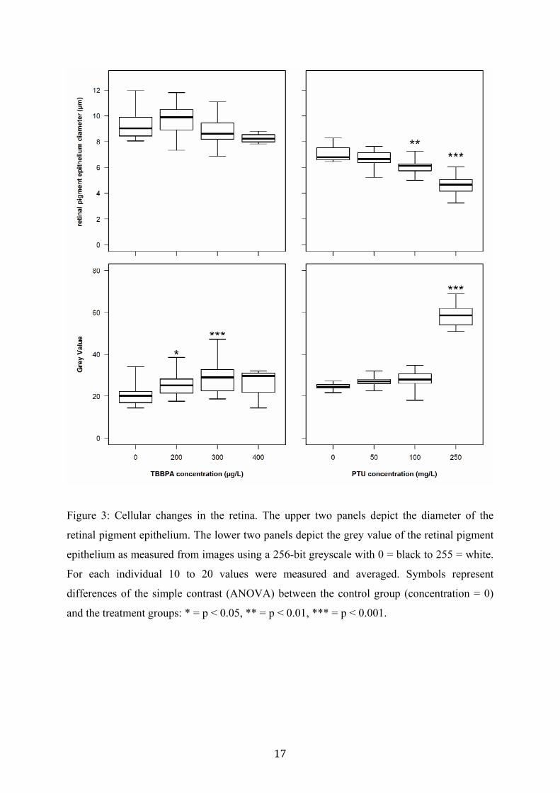
Both treatments had adverse effects on the development of the retinal pigment epithelium
(RPE) layer in the eye of exposed larvae (see fig. 4). Histological analyses of coronal sections
revealed a strong and significant concentration-dependent decrease in size and pigmentation
of the retinal cells for PTU-treated fish (see fig. 3; ANOVA: size, F[3,35] = 20.76, p < 0.001;
pigmentation, F[3,35] = 185.2, p < 0.001). Control fish had average RPE cell diameters of 7
µm, which significantly decreased to 5 µm in the highest exposure group (250 mg/L PTU),
while these changes were statistically not significant for TBBPA-treated fish. Grey values
(256-bit grayscale) increased from 25 (90% black) in the control to 60 (76% black) in the
highest exposure group. Similar observations were made for TBBPA-treated fish, but clearly
less pronounced than for PTU (ANOVA: size, F[3,50] = 3.33, p = 0.027; pigmentation,
F[3,50] = 4.61, p = 0.006). Measurements of the other cell layers in the retina revealed no
significant differences between control and treatments (data not shown).
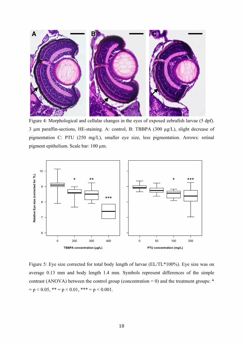
Both total body length and eye size of exposed larvae (5 dpf) decreased due to TBBPA and
PTU treatment (data not shown). To account for the changes in body morphology, eye size
(EL) was corrected for body length (TL) (see fig. 5). The relative eye size was significantly
decreased in both treatments (ANOVA EL/TL: TBBPA, F[3,28] = 8.47, p < 0.001; PTU,
F[3,38] = 5.74, p = 0.0024).
17
Figure 3: Cellular changes in the retina. The upper two panels depict the diameter of the
retinal pigment epithelium. The lower two panels depict the grey value of the retinal pigment
epithelium as measured from images using a 256-bit greyscale with 0 = black to 255 = white.
For each individual 10 to 20 values were measured and averaged. Symbols represent
differences of the simple contrast (ANOVA) between the control group (concentration = 0)
and the treatment groups: * = p < 0.05, ** = p < 0.01, *** = p < 0.001.
18
Figure 4: Morphological and cellular changes in the eyes of exposed zebrafish larvae (5 dpf).
3 µm paraffin-sections, HE-staining. A: control, B: TBBPA (300 µg/L), slight decrease of
pigmentation C: PTU (250 mg/L), smaller eye size, less pigmentation. Arrows: retinal
pigment epithelium. Scale bar: 100 µm.
Figure 5: Eye size corrected for total body length of larvae (EL/TL*100%). Eye size was on
average 0.13 mm and body length 1.4 mm. Symbols represent differences of the simple
contrast (ANOVA) between the control group (concentration = 0) and the treatment groups: *
= p < 0.05, ** = p < 0.01, *** = p < 0.001.
19
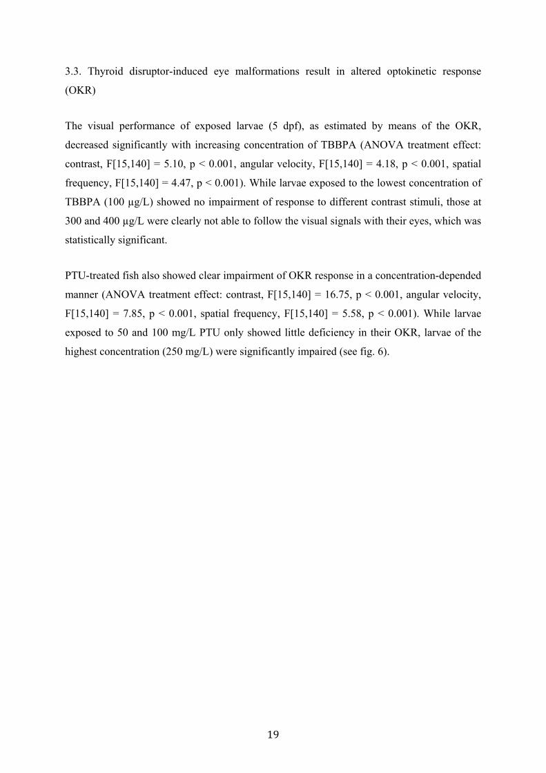
3.3. Thyroid disruptor-induced eye malformations result in altered optokinetic response
(OKR)
The visual performance of exposed larvae (5 dpf), as estimated by means of the OKR,
decreased significantly with increasing concentration of TBBPA (ANOVA treatment effect:
contrast, F[15,140] = 5.10, p < 0.001, angular velocity, F[15,140] = 4.18, p < 0.001, spatial
frequency, F[15,140] = 4.47, p < 0.001). While larvae exposed to the lowest concentration of
TBBPA (100 µg/L) showed no impairment of response to different contrast stimuli, those at
300 and 400 µg/L were clearly not able to follow the visual signals with their eyes, which was
statistically significant.
PTU-treated fish also showed clear impairment of OKR response in a concentration-depended
manner (ANOVA treatment effect: contrast, F[15,140] = 16.75, p < 0.001, angular velocity,
F[15,140] = 7.85, p < 0.001, spatial frequency, F[15,140] = 5.58, p < 0.001). While larvae
exposed to 50 and 100 mg/L PTU only showed little deficiency in their OKR, larvae of the
highest concentration (250 mg/L) were significantly impaired (see fig. 6).
20
21
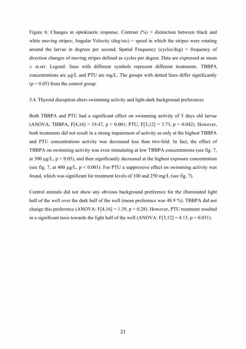
Figure 6: Changes in optokinetic response. Contrast (%) = distinction between black and
white moving stripes; Angular Velocity (deg/sec) = speed in which the stripes were rotating
around the larvae in degrees per second; Spatial Frequency (cycles/deg) = frequency of
direction changes of moving stripes defined as cycles per degree. Data are expressed as mean
± st.err. Legend: lines with different symbols represent different treatments. TBBPA
concentrations are µg/L and PTU are mg/L. The groups with dotted lines differ significantly
(p < 0.05) from the control group.
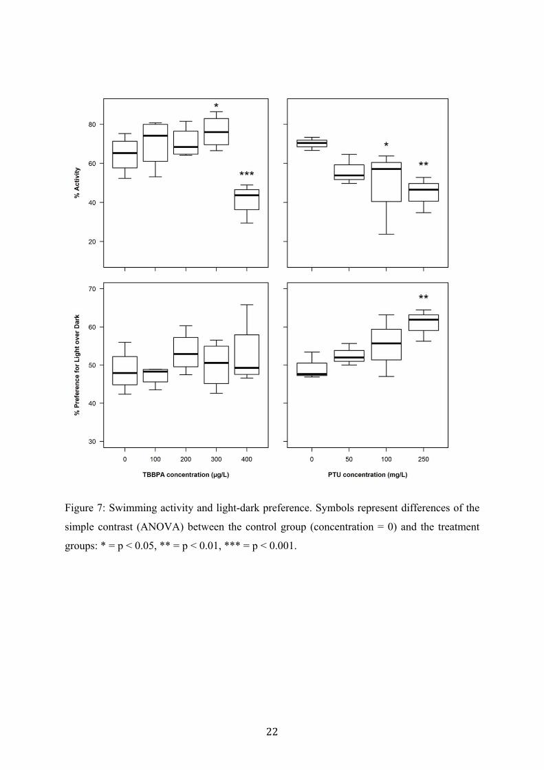
3.4. Thyroid disruption alters swimming activity and light-dark background preferences
Both TBBPA and PTU had a significant effect on swimming activity of 5 days old larvae
(ANOVA: TBBPA, F[4,16] = 19.47, p < 0.001; PTU, F[3,12] = 3.73, p = 0.042). However,
both treatments did not result in a strong impairment of activity as only at the highest TBBPA
and PTU concentrations activity was decreased less than two-fold. In fact, the effect of
TBBPA on swimming activity was even stimulating at low TBBPA concentrations (see fig. 7,
at 300 µg/L, p < 0.05), and then significantly decreased at the highest exposure concentration
(see fig. 7, at 400 µg/L, p < 0.001). For PTU a suppressive effect on swimming activity was
found, which was significant for treatment levels of 100 and 250 mg/L (see fig. 7).
Control animals did not show any obvious background preference for the illuminated light
half of the well over the dark half of the well (mean preference was 48.9 %). TBBPA did not
change this preference (ANOVA: F[4,16] = 1.39, p = 0.28). However, PTU treatment resulted
in a significant taxis towards the light half of the well (ANOVA: F[3,12] = 4.15, p = 0.031).
22
Figure 7: Swimming activity and light-dark preference. Symbols represent differences of the
simple contrast (ANOVA) between the control group (concentration = 0) and the treatment
groups: * = p < 0.05, ** = p < 0.01, *** = p < 0.001.
23
4. Discussion
The thyroid hormone system is known to be important for eye development of vertebrates
(Darras et al., 2015). Despite this knowledge, it remains unclear whether different molecular
changes of the thyroid axis can translate into the same disruption of eye development.
Moreover, the functional outcomes for the organism have not been clearly described yet. We
aimed to address these open questions by comparing the impact of two different thyroid
disruptors on the eye development of zebrafish embryos. On the one hand we hypothesized
that the disruption of eye morphology is a converging event that is independent of the
molecular MoA of the thyroid disruptor. Therefore, we chose two different thyroid disruptors
with known MoA: PTU as an inhibitor of thyroid hormone synthesis and TBBPA as disruptor
of the thyroid receptors. On the other hand, we hypothesized that the chemically-induced
dysfunctional eye development will result in disrupted visual physiology and vision-related
behaviour of the individual. We were able to confirm our hypotheses by demonstrating that
the induced molecular changes differed in our treatments, but that the morphological outcome
was very similar, and that the impaired eye development in fact was related to impaired visual
capacities.
The basic starting point of our study was to choose two well-described thyroid disruptors with
differing MoAs. As a proof-of-principle, we investigated the effects of both compounds on
expression of thyroid-related genes to demonstrate the influence on different key players of
the complex thyroid system. Both substances had significant impact on gene expression levels
of thyroid-related genes in exposed larvae, but the reaction pattern differed clearly, which
shows that the molecular changes affected different pathways within the thyroid hormone
system of the developing fish. We investigated the 3 different types of deiodinase enzymes,
which are important for regulation of thyroid hormone levels. Deiodinase type 1 (DIO1) and
type 2 (DIO2) increase and deiodinase type 3 (DIO3) decreases local intracellular levels of
T3, the most important active thyroid hormone. TBBPA had an up-regulating effect on the
expression of DIO1 transcripts while PTU up-regulated DIO2 and down-regulated DIO3.
Both indicate that either the enzymes themselves or the thyroid hormone levels were
disrupted by the treatment, which confirms that the chosen compounds act indeed as thyroid
disruptors. This is also obvious from the transcript changes found for the enzyme
thyroperoxidase (TPO), which was strongly and significantly up-regulated by PTU-treatment,
probably as a compensatory mechanism against the substance-induced inhibition of the
24
enzyme that is responsible for synthesis of thyroid hormones. The opposite effect was
observed for the TBBPA treatment. TSH expression levels were slightly up-regulated in both
exposures, which was not significant. Transcript changes for the thyroid receptors alpha and
beta clearly differed between both treatments: lower levels of TBBPA up-regulated the
thyroid receptor genes while PTU down-regulated these genes. These findings confirm the
current knowledge about the effects of both substances on the fish thyroid system (De Wit et
al., 2008; Quesada-García et al., 2014) and show that they were disrupting the thyroid system
in a different, partly even contrasting manner. This was the initial starting point for our further
investigations about the eye morphology and physiology of treated larvae.
Even though the molecular response obviously differed between both compounds used in our
study, the associated cellular changes in the retina of exposed larvae were very similar in their
response pattern, but not regarding the threshold: while PTU had a strong impact on the
development of the retinal pigment epithelium (decreased size and pigmentation), the effects
of TBBPA were similar but not as pronounced. The impact of PTU on eye development,
opsin expression and pigmentation in fish has been demonstrated previously (Gan and
Novales Flamarique, 2010; Macaulay et al., 2015; Raine et al., 2010). Effects of the PTU-
related compound phenylthiourea have also been reported (Bohnsack et al., 2011; Li et al.,
2012), and these authors describe reduced eye size and cellular changes after treatment with
this commonly used inhibitor of pigmentation in zebrafish larvae. The impact of TBBPA on
eye development has not been demonstrated yet. Only adverse effects on general and cardiac
development of zebrafish have been observed (Wu et al., 2015; Yang and K. Chan, 2015). In
our study, analyses of body size and eye size and the resulting ratio revealed that both
substances significantly decreased the relative eye size of the developing larvae.
The next evident step in our study was to demonstrate whether the morphological changes
have impact on the visual performance and vision-based behavioural capacities of the exposed
larvae. In fact, while several studies have demonstrated that thyroid disruptors have an
adverse effect on eye morphology (e.g. Reider and Connaughton, 2014), it has not been
clearly shown yet whether this implicates impaired vision. Visual performance represents a
key factor for the survival of developing fish embryos and larvae: behaviour concerning
predator avoidance and feeding success is highly dependent on an intact optical sense. Thus,
the present study provides novel information by verifying that impaired eye development
translates into impaired vision and behaviour. We used the OKR, swimming activity and
light-preference as fitness-related endpoints to investigate how general and vision-related
25
behaviours were altered by the treatment with the two different thyroid disruptors. The
reaction pattern of the OKR was very similar for both treatments, showing a significant
concentration-dependent decrease of visual capacities of the larvae. Partly comparable
observations have been made by Reider and Connaughton (2015) who treated zebrafish
embryos with the thyroid disruptor methimazol and observed increased anxiety behaviour. To
our knowledge, this is the only study that could demonstrate how thyroid disruption and the
resulting aberration of the eyes leads to altered behaviour in zebrafish larvae. Additional to
the impaired OKR, we also found changes in light seeking and swimming behaviour of
thyroid disruptor-treated larvae, which indicates that not only visual performance itself, but
also vision-related behaviour was disrupted. Thus, we are the first to provide evidence that
altered eye morphology, as we observed under exposure to different thyroid disruptors, indeed
implies altered visual capacity and vision-related behaviour.
5. Conclusion
The results of the present study provide initial evidence that thyroid disruptors which induce
different molecular changes in the thyroid system can lead to uniform alterations in eye
development of zebrafish larvae. This suggests that different molecular modes of action
eventually merge into identical morphological outcomes, a hypothesis which will have to be
further tested in detail in future studies. A second major finding from this study is that the
impaired eye development, as it arises under exposure to the thyroid-disrupting compounds,
indeed implies an impaired visual performance, as evidenced from physiological and
behavioral tests. These findings are especially relevant, as development and functionality of
the eyes are essential for the survival of fish larvae in the environment.
26
Competing interests: We have no competing interests.
Author´s contributions:
LB and AR are equally contributing authors. LB carried out parts of the molecular and
histological lab work, participated in data analysis, carried out OKR experiments, designed
the study and drafted the manuscript; AR carried out parts of the molecular lab work,
participated in data analysis, carried out the behavioural experiments, carried out the
statistical analyses and helped draft the manuscript; KR participated in molecular lab work
and fish maintenance, helped draft the manuscript; SN coordinated parts of the study and
helped draft the manuscript; HS conceived of the study, designed the study together with LB,
coordinated the study and helped draft the manuscript.
All authors gave final approval for publication.
Acknowledgments: The authors thank the team of the Neuhauss lab at the University of
Zürich for their input and help with molecular methods and OKR. Jitka Tumova helped with
the histology.
Funding: LB received a postdoctoral fellowship of the German Research Foundation DFG
(BA 5053/1-1) and the UniBE Initiator Grant of the University of Berne, Switzerland.
27
6. References: Bagci, E., Heijlen, M., Vergauwen, L., Hagenaars, A., Houbrechts, A.M., Esguerra, C.V.,
Blust, R., Darras, V.M., Knapen, D., 2015. Deiodinase knockdown during early zebrafish development affects growth, development, energy metabolism, motility and phototransduction. PLoS One 10, e0123285. doi:10.1371/journal.pone.0123285
Bertrand, S., Thisse, B., Tavares, R., Sachs, L., Chaumot, A., 2007. Unexpected novel relational links uncovered by extensive developmental profiling of nuclear receptor expression. PLoS Genetics 3, e188. doi:10.1371/journal.pgen.0030188
Boas, M., Feldt-Rasmussen, U., Main, K.M., 2012. Thyroid effects of endocrine disrupting chemicals. Mol Cell Endocrinol 355, 240–248. doi:10.1016/j.mce.2011.09.005
Bohnsack, B.L., Gallina, D., Kahana, A., 2011. Phenothiourea sensitizes zebrafish cranial neural crest and extraocular muscle development to changes in retinoic acid and IGF signaling. PLoS One 6, e22991. doi:10.1371/journal.pone.0022991
Bohnsack, B.L., Kahana, A., 2013. Thyroid hormone and retinoic acid interact to regulate zebrafish craniofacial neural crest development. Dev Biol 373, 300–309. doi:10.1016/j.ydbio.2012.11.005
Brar, N.K., Waggoner, C., Reyes, J.A., Fairey, R., Kelley, K.M., 2010. Evidence for thyroid endocrine disruption in wild fish in San Francisco Bay, California, USA. Relationships to contaminant exposures. Aquatic Toxicology 96, 203–215. doi:10.1016/j.aquatox.2009.10.023
Brouwer, A., Morse, D.C., Lans, M.C., Schuur, A.G., Murk, A.J., Klasson-Wehler, E., Bergman, A., Visser, T.J., 1998. Interactions of persistent environmental organohalogens with the thyroid hormone system: mechanisms and possible consequences for animal and human health. Toxicol Ind Health 14, 59–84.
Brown, S.B., Adams, B.A., Cyr, D.G., Eales, J.G., 2004. Contaminant effects on the teleost fish thyroid. Environmental toxicology and chemistry / SETAC 23, 1680–1701.
Carr, J.A., Patiño, R., 2011. The hypothalamus–pituitary–thyroid axis in teleosts and amphibians: Endocrine disruption and its consequences to natural populations. Gen Comp Endocrinol 170, 299–312. doi:10.1016/j.ygcen.2010.06.001
Chan, W., Chan, K., 2012. Disruption of the hypothalamic-pituitary-thyroid axis in zebrafish embryo–larvae following waterborne exposure to BDE-47, TBBPA and BPA. Aquatic Toxicology 108, 106–111. doi:10.1016/j.aquatox.2011.10.013
Chen, Q., Yu, L., Yang, L., Zhou, B., 2012. Bioconcentration and metabolism of decabromodiphenyl ether (BDE-209) result in thyroid endocrine disruption in zebrafish larvae. Aquatic Toxicology 110-111, 141–148. doi:10.1016/j.aquatox.2012.01.008
Chen, X., Engert, F., 2014. Navigational strategies underlying phototaxis in larval zebrafish. Frontiers in systems neuroscience. doi:10.3389/fnsys.2014.00039
Cheng, Y., Ekker, M., Chan, H.M., 2015. Relative developmental toxicities of pentachloroanisole and pentachlorophenol in a zebrafish model (Danio rerio). Ecotoxicol Environ Saf 112, 7–14. doi:10.1016/j.ecoenv.2014.10.004
Crofton, K.M., 2008. Thyroid disrupting chemicals: mechanisms and mixtures. Int J Androl 31, 209–223. doi:10.1111/j.1365-2605.2007.00857.x
Darras, V.M., Houbrechts, A.M., Van Herck, S.L.J., 2015. Intracellular thyroid hormone metabolism as a local regulator of nuclear thyroid hormone receptor-mediated impact on vertebrate development. Biochimica et Biophysica Acta (BBA) - Gene Regulatory Mechanisms 1849, 130–141. doi:10.1016/j.bbagrm.2014.05.004
De Wit, M., Keil, D., Remmerie, N., Ven, K.V.D., Brandhof, E.-J.V.D., Knapen, D., Witters, E., Coen, W.D., 2008. Molecular targets of TBBPA in zebrafish analysed through integration of genomic and proteomic approaches. Chemosphere 74, 96–105. doi:10.1016/j.chemosphere.2008.09.030
28
Environment Canada, H.C., 2013. Screening Assessment Report TBBPA. Government of Canada 1–178. ISBN: 978-1-100-22898-3
Fini, J.-B., Le Mével, S., Turque, N., Palmier, K., Zalko, D., Cravedi, J.-P., Demeneix, B.A., 2007. An In Vivo Multiwell-Based Fluorescent Screen for Monitoring Vertebrate Thyroid Hormone Disruption. Environ Sci Technol 41, 5908–5914. doi:10.1021/es0704129
Gan, K.J., Novales Flamarique, I., 2010. Thyroid hormone accelerates opsin expression during early photoreceptor differentiation and induces opsin switching in differentiated TRα-expressing cones of the salmonid retina. Dev Dyn 239, 2700–2713. doi:10.1002/dvdy.22392
Hamers, T., 2006. In Vitro Profiling of the Endocrine-Disrupting Potency of Brominated Flame Retardants. Toxicol. Sci. 92, 157–173. doi:10.1093/toxsci/kfj187
Heijlen, M., Houbrechts, A.M., Bagci, E., Van Herck, S.L.J., Kersseboom, S., Esguerra, C.V., Blust, R., Visser, T.J., Knapen, D., Darras, V.M., 2014. Knockdown of Type 3 Iodothyronine Deiodinase Severely Perturbs Both Embryonic and Early Larval Development in Zebrafish. Endocrinology 155, 1547–1559. doi:10.1210/en.2013-1660
Huber-Reggi, S.P., Mueller, K.P., Straumann, D., Huang, M.Y.-Y., Neuhauss, S.C.F., 2014. Individual Larvae of the Zebrafish Mutant belladonna Display Multiple Infantile Nystagmus-Like Waveforms that Are Influenced by Viewing ConditionsZebrafish Mutant Nystagmus-Like Waveforms. Invest. Ophthalmol. Vis. Sci. 55, 3971–3978. doi:10.1167/iovs.13-13576
Jagnytsch, O., Opitz, R., Lutz, I., Kloas, W., 2006. Effects of tetrabromobisphenol A on larval development and thyroid hormone-regulated biomarkers of the amphibian Xenopus laevis. Environmental Research 101, 340–348. doi:10.1016/j.envres.2005.09.006
Jomaa, B., 2014. Developmental toxicity of thyroid-active compounds in a zebrafish embryotoxicity test. ALTEX 31, 303–317. doi:10.14573/altex.1402011
Kitamura, S., Jinno, N., Ohta, S., Kuroki, H., Fujimoto, N., 2002. Thyroid hormonal activity of the flame retardants tetrabromobisphenol A and tetrachlorobisphenol A. Biochem. Biophys. Res. Commun. 293, 554–559. doi:10.1016/S0006-291X(02)00262-0
Kuiper, R.V., van den Brandhof, E.J., Leonards, P.E.G., van der Ven, L.T.M., Wester, P.W., Vos, J.G., 2006. Toxicity of tetrabromobisphenol A (TBBPA) in zebrafish (Danio rerio) in a partial life-cycle test. Arch. Toxicol. 81, 1–9. doi:10.1007/s00204-006-0117-x
Li, Z., Ptak, D., Zhang, L., Walls, E.K., Zhong, W., Leung, Y.F., 2012. Phenylthiourea Specifically Reduces Zebrafish Eye Size. PLoS One 7, e40132. doi:10.1371/journal.pone.0040132
Macaulay, L.J., Chen, A., Rock, K.D., Dishaw, L.V., Dong, W., Hinton, D.E., Stapleton, H.M., 2015. Developmental toxicity of the PBDE metabolite 6-OH-BDE-47 in zebrafish and the potential role of thyroid receptor β. Aquatic Toxicology 168, 38–47. doi:10.1016/j.aquatox.2015.09.007
Patrick, L., 2009. Thyroid disruption: mechanism and clinical implications in human health. Alternative Medicine Review Volume 14, Number 4
Pinto, P.I.S., Guerreiro, E.M., Power, D.M., 2013. Triclosan interferes with the thyroid axis in the zebrafish (Danio rerio). Toxicol. Res. 2, 60–69. doi:10.1039/C2TX20005H
Power, D.M., Llewellyn, L., Faustino, M., Nowell, M.A., Björnsson, B.T., Einarsdottir, I.E., Canario, A.V.M., Sweeney, G.E., 2001. Thyroid hormones in growth and development of fish. Comparative Biochemistry and Physiology Part C: Toxicology & Pharmacology 130, 447–459. doi:10.1016/S1532-0456(01)00271-X
Quesada-García, A., Valdehita, A., Kropf, C., Casanova-Nakayama, A., Segner, H., Navas, J.M., 2014. Thyroid signaling in immune organs and cells of the teleost fish rainbow trout (Oncorhynchus mykiss). Fish Shellfish Immunol 38, 166–174. doi:10.1016/j.fsi.2014.03.016
Raine, J.C., Coffin, A.B., Hawryshyn, C.W., 2010. Systemic thyroid hormone is necessary
29
and sufficient to induce ultraviolet-sensitive cone loss in the juvenile rainbow trout retina. J. Exp. Biol. 213, 493–501. doi:10.1242/jeb.036301
Raine, J.C., Hawryshyn, C.W., 2009. Changes in thyroid hormone reception precede SWS1 opsin downregulation in trout retina. J. Exp. Biol. 212, 2781–2788. doi:10.1242/jeb.030866
Reider, M., Connaughton, V.P., 2015. Developmental exposure to methimazole increases anxiety behavior in zebrafish. Behavioral Neuroscience 129, 634–642. doi:10.1037/bne0000087
Reider, M., Connaughton, V.P., 2014. Effects of Low-Dose Embryonic Thyroid Disruption and Rearing Temperature on the Development of the Eye and Retina in Zebrafish. Birth Defects Res B 101, 347–354. doi:10.1002/bdrb.21118
Sabaliauskas, N.A., Foutz, C.A., Mest, J.R., Budgeon, L.R., Sidor, A.T., Gershenson, J.A., Joshi, S.B., Cheng, K.C., 2006. High-throughput zebrafish histology. Methods 39, 246–254. doi:10.1016/j.ymeth.2006.03.001
Schmidt, F., Braunbeck, T., 2011. Alterations along the Hypothalamic-Pituitary-Thyroid Axis of the Zebrafish (Danio rerio) after Exposure to Propylthiouracil. Journal of Thyroid Research 2011, 1–17. doi:10.4061/2011/376243
Suzuki, S.C., Bleckert, A., Williams, P.R., Takechi, M., Kawamura, S., Wong, R.O.L., 2013. Cone photoreceptor types in zebrafish are generated by symmetric terminal divisions of dedicated precursors. Proc Natl Acad Sci U S A 110, 15109–15114. doi:10.1073/pnas.1303551110
Thienpont, B., Tingaud-Sequeira, A., Prats, E., Barata, C., Babin, P.J., Raldúa, D., 2011. Zebrafish Eleutheroembryos Provide a Suitable Vertebrate Model for Screening Chemicals that Impair Thyroid Hormone Synthesis. Environ Sci Technol 45, 7525–7532. doi:10.1021/es202248h
Van der Ven, L.T.M., van den Brandhof, E.-J., Vos, J.H., Power, D.M., Wester, P.W., 2006a. Effects of the Antithyroid Agent Propylthiouracil in a Partial Life Cycle Assay with Zebrafish. Environ Sci Technol 40, 74–81. doi:10.1021/es050972c
Van der Ven, L.T.M., van den Brandhof, E.-J., Vos, J.H., Power, D.M., Wester, P.W., 2006b. Effects of the Antithyroid Agent Propylthiouracil in a Partial Life Cycle Assay with Zebrafish. Environ Sci Technol 40, 74–81. doi:10.1021/es050972c
Wu, S., Ji, G., Liu, J., Zhang, S., Gong, Y., Shi, L., 2015. TBBPA induces developmental toxicity, oxidative stress, and apoptosis in embryos and zebrafish larvae (Danio rerio). Environ Toxicol n/a–n/a. doi:10.1002/tox.22131
Yang, J., Chan, K., 2015. Evaluation of the toxic effects of brominated compounds (BDE-47, 99, 209, TBBPA) and bisphenol A (BPA) using a zebrafish liver cell line, ZFL. Aquatic Toxicology 159, 138–147. doi:10.1016/j.aquatox.2014.12.011
Yu, L., Chen, M., Liu, Y., Gui, W., Zhu, G., 2013. Thyroid endocrine disruption in zebrafish larvae following exposure to hexaconazole and tebuconazole. Aquatic Toxicology 138-139, 35–42. doi:10.1016/j.aquatox.2013.04.001
Zhang, Y.-F., Xu, W., Lou, Q.-Q., Li, Y.-Y., Zhao, Y.-X., Wei, W.-J., Qin, Z.-F., Wang, H.-L., Li, J.-Z., 2014. Tetrabromobisphenol A Disrupts Vertebrate Development via Thyroid Hormone Signaling Pathway in a Developmental Stage-Dependent Manner. Environ Sci Technol 48, 8227–8234. doi:10.1021/es502366g


![physics-pages.wikispaces.com Review Problems Chapter Review Problems 8. c. How high is the imaoe'] — 4.0 cm = -0.33 do — 12.0 cm hi = mho = mm) = — 8.0 mm An object is 30.0 cm](https://static.fdocuments.in/doc/165x107/5b1b24fc7f8b9a19258e6bd3/physics-pages-review-problems-chapter-review-problems-8-c-how-high-is-the-imaoe.jpg)


![AQC source capture equipment · 2018. 11. 25. · single arm application Arm diameter [inches] / [mm] Fan model HP / KW 3 / 75 DFR-8 0.33 / 0.25 4 / 100 DFR-9 0.33 / 0.25 5 / 125](https://static.fdocuments.in/doc/165x107/60195fb1f987bb597135f23a/aqc-source-capture-equipment-2018-11-25-single-arm-application-arm-diameter.jpg)