Title Surgical Treatment of Femur-Shaft Fractures In ...
11
Title Surgical Treatment of Femur-Shaft Fractures In Children Author(s) MORITA, SHIN; ODA, HAJIME Citation 日本外科宝函 (1967), 36(5): 627-636 Issue Date 1967-09-01 URL http://hdl.handle.net/2433/207402 Right Type Departmental Bulletin Paper Textversion publisher Kyoto University
Transcript of Title Surgical Treatment of Femur-Shaft Fractures In ...
Title Surgical Treatment of Femur-Shaft Fractures In Children
Author(s) MORITA, SHIN; ODA, HAJIME
Citation 日本外科宝函 (1967), 36(5): 627-636
Issue Date 1967-09-01
URL http://hdl.handle.net/2433/207402
Right
Type Departmental Bulletin Paper
Textversion publisher
Kyoto University
臨床
Surgical Treatment of Femur-Shaft Fractures in Children
by
SHIN MoRIT A and HAJIME OoA
Clinic of Orthopaedic Surgery, Wakayama Re~ Cross Hospital, Wakayama
Received for Publication Sept. 12, 1967
627
The conservative treatment has been to be thought the best in the treatment of femur-
shaft fractures in children. But it has the disadvantages of uncertainty in the reduction
of displaced fragments and its maintenance. Marked overgrowth of the affected femur,
as had been expected, was not observed in the伺 ses of end to end apposition fixed by
intramedullary nailing, of which metal as the foreign body may hardly produce the stimuli
for the elongation of the femur. The elongation was of slight degree in our series.
When we have sufficient caution for the prevention of infectious complication, the surgical
treatment will be the method based upon the principles of the fracture management in
general.
The characteristic features in the course of femur-shaft fractures in children consist
in the overgrowth of the affected femur and the liability to fusion of fragments without
leaving behind the knee contracture. Then, the conservative treatment has been insisted
upon almost exclusively as the method of choice in the treatment. However, it encounters,
owing to the anatomical conditions of the thigh, some difficulties. It would be sometimes
necessary to treat surgically even femur-shaft fractures in the newborn. 7> The femur is
surrounded by the voluminous soft tissues to hinder the precise immobilization of frag-
ments. Furthermore, the muscles of the thigh are comparatively strong, which causes
easily the displacement of fragments at injury, and even after the reduction of fragments
the angulation deformity of the femur is apt to occur in the course of immobilization at
the site of fractures due to the unbalance of surrounding muscle tonus. Those are the
difficulties in the conservative treatment of femur-shaft fracture and in regards to the
certainty of the treatment, the conservative treatment has some disadvantages・ Many
devices and methods elaborated by many authors2>4>5> for traction and immobilization prove
the difficulty of conservative management. Then, with small series of femur-shaft fractures
in children surgically treated at hand, we show in the article that the surgical treatment
has enough advantages not to be abandoned.
TECHNIQUE
The osteosynthesis are attained with dia-pin (modified Rush-pin with a cross section
~f rhomboidal shape), intramedullary nails of V or clover-shape, plate, screws and wire
ror comminuted fracture. The plate and screws are usually used for the synthesis of
や加ochantericfracture. In older children, intramedullary nailing was 伺 rriedout with
the same method as that in adults but in younger children the dia-pin is often used for
628 日本外科宝函第36巻 第5号
the intramedullary fixation of which the procedure will be here described.
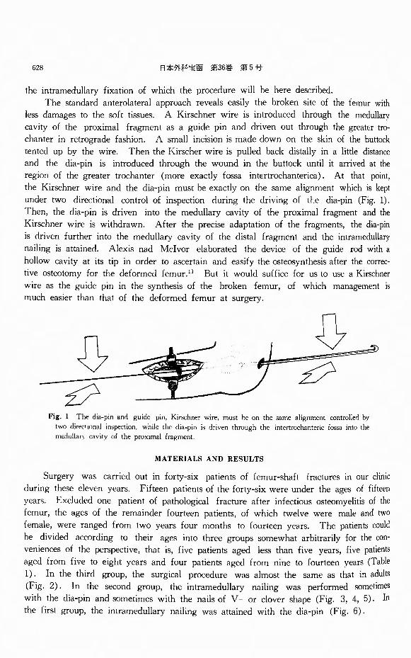
The standard anterolateral approach reveals easily the broken site of the femur with
less damages to the soft tissues. A Kirschner wire is introduced through the medullary
cavity of the proximal fragment as a guide pin and driven out through the greater tro・
chanter in retrograde fashion. A small incision is made down on the skin of the buttock
tented up by the wire. Then the Kirscher wire is pulled back distally in a little distance
and the dia-pin is introduced through the wound in the buttock until it arrived at the
region of the greater trochanter (more exactly fossa inttrtrochanterica). At that point,
the Kirschner wire and the dia-pin must be exactly on the same alignment which is kept
under two directional control of inspection <luring the driving of t上e dia-pin (Fig. 1).
Then, the dia-pin is driven into the medullary cavity of the proximal fragment and the
Kirschner wire is withdrawn. After the precise adaptation of the fragments, the dia-pin
is cl.riven further into the medullary伺 vityof the distal fragment and the intramedullary
nailing is attained. Alexis nad Mcivor elaborated the device of the guide rod with a
hollow cavity at its tip in order to ascertain and easify the osteosynthesis after the co汀ec-
tive ost印 tomyfor the deformed femur. 1 > But it would suffice for us to use a Kirschner
wire as the guide pin in the synthesis of the broken femur, of which management is
much easier than that of the deformed femur at surgery.
Fig. 1 The dia-pin anrl guide pin, Kirschner wire, must he on the団 mealignment controlled by two directional inspection, while the dia-pin is driven through the intertrochanteric fossa into the medullaηcavity of the proximal fragment
MATERIALS AND RESULTS
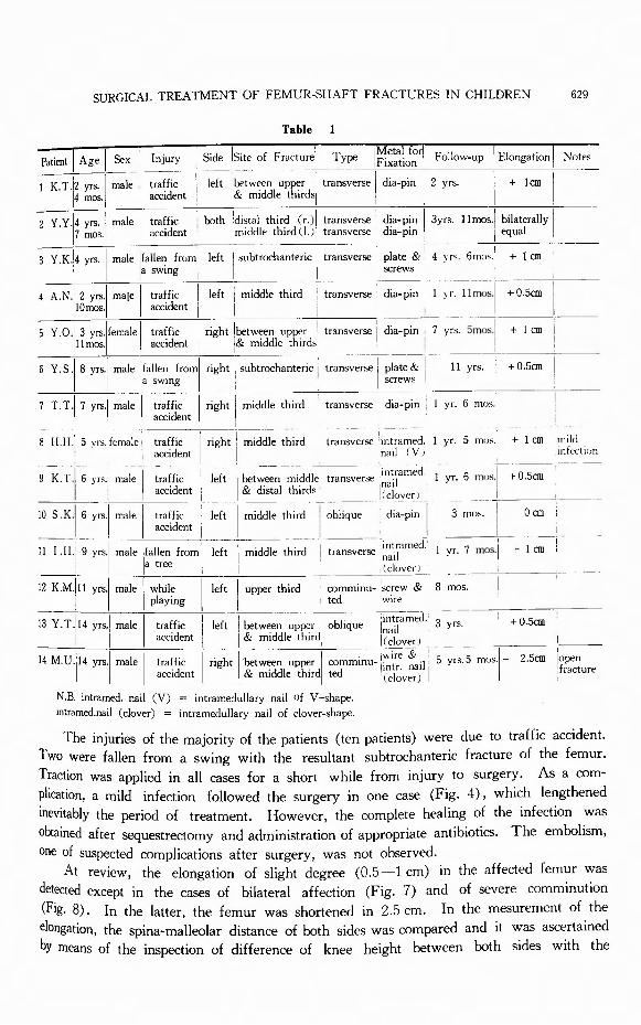
Surgery was carried out in forty-six patients of femur-shaft fractures in our clinic
during these eleven years. Fifteen patients of the forty-six were under the ages of fifteen
years. Excluded one patient of pathological fracture after infectious osteomyelitis of the
femur, the ages of the remainder fourteen patients, of which twelve were male and two
female, were ranged from two years four months to fourteen years. The patients could
be divided according to their ages into three groups somewhat arbitrarily for the con-
veniences of the perspective, that is, five patients aged less than five years, five patients
aged from five to eight years and four patients aged from nine to fourteen years (Table
1). In the third group, the surgical procedure was almost the same as that in adults
(Fig. 2). In the second group, the intramedullary nailing was performed sometimes
with the dia-pin and sometimes with the nails of V or clover shape (Fig. 3, 4, 5). In
the first group, the intramedullary nailing was attained with the dia-pin (Fig. 6).
SURGICAL TREATMENT OF FEMUR-SHAFT FRACTURES IN CHILDREN 629
同|州 Sex
1 K.T.!2 y瓜 Imale, 14 mos.
Table 1
T I I M何日記 , Injury [_ Side !Site of F ract吋 Type !Fixation I Foll側叩 |目。叩tionlNot白
traffic ¥ left !between upper ¥ transverse ¥ dia-pin I 2 y瓜 I+ 1佃|accident i I& middle出吋SI I I i I
z Y. Y .¥4 yrs. ! male traffic : both !distal出 rd(r.)¥ transve問 Idia-pin I 3yrs. 1 lm曲 Ibilaterally j 11 inos. [ accident • middle出吋(l.J]transverse i di叩 n] 同叫 !
3 Y.K.¥4 yrs. ¥ male ¥fallen from1 left ¥ subt町 hante巾\transverse 向旬& ' 4 y凡 6mos.1 + I佃
I I !a sw時 | | | 筑間WS ;
4 A.N .I 2 yrs.I maJe I traffic I left I m刷 lethird ・: transverse ; d恥 pin I yr. llm叫+0.5佃 |
l!Omos.I I m伽 tI I i I i I I
5 Y. 0 .I 3 yrs.lfemale I traffic I r凶 t!between叩阿 ! transv町田 id日・pin, 7 yrs. 5mos.I + I個 |
¥11 mos.¥ ¥ accident I !& middle出吋>I I i ' !
6 v.s.1 s yれ山]fallen~roml 帆 i 凱1btrocha c ¥ trar I I :a SW叩E I : I白:rews1
7 T目T.¥7 yrs. ! 1 1 accid町1tI I
8 H.H. 5 y目 i町ma!1 accident I I t】a』ICVJ .i『】i虻 t1m
9 K.T.16 yrιmale I traffic I left I between middle transverse -~~-t,~amed. I yr. 6 m s.I + 0.5佃
I • I accider】tI I & distal third ・ 1""" I ーー一」__ • I I I s I< cl~~er J _ よ
10 S.K目I6 y吋male¥ traffic ¥ left ¥ middle third i obli同ue d昨 pin¥ 3 mo•. I 0 an i し I I accident I I i , ! ~ __l_
II I .H.! 9刈male!fallen from I 凶 Imiddle 出i~<l- -i孟:ム i~~~f~~ecf I ~ mos. + I佃(, I la tree I I I I I I ,(clover)
12 K M・Illyrs.I male I while I left I upper third ・ comminu-!,;crew & 8 mos. I I I playing I I ! ted wi陀
13 Y. T .114 yrs.I male I traffic I left i出時nup阿 oblique j ~~,1;~amed. 3 yrs. l州知 !
! I I配:cidentI i & middle third i"0" i I I I I 川 1 IC clover J I
川 u.J14吋m町出払tI right I ~t;詰le"叫訂minu- j~~-~ail戸rs.5 mペー 2.5佃!?忠u陀
I I I I I I 1 (clover) I I
N.B. intramed. nail (V) = intramedullary nail of V-shape. intramed.nail (clover) = intramedullary nail of clov町-shape.
The injuries of the majority of the patients (ten patients) were due to traffic accident.
Two were fallen from a swing with the resultant subtrochanteric fracture of the femur.
Traction was applied in all四 sesfor a short while from injury to surgery. As a com-
plication, a mild infection followed the surgery in one case (Fig. 4), which lengthened
inevitably the period of treatment. However, the complete healing of the infection was
obtained after sequestrectomy and administration of appropriate antibiotics. The embolism,
one of suspected complications after surgery, was not observed.
At review, the elongation of slight degree (0.5ー Icm) in the affected femur was
detected except in the cases of bilateral affection (Fig. 7) and of severe comminution
(Fig. 8). In the latter, the femur was shortened in 2.5 cm. In the mesurement of the
elongation, the spina-malleolar distance of both sides was compared and it was ascertained
Dy means of the inspection of difference of knee height between both sides with the

' .
630 日本外科宝函第36巻第5号
(a) め)Fig. 2 Case 11. I. H., a boy aged nine y回 rsThe intramedullary nailin~ was carried out with the坦 memethod as that m adults. (a) After surgery. (b) Three months after surgery.
(])
Xコ
(a) ぬ)Fig. 3 Ca記 7.T. T., a boy aged seven y回 目.Aslender nail (dia-pin) not full of the medullary 国 vity suffic田 forthe ost田 synth田is. The an-gulation deformity did not developed owing to the early四 llusformation.
(a) After surgery.
(b) One y田 rsix months after surgery.
{a) (b) (c) (d) Fig. 4 Case 8. H. H., a girl aged five y回目. {a) After injury. (b) An intramedullary nail of V-shape was u,;ed for the osteosynthesis目 A mild infection was found as a complication after surller~o. roentgenogram was taken two months after surgery. {c) After the administration of appropriate antib10t1cs and目qu田tree-
r;ぷ郡山s~t~~~~e克r~d官部品t~~~~よ目立恒常立与l~~!mheiJぷSb~~:~~!~:.

SURGICAL TREATMENT OF FEMUR-SHAFT FRACTURES IN CHILDREN 631
(a) (b) (c)
Fig. 5 Case 9. K. T., a boy aged six Y田 rs. (a), (b) The intramedullary nail of
clover-shape was used for the ost田 synthesis. (c) The roentgenogram was taken at
review one y田rsix months after surgery.
(a) (b) (c) (d)
Fig. 6 Case 1. K. T., a boy aged two year,; and four months. (a) After injuηr. (b) The intramedullary
nailing was attained with the dia-pin. (c) One y田rafter surgery. (d) T山川 yearsafter surgery.

632 日本外科宝函第36巻 第5号
(a) (b) (C) (d) (e)
Fig. 7 Case 2. Y. Y ., a boy aged four years seven months with fracture of bilateral femur-shafts. (a) The
right femur after injury. (b) The osteosynth田 iswas carried out with a dia-pin. As the dia-pin was too
long, the after-treatment of long duration was n配 essaryfor the recovery of kn田 function. (c), (d) Eight
months after surgery. (e) At review three years and eleven months after surgery. The knee function was
normal.
(a) (b) (C) (d)
Fig. 8 Case 14目 M.U., a boy aged fourteen y回目 withthe comminuted fracture of the right femur. (a)
After injury. (b) After surgery. The comminuted fragments were gathered together with a wire. (c) One
y田 rafter surgery. The wire had been previously withdrawn. (d) At review five y回 rsand five months
after surgery. The shortening of the right femur was of 2.5 cm.

SURGICAL TREATMENT OF FEMUR-SHAFT FRACTURES IN CHILDREN 633
(a) (b)
(C) (d)
Fig. 9 Case 6. Y. S., a boy aged eight y白白
with subtrochanteric fracture of the right femur. (a) After injury. (b) After surgery. (c), (d) At review eleven y田 rsafter surgery. Any note-worthy varus or valgus deformity of the femoral neck w酷 notobserved in the affected side {c), compared with the uninjured side (d). But the overgrowth in the trochanter regicn was observed. The patient had refused to make the plate and screws withdrawn
patient in supine position and with the hips
and knees in flexed position that the femur
was responsible for the elongation. The
degree of the elongation seldom differs be-
tween the cases reviewed more than one year.
It suggests that the stimuli for the overgrowth
did endure only for a short while after injury
and the constancy of the degree of elongation
explains a firm maintenance of reduction after
surgery without producing superfluous stimuli
for overgrowth as in the cases of fallacious
and insufficient fixation. The slight elonga-
tion of the femur provides the patients in
their daily life with no inconveniences and is
also compatible with the vigorous activity.
The firm synthesis of fragments accomplished,
the immobilization with a plaster of parisαst
is often unnecessary and, if needed, it is for
the purpose of the repair of the soft tissues
at most during two weeks after surgery. In the majority of the cases, the callus formation
is apparent on the roentgenograms taken two
weeks after surgery and the after-treatment
αn be commenced by means of a hot bath
and active movement without weight聞 bearing.
Owing to the early callus formation, a slender
nail not full of the medullary cavity will suf-
fice especially in younger patients (Fig. 3).
It offers enough fixation to prピventthe angu-
lation deformity of the femur.
Any noteworthy varus or valgus deformity of the femoral neck was not observed at review in two国 sesof subtrochanteric fracture. But the roentgenograms revealed a rela-
tive overgrowth in the trochanter region (Fig. 9).
DISCUSSION
In the treatment of femur-shaft fractures in children, non-surgical treatment has been estimated better than surgical treatment, of which the basis is as follows :
( 1 ) The fragments do not fail to fuse in children, even if the exact reduction was not obtained. As to the displacement, slight overriding of fragments is favorable in regards
to the later developed overgrowth of the affected femur. ( 2 ) Surgical treatment is a sort of over-treatment and surgery is superfluous for
the fusion of the femur. Moreover, the possibility of danger for the complications followed by disastrous sequelae after surgery is great in comparison with non-surgical treatment.
Those are osteomyelitis, embolism and pseudarthrosis.

634 日本外科宝函第36巻 第5号
( 3 ) Non-surgical treatment needs inevitably longer period of traction and immobili同
zation. However, the knee contracture does not after all occur in children.
In this way, non-surgical treatment has been insisted upon with the fact that the
displacement of fragments could not be the hinderance of the bone fusion in children and
also with the presupposition of biological self-correctivity that some deformity would be
corrected in the course of the growth. Those are, however, of ten the pretext but not the
reason justifying a deformed healing. The chief deformity is the overriding of fragments
and angulation at the site of fracture. In fact, the degree of deformity, which is spon-
taneously overcome owing to the biological self-correctivity, is restricted in a narrow limit.
Blount stated that 1 cm of overriding was the ideal position for union and that side to
side apposition gives a more rapid and stronger union than end to end contact. He also denied intramedullary fixation because of the marked overgrowth produced by nails as
the forein body.2> However, the overgrowth of the femur in the cases of intramedullary
nailing is of constantly slight degree. Moreover, the slight overriding of fragments is
never the ideal position for the management of femur-shaft fractures in children. The lack of the firmness of fixation will bring forth the superfluous callus formation, which
would produce much more stimuli for the overgrowth. It needs for us meticulous control
over the affected thigh to keep the overriding of 1 cm or so from the beginning of treat-
ment to the consolidation of the femur, imposing superfluous and unnecessa巧rdiscomforts
upon the children. It would be better, if the primary be the prevention of the difference
of the femur-length, to excise beforehand the bone of 1 cm or so at the tip of fragment at surgery. However, we do not agree to such a petty conformation in the treatment of
the disorder in children. For the slight elongation of the femur is compatible without
any discomforts not only with the daily life but also with the vigorous activity of the
patients.
In the course of femur-shaft fractures in children the characteristic features, which
are the liability to fusion of fragments and no disabling knee contracture after traction
and immobilization of long duration, do not lead to the conclusion that the conservative
treatment is the only ideal and the best one but to the opinion that it is one of白
effective methods when performed by the skilful hands. However, the difficulty of
management still exists. On the other hand, the principles of fracture management in
general may be described as follows:
( 1 ) The method must be easy to be performed with certainty.
( 2 ) The period of the primary treatment shortened, the after-trrntment is to be
initiated as early as possible.
( 3 ) The danger followed by disastrous sequelae must be minimum.
( 4 ) The pain and discomforts must be minimum in the course of treatment. It
would be important for the improvement of disturbed local circulation and for the early
consっlidationof the broken bone.
The anatomical reconstruction of fragments leads generally to the recovery of physi圃
ological function. But in the fracture management, the problems of local circulation cannot
be too much stressed upon, as were pointed out by Schnek stating the role and importance
of plaster of paris cast without pad. 5> In this view-point, the traction applied until the
firm consolidation of the broken site is theoretically against the physiology. The firm

SURGICAL TREATMENT OF FEMUR-SHAFT FRACTURES IN CHILDREN 635
fixation of fragments and free movements of the joints of the proximity will much improve
the disturbed local circulation. The surgical treatment will satisfy this condition. Its
characteristics may be described as follows :
( 1) The procedure is carried out easily within a short time. The maintenance of
reduction is of certainty and its meticulous control is unnecessary.
( 2) The period of immobilization shortened, the after-treatment can be initiated in
the early stage of the course. The discomforts of the patients are much decreased.
( 3) Little damages to the soft tissues occun at surgery. Thus, the surgical treatment
of femur-shaft fractures may be sustained by the principles of the management of fractures
in general.
But we must pay much attention to the prevention of complications. Osteomyelitis,
embolism, although generally less common in children, 3J and pseudarthrosis were pointed
out as the possible complications followed by disastrous sequelae. However, osteomyelitis
and embolism are not always the possible complications only of surgical treatment of femur-
shaft fractures in children. Their danger may be suspected at all surgery. Therefore,
the unfortunate possible complictions do not afford the reason to abandon the surgical
treatment only of femur-shaft fractures in children. The prevention of complications
consists in the carefulness and skilfulness. Surgery must be carried out within shorter
duration without superfluous damages. Desinfection at surgery and during management
after surgeηis to be performed with extreme caution. The general status of patients
has to be properly controlled. As to pseudarthrosis, it is now of rare occurrence owing
to the progress of metal for fixation and also to the intramedullary nailing. The progress
of antibiotics, fixation-metal and anaesthetics enabled our active management of femur-shaft
fractures in children.
Although, at first glance, surgical intervention would give us an impression of the
superfluity of treatment, it decreases the discomforts and inconveniences of children discard-
ing longer lasting traction and immobilization.
SUMMARY
1) Small series of femur-shaft fractures in children surgically treated is reviewed.
2) In the majority of cases, a slight elongation (0.5-1 cm) of the affected femur
was observed at review, although it is practically negligible. Unfortunately, a mild infection
was observed in one case but completely healed with the administration of appropriate
antibiotics and sequestrectomy.
3) Surgical treatment, which is easily performed and enables the firm fixation of
fragments, shortens the period of traction and immobilization. It also decreases the pain
and discomforts of children.
REFERENCES
1) Alexis, P. G. & Mclvor. R. R. : A Practical Method of Insertion of the Ru'h Rod in the Femur. J. Bone and Joint Surg. 45-A : 1057-1058, 1963.
2) Blount. W. P. : Fractures in Child en. Williams & Wilkin,, Baltimore. 1955. 3) Crenshaw, A. H. (Editor) : Cambell’s Operative Orthopaedics. vol. one, fourth edition, Mosby, Saint Louis,
1963.
636 日本外科宝函第36巻 第5号
4) Dameron, T. B. & Thompson, H. A. ; Femoral Shaft Fractures in Children. Treatment by Closed Reduc-
tion anJ Double Spica Ca叫 Immobili回 tion.J. Bone anオJointSurg. 41-A : 1201-1212, 1959.
5) Ferry, A. M. & Edgar, M. S. : Moゴifie:I Bryant’s Traction. J. Bone and Joint Surg. 48-A : 533-536,
1966. 6) Schnek, F. G. : Die T吋rnil《 desungepolsterten Gipsverband白 WilhelmMaudrich, Wien, 1937.
7) Yoshitomi, J. & Oue, H.: Appli回 tionof Kirschner Wire to Femur-Shaft Fractures in the Newborn. Set-
keigeka (0比hopedicSurgery), 10 : 334-336, 1959. (Japanese)
和文抄録
小児の大腿骨々幹部骨折に対する観血的治療法
和歌山日赤整形外科
森田 信・小田
小児の大腿n々 幹部骨折l乙対する観血的治療法を過 た.
去11年間の経験14症例IC基いて述べた.症例の大多数 手術療法の主もな利点は整復並びに整復位の保持が
は交通事故による骨折であり,骨接合術は主として髄 確実かつ容易であり,牽引期間或いはギプス固定期間
内釘icょったが,ブランコより転落し受傷しだ 2例の を著るしく短縮でき,患児の終痛を軽減させる一方,
転子下骨折lζ対しては内副子及び螺子で固定した.手 早期lζ後療法を行ない得るため,障害された局所の循
術前の牽引及び術後のギプス固定は軟部組織の保護の 環状態を早期に積極的lζ改善させ,もって骨癒合を促
目的で極短期間行なった.術後 1年以上観察した症例 進させうる点にある.
の範闘では患側大腿骨lζ一様に軽度の延長が認められ