Title Page Differential block of sensory neuronal voltage...
48
JPET #133413 1 Title Page Differential block of sensory neuronal voltage-gated sodium channels by lacosamide, lidocaine and carbamazepine. Patrick L. Sheets, Cara Heers 1 , Thomas Stoehr, Theodore R. Cummins Department of Pharmacology and Toxicology, Indiana University School of Medicine, Indianapolis, Indiana (P.L.S., T.R.C.); Schwarz BioSciences, Department of Pharmacology/Toxicology, GmbH, Monheim, Germany (C.H., T.S.). JPET Fast Forward. Published on March 31, 2008 as DOI:10.1124/jpet.107.133413 Copyright 2008 by the American Society for Pharmacology and Experimental Therapeutics.
Transcript of Title Page Differential block of sensory neuronal voltage...

JPET #133413
1
Title Page
Differential block of sensory neuronal voltage-gated sodium channels by
lacosamide, lidocaine and carbamazepine.
Patrick L. Sheets, Cara Heers1, Thomas Stoehr, Theodore R. Cummins
Department of Pharmacology and Toxicology, Indiana University School of Medicine,
Indianapolis, Indiana (P.L.S., T.R.C.); Schwarz BioSciences, Department of
Pharmacology/Toxicology, GmbH, Monheim, Germany (C.H., T.S.).
JPET Fast Forward. Published on March 31, 2008 as DOI:10.1124/jpet.107.133413
Copyright 2008 by the American Society for Pharmacology and Experimental Therapeutics.

JPET #133413
2
Running Title Page
Running Title: Inhibition of sensory sodium channels by lacosamide
Address correspondence to: Contact Information: Theodore Cummins, Ph.D.,
Department of Pharmacology and Toxicology, Indiana University School of Medicine,
950 West Walnut St., R2 468, Indianapolis, IN. Tel. (317) 278-9342, Fax (317) 278-
5849, E-mail: [email protected]
Document Statistics:
Number of text pages: 38
Number of tables: 2
Number of figures: 8
Number of references: 40
Number of words in Abstract: 246
Number of words in Introduction: 724
Number or words in Discussion: 1263
Abbreviations: TTX, tetrodotoxin; TTX-R, tetrodotoxin-resistant; TTX-S, tetrodotoxin-
sensitive; HEK, human embryonic kidney; DRG, dorsal root ganglion; DMSO, dimethyl
sulfoxide; HEPES (4-(2-hydroxyethyl)-1-piperazineethanesulfonic acid);

JPET #133413
3
Abstract
Voltage-gated sodium channels play a critical role in excitability of nociceptors (pain-
sensing neurons). Several different sodium channels are thought to be potential targets
for pain therapeutics, including Nav1.7 which is highly expressed in nociceptors and
plays crucial roles in human pain and hereditary painful neuropathies, Nav1.3 which is
upregulated in sensory neurons following chronic inflammation and nerve injury, and
Nav1.8 which has been implicated in inflammatory and neuropathic pain mechanisms.
We compared the effects of lacosamide, a new pain therapeutic, with those of lidocaine
and carbamazepine on recombinant Nav1.7 and Nav1.3 currents and neuronal
tetrodotoxin-resistant (Nav1.8-type) sodium currents using whole-cell patch clamp
electrophysiology. Lacosamide is able to substantially reduce all three current types.
However, in contrast to lidocaine and carbamazepine, 1 mM lacosamide did not alter
steady-state fast-inactivation. Inhibition by lacosamide exhibited substantially slower
kinetics, consistent with the proposal that lacosamide interacts with slow inactivated
sodium channels. The estimated IC50 values for inhibition by lacosamide of Nav1.7,
Nav1.3 and Nav1.8-type channels following prolonged inactivation were 182, 415 and 16
µM, respectively. Nav1.7, Nav1.3, and Nav1.8-type channels in the resting state were
221, 123 and 257 fold less sensitive, respectively, to lacosamide than inactivated
channels. Interestingly, the ratio of resting to inactivated IC50’s for carbamazepine and
lidocaine were much smaller (ranging from 3 to 16). This suggests that lacosamide
should be more effective than carbamazepine and lidocaine at selectively blocking the
electrical activity of neurons that are chronically depolarized compared to those at more
normal resting potentials.

JPET #133413
4
Introduction
Neuropathic pain is a difficult type of pain to treat and it is estimated that as many
as half of all patients receive inadequate pain relief. More effective and better tolerated
treatments are needed. Lacosamide is a functionalized amino-acid that has shown
effectiveness as an anticonvulsant and analgesic and therefore might help fill this need
(Stohr et al., 2007; Beyreuther et al. 2007b). Lacosamide has shown efficacy in several
animal models of neuropathic pain (Beyreuther et al., 2006, 2007a) and is currently in
late stages of clinical development for epilepsy and diabetic neuropathic pain (Doty et al.
2007). Two modes of action have been identified for lacosamide, interaction with
collapsin-response mediator protein 2 and inhibition of voltage-gated sodium channel
activity by selective interaction with the slow inactivated conformation of the channel
(Beyreuther et al., 2007b, Errington et al. 2008). In this study we asked whether
lacosamide could inhibit voltage-gated sodium channels expressed in peripheral sensory
neurons and believed to be involved in pain mechanisms.
Voltage-gated sodium channels underlie the rapid action potentials characteristic
of neurons and muscle. They are transmembrane proteins with gated pores whose
opening and closing is controlled by changes in the voltage gradient across the membrane
(Catterall, 2000). The primary functional unit of the voltage-gated sodium channel is a
~240kD polypeptide α subunit. Nine distinct voltage-gated sodium channel α subunits
(Nav1.1-1.9) have been cloned from mammals (Goldin, 2002). Adult dorsal root ganglion
(DRG) sensory neurons can express multiple α subunits, including tetrodotoxin-sensitive

JPET #133413
5
(TTX-S) sodium channels (Nav1.1, Nav1.6 and Nav1.7) and tetrodotoxin-resistant
(TTX-R) sodium channels (Nav1.8 and Nav1.9).
Several sensory neuronal channels have been implicated in pain mechanisms. A
“nociceptor-specific” Nav1.7 knockout mouse showed significant deficits in responses to
inflammatory stimuli (Nassar et al., 2004) and humans that lack functional Nav1.7
channels exhibit a complete insensitivity to pain (Cox et al. 2006) with apparently no
other deficits, indicating that the loss of Nav1.7 activity selectively affects pain.
Additionally gain of function mutations in Nav1.7 cause two distinct inherited painful
neuropathies (Cummins et al., 2004; Drenth et al., 2005; Fertleman et al. 2006) and cause
hyperexcitability of DRG neurons (Rush et al., 2006; Sheets et al., 2007). These data
clearly indicate that Nav1.7 plays a crucial role in nociception. Nav1.3 might also play a
role in pain mechanisms. Nav1.3 is not normally expressed in adult sensory neurons but is
expressed following either chronic inflammation or nerve injury (Waxman et al. 1994;
Black et al. 2004) and has been connected to central neuropathic pain (Hains et al. 2003).
Based on these data, and because of its unique functional properties (Cummins et al.,
2001), Nav1.3 is hypothesized to play an important role in some types of pain. Two
distinct types of TTX-R currents (Nav1.8-type and Nav1.9-type) have been identified in
DRG neurons (Dib-Hajj et al., 1999; Cummins et al., 1999). Nav1.8 channels have been
implicated in both inflammatory (Akopian et al., 1999) and neuropathic pain (Lai et al.
2002) and selective block of Nav1.8 can alleviate both of these types of pain (Jarvis et al.,
2007). Nav1.8 currents are therefore also clearly of interest when considering the
mechanism of action of new pain therapeutics.

JPET #133413
6
All mammalian voltage-gated sodium channel α subunits have 4 domains, each
consisting of 6 transmembrane segments (S1-S6). The S6 segments are thought to be
important in binding local anesthetics such as lidocaine and anti-convulsants (Ragsdale
and Avoli, 1998; Clare et al., 2000). Many of the clinically relevant modulators exhibit a
pronounced state-dependent binding, where sodium channels that are rapidly and
repeatedly activated and inactivated are more readily blocked. This state-dependence is
thought to be very important in limiting the activity of cells exhibiting abnormal activity.
In a simplified scheme, voltage-gated sodium channels have four distinct states, open,
closed, fast-inactivated and slow-inactivated. The classic sodium channel modulators
such as lidocaine are believed to exhibit the highest affinity for the fast-inactivated state.
However, some studies have suggested that alteration of slow inactivation could be
clinically relevant too (Fukuda et al., 2005; Haeseler et al., 2006).
In this study we used whole-cell patch clamp electrophysiology to investigate the
effects of lacosamide on Nav1.7 and Nav1.3 voltage-gated sodium channels expressed in
HEK293 cells. In addition we studied the effects of lacosamide on Nav1.8-type TTX-R
currents from DRG neurons. We also compared the effects of lacosamide with those of
two common pain therapeutics, carbamazepine and lidocaine.

JPET #133413
7
Methods
Cell Culture
Human embryonic kidney 293 (HEK293) cells were grown under standard tissue
culture conditions (5% CO2; 37o C) in high-glucose DMEM (Invitrogen) supplemented
with 10% fetal bovine serum (Hyclone), 100 U/ml Penicillin and 100 µg/ml
Streptomycin. Two HEK293 cells lines were used. One line stably expressed the rat
Nav1.3 voltage-gated sodium channel (Cummins et al., 2001) and the other cell line
stably expressed the human Nav1.7 voltage-gated sodium channel (Cummins et al.,
1998). Stable cell lines were originally selected for using the antibiotic G418 (Geneticin;
Cellgro, Herndon, VA). No β subunits were transfected with the channels. At least 16
hours prior to electrophysiological recording experiments, the cells were harvested using
mechanical trituration and replated on 12mm glass covers slips.
DRG Culture
Adult male Sprague-Dawley rats (Harlan Laboratories, Indianapolis, IN) were
rendered unconscious by exposure to CO2 and decapitated. The L4 and L5 DRGs were
removed and incubated with a mixture of protease (12.5 mg/ml) and collagenase
(5 mg/ml) in bicarbonate-free DMEM for 40 minutes at 37 oC. The enzyme-treated
DRGs were then triturated using fire-polished glass pipettes, plated on poly-D-lysine
coated dishes, and maintained under standard tissue culture conditions (5% CO2; 37 oC)
in DMEM supplemented with 10% fetal bovine serum, 100 U/ml Penicillin and
100 µg/ml Streptomycin. TTX-R Nav1.8-type sodium currents were recorded from DRG
neurons 12-36 hours after plating.

JPET #133413
8
Whole-cell patch clamp recordings
Whole cell patch-clamp recordings were conducted at room temperature (~21oC)
using a HEKA EPC-10 amplifier. Data were acquired on a Windows-based Pentium IV
computer using the Pulse program (v 8.65, HEKA Electronic, Germany). Fire-polished
electrodes (0.9-1.3 MΩ) were fabricated from 1.7 mm capillary glass using a Sutter P-97
puller (Novato, CA, USA). The standard pipette solution contained (in mM): 140 CsF,
10 NaCl, 1.1 EGTA, 1 mM MgCl2 and 10 HEPES (4-(2-hydroxyethyl)-1-
piperazineethanesulfonic acid ), pH 7.3. The standard bathing solution contained (in
mM): 140 NaCl, 1 MgCl2, 3 KCl, 1 CaCl2, and 10 HEPES, pH 7.3 (adjusted with NaOH).
Series resistance errors were compensated to be less than 3 mV by using 80-85% series
resistance compensation and 1.0-1.5 MΩ pipettes. Cells on glass cover slips were
transferred into a recording chamber containing 250 µl of standard bathing solution. The
amplifier’s offset potential was zeroed with the electrode almost touching the cell of
interest. After obtaining a gigaseal, suction was used to establish the whole-cell
recording configuration. Cells were held at –80 mV for 3.5 minutes before initiating the
experimental protocols. Cells with less than 1 nA of peak sodium current were not used
for this study. Compounds were added to the bath compartment by first withdrawing 25
µl of bathing solution, then adding 25 µl of 10-fold concentrated compound and mixing
10-15 times with a 25 µl pipettor. Only one compound was tested on each cell, although
we typically tested two concentrations of a compound (without intervening washout) on
each cell. Cells were held at -80 mV for 2-3 minutes during initial application of a

JPET #133413
9
compound before running the specific voltage protocols described in the results section
and illustrated in the figures.
Chemicals and Solutions
Lacosamide ((2R)-2-(acetylamino)-N-benzyl-3-methoxypropanamide) was
obtained from Schwarz Pharma (Monheim, Germany). A 500 mM solution was made up
in dimethyl sulfoxide (DMSO). A 25 mM stock solution was made from this by 20-fold
dilution into standard bathing solution (see above). This stock solution was aliquoted and
stored at –20 °C. Lidocaine hydrochloride was purchased from Sigma Aldrich Co., St.
Louis, MO. Lidocaine stock solutions were prepared fresh daily. A 100 mM stock
solution was prepared in standard bathing solution and stored on ice. Carbamazepine was
purchased from Sigma-Aldrich Co. (St. Louis, MO). A 250 mM solution was made up in
DMSO. Frozen aliquots of this stock solution were stored at –20 °C. Each of the stock
solutions were diluted into standard bathing solution to create10-fold concentrated
solutions of 30 mM, 10 mM, 3 mM, 1 mM, 300 µM, 100 µM, 30 µM and 10 µM . The
concentrated solutions were diluted 1:10 into the recording chamber and thoroughly
mixed during the experiments to provide final concentrations of 3 mM, 1 mM, 300 µM,
100 µM, 30 µM, 10 µM, 3 µM and 1 µM for each of the three compounds. The 10-fold
concentrated solutions were prepared fresh daily and stored on ice. In a separate set of
control experiments, DMSO at 0.5% (v/v) had negligible effects on the functional
properties of Nav1.3 (n=4) or Nav1.7 (n=5) currents (see supplemental Figures 1 and 2) .
Data Analysis

JPET #133413
10
Voltage-clamp experimental data were analyzed using the Pulsefit (v 8.65, HEKA
Electronic, Germany) and Origin (OriginLab Corp., Northhampton, MA, USA) software
programs. Slope factors of steady-state inactivation curves were calculated using the
general Boltzmann function: I(V) = Offset + (Amplitude/1 + exp(-(V-Vhalf)/Slope). To
estimate inhibition potencies, dose response curves were fit using the Hill function:
fi = 1 – (fmax) / (1 + ([drug] / IC50)h) where fi is the fraction inhibited, fmax is the maximum
fraction that can be inhibited and h is the Hill coefficient. Error bars in the figures
represent the S.E.M. Data were assumed to be normally distributed and unpaired
Student’s t-test was used for statistical comparisons.
JPET #133413
11
Results
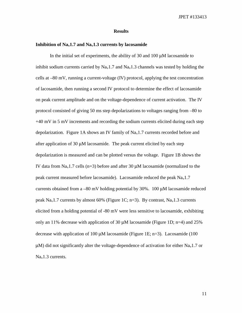
Inhibition of Nav1.7 and Nav1.3 currents by lacosamide
In the initial set of experiments, the ability of 30 and 100 µM lacosamide to
inhibit sodium currents carried by Nav1.7 and Nav1.3 channels was tested by holding the
cells at –80 mV, running a current-voltage (IV) protocol, applying the test concentration
of lacosamide, then running a second IV protocol to determine the effect of lacosamide
on peak current amplitude and on the voltage-dependence of current activation. The IV
protocol consisted of giving 50 ms step depolarizations to voltages ranging from –80 to
+40 mV in 5 mV increments and recording the sodium currents elicited during each step
depolarization. Figure 1A shows an IV family of Nav1.7 currents recorded before and
after application of 30 µM lacosamide. The peak current elicited by each step
depolarization is measured and can be plotted versus the voltage. Figure 1B shows the
IV data from Nav1.7 cells (n=3) before and after 30 µM lacosamide (normalized to the
peak current measured before lacosamide). Lacosamide reduced the peak Nav1.7
currents obtained from a –80 mV holding potential by 30%. 100 µM lacosamide reduced
peak Nav1.7 currents by almost 60% (Figure 1C; n=3). By contrast, Nav1.3 currents
elicited from a holding potential of -80 mV were less sensitive to lacosamide, exhibiting
only an 11% decrease with application of 30 µM lacosamide (Figure 1D; n=4) and 25%
decrease with application of 100 µM lacosamide (Figure 1E; n=3). Lacosamide (100
µM) did not significantly alter the voltage-dependence of activation for either Nav1.7 or
Nav1.3 currents.

JPET #133413
12
Comparison of lacosamide’s effects to lidocaine and carbamazepine effects
Inhibition of resting (closed) channels. Lidocaine and carbamazepine exhibit
differential binding to resting (or closed) channels and inactivated channels. We
examined the ability of lacosamide, lidocaine, and carbamazepine to inhibit resting
channels by holding cells expressing Nav1.7 and Nav1.3 currents at –120 mV for
10 seconds (to allow all channels to move to the resting state) and then eliciting currents
with a 20 ms step depolarization to 0 mV. Peak sodium current amplitude was measured
before and after application of lacosamide (1 to 3000 µM; n=3-7), lidocaine (1to 3000
µM; n=3-4), and carbamazepine (1 to 3000 µM; n=3-4). Fits to the concentration
response curves estimate that the IC50 of resting Nav1.7 channels is 0.7 mM for lidocaine,
1.6 mM for carbamazepine and roughly 40 mM for lacosamide (Figure 2A, Table 1). Fits
to the concentration response curves estimate that the IC50 of resting Nav1.3 channels is
1.5 mM for lidocaine, 2.5 mM for carbamazepine, and roughly 51 mM for lacosamide
(Figure 2B, Table 1). These data indicate that lacosamide has a much higher IC50 for
resting Nav1.3 and Nav1.7 channels than either carbamazepine or lidocaine. Although the
estimates of the affinity of resting Nav1.7 and Nav1.3 channels for lacosamide should
only be considered as very rough estimates due to lack of data at higher concentrations, at
1mM, lidocaine and carbamazepine inhibit Nav1.3 and Nav1.7 to a significantly (p <
0.0005) greater extent than 1 mM lacosamide.
Inhibition of inactivated channels. Next, we examined the ability of
lacosamide, lidocaine, and carbamazepine to interact with inactivated channels by
holding cells expressing Nav1.7 currents at –50 mV for 10 seconds (to allow all channels

JPET #133413
13
to move to inactivated states), then stepping the cells to –120 mV for 100ms (to allow
channels without drug bound to recover from fast-inactivation; Cummins and Waxman
1997; Cummins et al., 2001) and finally eliciting currents with a 20 ms step
depolarization to 0 mV to estimate channel availability (Figure 3C). Peak sodium current
amplitude was measured before and after application of lacosamide (1 to 3000 µM; n=3-
7), lidocaine (1 to 3000 µM; n=3-4) and carbamazepine (1 to 3000 µM; n=3-4). The
concentration response curve for inhibition of inactivated Nav1.7 channels is shown in
Figure 3A. The concentration response curve for inactivated Nav1.3 channels (Figure
3B) was similarly obtained, with the exception that we used a 10 second step to –40 mV
(due to differences in steady-state inactivation; see Figure 4) with the cells expressing
Nav1.3 currents. The different inactivation prepulse potentials were chosen to achieve
comparable extents of inactivation (a 500 ms prepulse to -40 mV inactivates 97.5% of
Nav1.3 channels and a 500 ms prepulse to -50 mV inactivates 96.7% of Nav1.7 channels).
Fits to the concentration response curves estimate that the IC50 for inhibition of
inactivated Nav1.7 channels is 44 µM for lidocaine, 406 µM for carbamazepine, and 182
µM for lacosamide. Fits to the concentration response curves estimate that the IC50 for
inhibition of inactivated Nav1.3 channels is 284 µM for lidocaine, 900 µM for
carbamazepine, and 415 µM for lacosamide. Table 1 shows the parameters for the Hill
equation fits. These data indicate that 1) lacosamide has a much higher affinity for
inactivated Nav1.7 and Nav1.3 channels than for resting Nav1.7 and Nav1.3 channels and
2) inactivated Nav1.3 channels are less sensitive to all three compounds than inactivated
Nav1.7 channels.

JPET #133413
14
Effects on voltage-dependence of steady-state fast and slow inactivation
Our data indicated that lacosamide, like lidocaine and carbamazepine, exhibits
preferential binding to inactivated channels. We asked whether lacosamide would
enhance fast inactivation of Nav1.7 and Nav1.3 channels in the same manner as lidocaine
and carbamazepine. Therefore we measured the effect of lacosamide, lidocaine, and
carbamazepine on the voltage-dependence of steady-state fast and slow inactivation. We
used 1 mM for each of the compounds in order to help maximize the binding of the
compound to the channels. Higher concentrations were not used for this as solubility
becomes a problem, especially with carbamazepine. Cells expressing Nav1.7 or Nav1.3
channels were held at -120 mV, stepped to inactivating prepulse potentials ranging from –
150 to –30 mV (in 10 mV increments) for 500 ms before stepping the cells to 0 mV for
20 ms to measure the available current (Figure 4C). In order to eliminate the bias of time
dependent shifts in the voltage-dependence of fast inactivation we compared data
recorded 4 minutes after establishing the whole-cell recording configuration from control
cells (in the absence of test compound; n=5) to data recorded 4 minutes after establishing
the whole-cell recording configuration from cells exposed to one of the three test
compounds (n=5-8). Both lidocaine and carbamazepine (1 mM) shifted the voltage-
dependence of steady-state fast inactivation by roughly 20 mV in the negative direction
for both Nav1.7 (Figure 4A) and Nav1.3 (Figure 4B) currents. The magnitude of the shift
in the presence of both lidocaine and carbamazepine is significant for both Nav1.3 and
Nav1.7 compared to control cells (P < 0.005). However, lacosamide (1 mM) did not
affect the voltage-dependence of steady-state fast inactivation for either Nav1.7 or Nav1.3
currents.

JPET #133413
15
Our data demonstrates that although lacosamide seems to exhibit preferential
binding to inactivated channels, this was not seen with 500 ms prepulses. Therefore we
next measured the effect of lacosamide, lidocaine, and carbamazepine (all at 1 mM) on
the voltage-dependence of steady-state slow inactivation. Cells expressing Nav1.7 or
Nav1.3 channels were held at –120 mV, stepped to inactivating prepulse potentials
ranging from –140 to 0 mV (in 10 mV increments) for 10 s, hyperpolarized to –160 mV
for 100 ms, then stepped to 0 mV for 20 ms to measure the available current (Figure 4F).
The brief hyperpolarization was used to allow channels (both with and without drug
bound) to recover from fast inactivation while limiting the recovery from slow
inactivation. In order to eliminate the bias of time dependent shifts in the voltage-
dependence of fast inactivation we compared data recorded 4 minutes after establishing
the whole-cell recording configuration from control cells (in the absence of test
compound; n=5) to data recorded 4 minutes after establishing the whole-cell recording
configuration from cells exposed to one of the three test compounds (n=5-8).
Carbamazepine did not alter the voltage-dependence of steady-state slow inactivation for
either Nav1.7 (Figure 4D) or Nav1.3 (Figure 4E) currents. However, both lacosamide and
lidocaine shifted the apparent voltage-dependence of steady-state slow inactivation for
both Nav1.7 and Nav1.3 currents.
Rate of development of inhibition
In order to better understand the interaction between lacosamide and Nav1.7 and
Nav1.3 sodium channels, we investigated the time course for development of block by
1 mM lacosamide, lidocaine and carbamazepine. Development of block was examined
by holding the cells (n=5-7 for each of the four conditions) expressing Nav1.7 (Figure
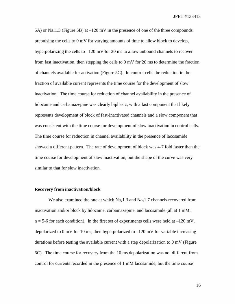
JPET #133413
16
5A) or Nav1.3 (Figure 5B) at –120 mV in the presence of one of the three compounds,
prepulsing the cells to 0 mV for varying amounts of time to allow block to develop,
hyperpolarizing the cells to –120 mV for 20 ms to allow unbound channels to recover
from fast inactivation, then stepping the cells to 0 mV for 20 ms to determine the fraction
of channels available for activation (Figure 5C). In control cells the reduction in the
fraction of available current represents the time course for the development of slow
inactivation. The time course for reduction of channel availability in the presence of
lidocaine and carbamazepine was clearly biphasic, with a fast component that likely
represents development of block of fast-inactivated channels and a slow component that
was consistent with the time course for development of slow inactivation in control cells.
The time course for reduction in channel availability in the presence of lacosamide
showed a different pattern. The rate of development of block was 4-7 fold faster than the
time course for development of slow inactivation, but the shape of the curve was very
similar to that for slow inactivation.
Recovery from inactivation/block
We also examined the rate at which Nav1.3 and Nav1.7 channels recovered from
inactivation and/or block by lidocaine, carbamazepine, and lacosamide (all at 1 mM;
n = 5-6 for each condition). In the first set of experiments cells were held at –120 mV,
depolarized to 0 mV for 10 ms, then hyperpolarized to –120 mV for variable increasing
durations before testing the available current with a step depolarization to 0 mV (Figure
6C). The time course for recovery from the 10 ms depolarization was not different from
control for currents recorded in the presence of 1 mM lacosamide, but the time course

JPET #133413
17
was more complex and slower in the presence of lidocaine and carbamazepine, indicating
that these two compounds can rapidly alter Nav1.7 (Figure 6A) and Nav1.3 (Figure 6B)
channel activity. In the second set of experiments cells (n=5-9) were held at –120 mV,
depolarized to 0 mV for 5 s, then hyperpolarized to –120 mV for a variable duration
before testing the available current with a step depolarization to 0 mV. The 5 s
depolarization provides time for the channels to undergo slow inactivation. The time
course for recovery from the 5 s depolarization was not different from control for currents
recorded in the presence of 1 mM lidocaine and carbamazepine. However, the time
constant for recovery from the 5 s prepulse was significantly greater in the presence of
1 mM lacosamide (P < 0.01 at 500 ms; Figure 6D,E).
Effects on TTX-R current
We wanted to examine the effects of lacosamide, carbamazepine and lidocaine on
Nav1.8 currents and determine how the effects compared with those on Nav1.3 and
Nav1.7 currents. Nav1.8 currents are very difficult to record from HEK293 cells,
therefore we recorded Nav1.8-type currents from rat L4/L5 DRG neurons 12-18 hours
after being cultured. Two distinct types of TTX-R currents (Nav1.8-type and Nav1.9-
type) have been identified in DRG neurons (Akopian et al., 1996; Dib-Hajj et al., 1999;
Cummins et al., 1999). We focused on Nav1.8 currents as its role in pain is much better
understood than that of Nav1.9 and because it is difficult to obtain stable recordings of
Nav1.9 currents, even in DRG sensory neurons (Leffler et al., 2005). In our recordings
we isolated Nav1.8-type currents by using 500 nM TTX to eliminate TTX-S currents and
a holding potential (-80 mV) which is efficient at eliminating the persistent TTX-R

JPET #133413
18
current carried by Nav1.9 (Cummins et al., 1999, Priest et al. 2005, Amaya et al. 2006).
The effects of lacosamide, carbamazepine, and lidocaine were tested on resting and
inactivated channels. Peak sodium current amplitude was measured before and after
application of drug (300 µM), and projected IC50 values from our results using the
equation: (% remaining current)/((1-% remaining current) x [drug]). As before, resting
channels were tested by holding the neurons at -80 mV and pulsing to –120 mV for 10
seconds (to allow all channels to move to the resting (closed state) and then eliciting
currents with a 20 ms step depolarization to 0 mV. Using this protocol we observed
decreases of 7.3 ± 4.3% (n = 6), 26.4 ± 3.2% (n = 4), 48.6 ± 7.1% (n = 5) for lacosamide,
carbamazepine, and lidocaine, respectively (Figure 7A). Based on these values, the
estimated IC50 values of lacosamide, carbamazepine, and lidocaine for resting Nav1.8-
type TTX-R channels are 4 mM, 840 µM, and 320 µM, respectively.
To test the ability of lacosamide, lidocaine, and carbamazepine to inhibit
inactivated Nav1.8-type TTX-R channels, we pulsed the neurons to –20 mV (which
produces ~95% inactivation) for 10 seconds (to allow all channels to move to the
inactivated state and to allow for drug binding to reach equilibrium), then stepped the
cells to –120 mV for 100ms (to allow channels without drug bound to recover from
inactivation; Cummins and Waxman, 1997) and finally elicited currents with a 20 ms step
depolarization to 0 mV to estimate channel availability. Peak sodium current amplitudes
were measured before and after application of lacosamide (n = 6), carbamazepine (n = 8),
or lidocaine (n = 9), all at a concentration of 300 µM. Using this protocol, we observed
decreases of 95.1 ± 1.3%, 68.5 ± 5.1%, 89.1 ± 2.6% for lacosamide, carbamazepine, and
lidocaine, respectively. Based on these values, we estimate that the IC50 for inhibition of

JPET #133413
19
inactivated Nav1.8-type TTX-R channels is 16 µM for lacosamide, 138 µM for
carbamazepine, and 37 µM for lidocaine. Figure 7B shows the comparison of current
inhibition on inactivated Nav1.7, Nav1.3, and Nav1.8-type channels by all three drugs at
concentrations of 300 µM. In this case each channel subtype was held at different pre-
pulse potentials, due to differences in their voltage dependence of steady-state
inactivation, to ensure >95% inactivation. Figure 7C shows the comparison of current
inhibition of Nav1.7, Nav1.3, and Nav1.8-type channels held at -50 mV, which would
simulate a slightly depolarized potential for a sensory neuron. Lacosamide inhibition at –
50 mV was similar for Nav1.8-type, Nav1.7 and Nav1.3 currents. Lidocaine inhibition at
–50 mV was also similar for all three types of currents. However, differences were still
observed between the current subtypes for carbamazepine inhibition at -50 mV.
We further investigated the effects lacosamide, lidocaine, and carbamazepine on
Nav1.8-type channels by looking at their effects on the voltage-dependence of steady-
state fast and slow inactivation. We used 1 mM for each of the compounds in order to
help maximize the interaction of the channels with the compound. Neurons were held at
-80 mV, stepped to inactivating prepulse potentials ranging from –100 to 30 mV (in
10 mV increments) for 500 ms before stepping the cells to 0 mV for 20 ms to measure the
available current. In order to eliminate the bias of time dependent shifts in the voltage-
dependence of fast inactivation we compared data recorded 3 minutes after establishing
the whole-cell recording configuration from control cells (in the absence of test
compound; n = 9) to data recorded 3 minutes after establishing the whole-cell recording
configuration from cells exposed to one of the three test compounds (n = 5-7).
Surprisingly, none of the drugs altered the voltage-dependence of steady-state fast

JPET #133413
20
inactivation (Figure 8A). The estimated V1/2 of steady-state fast inactivation (obtained
with Boltzmann fits) of Nav1.8-type current in the presence of lacosamide (-33.4 ±
2.0 mV; slope: 3.5 ± 0.1; n = 5), lidocaine (-39.5 ± 2.5 mV; slope: 4.5 ± 0.2, n = 7), and
carbamazepine (-37.4 ± 2.5 mV; slope: 4.1 ± 0.2, n = 6) was not statistically different
from that of control Nav1.8-type current (-34.7 ± 1.3 mV; slope: 4.0 ± 0.1; One-way
ANOVA analysis).
To study the effects of these drugs on Nav1.8-type current slow inactivation, we
stepped to inactivating prepulse potentials ranging from –100 to 30 mV (in 10 mV
increments) for 10 s, hyperpolarized to –140 mV for 100 ms, then stepped to 0 mV for
20 ms to measure the available current. Effects were observed with all of the drugs on
the steady-state slow inactivation curve of Nav1.8-type currents (Figure 8B). The
estimated V1/2 of steady-state slow inactivation of Nav1.8-type current in the presence of
lacosamide (-54.8 ± 3.2 mV; slope: 5.2 ± 0.2, n = 6), lidocaine (-57.9 ± 2.6 mV; slope:
5.1 ± 0.2, n = 7), and carbamazepine (-53.1 ± 3.2 mV; slope: 4.4 ± 0.1, n = 6) were
significantly (p < 0.001) hyperpolarized from that of control Nav1.8-type current (-37.1 ±
1.2 mV; slope: 10.4 ± 0.1; n = 9; One-way ANOVA analysis). In addition, the slope of
the steady-state slow inactivation curve for Nav1.8-type current was significantly (p <
0.001; One-way ANOVA analysis) different from the slope values observed in the
presence of lacosamide, lidocaine, or carbamazepine. Both lacosamide and lidocaine
markedly increased the maximal extent to which slow inactivation can reduce the
availability of Nav1.8, i.e. at 20 mV reduction of channel availability for control and
carbamazepine reached a maximum of about 50%, whereas it amounted to 80% and 90%
for lidocaine and lacosamide respectively.

JPET #133413
21
Discussion
This study was aimed at determining the inhibitory effects of a new drug,
lacosamide, on voltage-gated sodium channels that are expressed in peripheral
nociceptive neurons and that have been identified as playing important roles in pain. We
focused our efforts on examining the effects of lacosamide on Nav1.7 and Nav1.3
channels and comparing them to clinically used traditional analgesics carbamazepine and
lidocaine. We found clinically relevant concentrations of lacosamide substantially reduce
Nav1.7 current amplitudes at a holding potential of –80 mV, while Nav1.3 currents were
inhibited to a slightly lesser extent, indicating that they are less sensitive. We found that
lacosamide has a significantly higher IC50 for resting Nav1.3 and Nav1.7 channels than
either carbamazepine or lidocaine. Lacosamide exhibited substantially greater inhibition
of inactivated Nav1.7 and Nav1.3 channels than resting Nav1.7 and Nav1.3 channels.
Lacosamide is more potent at inhibiting inactivated channels than carbamazepine but
slightly less potent than lidocaine. These data show lacosamide has a much greater
ability to discriminate between resting and inactivated Nav1.7 and Nav1.3 channels than
either lidocaine or carbamazepine.
The effects of lacosamide on Nav1.8-type currents in DRG neurons were similar
to that seen with Nav1.7 and Nav1.3 as lacosamide inhibited inactivated Nav1.8-type
channels to a significantly greater extent then resting channels and suggest lacosamide is
the most potent of the three drugs at inhibiting inactivated Nav1.8-type channels.
Although the ratio of lacosamide resting IC50 to the –50 mV IC50 was smaller for Nav1.8-
type channels than for Nav1.3 or Nav1.7 channels, this ratio was still greater for
lacosamide than for either lidocaine or carbamazepine (Table 2). These data, showing

JPET #133413
22
that lacosamide is much more effective at distinguishing between resting and inactivated
Nav1.7, Nav1.3, and Nav1.8-type channels than lidocaine and carbamazepine, suggest
lacosamide should be selective at inhibiting the activity of neurons with depolarized
membrane potentials compared to neurons with normal resting membrane potentials than
either lidocaine or carbamazepine. This could be an important part of the analgesic
mechanism of action of lacosamide as nociceptor hyperexcitability and increased pain
sensations are likely to be associated with depolarized membrane potentials in neurons.
The differential effects of lacosamide, carbamazepine, and lidocaine on the
voltage-dependence of steady-state fast inactivation and steady-state slow inactivation are
intriguing. These data showed that the inhibition induced by the three drugs was
different depending on the length of the depolarizing pulse used to inactivate the
channels. When using 500 ms prepulses, we observed a significant hyperpolarizing shift
in the voltage-dependence of steady-state fast inactivation for Nav1.7 and Nav1.3 treated
with carbamazepine and lidocaine but not when treated with lacosamide. In contrast
while carbamazepine had no effect on the voltage-dependence of slow inactivation for
Nav1.7 and Nav1.3, lacosamide and lidocaine both produced a significant hyperpolarizing
shift in the voltage-dependence of slow inactivation of these channels. These results
suggest while carbamazepine and lidocaine interact with fast-inactivated Nav1.7 and
Nav1.3 channels, lacosamide does not. Our data are consistent with the proposal that
lacosamide interacts predominantly with slow inactivated sodium channels (Errington et
al., 2008) and that this effect could be a mechanistic rather than a kinetic difference
compared to lidocaine and carbamazepine. However, based on these data we cannot rule
out the alternative possibility that lacosamide may bind and unbind very slowly from
JPET #133413
23
fast-inactivated channels. Mutagenesis experiments that selectively alter fast- or slow-
inactivation of the sodium channels should help definitively determine the mechanism of
action for lacosamide. Interestingly, none of the 3 drugs had any effect on the voltage-
dependence of Nav1.8-type channel fast inactivation. This result is not surprising for
lacosamide due to its ineffectiveness on fast-inactivation of Nav1.7 and Nav1.3 channels,
but is somewhat surprising for lidocaine and carbamazepine. However, previous studies
have indicated that lidocaine does not alter fast-inactivation of Nav1.8 channels in the
same manner that it does Nav1.7 channels (Chevrier et al., 2004) and that carbamazepine
might even preferentially target slow inactivated Nav1.8 channels (Cardenas et al., 2006).
This raises the possibility that access to the drug-binding site in fast-inactivated state of
Nav1.8-type channels might be substantially different from the site in Nav1.7 and Nav1.3
channels.
An interesting finding was that carbamazepine and lidocaine show significant
inhibition of Nav1.7 and Nav1.3 channels prepulsed to 0 mV for durations ranging from 3
to100 ms (Figure 5A and 5B). In that same range of prepulse durations lacosamide
induces minimal inhibition of Nav1.7 and Nav1.3 channels. At prepulse durations ranging
100ms to 11s, lacosamide induces inhibition of Nav1.7 and Nav1.3 while inhibition by
carbamazepine and lidocaine levels off. All three drugs eventually reached maximal
inhibition around 9 to 11s for both channels. Interestingly, the time point where
development of slow inactivation starts to occur for control channels is near the time
point at which lacosamide begins to cause inhibition and also where carbamazepine and
lidocaine inhibition plateaus. This suggests lacosamide interaction with Nav1.7 and
Nav1.3 channels is dependent on the channels transitioning to slow inactivated states and,

JPET #133413
24
in contrast, that transition to the slow inactivated state might impede further Nav1.7 and
Nav1.3 channel inhibition by carbamazepine and lidocaine. While recovery from slow
inactivation appeared to be slower in the presence of lacosamide, due to an increase in the
steady state fraction of slow inactivated channels by lacosamide, it was not altered by
lidocaine or carbamazepine, providing additional evidence that the mechanism of block
by lacosamide is possibly distinct from that of lidocaine or carbamazepine.
Lacosamide has been shown in acute, inflammatory and neuropathic animal pain
models (Beyreuther et al. 2006, 2007a; Hao et al., 2006), as well as in phase II clinical
trials for painful diabetic neuropathy (Rauck et al., 2007), to effectively inhibit pain with
few adverse side-effects. Based on the data reported here, it is likely lacosamide’s effects
on sodium channel activity contribute to its analgesic activity. Tissue injury and
inflammatory modulators can cause prolonged depolarization of peripheral nociceptors
and this likely contributes to inflammatory and neuropathic pain. A recent study suggests
that the effectiveness of drugs at inhibiting peripheral nerve activity following nerve
injury does not depend so much on the frequency at which action potentials are
generated, but rather on the nature of the injury and the pattern of action potential activity
(Ritter et al., 2007). Since a substantial degree of slow inactivation can be observed for
TTX-R sodium channels at small, subthreshold membrane depolarizations without any
spiking activity (Blair and Bean, 2003), a compound like lacosamide which targets the
slow inactivated channel might show enhanced therapeutic benefit.
CNS side-effects can limit the usefulness of sodium channel blockers in treating
pain and therefore drugs that are better able to differentiate between normal and abnormal
activity are desired. The enhanced ability of lacosamide to discriminate between resting

JPET #133413
25
channels and channels that have been subjected to prolonged depolarizations should
contribute to a better safety profile and increased tolerability.
Overall, our data show lacosamide is effective at blocking inactivated voltage-
gated Nav1.3, Nav1.7 and Nav1.8-type channels, voltage-gated sodium channels that have
been implicated as playing important roles in pain mechanisms. Furthermore our data
show that the kinetics of lacosamide inhibition is much slower than that of two other
sodium channel blockers, carbamazepine and lidocaine, at least for Nav1.3 and Nav1.7
channels. Our data are consistent with the proposal that lacosamide inhibition of voltage-
gated sodium channels involves selective interactions with slow-inactivated channels
(Errington et al. 2008). Ratios of resting IC50 to inactivated IC50 suggest lacosamide
should be more effective than carbamazepine and lidocaine at selectively blocking the
electrical activity of neurons that are chronically depolarized while having little or no
effect on neurons with normal resting potentials and we propose that this could contribute
to the therapeutic action of lacosamide.

JPET #133413
26
References
Akopian AN, Sivilotti L and Wood JN (1996) A tetrodotoxin-resistant voltage-gated
sodium channel expressed by sensory neurons. Nature 379:257-62.
Akopian AN, Souslova V, England S, Okuse K, Ogata N, Ure J, Smith A, Kerr BJ,
McMahon SB, Boyce S, Hill R, Stanfa LC, Dickenson AH and Wood JN (1999) The
tetrodotoxin-resistant sodium channel SNS has a specialized function in pain pathways.
Nat Neurosci 2:541-548.
Amaya F, Wang H, Costigan M, Allchorne AJ, Hatcher JP, Egerton J, Stean T, Morisset
V, Grose D, Gunthorpe MJ, Chessell IP, Tate S, Green PJ and Woolf CJ. (2006) The
voltage-gated sodium channel Na(v)1.9 is an effector of peripheral inflammatory pain
hypersensitivity. J Neurosci 26:12852-12860.
Black JA, Liu S, Tanaka M, Cummins TR and Waxman SG. (2004) Changes in the
expression of tetrodotoxin-sensitive sodium channels within dorsal root ganglia neurons
in inflammatory pain. Pain 108:237-247.
Blair NT and Bean BP. (2003) Role of tetrodotoxin-resistant Na+ current slow
inactivation in adaptation of action potential firing in small-diameter dorsal root ganglion
neurons. J Neurosci 23:10338-10350.
Beyreuther B, Callizot N and Stohr T. (2006) Antinociceptive efficacy of lacosamide in
a rat model for painful diabetic neuropathy. Eur J Pharmacol. 539:64-70.

JPET #133413
27
Beyreuther BK, Callizot N, Brot MD, Feldman R, Bain SC and Stohr T. (2007a)
Antinociceptive efficacy of lacosamide in rat models for tumor- and chemotherapy-
induced cancer pain. Eur J Pharmacol 565:98-104.
Beyreuther BK, Freitag J, Heers C, Krebsfanger N, Scharfenecker U and Stohr T.
(2007b) Lacosamide: a review of preclinical properties. CNS Drug Rev. 13:21-42.
Cardenas CA, Cardenas CG, de Armendi A and Scroggs RS. (2006) Carbamazepine
interacts with a slow inactivation state of NaV1.8-like sodium channels. Neurosci Lett
408:129-134.
Catterall WA (2000) From ionic currents to molecular mechanisms: the structure and
function of voltage-gated sodium channels. Neuron 26:13-25.
Chevrier P, Vijayaragavan K and Chahine M (2004) Differential modulation of Nav1.7
and Nav1.8 peripheral nerve sodium channels by the local anesthetic lidocaine. Br J
Pharmacol 142:576-584.
Clare JJ, Tate SN, Nobbs M and Romanos MA (2000) Voltage-gated sodium channels as
therapeutic targets. Drug Discov Today 5:506-520.
Cox JJ, Reimann F, Nicholas AK, Thornton G, Roberts E, Springell K, Karbani G, Jafri
H, Mannan J, Raashid Y, Al-Gazali L, Hamamy H, Valente EM, Gorman S, Williams R,
McHale DP, Wood JN, Gribble FM and Woods CG (2006) An SCN9A channelopathy
causes congenital inability to experience pain. Nature 444:894-898.

JPET #133413
28
Cummins TR, Howe JR and Waxman SG (1998) Slow closed-state inactivation: a novel
mechanism underlying ramp currents in cells expressing the hNE/PN1 sodium channel. J
Neurosci 18:9607-9619.
Cummins TR, Dib-Hajj SD, Black JA, Akopian AN, Wood JN and Waxman SG (1999)
A novel persistent tetrodotoxin-resistant sodium current in SNS-null and wild-type small
primary sensory neurons. J Neurosci 19:RC43.
Cummins TR, Aglieco F, Renganathan M, Herzog RI, Dib-Hajj SD and Waxman SG
(2001) Nav1.3 sodium channels: rapid repriming and slow closed-state inactivation
display quantitative differences after expression in a mammalian cell line and in spinal
sensory neurons. J Neurosci 21:5952-5961.
Cummins TR, Dib-Hajj SD and Waxman SG (2004) Electrophysiological properties of
mutant Nav1.7 sodium channels in a painful inherited neuropathy. J Neurosci. 24:8232-
8236.
Cummins TR and Waxman SG (1997) Downregulation of tetrodotoxin-resistant sodium
currents and upregulation of a rapidly repriming tetrodotoxin-sensitive sodium current in
small spinal sensory neurons after nerve injury. J Neurosci 17:3503-3514.
Dib-Hajj SD, Tyrrell L, Cummins TR, Black JA and Wood PM, Waxman SG (1999)
Two tetrodotoxin-resistant sodium channels in human dorsal root ganglion neurons.
FEBS Lett 462:117-120.
Doty P, Rudd GD, Stoehr T and Thomas D (2007): Lacosamide. Neurother 4:145-148.

JPET #133413
29
Drenth JP, Te Morsche RH, Guillet G, Taieb A, Kirby RL and Jansen JB (2005) SCN9A
mutations define primary erythermalgia as a neuropathic disorder of voltage gated
sodium channels. J Invest Dermatol 124:1333-1338.
Errington AC, Stoehr T, Heers C, Lees G (2008): The investigational anticonvulsant
lacosamide selectively enhances slow inactivation of voltage-gated sodium channels.
Mol Pharmacol 73:157-169.
Fertleman CR, Baker MD, Parker KA, Moffatt S, Elmslie FV, Abrahamsen B, Ostman J,
Klugbauer N, Wood JN, Gardiner RM and Rees M (2006) SCN9A mutations in
paroxysmal extreme pain disorder: allelic variants underlie distinct channel defects and
phenotypes. Neuron 52:767-774.
Fukuda K, Nakajima T, Viswanathan PC and Balser JR. (2005) Compound-specific Na+
channel pore conformational changes induced by local anaesthetics. J Physiol 564:21-31.
Goldin AL (2002) Evolution of voltage-gated Na(+) channels. J Exp Biol 205:575-584.
Haeseler G, Foadi N, Ahrens J, Dengler R, Hecker H and Leuwer M. (2006) Tramadol,
fentanyl and sufentanil but not morphine block voltage-operated sodium channels. Pain
126:234-44.
Hains BC, Klein JP, Saab CY, Craner MJ, Black JA and Waxman SG. (2003)
Upregulation of sodium channel Nav1.3 and functional involvement in neuronal
hyperexcitability associated with central neuropathic pain after spinal cord injury. J
Neurosci 23:8881-8892

JPET #133413
30
Hao JX, Stohr T, Selve N, Wiesenfeld-Hallin Z and Xu XJ. (2006) Lacosamide, a new
anti-epileptic, alleviates neuropathic pain-like behaviors in rat models of spinal cord or
trigeminal nerve injury. Eur J Pharmacol 553:135-140.
Jarvis MF, Honore P, Shieh CC, Chapman M, Joshi S, Zhang XF, Kort M, Carroll W,
Marron B, Atkinson R, Thomas J, Liu D, Krambis M, Liu Y, McGaraughty S, Chu K,
Roeloffs R, Zhong C, Mikusa JP, Hernandez G, Gauvin D, Wade C, Zhu C, Pai M,
Scanio M, Shi L, Drizin I, Gregg R, Matulenko M, Hakeem A, Gross M, Johnson M,
Marsh K, Wagoner PK, Sullivan JP, Faltynek CR and Krafte DS. (2007) A-803467, a
potent and selective Nav1.8 sodium channel blocker, attenuates neuropathic and
inflammatory pain in the rat. Proc Natl Acad Sci U S A 104:8520-8525.
Lai J, Gold MS, Kim CS, Bian D, Ossipov MH, Hunter JC, Porreca F (2002) Inhibition
of neuropathic pain by decreased expression of the tetrodotoxin-resistant sodium channel,
NaV1.8. Pain 95:143-152.
Leffler A, Herzog RI, Dib-Hajj SD, Waxman SG and Cummins TR. (2005)
Pharmacological properties of neuronal TTX-resistant sodium channels and the role of a
critical serine pore residue. Pflugers Arch 451:454-463.
Nassar MA, Stirling LC, Forlani G, Baker MD, Matthews EA, Dickenson AH and Wood
JN (2004) Nociceptor-specific gene deletion reveals a major role for Nav1.7 (PN1) in
acute and inflammatory pain. Proc Natl Acad Sci U S A 101:12706-12711.
Priest BT, Murphy BA, Lindia JA, Diaz C, Abbadie C, Ritter AM, Liberator P, Iyer LM,
Kash SF, Kohler MG, Kaczorowski GJ, MacIntyre DE and Martin WJ. (2005)

JPET #133413
31
Contribution of the tetrodotoxin-resistant voltage-gated sodium channel NaV1.9 to
sensory transmission and nociceptive behavior. Proc Natl Acad Sci U S A 102:9382-
9387.
Ragsdale DS and Avoli M (1998) Sodium channels as molecular targets for antiepileptic
drugs. Brain Res Brain Res Rev 26:16-28.
Rauck RL, Shaibani A, Biton V, Simpson J and Koch B. (2007) Lacosamide in painful
diabetic peripheral neuropathy: a phase 2 double-blind placebo-controlled study. Clin J
Pain 23:150-158.
Ritter AM, Ritchie C and Martin WJ. (2007) Relationship between the firing frequency
of injured peripheral neurons and inhibition of firing by sodium channel blockers. J Pain
8:287-295.
Rush AM, Dib-Hajj SD, Liu S, Cummins TR, Black JA and Waxman SG. (2006) A
single sodium channel mutation produces hyper- or hypoexcitability in different types of
neurons. Proc Natl Acad Sci U S A. 103:8245-8250.
Sheets PL, Jackson II JO, Waxman SG, Dib-Hajj S and Cummins TR. (2007) A Nav1.7
Channel Mutation Associated with Hereditary Erythromelalgia Contributes to Neuronal
Hyperexcitability and Displays Reduced Lidocaine Sensitivity. J Physiol 581:1019-
1031.

JPET #133413
32
Stohr T, Kupferberg HJ, Stables JP, Choi D, Harris RH, Kohn H, Walton N and White
HS. (2007) Lacosamide, a novel anti-convulsant drug, shows efficacy with a wide safety
margin in rodent models for epilepsy. Epilepsy Res 74:147-154.
Waxman SG, Kocsis JD and Black JA. (1994) Type III sodium channel mRNA is
expressed in embryonic but not adult spinal sensory neurons, and is reexpressed
following axotomy. J Neurophysiol 72:466-470.

JPET #133413
33
Footnotes
This work was supported by Schwarz BioSciences. 1Current address: Merz Pharmaceuticals GmbH, Frankfurt am Main, Germany

JPET #133413
34
Legends for Figures Figure 1: A, Current-voltage (IV) family trace of an HEK293 cell stably expressing the
human Nav1.7 channel before (left) and after (middle) application of 30 µM lacosamide
(LCM). The voltage protocol is shown on the right. B and C, IV relationship of Nav1.7
current in the absence and presence of 30 µM (B) and 100 µM (C) lacosamide. D and E,
IV relationship of Nav1.3 current in the absence and presence of 30 µM (D) and 100 µM
(E) lacosamide.
Figure 2: A, Concentration-response curve for the inhibitory effects of lacosamide
(LCM), lidocaine, and carbamazepine (CBZ) on resting Nav1.7 channels. B,
Concentration-response curve for the inhibitory effects of lacosamide (LCM), lidocaine,
and carbamazepine (CBZ) on resting Nav1.3 channels. C, The voltage protocol for testing
current amplitude is shown.
Figure 3: A, Concentration-response curve for the inhibitory effects of lacosamide
(LCM), lidocaine, and carbamazepine (CBZ) on inactivated Nav1.7 channels. B,
Concentration-response curve for the inhibitory effects of lacosamide (LCM), lidocaine,
and carbamazepine (CBZ) on inactivated Nav1.3 channels. C, The voltage protocol for
testing current amplitude is shown.
Figure 4: Effects of lacosamide, lidocaine, and carbamazepine on the voltage-
dependence of steady-state fast and slow inactivation for Nav1.7 and Nav1.3 channels.
A and B, Lidocaine and carbamazepine (CBZ) caused a significant hyperpolarizing shift

JPET #133413
35
in the voltage-dependence of steady-state fast inactivation for Nav1.7 (A) and Nav1.3 (B)
channels. Lacosamide (LCM) had no effect on the voltage-dependence of steady-state
fast inactivation for either channel. Midpoints of Nav1.7 steady-state inactivation in (A)
were –76.1±1.9 (n=6), -99.3±1.5 (n=7), -97.0±3.3 (n=5), -78.7±1.2 (n=6) for control,
+lidocaine, +carbamazepine, and +lacosamide, respectively. Midpoints of Nav1.3
steady-state inactivation in (A) were –62.1±2.7 (n=9), -82.5±1.6 (n=9), -81.0±2.6 (n=8),
-65.4±1.8 (n=11) for control, +lidocaine, +carbamazepine, and +lacosamide, respectively.
C, Voltage protocol for steady-state fast inactivation is shown. Data sweeps were
acquired at 0.2 Hz. D and E, Lidocaine and lacosamide (LCM) caused a significant
hyperpolarizing shift in the voltage-dependence of slow inactivation for Nav1.7 (D) and
Nav1.3 (E) channels. Carbamazepine (CBZ) had no effect on the voltage-dependence of
slow inactivation for either channel. F, Voltage protocol for steady-state slow
inactivation is shown. The 100 ms pulse at –160 mV is used to allow for recovery of
fast-inactivated and drug-bound fast-inactivated channels, but not slow-inactivated or
drug-bound slow-inactivated channels. Data sweeps were acquired at 0.09 Hz.
Figure 5: Effects of lacosamide, lidocaine, and carbamazepine on the development of
slow inactivation/inhibition for Nav1.7 and Nav1.3 channels. For the control channels, a
brief (20 ms) hyperpolarizing pulse to -120 mV was given after the inactivating pulse to
0 mV at increasing durations. A and B, Lidocaine and carbamazepine (CBZ) caused an
increase in the rate of development of inactivation/inhibition at pre-pulse durations
ranging from 1 to 100 ms before slowing at pre-pulse durations greater than 100 ms for
both Nav1.7 (A) and Nav1.3 (B) channels. An increase in the rate of development of

JPET #133413
36
inactivation/inhibition for both Nav1.7 (A) and Nav1.3 (B) channels was observed in the
presence of lacosamide (LCM) but at pre-pulse durations longer than those seen with
lidocaine and carbamazepine. C, The voltage protocol is shown. Following a variable
conditioning pulse to 0 mV, a 20 ms pulse to –120 mV allows recovery from fast
inactivation (but not block) before the fraction of current available with the 20 ms pulse
to 0 mV. Data sweeps were acquired at 0.5 Hz for short pre-pulse durations and at
slower rates for the longer pre-pulse durations.
Figure 6: Effects of lacosamide, lidocaine, and carbamazepine on the recovery of Nav1.7
and Nav1.3 channels from inactivation. Cells were held at –120 mV, depolarized to 0 mV
for 10 ms or 5s, then hyperpolarized to –120 mV for variable increasing durations before
testing the available current with a step depolarization to 0 mV. A and B, Lidocaine and
carbamazepine (CBZ) caused an decrease in the rate of recovery from
inactivation/inhibition (10 ms) for both Nav1.7 (A) and Nav1.3 (B) channels while
lacosamide (LCM) had no effect. C, The voltage protocol used for (A) and (B) is shown.
Data sweeps were acquired at 0.5 Hz. D and E, Lidocaine and carbamazepine (CBZ) did
not alter recovery kinetics after induction of slow inactivation compared to control. In the
presence of lacosamide (LCM) the fraction of available channels after 5 s depolarization
was lower compared to control, lidocaine and carbamazepine and the duration of
recovery longer (significant at 500 ms) compared to the other conditions. F, The voltage
protocol used for (D) and (E) is shown. Data sweeps were acquired at 0.5 Hz for short
recovery durations and at slower rates for the longer recovery durations.

JPET #133413
37
Figure 7: Comparison of inhibition on resting and inactivated Nav1.7, Nav1.3, and TTX-
R channels by lacosamide, lidocaine, and carbamazepine (all at 300 µM). A, Lacosamide
(LCM) had the smallest inhibitory effect on resting Nav1.7, Nav1.3, and TTX-R channels
compared to lidocaine and carbamazepine. Lidocaine and carbamazepine (CBZ) had the
greatest inhibitory effect on resting TTX-R channels compared to Nav1.7 and Nav1.3
channels. B, Effects of lacosamide (LCM), lidocaine, and carbamazepine (CBZ) on
completely inactivated Nav1.7, Nav1.3, and TTX-R channels. For complete inactivation,
the voltage of the inactivating pre-pulse was varied due to differences in the voltage-
dependence of inactivation between channels. Nav1.7 channels were pre-pulsed to -50
mV, Nav1.3 channels to -40 mV, and TTX-R channels to -20 mV. Lacosamide showed
considerably more inhibition on TTX-R channels compared to Nav1.7 and Nav1.3
channels. Lidocaine and carbamazepine were effective at inhibiting TTX-R channels
more than Nav1.3 channels but not Nav1.7 channels. C, Effects of lacosamide (LCM),
lidocaine, and carbamazepine (CBZ) on Nav1.7, Nav1.3, and TTX-R channels pre-pulsed
to -50 mV. This pre-pulse voltage was chosen to theoretically represent a slightly
depolarized potential for a sensory neuron. Lacosamide and lidocaine showed
comparable inhibition on TTX-R channels, Nav1.7, and Nav1.3 channels. Carbamazepine
displayed more inhibition on TTX-R channels compared to Nav1.3 channels. Values and
statistical significance was determined using a one-way ANOVA followed with a
Tukey’s means comparison test (** = p < 0.01, *** = p < 0.001).

JPET #133413
38
Figure 8: Effects of lacosamide, lidocaine, and carbamazepine (all at 1 mM) on the
voltage-dependence of steady-state fast and slow inactivation for TTX-R channels. A,
Representative family of Nav1.8-type currents recorded from a small diameter DRG
neuron. B, Lacosamide (LCM), lidocaine, and carbamazepine (CBZ) had no effect on
the voltage-dependence of steady-state fast inactivation for TTX-R channels. Data were
acquired at 0.2 Hz. C, Lacosamide (LCM), lidocaine, and carbamazepine (CBZ) caused a
significant hyperpolarizing shift in the voltage-dependence of slow inactivation for TTX-
R channels. Data were acquired at 0.09 Hz. Lacosamide caused the greatest decrease in
elicited TTX-R current followed by lidocaine and carbamazepine, respectively. At
20 mV lacosamide and lidocaine reduced available current by 90 and 80%, whereas
maximal reduction by carbamazepine at this voltage was similar to control (about 50%).

JPET #133413
39
Table 1. Hill equation fit parameters to dose response relationships in Figures 2 and 3. Resting Inhibition (Figure 2) Inactivated Inhibition (Figure 3) fmax IC50 h R2 fmax IC50 h R2 Nav1.3 Lidocaine .997 1462 µM 1.35 .99 .949 284 µM .48 .95 Carbamazepine .996 2464 µM 1.10 .97 .942 900 µM .89 .95 Lacosamide .998 51,200 µM 1.0 .96 .986 415 µM .61 .98 Nav1.7 Lidocaine .994 718 µM 1.02 .99 .992 44 µM .43 .95 Carbamazepine 1.0 1584 µM 1.0 .96 .94 406 µM 1.12 .96 Lacosamide 1.0 40,300 µM .78 .95 .954 182 µM .90 .974
R2 is the square of the correlation between the fit and the actual inhibition values and is an indication of the goodness of the fit.

JPET #133413
40
Table 2. Ratio of IC50 for closed channels to IC50 at depolarized potentials Nav1.3
at -40 mV Nav1.3 at -50 mV
Nav1.7 at -50 mV
Nav1.8-type at -50 mV
Nav1.8-type at -20 mV
Lacosamide 123 130 221 16 250 Lidocaine 5 5 16 3 9 Carbamazepine 3 3 4 7 6