Title A Historical Review of Enzymatic Debridement: Revisited · A Historical Review of Enzymatic...
51
The following is an update to a book entitled, “A Historical Review of Enzymatic Debridement: Revisited”, which I wrote in 2003. Since its publication, while the relevant clinical evidence has remained consistent, the amount of biochemical research and knowledge gained has been impressive. In the ϐirst chapter a sampling of the typical topical enzymatic debriding agents that have been used in wound care are reviewed and interestingly enough only one remains on the market. The FDA has removed all others from the marketplace and an explanation is provided in chapter one along with descriptions of the use and mode of action (MoA) of these agents. Chapter two is a review of the many different types of collagen found in the body, including their structure, form, and function as so much additional insight into this molecule has been gained since 2003. In chapter three we see an account depicting the many advances in understanding matrix metalloproteinases (MMPs) reviewed in detail. Form, function, tissue orientation and preferred substrates are addressed. Finally, in chapter four we see the history of the MoA of MMPs as compared to bacterial collagenase starting in the early ‘80s to the time of this current publication. In addition we see the level of complexity of bacterial collagenases compared to MMPs, helping us to better understand why bacterial collagenase is much more efϐicient at removing necrotic tissue from wounds than are our own (endogenous) MMPs. I hope the reader ϐinds this review useful from an academic standpoint, but more importantly from a clinical framework helping to understand the role of these types of therapies in wound care. INTRODUCTION David W Brett* Science & Technology Manager, Advance Wound Care| Wound Management Division, Smith & Nephew, Fort Worth, TX, USA *Corresponding author: David W Brett, Science & Technology Manager, Advance Wound Care, Wound Management Division, Smith & Nephew, Fort Worth, TX, USA, Tel: 727-244-3883; Email: [email protected] Subject Category: Biotechnology and Biomedicine Title A Historical Review of Enzymatic Debridement: Revisited A Historical Review of Enzymatic Debridement: Revisited
Transcript of Title A Historical Review of Enzymatic Debridement: Revisited · A Historical Review of Enzymatic...
The following is an update to a book entitled, “A Historical Review of Enzymatic Debridement: Revisited”, which I wrote in 2003. Since its publication, while the relevant clinical evidence has remained consistent, the amount of biochemical research and knowledge gained has been impressive. In the irst chapter a sampling of the typical topical enzymatic debriding agents that have been used in wound care are reviewed and interestingly enough only one remains on the market. The FDA has removed all others from the marketplace and an explanation is provided in chapter one along with descriptions of the use and mode of action (MoA) of these agents. Chapter two is a review of the many different types of collagen found in the body, including their structure, form, and function as so much additional insight into this molecule has been gained since 2003. In chapter three we see an account depicting the many advances in understanding matrix metalloproteinases (MMPs) reviewed in detail. Form, function, tissue orientation and preferred substrates are addressed. Finally, in chapter four we see the history of the MoA of MMPs as compared to bacterial collagenase starting in the early ‘80s to the time of this current publication. In addition we see the level of complexity of bacterial collagenases compared to MMPs, helping us to better understand why bacterial collagenase is much more ef icient at removing necrotic tissue from wounds than are our own (endogenous) MMPs. I hope the reader inds this review useful from an academic standpoint, but more importantly from a clinical framework helping to understand the role of these types of therapies in wound care.
INTRODUCTION
David W Brett*Science & Technology Manager, Advance Wound Care| Wound Management Division, Smith & Nephew, Fort Worth, TX, USA
*Corresponding author: David W Brett, Science & Technology Manager, Advance Wound Care, Wound Management Division, Smith & Nephew, Fort Worth, TX, USA, Tel: 727-244-3883; Email: [email protected]
Subject Category: Biotechnology and Biomedicine
Title
A Historical Review of Enzymatic Debridement: Revisited
A Historical Review of Enzymatic Debridement: Revisited
A Historical Review of Enzymatic Debridement: RevisitedOpen Access
HTTPS://WWW.HEIGHPUBS.ORG
002
Table of Contents - 4 ChaptersSl No Title Pages
1 Types of Enzymes - David W Brett 3-8
2 Technical Review of Collagen - David W Brett 9-15
3 Endogenous Collagenase - David W Brett 16-32
4 Modes of Action of Enzymatic Débridement - David W Brett 33-51
*Corresponding author: David W Brett, Science & Technology Manager, Advance Wound Care, Wound Management Division, Smith & Nephew, Fort Worth, TX, USA, Tel: 727-244-3883; Email: [email protected]
I’d like to thank and credit Dr. Lei Shi, PhD, for his expertise in the editing of this book. Dr. Shi’s expertise and guidance were invaluable in writing this book and it
would not have been possible without his kind critique and assistance.
A Historical Review of Enzymatic Debridement: RevisitedOpen Access
HTTPS://WWW.HEIGHPUBS.ORG
003
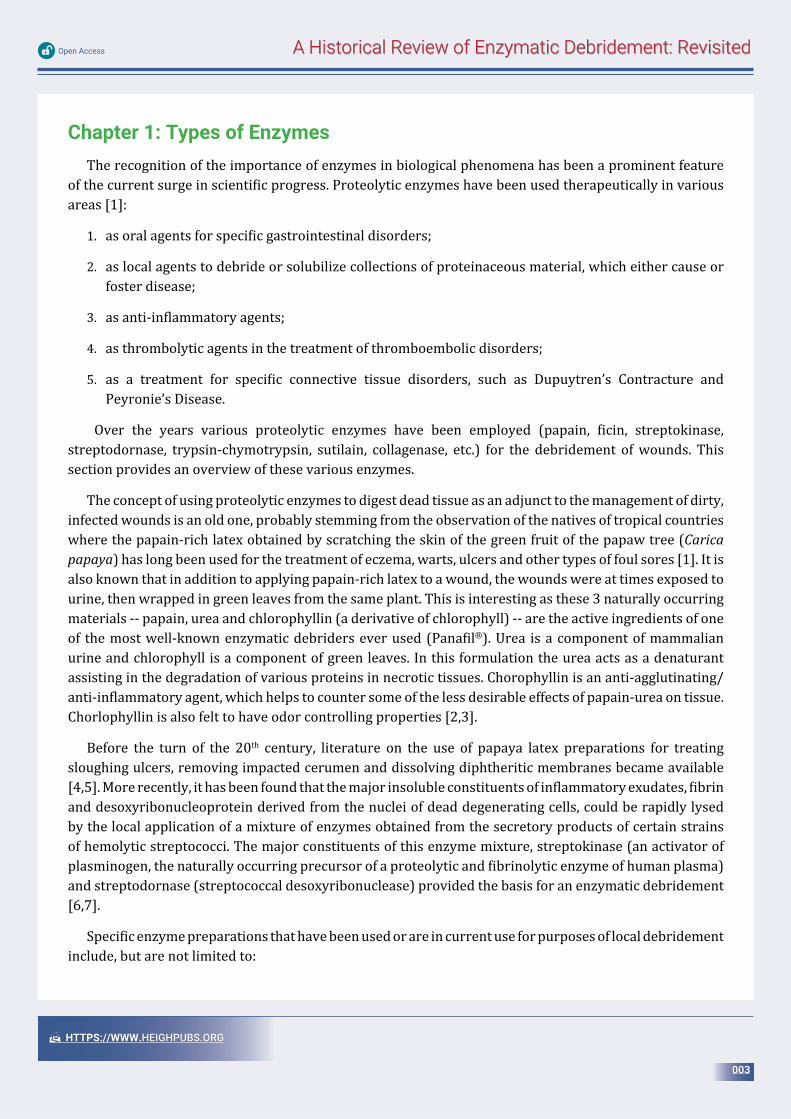
Chapter 1: Types of EnzymesThe recognition of the importance of enzymes in biological phenomena has been a prominent feature
of the current surge in scienti ic progress. Proteolytic enzymes have been used therapeutically in various areas [1]:
1. as oral agents for speci ic gastrointestinal disorders;
2. as local agents to debride or solubilize collections of proteinaceous material, which either cause or foster disease;
3. as anti-in lammatory agents;
4. as thrombolytic agents in the treatment of thromboembolic disorders;
5. as a treatment for speci ic connective tissue disorders, such as Dupuytren’s Contracture and Peyronie’s Disease.
Over the years various proteolytic enzymes have been employed (papain, icin, streptokinase, streptodornase, trypsin-chymotrypsin, sutilain, collagenase, etc.) for the debridement of wounds. This section provides an overview of these various enzymes.
The concept of using proteolytic enzymes to digest dead tissue as an adjunct to the management of dirty, infected wounds is an old one, probably stemming from the observation of the natives of tropical countries where the papain-rich latex obtained by scratching the skin of the green fruit of the papaw tree (Carica papaya) has long been used for the treatment of eczema, warts, ulcers and other types of foul sores [1]. It is also known that in addition to applying papain-rich latex to a wound, the wounds were at times exposed to urine, then wrapped in green leaves from the same plant. This is interesting as these 3 naturally occurring materials -- papain, urea and chlorophyllin (a derivative of chlorophyll) -- are the active ingredients of one of the most well-known enzymatic debriders ever used (Pana il®). Urea is a component of mammalian urine and chlorophyll is a component of green leaves. In this formulation the urea acts as a denaturant assisting in the degradation of various proteins in necrotic tissues. Chorophyllin is an anti-agglutinating/anti-in lammatory agent, which helps to counter some of the less desirable effects of papain-urea on tissue. Chorlophyllin is also felt to have odor controlling properties [2,3].
Before the turn of the 20th century, literature on the use of papaya latex preparations for treating sloughing ulcers, removing impacted cerumen and dissolving diphtheritic membranes became available [4,5]. More recently, it has been found that the major insoluble constituents of in lammatory exudates, ibrin and desoxyribonucleoprotein derived from the nuclei of dead degenerating cells, could be rapidly lysed by the local application of a mixture of enzymes obtained from the secretory products of certain strains of hemolytic streptococci. The major constituents of this enzyme mixture, streptokinase (an activator of plasminogen, the naturally occurring precursor of a proteolytic and ibrinolytic enzyme of human plasma) and streptodornase (streptococcal desoxyribonuclease) provided the basis for an enzymatic debridement [6,7].
Speci ic enzyme preparations that have been used or are in current use for purposes of local debridement include, but are not limited to:
A Historical Review of Enzymatic Debridement: Revisited
Published: June 25, 2019 004
Microbe derived enzymes
Sutilain: a water-soluble mixture of serine proteases derived from the bacteria Bacillis subtilis that is relatively nonspeci ic in its action and is capable of breaking down a variety of necrotic tissue types within an optimal pH range of 6.0-7.5 [8].
Clostridial collagenase: a water-soluble enzyme that speci ically attacks and breaks down undenatured (natural) collagen. In actuality, collagenase is known to degrade denatured collagen as well. Collagenase is commercially derived from bacterial (Clostridium histolyticum) sources. Collagenase is active over a pH range of 6.0-8.0. Bacterial collagenase, although a zinc metalloproteinase that uses calcium bears little structural relationship to mammalian collagenase. Bacterial collagenase rapidly attacks human collagen at many points, degrading it into small peptides. The commercially available collagenase is made up of proteolytic enzymes that break collagen into small peptides (oligopeptides) of differing molecular weights, most of which are tripeptides [9,10]. However, more recent work has shown that the oligopeptides may be larger [11]. Two genes, colG and colH transcribe for two C. collagenases. These collagenases uniquely cleave the interstitial collagens and exhibit both endopeptidase and tripeptidylcarboxypeptidase activities. The combined activity of endo- and tripeptidyl-C-peptidase makes these enzymes ideally suited for rapid collagen degradation. Clostripain is a cysteine-activated protease also found in culture iltrates of Clostridium histolyticum. However, the level of this enzyme is low and the effects on collagen may not be as pronounced as for C. collagenases. In contrast, the mammalian collagenase-1 (MMP-1) acts differently by cleaving interstitial collagen at a single locus within the triple helical structure, giving rise to 2 large fragments, TCA and TCB. These portions of the helix are then attacked by other less speci ic proteases, released by connective tissue cells, to be further degraded into small peptides [12].
Streptokinase-streptodornase mixtures: This preparation is only partially puri ied and contains a number of other streptococcal enzymes, such as a ribonuclease, hyaluronase, nucleotidase and nucleosidase, all of which may contribute to the effects observed. The enzyme mixture is essentially free of streptolysin and streptococcol proteinase. It does not contain any proteolytic enzymes in the conventional sense. The mixture contains enzymes, which act upon non-protein substrates; much of its virtue lies in its content of streptodornase, which rapidly reduces the viscosity of purulent exudates. Plasmin, the proteolytic enzyme formed from the latter precursor, is active at neutral pH and, though distinct from trypsin, resembles it in many respects (pH optima, types of links split, etc.). The major attribute of streptokinase lies in its special ibrin-dissolving properties. In contrast to the rapid inhibition of proteolytic enzymes by naturally-occurring humoral antiproteolytic substances, streptokinase is inactivated at a relatively slow rate (except in the presence of an excess of a speci ic antibody, antistreptokinase).
Streptokinase Streptodornase preparations are the agents of choice for liquefying clotted blood, loculated effusions and purulent exudate in closed body cavities. A signi icant incidence of pyrogenic [pyogenic] and in lammatory reactions to the locally administered enzyme mixture has limited its usefulness since the therapeutic procedure may be complicated by the patient’s discomfort and the need for frequent and repeated drainage [1].
Streptodornase: (streptococcal desoxyribonuclease) acts directly upon desoxyribonucleic acid (DNA), rapidly depolymerizing this highly complex substance into smaller units [1,13]. The activity of streptodornase is enhanced by the presence of Mg2+ or other divalent metallic ions and inhibited by the presence of substances, such as citrate, which form complexes with the metallic cofactor (i.e., chelating agents).
A Historical Review of Enzymatic Debridement: Revisited
Published: June 25, 2019 005
Fungal: Fungal proteases have also been employed as topically applied enzymatic debriders.
Animal-derived enzymes
Fibrinolysin: commercially obtained from bovine plasma, then activated by chloroform, it speci ically attacks and breaks down the ibrin component of blood clots and ibrinous exudates.
Desoxyribonuclease: obtained from bovine pancreatic tissue, acts speci ically on the nucleoprotein components of purulent exudates.
Trypsin: Crystalline trypsin preparations of bovine pancreatic origin have been used in the past. Trypsin is a serine protease and can directly hydrolyze a large number of naturally-occurring proteins. It is thought not to affect living cells or require any cofactors, and its action on denatured proteins is usually more extensive than on native proteins. Trypsin has advantages over streptokinase for surface wound debridement since it does not require additional factors for its action, acts upon a greater number of proteins than plasmin, and degrades them more extensively [1].
Chemotrypsin: This preparation is of bovine pancreatic origin and is the other major serine protease of the pancreas. Pancreatic enzymes are usually standardized in terms of their proteolytic activity. Though chymotrypsin acts upon different bonds in proteins than does trypsin or plasmin, its spectrum of activity on whole proteins is somewhat similar to that of trypsin [1].
Hyaluronidase: This is another common animal-derived enzyme used for topical enzymatic debridement.
Plant-derived enzymes
Bromelain: A mixture of water-soluble, cysteine proteases derived from the stem or fruit of the pineapple plant. This mixture of proteolytic enzymes is reported to be effective in breaking down a variety of different necrotic tissue substrates over a fairly wide pH range (5.5-8.5). It should be noted that cases of anaphylactic shock have been reported with enzymes derived from the pineapple plant, as well as with other plant-derived enzymes.
Papain: A latex protein obtained from the skin and green fruit of the papaw tree (Carica papaya). Papain is a cysteine protease and acts upon a wide variety of proteins; its activity can be considerably enhanced by the addition of cysteine or other reducing agents or by protein denaturants, such as urea. Indeed, without the presence of urea, papain displays lower proteolytic activity. The enzyme’s activity is optimal over a pH range of 3-9. It has been stated that at low pH, papain is capable of digesting collagen. Though papain preparations have been used occasionally in acetic acid solutions to digest collagenous tissue, the success of this method has not been established [1]. Other literature sources have described papain as having no effect on collagen. In 1958, J. Miller et al. [14], showed that papain-urea lacks the ability to degrade native collagen & states that only clostridial collagenase was able to adequately digest collagen.
Some feel that puri ied papain preparations eventually may prove to be the most practical for surface debridement. Others feel this is unlikely, given the mode of action of papain, its aggressive attack on viable tissue, and the associated stinging and burning reported in some literature sources. Miller [15], and Morrison et al. [16] all describe prolonged and intensi ied in lammatory responses as a result of treatment with papain-urea systems. Langer et al. 2013 [17], found in a prospective descriptive study on burns (mean TBS = 33.17%) that the combination of papain and urea caused so much pain (and fever) that only 2 of the 34 patients involved were able to complete the study.
Why were papain-urea based enzymatic deriders removed from the market?
A Historical Review of Enzymatic Debridement: Revisited
Published: June 25, 2019 006
As per the Federal Register, in 2008 the U.S. Food and Drug Administration (FDA) [18] ordered companies to stop marketing unapproved drug products that contain papain in a topical dosage form. Under this ruling, irms marketing any unapproved topical papain products had to stop manufacturing these products by November 24, 2008. Companies or others engaged in shipping these products had to stop shipping them by January 21, 2009. After these dates, all topical products containing papain must have FDA approval to be manufactured or shipped interstate. The FDA went on to state that topical drug products containing papain have historically been marketed without approval; there are no approved topical drug products containing papain. FDA took this action because adverse events with use of topical papain drug products reported to the agency raised serious safety concerns regarding these products. The FDA found that these drugs can produce harmful or near fatal effects including hypersensitivity resulting in anaphylactic reactions. Such cases have required emergency rooms visits, some requiring treatment with epinephrine. Hypersensitivity manifestations have also resulted in cardiovascular symptoms such as hypotension (low blood pressure) and tachycardia (rapid heart rate). Additionally, reports in the medical literature suggest that patients who are allergic to latex may also be allergic to papaya, the source of papain. Furthermore, the effectiveness of these products is not supported by scienti ically sound studies in the medical literature.
The FDA pointed out that papain is in fact a latex protein and sites cases of cross reactivity between latex and papaya have been documented in medical literature, and one of the cases reported to FDA involved anaphylactic shock in a patient with a history of allergy to latex. In addition, papain-containing drug products in topical form historically have been marketed without approval, and because no irm obtained an application for them prior to passage of the Drug Amendments of 1962, they were not included in the Drug Ef icacy Study Implementation (DESI) review. Adverse events associated with the use of topical papain products reported to FDA raise serious safety concerns regarding these products. Through January 2008, FDA had received 37 reports of adverse events associated with topical papain products. In addition to several complaints that the products were ineffective, the reports include cases of potentially life threatening hypersensitivity reactions. Reactions described include serious cases of anaphylaxis and anaphylactic shock that started within 15 minutes of topical papain use and resulted in hospitalizations, including admissions to the intensive care unit. Finally, the FDA was particularly concerned about adverse events associated with the use of papaincontaining topical drug products in light of the dearth of published, well-controlled studies demonstrating the effectiveness of those products. Given the absence of the kinds of scienti ic studies routinely conducted by sponsors and submitted for agency review as part of the FDA approval process, it was impossible for the agency to assess either the amount of risk associated with these products or the extent to which their bene its might justify their risks.
Products affected (by name) were Accuzyme®, Allan il®, Allanzyme®, Ethezyme®, Gladase®, Kovia®, Pana il®, Pap Urea®, and Ziox®. Other formulations were marketed under the names of the active ingredients, for instance papain-urea ointment. At the time of the FDA’s determination there are approximately 35 unapproved topical products containing papain on the market. This ruling in effect ended the use of papain-urea based enzymatic debriding agents in the US.
Actinidin: A member of the papain-like family of cysteine proteases, is abundant in kiwifruit. Chalabi et al. 2014 [19], investigated the proteolytic activity of actinidin compared to papain on several different ibrous and globular proteins under neutral, acidic and basic conditions. The indings showed that actinidin
has no or limited proteolytic effect on globular proteins such as immunoglobulins including sheep IgG, rabbit IgG, chicken IgY, and ish IgM, bovine serum albumin (BSA), lipid transfer protein (LTP), and whey proteins (α-lactalbumin and β-lactoglobulin) compared to papain. In contrast to globular proteins, actinidin could hydrolyze collagen and ibrinogen perfectly at neutral and mild basic pHs. Moreover, this enzyme could digest pure α-casein and major subunits of micellar casein especially at acidic pHs. Taken together,
A Historical Review of Enzymatic Debridement: Revisited
Published: June 25, 2019 007
the data (in this particular in vitro study) indicated that actinidin has narrow substrate speci icity with the highest enzymatic activity for the collagen and ibrinogen substrates. Hafezi et al. 2010 [20], found that debridement and scar contraction occurred faster in the kiwi-treated group than in the untreated group in acute burn wounds. Following rapid enzymatic debridement, healing appeared to progress normally, with no evidence of damage to adjacent healthy tissue. However, information on the clinical application as a topical enzymatic debrider is limited. In a randomized controlled clinical study on 17 neuropathic diabetic foot ulcers Mohajeri et al. 2014 [21], found that the mean reduction in surface area of foot ulcer in the experimental group was signi icantly higher than the control group (168.11 ± 22.31 vs. 88.80 ± 12.04 mm2 respectively, P < 0.0001). The amount of collagen and granulation tissues was signi icantly higher in the experimental groups than the control group (P value < 0.0001). Signi icantly higher levels of angiogenesis and vascularization were found in the kiwifruit treated patients (P value < 0.0001). No signi icant antibacterial effect was observed for kiwifruit in this study. However, in this particular study, all patients were surgically debrided prior to initiation of the study period.
Ficin: Ficin is another plant-derived cysteine protease found in igs.
Additional sources for enzymes such as avian, larva and crustaceans have been investigated, as well.
Effective collagen breakdown appears to be essential to optimum eschar removal. Collagen is a major component of chronic wound eschar, as collagen makes up 70%-80% of the dry weight of skin and is a major component of the extracellular matrix.
The history of enzymatic debriders has been a turbulent one, with only one enzymatic system currently used widely in clinic, clostridial collagenase. One reason for this turbulent history may be related to an enzyme’s ability to degrade collagen. Howes et al [22], and Rao et al. [23], have demonstrated that necrotic tissue is anchored to the wound surface by strands of undenatured collagen. Until these ibers are severed, débridement cannot take place, granulation is slowed, and thus no supportive base is available for proper epithelialization. Consequently, the wound fails to heal. It should be noted that though limited, studies suggest that actinidin (found in kiwi fruit) may have the ability to degrade collagen, which may warrant further study. Another aspect may be the fact that most enzymes used historically have not been highly selective in their catalytic activity. Non-selective being the inability to distinguish between healthy and necrotic tissue. The one exception would be clostridial collagenase, which is felt to be more selective than the enzymes mentioned, previously.
References1. Sherry S, Anthony P. Fleteher. Proteolytic Enzymes: a therapeutic evaluation. Clinical pharmacology and therapeutics.
1960; 1: 202-226. Ref.: https://tinyurl.com/y2s3po7w
2. Sack PW, Barnard RD. Studies on the hemagglutinating and infl ammatory properties of exudate from nonhealing wounds and their inhibition by chlorophyll derivatives. N Y State J Med. 1955; 55: 2952–2956.Ref.: https://tinyurl.com/yyowm8mc
3. Brett DW. Chlorophyllin—A Healer? A Hypothesis for its Activity. WOUNDS. 2005; 17: 190–195.Ref.: https://tinyurl.com/yy6qa9bq
4. Modder EE. On the medicinal uses of Carica papaya (Lin.), Ceylon MJ, February, 1887-1888, 115.
5. Jacobi A. Note on Papayotin. Therap. Gaz. 1886; 2:145-147. Ref.: https://tinyurl.com/y49nafaz
6. Johnson AJ. Cytological studies in association with local injections of streptokinase-streptodornase into patients. J Clin Invest. 1950; 29: 1376-1386. Ref.: https://tinyurl.com/y4yuxkao
7. Tillett WS, Sherry S. Effect in patients of streptococcal desoxyribonuclease in Fibrinous, Purulent & Sanguinous Pleural Exudations. J Clin Invest. 1949; 28: 173-190. Ref.: https://tinyurl.com/y4rq4o4x
A Historical Review of Enzymatic Debridement: Revisited
Published: June 25, 2019 008
8. Coopwood TB. Evaluation of a Topical Enzymatic Debridement Agent-Sutilains Ointment: A Preliminary Report. South Med J. 1976; 69: 834-836. Ref.: https://tinyurl.com/yxoyyluc
9. Cortivo R. Biological Activity of Human Collagen Breakdown Products on Fibroblasts. WOUNDS, A Compendium of Clinical Research and Practice. 1995; 7: 38A-44A.
10. Postlethwaite AE, Seyer JM, Kang AH. Chemotactic attraction of human fi broblasts to type I, II and III collagens and collagenderived peptides. Proc Natl Acad Sci. 1978; 75: 871-875. Ref.: https://tinyurl.com/yy4xeg4z
11. Sheets AR, Demidova-Rice TN, Shi L, Ronfard V, Grover KV, et al. Identifi cation and Characterization of Novel Matrix-Derived Bioactive Peptides: A Role for Collagenase from Santyl Ointment in Post- Debridement Wound Healing? PLoS One. 2016; 11: e0159598. Ref.: https://tinyurl.com/yycst5jr
12. Jeffrey J. Metalloproteinases and Tissue Turnover. WOUNDS, A Compendium of Clinical Research and Practice. Sup A. 1995; 7: 13A-22A.
13. Sherry S, Goeller JP. The extent of the enzymatic degradation of desoxyribonucleic acid (DNA) in purulent exudates by streptodornase. J Clin Invest. 1950; 29: 1588-1594. Ref.: https://tinyurl.com/yyzhep77
14. Miller JM. The interaction of Papain, Urea and Water-Soluble Chlorophyllin in a Proteolytic Ointment for Infected Wounds. Surgery. 1958; 43: 939-948. Ref.: https://tinyurl.com/y4lhwln3
15. Miller EW. Decubitus Ulcers Treated with Papain-Urea Chlorophyllin Ointment, NY State J Med. 1956; 56: 1446-1448. Ref.: https://tinyurl.com/yymutzun
16. Morrison JE, John L. Casali. Continuous Proteolytic Therapy for Decubitus Ulcers. Am J Surg. 1957; 93: 446-448. Ref.: https://tinyurl.com/yyppvd8z
17. Langer V, PS Bhandari, S Rajagopalan, MK Mukherjee. Enzymatic debridement of large burn wounds with papain-urea: Is it safe? Med J Armed Forces India. 2013; 69: 144-150. Ref.: https://tinyurl.com/y58qand4
18. Department of health and human services. Food and Drug Administration [Docket No. FDA–2008–N– 0481] Topical Drug Products Containing Papain; Enforcement Action Dates Federal Register. 2008; 73: 54831-54834.
19. Chalabi M, Khademi F, Yarani R, Mostafaie A. Proteolytic Activities of Kiwifruit Actinidin (Actinidia deliciosa cv. Hayward) on Different Fibrous and Globular Proteins: A Comparative Study of Actinidin with Papain. Appl Biochem Biotechnol. 2014; 172: 4025-4037. Ref.: https://tinyurl.com/yxvdhjub
20. Hafezi F, Rad HE, Naghibzadeh B, Nouhi A, Naghibzadeh G. Actinidia deliciosa (kiwifruit), a new drug for enzymatic debridement of acute burn wounds. Burns. 2010; 36: 352-355. Ref.: https://tinyurl.com/yx9nb9bq
21. Mohajeri G, Safaee M, Sanei MH. Effects of a topical Kiwifruit on healing of neuropathic diabetic foot ulcer. J Res Med Sci. 2014; 19: 520-524. Ref.: https://tinyurl.com/y6aovumu
22. Howes EL, Mandl I, Zaffuto S, Ackermann W. The Use of Clostridium histolyticum Enzymes in the treatment of Experimental 3rd Degree Burns. Surg Gynecol Obstet. 1959; 109: 177-188. Ref.: https://tinyurl.com/y4hfn7bc
23. Rao DB, Sane PG, Georgiev EL. Collagenase in the Treatment of Dermal and Decubitus Ulcers. J Am Geriatr Soc. 1975; 23: 22-30. Ref.: https://tinyurl.com/y2xz4plj
24. Sherry S, Johnson A, Tillett WS. The action of Streptococcal desoxyribonuclease (streptodornase), in vitro and on Purulent Pleural Exudations of Patients. J Clin Invest. 1949; 28: 1094-1104. Ref.: https://tinyurl.com/y3xkd3oq
A Historical Review of Enzymatic Debridement: RevisitedOpen Access
HTTPS://WWW.HEIGHPUBS.ORG
009
Chapter 2: Technical Review of CollagenProteins
Proteins are natural polymers, which make up about 15% of our bodies (dry weight). The building blocks of all proteins are α-amino acids. The alpha (α) is derived from the fact that the amino group (–NH2) is always attached to the α-carbon, which is bonded to the carboxyl group (–CO2H). Amino acids are joined together into proteins via condensation reactions in which the amine group of one amino acid reacts with the carbonyl group of another amino acid. In this reaction, a peptide bond is formed and a molecule of water is liberated (condensation). As the reaction proceeds repetitively, a polypeptide is produced and, eventually, a protein.
The structure of a given protein can be divided into 3 and sometimes 4 categories: primary, secondary, tertiary, and quaternary.
• Primary structure is simply the sequence and identity of amino acids making up the polypeptide.
• Secondary structure refers to the arrangement of the chain of the long molecule, which is determined to a great extent by hydrogen bonding (H-bonding) between lone electron pairs on the carbonyl oxygen of an amino acid and a hydrogen atom attached to nitrogen on another amino acid. Well-known examples are α-helices and β-strands.
• Tertiary structure refers to the overall 3-dimensional shape of the protein, which can be narrow and long or globular. Tertiary structure results from several types of interactions: charge based, hydrophobic based, and Van der Waals forces. A well-known example of a covalent bond occurs when 2 cysteines (amino acids) combine to form a disul ide linkage (S-S), resulting in a cystine residue.
• Quaternary structure refers to the interaction of 2 or more separate protein chains, resulting in a larger conglomeration with a speci ic function (hemoglobin is an example).
Collagen
Collagen plays an important structural role in many biological tissues such as, skin, tendon, bone, teeth, cartilage, and the cardiovascular system [1]. Two of the main classes of extracellular macromolecules that make up the extracellular matrix are the collagens and the heteropolysaccharides known as glycosaminoglycans (GAGs), which are usually covalently linked to protein to form proteoglycans [2]. Collagen is the major protein of the extracellular matrix and is the most abundant protein found in mammals, comprising 25% of the total protein and 70% to 80% of skin (dry weight). Collagen acts as a structural scaffold within tissues. The central feature of all collagen molecules is their stiff, triplestranded helical structure [3].
Three collagen polypeptide chains, called α-chains, are wound around each other in a regular triple-stranded helix to generate a ropelike collagen molecule approximately 300 nm (3,000 Å) long and 1.5 nm (15Å) in diameter. The length of the helical regions and individual αchains varies among collagen types. The major types of collagen molecules are referred to as types I, II, III, IV, and V. Types I, II, and III are the main types found in connective tissue and constitute 90% of all collagen in the body. After being secreted into the extracellular spaces, types I, II, and III assemble into insoluble micro- ibrils consisting of 5 triple helical molecules [4]. The micro- ibrils then form collagen ibrils, which are long (up to many micrometers), thin (10 to 300 nm in diameter), cablelike structures [2].
A Historical Review of Enzymatic Debridement: Revisited
Published: June 25, 2019 0010
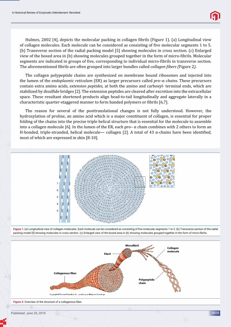
Hulmes, 2002 [4], depicts the molecular packing in collagen ibrils (Figure 1). (a) Longitudinal view of collagen molecules. Each molecule can be considered as consisting of ive molecular segments 1 to 5. (b) Transverse section of the radial packing model [5] showing molecules in cross section. (c) Enlarged view of the boxed area in (b) showing molecules grouped together in the form of micro- ibrils. Molecular segments are indicated in groups of ive, corresponding to individual micro- ibrils in transverse section. The aforementioned ibrils are often grouped into larger bundles called collagen ibers (Figure 2).
The collagen polypeptide chains are synthesized on membrane bound ribosomes and injected into the lumen of the endoplasmic reticulum (ER) as larger precursors called pro–α chains. These precursors contain extra amino acids, extension peptides, at both the amino and carboxyl- terminal ends, which are stabilized by disul ide bridges [2]. The extension peptides are cleaved after excretion into the extracellular space. These resultant shortened products align head-to-tail longitudinally and aggregate laterally in a characteristic quarter-staggered manner to form banded polymers or ibrils [6,7].
The reason for several of the posttranslational changes is not fully understood. However, the hydroxylation of proline, an amino acid which is a major constituent of collagen, is essential for proper folding of the chains into the precise triple helical structure that is essential for the molecule to assemble into a collagen molecule [6]. In the lumen of the ER, each pro– α chain combines with 2 others to form an H-bonded, triple-stranded, helical molecule— collagen [2]. A total of 43 α-chains have been identi ied, most of which are expressed in skin [8-10].
Figure 1: (a) Longitudinal view of collagen molecules. Each molecule can be considered as consisting of fi ve molecular segments 1 to 5. (b) Transverse section of the radial packing model [5] showing molecules in cross section. (c) Enlarged view of the boxed area in (b) showing molecules grouped together in the form of micro-fi brils.
Collagenous fiber
Fibril
MicrofibrilCollagenmolecule
Polypeptidechain
Figure 2: Overview of the structure of a collagenous fi ber.
A Historical Review of Enzymatic Debridement: Revisited
Published: June 25, 2019 011
Collagen is unique among proteins in that every third amino acid of the peptide chain is glycine, the smallest amino acid. Each of the 3 polypeptide chains (α chains) contains about 1,000 amino acids, so the structure of each chain can be considered to be approximately 330 repeating units of glycine–X–Y (where X and Y represent neutral amino acids). Although proline accounts for about 10% of the total amino acid content of collagen, it is found only rarely in other animal proteins [11]. One source describes 10.5% of the collagen molecule being comprised by the glycine-proline-hydroxyproline triplet [12]. Another source mentions that 23% of the molecule is comprised of a combination of proline & hydroxyproline [13]. Yet, a more recent source describes proline ~28%; hydroxyproline ~38% of the collagen molecule [14]. At any rate, this triplet is unique to collagen molecules. At least twenty nine types of collagen (designated) are found in vertebrates [8-10,15].
• The best-known types (I, II, and III) each consist of 3 polypeptides, called α chains. Each chain has the general structure (Gly-X-Y)330, with the 3 chains wrapped around each other in a ropelike triple helix.
• Type I collagen consists of 2 identical αchains (α1), and a slightly different chain, called α2.
• Types II and III collagen each contain distinctive α chains, but the 3 chains in each molecule of type II or type III collagen are identical.
• Basement membranes contain collagen that has been named type IV and there are several kinds of type IV (made up 6 different types of α chains) in different basement membranes.
As early as 1963 Haurowitz [11] described the rod-like structure of the collagen molecule as being distinguished from most proteins, which tend to be rounded or globular (i.e., the globulins). A number of features of collagen biosynthesis also distinguish it from other proteins. One unusual characteristic of collagen biosynthesis is that there are a large number of posttranslational modi ications made to the molecule; that is, the protein is irst synthesized as a precursor polypeptide chain. The polypeptide chains must then be “processed” through a number of enzymatic steps, all of which occur after the information carried by messenger RNA (mRNA) has been translated and which are essential to producing collagen in its inal form. Some of these posttranslational changes occur in the collagen-producing cell; others occur extracellularly [7].
Most of the enzymes involved in these modi ications of polypeptide chains have been well characterized, and their roles are well de ined. The irst step in the formation of the collagen molecule is the reading of the template mRNA by polysomes, or polyribosomes, bound to membranes of the rough ER. The polysomes assemble amino acids into polypeptide chains, which are in fact about 50% longer than the α chains of collagen and are called pro-α chains. The pro-α chains are longer than α chains, because they contain additional amino acid sequences at both ends and form the precursor molecule known as pro-collagen. These additional amino acid sequences at the end of pro-collagen must be cleaved by speci ic enzymes to yield the collagen molecule. As polysomes assemble pro-α chains, the newly formed amino-terminal ends pass into the cisterna/lumen of the rough ER, where the irst posttranslational steps begin to occur.
As previously mentioned, one step is the hydroxylation of peptidyl-proline. Approximately 100 residues are converted to hydroxyproline in this step. Hydroxylation of 5 to 20 peptidyl-lysine residues into hydroxylysine also begins. Hydroxylation probably continues even after the carboxyl-terminal extension (which is the inal portion of the pro-α chain) has been released by the polysome. After the pro-α chains enter into the cisternae, interchain disul ide bonds form.
While the molecules are still in the ER, galactose and glucose residues are added to the hydroxylysine residues, and still other sugars are attached to the terminal extensions. This glycosylation may continue
A Historical Review of Enzymatic Debridement: Revisited
Published: June 25, 2019 012
during or after the next step, which is the passage of the molecules from the ER into the Golgi vacuoles. Once posttranslational modi ications are completed, the collagen regions of the pro-α chains fold into a triple helix (this domain of the molecule becomes a rigid rod). Finally, the pro-collagen molecule is secreted from the cell in transport vesicles/vacuoles.
As previously mentioned, although the reasons for several of the posttranslational modi ications remain unclear, it is known that the hydroxylation of proline is essential for correct folding of the chains into the precise triple helical structure that is essential for the molecule to assemble into a collagen iber. If the chains do not form such helixes within the cell, they are secreted only very slowly and come out as nonfunctional protein. Additional posttranslational modi ications occur after the pro-collagen is secreted through the cell’s plasma membrane into the extracellular space.
The irst such modi ication is removal, by 2 or more proteases, of the amino- and carboxyterminal extensions from pro-collagen in order to convert it to collagen. These extensions probably have played important roles in the assembly of the triple helix, especially in controlling the rate. They probably also have other functions, such as preventing premature formation of ibers or formation before the protein is secreted. Then, following the removal of the extensions, the collagen molecules form into ibers.
The process by which collagen forms ibers is a dramatic, spontaneous self-assembly process. The information for determining the structure of the iber is provided entirely by the amino acid sequences and the conformation of the collagen molecule. For the iber to achieve its normal strength; however, chemical cross-links must be introduced to link the molecules in the iber to each other. This occurs through deamination of the hydroxylysine and lysine residues to produce aldehydes; cross-links are formed by reaction of either 2 aldehydes or 1 aldehyde and 1 amino group on adjacent molecules. This type of cross-linking (isopeptide bond formed by transglutaminase) is unique to collagen and elastin [7].
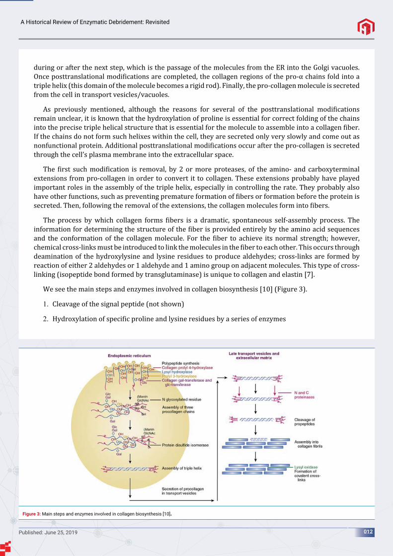
We see the main steps and enzymes involved in collagen biosynthesis [10] (Figure 3).
1. Cleavage of the signal peptide (not shown)
2. Hydroxylation of speci ic proline and lysine residues by a series of enzymes
Figure 3: Main steps and enzymes involved in collagen biosynthesis [10].
A Historical Review of Enzymatic Debridement: Revisited
Published: June 25, 2019 013
3. Glycosylation of certain asparagine residues in the C-peptide
4. Formation of intramolecular and intermolecular disul ide bonds via protein disul ide isomerase.
5. Assembly of the triple helix is formed in the C-terminal region after the C propeptides of three α-chains become registered with each other and ~ 100 proline residues in each α-chain have been hydroxylated to 4-hydroxyproline.
6. Triple helix formation proceeds toward the N-terminus in a zipper-like fashion.
7. Procollagen molecules are transported from the ER to Golgi, where they begin to associate laterally and exit the cell via secretory vesicles.
8. Cleavage of N and C propeptides and spontaneous self-assembly of the collagen molecules into ibrils, and formation of cross-links.
From here the collagen ibrils are organized into large collagen ibers (readily detected in the connective tissue of the dermis) which are well-organized polymers composed of speci ic and distinct collagen types, the most abundant being type I collagen. Collagen ibers are arranged at right angles or orthogonal laminae. The ibril diameter is remarkably constant in a given layer, and every layer is turned 900, so the ibers in any layer are arranged orthogonally to the layer immediately above and below it, thereby conferring 3600 resistance to physical stress. This orderly array of ibers is extraordinarily effective in maintaining the structural integrity of connective tissue [6].
The tensile strength of collagen is remarkable: a iber 1 mm in diameter can hold a load of 10 to 40 kg without breaking [11]. Collagen is physiologically stable. Disruption of ibrils only begins at temperatures above 500C. Onset of the transition occurs at (58 +/-10)0C and the main transition occurs at (65 +/- 10)0C. The main transition corresponds to the process of gelatinization of collagen in a hydrated environment and is caused by the breaking of internal cross-links [1]. Fibrillar collagen is chemically resistant as well. It is essentially insoluble under physiological conditions. It is resistant to the degradative effects of a wide range of naturally occurring enzymes such as trypsin and chymotrypsin. Collagen types I and III are principal connective tissue proteins of the dermal tissues and are abundant in tendon, bone, and blood vessels. Type II collagen is a cartilage-speci ic protein that is also present in vitreous humor and cornea of the eye. All forms of collagen, along with the other components of connective tissue, such as glycosaminoglycans, proteoglycans, elastin, micro ibrils, laminins, tenascins, ibronectin, and many others interact by speci ic chemical bonding and in a precise architectural orientation to yield the inal form of tissue [6].
Twenty-nine genetically distinct types of collagen comprising 43 unique α-chains have been identi ied in vertebrates. The vast majorities of these collagens exist in humans and based upon domain organization and other structural features can be categorized:
Fibril-forming collagens (types I, II, III, V, XI, XXIV, XXVII).
• Fibril-associated collagens with interrupted triple helices (IX, XII, XIV, XVI, XIX, XX, XXI, XXII, XXVI).
• Collagens capable of forming hexagonal network (e.g., VIII, X).
• Basement membrane collagen (IV).
• Collagens that assemble into beaded ilaments (e.g., type VI).
• Anchoring iber-forming collagens (e.g., VII).
• Plasma membrane-spanning collagens (XIII, XVII, XXIII, XXV).
• Collagens with unique domain organization (XV, XVIII).
A Historical Review of Enzymatic Debridement: Revisited
Published: June 25, 2019 014
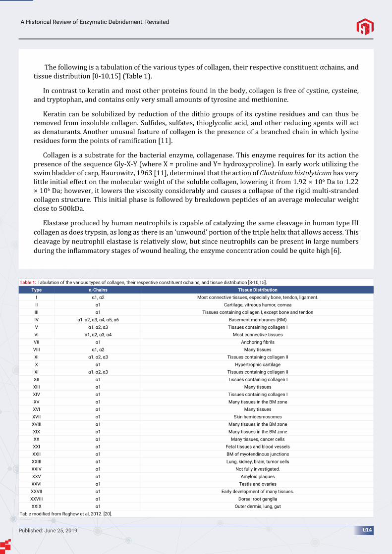
The following is a tabulation of the various types of collagen, their respective constituent αchains, and tissue distribution [8-10,15] (Table 1).
In contrast to keratin and most other proteins found in the body, collagen is free of cystine, cysteine, and tryptophan, and contains only very small amounts of tyrosine and methionine.
Keratin can be solubilized by reduction of the dithio groups of its cystine residues and can thus be removed from insoluble collagen. Sul ides, sulfates, thioglycolic acid, and other reducing agents will act as denaturants. Another unusual feature of collagen is the presence of a branched chain in which lysine residues form the points of rami ication [11].
Collagen is a substrate for the bacterial enzyme, collagenase. This enzyme requires for its action the presence of the sequence Gly-X-Y (where X = proline and Y= hydroxyproline). In early work utilizing the swim bladder of carp, Haurowitz, 1963 [11], determined that the action of Clostridum histolyticum has very little initial effect on the molecular weight of the soluble collagen, lowering it from 1.92 × 10⁶ Da to 1.22 × 10⁶ Da; however, it lowers the viscosity considerably and causes a collapse of the rigid multi-stranded collagen structure. This initial phase is followed by breakdown peptides of an average molecular weight close to 500kDa.
Elastase produced by human neutrophils is capable of catalyzing the same cleavage in human type III collagen as does trypsin, as long as there is an ‘unwound’ portion of the triple helix that allows access. This cleavage by neutrophil elastase is relatively slow, but since neutrophils can be present in large numbers during the in lammatory stages of wound healing, the enzyme concentration could be quite high [6].
Table 1: Tabulation of the various types of collagen, their respective constituent αchains, and tissue distribution [8-10,15]. Type α-Chains Tissue Distribution
I α1, α2 Most connective tissues, especially bone, tendon, ligament. II α1 Cartilage, vitreous humor, cornea III α1 Tissues containing collagen I, except bone and tendon IV α1, α2, α3, α4, α5, α6 Basement membranes (BM) V α1, α2, α3 Tissues containing collagen I VI α1, α2, α3, α4 Most connective tissues VII α1 Anchoring fi brils VIII α1, α2 Many tissues XI α1, α2, α3 Tissues containing collagen II X α1 Hypertrophic cartilage XI α1, α2, α3 Tissues containing collagen II XII α1 Tissues containing collagen I XIII α1 Many tissues XIV α1 Tissues containing collagen I XV α1 Many tissues in the BM zone XVI α1 Many tissues XVII α1 Skin hemidesmosomes XVIII α1 Many tissues in the BM zone XIX α1 Many tissues in the BM zone XX α1 Many tissues, cancer cells XXI α1 Fetal tissues and blood vessels XXII α1 BM of myotendinous junctions XXIII α1 Lung, kidney, brain, tumor cells XXIV α1 Not fully investigated. XXV α1 Amyloid plaques XXVI α1 Testis and ovaries XXVII α1 Early development of many tissues. XXVIII α1 Dorsal root ganglia XXIX α1 Outer dermis, lung, gut
Table modifi ed from Raghow et al, 2012. [20].
A Historical Review of Enzymatic Debridement: Revisited
Published: June 25, 2019 015
Once it was felt that collagen provided structural support, only. However, collagen and collagen derived fragments control many cellular functions, such as cell shape and differentiation, cell migration and the synthesis of a number of proteins necessary for wound closure [16-19].
References1. Bozec L, Odlyha M. Thermal denaturation studies of collagen by microthermal analysis and atomic force microscopy.
Biophys J. 2011; 101: 228-236. Ref.: https://tinyurl.com/yy67t3bx
2. Alberts B. In Molecular Biology of The Cell, 1. City, Publisher, NY and London, Garland Publishing, Inc. 1983, 692-701.
3. Krieg T. Collagen in the Healing Wound. Wounds. Sup A. 1995; 7: 5A-12A.
4. Hulmes DJ. Building collagen molecules, fi brils, and suprafi brillar structures. J Struct Biol. 2002; 137: 2-10. Ref.: https://tinyurl.com/y4m3cuku
5. Hulmes DJS, T J Wess, D J Prockop, P Fratzl. Radial packing, order, and disorder in collagen fi brils. Biophys J. 1995; 68: 1661-1670. Ref.: https://tinyurl.com/y3yootdr
6. Jeffrey J. Metalloproteinases and Tissue Turnover. Wounds, A Compendium of Clinical Research and Practice. Sup A. 1995; 7: 13A-22A.
7. Prockop D, Guzman NA. Collagen diseases and the biosynthesis of collagen. Hosp Prac. 1977; 12: 61-68. Ref.: https://tinyurl.com/y3efoh97
8. Misawa K, Kanazawa T, Imai A, Endo S, Mochizuki D, et al. Prognostic value of type XX and XXIV collagen mRNA expression in head and neck cancer patients. Mol Concol. 2014; 2: 285-291. Ref.: https://tinyurl.com/y3kztzdk
9. Ricard-Blum S. The collagen family. Cold Spring Perspect Biol. 2001; 3: a004978. Ref.: https://tinyurl.com/y5g95tcd
10. Myllyharju J, Kivirikko KI. Collagens, modifying enzymes and their mutations in humans, fl ies and worms. Trends Genet. 2004; 20: 33-43. Ref.: https://tinyurl.com/y4h9f5x7
11. Haurowitz F. Properties of Amino Acids: The Chemistry and Function of Protiens. 2nd Edition, 1963; 37-38, 193-196, 212-216.
12. Ramshaw JA, Shah NK, Brodsky B. Gly-X-Y Tripeptide Frequencies in Collagen: A Context for Host–Guest Triple-Helical Peptides. J Struct Biol. 1998; 122: 86–91. Ref.: https://tinyurl.com/y6sarb7w
13. Barbul A. Proline precursors to sustain Mammalian collagen synthesis. J Nutr. 2008; 138: 2021S-2024S. Ref.: https://tinyurl.com/y5cjurgc
14. Schönauer E, Kany AM, Haupenthal J, Hüsecken K, Hoppe IJ, et al. Discovery of a Potent Inhibitor Class with High Selectivity toward Clostridial Collagenases. J Am Chem Soc. 2017; 139: 12696-12703. Ref.: https://tinyurl.com/y56kp3ak
15. Soderhall C, Marenholz I, Kerscher T, Rüschendorf F, Esparza-Gordillo J, et al. Variants in a novel epidermal collagen gene (COL29A1) are associated with atopic dermatitis. PLoS Biol. 2007; 5: 1952-1961. Ref.: https://tinyurl.com/y47ln83n
16. Madri JA, Marx M. Matrix composition, organization, and soluble factors: modulators of microvascular cell differentiation in vitro. Kidney Int. 1992; 41: 560-565. Ref.: https://tinyurl.com/yx925rex
17. Montesano R, Orci L, Vassalli P. In vitro rapid organization of endothelial cells into capillary-like network is promoted by collagen matrices. J Cell Biol. 1983; 97: 1648-1651. Ref.: https://tinyurl.com/y27k6ylx
18. Albini A, Adelmann-Grill BC. Collagenolytic cleavage products of collagen type 1 as chemoattractants for human dermal fi broblasts. Eur J Cell Biol. 1985; 36: 104-107. Ref.: https://tinyurl.com/yygpn24z
19. Hynes RO. lntegrins: versatility, modulation, and signaling in cell adhesion. Cell. 1992; 69: 11-25. Ref.: https://tinyurl.com/yy6x9qkr
20. Raghow R. Connective Tissues of the Subendothelium. Vascular Medicine: A Companion to Braunwald’s Heart Disease. 2012. 2nd Edition. Ch. 4.
A Historical Review of Enzymatic Debridement: RevisitedOpen Access
HTTPS://WWW.HEIGHPUBS.ORG
0016
Chapter 3: Endogenous Collagenase The role of matrix metalloproteinases in wound repair
The serine proteinases comprise the largest family of extracellular enzymes and include plasmin, plasminogen activators, and leukocyte elastase, as well as the coagulation and digestive proteinases. Generally, these are potent enzymes with broad catalytic speci icity and are readily available when needed. Plasminogen, the inactive form of plasmin, is present in high concentrations in blood and tissue. Neutrophils store an abundance of leukocyte elastase. In contrast, the metalloproteinases in wounded tissues have more de ined substrate speci icity and are generally produced on demand.
The structural and functional diversity of matrix metalloproteinases (MMPs) rivals that of the superfamily of collagens (reviewed in the previous chapter). The MMPs belong to a large family of zinc-dependent endopeptidases, the irst of which was described over a half century ago. MMPs were discovered initially as the agents responsible for tail resorption during frog metamorphosis [1-3] and have since been identi ied as the main processors of extracellular matrix (ECM) components [4]. MMPs have also been implicated in more sophisticated processes than mere ECM turnover [5,6]. These include the activation or inactivation of other proteins through limited proteolysis of selected bonds, as well as the shedding of membrane-anchored forms into circulation. Substrates include other (pro-)proteases, protease inhibitors, clotting factors, antimicrobial peptides, chemotactic and adhesion molecules, and growth factors, hormones, cytokines, as well as, their receptors and binding proteins. In such shedding functions, MMPs overlap in substrate speci icity, and in spatial and temporal location [4,7,8].
Twenty three different MMPs (in human tissues) representing 24 distinct gene products have been characterized [9]. Based on their cellular localization, these enzymes can be broadly subdivided into secreted and membrane-bound MMPs. However, a more detailed analysis of their structural organization and substrate speci icities indicates that MMPs may be better classi ied as collagenases, gelatinases, stromelysins, matrilysins, and membrane-type MMPs [9].
The typical MMP consists of three subdomains: the pro-domain, the catalytic domain, and the hemopexin-like C-domain, connected to the catalytic domain via a short linker region. The catalytic domain of MMPs contains a Zn2+ ion-binding amino acid sequence motif and a substrate-speci ic site. The MMP is synthesized as a pre-proenzyme and is maintained in latent conformation by the pro-domain via interaction between a cysteine (located in prodomain) and a Zn2+ ion (located in the catalytic domain). Only when this interaction is disrupted, either by proteolysis of the pro-domain or by a chemical modi ication of the cysteine, the MMP becomes activated. A number of intracellular and extracellular proteinases, including other MMPs, are known to speci ically degrade the pro-domain to activate MMPs in vivo [9].
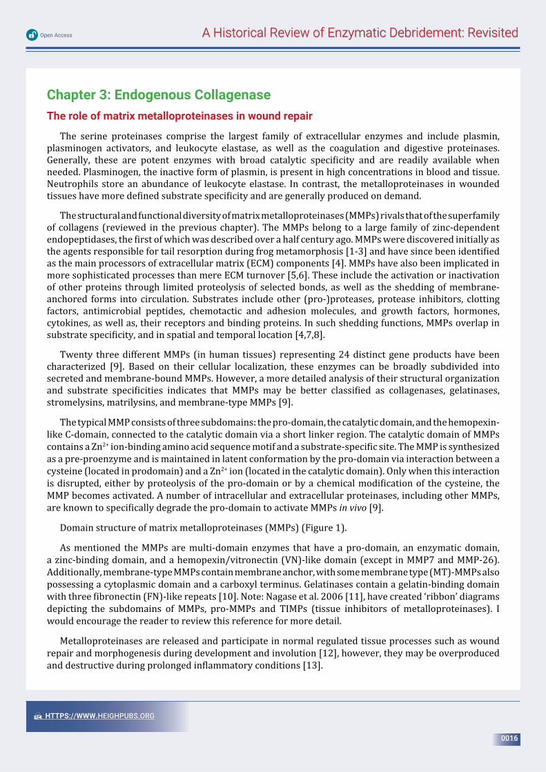
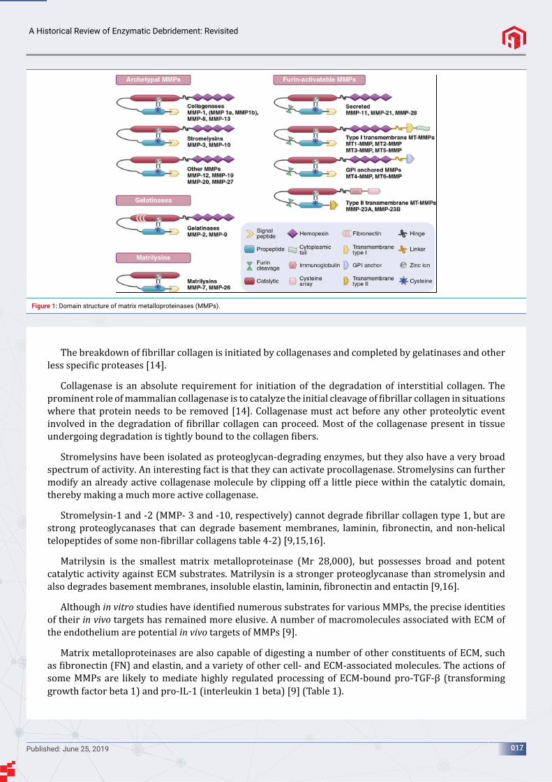
Domain structure of matrix metalloproteinases (MMPs) (Figure 1).
As mentioned the MMPs are multi-domain enzymes that have a pro-domain, an enzymatic domain, a zinc-binding domain, and a hemopexin/vitronectin (VN)-like domain (except in MMP7 and MMP-26). Additionally, membrane-type MMPs contain membrane anchor, with some membrane type (MT)-MMPs also possessing a cytoplasmic domain and a carboxyl terminus. Gelatinases contain a gelatin-binding domain with three ibronectin (FN)-like repeats [10]. Note: Nagase et al. 2006 [11], have created ‘ribbon’ diagrams depicting the subdomains of MMPs, pro-MMPs and TIMPs (tissue inhibitors of metalloproteinases). I would encourage the reader to review this reference for more detail.
Metalloproteinases are released and participate in normal regulated tissue processes such as wound repair and morphogenesis during development and involution [12], however, they may be overproduced and destructive during prolonged in lammatory conditions [13].
A Historical Review of Enzymatic Debridement: Revisited
Published: June 25, 2019 017
The breakdown of ibrillar collagen is initiated by collagenases and completed by gelatinases and other less speci ic proteases [14].
Collagenase is an absolute requirement for initiation of the degradation of interstitial collagen. The prominent role of mammalian collagenase is to catalyze the initial cleavage of ibrillar collagen in situations where that protein needs to be removed [14]. Collagenase must act before any other proteolytic event involved in the degradation of ibrillar collagen can proceed. Most of the collagenase present in tissue undergoing degradation is tightly bound to the collagen ibers.
Stromelysins have been isolated as proteoglycan-degrading enzymes, but they also have a very broad spectrum of activity. An interesting fact is that they can activate procollagenase. Stromelysins can further modify an already active collagenase molecule by clipping off a little piece within the catalytic domain, thereby making a much more active collagenase.
Stromelysin-1 and -2 (MMP- 3 and -10, respectively) cannot degrade ibrillar collagen type 1, but are strong proteoglycanases that can degrade basement membranes, laminin, ibronectin, and non-helical telopeptides of some non- ibrillar collagens table 4-2) [9,15,16].
Matrilysin is the smallest matrix metalloproteinase (Mr 28,000), but possesses broad and potent catalytic activity against ECM substrates. Matrilysin is a stronger proteoglycanase than stromelysin and also degrades basement membranes, insoluble elastin, laminin, ibronectin and entactin [9,16].
Although in vitro studies have identi ied numerous substrates for various MMPs, the precise identities of their in vivo targets has remained more elusive. A number of macromolecules associated with ECM of the endothelium are potential in vivo targets of MMPs [9].
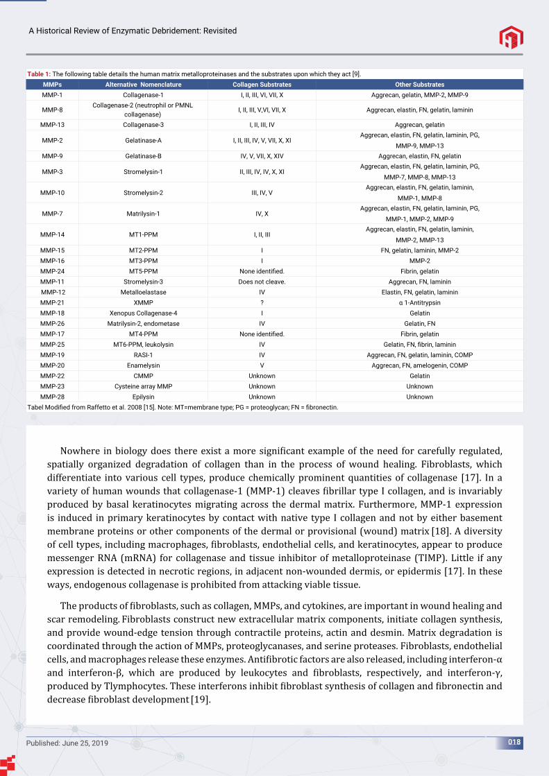
Matrix metalloproteinases are also capable of digesting a number of other constituents of ECM, such as ibronectin (FN) and elastin, and a variety of other cell- and ECM-associated molecules. The actions of some MMPs are likely to mediate highly regulated processing of ECM-bound pro-TGF-β (transforming growth factor beta 1) and pro-IL-1 (interleukin 1 beta) [9] (Table 1).
Figure 1: Domain structure of matrix metalloproteinases (MMPs).
A Historical Review of Enzymatic Debridement: Revisited
Published: June 25, 2019 018
Nowhere in biology does there exist a more signi icant example of the need for carefully regulated, spatially organized degradation of collagen than in the process of wound healing. Fibroblasts, which differentiate into various cell types, produce chemically prominent quantities of collagenase [17]. In a variety of human wounds that collagenase-1 (MMP-1) cleaves ibrillar type I collagen, and is invariably produced by basal keratinocytes migrating across the dermal matrix. Furthermore, MMP-1 expression is induced in primary keratinocytes by contact with native type I collagen and not by either basement membrane proteins or other components of the dermal or provisional (wound) matrix [18]. A diversity of cell types, including macrophages, ibroblasts, endothelial cells, and keratinocytes, appear to produce messenger RNA (mRNA) for collagenase and tissue inhibitor of metalloproteinase (TIMP). Little if any expression is detected in necrotic regions, in adjacent non-wounded dermis, or epidermis [17]. In these ways, endogenous collagenase is prohibited from attacking viable tissue.
The products of ibroblasts, such as collagen, MMPs, and cytokines, are important in wound healing and scar remodeling. Fibroblasts construct new extracellular matrix components, initiate collagen synthesis, and provide wound-edge tension through contractile proteins, actin and desmin. Matrix degradation is coordinated through the action of MMPs, proteoglycanases, and serine proteases. Fibroblasts, endothelial cells, and macrophages release these enzymes. Anti ibrotic factors are also released, including interferon-α and interferon-β, which are produced by leukocytes and ibroblasts, respectively, and interferon-γ, produced by Tlymphocytes. These interferons inhibit ibroblast synthesis of collagen and ibronectin and decrease ibroblast development [19].
Table 1: The following table details the human matrix metalloproteinases and the substrates upon which they act [9]. MMPs Alternative Nomenclature Collagen Substrates Other Substrates MMP-1 Collagenase-1 I, II, III, VI, VII, X Aggrecan, gelatin, MMP-2, MMP-9
MMP-8 Collagenase-2 (neutrophil or PMNL
collagenase) I, II, III, V,VI, VII, X Aggrecan, elastin, FN, gelatin, laminin
MMP-13 Collagenase-3 I, II, III, IV Aggrecan, gelatin
MMP-2 Gelatinase-A I, II, III, IV, V, VII, X, XI Aggrecan, elastin, FN, gelatin, laminin, PG,
MMP-9, MMP-13 MMP-9 Gelatinase-B IV, V, VII, X, XIV Aggrecan, elastin, FN, gelatin
MMP-3 Stromelysin-1 II, III, IV, IV, X, XI Aggrecan, elastin, FN, gelatin, laminin, PG,
MMP-7, MMP-8, MMP-13
MMP-10 Stromelysin-2 III, IV, V Aggrecan, elastin, FN, gelatin, laminin,
MMP-1, MMP-8
MMP-7 Matrilysin-1 IV, X Aggrecan, elastin, FN, gelatin, laminin, PG,
MMP-1, MMP-2, MMP-9
MMP-14 MT1-PPM I, II, III Aggrecan, elastin, FN, gelatin, laminin,
MMP-2, MMP-13 MMP-15 MT2-PPM I FN, gelatin, laminin, MMP-2 MMP-16 MT3-PPM I MMP-2 MMP-24 MT5-PPM None identifi ed. Fibrin, gelatin MMP-11 Stromelysin-3 Does not cleave. Aggrecan, FN, laminin MMP-12 Metalloelastase IV Elastin, FN, gelatin, laminin MMP-21 XMMP ? α 1-Antitrypsin MMP-18 Xenopus Collagenase-4 I Gelatin MMP-26 Matrilysin-2, endometase IV Gelatin, FN MMP-17 MT4-PPM None identifi ed. Fibrin, gelatin MMP-25 MT6-PPM, leukolysin IV Gelatin, FN, fi brin, laminin MMP-19 RASI-1 IV Aggrecan, FN, gelatin, laminin, COMP MMP-20 Enamelysin V Aggrecan, FN, amelogenin, COMP MMP-22 CMMP Unknown Gelatin MMP-23 Cysteine array MMP Unknown Unknown MMP-28 Epilysin Unknown Unknown
Tabel Modifi ed from Raffetto et al. 2008 [15]. Note: MT=membrane type; PG = proteoglycan; FN = fi bronectin.

A Historical Review of Enzymatic Debridement: Revisited
Published: June 25, 2019 019
Collagenase is not typically produced in dermal cells in acute human wounds or in healthy tissue [17]. Interstitial collagenase is produced by basal keratinocytes in wounded skin. It has often been assumed that the enzyme is produced primarily by ibroblasts, macrophages, and other cells at the leading edge of the granulation tissue [20].
Basal keratinocytes at the migrating front of re-epithelialization are the predominant sources of collagenase during active wound repair. Collagenase expression by migrating keratinocytes is an invariable feature of disrupted epidermis, both as a consequence of normal ulceration resulting from secondary intention and in ulceration resulting from a variety of disease processes [20]. This enzyme is also produced by migrating basal keratinocytes in full thickness burn wounds [17]. Although collagenase is always produced by epidermal cells at the wound edge, the amount varies considerably among wound types. In chronic ulcers, very high levels of expression are seen in the basal keratinocytes, with frequent production in the underlying dermis of these samples. Observations implicate keratinocytes as major participants in the degradation of collagen during wound healing and levels of collagenase produced in the epidermis and within the whole of the wound bed are much greater in non-healing wounds than in normal wounds [20].
Synthesis and structure
A variety of lines of human skin ibroblasts produce chemically signi icant quantities of collagenase. In human skin, collagenase is synthesized and secreted by these cells in culture as a zymogen, a pro-enzyme with a molecular mass of approximately 52,000 daltons (Da) [16,21]. The zymogen is incapable of catalytic activity or of binding to its eventual substrate, collagen. The process of activation of pro-collagenase to the active enzyme is all-important in the biology of collagen degradation. Other MMPs are also secreted as a proenzyme or zymogen with no catalytic activity. As previously mentioned, the pro-peptide contains a highly conserved cysteine residue that interacts with the Zn2+-binding region of the enzyme, thereby effectively blocking catalytic activity. The pro-peptide domain is linked to the catalytic domain, which is quite similar among the MMPs. All MMP pro-enzymes have a short signal peptide, as do most proteins secreted from cells, and they also contain a pro-peptide. Molecular speci icity of the MMPs resides in the hemopexin-like region at the C-terminal ends [22].
Active collagenase can be activated from the zymogen by a process of cleaving the signal peptide. Mammalian collagenase belongs to a family of extracellular MMPs that are capable of degrading connective tissue components. This family of enzymes is composed of collagenases with speci icity for the ibrillar collagens, gelatins, and types IV and V. Stromelysin has a wide speci icity, including ibronectin, laminin, type IV collagen and cartilage proteoglycan (Table 4-2) [9]. All collagenases require Ca2+ for activity. In the absence of Ca2+, collagenase appears to be less thermostable and more susceptible to proteolytic degradation. Zn2+ is required for proteolytic activity. Zn2+ is tightly bound within the protein and is not removed in dialysis. Zn2+ participates in the movement of electrons required for the hydrolysis of the peptide bond. The presence of Zn2+ in these proteases, coupled with the calcium requirement, provides the basis for the consistent inding that collagenases as a group are inhibited by chelating agents such as EDTA [21]. To this end, one collagen-based wound care dressing has been designed to deactivate excessive MMPs (found in chronic wounds) by the introduction of EDTA to the MMPs.
As previously mentioned, 23 MMPs are present in humans. They are numbered 1 to 3, 7 to 17, 19 to 21, and 23 to 28 for historical reasons (there are two identical forms for MMP-23, encoded by two genes). If MMPs are not subjected to spatial and temporal control, they become destructive, which can lead to pathologies such as arthritis, in lammation, and cancer. In 2010, Tallant described the catalytic domains of 13 MMPs and it is felt that there are similarities to the other 10 MMPs. Tallant details that the active site contains an extended zinc-binding motif, which contains three zinc-binding histidines and a glutamate that acts as a general base/acid during catalysis. In addition, a conserved methionine provides a hydrophobic
A Historical Review of Enzymatic Debridement: Revisited
Published: June 25, 2019 020
base for the zincbinding site. MMPs contain three α-helices and a ive-stranded β-sheet, as well as at least two calcium sites and a second zinc site with structural functions. Most MMPs are secreted as inactive zymogens with an N-terminal 80-residue pro-domain, which folds into a three-helix globular domain and inhibits the catalytic zinc through a cysteine imbedded in a conserved motif. Removal of the pro-domain enables access of a catalytic solvent molecule (water) and substrate molecules to the active-site cleft.
MMPs are mosaic proteins, each constituted by a modular combination of inserts and domains. These may include, from N- to C-terminus, a signal peptide for secretion; a 80-residue zymogenic pro-peptide; a 165-residue zinc- and calcium-dependent catalytic domain; a 15-to65-residue linker region; and a 200-residue hemopexin-like domain for collagen binding, pro-MMP activation, and dimerization [23].
TX-ray or NMR structures comprising at least the catalytic domains, isolated or in complexes with inhibitors are available. The catalytic domain structures are very similar, in the shape of a sphere with a diameter of 40 Å. A shallow active-site cleft lies on the front surface, which causes substrates to bind in an ‘approximately’ extended conformation relative to their standard orientation. This orientation entails that a substrate binds horizontally from left (N-terminal nonprimed side) to right (C-terminal primed side) of the catalytic metal ion [24,25]. The polypeptide chain creeps upwards along the molecular surface to enter the N-terminal sub-domain (NTS) and a ive-stranded twisted β-sheet that parallels and delimits the active-site cleft on its top. Two helices (αA, the “backing helix”, and αB, the “activesite helix”) are located in the concave side of the sheet. The residues at the interface between the sheet and the helices are mainly hydrophobic and give rise to an extended central hydrophobic core. On the convex side of the sheet, three elements protrude from the molecular surface: the loop connecting strands βII and βIII (LβIIβIII), LβIIIβIV, and LβIVβV [21].
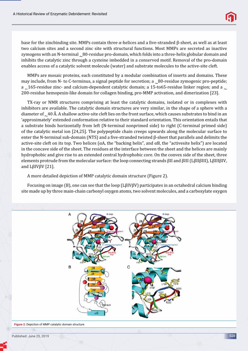
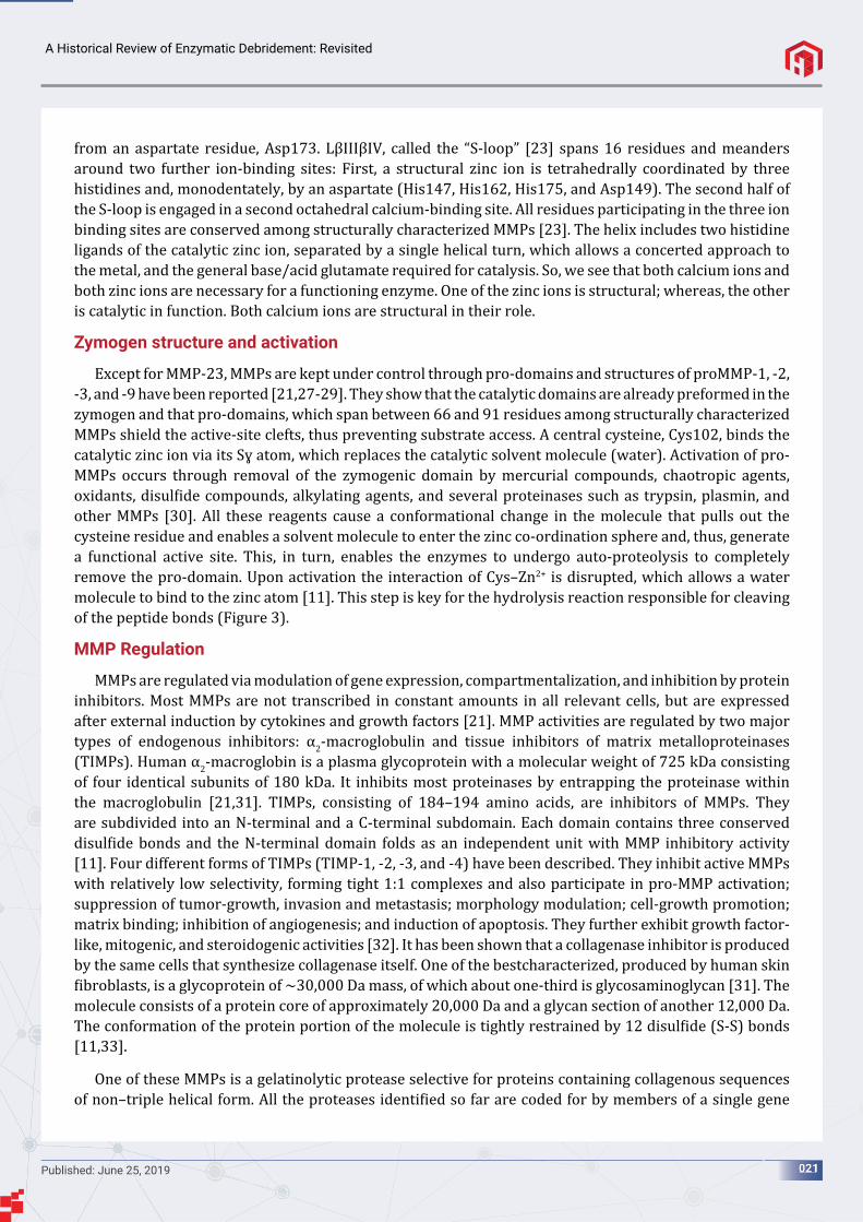
A more detailed depiction of MMP catalytic domain structure (Figure 2).
Focusing on image (B), one can see that the loop (LβIVβV) participates in an octahedral calcium binding site made up by three main-chain carbonyl oxygen atoms, two solvent molecules, and a carboxylate oxygen
Figure 2: Depiction of MMP catalytic domain structure
A Historical Review of Enzymatic Debridement: Revisited
Published: June 25, 2019 021
from an aspartate residue, Asp173. LβIIIβIV, called the “S-loop” [23] spans 16 residues and meanders around two further ion-binding sites: First, a structural zinc ion is tetrahedrally coordinated by three histidines and, monodentately, by an aspartate (His147, His162, His175, and Asp149). The second half of the S-loop is engaged in a second octahedral calcium-binding site. All residues participating in the three ion binding sites are conserved among structurally characterized MMPs [23]. The helix includes two histidine ligands of the catalytic zinc ion, separated by a single helical turn, which allows a concerted approach to the metal, and the general base/acid glutamate required for catalysis. So, we see that both calcium ions and both zinc ions are necessary for a functioning enzyme. One of the zinc ions is structural; whereas, the other is catalytic in function. Both calcium ions are structural in their role.
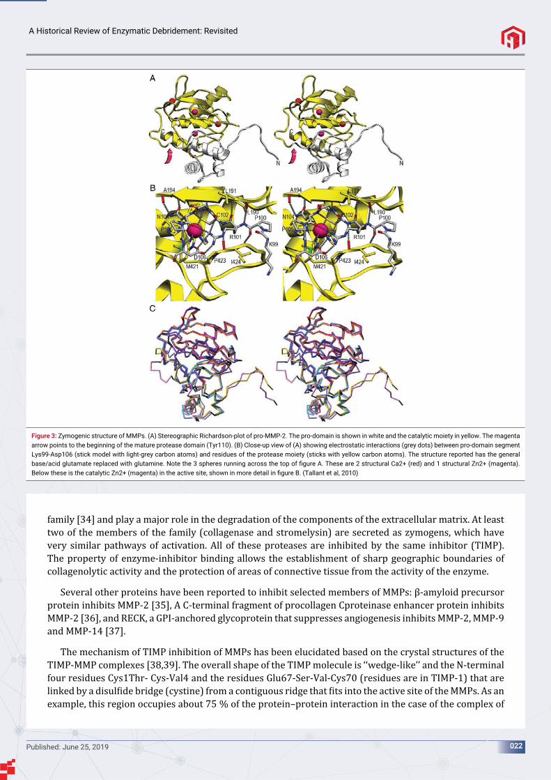
Zymogen structure and activation
Except for MMP-23, MMPs are kept under control through pro-domains and structures of proMMP-1, -2, -3, and -9 have been reported [21,27-29]. They show that the catalytic domains are already preformed in the zymogen and that pro-domains, which span between 66 and 91 residues among structurally characterized MMPs shield the active-site clefts, thus preventing substrate access. A central cysteine, Cys102, binds the catalytic zinc ion via its Sɣ atom, which replaces the catalytic solvent molecule (water). Activation of pro-MMPs occurs through removal of the zymogenic domain by mercurial compounds, chaotropic agents, oxidants, disul ide compounds, alkylating agents, and several proteinases such as trypsin, plasmin, and other MMPs [30]. All these reagents cause a conformational change in the molecule that pulls out the cysteine residue and enables a solvent molecule to enter the zinc co-ordination sphere and, thus, generate a functional active site. This, in turn, enables the enzymes to undergo auto-proteolysis to completely remove the pro-domain. Upon activation the interaction of Cys–Zn2+ is disrupted, which allows a water molecule to bind to the zinc atom [11]. This step is key for the hydrolysis reaction responsible for cleaving of the peptide bonds (Figure 3).
MMP Regulation
MMPs are regulated via modulation of gene expression, compartmentalization, and inhibition by protein inhibitors. Most MMPs are not transcribed in constant amounts in all relevant cells, but are expressed after external induction by cytokines and growth factors [21]. MMP activities are regulated by two major types of endogenous inhibitors: α2-macroglobulin and tissue inhibitors of matrix metalloproteinases (TIMPs). Human α2-macroglobin is a plasma glycoprotein with a molecular weight of 725 kDa consisting of four identical subunits of 180 kDa. It inhibits most proteinases by entrapping the proteinase within the macroglobulin [21,31]. TIMPs, consisting of 184–194 amino acids, are inhibitors of MMPs. They are subdivided into an N-terminal and a C-terminal subdomain. Each domain contains three conserved disul ide bonds and the N-terminal domain folds as an independent unit with MMP inhibitory activity [11]. Four different forms of TIMPs (TIMP-1, -2, -3, and -4) have been described. They inhibit active MMPs with relatively low selectivity, forming tight 1:1 complexes and also participate in pro-MMP activation; suppression of tumor-growth, invasion and metastasis; morphology modulation; cell-growth promotion; matrix binding; inhibition of angiogenesis; and induction of apoptosis. They further exhibit growth factor-like, mitogenic, and steroidogenic activities [32]. It has been shown that a collagenase inhibitor is produced by the same cells that synthesize collagenase itself. One of the bestcharacterized, produced by human skin ibroblasts, is a glycoprotein of ~30,000 Da mass, of which about one-third is glycosaminoglycan [31]. The
molecule consists of a protein core of approximately 20,000 Da and a glycan section of another 12,000 Da. The conformation of the protein portion of the molecule is tightly restrained by 12 disul ide (S-S) bonds [11,33].
One of these MMPs is a gelatinolytic protease selective for proteins containing collagenous sequences of non–triple helical form. All the proteases identi ied so far are coded for by members of a single gene
A Historical Review of Enzymatic Debridement: Revisited
Published: June 25, 2019 022
family [34] and play a major role in the degradation of the components of the extracellular matrix. At least two of the members of the family (collagenase and stromelysin) are secreted as zymogens, which have very similar pathways of activation. All of these proteases are inhibited by the same inhibitor (TIMP). The property of enzyme-inhibitor binding allows the establishment of sharp geographic boundaries of collagenolytic activity and the protection of areas of connective tissue from the activity of the enzyme.
Several other proteins have been reported to inhibit selected members of MMPs: β-amyloid precursor protein inhibits MMP-2 [35], A C-terminal fragment of procollagen Cproteinase enhancer protein inhibits MMP-2 [36], and RECK, a GPI-anchored glycoprotein that suppresses angiogenesis inhibits MMP-2, MMP-9 and MMP-14 [37].
The mechanism of TIMP inhibition of MMPs has been elucidated based on the crystal structures of the TIMP-MMP complexes [38,39]. The overall shape of the TIMP molecule is ‘‘wedge-like’’ and the N-terminal four residues Cys1Thr- Cys-Val4 and the residues Glu67-Ser-Val-Cys70 (residues are in TIMP-1) that are linked by a disul ide bridge (cystine) from a contiguous ridge that its into the active site of the MMPs. As an example, this region occupies about 75 % of the protein–protein interaction in the case of the complex of
Figure 3: Zymogenic structure of MMPs. (A) Stereographic Richardson-plot of pro-MMP-2. The pro-domain is shown in white and the catalytic moiety in yellow. The magenta arrow points to the beginning of the mature protease domain (Tyr110). (B) Close-up view of (A) showing electrostatic interactions (grey dots) between pro-domain segment Lys99-Asp106 (stick model with light-grey carbon atoms) and residues of the protease moiety (sticks with yellow carbon atoms). The structure reported has the general base/acid glutamate replaced with glutamine. Note the 3 spheres running across the top of fi gure A. These are 2 structural Ca2+ (red) and 1 structural Zn2+ (magenta). Below these is the catalytic Zn2+ (magenta) in the active site, shown in more detail in fi gure B. (Tallant et al, 2010)
A Historical Review of Enzymatic Debridement: Revisited
Published: June 25, 2019 023
the catalytic domain of MMP-3 (stromelysin-1) and TIMP-1. The catalytic zinc atom is bidentately chelated by the N-terminal amino group and the carbonyl group of Cys1, which expels the water molecule bound to the zinc atom. And as previously described, w/o the water molecule, the hydrolysis reaction responsible for peptide bond cleavage is not possible. It is unlikely that TIMP has any regulatory role related to topically apply bacterial collagenase [11] (Table 2).
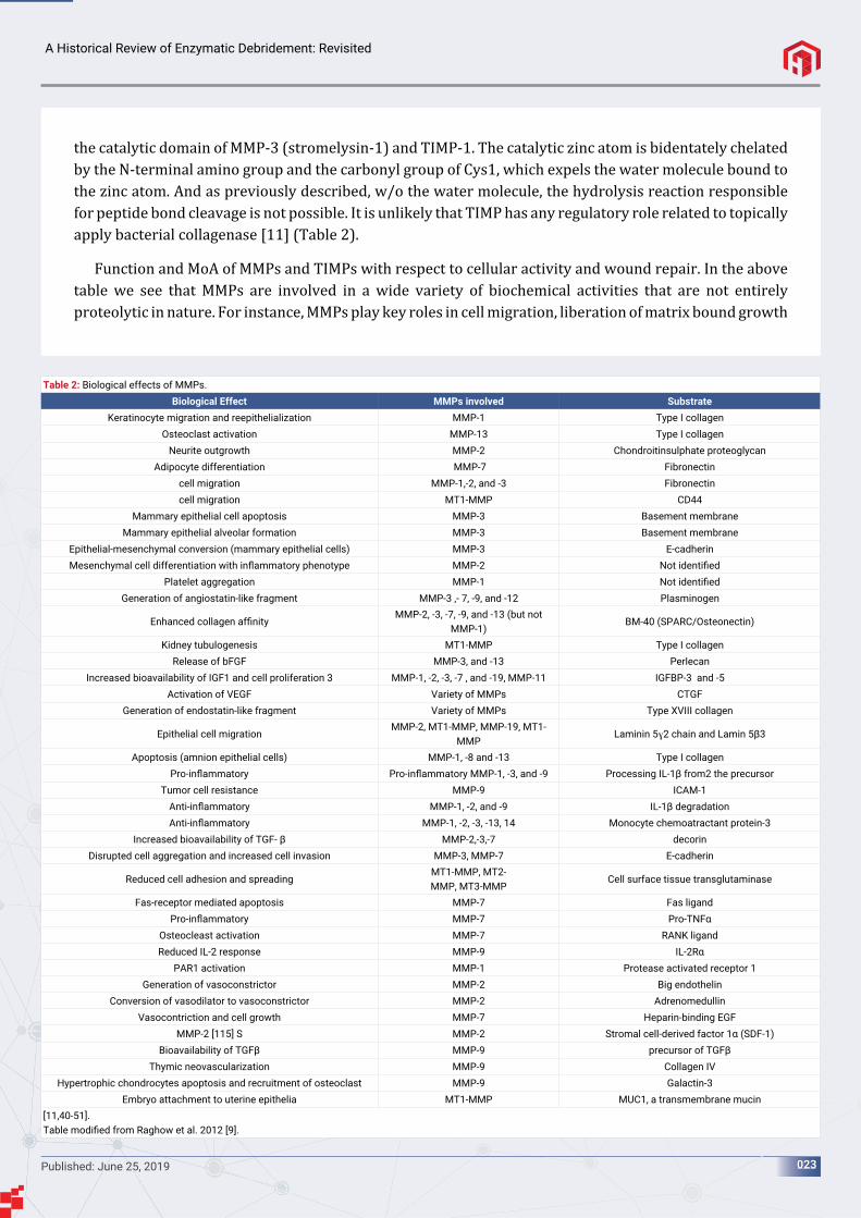
Function and MoA of MMPs and TIMPs with respect to cellular activity and wound repair. In the above table we see that MMPs are involved in a wide variety of biochemical activities that are not entirely proteolytic in nature. For instance, MMPs play key roles in cell migration, liberation of matrix bound growth
Table 2: Biological effects of MMPs.Biological Effect MMPs involved Substrate
Keratinocyte migration and reepithelialization MMP-1 Type I collagen Osteoclast activation MMP-13 Type I collagen
Neurite outgrowth MMP-2 Chondroitinsulphate proteoglycan Adipocyte differentiation MMP-7 Fibronectin
cell migration MMP-1,-2, and -3 Fibronectin cell migration MT1-MMP CD44
Mammary epithelial cell apoptosis MMP-3 Basement membrane Mammary epithelial alveolar formation MMP-3 Basement membrane
Epithelial-mesenchymal conversion (mammary epithelial cells) MMP-3 E-cadherin Mesenchymal cell differentiation with infl ammatory phenotype MMP-2 Not identifi ed
Platelet aggregation MMP-1 Not identifi ed Generation of angiostatin-like fragment MMP-3 ,- 7, -9, and -12 Plasminogen
Enhanced collagen affi nity MMP-2, -3, -7, -9, and -13 (but not
MMP-1) BM-40 (SPARC/Osteonectin)
Kidney tubulogenesis MT1-MMP Type I collagen Release of bFGF MMP-3, and -13 Perlecan
Increased bioavailability of IGF1 and cell proliferation 3 MMP-1, -2, -3, -7 , and -19, MMP-11 IGFBP-3 and -5 Activation of VEGF Variety of MMPs CTGF
Generation of endostatin-like fragment Variety of MMPs Type XVIII collagen
Epithelial cell migration MMP-2, MT1-MMP, MMP-19, MT1-
MMP Laminin 5ɣ2 chain and Lamin 5β3
Apoptosis (amnion epithelial cells) MMP-1, -8 and -13 Type I collagen Pro-infl ammatory Pro-infl ammatory MMP-1, -3, and -9 Processing IL-1β from2 the precursor
Tumor cell resistance MMP-9 ICAM-1 Anti-infl ammatory MMP-1, -2, and -9 IL-1β degradation Anti-infl ammatory MMP-1, -2, -3, -13, 14 Monocyte chemoatractant protein-3
Increased bioavailability of TGF- β MMP-2,-3,-7 decorin Disrupted cell aggregation and increased cell invasion MMP-3, MMP-7 E-cadherin
Reduced cell adhesion and spreading MT1-MMP, MT2-MMP, MT3-MMP
Cell surface tissue transglutaminase
Fas-receptor mediated apoptosis MMP-7 Fas ligand Pro-infl ammatory MMP-7 Pro-TNFα
Osteocleast activation MMP-7 RANK ligand Reduced IL-2 response MMP-9 IL-2Rα
PAR1 activation MMP-1 Protease activated receptor 1 Generation of vasoconstrictor MMP-2 Big endothelin
Conversion of vasodilator to vasoconstrictor MMP-2 Adrenomedullin Vasocontriction and cell growth MMP-7 Heparin-binding EGF
MMP-2 [115] S MMP-2 Stromal cell-derived factor 1α (SDF-1) Bioavailability of TGFβ MMP-9 precursor of TGFβ
Thymic neovascularization MMP-9 Collagen IV Hypertrophic chondrocytes apoptosis and recruitment of osteoclast MMP-9 Galactin-3
Embryo attachment to uterine epithelia MT1-MMP MUC1, a transmembrane mucin [11,40-51]. Table modifi ed from Raghow et al. 2012 [9].
A Historical Review of Enzymatic Debridement: Revisited
Published: June 25, 2019 024
factors, in lammation, anti-in lammation, platelet aggregation, cellular apoptosis, cellular differentiation, etc. In the following pages a few of these aspects will be discussed in more detail. It is hypothesized that the catalytic activity of MMP-1 is necessary for keratinocyte migration on type I collagen. To test this idea, keratinocyte motility on type I collagen by colony dispersion and colloidal gold migration assays was employed. In both assays, primary human keratinocytes migrated ef iciently on collagen. The speci icity of MMP-1 in promoting cell movement was demonstrated in 4 distinct experiments [18].
Keratinocyte migration was completely blocked by peptide hydroxymates, which are potent inhibitors of the catalytic activity of MMPs [52].
HaCaTs, a line of human keratinocytes that does not express MMP-1 in response to collagen, did not migrate on a type I collagen matrix, but moved ef iciently on denatured type I collagen (gelatin). Epidermal growth factor (EGF), which induces MMP-I production by HaCaT cells, resulted in the ability of these cells to migrate across type I collagen matrix.
Keratinocytes did not migrate on mutant type I collagen that lacked the MMP-1 cleavage site, even though this substrate induced MMP-1 expression.
Cell migration on collagen was completely blocked by recombinant TIMP-1 and by af inity-puri ied anti–MMP-1 antiserum.
In addition, the collagen-mediated induction of collagenase-1 and migration of primary keratinocytes on collagen were blocked by antibodies against the α2 integrin subunit, but not by antibodies against the α1 or α3 integrin subunit. It is proposed that interaction of the α2β1 integrin with dermal collagen mediates induction of collagenase-1 in keratinocytes at the onset of healing and that the activity of collagenase-1 is needed to initiate cell movement. Furthermore, it is proposed that cleavage of dermal collagen provides keratinocytes with a mechanism to maintain their directionality during re-epithelialization [18].
As previously described, endogenous collagenase activity is blocked by TIMP. TIMP-1 has already been discussed, but a related molecule (TIMP-2) displays a higher af inity for other members of the matrix proteinase family, particularly the 92- and 72-kDa gelatinases, MMP-9 and -2, respectively. However, researchers have suggested that TIMP-2 preferentially deactivates MMP-2 and TIMP-1 preferentially deactivates MMP-9 [12,33,53]. TIMP is nearly ubiquitous in human tissues and appears to form a barrier to incidental matrix degradative events [31,54]. Data suggest that collagenase and TIMP are temporally and spatially regulated during cutaneous wound repair.
Interstitial collagenase is a well-described zinc metalloproteinase produced by a variety of cell types involved in the healing process, such as ibroblasts, macrophages, endothelial cells, and keratinocytes [17,55-58]. Its substrates speci ically include the interstitial collagens, types I, II, and III [20]. Additionally, MMP-1 degrades anchoring ibril or type VII collagen, as well as, collagen type X [59-61]. However, it is felt that the majority (or a great deal) of collagenase is expressed by migrating basal keratinocytes at their leading edge of motility [20].
Data from in situ hybridization of tissue samples obtained by surgical débridement of second- and third-degree burns showed that speci ic signals for both collagenase and TIMP were restricted to the regions of the specimen that displayed histologic features characteristic of active wound healing (e.g., leading epidermal tips, surviving epidermal structures within the dermis, vascular proliferation). Little or no hybridization was noted in areas of severe necrosis (i.e., third-degree injury) or in histologically normal areas distal to the burn injury. Strong focal hybridization signals were detected in various cell types within the dermis (granulation tissue) of partial-thickness burn wounds. Expression of degradative matrix molecules within the granulation tissues appeared to be temporally and spatially regulated. Cells capable
A Historical Review of Enzymatic Debridement: Revisited
Published: June 25, 2019 025
of producing collagenase and TIMP were present in nearly identical locations within the mesenchymal tissues of human burn wounds [17].
During the continuum of wound repair, the collagenase- and TIMP-expressing cells were initially limited in spatial orientation to the perivascular and perifollicular regions and around the edges of burn wounds. As time progressed, cells expressing both collagenase and TIMP became widely dispersed throughout the viable dermis. Eventually, the number of cells expressing collagenase and TIMP diminished in the later reparative and remodeling phases in more super icial burns. In contrast, many of the deepest partial-thickness burns were still actively healing. Such deeper burns continued to show abundant labeled cells within the deep dermis 15 to 34 days after injury. Distinct spatial and temporal expressions were found within human burn wounds for interstitial collagenase and TIMP [17].
Transcription of mRNAs for collagenase and TIMP is a common and widespread event in the healing of burn wounds [17]. Synthesis and expression of collagenase and TIMP is tightly regulated during wound repair. Strong signals were detected within granulation tissue and at the edge of the epidermis, but were not seen in the more distal uninjured epidermis and dermis. The earliest time point after injury (2 days) was characterized by weak labeling for mRNAs, representing collagenase and TIMP. Because thermal injury is characterized by a 12- to 24-hour period of continued tissue destruction, the absence of mRNAs soon after wounding may re lect this period of continued tissue damage, as well as the time required for initiation of increased transcription. Peak intensities and number of cells expressing collagenase and TIMP were noted during the active phase of granulation tissue formation, in lammation, and re-epithelialization. More mature wounds showed a decline in the number of cells transcribing mRNAs for either of these proteins [17].
The presence of collagenase and TIMP in epithelial structures in situ was anticipated, because keratinocytes in culture synthesize both interstitial collagenase and TIMP [62]. Keratinocyte production of collagenase and TIMP appear to be inhibited by laminin and stimulated by collagen types I and IV. In situ data showing stimulated expression of these proteins in regions characterized by increased cell motility and activity of the basement membrane with no detectable expression in the adjacent hypertrophic epidermis are consistent with in vitro studies. The presence of hybridization signals in the vessel walls of capillaries demonstrated that endothelial cells could temporarily produce these proteins during angiogenesis associated with wound repair [17]. Cultured endothelial cells are copious producers of matrix MMPs and TIMP [57]. MMP may facilitate vessel growth, which may in turn be inhibited by TIMP.
With reference to chronic wounds such as venous stasis ulcers, diabetic ulcers, and decubiti, it seems likely that these persistent wounds may be characterized by the disruption of the carefully orchestrated process of matrix degradation and remodeling. It appears likely that remodeling events are tightly regulated by a multifactorial equilibrium between the synthesis of extracellular matrix proteins and their degradative enzymes and inhibitors.
The best-characterized and historically oldest subgroup of MMPs are the interstitial collagenases, which possess the unique ability to cleave the triple helix of native types I, II, and III collagen. MMPs display a high degree of structural similarity, with about 40% amino acid homology among all members of the family [14]. Another similarity of the collagenases is that they cleave collagen in the same manner.
Three interstitial collagenases have been extensively studied, types -1, -2 and -3 (MMP-1, -8 and -13, respectively). All cleave native type I collagen at a single locus (Gly775–Ile776 in the α1 chains; Gly775–Leu776 in α2 chain), which is located approximately three fourths of the distance from the N-terminus of the collagen molecule [17,63]. At physiologic temperature (37°C), the ¾ and ¼ length fragments, a 225-kDa fragment (TCA) and a 75-kDa fragment (TCB), respectively, denature spontaneously into randomly coiled gelatin
A Historical Review of Enzymatic Debridement: Revisited
Published: June 25, 2019 026
peptides and can be further attacked by a variety of enzymes, including the gelatinases [14]. It should be noted that these bonds are completely different from those cleaved by the collagenase for the bacterium Clostridium histolyticum [20].
The single cleavage of the collagen triple helix catalyzed by the three interstitial collagenases is the rate-limiting step of collagen degradation [14].
Collagenase-1 (MMP-1) is produced in humans by a variety of epithelial mesenchymal cell types, including keratinocytes, ibroblasts, macrophages, chondrocytes, and smooth muscle cells [60,64].
Collagenase-2 (MMP-8) is a product only of the polymorphonuclear leukocyte and is stored within neutrophil granules, in contrast to all other MMPs, which are rapidly secreted without signi icant intracellular stores [65].
Collagenase-3 (MMP-13) is an enzyme found in breast cancer, but appears to be the predominant interstitial collagenase in certain rodent species, such as the rat or mouse [63]. More recently it has been found that human smooth muscle cells produce MMP-13 [9].
Furthermore, the inhibition of collagenase can be judicially controlled by regulation of the timing, location, and amount of TIMP produced and by releasing collagenase with tight extracellular spaces that are not easily accessible to TIMP. Collagenase activity increases about 10-fold for every 10°C increase in temperature. This is a remarkable response, since most enzymes increase their activity by a factor of 2 for every 10°C increase [14].
Keratinocytes are capable of secreting TIMP-1 [54]. Most collagenase-producing cells also make TIMP-1, but it has been found that TIMP-1 mRNA does not co-localize with collagenase mRNA in migrating keratinocytes in chronic wounds. TIMP-1 is produced by stromal or perivascular cells, usually away from sites of collagenase activity. This distinct localization of enzyme and inhibitor suggests that keratinocyte-derived collagenase acts without impedance from TIMP-1. By means of cell surface receptors, the cell recognizes a particular matrix molecule and is instructed to produce the appropriate MMP. The protease is released in a protected pericellular compartment, where it degrades its substrate. TIMPs are present in the tissue environment to neutralize “spent” proteinases, thereby preventing excessive and unwanted degradation away from the sites of MMP production. In the process of re-epithelialization, keratinocytes interact with the dermal matrix generally and type I collagen in particular. These new cell-matrix contacts may provide an early and critical signal to initiate the epithelial response to wounding [20].
An interesting aspect of the epithelial expression of interstitial collagenase in wounded skin is that the enzyme is not produced in non-ulcerated samples in vitro. Basal keratinocytes normally rest on a basement membrane composed of various forms of laminin, entactin, proteoglycans, and type IV collagen [20]. In response to wounding, keratinocytes migrate from the edge of the wound under a provisional matrix of ibrin and ibronectin [20,66] and over the dermis, which includes structural macromolecules such as type I collagen. Loss of contact with the basement membrane and establishment of new cell: matrix interaction with components of the dermal and provisional matrices may be a critical determinant that affects keratinocyte phenotype and which in turn induces collagenase production. Collagenase production is induced in vitro in isolated human basal keratinocytes grown on a surface coated with type I collagen. Migrating keratinocytes also present a distinct pattern of matrix-binding receptors; they may also be involved in the regulation of collagenase production [20].
These receptors, integrins (mentioned previously), are heterodimeric surface molecules composed of distinct α and β protein chains that cells use to attach to matrix proteins and to each other. Integrins are also used by cells to move or migrate over the extracellular matrix. Keratinocytes at the wound edge; however,
A Historical Review of Enzymatic Debridement: Revisited
Published: June 25, 2019 027
selectively produce additional integrins, such as α5β1 and αvβ3, which are characteristic of migrating cells. These receptors are present on the same keratinocytes that produce interstitial collagenase [20,67-70]. Although the α5β1 integrin recognizes ibronectin, which is present in high concentrations in both the provisional and dermal matrices, but is absent from the epidermal basement membrane, it is doubtful that this receptor mediates induction of collagenase production in vitro by keratinocytes cultured on collagen. Most likely other integrins, such as α5β1, which interacts strongly with native type I collagen, participate in the induction of collagenase gene expression [20,68-70].
Cells most likely do not release proteases indiscriminately, especially an enzyme like interstitial collagenase, which has such de ined substrate speci icity, but rely on precise cell:matrix interactions to inform the cell that it is in contact with a particular matrix protein. Collagenase expression is modulated by numerous pro-in lammatory mediators, such as interleukin-1 (IL-1) and epidermal growth factor (EGF) [20].
Since many cytokines are present in the wound environment and because the epidermis is a source of so many soluble mediators [71], expression of collagenase in migrating basal keratinocytes may be in luenced by the presence of many of these factors. The overexpression of cytokines in chronic ulcers may lead to excessive production of protease. The invariant and prominent production of interstitial collagenase-1 by basal keratinocytes in both acute and chronic wounds indicates that this MMP serves a critical and required role in reepithelialization [20].
Type I collagen is the most abundant structural component of the dermal matrix. It is likely that migrating keratinocytes interact with this protein. Since the α2β1 integrin receptor for type I collagen binds native collagen much tighter than it does gelatin interstitial collagenase may aid in dissociating keratinocytes from collagen-rich matrix and thereby promote ef icient migration over the dermal and provisional matrices. Thus, in a cutaneous wound-healing response, collagenase may serve a bene icial function, unlike its potentially destructive role in arthritis and vascular disease [20].
Stromelysin-2 (MMP-10) may facilitate keratinocyte migration by removing damaged matrix basement membrane. It is also tempting to speculate that stromelysin-2 may be involved in the activation of co-secreted procollagenase [72]. Since it is produced by proliferating cells, stromelysin-1 (MMP-3) is probably not involved in reepithelialization per se, but rather is needed for restructuring the early basement membrane [20].
In the dermis, collagenase-1 and stromelysin-1 probably affect tissue repair at various stages, including remodeling during the formation and removal of granulation tissue and during resolution of scar tissue. Furthermore, these 2 MMPs may be needed for related processes such as angiogenesis and the extravasation and migration of in lammatory cells [20].
More recently is has been determined that MMP-3 is involved w/ anti-in lammatory processes, as well [11].
Parks, 1995, [20] found that more endogenous collagenase is produced in non-healing or poorly healing wounds than in wounds that will heal or are healing properly. The reason for this excess production of collagenase is probably a result of the massive and persistent in lammation associated with chronic ulcers. As is known, the expression of collagenase by any cell is greatly in luenced by cytokines released from in lammatory cells. Excess proteolysis by this enzyme may cause tissue damage that actually impairs healing.
There are two possible mechanisms that may control the cessation of epidermal proteinase production [20].
One is the reformation of the basement membrane. In intact skin, basal keratinocytes rest on a basement membrane and do not make collagenase. In studies of acute wounds, collagenase is not produced in
A Historical Review of Enzymatic Debridement: Revisited
Published: June 25, 2019 028
recently healed samples with a reformed basement membrane. Also, keratinocytes grown on basement membrane proteins do not make collagenase, whereas keratinocyte cells on type I collagen do. These observations strongly suggest that cell contact with a basement membrane protein down-regulates collagenase production.
The other mechanism that may be involved in the turning-off of collagenase production is cell-to-cell contact. It is known that migrating epithelial cells dissolve or disorganize the cell-to-cell contact and that these rapidly reform once re-epithelialization is complete. It has been found that collagenase production decreases markedly in keratinocytes at high con luence, even if the cells are plated on collagen.
As discussed, members of the MMP family are inhibited by TIMPs. In normal wounds, there is a temporal change in the concentration of TIMP-1 that is greatest at 2-3 days after wounding and remained elevated above normal serum level at 10 days. In contrast, the wound luid from the non-healing wounds contains less TIMP than was accumulated in the irst 24 hours by wounds that eventually healed. Angiogenesis is assumed to proceed by proteolysis of matrix that immediately surrounds vascular endothelial cells. Some angiogenic factors stimulate production of endothelial cell metalloproteinases. Excess antiproteinase, whether synthetic or natural could prevent angiogenesis. The addition of either TIMP-1 or TIMP-2 inhibits angiogenesis [73,74].
In this chapter, it has been shown that the human body contains a variety of proteins that function as degradative enzymes to support the natural debridement and remodeling of devitalized tissue, two key aspects of the wound-healing process. Examples of these proteins include serine proteases and metalloproteases. Removal of necrotic tissue is essential to reduce the bacterial burden in a wound, which in turn decreases the amount of in lammatory mediators. As exudate is an aspect of in lammation, exudate levels may also be reduced. In response to wounding, keratinocytes migrate inward from the edge of a wound under a provisional matrix of ibrin and ibronectin over the dermis, which includes structural macromolecules such as type I collagen, micro ibrils, and elastin, which are distinct from those found in the basement membrane. Removal of this provisional (i.e., wound) matrix is essential for the development of a clean wound bed upon which granulation can occur. In addition, healing of a completely debrided wound bed results in decreased scar formation [75,76].
Wound healing is a complex biological process that should proceed in a timely and orderly fashion under normal environmental conditions. However, the process can be impaired by a variety of both systemic and local factors. Systemic factors are related to impaired oxygenation, poor nutrition, concomitant medical conditions such as diabetes and cardiovascular disease, aging, and such medications as immunosuppressants, chemotherapeutics, and corticosteriods. Local factors associated with impaired healing include mechanical stressors, bacterial infection, cytotoxic agents, wound desiccation, and necrotic tissue. Necrotic tissue is anchored to the wound surface by strands of undenatured collagen [75,77], though it is safe to assume that partially denatured strands play a role, as well. Until these ibers are severed, débridement cannot take place and granulation tissue formation is slowed, thus slowing the rate of wound closure. Over the years, various topically applied proteolytic enzymes have been employed (papain, icin, streptokinase, streptodornase, trypsin–chymotrypsin, etc.) for the débridement of wounds. They have had only limited success because they are less ef icient at removing native collagen when compared to clostridial collagenase [78,79]. At physiologic pH values, papain and icin do not digest collagen at a signi icant rate, and denaturing agents such as urea must be incorporated in formulations containing these enzymes in order for them to attack collagen. However, in 1958, Miller et al. [80], showed that papain-urea lacks the ability to degrade native collagen and stated that only clostridial collagenase was able to adequately digest collagen. These and other aspects of topical enzyme debriders are detailed in chapter 1, entitled “Types of Enzymes.”
A Historical Review of Enzymatic Debridement: Revisited
Published: June 25, 2019 029
References1. Gross J, Lapiere CM. Collagenolytic activity in amphibian tissues: a tissue culture assay. Proc Natl Acad Sci. 1962; 48:
1014-1022. Ref.: https://tinyurl.com/y5wselrq
2. Gross J. How tadpoles lose their tails: path to discovery of the fi rst matrix metalloproteinase. Matrix Biol. 2004; 23: 3-13. Ref.: https://tinyurl.com/y5w77co2
3. Lapière CM. Tadpole collagenase, the single parent of such a large family. Biochimie. 2005; 87: 243-247. Ref.: https://tinyurl.com/yyzps8mj
4. Murphy G. The ADAMs: signaling scissors in the tumour microenvironment. Nat Rev Cancer. 2008; 8: 929-941. Ref.: https://tinyurl.com/yykf3tlw
5. Egeblad M, Werb Z. New functions for the matrix metalloproteinases in cancer progression. Nat Rev Cancer. 2002; 2:161-174. Ref.: https://tinyurl.com/y4vvgmzv
6. Brinckerhoff CE, Matrisian LM. Matrix metalloproteinase: A tail of a frog that became a prince. Nat Rev Mol Cell Biol. 2002: 207-214. Ref.: https://tinyurl.com/y64qjy4t
7. Edwards DR, Handsley MM, Pennington CJ. The ADAM, metalloproteinases. Mol Aspects Med. 2008; 29: 258-289. Ref.: https://tinyurl.com/y628ehdt
8. Overall CM, Blobel CP. In search of partners: linking extracellular proteases to substrates. Nat Rev Mol Cell Biol. 2007; 8: 245-257. Ref.: https://tinyurl.com/yxvm4fa7
9. Raghow R. Connective Tissues of the Subendothelium Vascular Medicine: A Companion to Braunwald's Heart Disease. 2012.
10. Nagase H, Woessner JF Jr. Matrix metalloproteinases. J Biol Chem. 1999; 274: 21491-21494. Ref.: https://tinyurl.com/yy88m882
11. Nagase H, Visse R, Murphy G. Structure and function of matrix metalloproteinases and TIMPs. Cardiovascular Research. 2006; 69: 562-573. Ref.: https://tinyurl.com/y4u8auu9
12. Matrisian LM. The matrix-degrading metalloproteinases. Bio Essays. 1992; 14: 455-463.
13. Mast BA, Schultz GS. Interactions of cytokines, growth factors and proteases in acute and chronic wounds. Wound Rep Reg. 1996; 4: 411-420. Ref.: https://tinyurl.com/yxpwboo5
14. Jeffrey J. Metalloproteinases and Tissue Turnover Wounds, a Compendium of Clinical Research and Practice. 1995; 7: 13A-22A.
15. Raffetto JD, Khalil RA. Matrix Metalloproteinases and their Inhibitors in Vascular Remodeling and Vascular Disease. Biochem Pharmacol. 2008; 75: 346-359. Ref.: https://tinyurl.com/y43kerg5
16. Brett DW. A Historic Review of Topical Enzymatic Debridement. The McMahon Publishing Group. 2003.
17. Stricklin GP, Li L, Jancic V, Wenczak BA, Nanney LB. Localization of mRNAs Representing Collagenase and TIMP in Sections of Healing Human Burn Wounds. Am J Pathol. 1993; 143: 1657-1666. Ref.: https://tinyurl.com/yyh4x9bf
18. Pilcher BK, Dumin JA, Sudbeck BD, Krane SM, Welgus HG, Parks WC. The activity of collagnese-1 is required for keratinocyte migration on a type 1 collagen matrix. J Cell Biol. 1997; 137: 1445-1457. Ref.: https://tinyurl.com/y6huqsew
19. Wang R, Ghahary A, Shen Q, Scott PG, Roy K, Tredget EE. Hypertrophic scar tissues and fi broblasts produce more transforming growth factor-β1 mRNA and protein than normal skin. Wound Repair Regen. 2000; 8: 128-137. Ref.: https://tinyurl.com/y3vg47xs
20. Parks WC. The Production, Role, and Regulation of Matrix Metalloproteinsases in the Healing Epidermis. Wounds Sup A. 1995; 7: 23A-A33.
21. Tallant C, Marrero A, Gomis-Rüth FX. Matrix metalloproteinases: Fold and function of their catalytic domains. Biochimica et Biophysica Acta. 2010; 1803: 20-28. Ref.: https://tinyurl.com/y3dbot63
22. Collier IE, Krasnov PA, Strongin AY, Birkedal-Hansen H, Goldberg GI. Alanine scanning mutagenesis and functional analysis of the fi bronectin-like collagenbinding domain from human 92-kDa type IV collagenase. J Biol Chem. 1992; 267: 6776-6791. Ref.: https://tinyurl.com/y3bbeh6r
A Historical Review of Enzymatic Debridement: Revisited
Published: June 25, 2019 030
23. Maskos K. Crystal structures of MMPs in complex with physiological and pharmacological inhibitors. Biochimie. 2005; 87: 249-263. Ref.: https://tinyurl.com/y3woxn7x
24. Schechter I, Berger A. On the size of active site in proteases I Papain. Biochem Biophys Res Commun. 1967; 27: 157-162. Ref.: https://tinyurl.com/yy2xz2u4
25. Gomis-Rüth FX, Stöcker W, Huber R, Zwilling R, Bode W. Refi ned 1.8 Å X-ray crystal structure of astacin, a zinc-endopeptidase from the crayfi sh Astacus astacus L. Structure determination, refi nement, molecular structure and comparison with thermolysin, J Mol Biol. 1993; 229: 945-968. Ref.: https://tinyurl.com/yymyubuq
26. Reinemer P, Grams F, Huber R, Kleine T, Schnierer S, Piper M, Tschesche H, Bode W. Structural implications for the role of the N terminus in the ‘superactivation’ of collagenases: A crystallographic study. FEBS Lett. 1994; 338: 227-233. Ref.: https://tinyurl.com/yxgacnsw
27. Morgunova E, Tuuttila A, Bergmann U, Tryggvason K. Structural insight into the complex formation of latent matrix metalloproteinase 2 with tissue inhibitor of metalloproteinase 2. Proc Natl Acad Sci. 2002; 99: 7414-7419. Ref.: https://tinyurl.com/yyqurqya
28. Becker JW, Marcy AI, Rokosz LL, Axel MG, Burbaum JJ. Stromelysin-1: three-dimensional structure of the inhibited catalytic domain and of the C-truncated proenzyme, Prot Sci. 1995; 4: 1966-1976. Ref.: https://tinyurl.com/y5p2b5pg
29. Jozic D, Bourenkov G, Lim NH, Visse R, Nagase H. X-ray structure of human proMMP-1: new insights into procollagenase activation and collagen binding. J Biol Chem. 2005; 280: 9578-9585. Ref.: https://tinyurl.com/y5omvmf2
30. Cha J, Pedersen MV, Auld DS. Metal and pH dependence of heptapeptide catalysis by human matrilysin. Biochemistry. 1996; 35: 15831-15838. Ref.: https://tinyurl.com/y4zbuou4
31. Stricklin GP, Welgus HG. Human skin fi broblast collagenase inhibitor. Purifi cation and biochemical characterization. J Biol Chem. 1983; 258: 12252-12258. Ref.: https://tinyurl.com/yxq4rojl
32. Hayakawa T, Yamashita K, Tanzawa K, Uchijima E, Iwata K. Growth-promoting activity of tissue inhibitor of metalloproteinases-1 (TIMP-1) for a wide range of cells. A possible new growth factor in serum. FEBS Lett. 1992; 298: 29-32. Ref.: https://tinyurl.com/y26ythw2
33. Stetler-Stevenson WG, Krutzsch HC, Liotta LA. Tissue inhibitor of metalloproteinase (TIMP-2): A new member of the metalloproteinase family. J Biol Chem. 1989; 264: 17374-17378. Ref.: https://tinyurl.com/yxuu7f6y
34. Collier IE. H-ras oncogene transformed human bronchial epithelial cells (TBC-1) secrete a single metalloprotease capable of degrading basement membrane collagen. J Biol Chem.1988; 263: 6579-6587.
35. Higashi S, Miyazaki K. Identifi cation of a region of beta-amyloid precursor protein essential for its gelatinase A inhibitory activity. J Biol Chem. 2003; 278: 14020-14028. Ref.: https://tinyurl.com/y559kakk
36. Mott JD, Thomas CL, Rosenbach MT, Takahara K, Greenspan DS. Post-translational proteolytic processing of procollagen C-terminal proteinase enhancer releases a metalloproteinase inhibitor. J Biol Chem. 2000; 275: 1384-1390. Ref.: https://tinyurl.com/y26d478h
37. Oh J, Takahashi R, Kondo S, Mizoguchi A, Adachi E. The membrane-anchored MMP inhibitor RECK is a key regulator of extracellular matrix integrity and angiogenesis. Cell. 2001; 107: 789-800. Ref.: https://tinyurl.com/y4upg6go
38. Gomis-Rüth FX, Maskos K, Betz M, Bergner A, Huber R. Mechanism of inhibition of the human matrix metalloproteinase stromelysin-1 by TIMP-1. Nature. 1997; 389: 77-81. Ref.: https://tinyurl.com/y28weu4v
39. Fernandez-Catalan C, Bode W, Huber R, Turk D, Calvete JJ, et al. Crystal structure of the complex formed by the membrane type 1-matrix metalloproteinase with the tissue inhibitor of metalloproteinases- 2, the soluble progelatinase a receptor. EMBO J. 1998; 17: 5238-5248. Ref.: https://tinyurl.com/y26sjrlz
40. Miyamoto S, Yano K, Sugimoto S, Ishii G, Hasebe T, et al. Matrix metalloproteinase-7 facilitates insulin-like growth factor bioavailability through its proteinase activity on insulin-like growth factor binding protein 3. Cancer Res. 2004; 64: 665-671. Ref.: https://tinyurl.com/yymocmqe
41. Sadowski T, Dietrich S, Koschinsky F, Sedlacek R. Matrix metalloproteinase 19 regulates insulin-like growth factor-mediated proliferation, migration, and adhesion in human keratinocytes through proteolysis of insulin-like growth factor binding protein-3. Mol Biol Cell. 2003; 14: 4569-4580. Ref.: https://tinyurl.com/y67devrn
42. Udayakumar TS, Chen ML, Bair EL, Von Bredow DC, Cress AE, et al. Membrane type-1-matrix metalloproteinase
A Historical Review of Enzymatic Debridement: Revisited
Published: June 25, 2019 031
expressed by prostate carcinoma cells cleaves human laminin-5 beta3 chain and induces cell migration. Cancer Res. 2003; 63:2292-2299. Ref.: https://tinyurl.com/y3xeg9js
43. Haro H, Crawford HC, Fingleton B, Shinomiya K, Spengler DM, et al. Matrix metalloproteinase-7-dependent release of tumor necrosis factor-alpha in a model of herniated disc resorption. J Clin Invest. 2000; 105: 143-150. Ref.: https://tinyurl.com/yy7s7743
44. Lynch CC, Hikosaka A, Acuff HB, Martin MD, Kawai N, et al. MMP-7 promotes prostate cancer-induced osteolysis via the solubilization of RANKL. Cancer Cell. 2005; 7: 485-496. Ref.: https://tinyurl.com/y4ob24ln
45. Boire A, Covic L, Agarwal A, Jacques S, Sherifi S, et al. PAR1 is a matrix metalloprotease-1 receptor that promotes invasion and tumorigenesis of breast cancer cells. Cell. 2005; 120: 303-313. Ref.: https://tinyurl.com/y3l824mt
46. Fernandez-Patron C, Radomski MW, Davidge ST. Vascular matrix metalloproteinase-2 cleaves big endothelin-1 yielding a novel vasoconstrictor. Circ Res. 1999; 85: 906-911. Ref.: https://tinyurl.com/yymmb74m
47. Hao L, Du M, Lopez-Campistrous A, Fernandez-Patron C. Agonistinduced vactivation of matrix metalloproteinase-7 promotes vasoconstriction through the epidermal growth factor-receptor pathway. Circ Res. 2004; 94: 68-76. Ref.: https://tinyurl.com/y5vmqvy5
48. Zhang K. HIV-induced metalloproteinase processing of the chemokine stromal cell derived factor1 causes neurodegeneration. Nat Neurosci. 2003; 6:1064-1071.
49. Odaka C, Tanioka M, Itoh T. Matrix metalloproteinase-9 in macrophages induces thymic neovascularization following thymocyte apoptosis. J Immunol. 2005; 174: 846-853. Ref.: https://tinyurl.com/y5vv74cv
50. Ortega N, Behonick DJ, Colnot C, Cooper DN, Werb Z. Galectin-3 is a downstream regulator of matrix metalloproteinase-9 function during endochondral bone formation. Mol Biol Cell. 2005; 16:3028-3039. Ref.: https://tinyurl.com/y4ozokm9
51. Thathiah A, Carson DD. MT1-MMP mediates MUC1 shedding independent of TACE/ADAM17. Biochem J. 2004; 382: 363-373. Ref.: https://tinyurl.com/yxtvofbb
52. Moore WM, Spilburg CA. Purifi cation of human collagenases with a hydroxamic acid affi nity column. Biochemistry. 1986; 25: 5189-5195. Ref.: https://tinyurl.com/y33jpg7g
53. Goldberg GI. Human 72kDa type IV collagenase forms a complex with a tissue inhibitor of metalloproteinase designated as TIMP-2. Proc Natl Acad Sci. 1989; 86: 8207-8211.
54. Welgus HG, Stricklin GP. Human collagenase inhibitor: comparative studies in human connective tissues, serum and amniotic fl uid. J Biol Chem. 1983; 258: 12259-12264. Ref.: https://tinyurl.com/y2uoj2xc
55. Welgus HG. Human skin fi broblast collagenase. Interactions with substrate and inhibitor. Collagen Rel Res. 1985; 5: 167-179.
56. Bauer EA, Stricklin GP, Jeffrey JJ, Eisen AZ. Collagenase production by human skin fi broblasts. Biochem Biophys Res Commun. 1975; 64: 232-240. Ref.: https://tinyurl.com/y63halxw
57. Herron GS, Banda MJ, Clark EJ, Gavrilovic J, Werb Z. Secretion of metalloproteinases by stimulated capillary endothelial cells. II. Expression of collagenase and stromelysin activities is regulated by endogenous inhibitors. J Biol Chem. 1986; 261: 2814-2818. Ref.: https://tinyurl.com/y2jzb6v2
58. Welgus GH. The collagen substrate specifi city of human skin fi broblast collagenases. J Biol Chem. 1981; 256:9511-9515.
59. Sage H. Collagens of basement membranes. J Invest Dermatol. 1982; 79: 51s-59s.
60. Schmid TM, H. Edward Conrad. A unique low MW collagen secreted by culture chick embryo chondrocytes. J Biol Chem. 1982; 259:12444-12450. Ref.: https://tinyurl.com/yy8h5t4p
61. Welgus HG. Differential susceptibility of type X collagen to cleavage by two mammalian collagenases and 72kDa type IV collagenases. J Biol Chem. 1990; 265: 13521-13527.
62. Petersen MJ, Woodley DT, Stricklin GP, O'Keefe EJ. Enhanced synthesis of collagenase by human keratinocytes cultured on type I or type IV collagen. J Invest Dermatol. 1990; 94: 341-346. Ref.: https://tinyurl.com/y2bounjc
63. Freije JMP. in-vitro cloning and expression of collagenase-3 human MMP products from breast carcinomas. J Biol Chem. 1994; 269: 1642-1650.
A Historical Review of Enzymatic Debridement: Revisited
Published: June 25, 2019 032
64. Arihiro S, Ohtani H, Hiwatashi N, Torii A, Sorsa T, et al. Vascular smooth muscle cells and pericytes express MMP-1, MMP-9, TIMP-1 and type I procollagen in infl ammatory bowel disease. Histopathology. 2001; 39: 50-59. Ref.: https://tinyurl.com/yynfx279
65. Hasty KA, Pourmotabbed TF, Goldberg GI, Thompson JP, Spinella DG, et al. Human neutrophil collagenase: A distinct product with homology to other MMPs. J Biol Chem. 1990; 265: 11421-11440. Ref.: https://tinyurl.com/y6eu7x8o
66. Clark RAF, Lanigan JM, DellaPelle P, Manseau E, Dvorak HF, et al. Fibronectin and fi brin provide a prvisoinal matrix for epidermal cell migration during wound re-epithleization. J Invest Dertmatol. 1982; 79: 264-269. Ref.: https://tinyurl.com/y2xwyggm
67. Saarialho-Kere UK, Kovacs SO, Pentland AP, Olerud JE, Welgus HG, et al. Cell-matrix interactions modulate interstitial collagenase expression by human keratinocytes actively involved in wound healing. J Clin In vest. 1993; 92: 2858-2866. Ref.: https://tinyurl.com/y6jh2bs6
68. Jokinen J, Dadu E, Nykvist P, Käpylä J, White DJ, et al. Integrin-mediated Cell Adhesion to Type I Collagen Fibrils. J Biol Chem. 2004; 279: 31956-31963. Ref.: https://tinyurl.com/yyq85te6
69. Taubenberger AV, Woodruff MA, Bai H, Muller DJ, Hutmacher DW. The effect of unlocking RGD-motifs in collagen I on pre-osteoblast adhesion and differentiation. Biomaterials. 2010; 31: 2827-2835. Ref.: https://tinyurl.com/yxkw3spd
70. Fong E, Tzlil S, Tirrell DA. Boundary crossing in epithelial wound healing. Proc Natl Acad Sci. 2010; 107: 19302-19307. Ref.: https://tinyurl.com/y3f9oepv
71. McKay IA, Leigh IM. Epidermal cytokines and their roles in cutaneous wound healing. Br Dermatol .1991; 124: 513-518. Ref.: https://tinyurl.com/yytmvpxd
72. Murphy G, Cockett MI, Stephens PE, Smith BJ, Docherty AJ. Stromelysin is an activator of procollagenase. A study with natural and recombinant enzymes. Biochem J. 1987; 248: 265-268. Ref.: https://tinyurl.com/y3k35qq6
73. Fisher C, Gilbertson-Beadling S, Powers EA, Petzold G, Poorman R, et al. Interstitial collagenase is required for angiogenesis in vitro. Devel Biol. 1994; 162: 499510. Ref.: https://tinyurl.com/yxelk9n2
74. Johnson MD, Kim HR, Chesler L, Tsao-Wu G, Bouck N, et al. Inhibition of angiogenesis by TIMP. J Cell Physiol. 1994; 160: 194-202. Ref.: https://tinyurl.com/y53asn8c
75. Rao DB, Sane PG, Georgiev EL. Collagenase in the Treatment of Dermal and Decubitus Ulcers. J Am Geriatric. 1975; 23: 22-30. Ref.: https://tinyurl.com/y2xz4plj
76. Boxer AM, Gottesman N, Bernstein H, Mandl I. Debridement of Dermal Ulcers and Decubiti with Collagenase. Geriatrics. 1969; 24: 75-86. Ref.: https://tinyurl.com/yxfe23ov
77. Howes EL. The Use of Clostridium histolyticum Enzymes in the treatment of Experimental 3rd Degree Burns, Surg Gyn and Obstet. 1959; 109: 177-188.
78. Sherry S. Proteolytic Enzymes: a therapeutic evaluation. Clinical pharmacology and therapeutics. 1960; 1: 202-226.
79. Mandl I. Collagenase. Science Publishers. 1972.
80. Miller JM, et al. The interaction of Papain, Urea and Water-Soluble Chlorophyllin in a Proteolytic Ointment for Infected Wounds. Surg. 1958; 43: 939-948. Ref.: https://tinyurl.com/y4lhwln3
81. Carmichael DF, Sommer A, Thompson RC, Anderson DC, Smith CG, et al. Primary structure and cDNA cloning of human fi broblast collagenase inhibitor. Proc Natl Acad Sci. 1986; 83: 2407-2411. Ref.: https://tinyurl.com/y2t5nbjx
82. Eeckhout T, Vaes G. Further studies on the activation of procollagenase the latent precursor of bone collagenase. Effects of lysosomal cathepsin B, plasmin and kallikrein and spontaneous activation. Biochem J. 1977; 166: 21-31. Ref.: https://tinyurl.com/y2gsszq3
83. Hu J, Van den Steen PE, Sang QX, Opdenakker G. Matrix metalloproteinase inhibitors as therapy for infl ammatory and vascular disease. Nat Rev Drug Discov. 2007; 6: 480-498. Ref.: https://tinyurl.com/yyrps5fk
84. Overall CM, López-Otín C. Strategies for MMP inhibition in cancer; innovations for the post-trial era. Nat Rev Cancer. 2002; 2: 657-672. Ref.: https://tinyurl.com/y4j8os7v
85. Welgus HG, Jeffrey JJ, Stricklin GP, Roswit WT, Eisen AZ. Characteristics of the action of human skin fi broblast collagenase on fi brillar collagen. J Biol Chem. 1980; 255: 6806-6813. Ref.: https://tinyurl.com/yylhrlgz
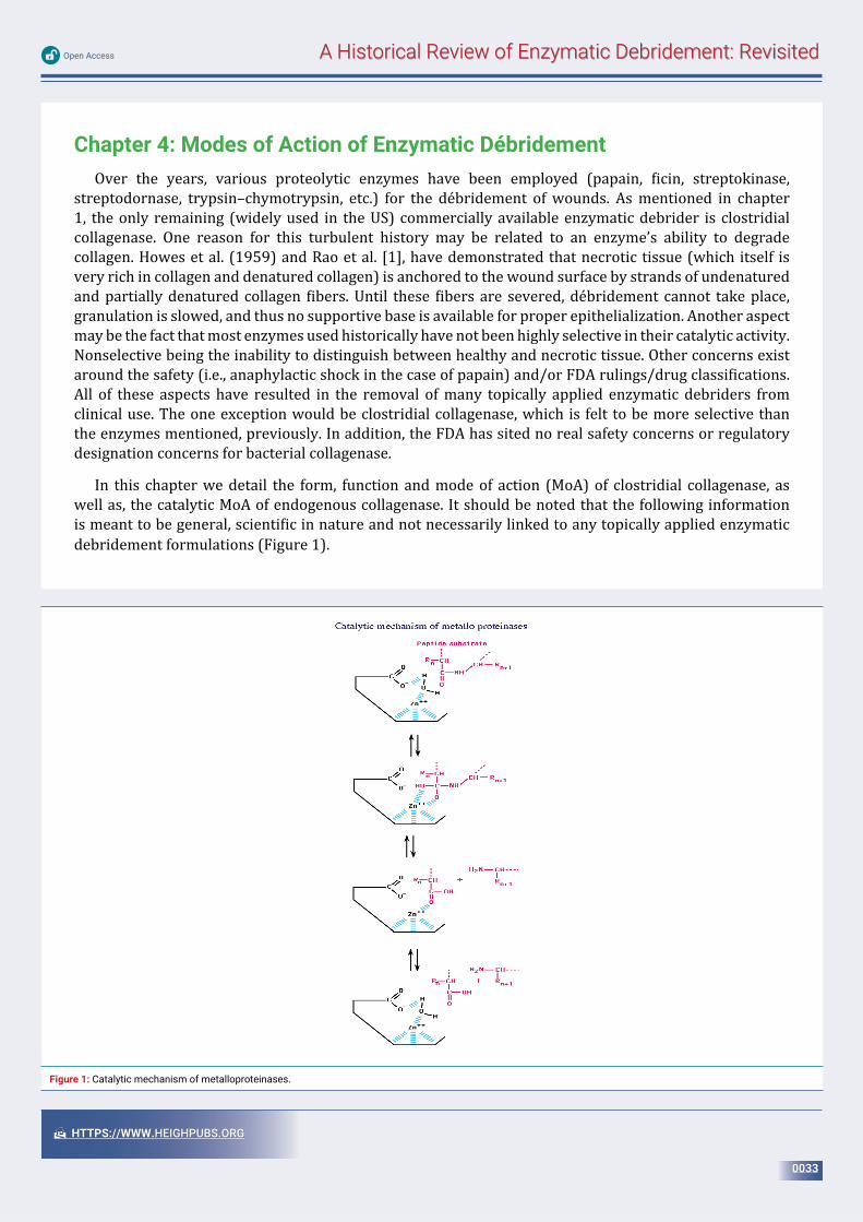
A Historical Review of Enzymatic Debridement: RevisitedOpen Access
HTTPS://WWW.HEIGHPUBS.ORG
0033
Chapter 4: Modes of Action of Enzymatic Débridement Over the years, various proteolytic enzymes have been employed (papain, icin, streptokinase,
streptodornase, trypsin–chymotrypsin, etc.) for the débridement of wounds. As mentioned in chapter 1, the only remaining (widely used in the US) commercially available enzymatic debrider is clostridial collagenase. One reason for this turbulent history may be related to an enzyme’s ability to degrade collagen. Howes et al. (1959) and Rao et al. [1], have demonstrated that necrotic tissue (which itself is very rich in collagen and denatured collagen) is anchored to the wound surface by strands of undenatured and partially denatured collagen ibers. Until these ibers are severed, débridement cannot take place, granulation is slowed, and thus no supportive base is available for proper epithelialization. Another aspect may be the fact that most enzymes used historically have not been highly selective in their catalytic activity. Nonselective being the inability to distinguish between healthy and necrotic tissue. Other concerns exist around the safety (i.e., anaphylactic shock in the case of papain) and/or FDA rulings/drug classi ications. All of these aspects have resulted in the removal of many topically applied enzymatic debriders from clinical use. The one exception would be clostridial collagenase, which is felt to be more selective than the enzymes mentioned, previously. In addition, the FDA has sited no real safety concerns or regulatory designation concerns for bacterial collagenase.
In this chapter we detail the form, function and mode of action (MoA) of clostridial collagenase, as well as, the catalytic MoA of endogenous collagenase. It should be noted that the following information is meant to be general, scienti ic in nature and not necessarily linked to any topically applied enzymatic debridement formulations (Figure 1).
Figure 1: Catalytic mechanism of metalloproteinases.
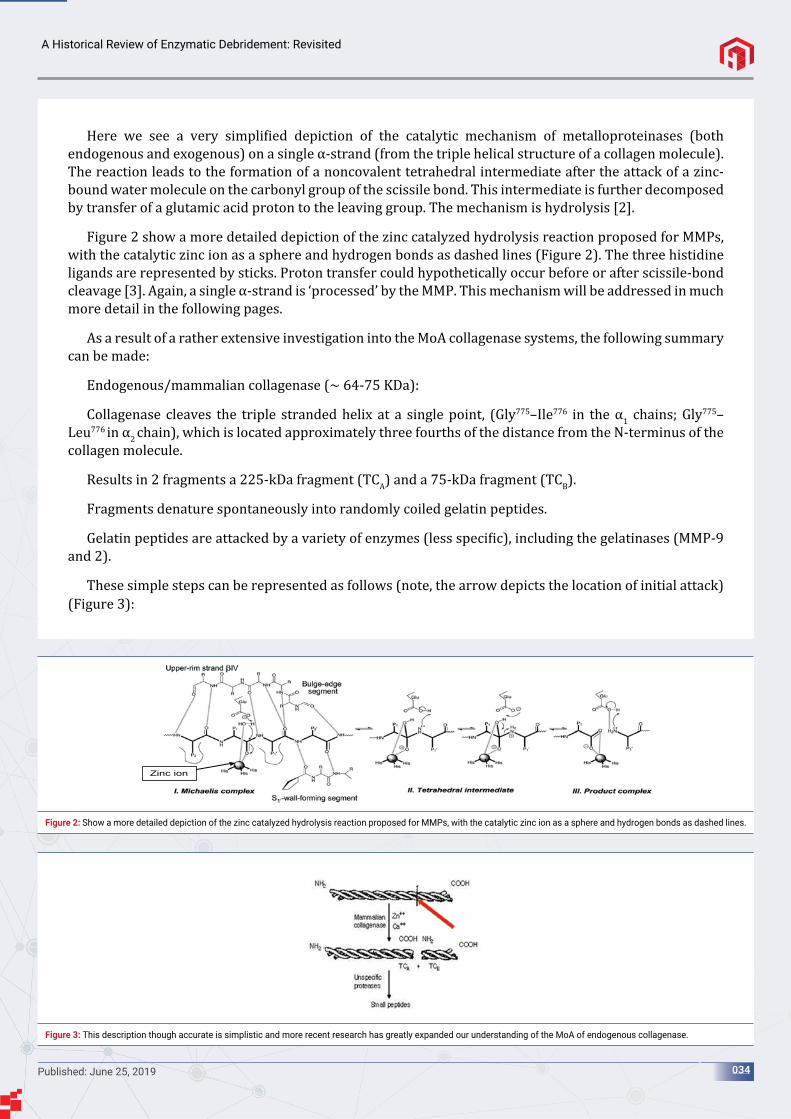
A Historical Review of Enzymatic Debridement: Revisited
Published: June 25, 2019 034
Here we see a very simpli ied depiction of the catalytic mechanism of metalloproteinases (both endogenous and exogenous) on a single α-strand (from the triple helical structure of a collagen molecule). The reaction leads to the formation of a noncovalent tetrahedral intermediate after the attack of a zinc-bound water molecule on the carbonyl group of the scissile bond. This intermediate is further decomposed by transfer of a glutamic acid proton to the leaving group. The mechanism is hydrolysis [2].
Figure 2 show a more detailed depiction of the zinc catalyzed hydrolysis reaction proposed for MMPs, with the catalytic zinc ion as a sphere and hydrogen bonds as dashed lines (Figure 2). The three histidine ligands are represented by sticks. Proton transfer could hypothetically occur before or after scissile-bond cleavage [3]. Again, a single α-strand is ‘processed’ by the MMP. This mechanism will be addressed in much more detail in the following pages.
As a result of a rather extensive investigation into the MoA collagenase systems, the following summary can be made:
Endogenous/mammalian collagenase (~ 64-75 KDa):
Collagenase cleaves the triple stranded helix at a single point, (Gly775–Ile776 in the α1 chains; Gly775–Leu776 in α2 chain), which is located approximately three fourths of the distance from the N-terminus of the collagen molecule.
Results in 2 fragments a 225-kDa fragment (TCA) and a 75-kDa fragment (TCB).
Fragments denature spontaneously into randomly coiled gelatin peptides.
Gelatin peptides are attacked by a variety of enzymes (less speci ic), including the gelatinases (MMP-9 and 2).
These simple steps can be represented as follows (note, the arrow depicts the location of initial attack) (Figure 3):
Zinc ion
Figure 2: Show a more detailed depiction of the zinc catalyzed hydrolysis reaction proposed for MMPs, with the catalytic zinc ion as a sphere and hydrogen bonds as dashed lines.
Figure 3: This description though accurate is simplistic and more recent research has greatly expanded our understanding of the MoA of endogenous collagenase.
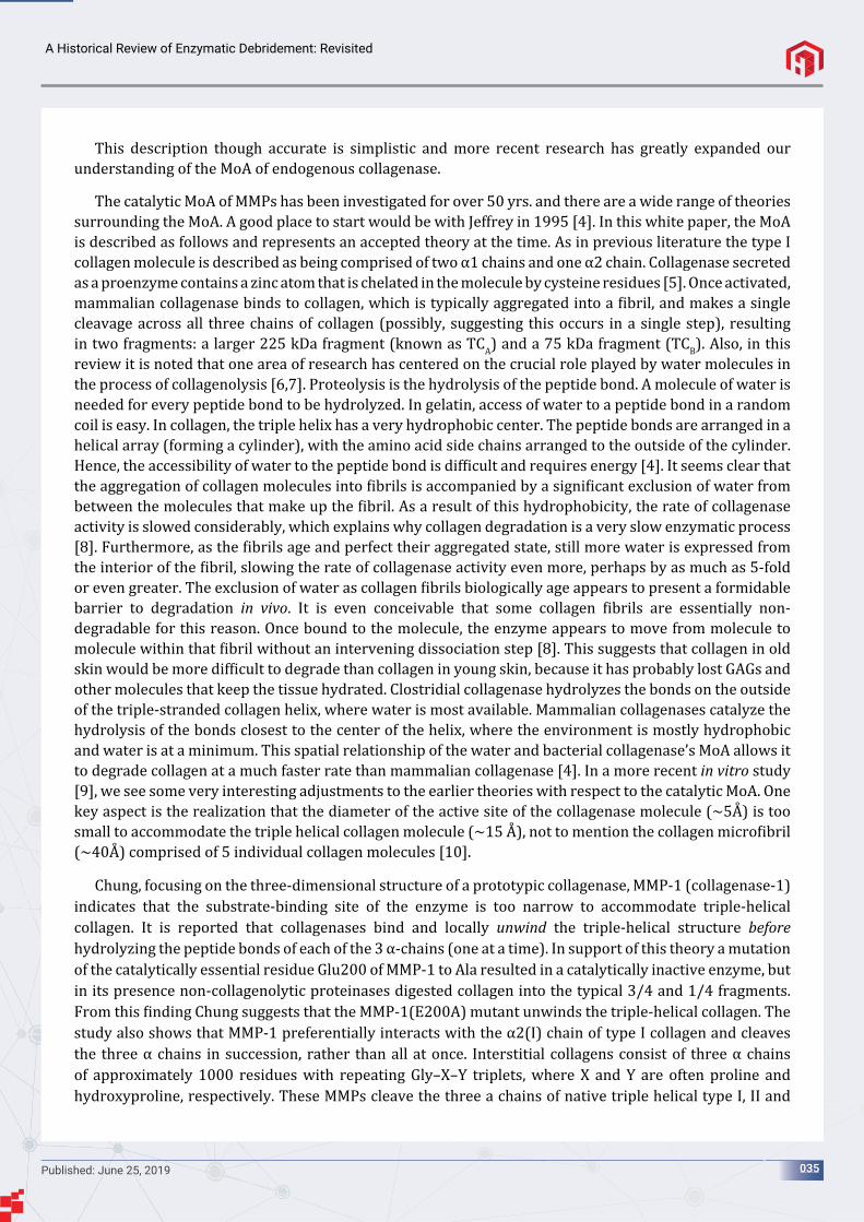
A Historical Review of Enzymatic Debridement: Revisited
Published: June 25, 2019 035
This description though accurate is simplistic and more recent research has greatly expanded our understanding of the MoA of endogenous collagenase.
The catalytic MoA of MMPs has been investigated for over 50 yrs. and there are a wide range of theories surrounding the MoA. A good place to start would be with Jeffrey in 1995 [4]. In this white paper, the MoA is described as follows and represents an accepted theory at the time. As in previous literature the type I collagen molecule is described as being comprised of two α1 chains and one α2 chain. Collagenase secreted as a proenzyme contains a zinc atom that is chelated in the molecule by cysteine residues [5]. Once activated, mammalian collagenase binds to collagen, which is typically aggregated into a ibril, and makes a single cleavage across all three chains of collagen (possibly, suggesting this occurs in a single step), resulting in two fragments: a larger 225 kDa fragment (known as TCA) and a 75 kDa fragment (TCB). Also, in this review it is noted that one area of research has centered on the crucial role played by water molecules in the process of collagenolysis [6,7]. Proteolysis is the hydrolysis of the peptide bond. A molecule of water is needed for every peptide bond to be hydrolyzed. In gelatin, access of water to a peptide bond in a random coil is easy. In collagen, the triple helix has a very hydrophobic center. The peptide bonds are arranged in a helical array (forming a cylinder), with the amino acid side chains arranged to the outside of the cylinder. Hence, the accessibility of water to the peptide bond is dif icult and requires energy [4]. It seems clear that the aggregation of collagen molecules into ibrils is accompanied by a signi icant exclusion of water from between the molecules that make up the ibril. As a result of this hydrophobicity, the rate of collagenase activity is slowed considerably, which explains why collagen degradation is a very slow enzymatic process [8]. Furthermore, as the ibrils age and perfect their aggregated state, still more water is expressed from the interior of the ibril, slowing the rate of collagenase activity even more, perhaps by as much as 5-fold or even greater. The exclusion of water as collagen ibrils biologically age appears to present a formidable barrier to degradation in vivo. It is even conceivable that some collagen ibrils are essentially non-degradable for this reason. Once bound to the molecule, the enzyme appears to move from molecule to molecule within that ibril without an intervening dissociation step [8]. This suggests that collagen in old skin would be more dif icult to degrade than collagen in young skin, because it has probably lost GAGs and other molecules that keep the tissue hydrated. Clostridial collagenase hydrolyzes the bonds on the outside of the triple-stranded collagen helix, where water is most available. Mammalian collagenases catalyze the hydrolysis of the bonds closest to the center of the helix, where the environment is mostly hydrophobic and water is at a minimum. This spatial relationship of the water and bacterial collagenase’s MoA allows it to degrade collagen at a much faster rate than mammalian collagenase [4]. In a more recent in vitro study [9], we see some very interesting adjustments to the earlier theories with respect to the catalytic MoA. One key aspect is the realization that the diameter of the active site of the collagenase molecule (~5Å) is too small to accommodate the triple helical collagen molecule (~15 Å), not to mention the collagen micro ibril (~40Å) comprised of 5 individual collagen molecules [10].
Chung, focusing on the three-dimensional structure of a prototypic collagenase, MMP-1 (collagenase-1) indicates that the substrate-binding site of the enzyme is too narrow to accommodate triple-helical collagen. It is reported that collagenases bind and locally unwind the triple-helical structure before hydrolyzing the peptide bonds of each of the 3 α-chains (one at a time). In support of this theory a mutation of the catalytically essential residue Glu200 of MMP-1 to Ala resulted in a catalytically inactive enzyme, but in its presence non-collagenolytic proteinases digested collagen into the typical 3/4 and 1/4 fragments. From this inding Chung suggests that the MMP-1(E200A) mutant unwinds the triple-helical collagen. The study also shows that MMP-1 preferentially interacts with the α2(I) chain of type I collagen and cleaves the three α chains in succession, rather than all at once. Interstitial collagens consist of three α chains of approximately 1000 residues with repeating Gly–X–Y triplets, where X and Y are often proline and hydroxyproline, respectively. These MMPs cleave the three a chains of native triple helical type I, II and
A Historical Review of Enzymatic Debridement: Revisited
Published: June 25, 2019 036
III collagens after Gly in a particular sequence (Gln/Leu)–Gly#(Ile/Leu)– (Ala/Leu) (# indicates the bond cleaved) located approximately three quarters away from the Nterminus of the collagen molecule. The action of these enzymes is critical for the initiation of collagen breakdown, as once collagens are cleaved into 3/4 and 1/4 fragments they denature at body temperature and are degraded by gelatinases and other nonspeci ic tissue proteinases.
The structural basis for collagen-degrading speci icity among certain members of MMPs is not clearly understood. An additional enigma is the mechanism by which collagenases cleave triple-helical collagens when the dimensions of the collagenase active site and the structure of interstitial collagens were considered in the same yr. as Jeffrey [11]. The substratebinding site of MMP-1 forms a deep cleft with the catalytic zinc located at the bottom, and the entrance of this groove is only ~5Å wide, suf icient to accommodate only a single polypeptide chain. Type I collagen, on the other hand, consisting of two a1(I) chains and one α2(I) chain, is ~3,000 Å in length and ~15A˚ in diameter [9]. Thus, the triple-helical collagen does not it into the active site cleft of the enzyme. Molecular docking attempts to place the triple-helical model of Kramer et al. [12], to the crystal structure of porcine MMP-1, indicated that the closest susceptible peptide bond is at least 7 Å away from the catalytic zinc atom. Chung et al. [9], concludes that either the active site of MMP-1 undergoes large conformational changes or that the triple-helical collagen needs to be unwound, so a single a α-chain can it into the active site of the enzyme. In an effort to demonstrate collagen unwinding by MMP-1, the researchers performed a series of in vitro experiments. Glu200, the residue essential for peptide hydrolysis, was mutated to Ala. They postulated that such a mutant would locally unwind collagen upon interaction with collagen, but would not cleave peptide bonds, and that the unwound collagen would then be susceptible to cleavage by a non-collagenolytic enzymes. The MMP-1(E200A) mutant was essentially inactive and unable to cleave the α1(I) and α2(I) chains of collagen I. As demonstrated previously [13], the catalytic domain of MMP-1 lacking the C-terminal Hpx (hemopexin) like (attachment) domain (MMP-1(ΔC)) also could not cleave collagen I. However, when collagen I was incubated with MMP-1(E200A) and MMP-1(ΔC) at 250C, it was cleaved into the typical 3/4 (TCA) and 1/4 (TCB). So, essentially the authors concluded that with MMP-1(E200A)... thought to unwind the triple helical molecule and MMP-1(ΔC)...catalytic domain without attachment domain (Hpxhemopexin like) was able to produce the two initial fragments (TCa and b). NH2-terminal sequencing of the TCB fragments indicated that MMP-1(ΔC) and MMP-3 (stromelysin-1, noncollagenolytic proteinases) cleaved the Gly775–Ile776 bond of the α1(I) chain(s) and the Gly775– Leu776 bond of the α2(I) chain in the presence of MMP-1(E200A). It was notable that the α1(I) chain(s) was cleaved more rapidly by non-collagenolytic proteinases in the presence of MMP-1(E200A) compared with the active MMP-1 alone. From this Chung suggests that the unwinder MMP-1(E200A) preferentially interacts with the α2(I) chain, which renders the α1(I) chains more exposed and susceptible to a cutter proteinase. This suggests that the unwinding of collagen by MMP-1 takes place only locally, and it does not affect the overall triple-helical structure. The requirement of higher concentrations of the unwinder and the cutter to cleave collagen suggests that both components must simultaneously bind to the collagen substrate. In the case of HpxMMP-1 and MMP-1(ΔC), the ratio of the α1(I) to α2(I) chain cleavage products was similar to that of full-length MMP-1, suggesting that together they behave like a full length collagenase most likely by associating with collagen in a similar manner.
In general terms the regions susceptible to proteinases are usually exposed on the surface of molecules and they are often lexible, so that the scissile bond can readily be accommodated within the active site of the enzyme. The interstitial collagens are long triple-helical structures consisting of three left-handed poly-Pro II-like helices stabilized by hydrogen bonds formed among the backbones of three α-chains and they are highly resistant to most proteinases. Chung goes on to mention that this is the irst demonstration that a single polypeptide proteinase induces signi icant structural changes in the substrate prior to peptide bond hydrolysis. Owing to the structural constraint between collagenase and the collagen
A Historical Review of Enzymatic Debridement: Revisited
Published: June 25, 2019 037
substrate, several hypotheses have been proposed to explain how collagenase may act on triple-helical collagens [11,14,15]. This includes: a proline zipper model, proposing that the proline-rich linker region of collagenases interacts with and unwinds the triple-helical collagen, and a collagen-trapping model in which the Hpx domain folds over the catalytic site sandwiching collagen [11]. However, the intact linker region may not be necessary as the catalytic domain and the Hpx domain added together can cleave collagen, as per Chung. According to Chung the collagen trapping model is also inconsistent with the observation that non-collagenolytic proteinases can cleave α1(I) and α2(I) chains in the presence of MMP-1(E200A), whereas in the aforementioned model they would be protected by the Hpx domain. Chung also considered the following two other possible mechanisms:
Collagenase stabilizes the partially unwound state of collagen that may occur spontaneously around the collagenase-susceptible region.
Conformational changes occur within the collagenase molecule in such a way that it accommodates the triple-helical collagen in the active site.
As these two possibilities are also inconsistent with Chung’s results, Chung concluded that the critical aspects of the collagenolytic speci icity rely on the structural changes in collagen, induced by interacting with collagenase.
Still, additional (and sometimes competing) theories on the MoA are described in a review paper by Duarte [16]. In this review more detail is added to the discussion. MMP-1 is described as having a C-terminal HPX- attachment domain that comprises a four-bladed βpropeller, and is linked to the catalytic domain via a lexible hinge region. It has been reported that this linker peptide has a critical role assisting collagen binding/unwinding before collagenolysis, either by direct binding of the substrate [11], or by allowing the proper alignment of the CAT (catalytic) and HXP (attachment) domains [17]. MMP-1, as well as the other ‘‘classic’’ collagenases (MMP-8 and MMP-13), hydrolyzes interstitial ( ibrillar) collagens I, II and III into the characteristic 1/4 and 3/4 length fragments, at a region on collagen molecule more susceptible to conformational changes. It was also shown that the conformational arrangement of the hinge region of MMP-1 is crucial in the accurate positioning of HPX and CAT domains prior collagenolysis [18]. Note, in earlier work Chung pointed out that this ‘linker region’ may not be necessary for catalysis. As previously mentioned, crystalline structures of MMP-1 [19,20] and MMP-8 showed that the catalytic cleft of these enzymes is too narrow (~5 Å) to accommodate the collagen triple helix (~15Å in diameter). The hypothesis that the triple-helical collagen needs to be unwounded, so that a single a chain can it into the active site of the enzyme has experimental support, as well [21-23]. Using NMR and small angle X-ray scattering, Bertini and co-workers have observed an open/extended and a closed conformation of MMP-1 [21,22]. Also, the structures of MMPs and MMPpeptide complexes showed speci ic interactions between the collagenase and a triple-helical peptide (THP, composed by three chains: 1T, 2T, and 3T), used as a collagen model [18]. Bertini’s data suggest that collagenolysis relies on multiple exosites (secondary binding sites, remote from the active site) interactions, where MMP-1 domains interact cooperatively with the three different α-chains of collagen. These interactions allow the scissile bond to be correctly positioned at the active site, and, at the same time, the molecular stretching of the substrate promotes the local unfolding of collagen that is required for cleavage. The irst proposition for a detailed mechanism describing the collagenolysis by MMP1 [18,21,22] considers four main steps:
In the extended (opened) conformation, MMP-1 binds to the triple-helical peptide 1T-2 T at Val23-Leu26 via the HPX domain: due to the lexibility of the linker region, the CAT domain is guided to the residues around the cleavage site (Gly16-Ile17 of chain 1T, corresponding to the Gly775–Ile776 bond on α1( I) collagen ).
A Historical Review of Enzymatic Debridement: Revisited
Published: June 25, 2019 038
Back-rotation of the CAT and HXP domains leads to the closed MMP-1 conformation; this promotes unwinding of the triple-helical peptide and docking of the 1T chain into the metalloproteinase active site.
Hydrolysis occurs and, initially, both peptide fragments remain bounded to the active site.
The C-terminal region of the N terminal peptide fragment is released; afterwards, MMP-1 hydrolyzes the peptide bonds of each remaining chains in succession.
Most data concerning the mechanisms of collagenolysis, including data from the action of bacterial collagenases, have been interpreted based on this paradigm – that there is a local unfolding of collagen prior to cleavage [16,24], which is detailed in the next section, ‘bacterial collagenase’. However, as is common in scienti ic research, there are alternative theories. Hydrolysis at room temperature – a temperature well below the melting temperature of type I collagen, Tm <360C [25] – is achieved without the HPX binding domain (non-catalytic) of MMPs that was known for being involved in the binding and the unwinding of triple-helical collagen [23,26]. Recall that Chung credits the unwinder MMP-1(E200A) mutant with the unwinding activity. In a break from the unwinding theory of collagenase activity it was also demonstrated that these collagenases preferentially recognize and hydrolyze partially unfolded states of collagen. These substrate-centered observations led the authors to propose an alternative mechanism of collagenolysis, in which collagenases do not act as triple-helicases. This proposal assumes that collagen is lexible in the vicinity of the cleavage site and in consequence, digestion occurs without collagenases actively unwinding the triple-helical collagen [26], an energetically costly task. It is possible to speculate that, within connective tissues, collagens and collagenases may interact with several other components of extracellular matrices like ibronectin, integrins, etc. Those interactions may induce conformational alterations on both enzyme and substrate which, in turn, impact the interaction between MMPs and their triplehelical substrates. So, clearly there are competing theories in the literature with respect to the details of the MoA of MMPs.
Bacterial collagenase
Bacterial collagenase, although a zinc metalloenzyme requiring calcium for its activity, bears little structural relationship to mammalian collagenase. Bacterial collagenase rapidly attacks and degrades human collagen into small peptides. In vitro, human types I and III collagen, extracted and puri ied from placental tissue, was digested by incubation with bacterial collagenase. After analysis on Superex-30 gel sieve chromatography, the breakdown products were shown to be of the size of di- and tri-peptides. The collagen-derived peptides were then added to rat ibroblast culture to evaluate the effects of these breakdown products on cell proliferation and biosynthetic activity. By means of the neutral red test, stimulated cell proliferation was demonstrated when collagen breakdown products, at a concentration of 5 to 50 ng/mL of medium, were added by Cortivo et al. [27], Postlethwaite et al. [28]. Other authors have observed migration of a variety of cell types (keratinocytes, endothelial vascular cells, ibroblasts, etc.) key for wound progression in response to exposure to clostridial and endogenous collagenases [28-33]. More recently it has been demonstrated (in vitro) that collagenase-driven digestion of human cellular-synthesized extracellular matrices yields several collagen and non-collagenous peptides with known and unexpected activities linked to wound progression and the cellular responses to injury, including cellular migration, proliferation and angiogenic activation. Pre-clinical cell based assays reveal that the bacterial collagenase elaborated and combinatorial peptides identi ied/synthesized possess signi icant growth-promoting, migration-enhancing and angiogenesis inducing activities when tested in the 10-100nM range. Also, in this work in vivo (murine) models were used to demonstrate cell migration/proliferation as a result of exposure to the aforementioned peptides. However, the peptides were much larger than those described by Cortivo et al, 1995 and Postlethwaite et al. 1978 [27,28]. In the more recent work the peptides ranged from 8 to 20 amino acids long [34]. Other literature suggests reduced scarring, acute/chronic wound progression, antiin lammatory properties, growth factor release, growth factor cascade
A Historical Review of Enzymatic Debridement: Revisited
Published: June 25, 2019 039
initiation, etc., as effects of bacterial collagenase interaction with wound matrix components. Though these aspects warrant much more investigation, it would appear that bacterial collagenase has the ability to rapidly digest collagen and promote other cellular functions bene icial in wound repair.
Bacterial collagenase is made up of proteolytic enzymes that break collagen into small peptides of differing molecular weights. Seven collagenases (isoforms) have been identi ied in the puri ied culture iltrate of C. histolyticum, and all have been puri ied to homogeneity. It should be noted that only two genes,
colG and colH transcribe for two metalloproteases. However, due to post-transcriptional autolytic events, seven truncated collagenases (isoforms) have been identi ied. The seven collagenases can be divided into two classes, I (α, β, γ, η) and II (δ, ε, ξ), which are classi ied based on their bacterial gene of origin (colG and ColH, respectively) and on their point of hydrolytic attack on the collagen molecule – class I enzymes act at loci near the ends of the collagen triple-helical domains, whereas class II enzymes make internal initial cleavages. These collagenases uniquely cleave the interstitial collagens and exhibit both endopeptidase and tripeptidylcarboxypeptidase activities. The combined activity of endo- and tripeptidyl-C-peptidase makes these enzymes ideally suited for rapid collagen degradation. Their combined action at many sites along the peptide chain results in the sequential cleavage into short segments. Both classes have speci ic binding domains that enable them to recognize and bind to triple helical collagen, in a variety of locations [35-38]. An interesting note, in the work by French who 1st studied the isoforms of collagenase described them as attacking at hyper-reactive cleavage sites suggesting that type I, II, and III collagens contain regions that have speci ic non-triple helical conformations [36]. In other words, there is no need to unwind the triple helix. So, again, we see competing theories with respect to the MoA of bacterial collagenase, as there are for endogenous collagenase.
As a reminder, in contrast to clostridium histolyticum collagenases, mammalian collagenases act differently by cleaving interstitial collagen at a single locus within the triple helical structure, giving rise to 2 large fragments, TCA and TCB
[4,39]. These portions of the helix are then attacked by other nonspeci ic proteases, released by connective tissue cells, to be further degraded into small peptides [27].
An interesting thought with respect to bacterial collagenase and endogenous collagenase was put forth by Parks in 1995 [29]. The migration (obligatorily occurring within a viable part of the wound bed) of cells responsible for removing non-viable collagen is hindered by necrotic (non-viable) tissue. For this reason, little if any collagenase expression (from migrating cells) would be detected in necrotic regions. Alternatively, bacterial collagenase, when topically applied, attacks proteins within the super icial and necrotic (non-viable) areas of the wound [29]. It could be argued that bacterial collagenase treatment is not simply augmentation therapy, but rather provides an essential biochemical activity to areas where in the cells are not producing proteases. Thus, because of these critical and signi icant spatial differences and because of the marked differences in the matrix proteins that these proteinases can degrade, it is easy to conceive that the catalytic activity of these two collagenases can result in different effects in repair [29]. This is an interesting thought; however, it should be noted that collagenase-2 (MMP-8, neutrophil/PMNL collagenase) would still be present in chronic wounds halted in the in lammatory phase of wound healing. In addition, other sources suggest the presence of collagenase expressed by other cell types in necrotic tissue (due to the expression of pro-in lammatory cytokines by in lammatory cells- Parks, 1995) [29], whereas, other sources state the lack of collagenase expression in necrotic tissue [30].
It has been shown in diabetic ulcers that necrotic tissue is anchored to the wound surface by a layer of perpendicular strands of undenatured collagen [1]. However, it is likely that some of these collagen strands are partially denatured. Collagenases, by de inition, are enzymes capable of solubilizing ibrous collagen (both native and denatured collagen) by peptide bond cleavage under physiologic pH and temperature conditions. Thus, collagenase attacks not only necrotic tissue, but also ibers of undenatured collagen. It
A Historical Review of Enzymatic Debridement: Revisited
Published: June 25, 2019 040
is suspected that the ibers of undenatured collagen anchor the eschar plug to the wound bed. With use of topically applied collagenase, the entire ‘nerotic plug’ could be released and the remaining anchoring ibers would be removed. This would tend to lead to a cleaner and more thoroughly prepared wound bed.
It has also been documented that, at times, collagenase treatments have resulted in a decrease in visible scarring [40,41].
Since necrotic tissue is an important local cause of failure of wound progression, it would seem obvious that such binding must be severed, so that debridement and wound progression can occur. Otherwise, granulation is slowed and no supportive base is available for epithelialization. Collagenase is irreversibly inactivated in a low-pH environment. It functions best in the pH range of 6 to 8 and temperatures below 56°C. Chelating agents (EDTA, citric acid, sodium citrate, etc.) also inactivate the enzyme by interacting with Ca2+ ions and Zn2+ ions, essential constituents of the structure (and function) of collagenase. It is well known that four Ca2+ ions play a role in stabilizing the 30 structure of the protein near the active site. It is also well known that a Zn2+ ion is located in the active site and is necessary for enzymatic activity. Collagenase hydrolyzes the peptide bonds in collagen. It does not attack other proteins such as hemoglobin or ibrin, important components in the formation of granulation tissue. In addition, collagenase does not attack growth factors, tissue inhibitors of metalloproteinase (TIMPs), and other critical components of the wound repair cycle [40], whereas, other, less speci ic enzymatic systems, such as papain–urea, were reported to have a negative effect on plateletderived growth factor (PDGF), and perhaps (based upon its MoA) other growth factors, TIMPS, integrins, etc. One could postulate that this lack of selectivity is one reason these historical topical enzymatic formulations are no longer widely used or even available.
Collagenase has been reported as an effective agent for the débridement of thermal burns. Although any protease would thoroughly digest degraded matter in the center of the burn eschar, only collagenase would effectively attack necrotic edges of the eschar, including the perpendicular ibers of undenatured collagen. These perpendicular collagen ibers anchor the eschar plug to the wound bed, and their removal is likely to be key for optimal wound bed preparation.
Collagenase has also been found to be useful in the débridement of third-degree burns. Otteman and Stahlgren compared the lytic effects of a number of enzymes on experimentally induced burns. The enzymes studied included streptokinase–streptodornase, trypsin– chymotrypsin, papain, icin, desoxyribonuclease– ibrinolysin, and collagenase. Of these enzymes, only collagenase and papain were more than 90% effective in the digestion of wound debris and necrotic material [42].
The design/structure of the active site of bacterial collagenase allows it to cleave the triple helical collagen at many different points. Anywhere a Gly-X-Y (where X = proline and Y= hydroxyproline) exists, it is felt bacterial collagenase can attack [29,35]. Collagen is unique among proteins in that every third amino acid of the peptide chain is glycine, the smallest amino acid. Each of the 3 polypeptide chains contains about 1,000 amino acids, so the structure of each chain can be considered to be 330 repeating units of glycine–X–Y; where X = proline and Y= hydroxyproline [43]. More recent sources provide even more information as to the point of attack. One source describes 10.5% of the collagen molecule being comprised by the glycine-proline-hydroxyproline triplet [44]. Another source mentions that 23% of the molecule is comprised of a combination of proline and hydroxyproline [45]. Yet, a more recent source describes proline ~28%; hydroxyproline ~38% of the collagen molecule [46].̈ Given this, it is possible that on a single α-chain, there could be ~100 to ~ 330 locals where bacterial collagenase might attack.
As a result of a rather extensive investigation into the MoA collagenase systems, the following summary can be made:
Bacterial collagenase (~ 115-120 KDa):
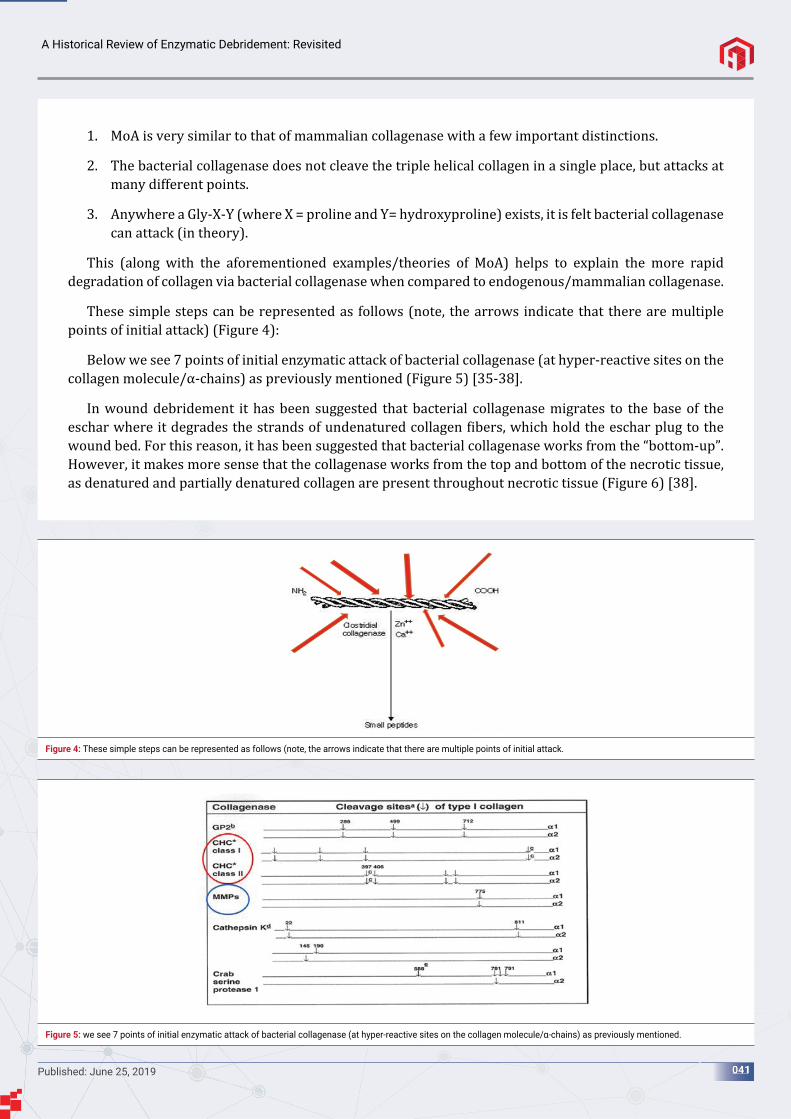
A Historical Review of Enzymatic Debridement: Revisited
Published: June 25, 2019 041
1. MoA is very similar to that of mammalian collagenase with a few important distinctions.
2. The bacterial collagenase does not cleave the triple helical collagen in a single place, but attacks at many different points.
3. Anywhere a Gly-X-Y (where X = proline and Y= hydroxyproline) exists, it is felt bacterial collagenase can attack (in theory).
This (along with the aforementioned examples/theories of MoA) helps to explain the more rapid degradation of collagen via bacterial collagenase when compared to endogenous/mammalian collagenase.
These simple steps can be represented as follows (note, the arrows indicate that there are multiple points of initial attack) (Figure 4):
Below we see 7 points of initial enzymatic attack of bacterial collagenase (at hyper-reactive sites on the collagen molecule/α-chains) as previously mentioned (Figure 5) [35-38].
In wound debridement it has been suggested that bacterial collagenase migrates to the base of the eschar where it degrades the strands of undenatured collagen ibers, which hold the eschar plug to the wound bed. For this reason, it has been suggested that bacterial collagenase works from the “bottom-up”. However, it makes more sense that the collagenase works from the top and bottom of the necrotic tissue, as denatured and partially denatured collagen are present throughout necrotic tissue (Figure 6) [38].
Figure 4: These simple steps can be represented as follows (note, the arrows indicate that there are multiple points of initial attack.
Figure 5: we see 7 points of initial enzymatic attack of bacterial collagenase (at hyper-reactive sites on the collagen molecule/α-chains) as previously mentioned.
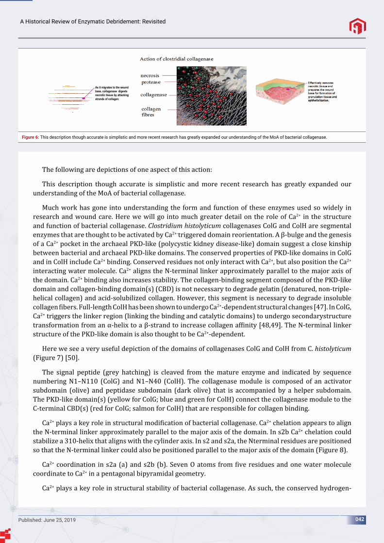
A Historical Review of Enzymatic Debridement: Revisited
Published: June 25, 2019 042
The following are depictions of one aspect of this action:
This description though accurate is simplistic and more recent research has greatly expanded our understanding of the MoA of bacterial collagenase.
Much work has gone into understanding the form and function of these enzymes used so widely in research and wound care. Here we will go into much greater detail on the role of Ca2+ in the structure and function of bacterial collagenase. Clostridium histolyticum collagenases ColG and ColH are segmental enzymes that are thought to be activated by Ca2+-triggered domain reorientation. A β-bulge and the genesis of a Ca2+ pocket in the archaeal PKD-like (polycystic kidney disease-like) domain suggest a close kinship between bacterial and archaeal PKD-like domains. The conserved properties of PKD-like domains in ColG and in ColH include Ca2+ binding. Conserved residues not only interact with Ca2+, but also position the Ca2+
interacting water molecule. Ca2+ aligns the N-terminal linker approximately parallel to the major axis of the domain. Ca2+ binding also increases stability. The collagen-binding segment composed of the PKD-like domain and collagen-binding domain(s) (CBD) is not necessary to degrade gelatin (denatured, non-triple-helical collagen) and acid-solubilized collagen. However, this segment is necessary to degrade insoluble collagen ibers. Full-length ColH has been shown to undergo Ca2+-dependent structural changes [47]. In ColG, Ca2+ triggers the linker region (linking the binding and catalytic domains) to undergo secondarystructure transformation from an α-helix to a β-strand to increase collagen af inity [48,49]. The N-terminal linker structure of the PKD-like domain is also thought to be Ca2+-dependent.
Here we see a very useful depiction of the domains of collagenases ColG and ColH from C. histolyticum (Figure 7) [50].
The signal peptide (grey hatching) is cleaved from the mature enzyme and indicated by sequence numbering N1–N110 (ColG) and N1–N40 (ColH). The collagenase module is composed of an activator subdomain (olive) and peptidase subdomain (dark olive) that is accompanied by a helper subdomain. The PKD-like domain(s) (yellow for ColG; blue and green for ColH) connect the collagenase module to the C-terminal CBD(s) (red for ColG; salmon for ColH) that are responsible for collagen binding.
Ca2+ plays a key role in structural modi ication of bacterial collagenase. Ca2+ chelation appears to align the N-terminal linker approximately parallel to the major axis of the domain. In s2b Ca2+ chelation could stabilize a 310-helix that aligns with the cylinder axis. In s2 and s2a, the Nterminal residues are positioned so that the N-terminal linker could also be positioned parallel to the major axis of the domain (Figure 8).
Ca2+ coordination in s2a (a) and s2b (b). Seven O atoms from ive residues and one water molecule coordinate to Ca2+ in a pentagonal bipyramidal geometry.
Ca2+ plays a key role in structural stability of bacterial collagenase. As such, the conserved hydrogen-
As it migrates to the woundbase, collagenase digestsnecrotic tissue by attackingstrands of collagen.
Figure 6: This description though accurate is simplistic and more recent research has greatly expanded our understanding of the MoA of bacterial collagenase.
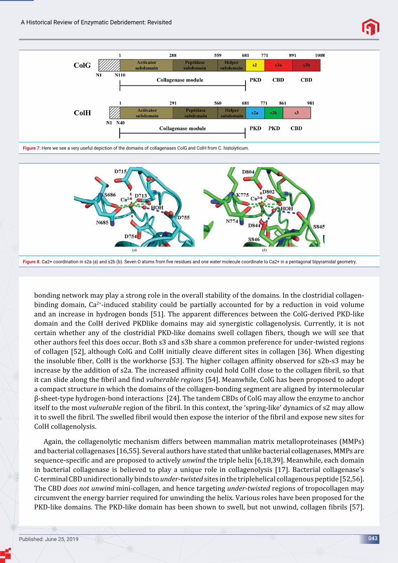
A Historical Review of Enzymatic Debridement: Revisited
Published: June 25, 2019 043
bonding network may play a strong role in the overall stability of the domains. In the clostridial collagen-binding domain, Ca2+-induced stability could be partially accounted for by a reduction in void volume and an increase in hydrogen bonds [51]. The apparent differences between the ColG-derived PKD-like domain and the ColH derived PKDlike domains may aid synergistic collagenolysis. Currently, it is not certain whether any of the clostridial PKD-like domains swell collagen ibers, though we will see that other authors feel this does occur. Both s3 and s3b share a common preference for under-twisted regions of collagen [52], although ColG and ColH initially cleave different sites in collagen [36]. When digesting the insoluble iber, ColH is the workhorse [53]. The higher collagen af inity observed for s2b-s3 may be increase by the addition of s2a. The increased af inity could hold ColH close to the collagen ibril, so that it can slide along the ibril and ind vulnerable regions [54]. Meanwhile, ColG has been proposed to adopt a compact structure in which the domains of the collagen-bonding segment are aligned by intermolecular β-sheet-type hydrogen-bond interactions [24]. The tandem CBDs of ColG may allow the enzyme to anchor itself to the most vulnerable region of the ibril. In this context, the ‘spring-like’ dynamics of s2 may allow it to swell the ibril. The swelled ibril would then expose the interior of the ibril and expose new sites for ColH collagenolysis.
Again, the collagenolytic mechanism differs between mammalian matrix metalloproteinases (MMPs) and bacterial collagenases [16,55]. Several authors have stated that unlike bacterial collagenases, MMPs are sequence-speci ic and are proposed to actively unwind the triple helix [6,18,39]. Meanwhile, each domain in bacterial collagenase is believed to play a unique role in collagenolysis [17]. Bacterial collagenase’s C-terminal CBD unidirectionally binds to under-twisted sites in the triplehelical collagenous peptide [52,56]. The CBD does not unwind mini-collagen, and hence targeting under-twisted regions of tropocollagen may circumvent the energy barrier required for unwinding the helix. Various roles have been proposed for the PKD-like domains. The PKD-like domain has been shown to swell, but not unwind, collagen ibrils [57].
Figure 7: Here we see a very useful depiction of the domains of collagenases ColG and ColH from C. histolyticum.
Figure 8: Ca2+ coordination in s2a (a) and s2b (b). Seven O atoms from fi ve residues and one water molecule coordinate to Ca2+ in a pentagonal bipyramidal geometry.
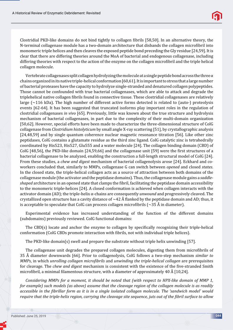
A Historical Review of Enzymatic Debridement: Revisited
Published: June 25, 2019 044
Clostridial PKD-like domains do not bind tightly to collagen ibrils [58,50]. In an alternative theory, the N-terminal collagenase module has a two-domain architecture that disbands the collagen micro ibril into monomeric triple helices and then cleaves the exposed peptide bond preceding the Gly residue [24,59]. It is clear that there are differing theories around the MoA of bacterial and endogenous collagenase, including differing theories with respect to the action of the enzyme on the collagen micro ibril and the triple helical collagen molecule.
Vertebrate collagenases split collagen hydrolyzing the molecule at a single peptide bond across the three α chains organized in its native triple-helical conformation [60,61]. It is important to stress that a large number of bacterial proteases have the capacity to hydrolyze single-stranded and denatured collagen polypeptides. Those cannot be confounded with true bacterial collagenases, which are able to attack and degrade the triplehelical native collagen ibrils found in connective tissue. These clostridial collagenases are relatively large (~116 kDa). The high number of different active forms detected is related to (auto-) proteolysis events [62-64]. It has been suggested that truncated isoforms play important roles in the regulation of clostridial collagenases in vivo [65]. Previously, little was known about the true structure and hydrolysis mechanism of bacterial collagenases, in part due to the complexity of their multi-domain organization [50,62]. However, special efforts have been made to characterize the three-dimensional structure of ColG collagenase from Clostridium histolyticum by small angle X-ray scattering [51], by crystallographic analysis [24,48,59] and by single quantum coherence nuclear magnetic resonance titration [56]. Like other zinc peptidases, ColG contains a glutamate residue as the third zinc ligand. ColG catalytic zinc is tetrahedrally coordinated by His523, His527, Glu555 and a water molecule [24]. The collagen binding domain (CBD) of ColG [48,56], the PKD-like domain [24,59,66] and the collagenase unit [59] were the irst structures of a bacterial collagenase to be analyzed, enabling the construction a full-length structural model of ColG [24]. From these studies, a chew and digest mechanism of bacterial collagenolysis arose [24]. Eckhard and co-workers concluded that, similarly to MMPs, collagenase G can switch between opened and closed states. In the closed state, the triple-helical collagen acts as a source of attraction between both domains of the collagenase module (the activator and the peptidase domains). Thus, the collagenase module gains a saddle-shaped architecture in an opened state that clamps the ibril, facilitating the peptidase domain accessibility to the monomeric triple-helices [24]. A closed conformation is achieved when collagen interacts with the activator domain (AD); the triple-helix α-chains are consequently unwound and progressively cleaved. The crystallized open structure has a cavity distance of ~42 Å lanked by the peptidase domain and AD; thus, it is acceptable to speculate that ColG can process collagen micro ibrils (~35 Å in diameter).
Experimental evidence has increased understanding of the function of the different domains (subdomains) previously reviewed. ColG functional domains:
The CBD(s) locate and anchor the enzyme to collagen by speci ically recognizing their triple-helical conformation (ColG CBDs promote interaction with ibrils, not with individual triple helices).
The PKD-like domain(s) swell and prepare the substrate without triple helix unwinding [57].
The collagenase unit degrades the prepared collagen molecules, digesting them from micro ibrils of 35 Å diameter downwards [66]. Prior to collagenolysis, ColG follows a two-step mechanism similar to MMPs, in which unrolling collagen micro ibrils and unwinding the triple-helical collagen are prerequisites for cleavage. The chew and digest mechanism is consistent with the existence of the ive-stranded Smith micro ibril, a minimal ilamentous structure, with a diameter of approximately 40 Å [10,24].
Considering MMPs for a moment, it should be noted that (with respect to HPX-like domain of MMP 1, for example) such models (as above) assume that the cleavage region of the collagen molecule is as readily accessible in the ibrillar form as it is in a single isolated collagen molecule. The ‘sandwich model’ would require that the triple-helix region, carrying the cleavage site sequence, juts out of the ibril surface to allow
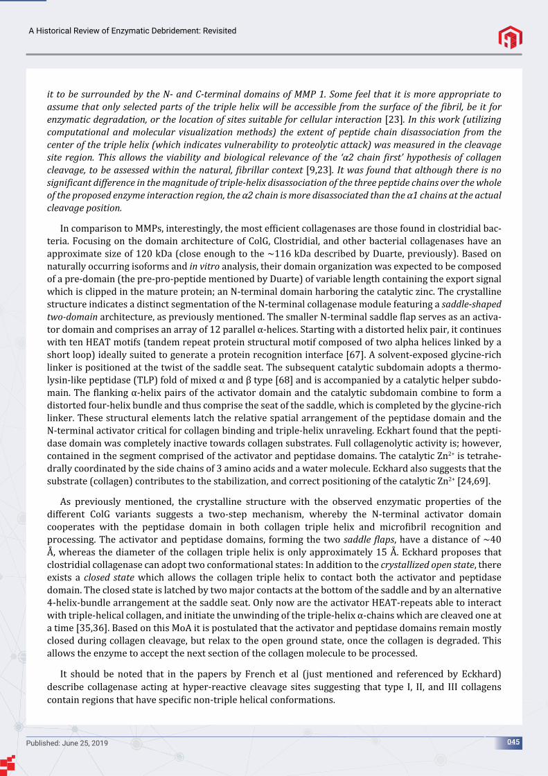
A Historical Review of Enzymatic Debridement: Revisited
Published: June 25, 2019 045
it to be surrounded by the N- and C-terminal domains of MMP 1. Some feel that it is more appropriate to assume that only selected parts of the triple helix will be accessible from the surface of the ibril, be it for enzymatic degradation, or the location of sites suitable for cellular interaction [23]. In this work (utilizing computational and molecular visualization methods) the extent of peptide chain disassociation from the center of the triple helix (which indicates vulnerability to proteolytic attack) was measured in the cleavage site region. This allows the viability and biological relevance of the ‘α2 chain irst’ hypothesis of collagen cleavage, to be assessed within the natural, ibrillar context [9,23]. It was found that although there is no signi icant difference in the magnitude of triple-helix disassociation of the three peptide chains over the whole of the proposed enzyme interaction region, the α2 chain is more disassociated than the α1 chains at the actual cleavage position.
In comparison to MMPs, interestingly, the most ef icient collagenases are those found in clostridial bac-teria. Focusing on the domain architecture of ColG, Clostridial, and other bacterial collagenases have an approximate size of 120 kDa (close enough to the ~116 kDa described by Duarte, previously). Based on naturally occurring isoforms and in vitro analysis, their domain organization was expected to be composed of a pre-domain (the pre-pro-peptide mentioned by Duarte) of variable length containing the export signal which is clipped in the mature protein; an N-terminal domain harboring the catalytic zinc. The crystalline structure indicates a distinct segmentation of the N-terminal collagenase module featuring a saddle-shaped two-domain architecture, as previously mentioned. The smaller N-terminal saddle lap serves as an activa-tor domain and comprises an array of 12 parallel α-helices. Starting with a distorted helix pair, it continues with ten HEAT motifs (tandem repeat protein structural motif composed of two alpha helices linked by a short loop) ideally suited to generate a protein recognition interface [67]. A solvent-exposed glycine-rich linker is positioned at the twist of the saddle seat. The subsequent catalytic subdomain adopts a thermo-lysin-like peptidase (TLP) fold of mixed α and β type [68] and is accompanied by a catalytic helper subdo-main. The lanking α-helix pairs of the activator domain and the catalytic subdomain combine to form a distorted four-helix bundle and thus comprise the seat of the saddle, which is completed by the glycine-rich linker. These structural elements latch the relative spatial arrangement of the peptidase domain and the N-terminal activator critical for collagen binding and triple-helix unraveling. Eckhart found that the pepti-dase domain was completely inactive towards collagen substrates. Full collagenolytic activity is; however, contained in the segment comprised of the activator and peptidase domains. The catalytic Zn2+ is tetrahe-drally coordinated by the side chains of 3 amino acids and a water molecule. Eckhard also suggests that the substrate (collagen) contributes to the stabilization, and correct positioning of the catalytic Zn2+ [24,69].
As previously mentioned, the crystalline structure with the observed enzymatic properties of the different ColG variants suggests a two-step mechanism, whereby the N-terminal activator domain cooperates with the peptidase domain in both collagen triple helix and micro ibril recognition and processing. The activator and peptidase domains, forming the two saddle laps, have a distance of ~40 Å, whereas the diameter of the collagen triple helix is only approximately 15 Å. Eckhard proposes that clostridial collagenase can adopt two conformational states: In addition to the crystallized open state, there exists a closed state which allows the collagen triple helix to contact both the activator and peptidase domain. The closed state is latched by two major contacts at the bottom of the saddle and by an alternative 4-helix-bundle arrangement at the saddle seat. Only now are the activator HEAT-repeats able to interact with triple-helical collagen, and initiate the unwinding of the triple-helix α-chains which are cleaved one at a time [35,36]. Based on this MoA it is postulated that the activator and peptidase domains remain mostly closed during collagen cleavage, but relax to the open ground state, once the collagen is degraded. This allows the enzyme to accept the next section of the collagen molecule to be processed.
It should be noted that in the papers by French et al (just mentioned and referenced by Eckhard) describe collagenase acting at hyper-reactive cleavage sites suggesting that type I, II, and III collagens contain regions that have speci ic non-triple helical conformations.
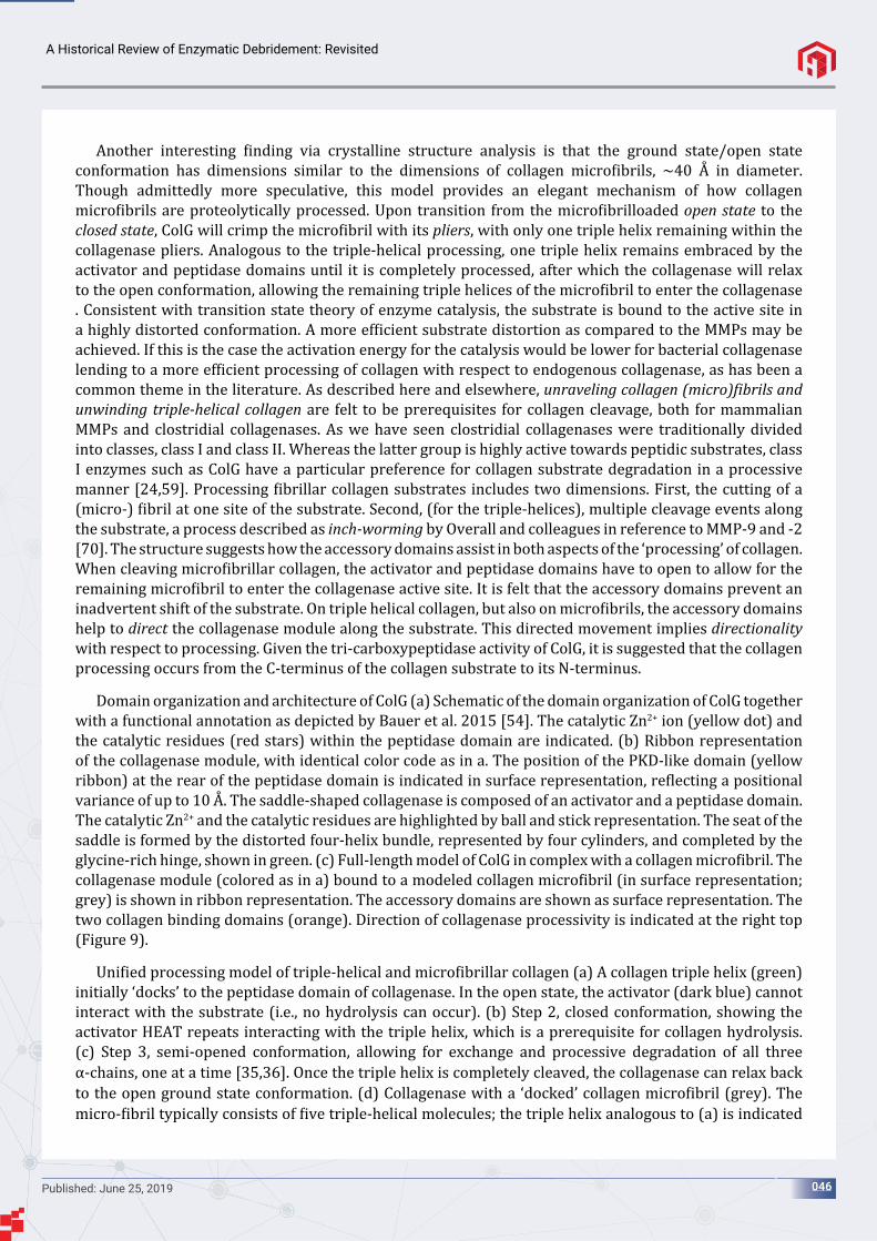
A Historical Review of Enzymatic Debridement: Revisited
Published: June 25, 2019 046
Another interesting inding via crystalline structure analysis is that the ground state/open state conformation has dimensions similar to the dimensions of collagen micro ibrils, ~40 Å in diameter. Though admittedly more speculative, this model provides an elegant mechanism of how collagen micro ibrils are proteolytically processed. Upon transition from the micro ibrilloaded open state to the closed state, ColG will crimp the micro ibril with its pliers, with only one triple helix remaining within the collagenase pliers. Analogous to the triple-helical processing, one triple helix remains embraced by the activator and peptidase domains until it is completely processed, after which the collagenase will relax to the open conformation, allowing the remaining triple helices of the micro ibril to enter the collagenase . Consistent with transition state theory of enzyme catalysis, the substrate is bound to the active site in a highly distorted conformation. A more ef icient substrate distortion as compared to the MMPs may be achieved. If this is the case the activation energy for the catalysis would be lower for bacterial collagenase lending to a more ef icient processing of collagen with respect to endogenous collagenase, as has been a common theme in the literature. As described here and elsewhere, unraveling collagen (micro) ibrils and unwinding triple-helical collagen are felt to be prerequisites for collagen cleavage, both for mammalian MMPs and clostridial collagenases. As we have seen clostridial collagenases were traditionally divided into classes, class I and class II. Whereas the latter group is highly active towards peptidic substrates, class I enzymes such as ColG have a particular preference for collagen substrate degradation in a processive manner [24,59]. Processing ibrillar collagen substrates includes two dimensions. First, the cutting of a (micro-) ibril at one site of the substrate. Second, (for the triple-helices), multiple cleavage events along the substrate, a process described as inch-worming by Overall and colleagues in reference to MMP-9 and -2 [70]. The structure suggests how the accessory domains assist in both aspects of the ‘processing’ of collagen. When cleaving micro ibrillar collagen, the activator and peptidase domains have to open to allow for the remaining micro ibril to enter the collagenase active site. It is felt that the accessory domains prevent an inadvertent shift of the substrate. On triple helical collagen, but also on micro ibrils, the accessory domains help to direct the collagenase module along the substrate. This directed movement implies directionality with respect to processing. Given the tri-carboxypeptidase activity of ColG, it is suggested that the collagen processing occurs from the C-terminus of the collagen substrate to its N-terminus.
Domain organization and architecture of ColG (a) Schematic of the domain organization of ColG together with a functional annotation as depicted by Bauer et al. 2015 [54]. The catalytic Zn2+ ion (yellow dot) and the catalytic residues (red stars) within the peptidase domain are indicated. (b) Ribbon representation of the collagenase module, with identical color code as in a. The position of the PKD-like domain (yellow ribbon) at the rear of the peptidase domain is indicated in surface representation, re lecting a positional variance of up to 10 Å. The saddle-shaped collagenase is composed of an activator and a peptidase domain. The catalytic Zn2+ and the catalytic residues are highlighted by ball and stick representation. The seat of the saddle is formed by the distorted four-helix bundle, represented by four cylinders, and completed by the glycine-rich hinge, shown in green. (c) Full-length model of ColG in complex with a collagen micro ibril. The collagenase module (colored as in a) bound to a modeled collagen micro ibril (in surface representation; grey) is shown in ribbon representation. The accessory domains are shown as surface representation. The two collagen binding domains (orange). Direction of collagenase processivity is indicated at the right top (Figure 9).
Uni ied processing model of triple-helical and micro ibrillar collagen (a) A collagen triple helix (green) initially ‘docks’ to the peptidase domain of collagenase. In the open state, the activator (dark blue) cannot interact with the substrate (i.e., no hydrolysis can occur). (b) Step 2, closed conformation, showing the activator HEAT repeats interacting with the triple helix, which is a prerequisite for collagen hydrolysis. (c) Step 3, semi-opened conformation, allowing for exchange and processive degradation of all three α-chains, one at a time [35,36]. Once the triple helix is completely cleaved, the collagenase can relax back to the open ground state conformation. (d) Collagenase with a ‘docked’ collagen micro ibril (grey). The micro- ibril typically consists of ive triple-helical molecules; the triple helix analogous to (a) is indicated
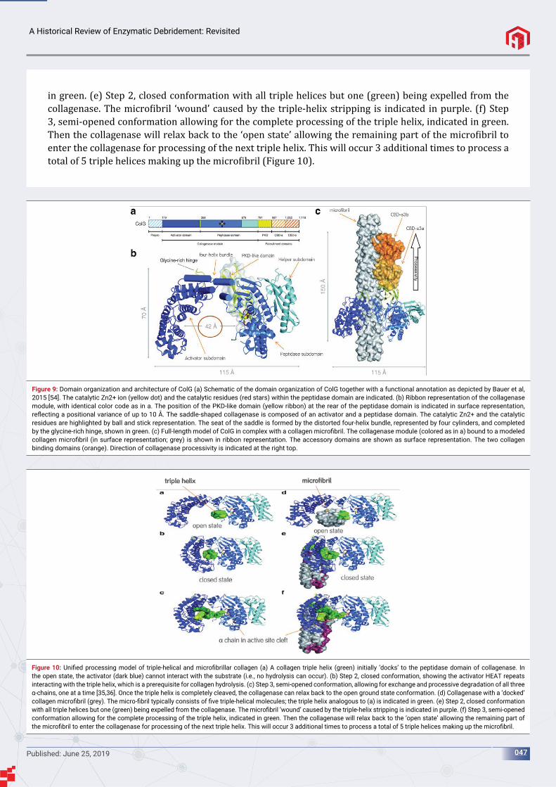
A Historical Review of Enzymatic Debridement: Revisited
Published: June 25, 2019 047
in green. (e) Step 2, closed conformation with all triple helices but one (green) being expelled from the collagenase. The micro ibril ‘wound’ caused by the triple-helix stripping is indicated in purple. (f) Step 3, semi-opened conformation allowing for the complete processing of the triple helix, indicated in green. Then the collagenase will relax back to the ‘open state’ allowing the remaining part of the micro ibril to enter the collagenase for processing of the next triple helix. This will occur 3 additional times to process a total of 5 triple helices making up the micro ibril (Figure 10).
Figure 9: Domain organization and architecture of ColG (a) Schematic of the domain organization of ColG together with a functional annotation as depicted by Bauer et al, 2015 [54]. The catalytic Zn2+ ion (yellow dot) and the catalytic residues (red stars) within the peptidase domain are indicated. (b) Ribbon representation of the collagenase module, with identical color code as in a. The position of the PKD-like domain (yellow ribbon) at the rear of the peptidase domain is indicated in surface representation, refl ecting a positional variance of up to 10 Å. The saddle-shaped collagenase is composed of an activator and a peptidase domain. The catalytic Zn2+ and the catalytic residues are highlighted by ball and stick representation. The seat of the saddle is formed by the distorted four-helix bundle, represented by four cylinders, and completed by the glycine-rich hinge, shown in green. (c) Full-length model of ColG in complex with a collagen microfi bril. The collagenase module (colored as in a) bound to a modeled collagen microfi bril (in surface representation; grey) is shown in ribbon representation. The accessory domains are shown as surface representation. The two collagen binding domains (orange). Direction of collagenase processivity is indicated at the right top.
Figure 10: Unifi ed processing model of triple-helical and microfi brillar collagen (a) A collagen triple helix (green) initially ‘docks’ to the peptidase domain of collagenase. In the open state, the activator (dark blue) cannot interact with the substrate (i.e., no hydrolysis can occur). (b) Step 2, closed conformation, showing the activator HEAT repeats interacting with the triple helix, which is a prerequisite for collagen hydrolysis. (c) Step 3, semi-opened conformation, allowing for exchange and processive degradation of all three α-chains, one at a time [35,36]. Once the triple helix is completely cleaved, the collagenase can relax back to the open ground state conformation. (d) Collagenase with a ‘docked’ collagen microfi bril (grey). The micro-fi bril typically consists of fi ve triple-helical molecules; the triple helix analogous to (a) is indicated in green. (e) Step 2, closed conformation with all triple helices but one (green) being expelled from the collagenase. The microfi bril ‘wound’ caused by the triple-helix stripping is indicated in purple. (f) Step 3, semi-opened conformation allowing for the complete processing of the triple helix, indicated in green. Then the collagenase will relax back to the ‘open state’ allowing the remaining part of the microfi bril to enter the collagenase for processing of the next triple helix. This will occur 3 additional times to process a total of 5 triple helices making up the microfi bril.
A Historical Review of Enzymatic Debridement: Revisited
Published: June 25, 2019 048
Summary
From this chapter one gets a sense of the amount of investigative work performed over the past 50 to 60 years in the area of wound débridement via topical application of bacterial collagenase as a debriding agent. From this and previous chapters one also gets a sense of the variety of differing viewpoints on mechanisms of action of both endogenous and bacterial collagenases, as well as, on the effects on viable tissue of the various enzymatic debriders used clinically in the past and present. Eckhard et al., provide an elegant and easy to follow model (i.e., addresses the geometric aspects of active site cleft vs. collagen micro ibrils/triple helix; open/closed conformation of enzymes; unwinding of micro ibrils/helices; cleavage of individual α chains). This theory is applied in explaining the MoA of endogenous collagenases, as well. Here we see a similar MoA including unique functional ‘domains’, unwinding of micro ibrils/helices, open/closed enzyme geometries, cleaving individual α chains (one at a time). However, as demonstrated in this and in previous chapters, bacterial collagenase is far more ef icient in its MoA for a variety of reasons.
As is the nature of scienti ic research, old ideas make way for new ideas generated as analytical technologies/techniques improve. However, we should not discount the work of so many others over the yrs. out of hand. It is likely that the ‘true’ MoA is a conglomeration of bits and pieces of the theories discussed thus far (and those yet to come). In time, and as more studies using modern analytical methods are performed, perhaps we will see more agreement in the literature. Although the mechanisms of action and the substrates upon which various enzymes act are interesting from an academic standpoint, they more importantly provide an insight into their effects on the wound bed and subsequent wound progression.
References1. Rao DB, Sane PG, Georgiev EL. Collagenase in the Treatment of Dermal and Decubitus Ulcers. J Am Geriatric. 1975;
23: 22-30. Ref.: https://tinyurl.com/y2xz4plj
2. Brett DW. A Historic Review of Topical Enzymatic Debridement. The McMahon Publishing Group. 2003: 33.
3. Tallant C, Marrero A, Gomis-Rüth FX. Matrix metalloproteinases: Fold and function of their catalytic domains. Biochimica Biophysica Acta. 2010; 1803: 20–28. Ref.: https://tinyurl.com/y3dbot63
4. Jeffrey J. Metalloproteinases and Tissue Turnover. Wounds. A Compendium of Clinical Research and Practice. 1995; 7: 13A-22A.
5. Seltzer JL, Jeffrey JJ, Eisen AZ. Evidence for mammalian collagenase as zinc metalloenzymes. Biochem Biophys Acta.1977; 485: 178-187. Ref.: https://tinyurl.com/y4p7ydhm
6. Welgus HG, Jeffrey JJ, Eisen AZ. Human skin fi broblast collagenase. Assessment of activation energy and deuterium isotope effect with collagenous substrates. J Biol Chem. 1981b; 256: 9516–9521. Ref.: https://tinyurl.com/y6cdbfck
7. Jeffrey JJ, Welgus HG, Burgeson RE, Eisen AZ. Activation energy and deuterium isotope effect on human skin collagen using homologous collagen substrates. J Biol Chem. 1983; 258: 11123-11127. Ref.: https://tinyurl.com/y679g379
8. Welgus HG, Jeffrey JJ, Stricklin GP, Roswit WT, Eisen AZ. Characteristics of the action of human skin fi broblast collagenase on fi brillar collagen. J Biol Chem. 1980; 255: 6806-6813. Ref.: https://tinyurl.com/yylhrlgz
9. Chung L, Deendayal Dinakarpandian, Naoto Yoshida, Janelle L Lauer-Fields, Gregg B Fields, et al. Collagenase unwinds triple-helical collagen prior to peptide bond hydrolysis. The EMBO Journal. 2004; 23: 3020–3030. Ref.: https://tinyurl.com/y3tvf7co
10. Hulmes DJ. Building collagen molecules, fi brils, and suprafi brillar structures. J Struct Biol. 2002; 137: 2-10. Ref.: https://tinyurl.com/y4m3cuku
11. Bode W. A helping hand for collagenases: the haemopexinlike domain. Structure. 1995; 3: 527-530. Ref.: https://tinyurl.com/y37ctxbr
12. Kramer RZ, Bella J, Brodsky B, Berman HM. The crystal and molecular structure of a collagen-like peptide with a biologically relevant sequence. J Mol Biol. 2001; 311: 131–147. Ref.: https://tinyurl.com/y3u9uqcb

A Historical Review of Enzymatic Debridement: Revisited
Published: June 25, 2019 049
13. Clark IM, Cawston TE. Fragments of human fi broblast collagenase. Purifi cation and characterization. Biochem J. 1989; 263: 201–206. Ref.: https://tinyurl.com/y25onr7y
14. Ottl J, Gabriel D, Murphy G, Knäuper V, Tominaga Y, et al. Recognition and catabolism of synthetic heterotrimeric collagen peptides by matrix metalloproteinases. Chem Biol. 2000; 7: 119–132. Ref.: https://tinyurl.com/y4gumzxv
15. Overall CM. Molecular determinants of metalloproteinase substrate specifi city: matrix metalloproteinase substrate binding domains, modules, and exosites. Mol Biotechnol. 2002; 22: 51–86. Ref.: https://tinyurl.com/y5dd27wh
16. Duarte AS, Correia A, Esteves AC. Bacterial collagenases - A review. Crit Rev Microbiol. 2014; 42: 106-126. Ref.: https://tinyurl.com/y2zjz6j7
17. Fields GB. Interstitial Collagen Catabolism. J Biol Chem. 2013; 288: 8785-8793. Ref.: https://tinyurl.com/y5jvwugk
18. Bertini I, Fragai M, Luchinat C, Melikian M, Toccafondi M, et al. Structural basis for matrix metalloproteinase 1-catalyzed collagenolysis. J Am Chem Soc. 2012; 134: 2100-2110. Ref.: https://tinyurl.com/y3n3xdsj
19. Iyer S, Visse R, Nagase H, Acharya KR. Crystal structure of an active form of human MMP-1. J Mol Biol. 2006; 362: 78–88. Ref.: https://tinyurl.com/y354mgat
20. Li J, Brick P, O'Hare MC, Skarzynski T, Lloyd LF, et al. Structure of full-length porcine synovial collagenase reveals a C-terminal domain containing a calciumlinked, four-bladed P-propeller. Structure. 1995, 3: 541-549. Ref.: https://tinyurl.com/yykaf7qa
21. Bertini I, Fragai M, Giachetti A, Luchinat C, Maletta M, et al. Combining in silico tools and NMR data to validate protein-ligand structural models: application to matrix metalloproteinases. J Med Chem. 2005; 48: 7544-7559. Ref.: https://tinyurl.com/y2nq8mrd
22. Bertini I, Calderone V, Fragai M, Luchinat C, Maletta M, et al. Snapshots of the reaction mechanism of matrix metalloproteinases. Angew Chem Int Ed Engl. 2006; 45: 7952-7955. Ref.: https://tinyurl.com/y3q77w2c
23. Perumal S, Antipova O, Orgel JP. Collagen fi bril architecture, domain organization, and triple-helical conformation govern its proteolysis. Proc Natl Acad Sci. 2008; 105: 2824–2829. Ref.: https://tinyurl.com/y5lourew
24. Eckhard U, Schönauer E, Nüss D, Brandstetter H. Structure of collagenase G reveals a chew and digest mechanism of bacterial collagenolysis. Nat Struct Mol Biol. 2012; 18: 1109–1114. Ref.: https://tinyurl.com/y5k57r6j
25. Leikina E, Mertts MV, Kuznetsova N, Leikin S. Type I collagen s thermally unstable at body temperature. Proc Natl Acad Sci USA. 2002; 99: 1314–1318. Ref.: https://tinyurl.com/y4tnt42u
26. Salsas-Escat R, Nerenberg PS, Stultz CM. Cleavage site specifi city and conformational selection in type I collagen degradation. Biochemistry. 2010; 49: 4147–4158. Ref.: https://tinyurl.com/yygr9pnk
27. Cortivo R. Biological Activity of Human Collagen Breakdown Products on Fibroblasts. WOUNDS, A Compendium of Clinical Research and Practice. 1995; 7: 38A-44A.
28. Postlethwaite AE, Seyer JM, Kang AH. Chemotactic attraction of human fi broblasts to type I, II and III collagens and collagenderived peptides. Proc Natl Acad Sci. 1978; 75: 871-875. Ref.: https://tinyurl.com/yy4xeg4z
29. Parks WC. The Production, Role, and Regulation of Matrix Metalloproteinases in the Healing Epidermis. WOUNDS, A Compendium of Clinical Research and Practice. 1995; 7: 23A-A33.
30. Pilcher BK, Dumin JA, Sudbeck BD, Krane SM, Welgus HG, et al. The activity of collagnese-1 is required for keratinocyte migration on a type 1 collagen matrix. J Cell Biol. 1997; 137: 1445-1457. Ref.: https://tinyurl.com/y6huqsew
31. Herman IM. Molecular Mechanisms Regulating the Vascular Endothelial Cell Motile Response to Injury. J Cardiovascu Pharmacol. 1993; 22: S25-S36. Ref.: https://tinyurl.com/yykmadda
32. Herman IM. Stimulation of Human Keratinocyte Migration and Proliferation In Vitro: Insights into the cellular Processes to Injury and Wound Healing. Wounds. 1996; 8: 33-41.
33. Shi L, Ermis R, Garcia A, Telgenhoff D, Aust D. Degradation of human collagen isoforms by Clostridium collagenase and the effects of degradation products on cell migration. Int Wound J. 2010; 7: 87–95. Ref.: https://tinyurl.com/y659kzuy
34. Sheets AR, Demidova-Rice TN, Shi L, Ronfard V, Grover KV, et al. Identifi cation and Characterization of Novel Matrix-
A Historical Review of Enzymatic Debridement: Revisited
Published: June 25, 2019 050
Derived Bioactive Peptides: A Role for Collagenase from Santyl1 Ointment in Post- Debridement Wound Healing? PLoS One. 2016; 11: 1-22. Ref.: https://tinyurl.com/yycst5jr
35. French MF, Mookhtiar KA, Van Wart HE. Limited Proteolysis of Type I Collagen at Hyperreactive Sites by Class I and II Clostridium histolyticum Collagenases: Complementary Digestion Patternst. Biochemistry. 1987; 26: 681-687. Ref.: https://tinyurl.com/y64ewgmr
36. French MF, Bhown A, Van Wart HE. Identifi cation of clostridium histolyticum collagenase hyperreactive sites in type I, II, II collagens: lack of correlation with local triple helical stability. J Protein Chem. 1992; 11: 83-95. Ref.: https://tinyurl.com/y6fc7pwo
37. Mookhtiar KA, Van Wart HE. Clostridium histolyticum collagenases: a new look at some old enzymes. Matrix. 1992; 1: 116–126. Ref.: https://tinyurl.com/y4txxab3
38. Kim M, Hamilton SE, Guddat LW, Overall CM. Plant collagenase: unique collagenolytic activity of cysteine proteases from ginger. Biochim Biophys Acta. 2007; 1770: 1627-1635. Ref.: https://tinyurl.com/yykkwl2p
39. Welgus GH, Jeffrey JJ, Eisen AZ. The collagen substrate specifi city of human skin fi broblast collagenasees. J Biol Chem. 1981a; 256: 9511-9515. Ref.: https://tinyurl.com/y54hof3l
40. Altman MI, Goldstein L, Horowitz S. Collagenase-an adjunct to healing trophic ulcerations in the diabetic patient. J Am Podiatric Assoc. 1978; 68: 11-15. Ref.: https://tinyurl.com/y45tlsao
41. Boxer AM, Gottesman N, Bernstein H, Mandl I. Debridement of Dermal Ucers and Decubiti with Collagenase. Geriatrics. 1969; 24: 75-86. Ref.: https://tinyurl.com/yxfe23ov
42. Otteman, Stahlgren LH. A laboratory method of the quantitative measurements of the lysis of burn eschars in animals by chemical debriding agents. Surg Forum. 1962; 13: 41-43. Ref.: https://tinyurl.com/y2vsdva6
43. Prockop DJ. Collagen diseases and the biosynthesis of collagen, Hosp Prac. 1977; 12: 61-68.
44. Ramshaw JA, Shah NK, Brodsky B. Gly-X-Y Tripeptide Frequencies in Collagen: A Context for Host–Guest Triple-Helical Peptides. J Struct Biol. 1998; 122: 86–91. Ref.: https://tinyurl.com/yy4pdjrw
45. Barbul A. Proline precursors to sustain Mammalian collagen synthesis. J Nutr. 2008; 138: 2021S-2024S. Ref.: https://tinyurl.com/yxl8q8vr
46. Schonauer E, Kany AM, Haupenthal J, Hüsecken K, Hoppe IJ, et al. Discovery of a Potent Inhibitor Class with High Selectivity toward Clostridial Collagenases. J Am Chem Soc. 2017; 139: 12696−12703. Ref.: https://tinyurl.com/y56kp3ak
47. Ohbayashi, Matsumoto T, Shima H, Goto M, Watanabe K, et al. Biophys. Solution Structure of Clostridial Collagenase H and Its Calcium-Dependent Global Conformation Change. Biophysical J. 2013; 104: 1538–1545. Ref.: https://tinyurl.com/y3ox7lu4
48. Wilson JJ, Matsushita O, Okabe A, Sakon J. A bacterial collagen-binding domain with novel calcium-binding motif controls domain orientation. EMBO J. 2003; 22: 1743–1752. Ref.: https://tinyurl.com/y6kbvclk
49. Sides CR, Liyanage R, Lay JO Jr, Philominathan ST, Matsushita O, et al. Probing the 3-D Structure, Dynamics, and Stability of Bacterial Collagenase Collagen Binding Domain (apo- versus holo-) by Limited Proteolysis MALDI-TOF MS. J Am Soc Mass Spectrom. 2012; 23: 505–519. Ref.: https://tinyurl.com/y6cf4o7l
50. Matsushita O, Koide T, Kobayashi R, Nagata K, Okabe A. Substrate recognition by the collagen-binding domain of Clostridium histolyticum class I collagenase. J Biol Chem. 2001; 276: 8761–8770. Ref.: https://tinyurl.com/y2zfpetz
51. Philominathan STL, Matsushita O, Gensure R, Sakon J. Ca2+ induced linker transformation leads to compact and rigid collagen binding domain of Clostridium histolyticum collagenase. FEBS J. 2009; 276: 3589–3601. Ref.: https://tinyurl.com/yxahkwkm
52. Bauer R, Wilson JJ, Philominathan ST, Davis D, Matsushita O, et al. Structural Comparison of ColH and ColG Collagen-Binding Domains from Clostridium histolyticum. J Bacteriol. 2013; 195: 318–327. Ref.: https://tinyurl.com/y26euh9b
53. Breite AG. Transplant Proc. 2011; 43: 3171–3175.
54. Bauer R, Janowska K, Taylor K, Jordan B, Gann S, et al. Structures of three polycystic kidney disease-like domains from Clostridium histolyticum collagenases ColG and ColH. Acta Crystallogr D Biol Crystallogr. 2015; 71: 565-577. Ref.: https://tinyurl.com/y3dxsmk3
A Historical Review of Enzymatic Debridement: Revisited
Published: June 25, 2019 051
55. Adhikari AS, Glassey E, Dunn AR. Conformational dynamics accompanying the proteolytic degradation of trimeric collagen I by collagenases. J Am Chem Soc. 2012; 134: 13259–13265. Ref.: https://tinyurl.com/y5hoq3vf
56. Philominathan STL, Koide T, Matsushita O, Sakon J. Bacterial collagen-binding domain targets under-twisted regions of collagen. Protein Sci. 2012; 21: 1554–1565. Ref.: https://tinyurl.com/yxczrhfw
57. Wang YK, Zhao GY, Li Y, Chen XL, Xie BB, et al. Mechanistic insight into the function of the C-terminal PKD domain of the collagenolytic serine protease deseasin MCP-01 from deep sea Pseudoalteromonas sp. SM9913: binding of the PKD domain to collagen results in collagen swelling but does not unwind the collagen triple helix. J Biol Chem. 2010; 285: 14285–14291. Ref.: https://tinyurl.com/yyzgtkx5
58. Matsushita O, Jung CM, Minami J, Katayama S, Nishi N, et al. A Study of the Collagen-binding Domain of a 116-kDa Clostridium histolyticum Collagenase. J Biol Chem. 1998; 273: 3643–3648. Ref.: https://tinyurl.com/y6cwxjwk
59. Eckhard U, Schönauer E, Brandstetter H. Structural Basis for Activity Regulation and Substrate Preference of Clostridial Collagenases G, H, and T. J Biol Chem. 2013; 288: 20184–20194. Ref.: https://tinyurl.com/y3cbdjvw
60. Fasciglione GF, Stefano Marini, Silvana D’Alessio, Vincenzo Politi, Massimo Coletta. pH- and temperature-dependence of functional modulation in metalloprotei-nases. A comparison between neutrophil collagenase and gelatinases A and B. Biophys J. 2000; 79: 2138–2149. Ref.: https://tinyurl.com/y2922yva
61. Stack MS, Gray RD. Comparison of vertebrate collagenase and gelatinase using a new fl uorogenic substrate peptide. J Biol Chem. 1989; 264: 4277–4281. Ref.: https://tinyurl.com/y3qjuf6m
62. Matsushita O, Jung CM, Katayama S, Minami J, Takahashi Y, et al. Gene duplication and multiplicity of collagenases in Clostridium histolyticum. J Bacteriol. 1999; 181: 923–933. Ref.: https://tinyurl.com/y4uofth2
63. Teramura N, Tanaka K, Iijima K, Hayashida O, Suzuki K, et al. Cloning of a novel collagenase gene from the gram-negative bacterium Grimontia (Vibrio) hollisae 1706B and its effi cient expression in Brevibacillus choshinensis. J Bacteriol. 2011; 193: 3049–3056. Ref.: https://tinyurl.com/y5tto83n
64. Vaitkevicius K, Rompikuntal PK, Lindmark B, Vaitkevicius R, Song T, et al. The metalloprotease PrtV from Vibrio cholerae. FEBS J. 2008; 275: 3167–3177. Ref.: https://tinyurl.com/y49fzakd
65. Ducka P, Eckhard U, Schönauer E, Kofl er S, Gottschalk G, et al. A universal strategy for high-yield production of soluble and functional clostridial collage- nases in E. coli. Appl Microbiol Biot. 2009; 83: 1055–1065. Ref.: https://tinyurl.com/y4kj5qrk
66. Eckhard U, Brandstetter H. Polycystic kidney disease-like domains of clostridial collagenases and their role in collagen recruitment. Biol Chem. 2011; 392: 1039–1045. Ref.: https://tinyurl.com/y6fthdmj
67. Groves MR, Barford D. Topological characteristics of helical repeat proteins. Curr Opin Struct Biol. 1999; 9: 383–389. Ref.: https://tinyurl.com/y4qhqfs5
68. Thunnissen MM, Nordlund P, Haeggström JZ. Crystal structure of human leukotriene A (4) hydrolase, a bifunctional enzyme in infl ammation. Nat Struct Biol. 2001; 8: 131–135. Ref.: https://tinyurl.com/y49fzakd
69. Eckhard U, Schönauer E, Ducka P, Briza P, Nüss D, et al. Biochemical characterization of the catalytic domains of three different Clostridial collagenases. Biol Chem. 2009; 390: 11–18. Ref.: https://tinyurl.com/yxd4acyh
70. Overall CM, Butler GS. Protease yoga: extreme fl exibility of a matrix metalloproteinase. Structure. 2007; 15: 1159–1161. Ref.: https://tinyurl.com/y48ua8wt


![Enzymatic Cellulose Hydrolysis: Enzyme Reusability and ... · novel engineered fungal strains to produce more efficient lignocellulolytic enzyme systems [13]. However, recycling or](https://static.fdocuments.in/doc/165x107/5f1b5c0af788e165636ba193/enzymatic-cellulose-hydrolysis-enzyme-reusability-and-novel-engineered-fungal.jpg)