Tissue Processing for Histopathological Analysis
40
A Preface to Histological and Histopathological Analysis Tissue Processing Dr. Komal Parmar PG- MS Anatomy, 1 st year. 09.09.2016
-
Upload
komal-parmar -
Category
Health & Medicine
-
view
134 -
download
2
Transcript of Tissue Processing for Histopathological Analysis

A Preface to
Histological and
Histopathological
Analysis
Tissue
ProcessingDr. Komal Parmar
PG- MS Anatomy, 1st year.
09.09.2016
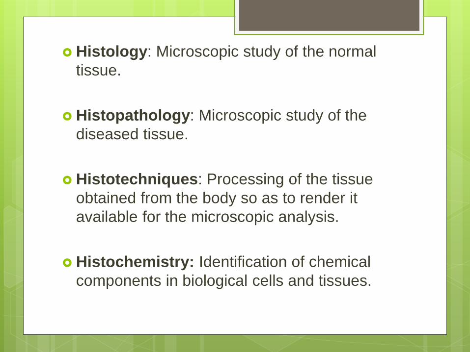
Histology: Microscopic study of the normal
tissue.
Histopathology: Microscopic study of the
diseased tissue.
Histotechniques: Processing of the tissue
obtained from the body so as to render it
available for the microscopic analysis.
Histochemistry: Identification of chemical
components in biological cells and tissues.
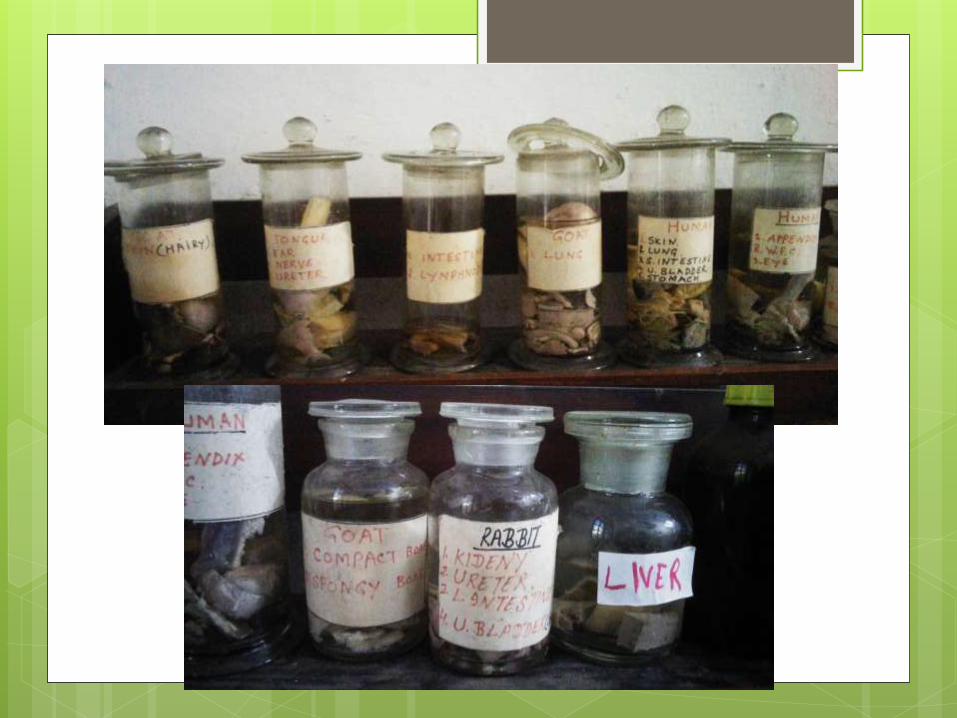
Sources for tissue study in
Histology
Autopsy
Animal tissue
Biopsy
Cadavers
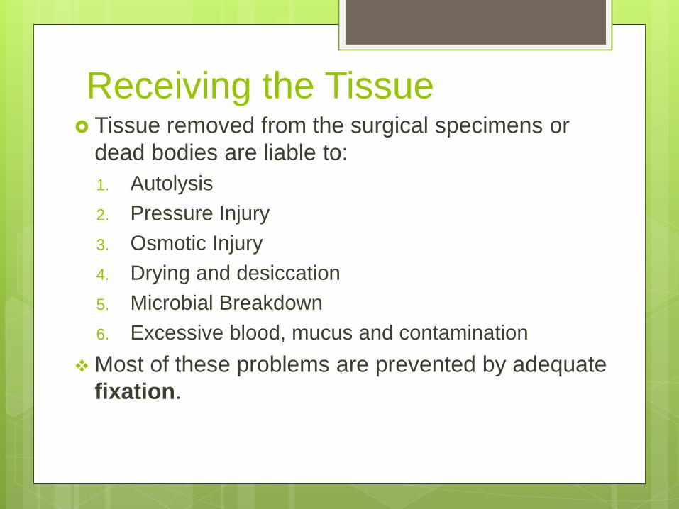
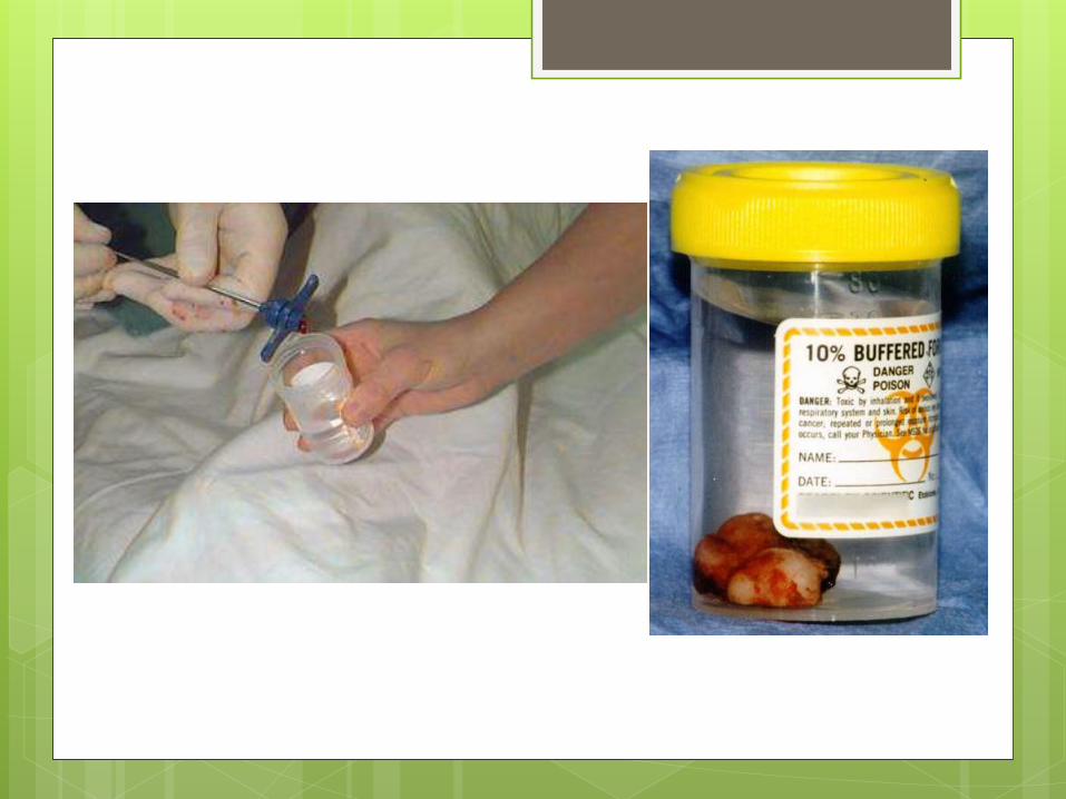
Receiving the Tissue Tissue removed from the surgical specimens or
dead bodies are liable to:
1. Autolysis
2. Pressure Injury
3. Osmotic Injury
4. Drying and desiccation
5. Microbial Breakdown
6. Excessive blood, mucus and contamination
Most of these problems are prevented by adequate
fixation.
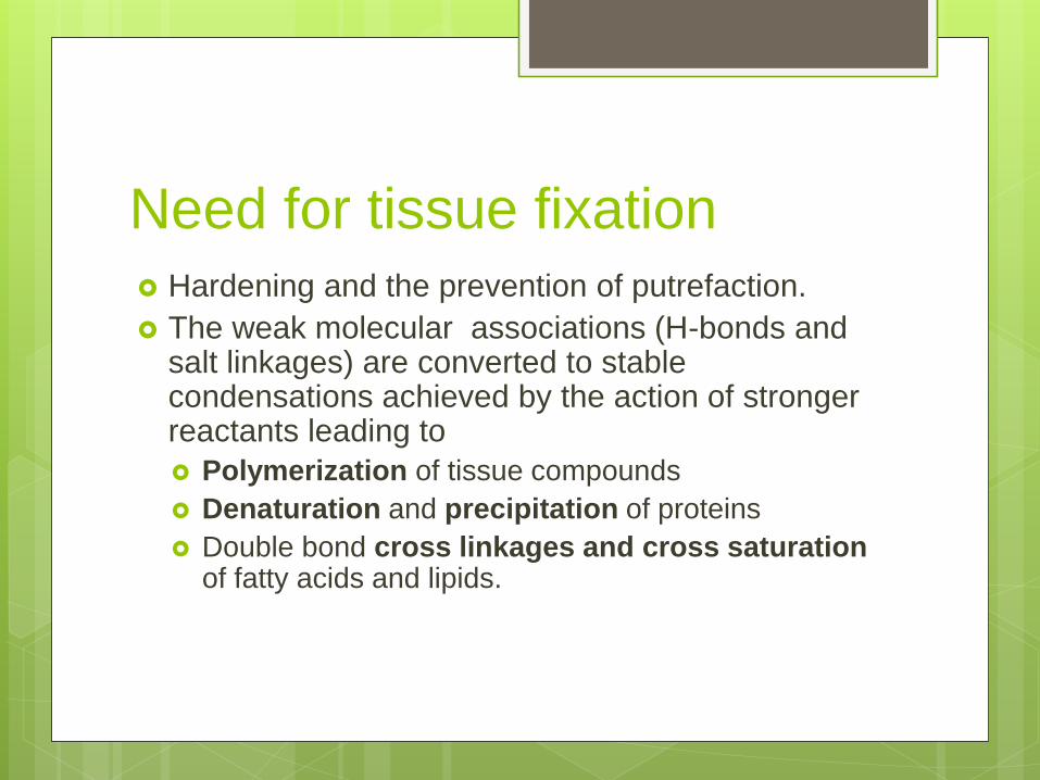
Need for tissue fixation
Hardening and the prevention of putrefaction.
The weak molecular associations (H-bonds and salt linkages) are converted to stable condensations achieved by the action of stronger reactants leading to
Polymerization of tissue compounds
Denaturation and precipitation of proteins
Double bond cross linkages and cross saturation of fatty acids and lipids.
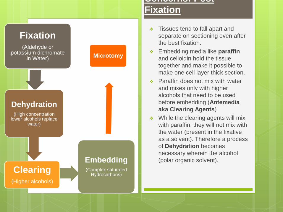
Concerns: Post
Fixation
Fixation(Aldehyde or
potassium dichromate in Water)
Microtomy
Embedding(Complex saturated
Hydrocarbons)
Dehydration(High concentration
lower alcohols replace water)
Clearing(Higher alcohols)
Tissues tend to fall apart and
separate on sectioning even after
the best fixation.
Embedding media like paraffin
and celloidin hold the tissue
together and make it possible to
make one cell layer thick section.
Paraffin does not mix with water
and mixes only with higher
alcohols that need to be used
before embedding (Antemedia
aka Clearing Agents)
While the clearing agents will mix
with paraffin, they will not mix with
the water (present in the fixative
as a solvent). Therefore a process
of Dehydration becomes
necessary wherein the alcohol
(polar organic solvent).
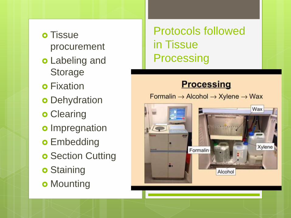
Tissue
procurement
Labeling and
Storage
Fixation
Dehydration
Clearing
Impregnation
Embedding
Section Cutting
Staining
Mounting
Protocols followed
in Tissue
Processing
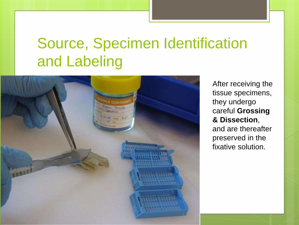
Source, Specimen Identification
and Labeling
After receiving the
tissue specimens,
they undergo
careful Grossing
& Dissection,
and are thereafter
preserved in the
fixative solution.
Unfixed Tissue: Temporary
Mounts Direct Examination: Thin membranes
Smear and Squash Preparations: Friable
material
Freehand Sectioning
Frozen Sections
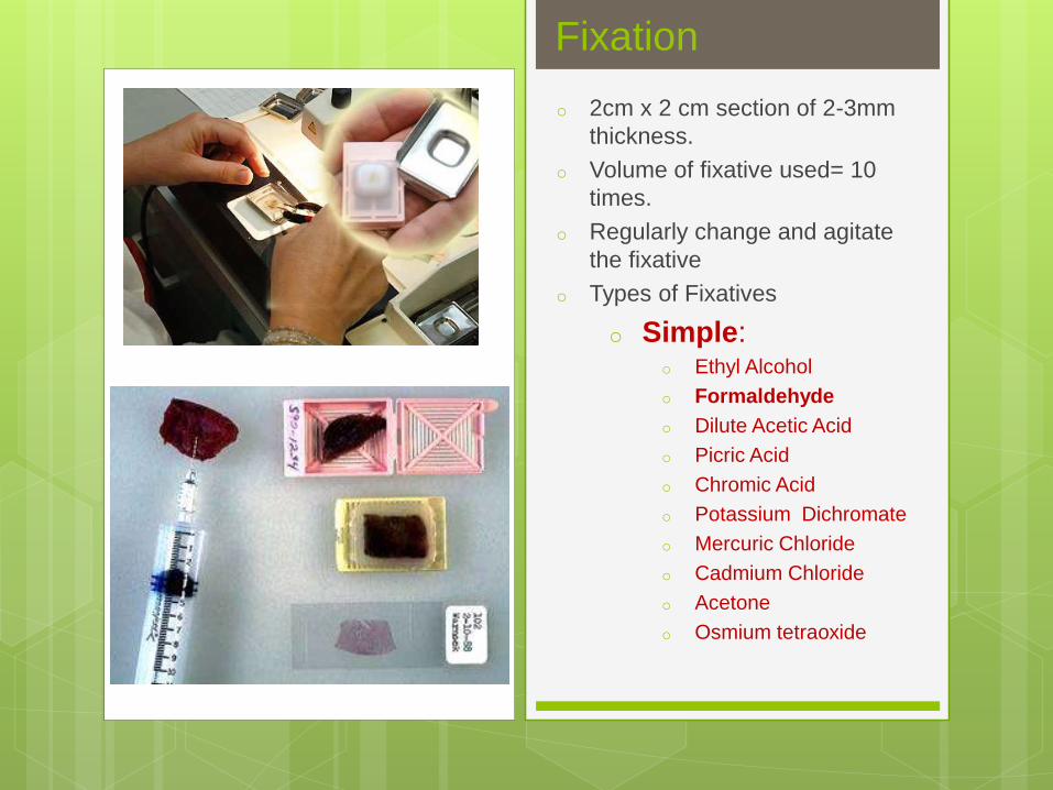
Fixation
o 2cm x 2 cm section of 2-3mm
thickness.
o Volume of fixative used= 10
times.
o Regularly change and agitate
the fixative
o Types of Fixatives
o Simple:o Ethyl Alcohol
o Formaldehyde
o Dilute Acetic Acid
o Picric Acid
o Chromic Acid
o Potassium Dichromate
o Mercuric Chloride
o Cadmium Chloride
o Acetone
o Osmium tetraoxide

FixativesGeneral Purpose Fixatives:
Ten Percent Formalin
Neutral Buffered Formalin
Zenker’s Fluid
Helly’s Fluid
Bouin’s Fluid
Carnoy’s Fluid
Alcohol- Formanlin
Plain Alcohol
Van de Grift’s Solution
Fixatives for Particular Purpose:
Aoyama’s Fluid: Golgi element.
Baker’s Calcium cadmium FormolSolution: Complex Lipids
Cobalt Calcium Formol Fixation: Complex Lipids
Gendre’s Fluid: Polysaccharides
Rossman’s Fluid: Polysaccharides.

Care During Fixation
Prepare ahead of time
Refrigerate
Placing the tissue in the bottle
Injecting and permeating the fixative through alternate approach.
Duration: Depends on the Tissue and the Fixative.
• Neutral Buffered Formalin fixes a block of tissue 1x2x0.4 cm in 4-6 hours.
• Time duration varies from 6-48 hours.
• Usually kept overnight.
Aftertreatment
Formalin requires no particular aftertreatment, and fixed tissues may be placed directly into the dehydrant. If using NBF, it is recommended that an alcoholconcentration of about 60% or so be used first.
If Picric acid is used, wash the tissue in 70% ethanol.
Treat mercury fixative containing sections with iodine mixture (Lugol’s Iodine + Sodium thiosulphate.
On using Zenker or Helley’s fluid the tissue needs to be washed for 10-15 hrs under tap water to get rid of excess chromium.
Decalcification
Immerse tissue cassette in 10% formic acid
with a stir bar overnight in a fume hood.
Rinse in running water for 30-60 minutes (the
smell should be gone).
Other decalcifying agents: Nitric Acid
Hydrochloric Acid
Picric Acid
Acetic Acid
Citric Acid
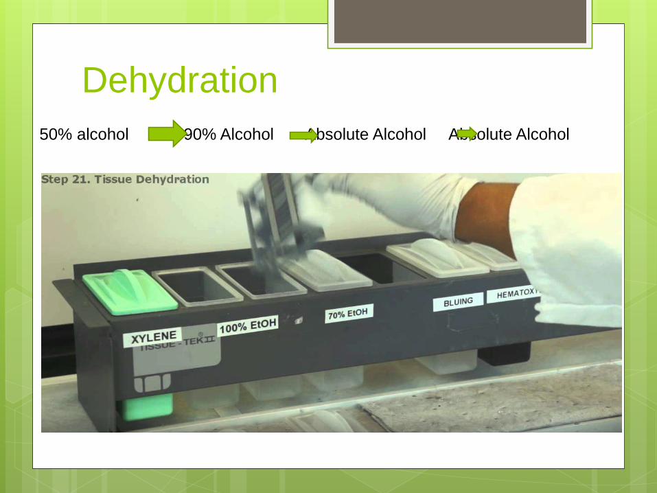
Dehydration
50% alcohol 90% Alcohol Absolute Alcohol Absolute Alcohol
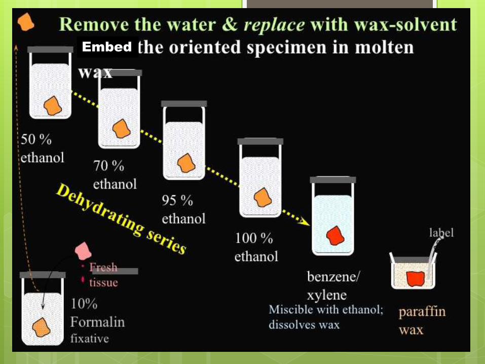
Embed
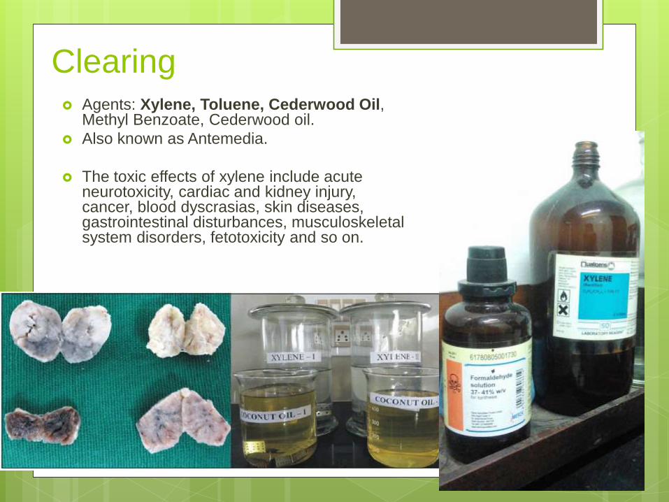
Clearing Agents: Xylene, Toluene, Cederwood Oil,
Methyl Benzoate, Cederwood oil.
Also known as Antemedia.
The toxic effects of xylene include acute neurotoxicity, cardiac and kidney injury, cancer, blood dyscrasias, skin diseases, gastrointestinal disturbances, musculoskeletal system disorders, fetotoxicity and so on.
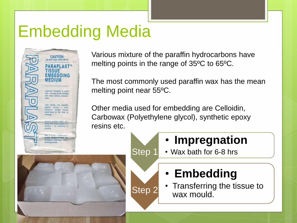
Embedding Media
Various mixture of the paraffin hydrocarbons have
melting points in the range of 35ºC to 65ºC.
The most commonly used paraffin wax has the mean
melting point near 55ºC.
Other media used for embedding are Celloidin,
Carbowax (Polyethylene glycol), synthetic epoxy
resins etc.
Step 1
• Impregnation• Wax bath for 6-8 hrs
Step 2
• Embedding• Transferring the tissue to
wax mould.
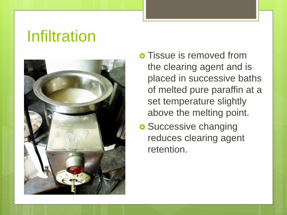
Infiltration Tissue is removed from
the clearing agent and is
placed in successive baths
of melted pure paraffin at a
set temperature slightly
above the melting point.
Successive changing
reduces clearing agent
retention.
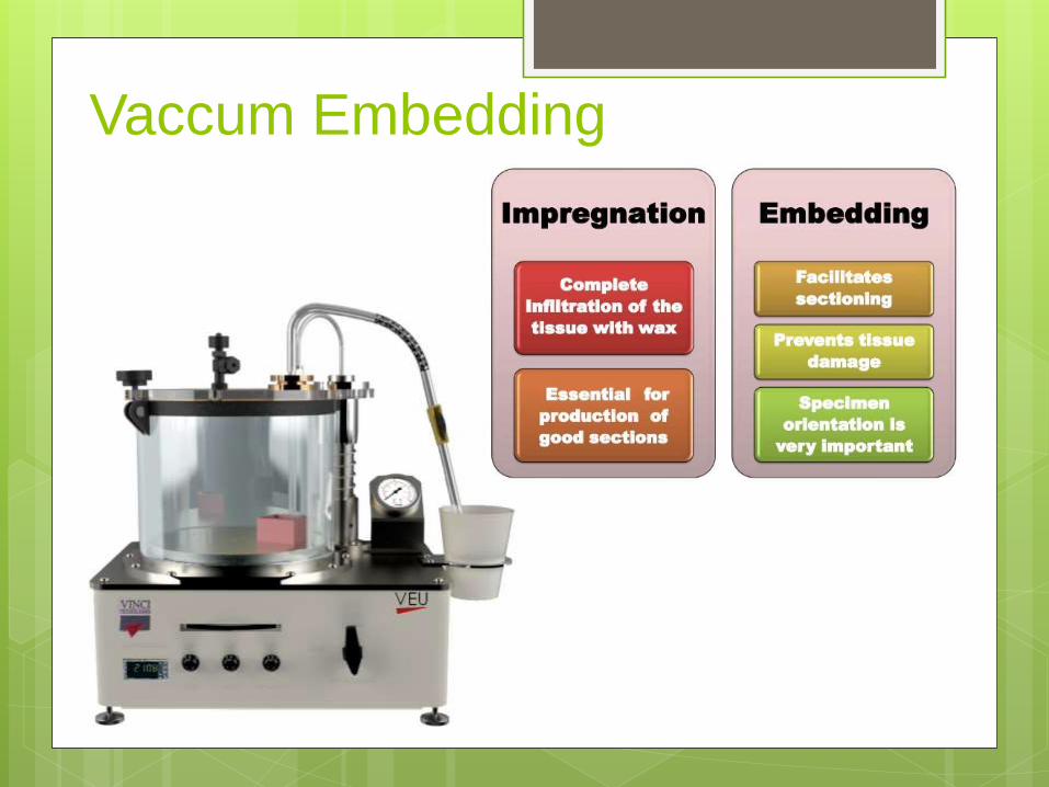
Vaccum Embedding
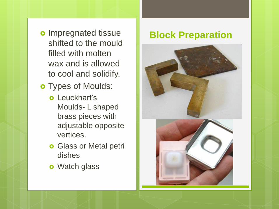
Impregnated tissue
shifted to the mould
filled with molten
wax and is allowed
to cool and solidify.
Types of Moulds:
Leuckhart’s
Moulds- L shaped
brass pieces with
adjustable opposite
vertices.
Glass or Metal petri
dishes
Watch glass
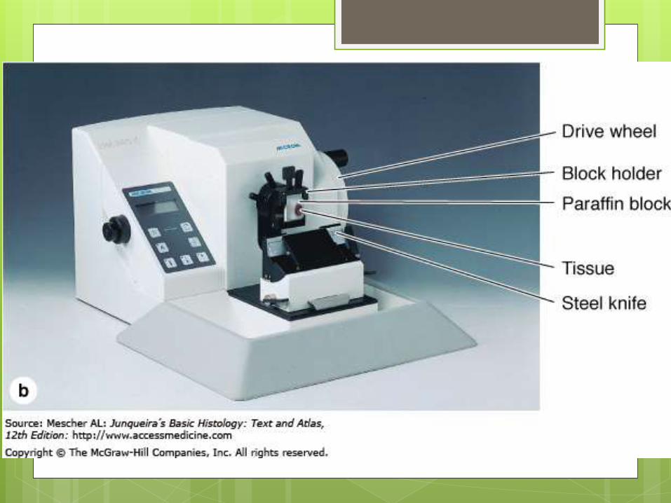
Block Preparation

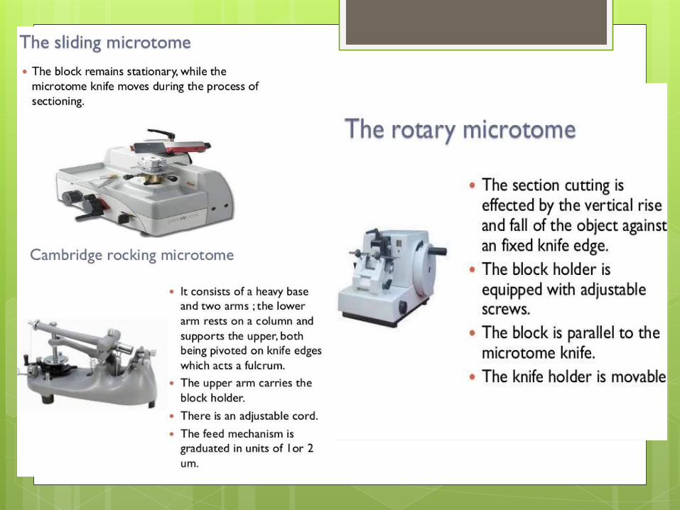
Tissue Sectioning

Microtome
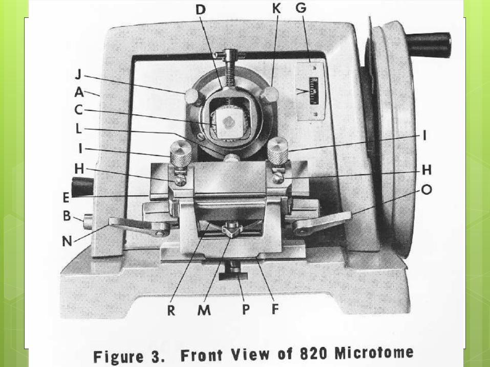
Front View

-Utilize safety features properly.
-Clamps and Brakes
-Camel brush- Soft and coarse
-Ice blocks
-Warm water
-Hot plate and oven

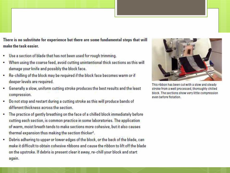
Trimming: The goal of properly trimming a
block is to conservatively expose the tissue
do to a level where a representative section
can be obtained.
Trimming is usually done at thicknesses
between 10 and 30 μm.
Consider Factors Affecting Section Thickness.
Floating Out The Sections Adhesive Solution: Mayer Glycerine-egg albumin
Glycerine-Egg-Albumin: GEA
Egg white: 1 part
Glycerin: 1 part
Thymol:1 crystal
The adhesive mixture used for routine purposes is a dilution of the above. 15ml of water with 3 drops of GEA.
Flattening:-Water Bath
-Individual Slide flattening on hot plate
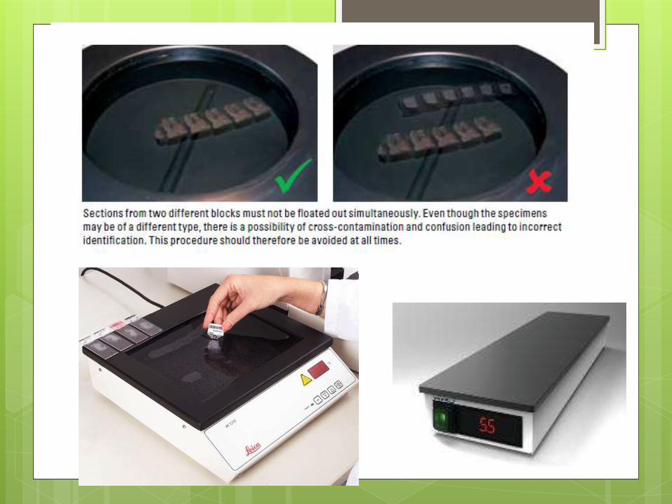
Drain excess water from beneath the section before drying. This is vital if slides are dried flat on a hot plate.
Slides can be stored in racks in an upright position, then dried in an oven.
Generally drying temperatures should not exceed 65 ˚C.
Excessive heat can cause droplets of water underneath a section to boil and this will cause damage.
Dry sections for between 10 and 30 minutes.
Some delicate specimens will produce best results when dried at 37 ˚C for a longer time (several hours to overnight).
Clean and maintain the Microtome thoroughly.
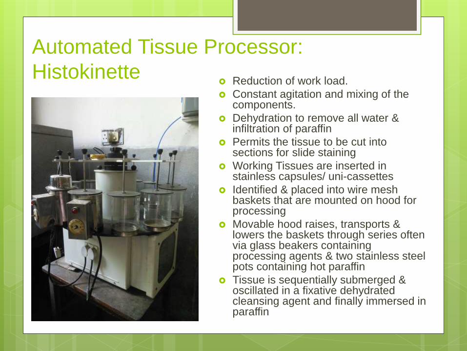
Automated Tissue Processor:
Histokinette Reduction of work load.
Constant agitation and mixing of the components.
Dehydration to remove all water & infiltration of paraffin
Permits the tissue to be cut into sections for slide staining
Working Tissues are inserted in stainless capsules/ uni-cassettes
Identified & placed into wire mesh baskets that are mounted on hood for processing
Movable hood raises, transports & lowers the baskets through series often via glass beakers containing processing agents & two stainless steel pots containing hot paraffin
Tissue is sequentially submerged & oscillated in a fixative dehydrated cleansing agent and finally immersed in paraffin

Programmable 12 stage timing sequence, having 24 hours calibrated disc
Delay action of 24 hours
Precision gear mechanism for trouble frees working
Safety device for tissue protection
Thermostatically controlled wax bath
Each Station with capacity of 1 Liter/2 Liter
Automatic agitations allow the effective processing of tissues
Assure utmost convenience in operation
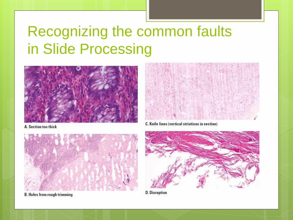
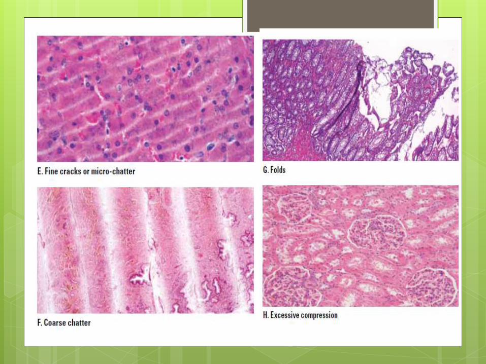
Recognizing the common faults
in Slide Processing